Embed Size (px)
Citation preview

Urinary System
Mr. Visanth V SMayo school of NursingLucknow
The Urinary System
• Urinary System is one of the four excretory systems in our body. The other three are bowel, lungs & skin.

Components of Urinary System
2 Kidneys
2 Ureters
Urethra
Urinary Bladder
Fig 7.1 Components of Urinary System
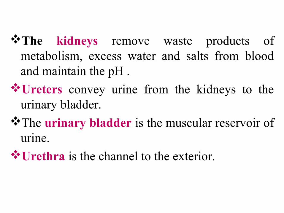
The kidneys remove waste products of metabolism, excess water and salts from blood and maintain the pH .
Ureters convey urine from the kidneys to the urinary bladder.
The urinary bladder is the muscular reservoir of urine.
Urethra is the channel to the exterior.
The Kidneys/Renes• Definition
– The kidneys are a pair of excretory organs
situated on the posterior abdominal wall,
one on each side of the vertebral column,
behind the peritoneum.
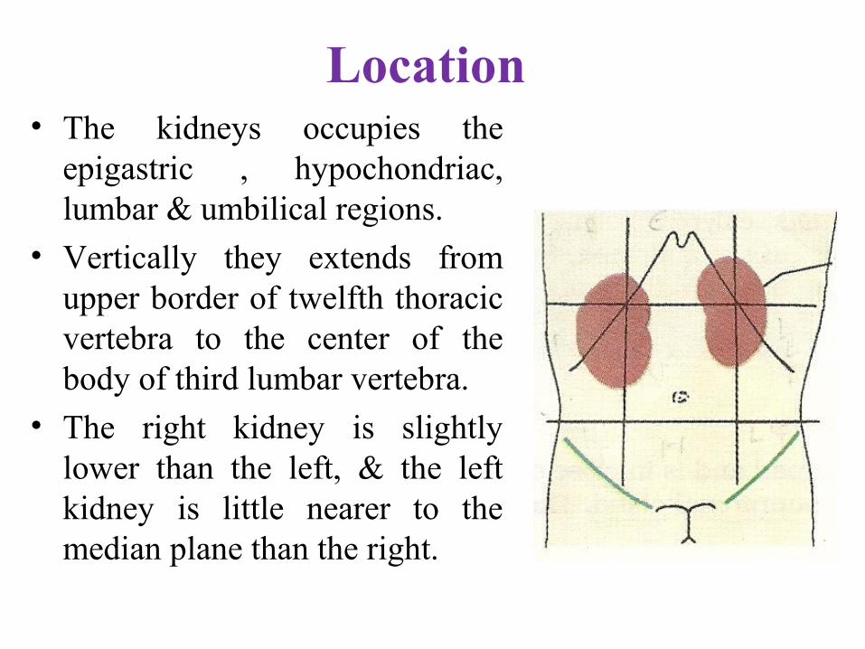
Location • The kidneys occupies the
epigastric , hypochondriac, lumbar & umbilical regions.
• Vertically they extends from upper border of twelfth thoracic vertebra to the center of the body of third lumbar vertebra.
• The right kidney is slightly lower than the left, & the left kidney is little nearer to the median plane than the right.

The Kidneys- Surface Anatomy
• External Features– Each kidney is bean shaped.– It has upper & lower poles,
medial and lateral borders, and anterior and posterior surfaces.
– The upper pole is broad & is in close contact with the corresponding suprarenal glands.
– The lower pole is pointed.
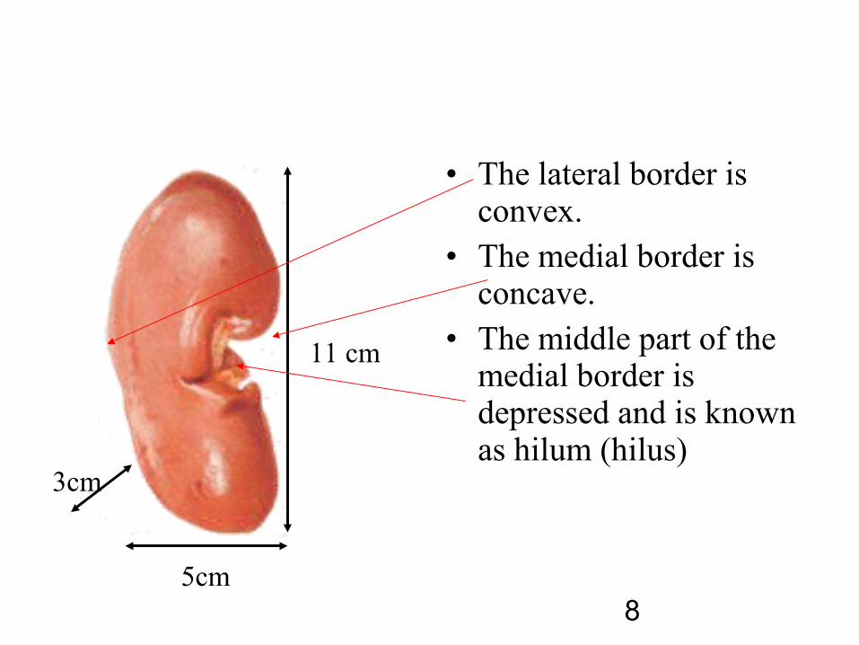
• The lateral border is convex.
• The medial border is concave.
• The middle part of the medial border is depressed and is known as hilum (hilus)
11 cm
5cm
3cm
8
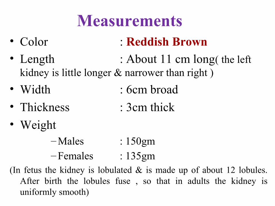
Measurements • Color : Reddish Brown• Length : About 11 cm long( the left
kidney is little longer & narrower than right )
• Width : 6cm broad
• Thickness : 3cm thick
• Weight – Males : 150gm– Females : 135gm
(In fetus the kidney is lobulated & is made up of about 12 lobules. After birth the lobules fuse , so that in adults the kidney is uniformly smooth)

Surface Marking- Morris Parallelogram
Anterior aspect Posterior aspect
Coverings
• It has 3 coverings1. Innermost fibrous capsule or true capsule
2. Middle fatty capsule / perinephric fat-it is a collection of fatty tissue. (It acts as a shock absorber & helps to maintain the kidney in its position)
3. The false capsule – made of renal fascia .It has two layers-Anterior & Posterior.
(Superiorly the two layers enclose the supra renal gland & then merge with diaphragmatic fascia, that is why the kidneys move
with respiration)

Relations - Anterior
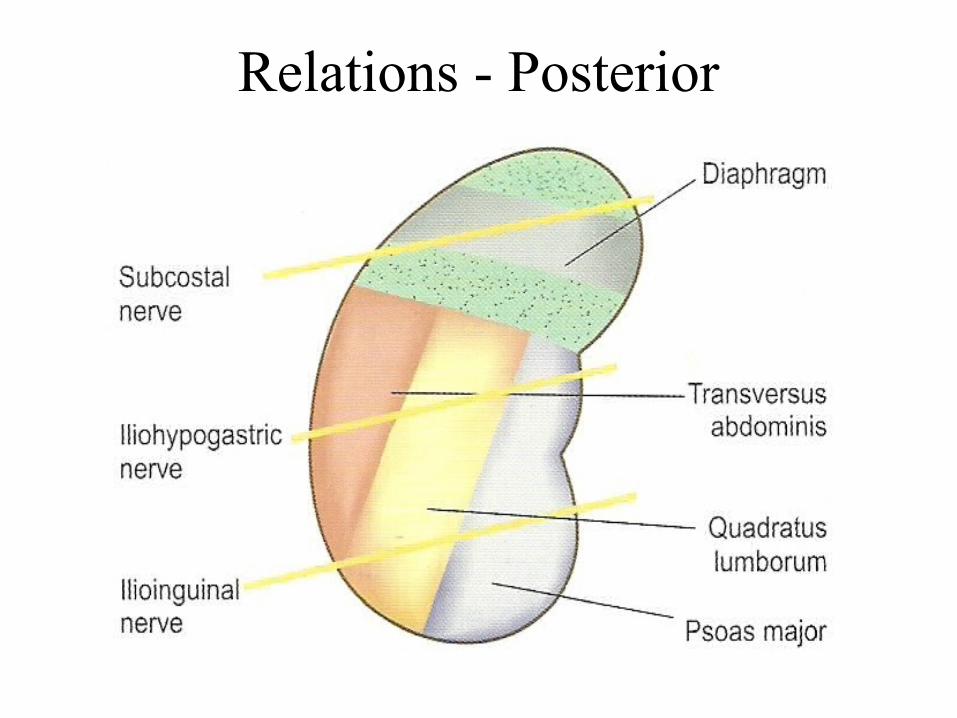
Relations - Posterior
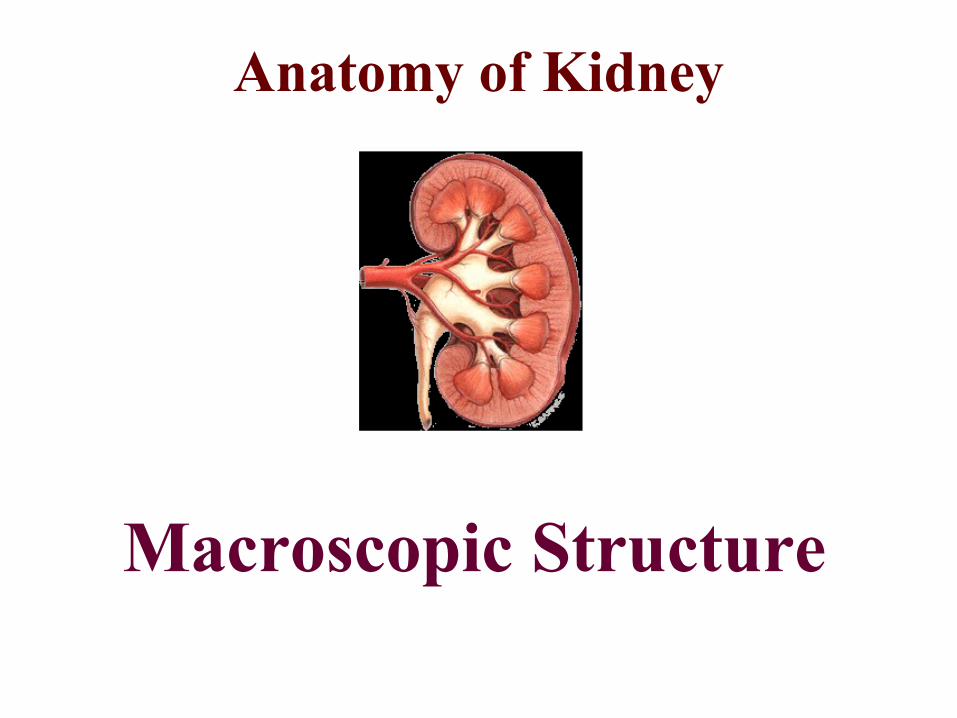
Anatomy of Kidney
Macroscopic Structure
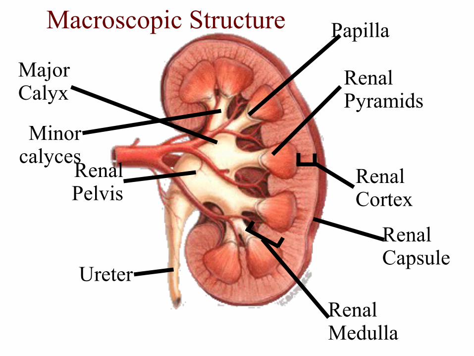
Renal Capsule
Renal Cortex
Renal Medulla
Renal Pelvis
Renal Pyramids
Ureter
Macroscopic Structure
Major Calyx
Minor calyces
Papilla

Kidney: Frontal Section
• Minor calyx: cup over papilla collects urine

Renal Artery
Renal Vein
NephronNephron
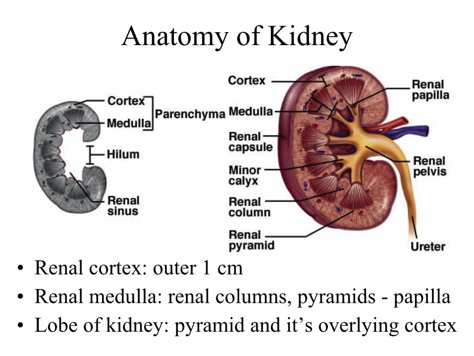
Anatomy of Kidney
• Renal cortex: outer 1 cm • Renal medulla: renal columns, pyramids - papilla• Lobe of kidney: pyramid and it’s overlying cortex
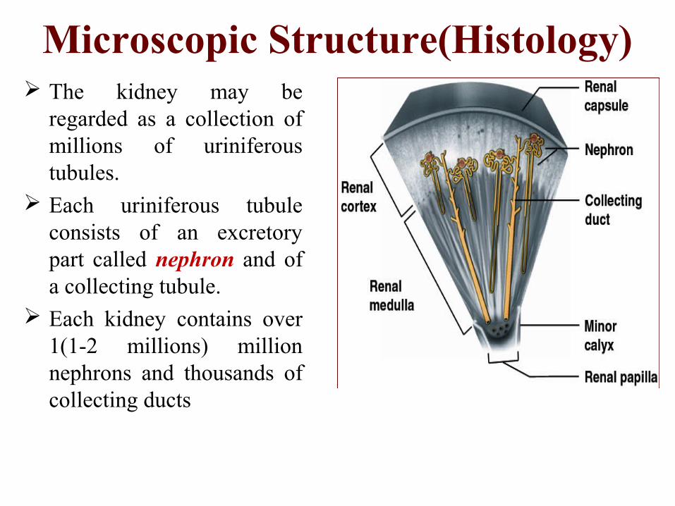
Microscopic Structure(Histology) The kidney may be
regarded as a collection of millions of uriniferous tubules.
Each uriniferous tubule consists of an excretory part called nephron and of a collecting tubule.
Each kidney contains over 1(1-2 millions) million nephrons and thousands of collecting ducts

Structure of Nephron

vein
artery
afferent arteriole
efferent arteriole
glomerulus
peritubular capillaries
Bowman’s capsule
proximal convoluted tubuledistal convoluted tubule
loop of Henle
collecting duct

urine
blood
filtration
tubular reabsorption and secretion
Nephron Functioning
“refreshed” blood
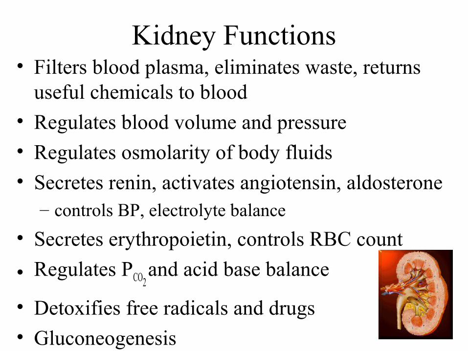
Kidney Functions• Filters blood plasma, eliminates waste, returns
useful chemicals to blood
• Regulates blood volume and pressure
• Regulates osmolarity of body fluids
• Secretes renin, activates angiotensin, aldosterone – controls BP, electrolyte balance
• Secretes erythropoietin, controls RBC count
• Regulates PCO2 and acid base balance
• Detoxifies free radicals and drugs
• Gluconeogenesis
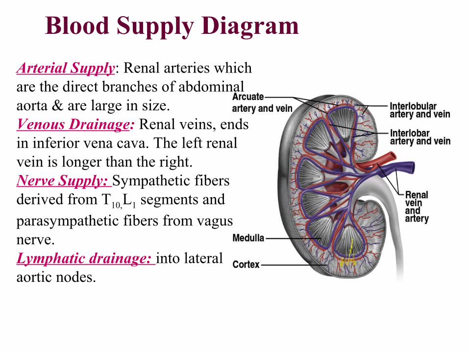
Blood Supply Diagram
Arterial Supply: Renal arteries which are the direct branches of abdominal aorta & are large in size.Venous Drainage: Renal veins, ends in inferior vena cava. The left renal vein is longer than the right.Nerve Supply: Sympathetic fibers derived from T10,L1 segments and parasympathetic fibers from vagus nerve.Lymphatic drainage: into lateral aortic nodes.
Applied Anatomy1. Congenital Anomalies
1. Unilateral/ Bilateral agenesis
2. Horse shoe kidney
3. Congenital poly cystic disease
4. Ectopic kidney
5. Abnormal renal arteries may arise from aorta or superior mesenteric artery
2. Infections
3. Renal Failure
THE URETERS
• Definition
The Ureters are a pair of narrow , thick
walled muscular tubes which convey
urine from the kidneys to urinary bladder.
Dimensions
• Each Ureters is about 25cm (10 inch)long.
• The upper half lies in the abdomen and the lower half in the pelvis.
• It measures 3mm diameter, but it slightly constricted at three places.– At the pelviureteric junction– At the brim of lesser pelvis– At its passage through the bladder wall
Parts
• For the purpose of description, ureter is divided into 2 parts– From the site of origin to pelvic brim- abdominal part– From pelvic brim to entry into urinary bladder- pelvic
part
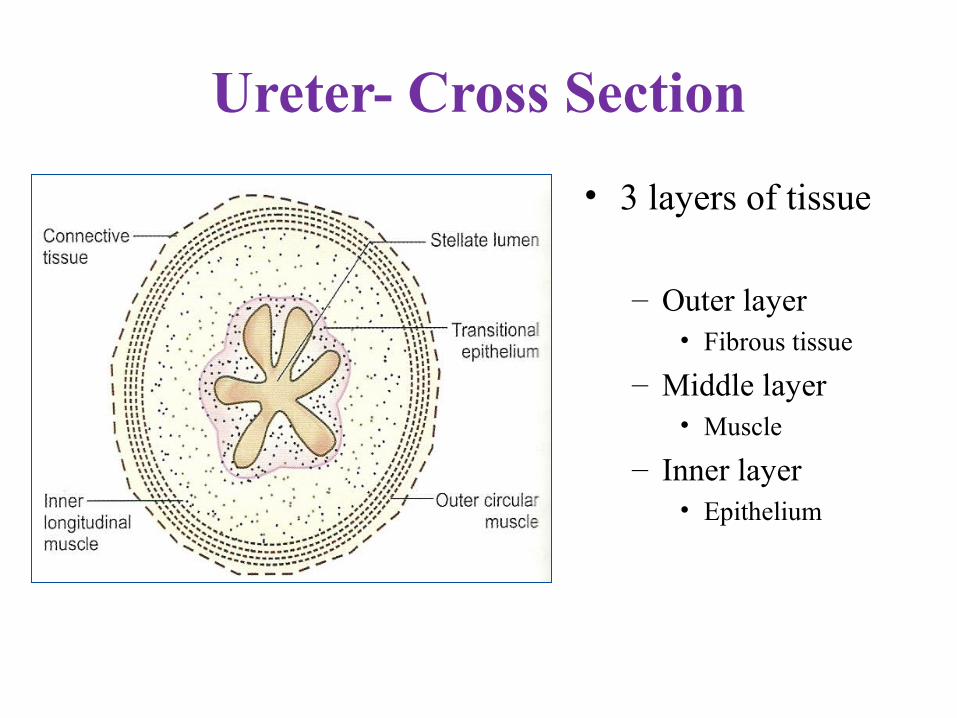
Ureter- Cross Section
• 3 layers of tissue
– Outer layer• Fibrous tissue
– Middle layer• Muscle
– Inner layer• Epithelium

• Blood Supply– Ureter is supplied by branches of
» Renal artery» Abdominal aorta» Gonadal artery» Common iliac artery» Internal iliac artery» Inferior vesical artery
• Nerve Supply– Autonomic nervous system

Urinary Bladder• The urinary
bladder is a hollow , muscular organ , which functions as the reservoir for the urine received from the kidneys and to discharge it out periodically
Position
• Empty bladder , in the adult situated within the pelvis . When distended , it rises up to the abdominal cavity and becomes an abdomino-pelvic organ.
• Capacity– The mean capacity of the bladder is 220 ml, filling
beyond 220ml causes a desire to micturate. Filling upto 500ml may be tolerated, buut it becomes painful.

Shape • An empty bladder is 4 sided pyramid in shape and has
– 4 angles -an apex, neck & 2 lateral angles– 4 surfaces
• Base (posterior surface)
• 2 inferiolateral surfaces
• Superior surface
When distended it is ovoid in shape

Relations
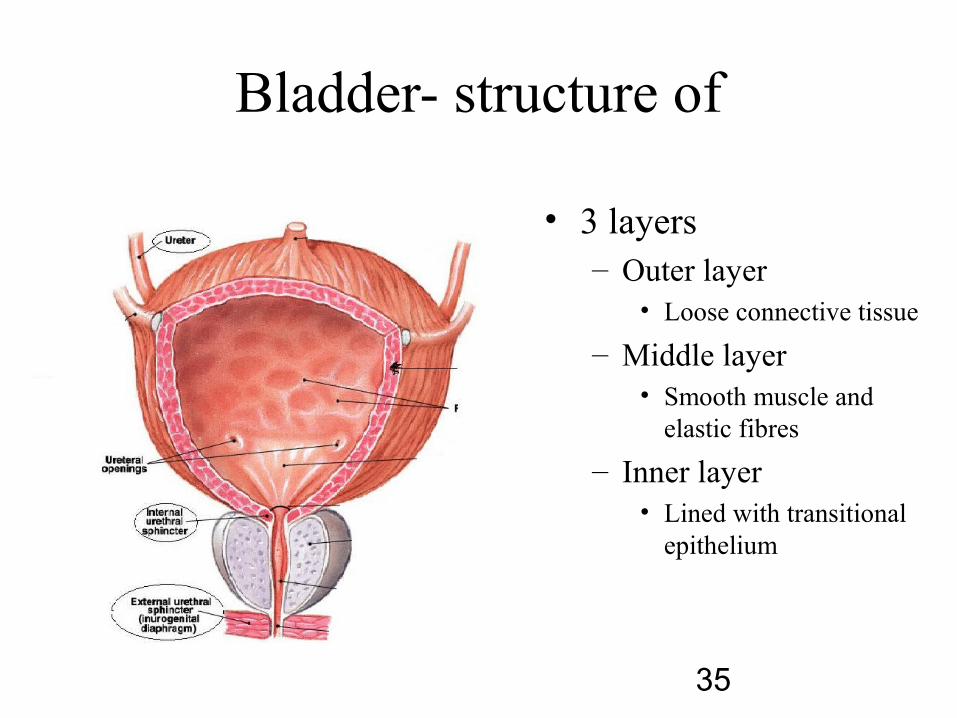
Bladder- structure of
• 3 layers– Outer layer
• Loose connective tissue
– Middle layer• Smooth muscle and
elastic fibres
– Inner layer• Lined with transitional
epithelium
35

Interior of Bladder• The mucous membrane is straw
colored & is thrown into folds. When bladder is distended, these folds disappear.
• The posterior wall shows a smooth triangular area called trigone. There are no mucous folds in this region.
• At the upper lateral angles of the trigone are the ureteric openings.
• At its inferior angle is the internal urethral orifice

Histology
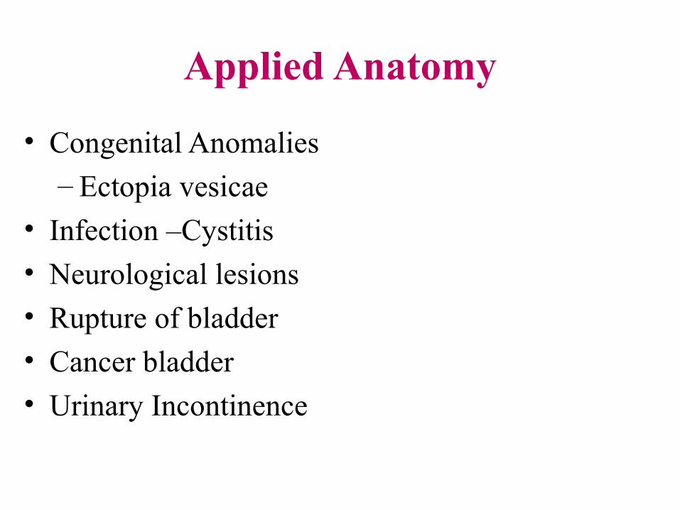
Applied Anatomy
• Congenital Anomalies
– Ectopia vesicae
• Infection –Cystitis
• Neurological lesions
• Rupture of bladder
• Cancer bladder
• Urinary Incontinence
39
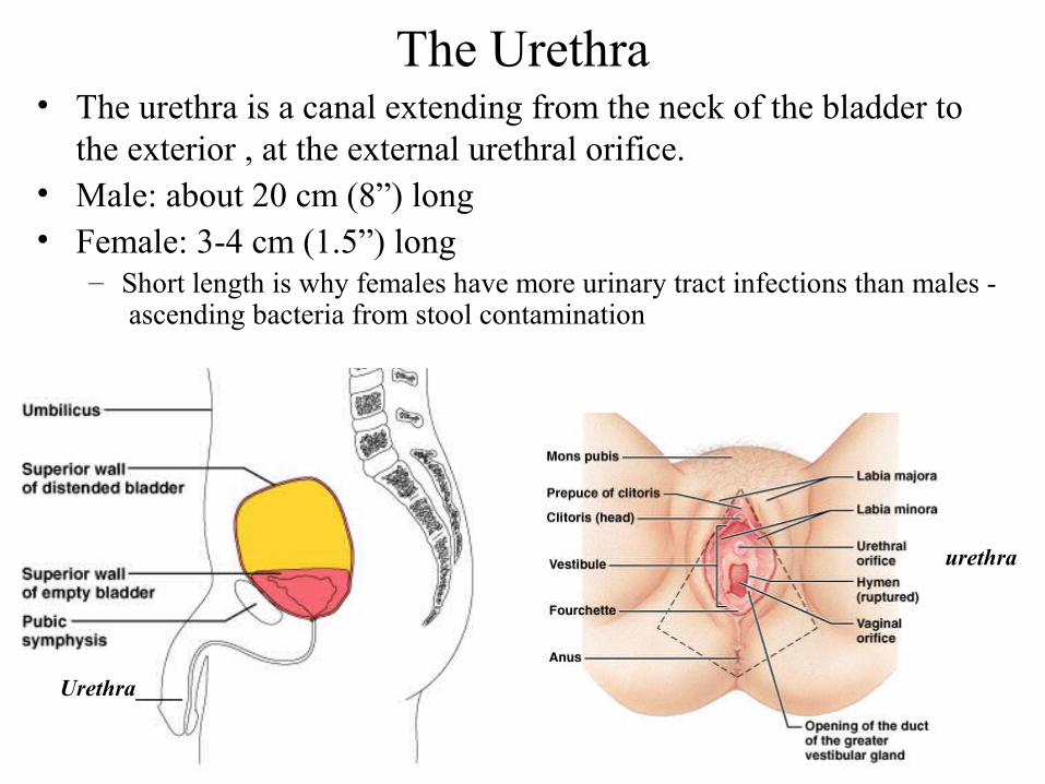
The Urethra• The urethra is a canal extending from the neck of the bladder to
the exterior , at the external urethral orifice.• Male: about 20 cm (8”) long• Female: 3-4 cm (1.5”) long
– Short length is why females have more urinary tract infections than males - ascending bacteria from stool contamination
Urethra____
urethra
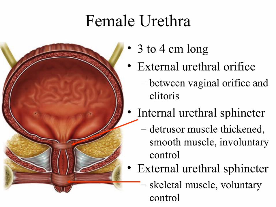
Female Urethra
• 3 to 4 cm long
• External urethral orifice – between vaginal orifice and
clitoris
• Internal urethral sphincter– detrusor muscle thickened,
smooth muscle, involuntary control
• External urethral sphincter– skeletal muscle, voluntary
control

Male Bladder and Urethra
• 18 cm long• Internal urethral sphincter• External urethral sphincter
• 3 regions– prostatic urethra
• during orgasm receives semen
– membranous urethra• passes through pelvic cavity
– penile urethra