Embed Size (px)
Citation preview
Urinary systemUrinary systemUrinary system
Dr. Carmen E. RexachDr. Carmen E. RexachAnatomy 35Anatomy 35
Mt. San Antonio CollegeMt. San Antonio College
Functions• Storage of urine
– Bladder stores up to 1 L of urine• Excretion of urine
– Transport of urine out of body• Blood volume regulation
– Effects of hormones on kidneys• Regulation of erythrocyte production
– Kidneys• Monitor oxygen content of blood• Produce EPO = erthrocyte production
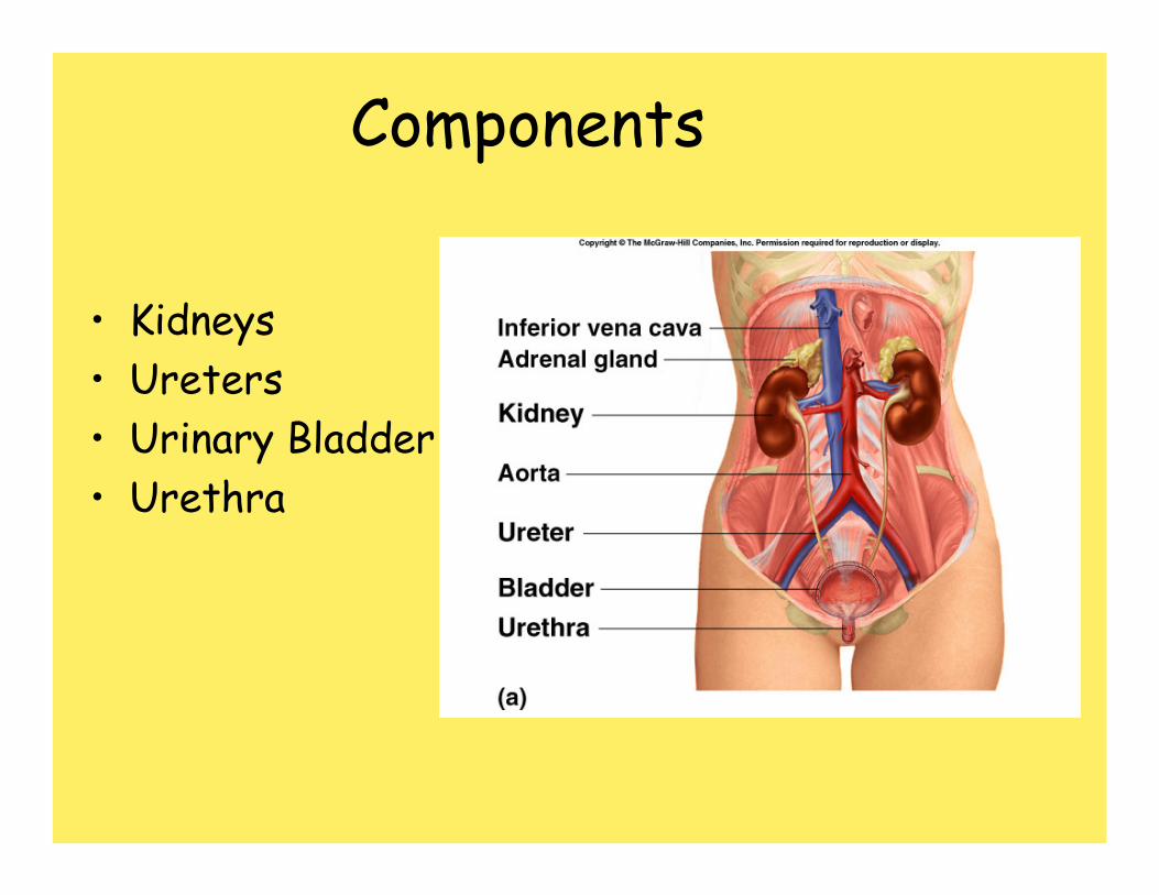
Components
• Kidneys• Ureters• Urinary Bladder• Urethra
Kidneys
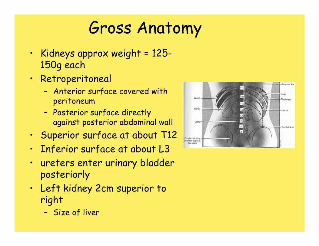
Gross Anatomy• Kidneys approx weight = 125-
150g each• Retroperitoneal
– Anterior surface covered with peritoneum
– Posterior surface directly against posterior abdominal wall
• Superior surface at about T12• Inferior surface at about L3• ureters enter urinary bladder
posteriorly• Left kidney 2cm superior to
right– Size of liver
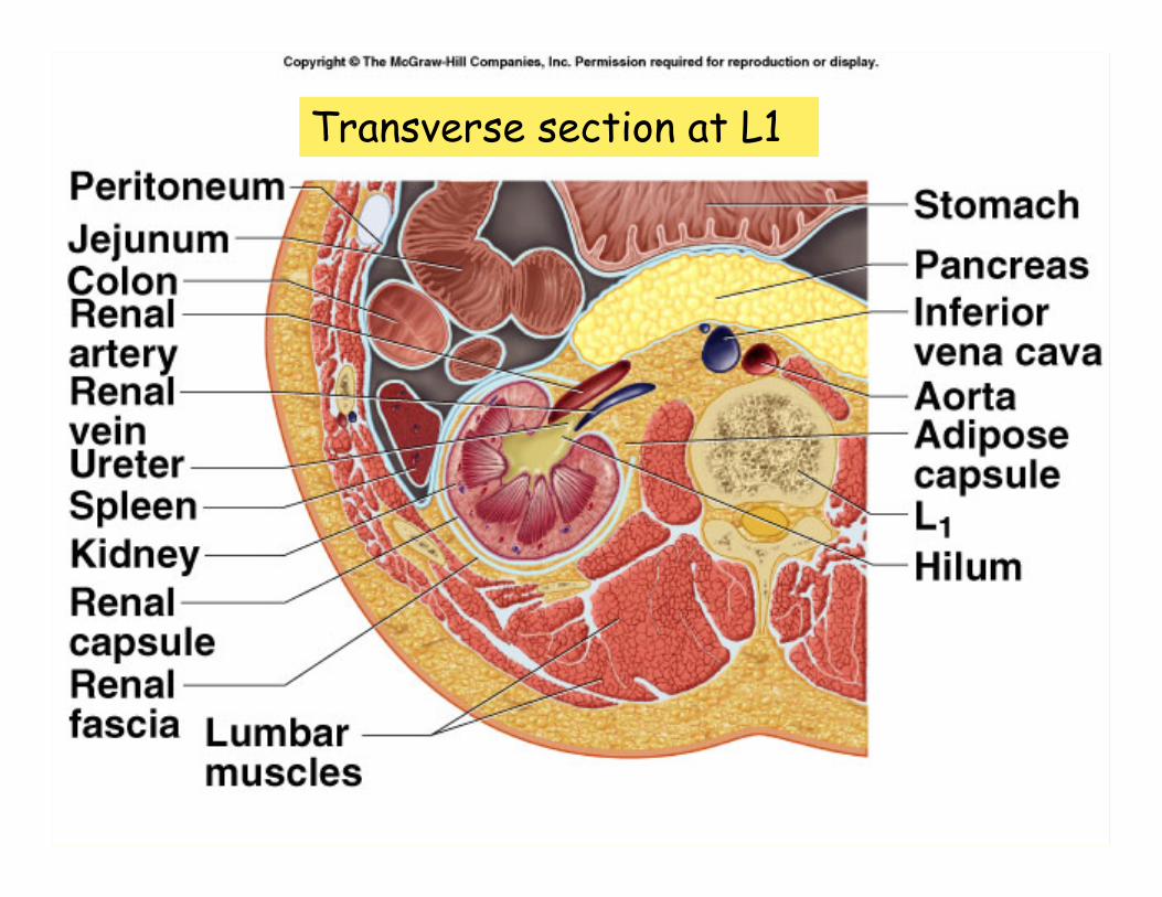
Transverse section at L1
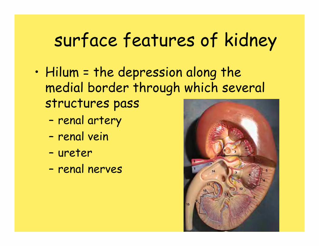
surface features of kidney• Hilum = the depression along the
medial border through which several structures pass– renal artery– renal vein– ureter– renal nerves
Surrounding structures• Fibrous capsule
– Innermost layer of dense irregular CT– Maintains shape, protection
• Adipose capsule (perinephric fat)– Adipose ct of varying thickness– Cushioning and insulation
• Renal fascia– Dense irregular CT– Anchors kidney to peritoneum & abdominal wall
• Paranephric fat– Outermost, adipose CT between renal fascia and
peritoneum
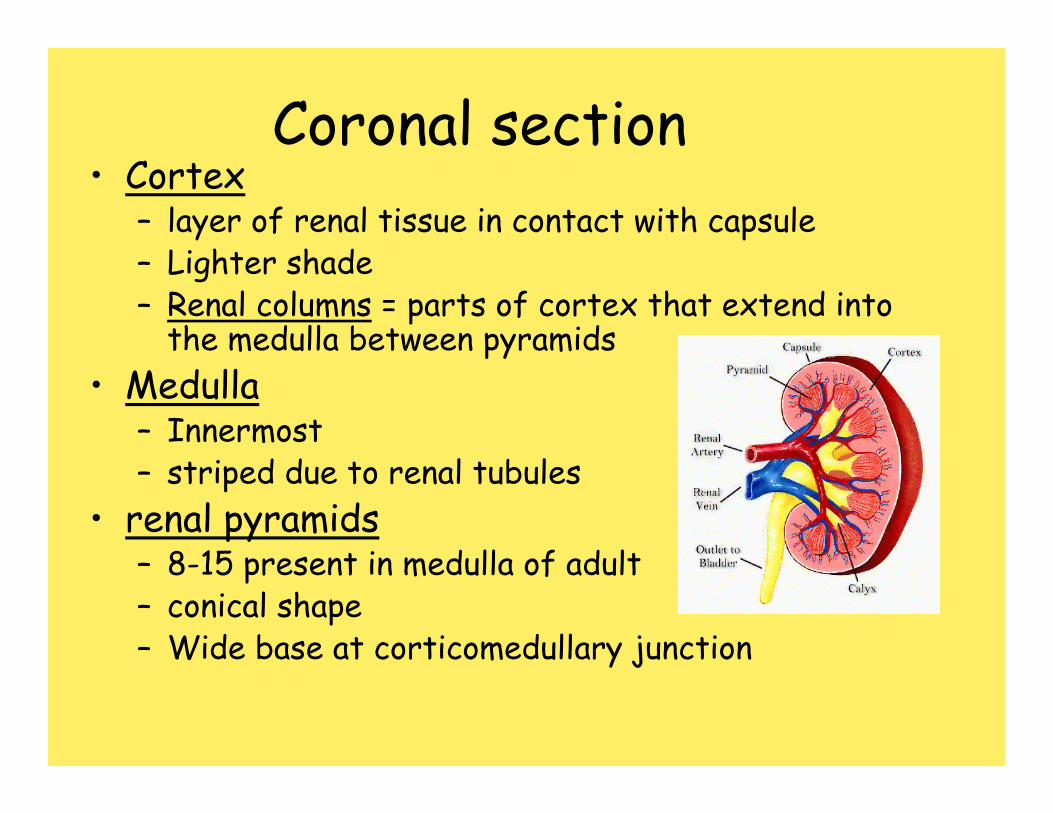
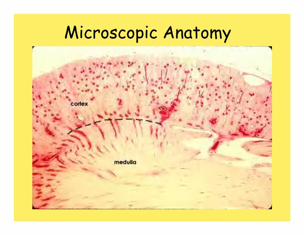
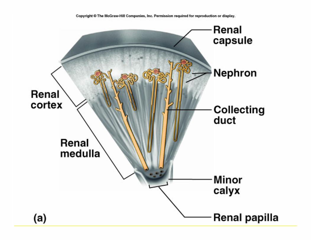
Coronal section• Cortex
– layer of renal tissue in contact with capsule– Lighter shade– Renal columns = parts of cortex that extend into
the medulla between pyramids• Medulla
– Innermost– striped due to renal tubules
• renal pyramids– 8-15 present in medulla of adult– conical shape– Wide base at corticomedullary junction
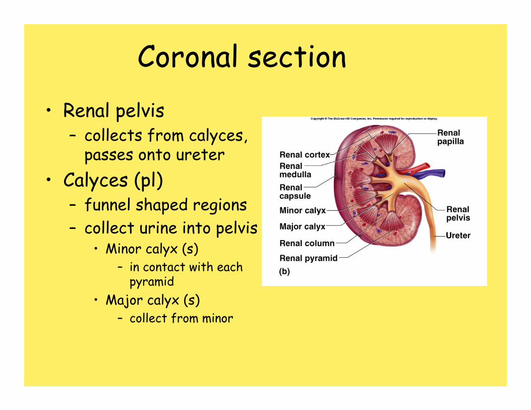
Coronal section• Renal pelvis
– collects from calyces, passes onto ureter
• Calyces (pl)– funnel shaped regions– collect urine into pelvis
• Minor calyx (s)– in contact with each
pyramid• Major calyx (s)
– collect from minor
Microscopic Anatomy
Microscopic anatomy
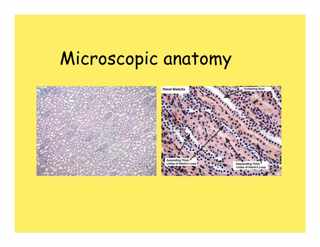
Renal tubules• Nephron
– functional unit of the kidney.• Each kidney contains approximately 1
million nephrons• Form urine by filtering and adjusting
composition of blood carried by renal vasculature.
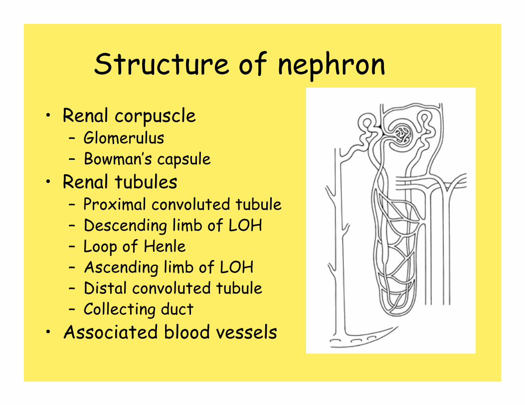
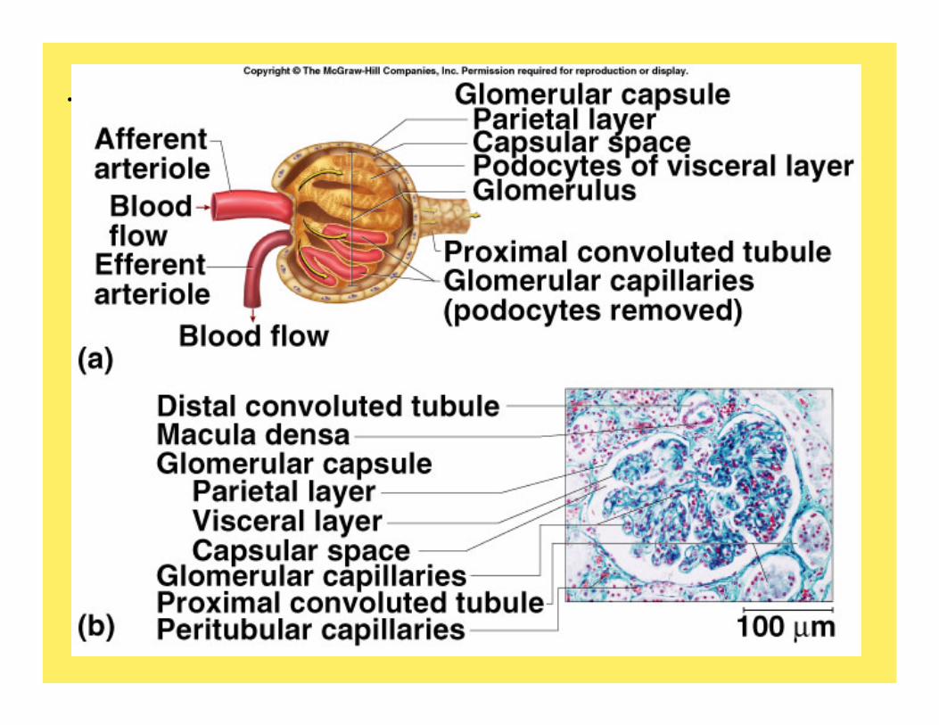
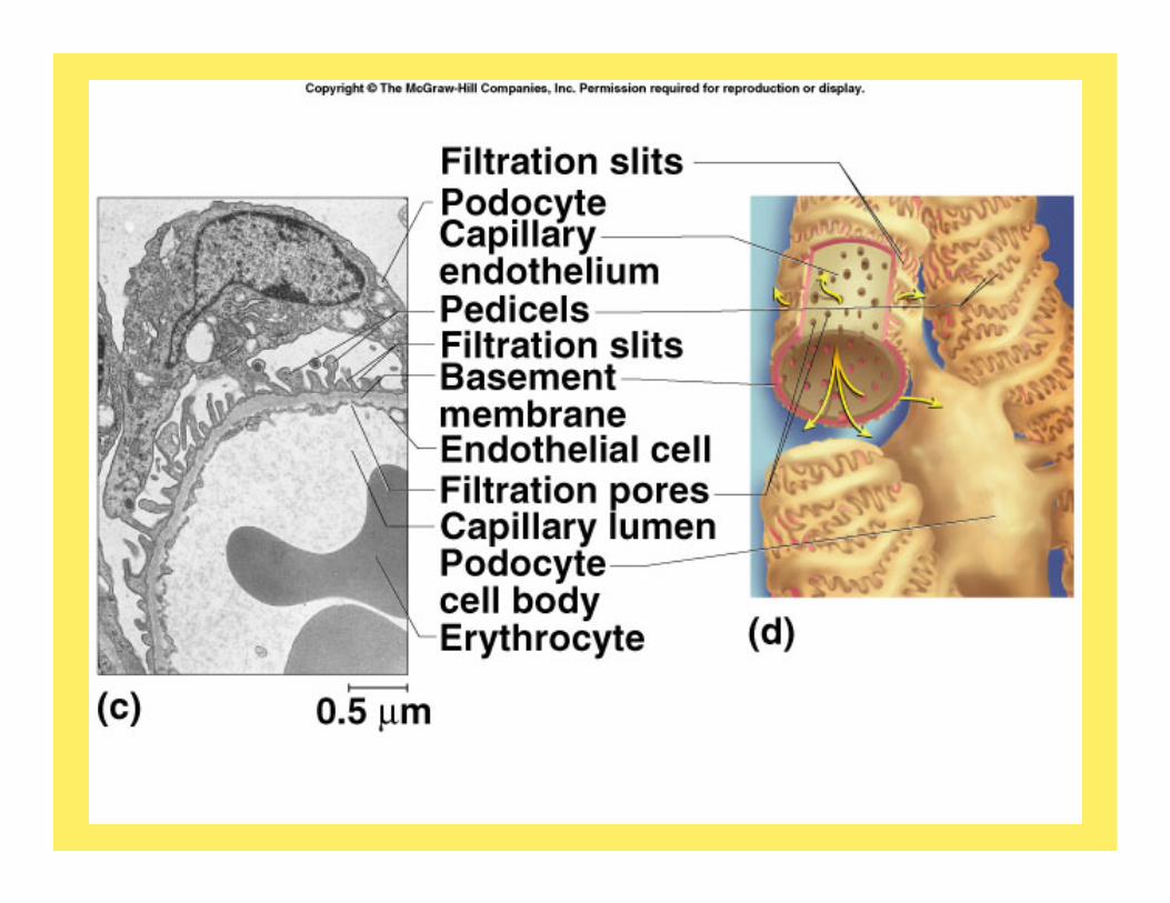
Structure of nephron• Renal corpuscle
– Glomerulus– Bowman’s capsule
• Renal tubules– Proximal convoluted tubule– Descending limb of LOH– Loop of Henle– Ascending limb of LOH– Distal convoluted tubule– Collecting duct
• Associated blood vessels
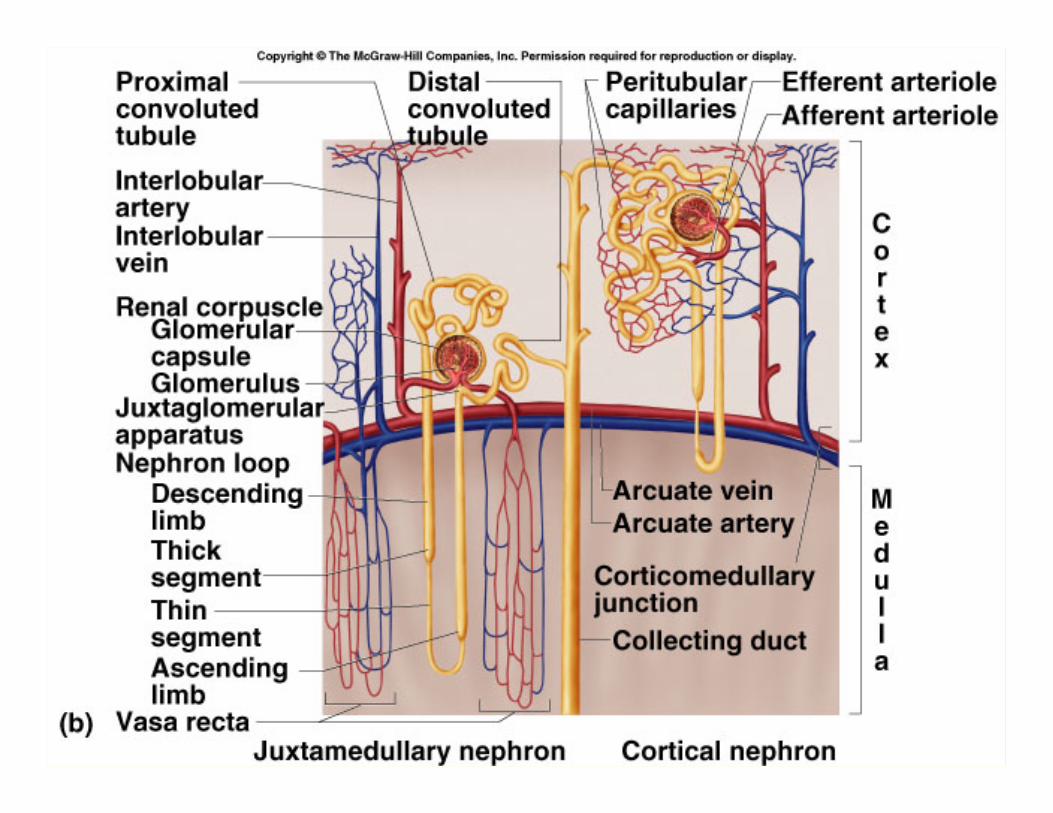
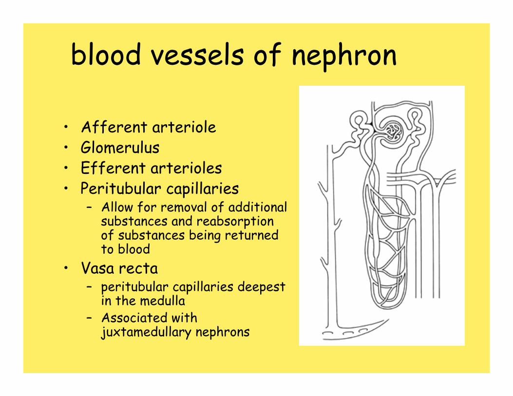
blood vessels of nephron
• Afferent arteriole• Glomerulus• Efferent arterioles • Peritubular capillaries
– Allow for removal of additional substances and reabsorptionof substances being returned to blood
• Vasa recta– peritubular capillaries deepest
in the medulla– Associated with
juxtamedullary nephrons
.
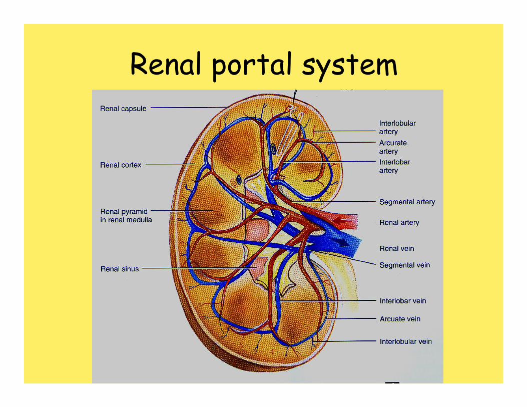
Renal portal system
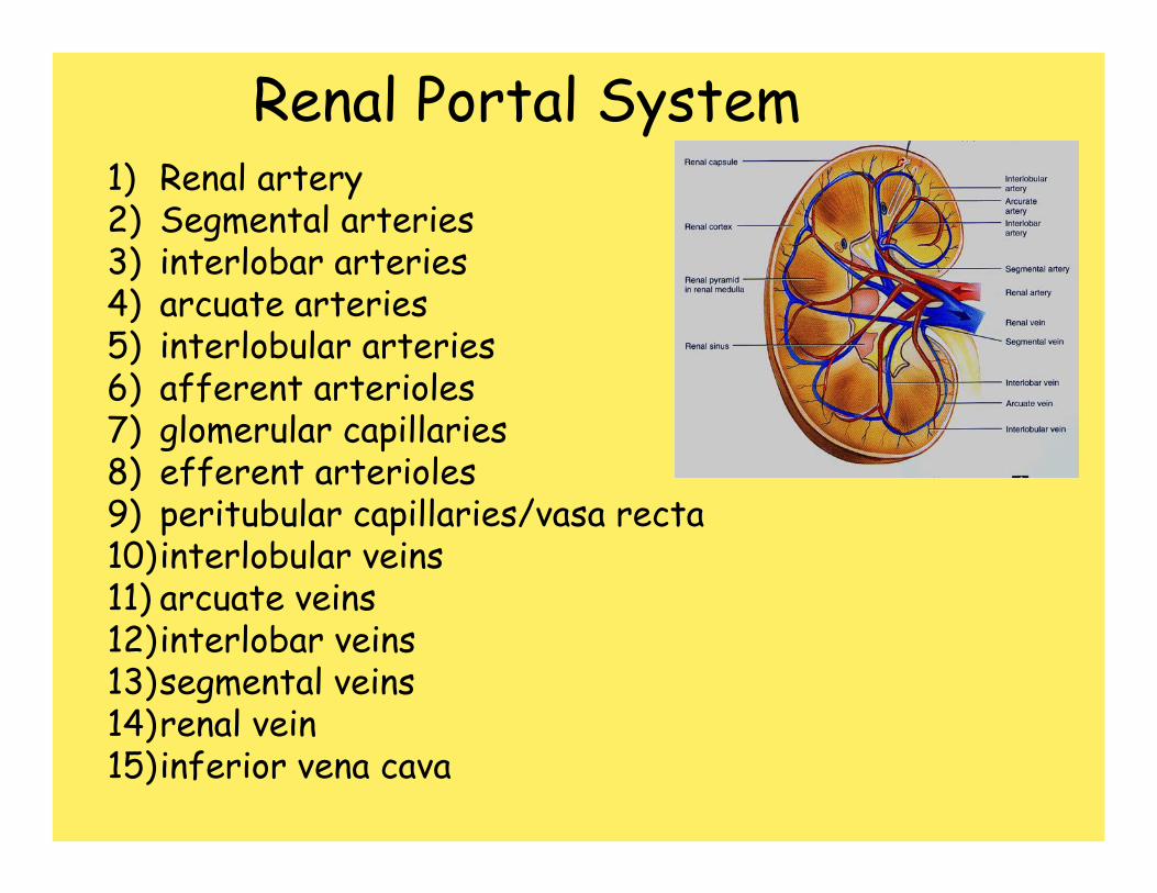
Renal Portal System1) Renal artery2) Segmental arteries3) interlobar arteries4) arcuate arteries5) interlobular arteries6) afferent arterioles7) glomerular capillaries8) efferent arterioles9) peritubular capillaries/vasa recta 10)interlobular veins 11) arcuate veins 12)interlobar veins 13)segmental veins 14)renal vein 15)inferior vena cava
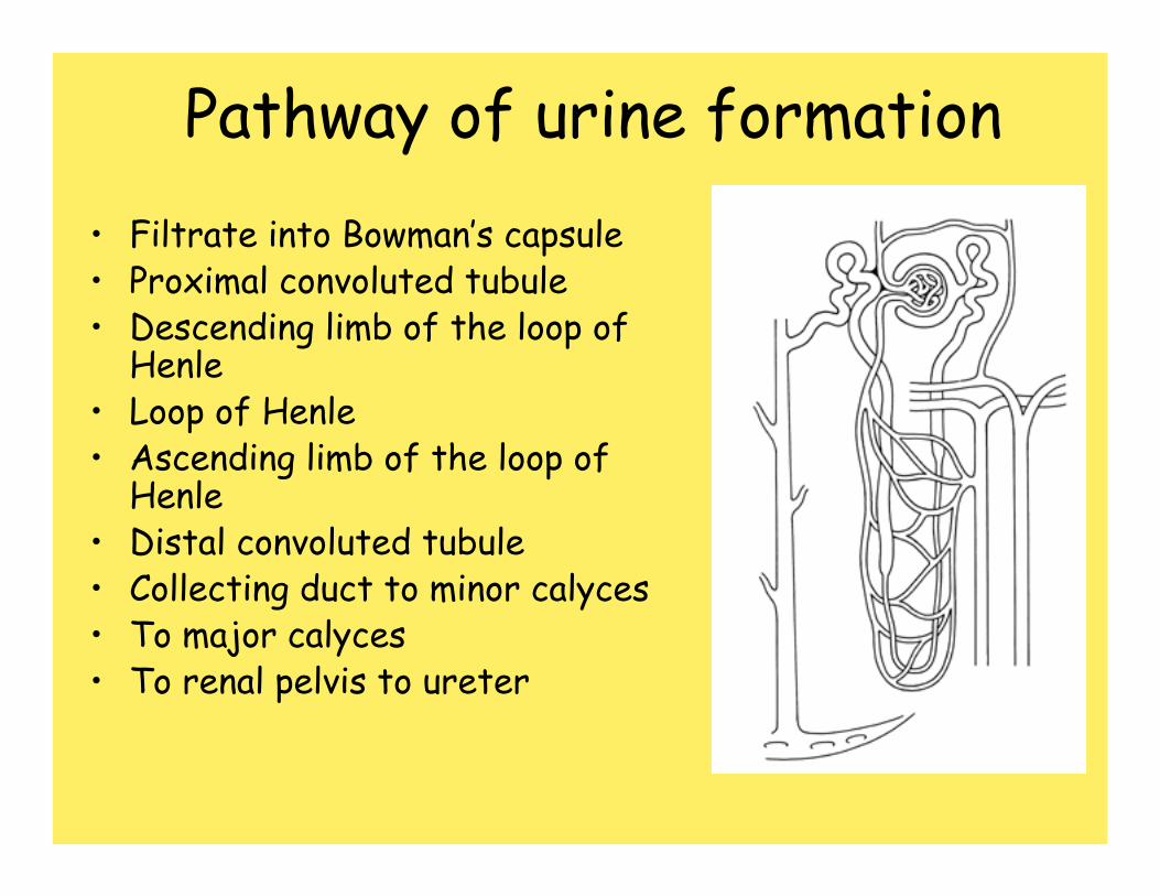
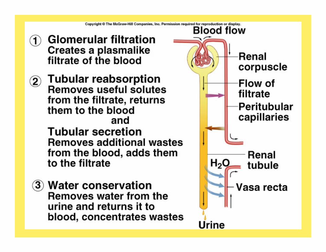
Pathway of urine formation• Filtrate into Bowman’s capsule• Proximal convoluted tubule• Descending limb of the loop of
Henle• Loop of Henle• Ascending limb of the loop of
Henle• Distal convoluted tubule• Collecting duct to minor calyces• To major calyces• To renal pelvis to ureter
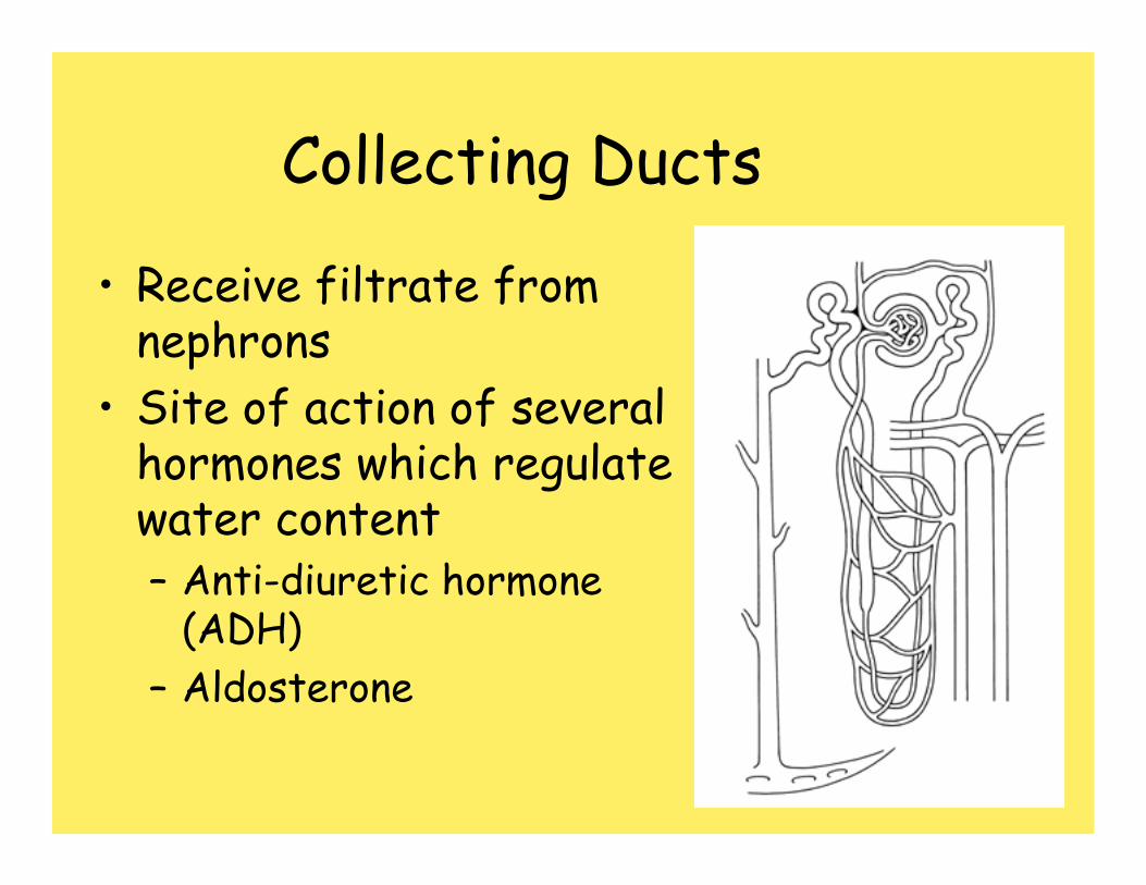
Collecting Ducts
• Receive filtrate from nephrons
• Site of action of several hormones which regulate water content– Anti-diuretic hormone
(ADH)– Aldosterone
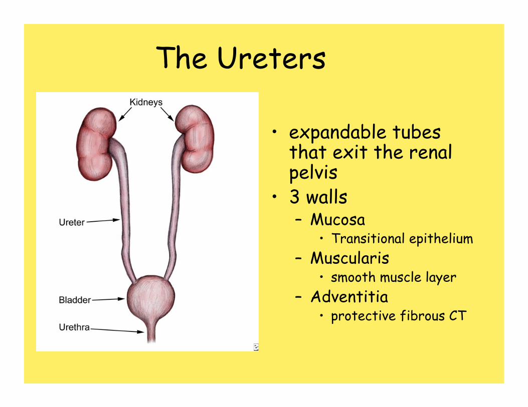
The Ureters
• expandable tubes that exit the renal pelvis
• 3 walls– Mucosa
• Transitional epithelium– Muscularis
• smooth muscle layer– Adventitia
• protective fibrous CT
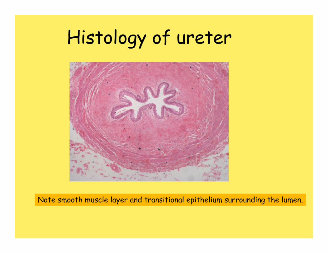
Histology of ureter
Note smooth muscle layer and transitional epithelium surrounding the lumen.
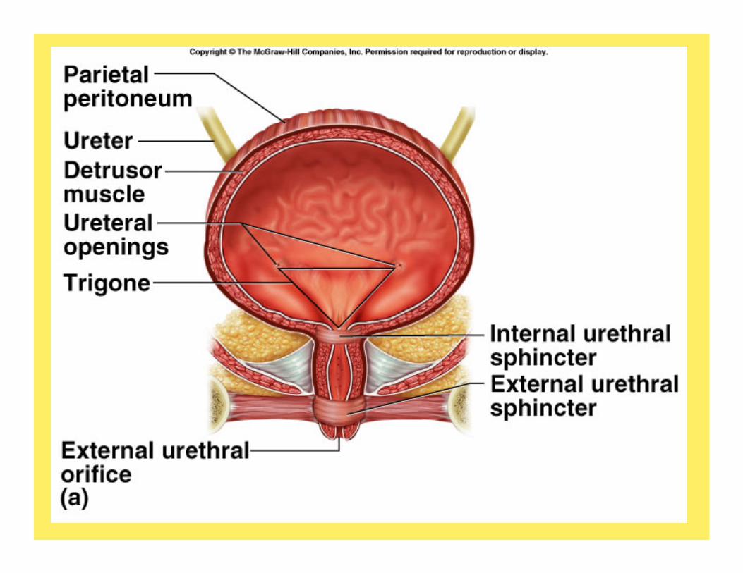
The Urinary bladder• Functions to store urine • Structure
– Rugae• macroscopic folds as in the stomach• flatten when the urinary bladder is distended
– Trigone• triangular region of the bladder• no rugae• location of openings to the ureters and urethra
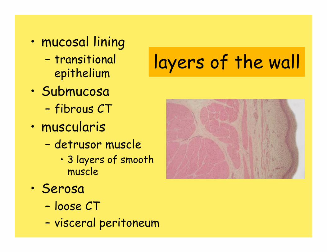
layers of the wall• mucosal lining
– transitional epithelium
• Submucosa– fibrous CT
• muscularis– detrusor muscle
• 3 layers of smooth muscle
• Serosa– loose CT – visceral peritoneum
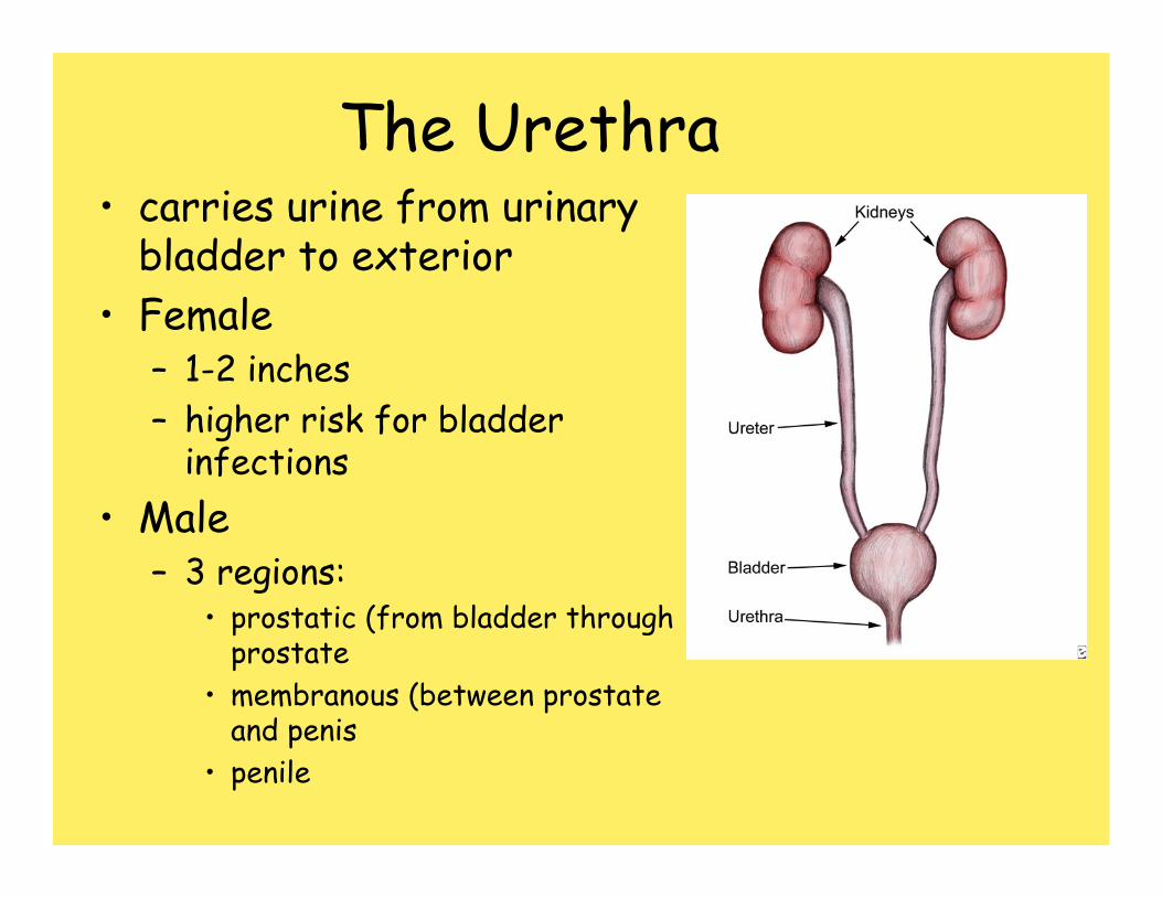
The Urethra• carries urine from urinary
bladder to exterior• Female
– 1-2 inches– higher risk for bladder
infections• Male
– 3 regions:• prostatic (from bladder through
prostate• membranous (between prostate
and penis• penile
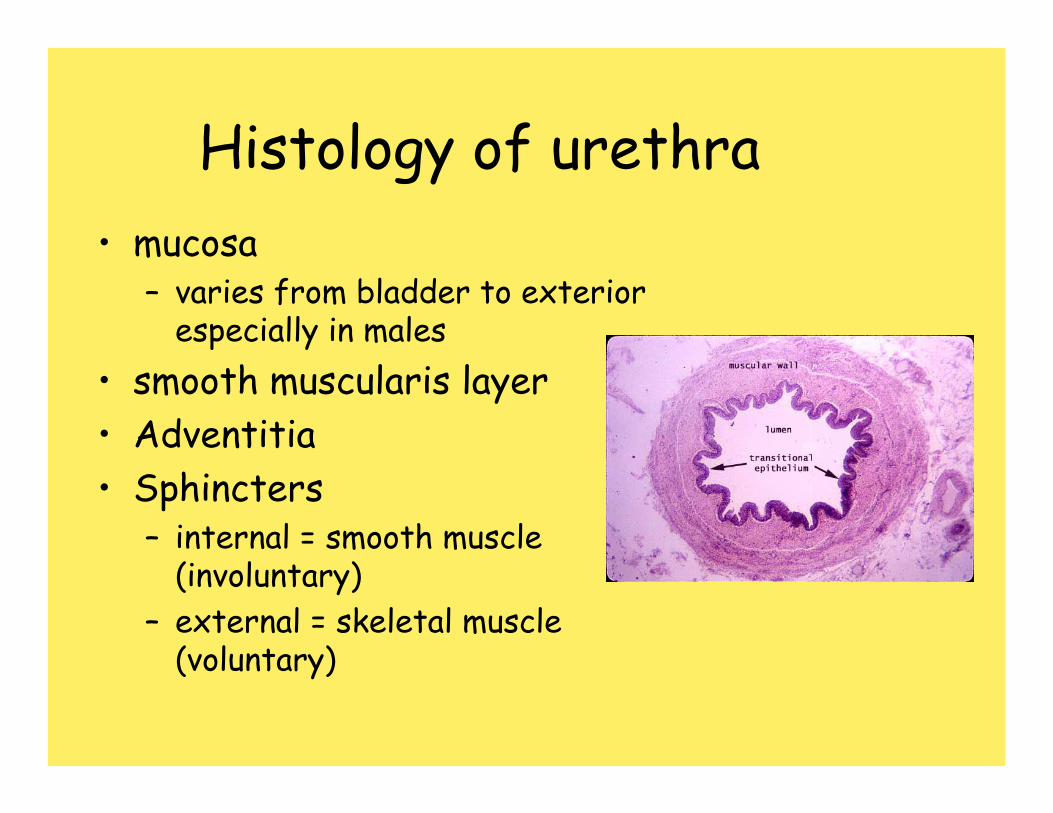
Histology of urethra• mucosa
– varies from bladder to exterior especially in males
• smooth muscularis layer• Adventitia• Sphincters
– internal = smooth muscle (involuntary)
– external = skeletal muscle (voluntary)
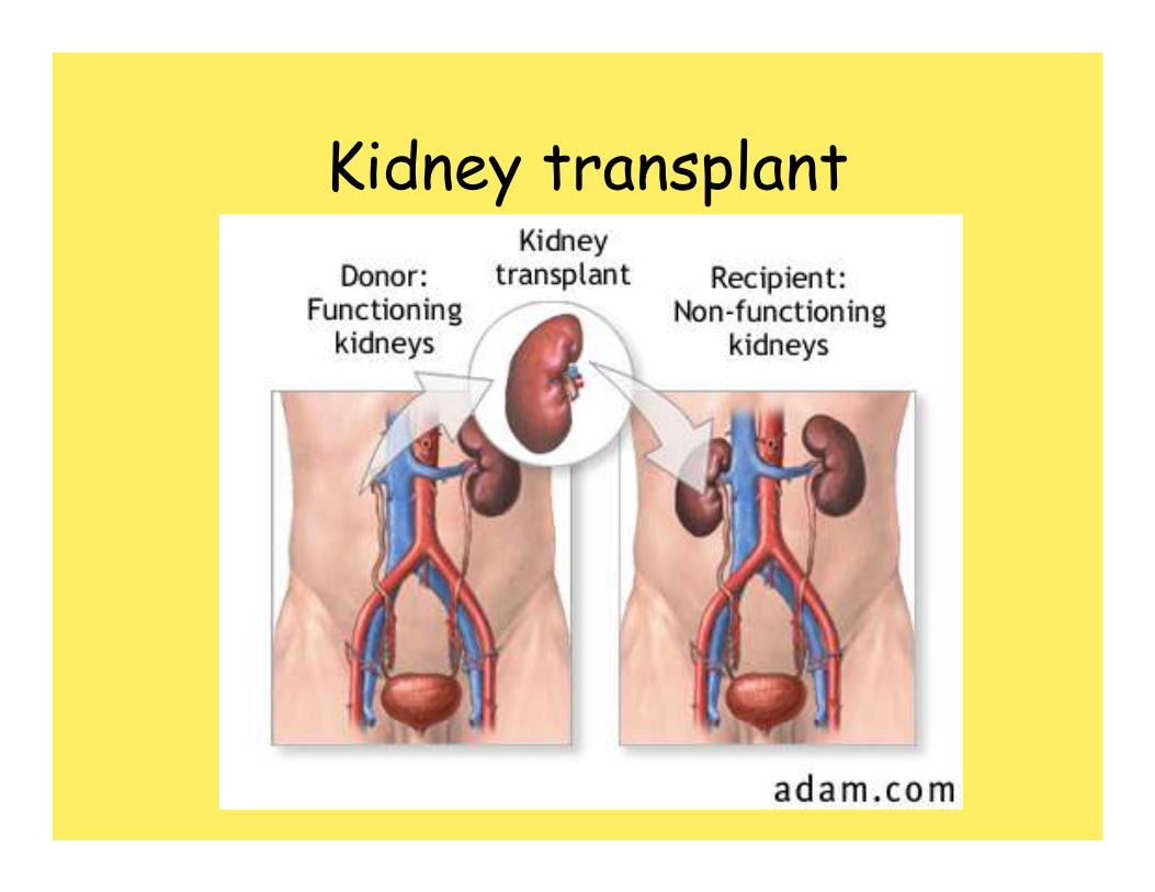
Kidney transplant

Kidney transplant
• Chronic kidney failure ≈ 260,000 patients
• Major causes of kidney disease– Diabetes (32%)– High blood pressure (25%)– Autoimmune disorders– Congenital abnormalities– Acute infections
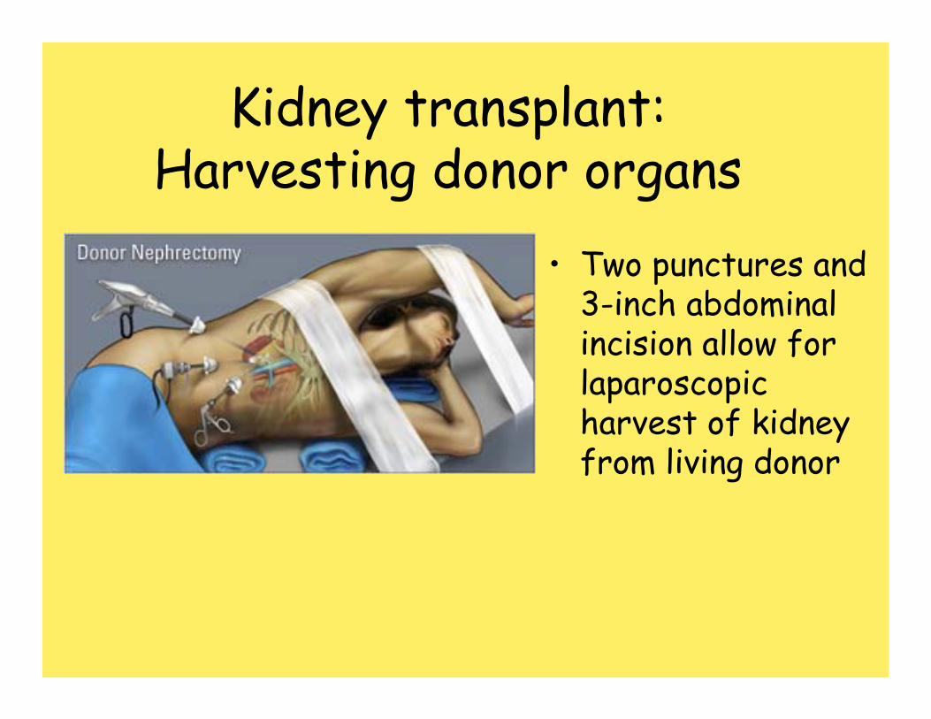
Kidney transplant:Harvesting donor organs
• Two punctures and 3-inch abdominal incision allow for laparoscopic harvest of kidney from living donor
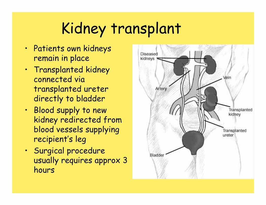
Kidney transplant• Patients own kidneys
remain in place• Transplanted kidney
connected via transplanted ureterdirectly to bladder
• Blood supply to new kidney redirected from blood vessels supplying recipient’s leg
• Surgical procedure usually requires approx 3 hours