Embed Size (px)
Citation preview

8/8/2019 Urinary Organs
http://slidepdf.com/reader/full/urinary-organs 1/18
URINARY ORGANS
8/8/2019 Urinary Organs
http://slidepdf.com/reader/full/urinary-organs 2/18
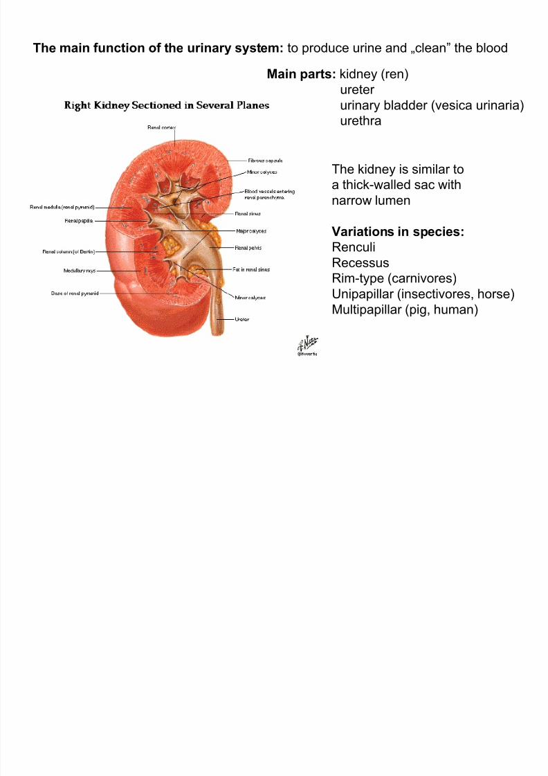
The main function of the urinary system: to produce urine and Äclean´ the blood
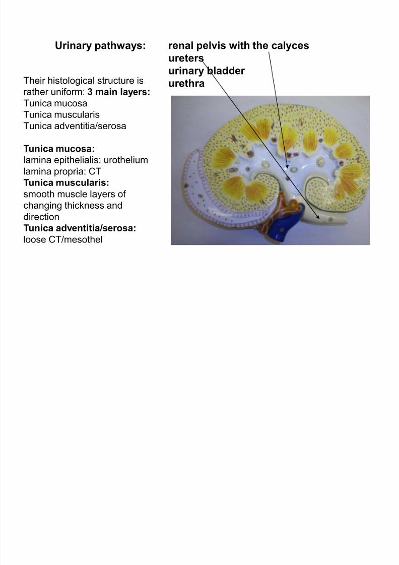
Main parts: kidney (ren)
ureter urinary bladder (vesica urinaria)
urethra
The kidney is similar to
a thick-walled sac withnarrow lumen
Variations in species:
Renculi
Recessus
Rim-type (carnivores)
Unipapillar (insectivores, horse)
Multipapillar (pig, human)
8/8/2019 Urinary Organs
http://slidepdf.com/reader/full/urinary-organs 3/18
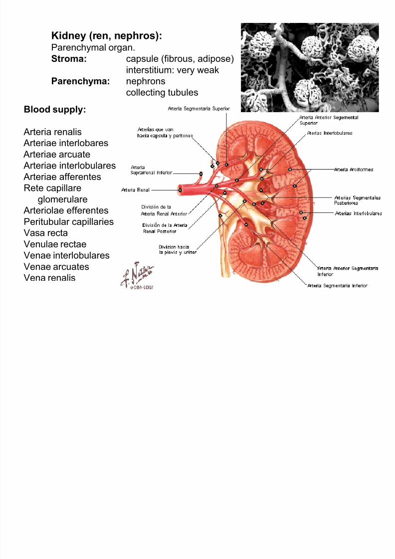
Kidney (r en, nephros):Parenchymal organ.
Stroma: capsule (fibrous, adipose)
interstitium: very weakPar enchyma: nephrons
collecting tubules
Blood supply:
Arteria renalis
Arteriae interlobares
Arteriae arcuate
Arteriae interlobulares
Arteriae afferentes
Rete capillare
glomerulare
Arteriolae efferentes
Peritubular capillaries
Vasa recta
Venulae rectae
Venae interlobulares
Venae arcuates
Vena renalis

8/8/2019 Urinary Organs
http://slidepdf.com/reader/full/urinary-organs 4/18
The nephron:Structural and functional unit of kidney parenchyma.
Main parts: renal corpuscle (Bowman¶s capsule,) glomerulus
tubulus renalis (proximal and distal convoluted tubules,
Henle¶s loop)
8/8/2019 Urinary Organs
http://slidepdf.com/reader/full/urinary-organs 5/18
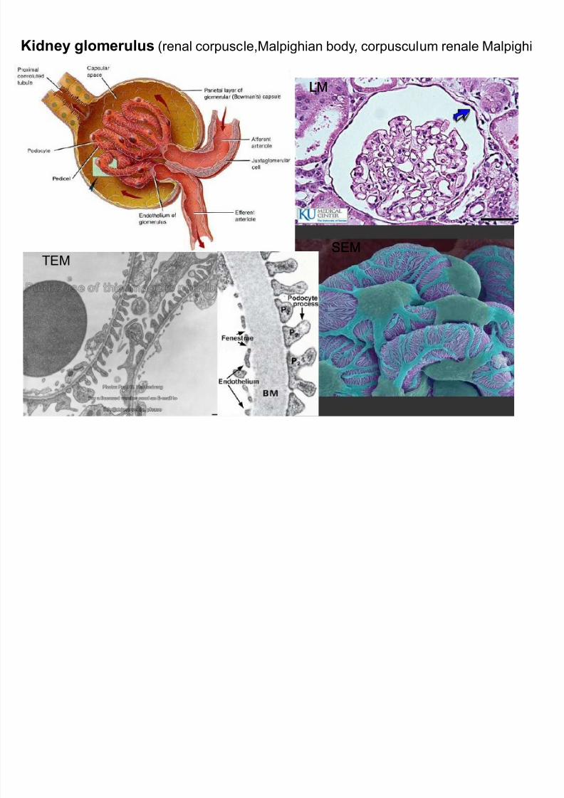
Kidney glomerulus (renal corpuscle,Malpighian body, corpusculum renale Malpighi
LMLM
SEMSEMTEM
8/8/2019 Urinary Organs
http://slidepdf.com/reader/full/urinary-organs 6/18
Ultrafiltration apparatus: fenestrated endothelium of capillaries
basal lamina (molecular filter)
filtration slits between podocyte pedicels
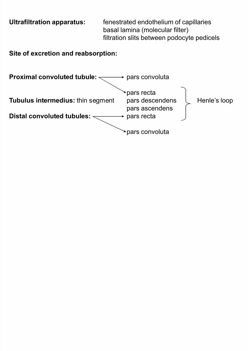
Site of excr etion and r eabsorption:
Proximal convoluted tubule: pars convoluta
pars recta
Tubulus intermedius: thin segment pars descendens Henle¶s loop
pars ascendens
Distal convoluted tubules: pars recta
pars convoluta
8/8/2019 Urinary Organs
http://slidepdf.com/reader/full/urinary-organs 7/18
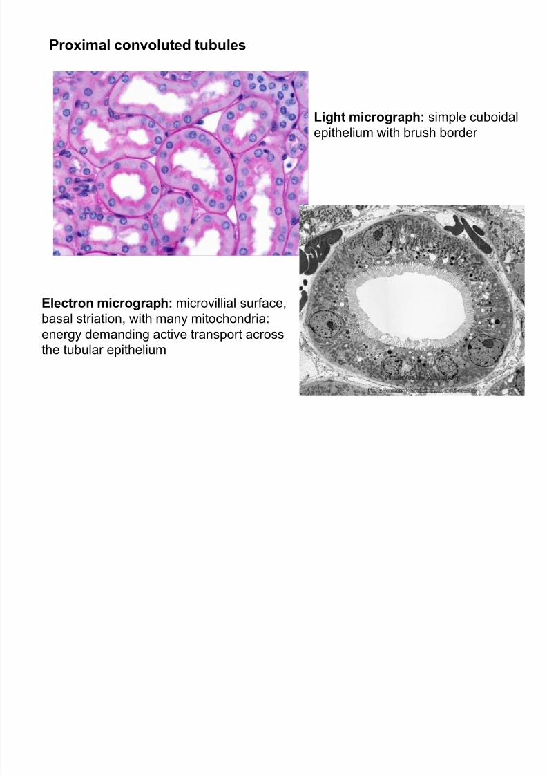
Proximal convoluted tubules
Light micrograph: simple cuboidal
epithelium with brush border
Electron micrograph: microvillial surface,
basal striation, with many mitochondria:
energy demanding active transport across
the tubular epithelium
8/8/2019 Urinary Organs
http://slidepdf.com/reader/full/urinary-organs 8/18
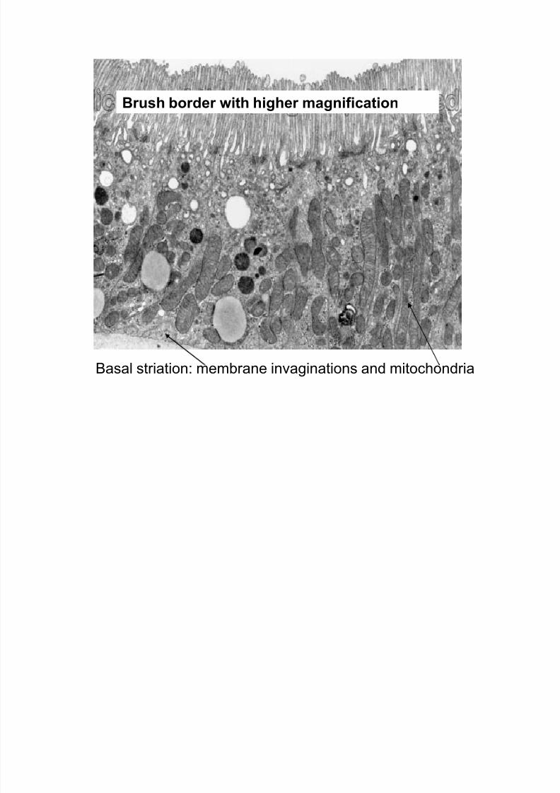
Brush border with higher magnification
Basal striation: membrane invaginations and mitochondria
8/8/2019 Urinary Organs
http://slidepdf.com/reader/full/urinary-organs 9/18
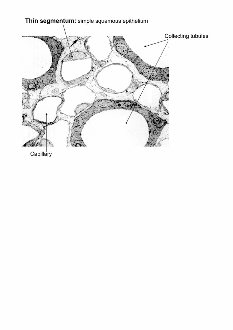
Thin segmentum: simple squamous epithelium
Capillary
Collecting tubules
8/8/2019 Urinary Organs
http://slidepdf.com/reader/full/urinary-organs 10/18
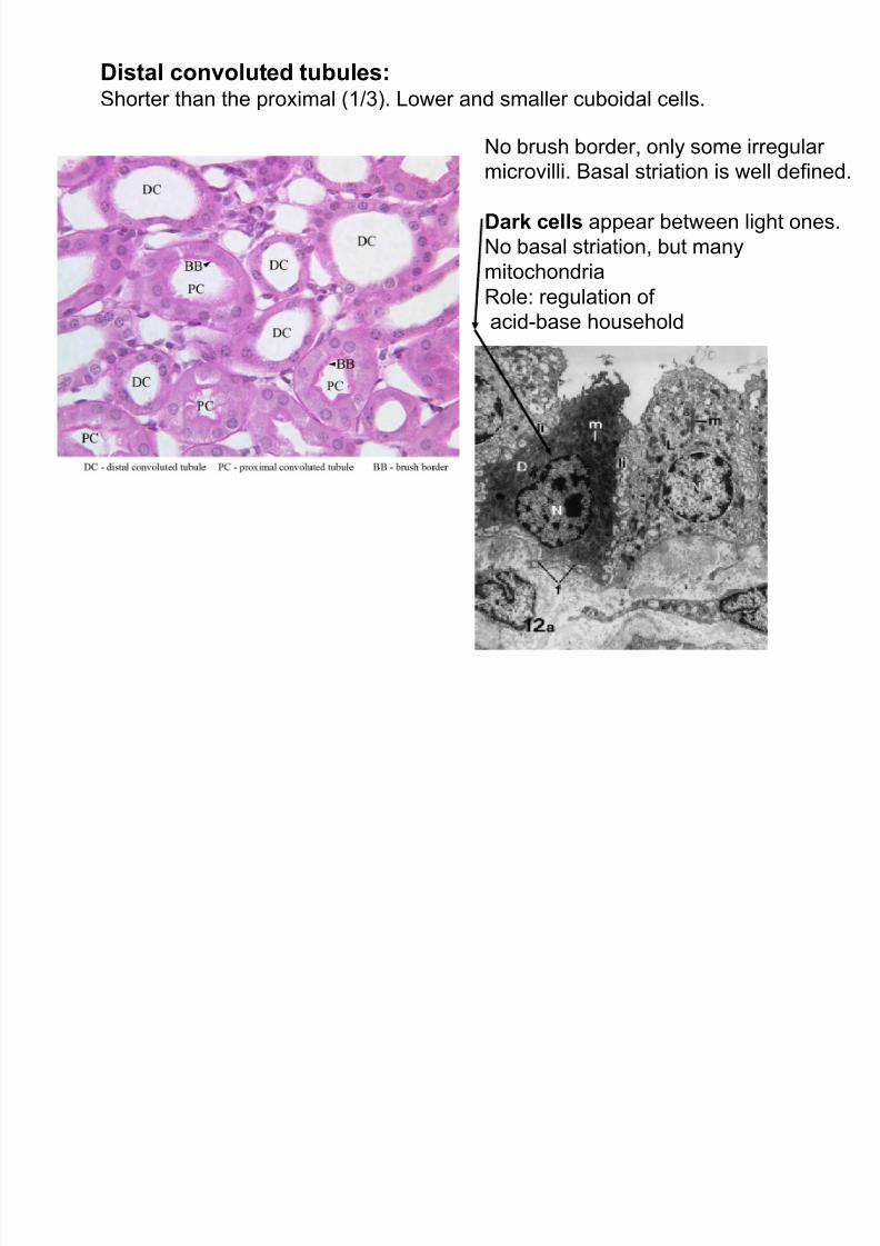
Distal convoluted tubules:Shorter than the proximal (1/3). Lower and smaller cuboidal cells.
No brush border, only some irregular microvilli. Basal striation is well defined.
Dark cells appear between light ones.
No basal striation, but many
mitochondria
Role: regulation of acid-base household
8/8/2019 Urinary Organs
http://slidepdf.com/reader/full/urinary-organs 11/18
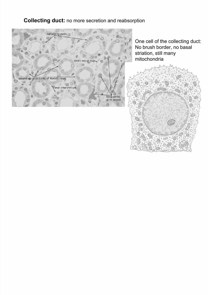
Collecting duct: no more secretion and reabsorption
One cell of the collecting duct:
No brush border, no basal
striation, still many
mitochondria
8/8/2019 Urinary Organs
http://slidepdf.com/reader/full/urinary-organs 12/18
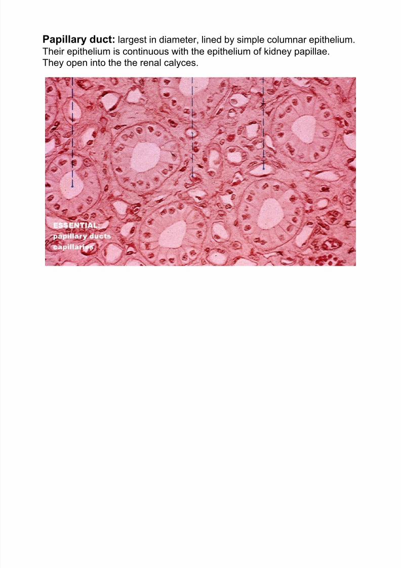
Papillary duct: largest in diameter, lined by simple columnar epithelium.
Their epithelium is continuous with the epithelium of kidney papillae.
They open into the the renal calyces.

8/8/2019 Urinary Organs
http://slidepdf.com/reader/full/urinary-organs 13/18
Juxtaglomerular apparatus (JGA): macula densa, juxtaglomerular cells,
extraglomerular mesangial cells.
Macula densa (MD): the distal convoluted tubule (DC) returns to the vascular pole
of its glomerulus: there the tubular epithelium becomes higher, nuclei are arranged in
a dense group. Function: osmoregulation via sensation of Na+ cc. in the distal tubule.Juxtaglomerular cells (JG): strongly modified myoepithelial cells in the wall of the
afferent arteriole. They sense the decrease in blood pressure, then produce renin,
an enzyme catalysing the production of active angiotensin, a potent raiser of
systemic blood pressure.
Extraglomerular mesangial cells:
Flat elongates cells forming fineMeshwork . Their function is obscure.
?Production of erythropoietin?
(stimulation of the erythropoiesis
in the red bone marrow)

8/8/2019 Urinary Organs
http://slidepdf.com/reader/full/urinary-organs 14/18
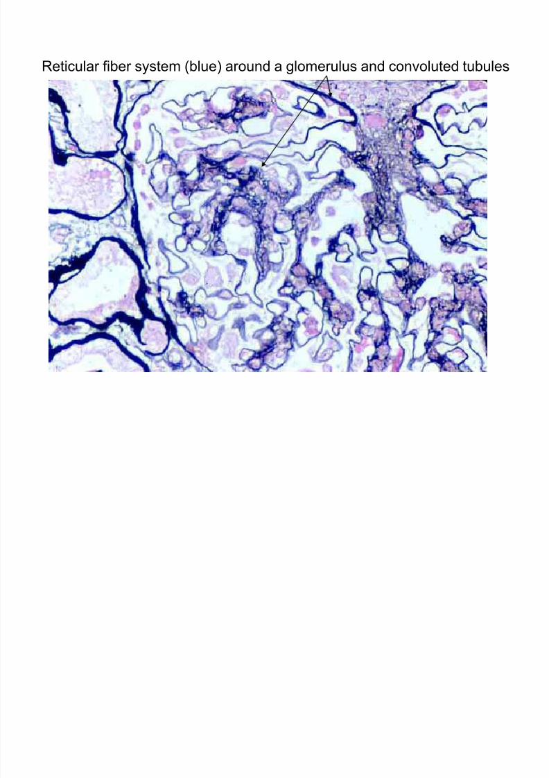
Interstitium of the kidney:Occupies very small volume, but it has importance in function.
All transport processes take place across the thin interstitum.
Its amount is lowest in the cortex, increases in medulla, towards the papillae.
Main fibrous component: meshwork of fine reticular fibers around the tubular
system.
Interstitial cells: fibroblasts, macrophages,dendritic cells, lymphocytes,
lipid-storing interstitial cells.
Erythropoietin (EPO): produced by intertubular fibroblast cells.Low oxygen tension stimulates the production of EPO.
8/8/2019 Urinary Organs
http://slidepdf.com/reader/full/urinary-organs 15/18
Reticular fiber system (blue) around a glomerulus and convoluted tubules
8/8/2019 Urinary Organs
http://slidepdf.com/reader/full/urinary-organs 16/18
8/8/2019 Urinary Organs
http://slidepdf.com/reader/full/urinary-organs 17/18
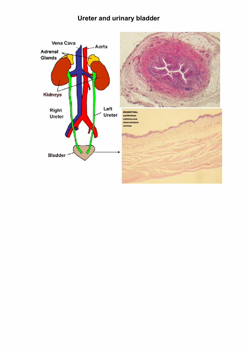
Ur eter and urinary bladder

8/8/2019 Urinary Organs
http://slidepdf.com/reader/full/urinary-organs 18/18
Female ur ethra
Tunica mucosa: folded
Lamina epithelialis: urothelium,
or stratified squamous keratinized
Lamina propria: loose CT with
wide lumened blood vessels
(cavernous tissue)Tunica muscularis:
Inner circular and outer longitudinal
smooth muscle layers
Tunica adventitia: loose CT,
blood and lymphatic vessels, nerves
Male ur ethra: see male genital organs


![DOCUMENT RESUME The Genitourinary System (and] … · · 2014-02-18The major organs of the urinary system that filter the blood and produce urine. ... Name the urinary ducts which](https://img.pdfslide.us/doc/110x75/5ace38057f8b9a6a678e779d/document-resume-the-genitourinary-system-and-major-organs-of-the-urinary-system.jpg)