Embed Size (px)
Citation preview

J. Neurol. Neurosurg. Psychiat., 1950, 13, 106.
UR/EMIC AND TROPHIC DEATHS FOLLOWING LEUCOTOMY:NEURO-ANATOMICAL FINDINGS
BYTURNER McLARDY
From the Institute of Psychiatry, University ofLondon
Little has been published concerning theoccurrence of delayed operative death afterbilateral prefrontal leucotomy. The term " delayedoperative death" in relation to leucotomy wasintroduced by Meyer and McLardy (1948;1949) in analysing undesirable clinical sequelesuffered by patients submitted to leucotomy whohad survived the danger of fatal operative haemor-rhage, and whose brains had in due course comeunder systematic investigation at the MaudsleyResearch Laboratory. I have elsewhere (McLardy,1948) described how I deduced from a study of thebrains and clinical records of 101 leucotomy casesthat probably as high a proportion of " the leucoto-mized population" dies a delayed operative deathwithin five months of the operation as fromhxmorrhage within two weeks of the operation.
In these three previous studies it was shown thatdelayed operative death was clearly related to thepost-mortem finding of leucotomy lesions extendingposterior to prefrontal domains in both hemi-spheres. It was not attempted, however, to decidewhether the most responsible part of the lesionsmight be damage to the premotor regions, theputamen, the caudate nucleus, the posterior partsof the orbital region, or structures lying betweenany of these. Such a more precise anatomicalanalysis has now been carried out on a larger seriesof cases. The findings, which are tabulated anddiscussed in the present paper, seem to providestrong evidence that at least bilateral substantialdamage to agranular orbital cortex, or to the regionof the subcallosal fasciculus at certain levels, isfatal.
Material and DataThe brain material and clinical records of 122
leucotomy cases have been collected from some 40British hospitals. Some of the records are ex-tremely sketchy, due to war-time circumstances.Most of the patients had not shown sufficient,clinical improvement to warrant discharge and haddied in the hospital in which their leucotomy had
been performed. The shortest post-operative sur-vival period in the series was five hours, the longestfive and a half years. Thirty-one of the patientsdied from cerebral hvmorrhage due to the opera-tion, all within 14 days (average 5 days). Fourdied from intracranial infection, all within 16 days(average 10 days). Of the remaining 87 cases,27 survived for a period of less than six months.Of these 27 cases, two are discarded from theanalysis because of undisputed death from organicbrain disease, one on account of death by suicide,and two others because they are almost certainlyexamples of the liability of post-encephalitic patientsto die after any surgical operation. This leaves 22cases where the particular nature of the early deathafter operation might be causally related to theparticular leucotomy lesions.The 22 cases are listed and numbered serially in
the Table in the order of length of post-operativesurvival. The only other clinical data tabulatedare the age at death, the apparent cause of death,and, where relevant, the date of recorded onsetof trophic phenomena. Sex and the nature andduration of illness showed no correlation trendwhatever with the cause or time of death (nor,actually, did age at death). On the anatomical side,involvement of " dorsal non-granular region "implies extension of the lesion into cortex or digitalwhite matter of the superior frontal gyrus at thetopographical level of Brodmann's (dysgranular)area 8 or (agranular) area 6 (Fig. 1). The cyto-architecture was not checked histologically in everycase. Involvement of " orbital non-granular region "implies ventral extension of the lesion into thosedysgranular and agranular zones of cortex, orrelated white matter, which Beck (1949) has deline-ated cytoarchitecturally within the posterior halfof the orbital region in the human brain. Thefigures " 3 " and " 4 " plotted in the last columnof the Table denote involvement of the third orfourth antero-posterior quarter respectively of theorbital region (Fig. 2): " c " indicates wherecortex as well as white matter was affected. By
106
by copyright. on 24 M
ay 2018 by guest. Protected
http://jnnp.bmj.com
/J N
eurol Neurosurg P
sychiatry: first published as 10.1136/jnnp.13.2.106 on 1 May 1950. D
ownloaded from

URL4EMIC AND TROPHIC DEATHS AFTER LEUCOTOMY
No.56 longitudinal bundle (Fig. 3), the external capsule,the uncinate fasciculus, the claustrum, the extremecapsule, and the insular cortex. Penetration of the
No.86 ,, ventricles per se likewise displayed no suggestivepositionoftassociation with delayed operative death.
10~~~~~~~~~~~~~~~~~~~~~~
aridctdyhinerpelns c\nt.
~~2/Il~~42Fii~~~~~~~~_
No.31A#
FIG. 2.-Beck's cytoarchitectural map showing theagranular (heavy stippling) and dysgranular (light
FIG. 1I.-Brodmann's standard cytoarchitectural map stippling) zones of cortex in the human orbitalof the frontal convexity and orbital region. The region. The interrupted lines and marginal numeralsposition of the head of the caudate nucleus is indicate the portioning into quarters employed inindicated in black. The relative position of the the present analysis.anterior horn and lateral angle of the lateralventricle is represented by the full line. Theposition and extent of the leucotomy lesions (ineach hemisphere) in cases Nos. 31, 56, and 86,are indicated by the interrupted lines.
striatumn" is meant caudate nucleus and putamen.For the purpose of this analysis the combinedvolume of these two nuclei which lie rostral to theanterior commissure was allotted the figure 100.The percentage fraction of this volume which wasdamaged in each hemisphere was then estimated /and tabulated. The two nuclei are not distinguished ,in the Table because separate correlation with /(.LBnature and time of death revealed no suggestive --trend. The " subcallosal fasciculus " is a fairlydistinctive structure visible even in fresh materialat the lateral angle of the lateral ventricle (Fig. 3)lying between the corpus callosum and caudatenucleus. In most cases its involvement or non-involvement could be determined by inspection. SWhen its damage by softening around the lesionwas doubtful, it was studied in coronal sectionsstained for cells, myelin, and axis cylinders.
Other anatomical variables which were taken FIG. 3.-Coronal section at the level of the middle ofinto account during the analysis but found to be the head of the caudate nucleus, showing theirrelevant in relation to survival time and nature position of the subcallosal fasciculus (S), theofdet,wredmgohiglaegrs cingulate fasciculus (C), the superior longitudinalof death, were cdamage to thle cingulate gyrus bundle (S.L.B.), the superior occipito-frontal
(area 24), the cingular belt (area 32), the superior fasciculus (0), and the uncinate fasciculus (U).
107
r
by copyright. on 24 M
ay 2018 by guest. Protected
http://jnnp.bmj.com
/J N
eurol Neurosurg P
sychiatry: first published as 10.1136/jnnp.13.2.106 on 1 May 1950. D
ownloaded from

TURNER McLARDY
DiscussionThe absence of any death between the twenty-
ninth day and the seventh week after operationdivides the cases in the Table into two groups.The eight cases ranging in survival period from5 hours to 29 days will be called Group I, theremaining 14 cases Group II. That the gapbetween the groups is not due entirely to chance willbecome apparent.
Group I: Uremia and Bilateral Damage to Cortexof Posterior Parts of Area 47
A striking feature of Group I is the occurrenceof four consecutive cases, dying from 20-24 daysafter operation, all from uremia. The concentrationof urea in the blood was investigated only inNos. 5 and 6, but Nos. 4 and 7 showed very similarclinical symptoms. The essentials of the protocolsafter operation are as follows:Case No. 4.Day after operation 1-9. Very restless.
9. Generalized jerky movements.,, ,, ,, 16. Very drowsy; semicoma-
tose; blood pressure 116/87(pre-operative blood pressure,124/90).
,, ,, ,, 17. Deep coma; temperature,990 F.; pulse, 124; respira-tion, 30.
19. Still in coma; slightly cya-nosed.
20. Died in coma. (No bloodurea recordings. Kidneys notremarked upon at necropsy.)
Case No. 5.Day after operation 3. Blood pressure back to nor-
mal (160/100); no speech;blood urea rising.
,, ,, ,, 5. Drowsy; retention overflowof urine; blood pressurenormal.
,, 6. Blood urea reached peak of500 mg. %.
,,,, ,, 9. Bladder function normal;blood urea falling; bloodpressure, 140/85; still nospeech.
,, ,, ,, 13. Very pale; temperature,990 F.
14. Blood urea down to 105mg%.
16. Pupils sluggish; hand-fed;urine normal; blood pressure,120/60.
17. Blood urea rising again;blood pressure, 130/90; verystuporose; continual twitchingof lips; occasional hiccough;breath smells of urine ; lumbarand ankle oedema; pulse, 120.
18. Comatose; blood urea 305mg%; blood pressure too lowto measure.
Day after operation 20. Died after generalized con-vulsion. (Bilateral, unsuspected,advanced pyelonephritis foundat necropsy.)
Case No. 6.Day after operation 3. Drowsy; pyrexial with slight
neck rigidity; temperature,99-101° F.
5. Drowsy; C.S.F. heavily blood-stained; temperature, 98.20 F.;pulse, 136; respiration, 23.
6. Now no signs of meningealirritation; blood urea 86-5mg%.
17. Very drowsy; doubly in-continent; blood urea 288mg%; temperature, 96.40 F.;pulse, 88; respiration, 20.
20. Comatose; blood urea 333mg%; temperature, 1000 F.;pulse, 130; respiration, 34.
21. Died in coma. (No bloodpressure recordings. Tubercu-lous left kidney removed sixmonths before operation, butright normal at necropsy.)
Case No. 7.Day after operation 6. Sharp rise in temperature to
1030 F.9. Sharp rise in temperature to
1030 F. again.11. Looks very well now, but
rather drowsy and not eatingwell.
15. Temperature still down.16. Up from bed for two hours.
20-23. Rather drowsy and unwell;twitches from time to time;sluggish pupillary light reaction.
,,,, ,, 24. Died in coma. (No bloodurea or blood pressure record-ings. Kidneys not remarkedupon at necropsy.)
A second notable feature is that these four casesall have bilateral damage to the cortex within thethird or fourth quarter of the orbital region as theironly common (bilateral) anatomical characteristicand as a characteristic which distinguishes themfrom practically all other cases in the Table. CaseNo. 9, in Group II, had bilateral involvement ofthe fourth quarter of the orbital region, but it wasconfined to slits in, and running parallel with, thefibres of the digital white matter. Case No. 12was the only case in Group II having bilateraldamage to the cortex within the posterior half ofthe orbital region and, significantly, the onlycase in Group IL to die of uremia. This case, itwill be noted, had considerably less damage to otherposterior structures than the cases in Group I.Among the 60 cases surviving over six months,there are only two (Nos. 56 and 86) which hadbilateral damage to the cortex in the third or fourth
108
by copyright. on 24 M
ay 2018 by guest. Protected
http://jnnp.bmj.com
/J N
eurol Neurosurg P
sychiatry: first published as 10.1136/jnnp.13.2.106 on 1 May 1950. D
ownloaded from

U-RA3T,MIC AND TROPHIC DEATHS AFTER LEUCOTOMY10
quarter. No. 56, dying nine months after operation
without manifest evidence of uremia, will be men-
tioned later. No. 86 had no other posterior
structures involved : the patient, a woman, died
from uriemia of unknown origin 11I months after
operation, aged 65 years.
These facts would seem to point strongly to
bilateral damage of the third or fourth quarter of
the orbital cortex being a determinant of fatal
urremia, and to the probability that coincidental
damage to other extra-prefrontal structures acceler-
ates such an outcome. Scoville's (1949 a, b)
experience of successful therapy by bilateral under-
cutting of the posterior orbital cortex, right up to
the optic chiasma would seem at first sight to
militate against such a conclusion ; likewise
Poppen's (1948) experience with the special pos-
terior ventral extension of his cuts in treatingintractable pain. It must be remembered, however,
that these two surgeons confined their lesions
essentially to the white matter, whereas it was by
irritating the cortex of area 13 in the posterior
orbital region, by electrical stimulation in cats,
that Cort (quoted by Livingston, 1948 a), claimed
to reproduce Trueta's kidney shunt mechanism in
his laboratory. It may be, therefore, that only
direct interference with the cortex produces striking
autonomic disturbances. Whether the uremia was
due to direct neural influence on kidney circulation,
or was dependent on blood pressure changes (as
suggested by Donovan, Galbraith, and Jackson,
1949) or on other factors, is impossible to judge
from the information available in the present six
cases. Certainly none of these cases gave any
history of post-operative ileus or of ulceration in
the gastro-intestinal tract (cf. Sweet and others,
1948). Only one had definite pre-operative renal in-
sufficiency.
More minute analysis of the position of the
orbital lesions in cases Nos. 4 to 7, 12 and 86 shows
only the lateral half, i.e., posterior area 47 (Fig. 2),
to be consistently bilaterally involved in all six.
It is within this region that in all six cases there was
substantial involvement, on at least one side,, of
cortex as well as of white matter. This would
suggest that the whole of the orbital non-granular
strip of cortex may well play as important a role in
autonomic affairs in man as does area 13 in monkeys.
TABLERELEVANT CLINICAL AND ANATOMICAL DATA IN 22 CASES SURVIVING LEUCOTOMY BY LESS THAN SIX MONTHS
Clinical Data Anatomical Structures Involved
Dorsal IOrbitalNon- Sb titm NnSerialAggrnlr cloa (prcn.gaurNumber Survival ageas o et Rgranua Fsciub-s dariatum Negon-Period Dah(Brodmann's FaccSueaagdeFgionDeath ~~~~~~~~Areas)(eFg2L R L R L R L R
I 5 hours 29 Respiratory inhibition .. 8 - 0 0 4, c 4, c2 7 days 73 ? Uremia and hxmorrhage 8 8 + - 20 20 4, c -3 13 days 66 ? Uremia and hxemorrhage - - - - 25 3013 4, c4 20 days 25 Uriemia. 8 8 -- 12 8 4, c 4, c5 20 days 49 Uremia 8 - + + 10 12 4, c 4, c6 21 days 29 iUremia. 8 8 ± + 20 60 4, c 3, c7 24 days 18 'Uremia. .. - + + 10 0 4, c 3, c8 29 days 45 Gastric perforation 6 6, 8 + - 12 50 4 -
9 7 weeks 37 ? Over-sedation. 8 - + ± 5 0 4 410 7 weeks 44 ? Trophic deterioration - 8 + + 20 30 - 4, cil 8 weeks 48 Trophic deterioration (D. 32) ..6 8, 6 ± + 0 0 - -
12 8l weeks 72 Uremia. - - 0 5 3, c 4, c13 9 weeks 62 Trophic deterioration (D. 26) - 8,6,4 + + 10 5 - 314 91 weeks 48 Trophic deterioration (D. 29) 8, 6 - + + 55 50 - -
15 91 weeks 71 Lobar pneumonia . .- - + - 0 216 10 weeks 64 Trophic deterioration (D. 31) ..- - + + 0 0 3 -
17 101 weeks 31 Trophic deterioration (D. ?) ..- - + + 5 55 4 -
18 14 weeks 50 Trophic deterioration (D. 33) ..- - + + 20 15 3 -
19 15 weeks 42 Trophic deterioration (D. 42) ..- 8 + + 30 30 - -
20 19 weeks 37 ? Trophic deterioration ..- - + +1 15 20 4, c -21 201 weeks 60 Empyema .. . .- - - + 10 5 3 322 241 weeks1 35 Phthisis.. . . . 6 - - - 0 0 - -
"D" gives the day after operation of the first appearance of trophic symptoms.
109
by copyright. on 24 M
ay 2018 by guest. Protected
http://jnnp.bmj.com
/J N
eurol Neurosurg P
sychiatry: first published as 10.1136/jnnp.13.2.106 on 1 May 1950. D
ownloaded from

110
Livingston and his associates (1948 a a
indeed already recorded an impressioiorbital autonomic "active focus in ma
what more laterally situated than in lowerPercival Bailey (personal communicationfound the most active focus to lie in t]insular cortex. The absence of manifin case No. 56 might indicate that 4between posterior area 47 and the h(caudate nucleus must be cut to prc
uraEmia.Area 13 within the medial half of th
orbital region was in none of these ur
substantially damaged. Case No. 1, v
lesions did extend into area 13, has bee]elsewhere (Meyer and McLardy, 1948,In No. 8, dying from acute perforatistomach, there was softening of the wI
right putamen due to vascular damage.
Group II: Trophic Deterioration an
Damage to the Region of the Subcaliosal
A most striking feature of GroupII itis the remarkable similarity of the natuiin Cases 11, 13, 14, 16, 17, 18, and 19.only all became marasmic and died wicutaneous septic sores within two to fiveoperation (average survival, 11 weeks), Imanifested such cutaneous "trophic "the fifth or sixth post-operative weel
protocols are as follows:
Case No. 11.Day after operation 5. Doubly incontin
voraciously.6. Singing and tal
fully.32. Quieter; not
doubly incontinenveloped blisters oi
34. Shows extremeearly stage bedsordehydrated; appe
pain if moved.41. Early gangrene
and toes.48. Hand improvir
gangrene progressii50. Temperature, c
grade bronchialslight albuminuria.
54. Urine negative55. Marked wasting57. Died with brormonia and chron(Given post-operatvitamins.)
Case No. 14.Day after operation 12. Responding slc
education; takingself.
TURNER McLARD Y
nd b) have Day after operation 26. Weight fallen to 6st. fromthat the 7 st. 4 lb. at operation.
In is some- ' ' ,, 29. Large bedsores; blisters onhands.^primates". ,, ,, ,, 40. Blisters now purulent.
l, 1950) has ,, ,, ,, 56. Temperature beginning tohe anterior rise slowly; average pulse,est urwemia 110; respiration, 24.
68. Died with a temperature ofconnexions 103° F. (Given post-operativeead of the multiple vitamins.)duce fatal These seven cases will therefore be spoken of as
dying from "trophic deterioration ".e posterior On the anatomical side is the equally striking-emic cases fact that these seven are the only cases in the wholewhere both series with a post-operative survival of over sevenn discussed weeks and bilateral involvement of the subcallosal, Case 59). fasciculus at the level of the head of the caudateion of the nucleus (Fig. 1), with the exception of No. 56.hole of the Five other cases in the total series had bilateral
destruction of practically all white matter lateraland dorso-lateral to the subcallosal fasciculus at
d Bilateral the level of the head of the caudate nucleus, butI Fasciculus lived from 11 months to 51 years (average, 2-1 years)n the Table after the operation. There would thus seem to bere of death strong evidence that severe bilateral damage to thisThey not region of the subcallosal fasciculus is a determinant
th multiple of delayed operative death from marasmus accom-
months of panied by trophic skin lesions. It is difficult tobut all first exclude definitely the superior occipito-frontal bundlelesions in which lies just lateral and ventral to the subcallosal
k. Typical fasciculus. Detailed histological investigation ofseveral crucial cases is in progress.
Post-leucotomy trophic lesions have been system-atically studied only by Ziegler and Osgood (1945)
ent; eating and Meyer and McLardy (1948, 1949). From
[king cheer- purely clinical and x-ray investigations Ziegler andOsgood concluded that (non-fatal) post-operative
well; still oedema and bullae might be associated with moreit; has de- posteriorly placed lesions, especially those involvingn left hand. areas 8 and 6. Meyer and McLardy did not relate
inanition *ies tongue the clinical phenomena to any particular structure
ars to be in involved in posterior cuts.There appear to be no experimental observations
in left hand on the result of selective bilateral damage to theng but toe subcallosal fasciculus (Mettler, 1942). Whetherng. this structure was damaged in the bilateral operations:ough, low- on the striatum performed in man (Meyers, 1942 apneumonia; and b; Hamby, 1947) is not specifically stated in
for sugar. the descriptions published by the surgeons. It is,however, noteworthy that Hamby's operations
ichial pneu- proved fatal, even when done in two stages, whereastic nephritis. Meyers' patients survived. According to myive multiple measurements on numerous dissected brains, Meyers'
sagittal cut through dorsal premotor cortex 2 cm.from the midline and parallel to the falx should
food him- enter the roof of the lateral ventricle through thecorpus callosum, whilst Hamby's similar cut
by copyright. on 24 M
ay 2018 by guest. Protected
http://jnnp.bmj.com
/J N
eurol Neurosurg P
sychiatry: first published as 10.1136/jnnp.13.2.106 on 1 May 1950. D
ownloaded from
UR14iMIC AND TROPHIC DEATHS AFTER LEUCOTOMY
(described by Browder, 1947, 1948) made 3 cm.from the midline, should enter the angle of thelateral ventricle by passing through the subcallosalfasciculus. Hamby's case No. 13, we are told,died three months after the second operation " ina state of inanition". Not enough is known ofthe blood supply of the subcallosal fasciculus topermit of more than speculation that Dandy's (1946)nine cases of death within 51 days after ligature ofone or both anterior cerebral arteries may havebeen due to ischemic softenings affecting bothsubcallosal fasciculi.A subsidiary clinico-anatomical point illustrated
in Group II, but not shown in the Table, is that inthe cases dying from trophic deterioration the onlytwo with complete sparing of the striatum (Nos. 11and 16) were the only two in which the clinicalrecords reported that the patient experienced acutepain when moved after the appearance of cutaneous
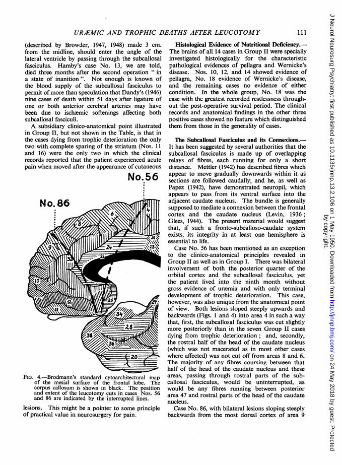
No.56
FIG. 4.-Brodmann's standard cytoarchitectural map
of the mesial surface of the frontal lobe. Thecorpus callosum is shown in black. The positionand extent of the leucotomy cuts in cases Nos. 56and 86 are indicated by the interrupted lines.
lesions. This might be a pointer to some principleof practical value in neurosurgery for pain.
Histological Evidence of Nutritional Deficiency.-The brains of all 14 cases in Group II were speciallyinvestigated histologically for the characteristicpathological evidences of pellagra and Wernicke'sdisease. Nos. 10, 12, and 14 showed evidence ofpellagra, No. 18 evidence of Wernicke's disease,and the remaining cases no evidence of eithercondition. In the whole group, No. 18 was thecase with the greatest recorded restlessness through-out the post-operative survival period. The clinicalrecords and anatomical findings in the other threepositive cases showed no feature which distinguishedthem from those in the generality of cases.
The Subcaliosal Fasciculus and its Connexions.-It has been suggested by several authorities that thesubcallosal fasciculus is made up of overlappingrelays of fibres, each running for only a shortdistance. Mettler (1942) has described fibres whichappear to move gradually downwards within it assections are followed caudally, and he, as well asPapez (1942), have demonstrated neuropil, whichappears to pass from its ventral surface into theadjacent caudate nucleus. The bundle is generallysupposed to mediate a connexion between the frontalcortex and the caudate nucleus (Levin, 1936;Glees, 1944). The present material would suggestthat, if such a fronto-subcalloso-caudate systemexists, its integrity in at least one hemisphere isessential to life.
Case No. 56 has been mentioned as an exceptionto the clinico-anatomical principles revealed inGroup II as well as in Group I. There was bilateralinvolvement of both the posterior quarter of theorbital cortex and the subcallosal fasciculus, yetthe patient lived into the ninth month withoutgross evidence of urnemia and with only terminaldevelopment of trophic deterioration. This case,however, was also unique from the anatomical pointof view. Both lesions sloped steeply upwards andbackwards (Figs. 1 and 4) into area 4 in such a waythat, first, the subcallosal fasciculus was cut slightlymore posteriorly than in the seven Group IL casesdying from trophic deterioration; and, secondly,the rostral half of the head of the caudate nucleus(which was not macerated as in most other caseswhere affected) was not cut off from areas 8 and 6.The majority of any fibres coursing between thathalf of the head of the caudate nucleus and theseareas, passing through rostral parts of the sub-callosal fasciculus, would be uninterrupted, aswould be any fibres running between posteriorarea 47 and rostral parts of the head of the caudatenucleus.
Case No. 86, with bilateral lesions sloping steeplybackwards from the most dorsal cortex of area 9
III
by copyright. on 24 M
ay 2018 by guest. Protected
http://jnnp.bmj.com
/J N
eurol Neurosurg P
sychiatry: first published as 10.1136/jnnp.13.2.106 on 1 May 1950. D
ownloaded from

TURNER McLARD Y
into the most rostral part of the angle of the lateralventricle (Figs. 1 and 4), surviving for 11 monthsand dying from uremia without trophic symptoms,would seem to exclude the participation of granularcortex in any fronto-subcalloso-caudate systemconcerned in fatal trophic deterioration. Again,No. 31 (Fig. 1) and two other cases, with survivalperiods of 9 months, 16 months, and 18 monthsrespectively, and bilateral deep ventral cuts sweepingupwards into the ventral parts of the anterior hornat about the level of the tip of the caudate nucleus,would seem to exclude the possibility that the" vital " fibres curve round the anterior horn fromorbital regions.
Finally, as already pointed out, all patients withbilateral destruction of the subcallosal fasciculusbetween the levels involved in cases Nos. 56 and 86died within five months of the operation. All theevidence, therefore, points to the existence of vitalstructures lying between the rostral half of the headof the caudate nucleus, the related portion of thesubcallosal fasciculus and cortex at the level ofBrodmann's areas 8 and 6, i.e., the structures lyingbetween the lesions in case No. 86 and case No. 56as illustrated in Figs. 1 and 4.
If these three regions contributed to a connectedvital system one might expect destruction of any oneof the elements to be equally fatal. The sub-callosal fasciculus, the bottle neck of the system,would be, as suggested by the present material, thepoint of greatest disruption by a minimal lesion.So far as damage to the caudate nucleus is con-
cerned, although survival in the present series doesnot appear to vary according to the (macroscopic)amount of its damage, there is no case of its bilateralsubstantial damage with survival over five months,with the one exception of case No. 87 living forone and a half years after the operation. In thiscase the damage to each caudate nucleus is confinedmore than in any other case to its ventricularsurface and would not presumably interfere with themajority of any connexions between the head of thecaudate nucleus and the cortex. Heath, Freedman,and Mettler (1947), it is interesting to recall, con-
sidered that the invariable death of felines subjectedto bilateral striatal ablation was most probably dueto metabolic dysfunction.The fact that, within the seven clear cases of fatal
trophic deterioration, brevity of survival is directlyproportionate, roughly, to the amount of dorsalnon-granular region involved in the lesions (seeTable), would seem in general to support theconception of such a vital fronto-subcalloso-caudatesystem. Yet, as is well known, very substantialbilateral destruction of dorsal non-granular cortexis not by itself lethal. This might imply merely that
the system is least vulnerable within its most diffuse,cortical, element. On the other hand, it mightindicate that the lethal factor in the dorsal leucotomylesions lies in a region not usually affected in clinicalor experimental surgery on the premotor cortex,namely the adjacent mesial cortex (Fig. 4). That theportion of area 32 lying rostral to the genu of thecorpus callosum (and premotor cortex) is not theregion concerned, would seem evidenced by theirrelevancy to survival of its involvement or non-involvement in the present series. There has beenequal absence of untoward incident, apparently,after fairly full ablation (Pool, 1949 a and b;personal communication, 1950) of this part ofarea 32, and after undercutting (Scoville, 1949 aand b; personal communication, 1950) of practi-cally the whole area. The cortical end of thepresumptive " vital system" cannot therefore beconfined to the whole of that area which is regardedas cytoarchitecturally homologous with the cortex(areas 32 and 31) to which all the suppressor areasin monkeys send connexions (McCulloch, 1944).Almost identical arguments are available againstarea 24 being the only region concerned. Extensionof investigations such as those of Meyers and hisassociates (1949) on electrical activity of the neo-striatum might clarify this problem. In the mean-time it may be relevant that these workers foundsuch uniformity of electrical activity in the caudatenucleus, the subcallosal fasciculus, and neighbouringparts as to suggest a common source of the dis-charge.The essentially metabolic nature of the trophic
deterioration might point to importance in itscausation of those cortical areas known to besignificant in autonomic affairs, such as areas 6and 24. The connexions concerned would thenpresumably be fibres such as those which haverecently been shown (M. Meyer, 1949) to projectfrom area 6 into the hypothalamus. Most of anysuch fibres from area 6 or area 24 to the hypo-thalamus might be expected to take the shortestcourse through, or immediately around, the sub-callosal fasciculus at the level of the head of thecaudate nucleus.
Experimental investigation of the subcallosalfasciculus from the point of view of its part indelayed operative death is in progress.
Significance of Grouping in Survival Times.-Whatever the validity of the more tenuous of thesedeductions from the data, it is clear that the timegap between Groups I and IL is due to more thanchance, and that a similar gap, or relative attenuationof cases when graded by survival period, is likewiseto be expected between Group II and cases dying at
112
by copyright. on 24 M
ay 2018 by guest. Protected
http://jnnp.bmj.com
/J N
eurol Neurosurg P
sychiatry: first published as 10.1136/jnnp.13.2.106 on 1 May 1950. D
ownloaded from

URA3MIC AND TROPHIC DEATHS AFTER LEUCOTOMY
random from intercurrent illness, or from any thirdgroup which might emerge in a larger series if therewere any tendency for cases with unilateral damageto extra-prefrontal structures to succumb to en-vironmental stresses which are not normally fatal.
Practical Implications of the Findings.-It is notproposed to speculate here on possible physiologicalmechanisms involved. Much of the relevantexperimental work has been referred to in theprevious papers quoted, and the elucidation ofsuppressor and other feed-back mechanisms pro-bably concerned is not sufficiently advanced to bearprofitable discussion. My practical concern is toutter a warning regarding two dangers additionalto hxmorrhage of blind leucotomy operations:delayed post-operative death from uremia andfrom trophic deterioration. The original trans-orbital leucotomy, as well as all the open methodsof leucotomy and cortical ablation, are almost*free of all three sources of mortality, but the newly-devised " deep " transorbital cut (Williams, 1949;Freeman, 1949) may ominously increase them.I have seen one such case with unilateraldamage to the striatum and subcallosal fasciculus.There would seem no good reason why the striatumand subcallosal fasciculus might not have beenequally damaged in both hemispheres, thus deter-mining the patient's death within five months.
SummaryIn a series of 22 leucotomy cases dying within
six months of the operation, analysis of the causesof death reveals two distinct groups : Group l,dying predominantly from uremia in the thirdpost-operative week, and Group Il, dying pre-dominantly from trophic deterioration (marasmuswith trophic skin lesions), between two and fivemonths after the operation. In the latter groupblisters or bulls almost always appeared on theextremities between the fourth and sixth post-operative weeks.Post-mortem anatomical (mainly macroscopic)
analysis of the lesions in the frontal lobes of these22 cases revealed a striking association of deathfrom uremia with bilateral damage to the posterior(non-granular) orbital cortex, and of death fromtrophic deterioration with bilateral cutting of theregion of the subcallosal fasciculus at the level ofthe head of the caudate nucleus.The orbital cortex concerned in the uremic
deaths appeared to be posterior area 47, not
area 13. Histopathological evidence of nutritionaldeficiency was not conspicuous among the cases oftrophic deterioration.From evidence supplied by these and other
relevant cases, in a total series of 122, it is deducedthat connexions between the caudate nucleus andBrodmann's (non-granular) cortical areas 8 and 6(including their mesial extension towards thecorpus callosum), via the rostral parts of thesubcallosal fasciculus, may be the structures whosedamage determined the fatal trophic deterioration.The practical intention of the paper is to emphasize
the serious danger of delayed operative death fromthe use of blind leucotomy techniques, includingthe " deep " transorbital cut.
The writer wishes to express his thanks to ProfessorAlfred Meyer for his interest in this investigation. Forthe opportunity to pursue all such studies he is indebtedto those medical superintendents and other medicalofficers who, realizing the potentialities of systematiccentralized investigation of such leucotomy material,contributed to the series.
REFERENCES
Beck, E. (1949). J. Anat., Lond., 83, 147.Browder, J. (1947). N. Y. St. J. Med., 47, 2589.--(1948). Amer. J. Surg., 75, 264.Dandy, W. E. (1946). Bull. Johns Hopk. Hosp., 79, 34.Donovan, J. F., Galbraith, A. J., and Jackson, H. (1949).
J. ment. Sci., 95, 655.Freeman, W. (1949). Proc. R. Soc. Med., Suppl. 42, 8.Glees, P. (1944). J. Anat., Lond., 78, 47.Hamby, W. B. (1947). N. Y. St. J. Med., 47, 2592.Heath, R. G., Freedman, D. A., and Mettler, F. A.
(1947). Fed. Proc., 6, 126.Levin, P. M. (1936). J. comp. Neurol., 63, 369.Livingston, R. B., Fulton, J. F., Delgado, J. M. R.,
Sachs, E., Brendler, S. J., and Davis, G. D. (1948a).Res. Pub. Ass. Res. nerv. ment. Dis., 27, 405.
--, Chapman, W. P., Livingston, K. E., and Kraintz,L. (1948b). Ibid., 27, 421.
McCulloch, W. S. (1944). In Buly, P. C. (Editor)'The Precentral Motor Cortex'. Univ. of IllinoisPress.
McLardy, T. (1948). 'Clinico-Pathological Studies on101 Cases of Leucotomy', M.D. Thesis, Univ. Glasgow.
Mettler, F. A. (1942). Res. Pub. Ass. Res. nerv. ment.Dis., 21, 150.
Meyer, A., and McLardy, T. (1948). J. ment. Sci., 94,555.
,(1949). Ibid., 95, 403.Meyer, M. (1949). Brain, 72, 265.Meyers, R. (1942a). Res. Pub. Ass. Res. nerv. ment. Dis.,
21, 602.(1942b). N. Y. St. J. Med., 42, 317., Hayne, R., and Knott, J. (1949). Journal of
Neurology, Neurosurgery and Psychiatry, 12, 111.
* One brain has already been examined where the original trans-orbital operation resulted in fatal hemorrhage from the lenticulo-striate vessels on one side.
113
by copyright. on 24 M
ay 2018 by guest. Protected
http://jnnp.bmj.com
/J N
eurol Neurosurg P
sychiatry: first published as 10.1136/jnnp.13.2.106 on 1 May 1950. D
ownloaded from

Papez, J. W. (1942). Res. Pub. Ass. Res. nerv. ment. Dis., Scoville, W. B. (1949b). Proc. R. Soc. Med., Suppl., 42, 3.21, 21. Sweet, W. H., Cotzias, G. C., Seed, J., and Yakovlev, P.
Pool. J. L. (1949a). Lancet, 2, 776. (1948). Res. Pub. Ass. Res. nerv. ment. Dis., 27, 795.-(1949b). Proc. R. Soc. Med., Suppl., 42, 1. Williams, J. M. (1949). Dig. Neurol. Psychiat., 17, 415.Poppen, J. L. (1948). Dig. Neurol. Psychiat., 16, 403. Ziegler, L. H., and Osgood, C. W. (1945). Arch. Neurol.Scoville, W. B. (1949a). J. Neurosurg., 6, 65. Psychiat., Chicago, 53, 262.
4:
114 TURNER McLARD Y
by copyright. on 24 M
ay 2018 by guest. Protected
http://jnnp.bmj.com
/J N
eurol Neurosurg P
sychiatry: first published as 10.1136/jnnp.13.2.106 on 1 May 1950. D
ownloaded from