Embed Size (px)
Citation preview
Uptake, metabolism, and cytotoxicity of isomeric cholesterol-5,6-epoxides in rabbit aortic endothelial cells
Alex Sevanian, Judith Berliner,* and Hazel Peterson
Institute for Toxicology and Department of Pathology, University of Southern California, Los Angeles, CA 90033 and Department of Pathology,* University of California School of Medicine, Los Angeles, CA 90024-1732
Abstract The isomeric cholesterol-5,6-epoxides represent two common cholesterol autoxidation products and along with their principal metabolic product, 3fi,5a,6P-cholestane triol, are pur- portedly angiotoxic. The uptake and cytotoxic action of these compounds was examined in cultured rabbit aortic endothelial cells emphasizing mechanisms of uptake and metabolic fate. The isomeric cholesterol epoxides are incorporated with equal facility and in a dose-dependent manner. The pattern of uptake, which is markedly influenced by media serum concentration and by temperature, suggests that these compounds are partly incor- porated through association with serum lipoproteins. After in- corporation, both epoxide isomers are rapidly converted to cholestane triol which readily exits the cells. Cholestane triol is further metabolized to an ester-type product representing up to 10% of the added cholesterol epoxides by 24 h of incubation. The order of cytotoxic potency of these cholesterol oxides is: cholestane triol > cholesterol-&epoxide > cholesterol-a-ep- oxide, with LD50 concentrations ranging from 23 to > 150 pM in confluent cells. Cholestane triol and cholesterol-@-epoxide are twice as cytotoxic to subconfluent cells as compared to confluent cells, whereas cholesterol-a-epoxide is essentially equitoxic to confluent and subconfluent cells. Cholesterol epoxide cytotoxic- ity is significantly reduced by treatments in the absence of serum in accord with substantial reduction in uptake when incubations are performed in serum-free media. Our findings show that these cytotoxic cholesterol oxides are incorporated by endothelial cells through a combination of receptor-mediated and nonspecific or passive mechanisms; however, the efficacy of up- take and resulting toxicity is substantially influenced by serum lipoproteins. - Sevanian, A., J. Berliner, and H. Peterson. Uptake, metabolism, and cytotoxicity of isomeric cholesterol-5,6-epoxides in rabbit aortic endothelial cells. J. Lipid Res. 1991. 32: 147-155.
Supplementary key words low density lipoproteins cholesterol epoxide cholestane triol cholesterol serum lipids
part, peroxidative damage to lipid components. Peroxida- tion of serum lipoproteins is thought to facilitate their rapid uptake and accumulation in resident macrophages and other cells of the vessel wall, and may also account for toxicity to the endothelium and underlying tissues (5-7). Several investigators have turned their attention to cholesterol oxidation as a contributing factor to atherosclerosis based on studies that showed that cholesterol oxides were angiotoxic and diets containing cholesterol oxidation products were highly atherogenic while similar diets free of these products were relatively nonatherogenic (8, 9).
Cholesterol oxides are formed during free radical- induced lipid peroxidation wherein cholesterol is a major component (10-13). Many of these cholesterol oxidation products are detected in human serum (14), are sig- nificantly elevated in humans with hypercholesterolemia (15), and can be isolated from foods (16, 17), tissues (18, 19), serum lipoproteins (ZO), and in human atheromatous plaques (21). However, it is unclear as to whether these cholesterol oxides are merely passive products of lipopro- tein lipid peroxidation (in which case they may serve as markers for oxidatively modified lipoproteins) or whether they contribute to the toxicity of oxidized lipoproteins.
Cholesterol oxidation in biological systems gives rise to numerous products, some of which predominate when biomembranes are subjected to lipid peroxidation (10, 13). Common cholesterol oxidation products include: cholesterol-5a,6a-epoxide (aCE), cholesterol-5P,6/3-
Hypercholesterolemia has long been considered a ma- jor risk factor for the development of atherosclerosis (1, 2). More recent studies suggest that progression of atherosclerosis may be mediated via free radical-induced modification of serum lipoproteins (3-5) involving, in
Abbreviations: LDL, low density lipoproteins; CE, cholesterol epox- ide; aCE, cholesterol-5a,6a-epoxide; BCE, cholesterol-5@,6fl-epoxide; CT, 3@,5a,6&cholestane triol; FBS, fetal bovine serum; REC, rabbit aortic endothelial cells; E A , trichloroacetic acid; VLDL, very low den- sity lipoproteins; HDL, high density lipoproteins; PBS, phosphate- buffered saline.
Journal of Lipid Research Volume 32, 1991 147
by guest, on April 9, 2019
ww
w.jlr.org
Dow
nloaded from
epoxide (PCE), and SP,5a,GP-cholestane triol (CT). The toxicities of these oxides, as well as other cholesterol ox- idation products, have been examined in vascular ex- plants (22), aortic smooth muscle cells (23), endothelial cells (24), and fibroblasts (25). Although some under- standing of their general toxicity is at hand, relatively lit- tle is known about the influence of cholesterol oxide uptake and metabolism on endothelial cell toxicity. En- dothelial cells represent a first line of exposure to cholesterol oxides as presented in serum lipoproteins. We describe in this report the relationship between uptake, metabolism, and cytotoxicity of three major cholesterol oxides, i.e., aCE, PCE, and CT, using rabbit aortic en- dothelial cells.
MATERIALS AND METHODS
Preparation of cholesterol oxides
Cholesterol was purchased from Sigma Chemical Co. (St. Louis, MO) and was found to be > 99% pure as de- termined by gas chromatography using chromatographic conditions described by Maerker and Unruh (26). Cholesterol epoxides were prepared from cholesterol by reaction with m-chloroperoxybenzoic acid as described previously (27). This procedure was also used for the preparation of radiolabeled cholesterol epoxides. Accord- ingly, la,2a-[3H]cholesterol, obtained from Amersham, Inc. (Arlington Heights, IL), was mixed with nonradioac- tive cholesterol to give a stock solution with a specific ac- tivity of 100 pCi/mol. The reaction yielded a nearly complete conversion to cholesterol epoxides (CE) produc- ing aCE and PCE at a ratio of 75:25, respectively. The isomeric mixture was separated by high performance li- quid chromatography (27) resulting in isolation of each isomer at > 98% purity. The mobile phase (composed of hexane-isopropanol) was removed by rotary evaporation, leaving the purified cholesterol epoxide crystals which were dissolved in absolute ethanol. C T was prepared from radioactive or nonradioactive aCE by mild hydrolysis using perchloric acid, as described previously (25), and dissolved in absolute ethanol. The specific activities of aCE, PCE, and CT were adjusted with corresponding unlabeled compounds immediately prior to their addition to cultured cells. Final specific activities of these com- pounds are presented in the figure legends. All com- pounds were added in ethanol (0.5% by volume) to the culture medium which was prepared at least 30 min prior to addition to cells.
Cell culture
Rabbit aortic endothelial cells (REC), obtained from New Zealand Albino rabbits, were used between passages 9 and 13. Their phenotypic characteristics have been
described previously (28) and were checked routinely via positive factor VI11 surface antigen, angiotensin convert- ing enzyme activity, and morphological appearance. These phenotypic markers were expressed at consistent levels throughout the study. Cells were maintained in 80/20 DMEM/M199 with 15% heat-inactivated fetal bovine serum (FBS) (Gibco, NY). Conditioned medium was also added at the time of medium change at a 1:4 ratio with fresh medium. Cells were passaged using a 1:3 split ratio, were allowed to grow to confluence, and transferred by mechanical disruption using a rubber policeman with dispersion by vigorous pipetting. Cultures were main- tained by weekly media changes and had a doubling time of approximately 28 h. Fresh complete media was added to the cells 24 h prior to each experiment.
Analysis of cholesterol epoxide distribution in serum components
To analyze the distribution of cholesterol epoxide among the serum components of the cell culture medium, 0.50 pCi of [3H]aCE (100 pCi/pmol) was added to 22 m1 of heat-inactivated FBS. The serum was then incubated in a shaker bath for 30 min at 37OC. Immediately after in- cubation the serum was subjected to ultracentrifugal frac- tionation of lipoproteins (29). Briefly, this involved layering the medium over 12.5% sucrose and centrifuga- tion for 21 h at 50,000 g. Fractions corresponding to den- sities ranging from 1.020 to 1.050 g/ml were collected and layered onto a 0-15% sucrose gradient and recentrifuged as above. Fourteen fractions were then collected, their densities were determined and aliquots were taken for measurement of radioactivity and protein content (Bio- Rad protein assay kit).
Uptake and metabolism of cholesterol epoxide
Studies on the uptake and metabolism of the cholesterol oxides involved addition of the radiolabeled compounds to complete media (medium containing 15% FBS) at specified concentrations in the presence of 10 pM [3H]aCE or PCE. The dishes were harvested at 1-2 h in- tervals up to 8 h and at selected intervals from 8 to 24 h. In experiments designed to test the effects of serum on up- take into cellular and extracellular components, cells were incubated in media prepared with FBS ranging from 15 to 0.5%, in the presence of a C E at 100 pCi/pmol. After 4 h incubation, cells were divided into two identical groups and washed three times with serum-free medium. To one group 0.1% trypsin was added for 10 min at 37OC, the cells were pelleted, and the supernatant was analyzed for trypsin-releasable counts. The cells from the second group were recovered by centrifugation, washed three times with serum-free medium, and aliquots were col- lected for measurement of radioactivity. This represents
148 Journal of Lipid Research Volume 32, 1991
by guest, on April 9, 2019
ww
w.jlr.org
Dow
nloaded from
total cellular radioactivity while the difference between total and trypsin releasable counts represents intracellular radioactivity.
Cholesterol oxide metabolites were analyzed by extrac- tion of the culture medium or cell pellet. Extraction of lipids was performed as described by Weithmann, Peter- son and Sevanian (30). Aliquots of the total lipid extracts were applied to silica gel-coated plates which were devel- oped in toluene-ethyl acetate 3:2. Measurements of radioactivity associated with zones correpsonding to authentic aCE, PCE, and C T standards were carried out as described previously (31). In those instances where up- take and/or metabolism of C T was of interest, plates were developed in a solvent system consisting of toluene-ethyl acetate-methanol 1:1:0.2. Plates were scanned using a Berthold radiochromatogram scanner (Weibald, Ger- many), and radioactive zones were recovered for more precise measurements using a liquid scintillation counter.
Cytotoxicity assays
Cholesterol oxides were added to either confluent (i.e., stationary) or subconfluent (logarithmically growing) cell cultures at various concentrations in order to establish cytotoxic dose ranges. Cytotoxic potencies were estimated on subconfluent and confluent cells from 72-h growth curves in the presence of the test compound (see Table 4), or by 24-h treatments of confluent cells with the test com- pound, replating, and measurement of plating efficiency at 24 h and cell growth for the following 72 h. Cell den- sities were typically IO5 cells/ml for confluent cells and 5 x lo4 cells/ml for subconfluent cells.
Duplicate dishes were used for each experimental con- dition and the data represent results from three to five in- dependent experiments. Results are expressed as the mean and standard error for each condition presented.
RESULTS
Uptake and metabolism
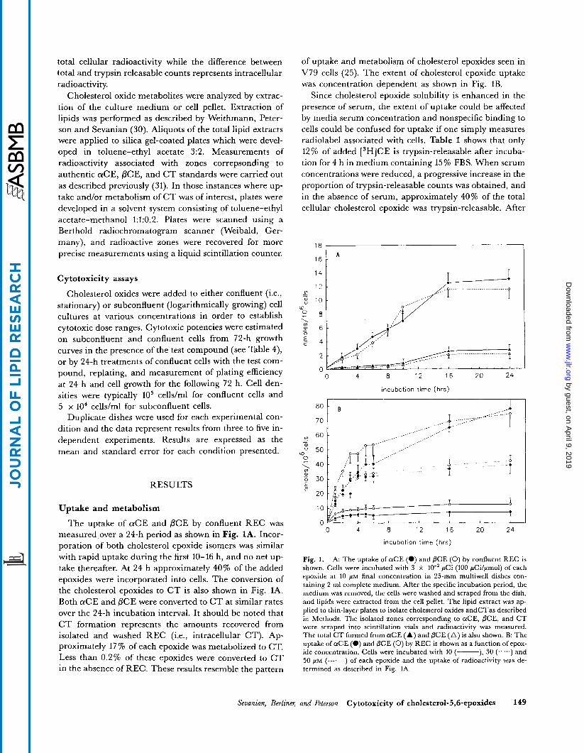
The uptake of aCE and PCE by confluent REC was measured over a 24-h period as shown in Fig. 1A. Incor- poration of both cholesterol epoxide isomers was similar with rapid uptake during the first 10-16 h, and no net up- take thereafter. At 24 h approximately 40% of the added epoxides were incorporated into cells. The conversion of the cholesterol epoxides to C T is also shown in Fig. 1A. Both aCE and PCE were converted to C T at similar rates over the 24-h incubation interval. It should be noted that C T formation represents the amounts recovered from isolated and washed REC (i.e., intracellular CT). Ap- proximately 17% of each epoxide was metabolized to CT. Less than 0.2% of these epoxides were converted to CT in the absence of REC. These results resemble the pattern
of uptake and metabolism of cholesterol epoxides seen in V79 cells (25). The extent of cholesterol epoxide uptake was concentration dependent as shown in Fig. 1B.
Since cholesterol epoxide solubility is enhanced in the presence of serum, the extent of uptake could be affected by media serum concentration and nonspecific binding to cells could be confused for uptake if one simply measures radiolabel associated with cells. Table 1 shows that only 12% of added [3H]CE is trypsin-releasable after incuba- tion for 4 h in medium containing 15% FBS. When serum concentrations were reduced, a progressive increase in the proportion of trypsin-releasable counts was obtained, and in the absence of serum, approximately 40% of the total cellular cholesterol epoxide was trypsin-releasable. After
1 8 ,
1 6 1 A
vi
W v
0
- -
W
c
- .....- I ._.
0 4 8 12 16 20 24
incubation time (hrs)
0 4 8 12 16 20 24
incubation time (hrs)
Fig. 1. A: The uptake of a C E (e) and PCE (0) by confluent REC is shown. Cells were incubated with 3 x 10" pCi (100 pCilpmol) of each epoxide at 10 PM final concentration in 25-mm multiwell dishes con- taining 2 m1 complete medium. After the specific incubation period, the medium was removed, the cells were washed and scraped from the dish, and lipids were extracted from the cell pellet. The lipid extract was ap- plied to thin-layer plates to isolate cholesterol oxides andCTas described in Methods. The isolated zones corresponding to aCE, PCE, and CT were scraped into scintillation vials and radioactivity was measured. The total C T formed from a C E (A) and PCE ( A ) is also shown. B: The uptake of a C E (0) and PCE (0) by REC is shown as a function of epox- ide concentration. Cells were incubated with 10 (-), 30 (......) and 50 FM (-------) of each epoxide and the uptake of radioactivity was de- termined as described in Fig. 1A.
Seuanian, Berliner, and Peterson Cytotoxicity of cholesterol-5,6-epoxi&s 149
by guest, on April 9, 2019
ww
w.jlr.org
Dow
nloaded from
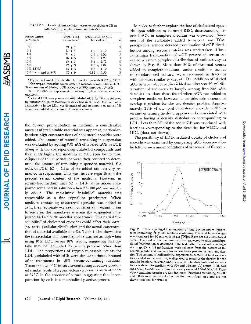
TABLE 1. Levels of intracellular versus extracellular a C E as In order to further explore the fate of cholesterol epox- influenced by media serum concentrations ide upon addition to cultured REC, distribution of la-
Percent Serum Percent Total pmoles olCE/lO6 Cells beled a C E in complete medium was examined. Since in Medium Extracellulara Intracellularb nc most of the radiolabel added to media was E A -
0 0.1 1 .o 5.0
10.0 15.0
10.0 Incubated at 4OC 10% L D L ~
39 k 7 23 f 4 21 f 3 16 f 5 15 f 3 12 3 13 f 3 32 f 9
3 1.0 + 0.40 3 2.9 & 0.50 3 5.0 f 1.23 3 9.1 k 2.72 5 8.6 * 3.04 3 2.3 * 1.10 5
0.82 f 0.30 5
"Trypsin-releasable counts after 4 h incubation with REC at 37OC. bNon-trypsin-releasable counts after 4 h incubation with REC at 37OC.
Total amount of labeled aCE added was l00 pmol per lo6 cells. 'n = Number of experiments involving duplicate cultures per ex-
periment. dIsolated LDL was pretreated with labeled aCE for 30 min followed
by ultracentrifugal re-isolation as described in the text. The content of radioactivity in the LDL was determined and an amount equal to 10% serum was added on the basis of protein content.
the 30-min preincubation in medium, a considerable amount of precipitable material was apparent, particular- ly when high concentrations of cholesterol epoxides were added. The amount of material remaining in suspension was evaluated by adding 0.01 ,uCi of labeled aCE or PCE along with the corresponding unlabeled compounds and then centrifuging the medium at 3000 rpm for 15 min. Aliquots of the supernatant were then counted to deter- mine the amount of remaining suspended material. For aCE or PCE, 62 1.2% of the added radioactivity re- mained in suspension. This was the case regardless of the percent serum content of the medium. However, in serum-free medium only 52 + 1.4% of the added com- pound remained in solution when 25-100 PM was initial- ly added. The remaining ''insoluble'' material was recoverable as a fine crystalline precipitate. When medium containing cholesterol epoxides was added to cells, the precipitate was seen by microscopic examination to settle on the monolayer whereas the suspended com- pound had a cloudy micellar appearance. This partial "in- solubility" of cholesterol epoxides could affect their intra- (vs. extra-) cellular distribution and the actual concentra- tion of material available to cells. Table 1 also shows that the intracellular cholesterol epoxide was not as high when using 10% LDL versus 10% serum, suggesting that up- take may be facilitated by serum proteins other than LDL. The proportions of trypsin-releasable counts for LDL prelabeled with a C E were similar to those obtained after treatments in 10% serum-containing medium. Treatments at 4OC in serum-containing medium produc- ed similar levels of trypsin-releasable counts as treatments at 37°C in the absence of serum, suggesting that incor- poration by cells is a metabolically active process.
precipitable, a more detailed examination of a C E distri- bution among serum proteins. was undertaken. Ultra- centrifugal fractionation of a C E prelabeled serum re- vealed a rather complex distribution of radioactivity as shown in Fig. 2. More than 80% of the total counts added to complete medium, under conditions similar to standard cell culture, were recovered in fractions with densities similar to that of LDL. Addition of labeled a C E to serum-free media yielded an ultracentrifugal dis- tribution of radioactivity largely among fractions with densities less than those found when a C E was added to complete medium; however, a considerable amount of overlap is evident for the two density profiles. Approx- imately 25% of the total cholesterol epoxide added to serum-containing medium appears to be associated with protein having a density distribution corresponding to LDL. Less than 3% of the added CE was associated with fractions corresponding to the densities for VLDL and HDL (data not shown).
The possibility of LDL-mediated uptake of cholesterol epoxide was examined by comparing a C E incorporation by REC grown under conditions of decreased LDL recep-
2 5 1""- l 1
! *OI 15
i o 0 0 1 01 0 1 020 1 030 1 040 1 050
Denslty 9/trn3
Fig. 2. Ultracentrifugal fractionation of fetal bovine serum lipopro- teins containing [3H]aCE. medium containing 15% fetal bovine serum was incubated for 30 min with 10 FM [3H]aCE (sp act 6.8 pCi/pmol) at 37%. Three m1 of this medium was then subjected to ultracentrifuga- tional fractionation as described in the text. After the second centrifuga- tion' step, 15 x 1.5 m1 fractions were collected from the bottom of the centrifuge tube and analyzed for radioactivity, protein content, and den- sity. The content of radioactivity, expressed as percent of total radioac- tivity added to the medium, is displayed in terms of the density for the specific fractions collected and measured. The distribution of radioac- tivity is shown for medium with (0) and without (A) serum. LDL was considered to sediment within the density range of 1.02-1.06 g/ml. Frac- tions containing protein are also indicated. Fractions containing VLDL and HDL were recovered after the first centrifugal step and are not shown (see text for details).
150 Journal of Lipid Research Volume 32, 1991
by guest, on April 9, 2019
ww
w.jlr.org
Dow
nloaded from
TABLE 2. Uptake of &E" by REC and effect of temperature and inhibitors
Condition % [SHIolCE Intracellular Uptake* n
37% 0.21 * 0.05 5 4% 0.05 f 0.02 3
25-OH cholesterol' 0.13 f 0.05 4
n~ total of 10 pmol/ml of [3H]aCE (100 pCilpmo1) was added to se- rum containing medium 30 min before addition to REC. Dishes were then incubated for the indicated periods.
*Non-trypsin-releasable counts incorporated in the presence of 2 %
dothelid cells. Values shown are the percent of total labeled &E that serum-containing medium. Measurements were made using confluent en-
is recovered with cells as non-trypsin-releasable counts. '25-Hydroxycholesterol was added to confluent REC at a concentra-
tion of 3 pg/ml3 days prior to analysis. Cells were then incubated in the presence of both aCE and 25-hydroxycholestero1.
tor function. The uptake of cholesterol epoxide was com- pared to the uptake of lZ5I-LDL (provided by Dr. Margaret Haberland, UCLA). As shown in Table 2, the uptake of cholesterol epoxide was reduced by 75% when cells were incubated at 4OC similarly to the reduction in LDL uptake. Treatment of cells with 25-hydroxycholes- terol for 3 days to down-regulate the LDL receptor (32) caused a 29 + 7.1% inhibition of LDL uptake and a similar, but not significant, decrease in cholesterol epox- ide uptake. There was no difference in the number or viability of REC among the various treatment groups. Moreover, no differences were measured in the uptake of a-aminoisobutyric acid (an analog of alanine) (33) sug- gesting that general membrane transport functions were not markedly affected by the treatment protocol (data not shown). Incubation of REC with labeled aCE in the pre- sence of 30 p M polyinosinic acid, an inhibitor of the LDL scavenger receptor (34), reduced the incorporation of label by 5-15% suggesting minimal involvement of scavenger receptors in the uptake process.
Cytotoxicity of cholesterol oxides
The cytotoxicity of the cholesterol epoxides and CT have been examined with aortic smooth muscle cells (23, 35) and V79 and C3H/lOT1/2 fibroblasts (36, 37), however, limited data exist on endothelial cell toxicity. The effects of serum on the cytotoxicity of cholesterol ox- ides was examined using aCE as a test compound. Table 3 compares the effects of various doses of aCE on the sur- vival of confluent REC for treatments in the presence and absence of 10% fetal bovine serum. The plating efficiency of REC was markedly affected by the absence of serum during the treatment period, therefore, all values for sur- viving fraction of cells were corrected on the basis of con- trol incubations (involving addition of ethanol vehicle alone) which were assigned a surviving fraction value of 1.0. A significant decrease in survival was observed at
&E concentrations above 150 pM in serum-containing medium whereas no toxicity was seen with Serum-free medium, even at the highest aCE concentrations tested (300 p ~ ) . Hence, the presence of serum components aP- pears to facilitate cholesterol epoxide toxicity.
Cytotoxic potencies were also examined by means of 72-h growth curves in the presence of these compounds at various doses (Table 4). The data indicate that sub- confluent cells are substantially more sensitive than con- fluent cells. The estimated LD50s in subconfluent cells for CT, PCE, and aCE are 15 pM, 25 pM, and 108 pM, respectively. These LD50 concentrations are similar to those reported previously for V79 cells (25) which are maintained as logarithmically growing cultures. By con- trast, the cytotoxic potencies of these compounds were lower in confluent cells; the LD50s being 23 pM, 55 pM, and > 150 p~ for CT, PCE, and aCE, respectively. Fur- ther examination of subconfluent cells treated with the cholesterol oxides revealed that about 20-50% of the cells that detached during the 24-h incubation period were not dead since after harvesting and washing these cells were able to reattach and grow. Cytotoxic analyses using the 24-h treatment protocol were also not, strictly speaking, measurements of cell death since about 80% of the treated cells remained attached to dishes, but did not divide, while control cells continued to divide. Thus, a major effect of these compounds may be inhibition of cell division which is subsequently manifested as cell death.
Uptake and metabolism of cholestane triol
We reported previously that intracellularly formed CT was able to readily exit cells (37), indicating that the cell
TABLE 3. Effect of media serum on cytotoxicity of cholesterol epoxide
Surviving Fraction"
Treatment Dose + Serum - Serum
PM
0 50 100 150 300
1 .oo 1 .oo 0.96 f 0.03 0.89 t 0.09 0.93 0.09 0.80 f 0.07 0.79 * 0.12 1.20 f 0.29 0.48 f 0.06 1.03 f 0.19
"The surviving fraction is obtained by replating all cells (i.e., detached and attached) at 10' per dish after 24 h treatment with aCE a t the treat- ment doses indicated. Subsequently, the cell number after 24 h culture in complete medium is divided by the number for untreated controls to obtain the value for surviving fraction. Control cultures are arbitrarily assigned a value of 1.00. Treatments were in the presence ( + serum) or absence ( - serum) of 10% fetal bovine serum. The indicated treat- ment concentrations of a C E were added to media 30 min prior to ad- ding the media to confluent REC. The plating efficiencies after 24 h treatment were 73 and 64% for ( + serum) and ( - serum), respectively.
Sevanian, Berliner, and Peterson Cytotoxicity of cholesteml-5,6-epoxides 151
by guest, on April 9, 2019
ww
w.jlr.org
Dow
nloaded from
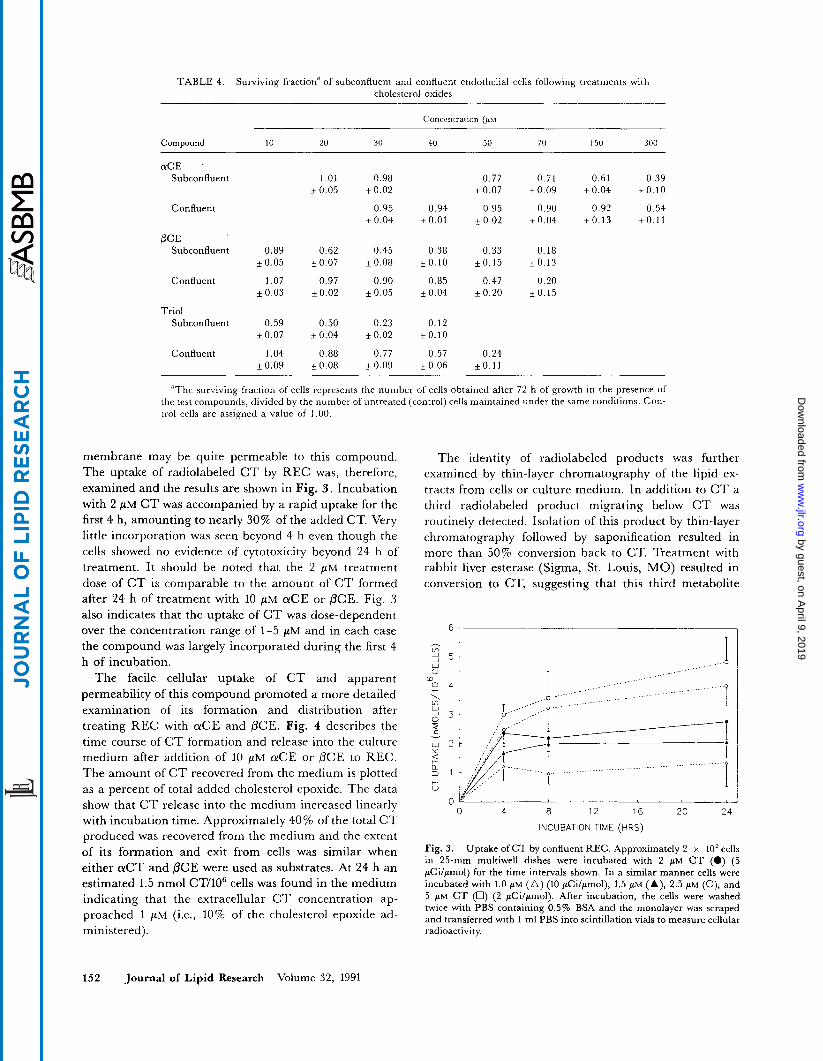
TABLE 4. Surviving fraction" of subconfluent and confluent endothelial cells following treatments with cholesterol oxides
Concentratmn (PM
Compound 1 0 20 30 40 50 70 150 300
a C E Subconfluent 1.01 0.98 0.77 0.71 0.61 0.39
k 0.05 f 0.02 i O . 0 7 i0 .09 +0.04 kO.10
Confluent 0.95 0.94 0.95 0.90 0.92 0.54 kO.04 iO.01 kO.02 kO.04 kO.13 t O . 1 1
PCE Subconfluent 0.89 0.62 0.45 0.38 0.33 0.18
iO.05 kO.07 kO.08 kO.10 kO.15 iO.13
Confluent 1.07 0.97 0.90 0.85 0.47 0.20 kO.03 t 0 . 0 2 iO.05 iO .04 ~ 0 . 2 0 kO.15
Triol Subconfluent 0.59 0.50 0.23 0.12
kO.07 kO.04 k0.02 kO.10
Confluent 1.04 0.88 0.77 0.57 0.24 kO.09 cO.08 c0 .09 kO.06 cO.11
'The surviving fraction of cells represents the number of cells obtained after 72 h of growth in the presence of the test compounds, divided by the number of untreated (control) cells maintained under the same conditions. Con- trol cells are assigned a value of 1 .OO.
membrane may be quite permeable to this compound. The uptake of radiolabeled C T by REC was, therefore, examined and the results are shown in Fig. 3 . Incubation with 2 FM C T was accompanied by a rapid uptake for the first 4 h, amounting to nearly 30% of the added CT. Very little incorporation was seen beyond 4 h even though the cells showed no evidence of cytotoxicity beyond 24 h of treatment. It should be noted that the 2 PM treatment dose of C T is comparable to the amount of C T formed after 24 h of treatment with 10 PM a C E or PCE. Fig. 3 also indicates that the uptake of C T was dose-dependent over the concentration range of 1-5 pM and in each case the compound was largely incorporated during the first 4 h of incubation.
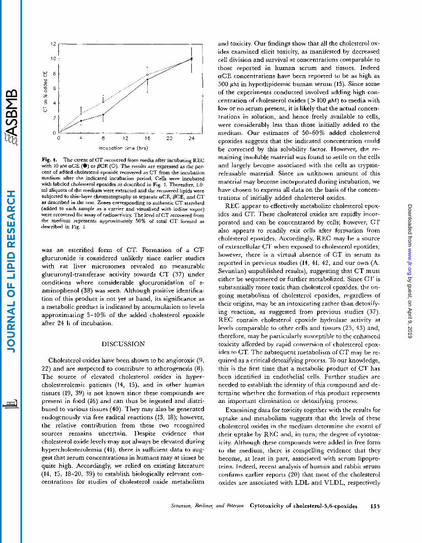
The facile cellular uptake of CT and apparent permeability of this compound promoted a more detailed examination of its formation and distribution after treating REC with aCE and PCE. Fig. 4 describes the time course of C T formation and release into the culture medium after addition of 10 pM aCE or PCE to REC. The amount of C T recovered from the medium is plotted as a percent of total added cholesterol epoxide. The data show that C T release into the medium increased linearly with incubation time. Approximately 40% of the total C T produced was recovered from the medium and the extent of its formation and exit from cells was similar when either a C T and PCE were used as substrates. At 24 h an estimated 1.5 nmol CT/106 cells was found in the medium indicating that the extracellular C T concentration ap- proached 1 pM (i.e., 10% of the cholesterol epoxide ad- ministered).
The identity of radiolabeled products was further examined by thin-layer chromatography of the lipid ex- tracts from cells or culture medium. In addition to C T a third radiolabeled product migrating below C T was routinely detected. Isolation of this product by thin-layer chromatography followed by saponification resulted in more than 50% conversion back to CT. Treatment with rabbit liver esterase (Sigma, St. Louis, MO) resulted in conversion to CT, suggesting that this third metabolite
6 , - v,
W V
2 5
Io 0 4 \ v, 2 3
f v
W 2
a 3 1
Y
c Q
+ V
0 0 4 a 12 16 20 24
INCUBATION TIME (HRS)
Fig. 3. Uptake of CT by confluent REC. Approximately 2 x 10' cells in 25-mm multiwell dishes were incubated with 2 PM C T (0) (5 pCilpmol) for the time intervals shown. In a similar manner cells were incubated with 1.0 pM (A) (10 pcilpmol), 1.5 pM (A), 2.5 PM (o), and 5 PM CT (U) (2 pCilpmol). After incubation, the cells were washed twice with PBS containing 0.5% BSA and the monolayer was scraped and transferred with 1 m1 PBS into scintillation vials to measure cellular radioactivity.
152 Journal of Lipid Research Volume 32, 1991
by guest, on April 9, 2019
ww
w.jlr.org
Dow
nloaded from
12 I
4 8 12 16 20 24
incubation time (hrs)
Fig. 4. The extent of C T recovered from media after incubating REC with 10 pM aCE (0) or PCE (0). The results are expressed as the per- cent of added cholesterol epoxide recovered as CT from the incubation medium after the indicated incubation period. Cells were incubated with labeled cholesterol epoxides as described in Fig. 1. Thereafter, 1.0- m1 aliquots of the medium were extracted and the recovered lipids were subjected to thin-layer chromatography to separate aCE, PCE, and CT as described in the text. Zones corresponding to authentic C T standard (added to each sample as a carrier and visualized with iodine vapor) were recovered for assay of radioactivity. The level of CT recovered from the medium represents approximately 50% of total CT formed as described in Fig. 1.
was an esterified form of CT. Formation of a CT- glucuronide is considered unlikely since earlier studies with rat liver microsomes revealed no measurable glucuronyl-transferase activity towards C T (37) under conditions where considerable glucuronidation of o- aminophenol (38) was seen. Although positive identifica- tion of this product is not yet at hand, its significance as a metabolic product is indicated by accumulation to levels approximating 5-10% of the added cholesterol epoxide after 24 h of incubation.
DISCUSSION
Cholesterol oxides have been shown to be angiotoxic (9, 22) and are suspected to contribute to atherogenesis (8). The source of elevated cholesterol oxides in hyper- cholesterolemic patients (14, 15), and in other human tissues (19, 39) is not known since these compounds are present in food (16) and can thus be ingested and distri- buted to various tissues (40). They may also be generated endogenously via free radical reactions (13, 18); however, the relative contribution from these two recognized sources remains uncertain. Despite evidence that cholesterol oxide levels may not always be elevated during hypercholesterolemia (41), there is sufficient data to sug- gest that serum concentrations in humans may at times be quite high. Accordingly, we relied on existing literature (14, 15, 18-20, 39) to establish biologically relevant con- centrations for studies of cholesterol oxide metabolism
and toxicity. Our findings show that all the cholesterol ox- ides examined elicit toxicity, as manifested by decreased cell division and survival at concentrations comparable to those reported in human serum and tissues. Indeed aCE concentrations have been reported to be as high as 500 FM in hyperlipidemic human serum (15). S’ mce some of the experiments conducted involved adding high con- centration of cholesterol oxides (> 100 PM) to media with low or no serum present, it is likely that the actual concen- trations in solution, and hence freely available to cells, were considerably less than those initially added to the medium. Our estimates of 50-60% added cholesterol epoxides suggests that the indicated concentration could be corrected by this solubility factor. However, the re- maining insoluble material was found to settle on the cells and largely become associated with the cells as trypsin- releasable material. Since an unknown amount of this material may become incorporated during incubation, we have chosen to express all data on the basis of the concen- trations of initia!ly added cholesterol oxides.
REC appear to effectively metabolize cholesterol epox- ides and CT. These cholesterol oxides are rapidly incor- porated and can be concentrated by cells; however, C T also appears to readily exit cells after formation from cholesterol epoxides. Accordingly, REC may be a source of extracellular C T when exposed to cholesterol epoxides; however, there is a virtual absence of CT in serum as reported in previous studies (14, 41, 42, and our own (A. Sevanian) unpublished results), suggesting that C T must either be sequestered or further metabolized. Since C T is substantially more toxic than cholesterol epoxides, the on- going metabolism of cholesterol epoxides, regardless of their origins, may be an intoxicating rather than detoxify- ing reaction, as suggested from previous studies (37). REC contain cholesterol epoxide hydrolase activity at levels comparable to other cells and tissues (25, 43) and, therefore, may be particularly susceptible to the enhanced toxicity afforded by rapid conversion of cholesterol epox- ides to CT. The subsequent metabolism of C T may be re- quired as a critical detoxifying process. To our knowledge, this is the first time that a metabolic product of C T has been identified in endothelial cells. Further studies are needed to establish the identity of this compound and de- termine whether the formation of this product represents an important elimination or detoxifying process.
Examining data for toxicity together with the results for uptake and metabolism suggests that the levels of these cholesterol oxides in the medium determine the extent of their uptake by REC and, in turn, the degree of cytotox- icity. Although these compounds were added in free form to the medium, there is compelling evidence that they become, at least in part, associated with serum lipopro- teins. Indeed, recent analysis of human and rabbit serum confirms earlier reports (20) that most of the cholesterol oxides are associated with LDL and VLDL, respectively
Seuanian, Berliner, and Peterson Cytotoxicity of cholesterol-5,6-epoxides 153
by guest, on April 9, 2019
ww
w.jlr.org
Dow
nloaded from
(A. Sevanian and H. Hodis, unpublished results). Four sets of observations suggest that cholesterol oxides may become associated with serum lipoproteins prior to their uptake by REC. I ) Addition of labeled cholesterol epoxide to fetal bovine serum results in association of approx- imately 25% of the radioactivity with LDL fraction after ultracentrifugation. 2) Incubation of aCE-prelabeled medium with serial dilutions of unlabeled serum- containing medium produces a progressive inhibition of a C E uptake (Table 1). Uptake of otCE by REC appears to be saturable (Fig. 1) and increasing cell numbers in- creases the amount of cholesterol oxide incorporation (data not shown). 3) The presence of serum in the medium, or addition of LDL to serum-free medium, sig- nificantly enhances cholesterol epoxide toxicity as com- pared to treatments in serum-free medium (Table 3 ) . Cox, Comai, and Goldstein (24) also noted that the cyto- toxic action of 25-hydroxycholesterol was enhanced when endothelial cells were treated with fetal bovine serum ve- hicle as compared to an ethanolic vehicle. 4 ) The uptake of aCE is markedly reduced when cells are incubated at 4OC. Based on these findings, we propose that transport via serum lipoproteins (and particularly LDL) may be an effective means by which cholesterol oxides are delivered to and incorporated by endothelial cells; however, con- siderable uptake may occur via nonspecific or passive (ab- sorption) processes under standard cell culture conditions.
A genotoxic mechanism for cholesterol epoxide toxicity has been proposed from several studies (25, 37, 44) and the potential for disrupting DNA synthesis (45) could ac- count for the greater toxicity seen with subconfluent cells. It should be noted that subconfluent smooth muscle cells are also more sensitive to 25-hydroxycholesterol (24). Since subconfluent cultures have a large proportion of cells in log phase, it is plausible that their sensitivity to cholesterol epoxides and CT may be due to interference of DNA synthesis and replication. There is no evidence that CT is directly genotoxic; however, the possibility re- mains that its toxicity is exerted by inhibition of cholesterol biosynthesis and/or metabolism (35, 46) or by membrane disruption. In any event, incorporation and cytotoxicity are markedly influenced by serum lipopro- teins suggesting that assimilation into cellular compart- ments is an important criterion for the toxic action of these compounds. Injury to endothelial cells via DNA or membrane damage may provide a locus for lesion deve- lopment on the vessel wall.
There is at present a paucity of evidence for oxidative modification of serum lipoproteins obtained directly from animals or humans (47). Nevertheless, compelling evi- dence is emerging to suggest that oxidative modification of lipoproteins (particularly LDL) renders toxicity to smooth muscle and endothelial cells and alters the mode of LDL uptake by endothelial cells and monocytes (48,
154 Journal of Lipid Research Volume 32, 1991
49). We propose that measurement of cholesterol oxides be considered as another means for identifying oxidative- ly modified lipoproteins in addition to providing evidence for lipid peroxidation in tissues. The active uptake oflipo- proteins containing these cholesterol oxides may be a pro- cess through which these cytotoxic compounds contribute to atherosclerosis. I
This study was supported by a grant ES03466 from the National Institutes of Health. Manuscript received 8 August 1990 and in revisedform 16 October 1990
1.
2.
3.
4.
5.
6.
7.
8.
9.
10.
11.
12.
13.
14.
REFERENCES
Wissler, R. W., M. Armstrong, D. Belheimer, K. Hayes, P. Hill, R. Jackson, P. Kritchevsky, R. W. Mahley, R. Minich, L. Rudel, and E. Smith. 1979. Conference on the health effects of blood lipids. Preventive Med. 8: 715-722. Anderson, K. M., W. P. Castelli, and D. Levy. 1987. Cholesterol and mortality: 30 years of follow-up from the Framingham study. J Am. Med. Assoc. 257: 2176-2180. Morel, D. W., P. E. DiCorleto, and G. M. Chisolm. 1984. Endothelial and smooth muscle cells alter low density lipo- protein in vitro by free radical oxidation. Arteriosclerosis. 4:
Hiramatsu, K., H. Rosen, J. W. Heinecke, G. Wollbauer, and A. Chait. 1987. Superoxide initiates oxidation of low density lipoproteins by human monocytes. Arteriosclerosis. 7:
Steinbrecher, U. P., S. Parthasarathy, D. S. Leake, J. L. Witztum, and D. Steinberg. 1984. Modification of low den- sity lipoprotein by endothelial cells involves lipid peroxida- tion and degradation of low density lipoprotein phospholipids. Proc. Natl. Acad. Sci. USA. 83: 3883-3887. Henrickson, T. S., S. A. Evensen, and B. Carlander. 1979. Injury to human endothelial cells in culture induced by low density lipoproteins. Scand. J . Lab. Incest. 39: 361-368. Hessler, J. R., D. W. Morel, L. J. Lewis, and G. M. Chisolm. 1983. Lipoprotein oxidation and lipoprotein- induced cytotoxicity. Arteriosclerosis. 3: 215-222. Imai, H., M. T. Werthessen, C. B. Taylor, and K. T. Lee. 1976. Angiotoxicity and atherosclerosis due to contami- nants of USP grade cholesterol. Arch. Pathol. Lab Med. 100:
Peng, S-K., C. B. Taylor, J. C. Hill, and R. J. Morin. 1985. Cholesterol oxidation derivatives and arterial endothelial damage. Atherosclerosis. 54: 121-133. Smith, L. L., and M. J. Kulig. 1975. Sterol metabolism. XXXIV. On the derivation of carcinogenic sterols from cholesterol. Cancer Biochem. Biophys. 1: 79-84. Aringer, L., and P. Eneroth. 1973. Studies on the formation of C7-oxygenated cholesterol and P-sitosterol metabolites in cell-free preparations of rat liver. J Lip id Res. 14: 563-572. Mitton, J. R., N. A. Scholan, and G. S. Boyd. 1971. The oxidation of cholesterol in rat liver subcellular particles. Eur. J Biochem. 20: 569-579. Watabe, T., M. Isobe, and A. Tsubaki. 1982. Epoxidation of cholesterol by hepatic microsomal lipid hydroperoxides. Biochem. Biophys. Res. Commun. 108: 724-730. Brooks, C. J. W., R. M. McKenna, W. J. Cole, J. MacLachlan, and T. D. V. Lawrie. 1983. Profile analysis of
357-364.
55-60.
565-569.
by guest, on April 9, 2019
ww
w.jlr.org
Dow
nloaded from
oxygenated sterols in plasma and serum. Biochem. SOC. Eans.
15. Gray, M. F., T. D. V. Lawrie, and C. J. W. Brooks. 1971. Isolation and identification of cholesterol a-oxide and other minor sterols in human serum. Lipidr. 6: 836-843.
16. Tsai, L. S., K. Ijichi, C. A. Hudson, and J. J. Meehan. 1980. A method for the quantitative estimation of cholesterol a-oxide in eggs. Lipidr. 15: 124-128.
17. Finocchiaro, E. T., K. Lee, and T. Richardson. 1984. Iden- tification and quantification of cholesterol oxides in grated cheese and bleached butteroil. J. Am. Oil Chem. SOC. 61:
18. Sevanian, A., N. Elsayed, and A. D. Hacker. 1982. Effects of vitamin E deficiency and nitrogen dioxide exposure on lung lipid peroxidation: use of lipid epoxides and malm- dialdehyde as measures of peroxidation. J. Toxicol. Enuimn. Health. 10: 743-756.
19. Petrakis, N. L., L. D. Gruenke, and J. C. Craig. 1981. Cholesterol and cholesterol epoxides in nipple aspirates of human breast fluid. Cancer Res. 41: 2563-2565.
20. Peng, S-K., G. A. Philips, G-Z. Xia, and R. J. Morin. 1987. Transport of cholesterol autoxidation products in rab- bit lipoproteins. Atheroslerosis. 64: 1-6.
21. Henderson, A. E. 1956. A histological and chromato- graphic study of normal and atheromatous human arteries.
22. Imai, H., N. T. Werthessen, P. W. LaQuesne, A. H. Soloway, and M. Kanisawa. 1980. Angiotoxicity of ox- ygenated sterols and possible precursors. Science. 207:
23. Peng, S-K., C. B. Taylor, P. Tham, N. T. Werthessen, and B. Mikkelson. 1978. Effect of auto-oxidation products from cholesterol on aortic smooth muscle cells. Arch. Pathol. Lab Med. 102: 57-61.
24. Cox, D. C., K. Comai, and A. L. Goldstein. 1988. Effects of cholesterol and 25-hydroxycholestero1 on smooth muscle cell and endothelial cell growth. Lipidr. 23: 85-88.
25. Sevanian, A., and A. R. Peterson. 1984. Cholesterol epox- ide is a direct-acting mutagen. Proc. Natl. Acad. Sci. USA. 81:
26. Maerker, G., and J. Unruh. 1986. Cholesterol oxides. I. Isolation and determination of some cholesterol oxidation products. J Am. Oil Chem. Soc. 63: 767-771.
27. Sevanian, A., and L. L. McLeod. 1987. Cholesterol autox- idation in phospholipid membrane bilayers. Lipidr. 22:
28. Baker, D. P,, B. J. Van Lenten, A. M. Fogelman, P. A. Ed- wards, C. Kean,'and J. A. Berliner. 1984. LDL, Scavenger, and P-VLDL receptors on aortic endothelial cells. Arteriosclerosis. 4: 248-255.
29. Manual of Laboratory Operations, Lipid Research Clinic Program. Vol I. Lipid and Lipoprotein Analysis. 1974. US DHEW, Publication no. (NIH) 75-628.
30. Weithmann, K. U., H. Peterson, and A. Sevanian. 1989. Incorporation of arachidonic, dihomogamma linolenic and eicosapentaenoic acids into cultured V79 cells. Lipids. 24:
31. Sevanian, A., and L. L. McLeod. 1986. Catalytic proper- ties and inhibition of hepatic cholesterol epoxide hydrolase. J. Biol. Chern. 261: 54-59.
32. Van Lenten, B. J., A. M. Fogelman, M. M. Hokom, L. Benson, M. E. Haberland, and P. A. Edwards. 1983. Regulation of the uptake and degradation of 0-very low density lipoprotein in human monocyte macrophages. J. Biol. Chem. 258: 5151-5157.
11: 700-701.
877-883.
J Histochem. Cytochem. 4: 153-159.
651-653.
4198-4202.
627-636.
173-178.
33. Kwok, C. F., B. J. Goldstein, D. Muller-Wieland, TS. Lee, R. Kahn, and G. L. King. 1989. Identification of persistent defects in insulin receptor structure and function in capil- lary endothelial cells from diabetic rats. J. Clin. Invest. 83:
34. Haberland, M. E., R. R. Rasmussen, C. L. Olch, and A. M. Fogelman. 1986. Two distinct receptors account for recognition of maleyl-albumin in human monocytes during differentiation in vitro. J. Clin. Invest. 77: 681-689.
35. Peng, S-K., P. Tham, C. B. Taylor, and B. Mikkelson. 1979. Cytotoxicity of cholesterol oxidation derivatives on cultured aortic smooth muscle cells and ,their effect on cholesterol biosynthesis. Am. J. Clin. Nutz 32: 1033-1042.
36. Raaphorst, G. P., E. I. Azzam, R. Langlois, and J. E. Van Lier. 1987. Effect of cholesterol a- and 0-epoxides on cell killing and transformation. Biochem. Pharmacol. 36:
37. Sevanian, A., and A. R. Peterson. 1986. The cytotoxicity and mutagenic properties of cholesterol oxidation products. Food C h . Toxicol. 24: 1103-1110.
38. Dutton, G. J., and I. D. E. Storey. 1962. Glucuronide form- ing enzymes. Methodr Enzymol. 5: 159-164.
39. Sporer, A., D. R. Brill, and C. P. Schaffner. 1982. Epox- ycholesterols in secretions and tissues of normal, benign and cancerous human prostate glands. Urology. 55:
40. Bascoul, J., N. Domergue, J. Mourot, G. Debry, and A. Crastes de Paulet. 1986. Intestinal absorption and fecal secretion of 5,6-epoxy-5a-cholesta-3-/3-ol by the male Wistar rat. Lipids. 21: 744-747.
41. Bjorkhem, I., 0. Breuer, B. Angelin, and S-A. Wikstrom. 1988. Assay of unesterified cholesterol-5,6-epoxide in hu- man serum by isotope dilution mass spectrometry. Levels in the healthy state and in hyperlipoproteinemia. J. Lipid Res.
42. Addis, P. B., H. A. Emanuel, S. D. Bergmann, and J. H. Zavoral. 1989. Capillary GC quantification of cholesterol oxidation products in plasma lipoproteins of fasted hu- mans. Free Radical Biol. Med. J . 7: 179-182.
43. Levin, W., D. P. Michaud, P. E. Thomas, and D. M. Jerina. 1983. Distinct rat hepatic microsomal epoxide hydrolases catalyze the hydration of cholesterol-5,6-oxide and certain xenobiotic alkene and arene oxides. Arch. Biochem. Biophys. 220: 485-494.
44. Peterson, A. R., H. Peterson, C. P. Spears, J. E. Trosko, and A. Sevanian. 1988. Mutagenic characterization of cholesterol epoxides in Chinese hamster V79 cells. Mutat. Res. 203: 355-366.
45. Parsons, P. G., and P. Goss. 1978. Chromosome damage and DNA repair induced in human fibroblasts by UV and cholesterol oxide. A u t . J . Exp. Biol. Med. Sci. 56: 287-296.
46. Morin, R. J., and S-K. Peng. 1989. Effects of cholesterol oxidation derivatives on cholesterol esterifying and cholesterol ester hydrolytic enzyme activity of cultured rab- bit aortic smooth muscle cells. Lipids. 24: 217-220.
47. Avogaro, P,, G. Bittolo-Bon, and G. Cazzolato. 1988. Pre- sence of a modified low density lipoprotein in humans. Arteriosclerosis. 8: 79-87.
48. Brown, M. S., Y. K. Ho, and J. L. Goldstein. 1980. The cholesterol ester cycle in macrophage foam cells. J. Biol. Chem. 255: 9344-9352.
49. Haberland, M. E., A. M. Fogelman, and P. A. Edwards. 1982. Specificity and receptor-mediated recognition of malondialdehyde-modified low density lipoprotein. Proc. Natl. Acad. Sci. USA. 79: 1712-1716.
127-136.
2369-2372.
244-250.
29: 1031-1038.
Sevanian, Berliner, and Peterson Cytotoxicity of cholesteml-5,6-epoxides 155
by guest, on April 9, 2019
ww
w.jlr.org
Dow
nloaded from