Embed Size (px)
Citation preview
661VOLUME L NUMBER 11 © 2016 JCO, Inc.
MICHAEL A. WEBB, DDS, MSFRANK E. CORDRAY, DDS, MSP. EMILE ROSSOUW, BSc, BChD (Hons.), PhD, FRCD
Dr. Cordray Dr. RossouwDr. Webb
Dr. Webb is in the private practice of orthodontics in Charlotte, NC. Dr. Cordray is an Assistant Clinical Professor, School of Dentistry, Ohio State University, Columbus, OH, and in the private practice of orthodontics at Cordray Orthodontics, 96 Northwoods Blvd., Columbus, OH 43235; e-mail: [email protected]. Dr. Rossouw is a Professor and Chair, Department of Orthodontics and Dentofacial Orthopedics, Eastman Institute for Oral Health, University of Rochester, Rochester, NY.
ships of the face and principles established in antiquity. Bony landmarks are sometimes difficult to locate, however, and positions of soft-tissue landmarks can vary from patient to patient. More-over, when taking a lateral cephalogram, the op-erator must accurately capture natural head posi-tion (NHP) to avoid errors introduced by head tipping.
Various facial analyses have attempted to quantify facial beauty by measuring lip position, but few authors have used the upper incisor as the alpha point in developing an analysis for facial esthetics. The present study was designed to inves-tigate whether planes established by bony land-marks and vertical perpendicular lines from the soft-tissue forehead midpoint and soft-tissue gla-bella could be used to determine the position of the upper incisor in an optimal profile.
Facial esthetics play an important role in con-temporary orthodontics. Having an attractive
face is perceived as an advantage in society with regard to competence, likeability, and potential for success—and this advantage can begin early in life.1 Orthodontists tend to view a patient in terms of correcting a malocclusion, but the patient may simply want improved function and esthetics. Al-though Angle maintained that correcting the mal-occlusion would inevitably improve facial esthet-ics, attempts to create a mathematical formula for beauty based on soft- and hard-tissue landmarks have proven to be inadequate.2
In the past, orthodontists used photographs to evaluate facial esthetics and dental casts to study the occlusion as it relates to the soft tissue. This changed with the development of cephalo-metric analyses based on proportional relation-
Upper-Incisor Position as a Determinant of the Ideal Soft-Tissue Profile

662 JCO/NOVEMBER 2016
Upper-Incisor Position as a Determinant of the Ideal Soft-Tissue Profile
Materials and Methods
An initial sample was obtained by screening the final orthodontic records of 400 adolescent Caucasian patients who had recently completed fixed-appliance therapy with one clinician, all un-der the same treatment philosophy. From this ini-tial sample, 100 subjects (48 male, 52 female) with harmonious facial profiles were selected regardless of their initial Class I, II, or III soft-tissue relation-ships. Every patient had a Class I occlusion with normal overbite and overjet at the end of treatment.
Following standard office procedure, each subject had a final digital lateral cephalogram taken with barium sulfate paste* applied at trich-ion (the hairline) to facilitate soft-tissue identifica-tion. Standardized profile, frontal, and smiling frontal photographs were taken in NHP, using a Canon PowerShot G5** digital camera (105mm macro lens) held level with the face.3 The photos were then transferred to PhotoScape*** and com-parably sized, with three subjects to a page.
Photos of 70 randomly selected patients from the sample of 100 were provided to four ABO-certified orthodontists for independent evaluation. The instructions were to select patients with an esthetically pleasing profile, which was defined as a normal profile in which the lips and chin were in harmony with the rest of the face.4-8 Care was taken not to mention preexisting skeletal or dental relationships, or whether the subject had under-gone extraction or nonextraction treatment. A sub-ject was included in the study sample if three of the four orthodontists viewed the profile as es-thetically pleasing.
Two weeks later, another set of 40 photo-graphs was sent for evaluation by the same four orthodontists. Ten of those patients were repeated from the previous sample of 70 subjects (to verify the consistency of the selection process), and 30 new subjects were added. The final sample of sub-
jects who were judged to have balanced, estheti-cally pleasing profiles consisted of 31 males and 33 females. Of these 64 patients, 19 males and 21 females were considered by all four orthodontists to have esthetically pleasing profiles.
Each digital cephalogram was calibrated and grayscale-adjusted to aid in landmark location, then printed on glossy photo paper. After .003" tracing paper† was placed over the cephalogram, the bony landmarks of anterior nasal spine, cli-noidale, floor of sella, gonion, menton, and roof of orbit were marked9 (Fig. 1). The soft-tissue fore-head was traced from trichion to glabella, and the crown of the upper central incisor was also traced. Pinholes were made through the tracing paper and radiograph in the four corners for later use in ver-ifying landmark locations.
Sassouni’s anterior cranial base was con-structed by drawing a line from the roof of the orbit to clinoidale10 (Fig. 2). A parallel line was drawn through the floor of sella and extended dis-tally; a mandibular plane was drawn from menton through gonion and extended distally until it inter-sected the cranial-base plane. A horizontal refer-ence plane was then drawn from this point of in-tersection anteriorly to ANS.
A line from trichion to glabella was bisected,
Fig. 1 Anatomical landmarks used for reference-plane construction.
*E-Z Paste, registered trademark of Bracco Diagnostics, Inc., Monroe Township, NJ; www.braccoimaging.com.**Canon USA, Inc., Melville, NY; www.canon.com.***MOOII Tech, Seoul, Korea; www.photoscape.org/ps/main/index.php.†Dentsply GAC, Islandia, NY; www.dentsply.com.‡Orthopli Corporation, Philadelphia, PA; www.orthopli.com.††MathWorks, Natick, MA; www.mathworks.com.

663VOLUME L NUMBER 11
Webb, Cordray, Rossouw
and a perpendicular line was projected from this midpoint onto the soft-tissue forehead to establish the forehead midpoint. A line perpendicular to the horizontal reference plane, called the forehead fa-cial plane (FFP), was drawn from soft-tissue gla-bella inferiorly past the upper central incisor. A line designated as the forehead midpoint plane (FMP) was drawn from the forehead midpoint inferiorly and perpendicular to the horizontal reference plane.
Using a digital caliper,‡ measurements were made to the nearest tenth of a millimeter from the FFP and FMP to the most facial aspect of the upper central incisor. Only the AP position was recorded, since the patient’s vertical relationship to soft tissue had already been established as esthetically pleas-
ing. A positive number was assigned if the incisor was located anterior to the plane, a negative num-ber if the incisor was posterior to the plane.
Statistical analyses were completed using Matlab 2012b†† software, with the significance level set at p < .05. Descriptive statistics were cal-culated and t-tests performed to assess the differ-ence between data sets.
Two weeks later, 10 of the subjects (five males, five females) were selected at random, and their cephalograms were retraced and measured by the same examiner. The systematic error be-tween the first and second measurements was calculated using a paired t-test (p < .05), and the error variance was calculated according to the Dahlberg formula. No significant differences were found (Table 1).
Results
In the esthetically pleasing group, the fe-males (N = 33) had a mean age of 14.38, and the males (N = 31) a mean age of 14.85 (Table 2). Since there was no significant age difference be-tween the groups (p = .087), they could be com-bined for further analysis. The mean upper-incisor position behind the FFP was −2.2mm in females and −2.6mm in males; the mean incisor position in front of the FMP was 1.6mm in females and 3.4mm in males (Table 3). Again, because there was no significant gender difference (p > .05), we combined the patients to evaluate the mean dis-tances of the upper incisors from the FFP and FMP. T-tests were then used to determine statisti-cal significance for each data set (FFP < 0mm and FMP > 0mm). Overall, the upper incisors were
TABLE 1MEASUREMENT ERROR ANALYSIS
First Measurement Second Measurement
Mean S.D Mean S.D. P Dahlberg FormulaForehead facial plane −2.97mm 2.05mm −2.95mm 2.03mm 0.98 0.27
Forehead midpoint plane 1.09mm 2.45mm 1.17mm 2.39mm 0.94 0.26
Fig. 2 Construction of analysis, showing fore-head facial plane (FFP) and forehead midpoint plane (FMP).

664 JCO/NOVEMBER 2016
Upper-Incisor Position as a Determinant of the Ideal Soft-Tissue Profile
Case 1
This 15-year-old female had a skeletal and dental Class III pattern (maxillary retrusion and mandibular protrusion) with a mild mandibular asymmetry (Fig. 3). The pretreatment cephalo-metric tracing showed that while a single-jaw maxillary advancement might have corrected the
positioned behind the FFP and in front of the FMP with a 95% confidence interval (p < .05). This analysis clearly demonstrates that in an estheti-cally pleasing profile, the upper incisor will be positioned between the FFP and FMP.
Potential uses of the analysis are described here in three patients who were not part of the study sample.
TABLE 2AGE OF SAMPLE*
Mean S.D. Minimum MaximumFemale (N = 33) 14.38 0.98 11.40 15.90
Male (N = 31) 14.85 1.15 12.60 17.50
*P = .087.
Fig. 3 Case 1. 15-year-old female patient with skeletal and dental Class III pattern (maxillary retrusion and mandibular protrusion) before treatment.

665VOLUME L NUMBER 11
Webb, Cordray, Rossouw
TABLE 3MEAN MEASUREMENTS BY GENDER
Forehead Facial Plane
Forehead Midpoint Plane
Female (N = 33) −2.20mm 1.64mm
Male (N = 31) −2.59mm 3.37mm
P 0.64 0.07
Fig. 4 Case 1. After 21 months of treatment, acceptable horizontal/anteroposterior (AP) positioning of upper incisors.

666 JCO/NOVEMBER 2016
Upper-Incisor Position as a Determinant of the Ideal Soft-Tissue Profile
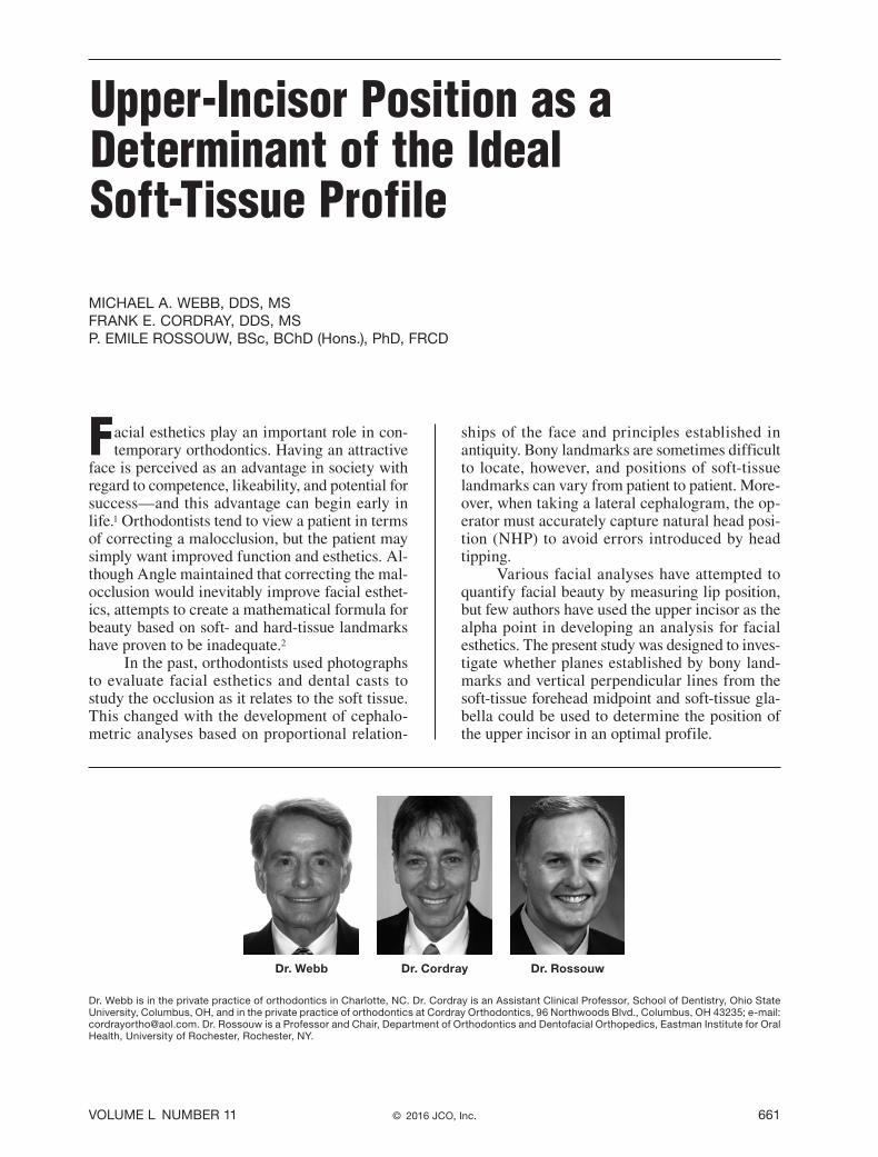
Case 2
An 11-year-old female presented with a skel-etal and dental Class I bimaxillary protrusive pat-tern (Fig. 5). She had a flat forehead and, conse-quently, a relatively narrow trough between the FMP and FFP. Both upper and lower incisors were flared, and the upper incisors were positioned well forward of the FFP. Incisor retraction in this case required four first-premolar extractions. After 27 months of orthodontic treatment, the patient showed acceptable horizontal/AP positioning of the upper incisors (Fig. 6).
AP discrepancy, it would have advanced the inci-sors ahead of the FFP, possibly making the pa-tient appear too maxillary protrusive. This could have created a bimaxillary Class III without ad-dressing the underlying mandibular asymmetry. Therefore, skeletal correction in this case re-quired double-jaw surgery involving maxillary advancement of 4mm (to the FFP) and mandib-ular retrusion and rotation. After 21 months of orthodontic treatment, the patient displayed ac-ceptable horizontal/AP positioning of the upper incisors (Fig. 4).
Fig. 5 Case 2. 11-year-old female patient with skeletal and dental Class I bimaxillary protrusive pattern and relatively narrow trough between FMP and FFP (red line) before treatment.
667VOLUME L NUMBER 11
Webb, Cordray, Rossouw
Case 3
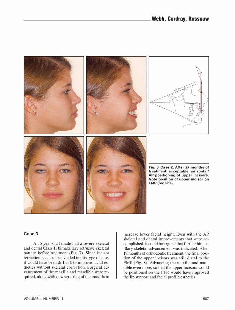
A 15-year-old female had a severe skeletal and dental Class II bimaxillary retrusive skeletal pattern before treatment (Fig. 7). Since incisor retraction needs to be avoided in this type of case, it would have been difficult to improve facial es-thetics without skeletal correction. Surgical ad-vancement of the maxilla and mandible were re-quired, along with downgrafting of the maxilla to
increase lower facial height. Even with the AP skeletal and dental improvements that were ac-complished, it could be argued that further bimax-illary skeletal advancement was indicated. After 19 months of orthodontic treatment, the final posi-tion of the upper incisors was still distal to the FMP (Fig. 8). Advancing the maxilla and man-dible even more, so that the upper incisors would be positioned on the FFP, would have improved the lip support and facial profile esthetics.
Fig. 6 Case 2. After 27 months of treatment, acceptable horizontal/AP positioning of upper incisors. Note position of upper incisor on FMP (red line).

668 JCO/NOVEMBER 2016
Upper-Incisor Position as a Determinant of the Ideal Soft-Tissue Profile
plane anteriorly; similarly, smaller or flatter noses or chins projected the esthetic plane posteriorly. The present study supports Ricketts’s observation that a case with proper incisor position and skeletal structure will have the lips positioned behind the E-line.12 Only three subjects (one male and two female) in our sample of esthetically pleasing pro-files exhibited lower lips in front of the E-line.
Burstone drew a plane from subnasale to soft-tissue pogonion to measure lip protrusion or retrusion in 32 “normal” adolescent males and females.4 He found that the average upper lip was
Discussion
Downs’s landmark cephalometric analysis included linear and angular measurements of both hard- and soft-tissue relationships.11 Ricketts intro-duced an “esthetic plane” (E-line) to evaluate the lips in relation to the nose tip and soft-tissue pogo-nion.12 In female adults, the lower lips ideally mea-sured 2mm and the upper lips 4mm posterior to the E-line; male lips were more retrusive. Large noses or soft-tissue chins required alterations in treatment plans because they tended to project the esthetic
Fig. 7 Case 3. 15-year-old female patient with skeletal and dental Class II bimaxillary retrusive pattern before treatment.

669VOLUME L NUMBER 11
Webb, Cordray, Rossouw
3.5mm and the average lower lip 2.2mm anterior to this plane (with no difference between males and females), but he did not consider the position of the incisors in the face, which can change de-pending on the malocclusion.
González-Ulloa and Stevens used Frankfort Horizontal (FH) as a reference plane, drawing a vertical line perpendicular to FH from soft-tissue nasion.13 In most faces considered “beautiful”, soft-tissue pogonion was positioned on this line. Both hard- and soft-tissue landmarks were used,
but identification of porion and orbitale was dif-ficult, especially when double images required bisection. Moreover, any variability of the FH plane could alter the position of the vertical (per-pendicular) line.
Holdaway used a soft-tissue facial plane (fa-cial angle relative to FH) to assess the profile chin position (ideal = 91° ± 7°).14 He stated that his harmony line (H-line) should lie 3-7mm anterior to subnasale, and that the lower lip should fall .5mm anterior to this plane. Holdaway utilized his
Fig. 8 Case 3. After 19 months of treatment, upper incisors still dis-tal to FMP.

670 JCO/NOVEMBER 2016
Upper-Incisor Position as a Determinant of the Ideal Soft-Tissue Profile
rigid rules for facial esthetics would be impossible to determine, general guidelines could be estab-lished.17 Optimal upper-incisor position in relation to adjacent soft tissue, both anteroposteriorly and vertically, was paramount to these authors. Their paper introduced the concept of positioning the upper incisors as the first step in diagnosis.
Andrews moved further away from hard-tissue internal cephalometric landmarks toward soft-tissue profile landmarks.18 He, too, found up-per-incisor placement critical to facial esthetics from the frontal and lateral views. For his control sample, Andrews used smiling profile photos of 94 Caucasian adult females from various publica-tions. With the patient standing in NHP, he esti-mated the AP distance of the upper incisor to a vertical line from an estimated forehead midpoint. A second vertical line, parallel to the first, was then drawn from soft-tissue glabella. In 93% of these patients, the upper incisors were positioned between the two constructed vertical lines, com-pared to 21% of a group of 94 treated female pa-tients.18 There was a strong correlation between harmonious profiles and incisor positioning be-tween the two lines. A strong correlation was also found between incisor position and forehead incli-nation: the more the forehead was inclined, the more forward the incisors could be positioned. The disadvantage of Andrews’s technique was that it still depended on accurately capturing NHP, whether positioning a patient in a cephalostat or taking profile photographs.
In our study, the mean distance between the FFP and FMP was greater in males (6.0mm) than in females (3.8mm), which can be attributed to the difference in their forehead shape and the promi-nence of soft-tissue glabella. A more posteriorly sloping forehead or an increased projection of glabella results in a greater range of acceptable upper-incisor positioning. “Feminine” faces tend to be more sensitive to excessive incisor retraction. These findings support the contention of Sarver and Ackerman17 and Andrews18 that diagnosis be-gins with proper vertical and AP positioning of the upper incisors.
In another study, Schlosser and colleagues took a smiling profile photo of a female with pleas-
H-angle, measured between the soft-tissue facial plane and the H-line, as well as A point convexity relative to the H-angle, to determine lip balance. His visual treatment objective established an un-strained soft-tissue lip balance relative to upper-incisor position.14 Holdaway’s admitted drawback was in cases with severe Class II and Class III skeletal structures; moreover, lip and chin thick-ness could affect his measurements. Evaluating patients in our sample by means of the H-line, only one male had a line that fell in front of the tip of the nose. The best profiles confirmed the Hold-away standards.14,15
Similar to the present study, Spradley and colleagues studied 25 Caucasian males and 25 Caucasian females previously considered by four of five judges (three orthodontists and two oral surgeons) to have esthetically pleasing or “nor-mal” profiles.5 Lateral cephalograms were taken in NHP, using a true vertical plumb line lateral to the profile. A true horizontal line was constructed from the true vertical, and a second vertical line was drawn from the true horizontal through sub-nasale. Lip position, sulcus depth, and soft-tissue pogonion were measured relative to this subna-sale true vertical. On average, the lips were in front of the subnasale vertical line, with both lips more procumbent in females (upper 2.1mm, low-er .4mm) than in males (upper 1.6mm, lower .2mm). Unlike Ricketts12 and Holdaway,14,15 this combination of soft-tissue landmarks and con-structed lines did not rely on the position of the chin, which can vary in hard- and soft-tissue thick-ness, nor on bony structures. It was technique-sensitive, however, because of the potential effect of head tipping on NHP.
Bergman, who used 16 soft-tissue landmarks to design a Soft Tissue Assessment Sheet, indi-cated that the lips would normally lie in front of a line from soft-tissue subnasale to pogonion (upper 3.5mm, lower 2.2mm).16 This method evaluated lip position within the soft-tissue envelope, but did not address upper-incisor AP position within the facial profile. Bergman acknowledged that relying exclusively on dentoskeletal analysis could lead to esthetic problems.
Sarver and Ackerman observed that while

671VOLUME L NUMBER 11
Webb, Cordray, Rossouw
ant facial features and altered the upper incisor and lip positions by 1mm anteriorly and posteriorly.19 Nine of these altered photos were shown to a pan-el of 20 orthodontists and 20 lay persons. Both groups favored the normal-to-protrusive individu-als over the retrusive individuals (with no signifi-cant difference between groups). In other words, upper-incisor position had a direct effect on per-ceived facial attractiveness. This analysis was en-tirely subjective; no cephalometric or soft-tissue measurements were used.
Our study confirms the validity of horizontal and vertical reference planes based on soft-tissue glabella and the forehead midpoint in determining the ideal AP position of the upper incisors for profile esthetics and lip support. Because it relies on internal bony and external soft-tissue land-marks, this technique does not depend on the ac-curacy of head positioning. We decided not to include lay persons in the study because other authors have shown dental professionals and lay persons to have similar opinions when it comes to evaluating profiles.5,20-22
In a follow-up evaluation, photographs of 100 faces (47 males and 53 females) that were not esthetically pleasing (Class II) were sent to the same four orthodontists. Only seven males and three females were judged by three of the four evaluators to meet the criteria of not being es-thetically pleasing. Among these 10 patients, the central incisors were a mean 2mm behind the FMP in the males and .9mm behind the FMP in the females. (Further statistical analysis was not possible due to the small sample size.) By con-trast, in our esthetically pleasing patients, the central incisors were clearly in front of the FMP. It appears that while we as professionals have a clear concept of an esthetically pleasing profile, we are unable to agree on faces that are not es-thetically pleasing.
Conclusion
Orthodontists need more definitive esthetic guidelines to determine the optimal position of the dentition within the face in three planes of space. A key element is the upper lip, which is directly
affected by the AP position of the upper incisors. The present study verified the clinical applicabil-ity of a cephalometric analysis using a hard-tissue-based horizontal plane from ANS and soft-tissue-based vertical planes from the forehead midpoint and glabella.
Analysis of upper-incisor position relative to the FFP and FMP can be useful in orthodontic treatment planning—for example, in determining whether extraction or nonextraction treatment is indicated for proper horizontal positioning of the upper incisors. In surgical-orthodontic cases, it can aid the oral surgeon and orthodontist in positioning the maxilla, based on the AP relationship of the upper incisors to the forehead, for optimal facial profile esthetics.
ACKNOWLEDGMENT: The authors are indebted to Dr. Dan Shen, University of South Florida, for statistical assistance with the research data. Ethics committee approval, informed consent, per-missions, and releases were obtained for all subjects (IRB study #13-3650).
REFERENCES
1. Alley, T.R. and Hildebrandt, K.: Determinants and conse-quences of facial esthetics, in Social and Applied Aspects of Perceiving Faces, ed., T.R. Alley, Lawrence Erlbaum Associates, Hillsdale, NJ, 1988, pp.101-140.
2. Angle, E.H.: Malocclusion of the Teeth, 7th ed., S.S. White Dental Manufacturing Co., Philadelphia, 1907.
3. Tulloch, C.; Phillips, C.; and Dann, C. IV: Cephalometric measures as indicators of facial attractiveness, Int. J. Adult Orthod. Orthog. Surg. 8:171-179, 1993.
4. Burstone, C.J.: Lip posture and its significance to treatment planning, Am. J. Orthod. 53:262-284, 1967.
5. Spradley, F.L.; Jacobs, J.D.; and Crowe, D.P.: Assessment of the anteroposterior soft-tissue contour of the lower facial third in the ideal young adult, Am. J. Orthod. 79:316-325, 1981.
6. Reidel, R.A.: Esthetics and its relation to orthodontic therapy, Angle Orthod. 20:168-178, 1950.
7. Burstone, C.J.: The integumental profile, Am. J. Orthod. 44:1-25, 1958.
8. Peck, H. and Peck, S.: A concept of facial esthetics, Angle Orthod. 40:284-318, 1970.
9. Jacobson, A. and Caufield, P.W.: Introduction to Radiographic Cephalometry, Lea & Febiger, Philadelphia, 1985.
10. Sassouni, V.: Diagnosis and treatment planning via roentgen-ographic cephalometry, Am. J. Orthod. 44:433-465, 1958.
11. Downs, W.B.: Variations in facial relationships: Their signifi-cance in treatment and prognosis, Am. J. Orthod. 34:812-840, 1948.
12. Ricketts, R.M.: Planning treatment on the basis of the facial pattern and an estimate of its growth, Angle Orthod. 427:14-37, 1957.
13. González-Ulloa, M. and Stevens, E.: Role of chin correction

672 JCO/NOVEMBER 2016
Upper-Incisor Position as a Determinant of the Ideal Soft-Tissue Profile
19. Schlosser, J.B.; Preston, C.B.; and Lampasso, J.: The effects of computer-aided anteroposterior maxillary incisor move-ment on ratings of facial attractiveness, Am. J. Orthod. 127:17-24, 2005.
20. Cohn, E.R.; Eigenbrode, C.R.; Dongelli, P.; Ferketic, M.; Close, J.M.; Sassouni, V.; and Sassouni, A.: A simple proce-dure to assess esthetic preference for dentofacial treatment, Am. J. Orthod. 89:223-227, 1986.
21. Shelley, A.D.; Southard, T.E.; Southard, K.A.; Casko, J.S.; Jakobsen, J.R.; Fridrich, K.L.; and Mergen, J.L.: Evaluation of profile esthetic change with mandibular advancement sur-gery, Am. J. Orthod. 117:630-637, 2000.
22. Spyropoulos, M.N. and Halazonetis, D.J.: Significance of the soft tissue profile on facial esthetics, Am. J. Orthod. 119:464-471, 2001.
in profileplasty, Plast. Reconstr. Surg. 41:477-486, 1968.14. Holdaway, R.A.: A soft tissue cephalometric analysis and its
use in orthodontic treatment planning: Part I, Am. J. Orthod. 84:1-28, 1983.
15. Holdaway, R.A.: A soft tissue cephalometric analysis and its use in orthodontic treatment planning: Part II, Am. J. Orthod. 85:279-293, 1984.
16. Bergman, R.: Cephalometric soft tissue facial analysis, Am. J. Orthod. 116:373-389, 1999.
17. Sarver, D.M. and Ackerman, J.L.: Orthodontics about face: The re-emergence of the esthetic paradigm, Am. J. Orthod. 117:575-576, 2000.
18. Andrews, W.A.: AP relationship of the maxillary central inci-sors to the forehead in adult white females, Angle Orthod. 78:662-669, 2008.