Embed Size (px)
Citation preview
Original articleArticle original
� 2011 CEOPublished by / Edite par Elsevier Masson SAS
All rights reserved / Tous droits reserves
Upper incisor intrusion: An anatomicalanalysis via CBCT
Ingression des incisives sup�erieures : uneanalyse anatomique a l’aide de CBCT
Antonio GRACCOa,*, Stefano GEMELLIb, Luca LOMBARDOa, Giuseppe SICILIANIa
aUniversity of Ferrara, Via Montebello 31, 44100 Ferrara, Italyb45100 Adige 24, Granzette, Rovigo, Italy
Available online: 29 April 2011 / Disponible en ligne : 29 avril 2011
© 2011 Elsevier M
SummaryObjective: To verify, via CBCT, the existence of a definabledistance for bodily intrusion of maxillary incisors, and itscorrelation to age, sex and/or facial biotype.
Materials and methods: The sample consisted of sagittal sec-tions obtained from 79 CBCTs of 220 maxillary incisors cor-rectly aligned on their osseous base (maxillary plane/long axisbetween 105� and 115�). The same sagittal sections were thenused to measure the distance between the root apex and upperinternal cortical bone of the jaw at each incisor, along theextension of the long axis. The patient sample was divided intothree distinct subgroups based on the degree of divergence; itwas also subdivided by gender and by age.
Results: The mean distances between the apex and upper inter-nal cortical bone were higher than 5 mm for the four maxillaryincisors. Statistically significant differences were evidencedbetween the two age groups in the means measured for the rightlateral incisor, left central and lateral incisor. The mean of thedistance measured for the right lateral incisor in the hyperdi-vergent patients was significantly greater with respect to that ofthe hypodivergent patients.
Conclusion: The CBCT data obtained permitted identificationof the alveolar anatomy and quantification of the intrusionpossible. In the older age group, potential intrusion was signif-icantly greater for teeth 12, 21 and 22. Furthermore, a directly
210
asson SAS. Tous droits réservés. - Document téléchargé le 27/05/2011 par ANTONIO GRACCO
R�esum�e
Objectif : V�erifier a l’aide de la tomographie a faisceau conique(CBCT) l’existence d’une distance quantifiable permettantl’ingression axiale des incisives maxillaires, de meme que sacorr�elation avec l’age, le sexe et/ou le biotype facial du patient.Mat�eriels et m�ethodes: L’�echantillon consistait en des coupessagittales obtenues a partir de 79 CBCT de 220 incisivesmaxillaires correctement align�ees sur leur base osseuse (planmaxillaire/axe long entre 105 � et 115 � ). Ensuite, les memescoupes sagittales ont �et�e utilis�ees pour mesurer la distanceentre l’apex et la corticale sup�ero-interne de la machoire auniveau de chaque incisive, dans le prolongement du grandaxe. La cohorte de patients a �et�e divis�ee en trois sous-groupes distincts en fonction de leur type de divergence ; ellea �egalement �et�e subdivis�ee en fonction du sexe et de l’agedes patients.R�esultats : Les distances moyennes entre l’apex et le cortexinterne sup�erieur �etaient sup�erieures a 5 mm pour les quatreincisives maxillaires. Des diff�erences statistiquement signifi-catives ont �et�e observ�ees entre les deux groupes d’age pourles mesures moyennes de l’incisive lat�erale droite ainsi quedes incisives centrale et lat�erale gauches. La distance mo-yenne relev�ee pour la lat�erale droite chez les patients hyper-divergents �etait significativement plus �elev�ee que chez leshypodivergents.Conclusion : Les donn�ees CBCT ont permis d’identifier l’ana-tomie alv�eolaire et de quantifier la longueur possible del’ingression. Chez les patients du groupe plus ag�e, le potentield’ingression �etait significativement plus �elev�e pour les 12, 21
*Correspondence and reprints / Correspondance et tir�es a part.
e-mail address / Adresse e-mail : [email protected] (Antonio Gracco)
International Orthodontics 2011 ; 9 : 210-223doi:10.1016/j.ortho.2011.03.003
(311063)
Upper incisor intrusion: An anatomical analysis via CBCTIngression des incisives sup�erieures : une analyse anatomique a l’aide de CBCT
© 2011 Elsevie
proportional relationship between the measurements pertainingto each tooth in a single patient was noted.
� 2011 CEO. Published by Elsevier Masson SAS. All rightsreserved
Key-words
·Maxillary incisors.
·Intrusion.
·CBCT.IntroductionWhile upper incisor intrusion is one of the most commonmovements required during orthodontic treatment, it is alsoone of the most difficult to achieve and to measure. Intrusion,the movement of a tooth along its long axis (LA) in an apicaldirection 1, is exploited in the upper frontal group to correctdeep bite in hyperdivergent and Class II patients in whichmandibular growth has ceased [1]; to compensate for over-eruption of the upper incisors in cases of gummy smile; tofacilitate restorative therapy of uneven gingival margins andabraded incisal edges [2,3]; and to accompany periodontalsurgery in treatment of migration and extrusion of the incisorsdue to periodontal disease [4,5].
The orthodontic mechanics proposed to perform upper incisorintrusion are numerous, including techniques utilizing mod-elled continuous archwires with accentuated Spee curves [6],intrusion loops [7] and utility archwires [8], as well as intru-sion wires according to the Burstone technique [1].Furthermore, techniques using aligners [9], sectional wiresand skeletal anchorage [10] have also recently beendescribed.All of the above techniques have their advantages and dis-advantages, but their main limitations are the undesirableapplication of force to the crowns of the teeth and problematicanchorage control. The intrusive force applied has a vector ofdirection distant from the centre of resistance (CR) of the toothand determine a moment which, in addition to intruding thetooth, cause inclination of its axis. Moreover, the reactionforce generated during intrusion tends to generate extrusiveforces and undesirable moments on the rest of the arch, whichcan be counteracted via extraoral support or skeletalanchorage.Thus, in clinical practice, intrusion is achieved partly via‘pure’ intrusion, partly via pseudo-intrusion (due to inclina-tion of the tooth), and partly by relative intrusion (extrusion ofthe adjacent teeth). It is therefore particularly difficult toverify whether ‘pure’ intrusion has occurred using clinicalexamination and/or teleradiogram superimposition alone,although a recent review of the relevant literature [11]
International Orthodontics 2011 ; 9 : 210-223
r Masson SAS. Tous droits réservés. - Document téléchargé le 27/05/2011 par ANTONIO GRAC
et 22. Par ailleurs, on a observ�e, chez un patient donn�e, unerelation directement proportionnelle entre les mesures rela-tives a chaque dent.� 2011 CEO. Edite par Elsevier Masson SAS. Tous droitsreserves
Mots-cl�es
·Incisives maxillaires.
·Ingression.
·CBCT.IntroductionSi l’ingression des incisives sup�erieures constitue l’un desmouvements les plus fr�equents lors d’un traitement orthodon-tique, elle est �egalement l’une des plus difficiles a r�eussir eta quantifier. L’ingression, le mouvement d’une dent sur songrand axe (LA) dans une direction apicale, est r�ealis�ee pourles incisives sup�erieures afin de (1) corriger une supraclusionchez des patients hyperdivergents et en Classe II n’ayant plusde potentiel de croissance mandibulaire [1] ; (2) compenser lasupraclusion des incisives sup�erieures en pr�esence d’un sour-ire gingival ; (3) faciliter la restauration de cretes gingivalesirr�eguli�eres et de bords incisifs us�es [2,3] ; elle peut etre�egalement associ�ee a la chirurgie lors de traitements demigration et d’�egression des incisives a la suite d’une patho-logie parodontale [4,5].Il existe de nombreuses m�ecaniques pour la r�ealisation d’uneingression des incisives sup�erieures. Parmi elles, les techni-ques faisant appel aux fils a m�emoire de forme avec descourbes de Spee accentu�ees [6], aux boucles d’ingression[7], aux arcs de base [8], ainsi qu’aux fils d’ingression de latechnique de Burstone [1]. Par ailleurs, des techniques utili-sant des aligneurs [9] et des fils segmentaires associ�es a unancrage squelettique [10] ont r�ecemment �et�e rapport�ees.Toutes ces techniques ont leurs avantages et leurs inconv�e-nients. Leurs principales limitations, cependant, rel�event del’application d’une force ind�esirable au niveau des couronnesdentaires et des difficult�es a controler l’ancrage. La forced’ingression appliqu�ee a un vecteur de direction �eloign�e ducentre de r�esistance (CR) de la dent et d�etermine un momentqui, outre l’ingression de la dent, provoque l’inclinaison de sonaxe. De plus, la force de r�eaction g�en�er�ee lors de l’ingression atendance a engendrer des forces �egressives et des momentsind�esirables partout ailleurs sur l’arcade, ce qui peut etre palli�ea l’aide d’un support extra-oral ou d’un ancrage squelettique.Ainsi, dans notre pratique clinique, l’ingression est r�ealis�eeen partie par une ingression « pure », en partie par unepseudo-ingression (due a l’inclinaison des dents), et en partiepar une ingression relative (�egression des dents voisines).Par cons�equent, il est particuli�erement difficile de v�erifier,a l’aide d’un examen clinique et/ou de superpositionst�el�eradiographiques seules, si une ingression « pure » a �et�e
211
CO (311063)
Antonio GRACCO et al.
© 2011 Elsevier M
suggested that the Burstone technique is the most efficacious,determining a mean intrusion of 1.46 mm (1.05 to 1.86 mm).
Recently, the introduction and rapid diffusion of cone-beamcomputed tomography (CBCT), has led to the possibility ofmore precision in the identification of anatomical referencepoints [12], and consequently more accurate measurement[13] with respect to traditional radiographic techniques.The aim of the present study was to verify, via CBCT, theexistence of a definable distance for bodily intrusion of inci-sors correctly aligned on the maxillary base, and whether sucha distance can be correlated to age, sex and/or facial biotype.
Materials and methods
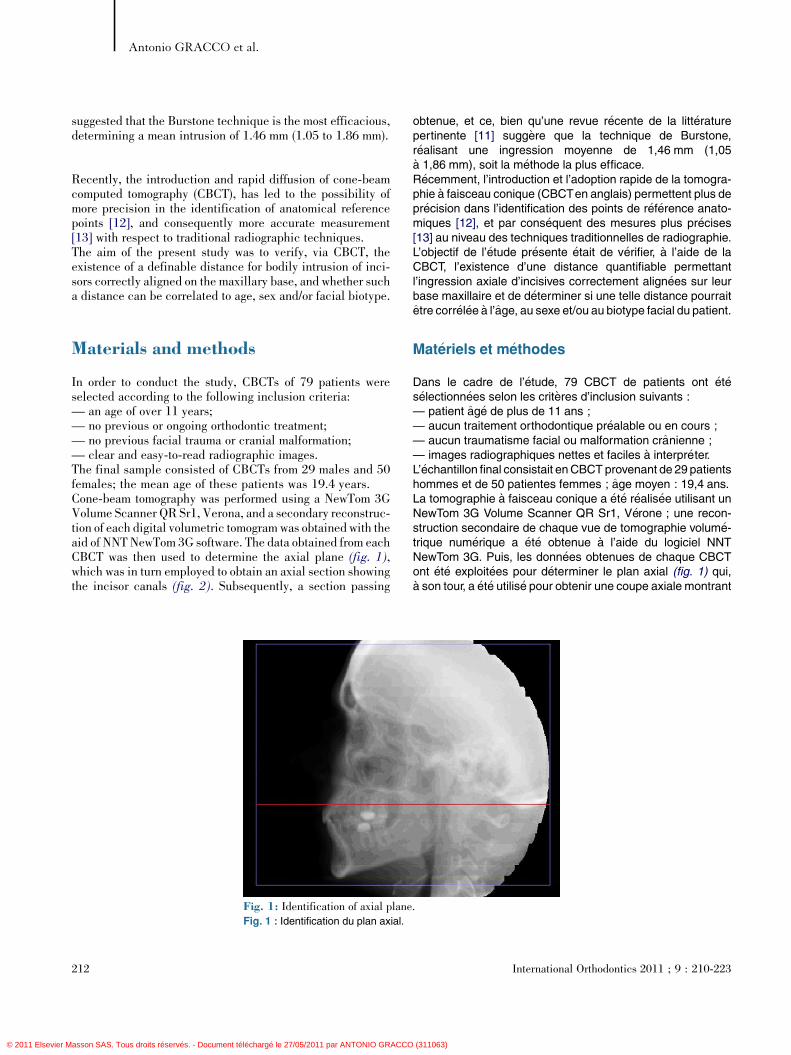
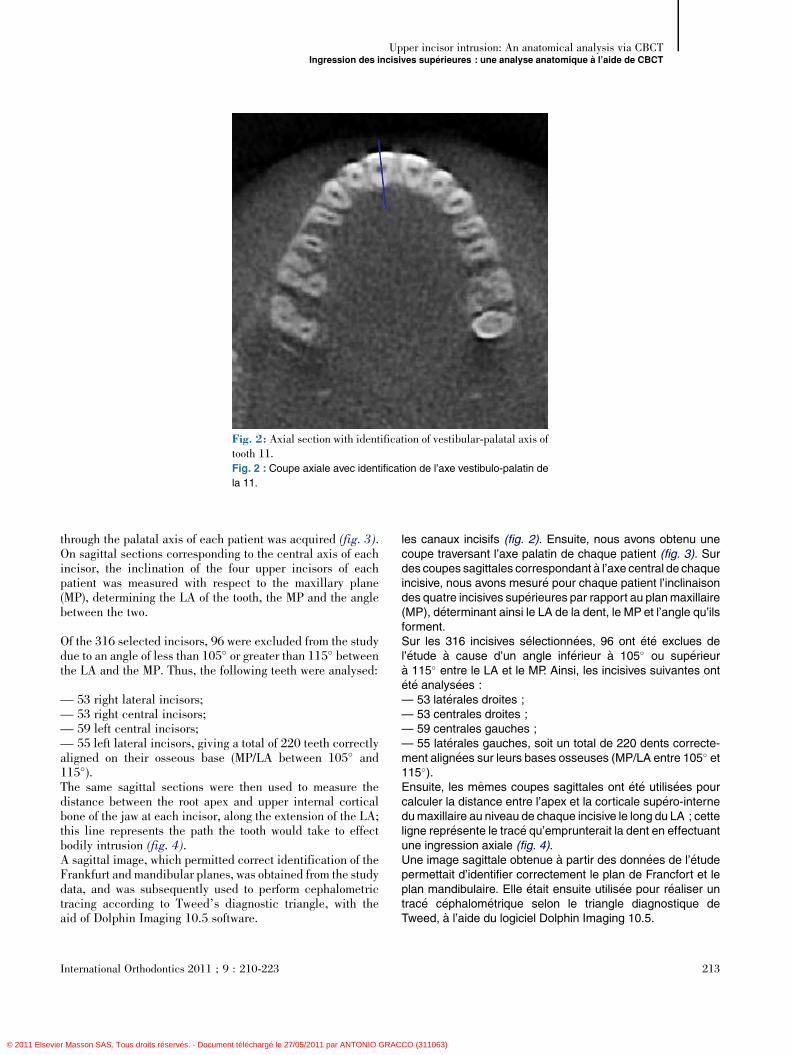
In order to conduct the study, CBCTs of 79 patients wereselected according to the following inclusion criteria:— an age of over 11 years;— no previous or ongoing orthodontic treatment;— no previous facial trauma or cranial malformation;— clear and easy-to-read radiographic images.The final sample consisted of CBCTs from 29 males and 50females; the mean age of these patients was 19.4 years.Cone-beam tomography was performed using a NewTom 3GVolume Scanner QR Sr1, Verona, and a secondary reconstruc-tion of each digital volumetric tomogram was obtained with theaid of NNT NewTom 3G software. The data obtained from eachCBCT was then used to determine the axial plane (fig. 1),which was in turn employed to obtain an axial section showingthe incisor canals (fig. 2). Subsequently, a section passing
[(Fig._1)TD$FIG]
Fig. 1: Identification of axial planeFig. 1 : Identification du plan axial.
212
asson SAS. Tous droits réservés. - Document téléchargé le 27/05/2011 par ANTONIO GRACCO
obtenue, et ce, bien qu’une revue r�ecente de la litt�eraturepertinente [11] sugg�ere que la technique de Burstone,r�ealisant une ingression moyenne de 1,46 mm (1,05a 1,86 mm), soit la m�ethode la plus efficace.R�ecemment, l’introduction et l’adoption rapide de la tomogra-phie a faisceau conique (CBCTen anglais) permettent plus depr�ecision dans l’identification des points de r�ef�erence anato-miques [12], et par cons�equent des mesures plus pr�ecises[13] au niveau des techniques traditionnelles de radiographie.L’objectif de l’�etude pr�esente �etait de v�erifier, a l’aide de laCBCT, l’existence d’une distance quantifiable permettantl’ingression axiale d’incisives correctement align�ees sur leurbase maxillaire et de d�eterminer si une telle distance pourraitetre corr�el�ee a l’age, au sexe et/ou au biotype facial du patient.
Mat�eriels et m�ethodes
Dans le cadre de l’�etude, 79 CBCT de patients ont �et�es�electionn�ees selon les crit�eres d’inclusion suivants :— patient ag�e de plus de 11 ans ;— aucun traitement orthodontique pr�ealable ou en cours ;— aucun traumatisme facial ou malformation cranienne ;— images radiographiques nettes et faciles a interpr�eter.L’�echantillon final consistait enCBCT provenant de 29 patientshommes et de 50 patientes femmes ; age moyen : 19,4 ans.La tomographie a faisceau conique a �et�e r�ealis�ee utilisant unNewTom 3G Volume Scanner QR Sr1, V�erone ; une recon-struction secondaire de chaque vue de tomographie volum�e-trique num�erique a �et�e obtenue a l’aide du logiciel NNTNewTom 3G. Puis, les donn�ees obtenues de chaque CBCTont �et�e exploit�ees pour d�eterminer le plan axial (fig. 1) qui,a son tour, a �et�e utilis�e pour obtenir une coupe axiale montrant
.
International Orthodontics 2011 ; 9 : 210-223
(311063)
[(Fig._2)TD$FIG]
Fig. 2: Axial section with identification of vestibular-palatal axis oftooth 11.Fig. 2 : Coupe axiale avec identification de l’axe vestibulo-palatin de
la 11.
Upper incisor intrusion: An anatomical analysis via CBCTIngression des incisives sup�erieures : une analyse anatomique a l’aide de CBCT
© 2011 Elsevie
through the palatal axis of each patient was acquired (fig. 3).On sagittal sections corresponding to the central axis of eachincisor, the inclination of the four upper incisors of eachpatient was measured with respect to the maxillary plane(MP), determining the LA of the tooth, the MP and the anglebetween the two.
Of the 316 selected incisors, 96 were excluded from the studydue to an angle of less than 105� or greater than 115� betweenthe LA and the MP. Thus, the following teeth were analysed:
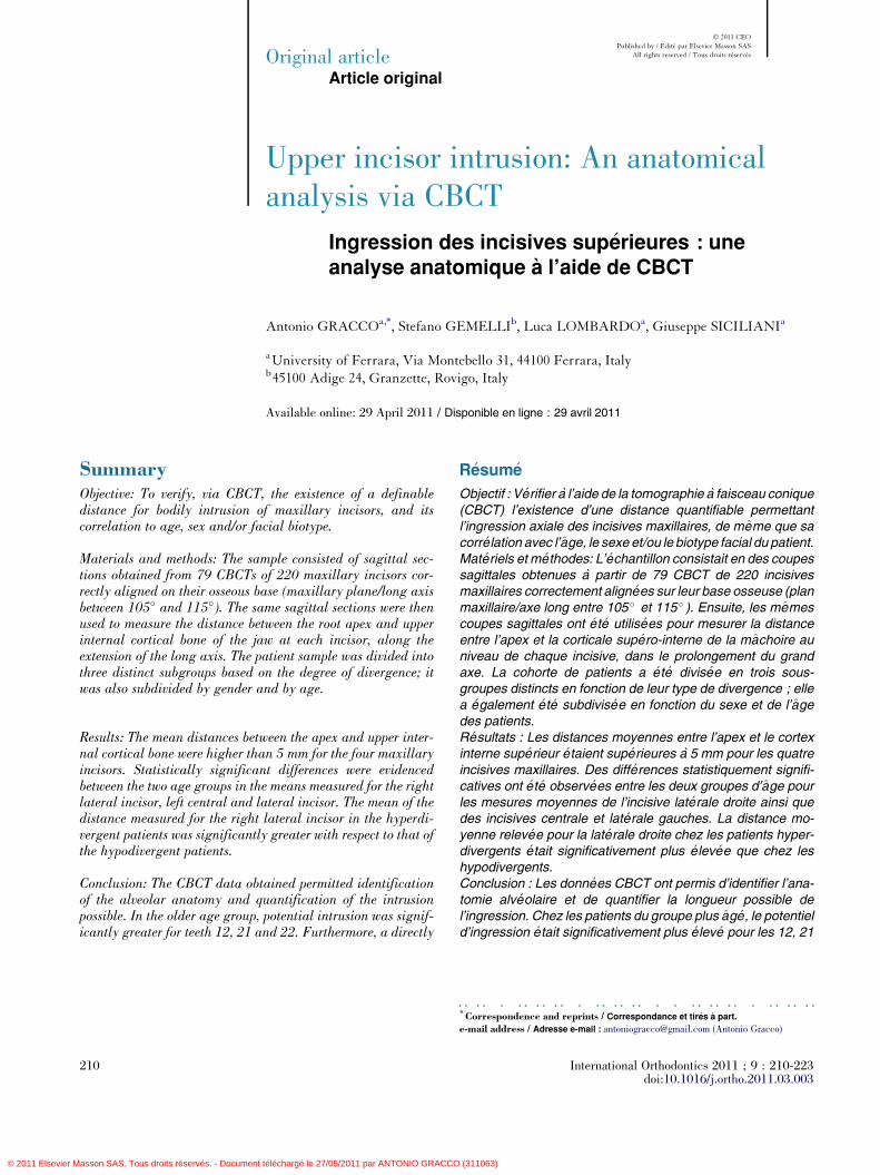
— 53 right lateral incisors;— 53 right central incisors;— 59 left central incisors;— 55 left lateral incisors, giving a total of 220 teeth correctlyaligned on their osseous base (MP/LA between 105� and115�).The same sagittal sections were then used to measure thedistance between the root apex and upper internal corticalbone of the jaw at each incisor, along the extension of the LA;this line represents the path the tooth would take to effectbodily intrusion (fig. 4).A sagittal image, which permitted correct identification of theFrankfurt and mandibular planes, was obtained from the studydata, and was subsequently used to perform cephalometrictracing according to Tweed’s diagnostic triangle, with theaid of Dolphin Imaging 10.5 software.
International Orthodontics 2011 ; 9 : 210-223
r Masson SAS. Tous droits réservés. - Document téléchargé le 27/05/2011 par ANTONIO GRAC
les canaux incisifs (fig. 2). Ensuite, nous avons obtenu unecoupe traversant l’axe palatin de chaque patient (fig. 3). Surdes coupes sagittales correspondant a l’axe central de chaqueincisive, nous avons mesur�e pour chaque patient l’inclinaisondes quatre incisives sup�erieures par rapport au planmaxillaire(MP), d�eterminant ainsi le LA de la dent, le MP et l’angle qu’ilsforment.Sur les 316 incisives s�electionn�ees, 96 ont �et�e exclues del’�etude a cause d’un angle inf�erieur a 105� ou sup�erieura 115� entre le LA et le MP. Ainsi, les incisives suivantes ont�et�e analys�ees :— 53 lat�erales droites ;— 53 centrales droites ;— 59 centrales gauches ;— 55 lat�erales gauches, soit un total de 220 dents correcte-ment align�ees sur leurs bases osseuses (MP/LA entre 105� et115�).Ensuite, les memes coupes sagittales ont �et�e utilis�ees pourcalculer la distance entre l’apex et la corticale sup�ero-internedumaxillaire au niveau de chaque incisive le long du LA ; cetteligne repr�esente le trac�e qu’emprunterait la dent en effectuantune ingression axiale (fig. 4).Une image sagittale obtenue a partir des donn�ees de l’�etudepermettait d’identifier correctement le plan de Francfort et leplan mandibulaire. Elle �etait ensuite utilis�ee pour r�ealiser untrac�e c�ephalom�etrique selon le triangle diagnostique deTweed, a l’aide du logiciel Dolphin Imaging 10.5.
213
CO (311063)

[(Fig._4)TD$FIG]
Fig. 4: Example of measurement on tooth 11.Fig. 4 : Exemple d’une prise de mesure sur la 11.
[(Fig._3)TD$FIG]
Fig. 3: Sagittal section used for measurement.Fig. 3 : Coupe sagittale utilis�ee pour faire les mesures.
Antonio GRACCO et al.
© 2011 Elsevier M
The patient sample was then divided into three distinct sub-groups based on the degree of divergence: 23 hypodivergent(FMA < 22�), 27 normodivergent (22 < FMA < 28�) and 29hyperdivergent (FMA > 28�) patients.The sample was also subdivided by gender: the female groupcomposed of 50 subjects and the male group of 29 subjects.Moreover, two age groups were created: Group 1 comprising53 subjects aged less than 20, and Group 2 made up of 26over-twenty-year-olds.
214
asson SAS. Tous droits réservés. - Document téléchargé le 27/05/2011 par ANTONIO GRACCO
L’�echantillon de patients a ensuite �et�e divis�e en trois sous-groupes distincts selon leur type de divergence : 23 patients�etaient hypodivergents (FMA < 22�), 27 normodivergents(22 < FMA < 28�) et 29 hyperdivergents (FMA > 28�).L’�echantillon a �egalement �et�e subdivis�e selon le sexe : legroupe f�eminin comprenait 50 sujets et le groupe masculin29 sujets. Par ailleurs, deux groupes ont �et�e cr�e�es en fonctionde l’age des patients avec, dans le groupe 1, 53 sujets ag�es demoins de 20 ans et, dans le groupe 2, 26 patients ag�es de plusde 20 ans.
International Orthodontics 2011 ; 9 : 210-223
(311063)
Upper incisor intrusion: An anatomical analysis via CBCTIngression des incisives sup�erieures : une analyse anatomique a l’aide de CBCT
© 2011 Elsevie
Statistical analysis
The mean values and standard deviations were calculated foreach tooth, and Fisher’s F Anova was employed to verifywhether the factors of age, gender or divergence had a statis-tically significant influence on the central tendency (mean) ofthe measurements taken for each tooth. Results are expressedas mean W standard deviation. Fisher’s post hoc test wasapplied in order to identify which principal effects yielded astatistical significance and Anova F for repeated measures tocompare the central tendencies (means) among the four teeth.Pearson’s r coefficient was calculated so as to analyse theproportional relationship between the four incisors: thisanalysis was applied to the single data points along the fourmeasurements (n = 27).
Results
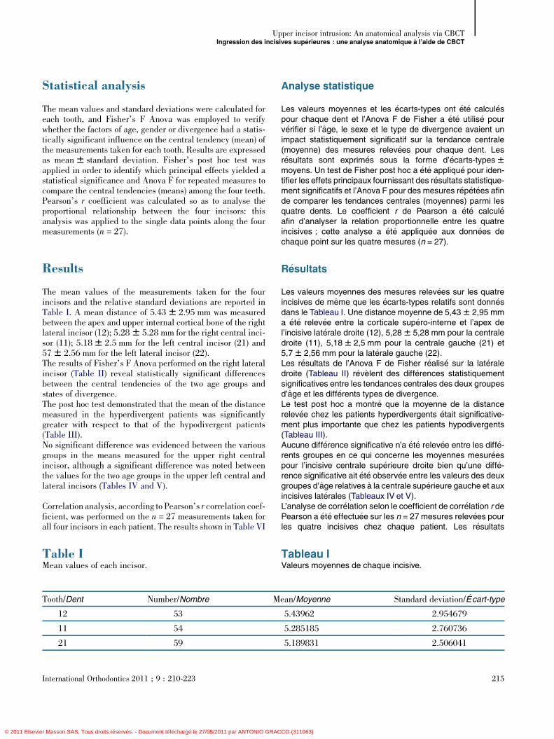
The mean values of the measurements taken for the fourincisors and the relative standard deviations are reported inTable I. A mean distance of 5.43 W 2.95 mm was measuredbetween the apex and upper internal cortical bone of the rightlateral incisor (12); 5.28 W 5.28 mm for the right central inci-sor (11); 5.18W 2.5 mm for the left central incisor (21) and57 W 2.56 mm for the left lateral incisor (22).The results of Fisher’s F Anova performed on the right lateralincisor (Table II) reveal statistically significant differencesbetween the central tendencies of the two age groups andstates of divergence.The post hoc test demonstrated that the mean of the distancemeasured in the hyperdivergent patients was significantlygreater with respect to that of the hypodivergent patients(Table III).No significant difference was evidenced between the variousgroups in the means measured for the upper right centralincisor, although a significant difference was noted betweenthe values for the two age groups in the upper left central andlateral incisors (Tables IV and V).
Correlation analysis, according to Pearson’s r correlation coef-ficient, was performed on the n = 27 measurements taken forall four incisors in each patient. The results shown in Table VI
able Iean values of each incisor.
Tableau IValeurs moyennes de chaque incisive.
ooth/Dent Number/Nombre Mean/Moyenne Standard deviation/ �Ecart-type
12 53 5.43962 2.954679
11 54 5.285185 2.760736
TM
T
21 59
International Orthodontics 2011 ; 9 : 210-223
r Masson SAS. Tous droits réservés. - Document téléchargé le 27/05/2011 par ANTONIO GRAC
Analyse statistique
Les valeurs moyennes et les �ecarts-types ont �et�e calcul�espour chaque dent et l’Anova F de Fisher a �et�e utilis�e pourv�erifier si l’age, le sexe et le type de divergence avaient unimpact statistiquement significatif sur la tendance centrale(moyenne) des mesures relev�ees pour chaque dent. Lesr�esultats sont exprim�es sous la forme d’�ecarts-types Wmoyens. Un test de Fisher post hoc a �et�e appliqu�e pour iden-tifier les effets principaux fournissant des r�esultats statistique-ment significatifs et l’Anova F pour des mesures r�ep�et�ees afinde comparer les tendances centrales (moyennes) parmi lesquatre dents. Le coefficient r de Pearson a �et�e calcul�eafin d’analyser la relation proportionnelle entre les quatreincisives ; cette analyse a �et�e appliqu�ee aux donn�ees dechaque point sur les quatre mesures (n = 27).
R�esultats
Les valeurs moyennes des mesures relev�ees sur les quatreincisives de meme que les �ecarts-types relatifs sont donn�esdans le Tableau I. Une distance moyenne de 5,43 W 2,95 mma �et�e relev�ee entre la corticale sup�ero-interne et l’apex del’incisive lat�erale droite (12), 5,28 W 5,28 mm pour la centraledroite (11), 5,18 W 2,5 mm pour la centrale gauche (21) et5,7 W 2,56 mm pour la lat�erale gauche (22).Les r�esultats de l’Anova F de Fisher r�ealis�e sur la lat�eraledroite (Tableau II) r�ev�elent des diff�erences statistiquementsignificatives entre les tendances centrales des deux groupesd’age et les diff�erents types de divergence.Le test post hoc a montr�e que la moyenne de la distancerelev�ee chez les patients hyperdivergents �etait significative-ment plus importante que chez les patients hypodivergents(Tableau III).Aucune diff�erence significative n’a �et�e relev�ee entre les diff�e-rents groupes en ce qui concerne les moyennes mesur�eespour l’incisive centrale sup�erieure droite bien qu’une diff�e-rence significative ait �et�e observ�ee entre les valeurs des deuxgroupes d’age relatives a la centrale sup�erieure gauche et auxincisives lat�erales (Tableaux IV et V).L’analyse de corr�elation selon le coefficient de corr�elation r dePearson a �et�e effectu�ee sur les n = 27 mesures relev�ees pourles quatre incisives chez chaque patient. Les r�esultats
5.189831 2.506041
215
CO (311063)
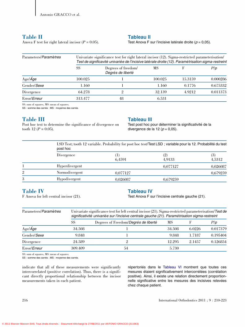
Table IVF Anova for left central incisor (21).
Tableau IVTest Anova F sur l’incisive centrale gauche (21).
Parameters/Param�etres Univariate significance test for left central incisor (21). Sigma-restricted parameterisation/Test designificativit�e univari�ee sur l’incisive centrale gauche (21). Param�etrisation sigma-restreint
SS Degrees of Freedom/Degr�es de libert�e MS F P/p
Age/Age 34.508 1 34.508 6.0226 0.017379
Gender/Sexe 9.848 1 9.848 1.7187 0.195404
Divergence 24.589 2 12.295 2.1457 0.126854
Error/Erreur 309.409 54 5.730
SS: sum of squares, MS: mean of squares.SS : somme des carr�es ; MS : moyenne des carr�es.
Table IIAnova F test for right lateral incisor (P = 0.05).
Tableau IITest Anova F sur l’incisive lat�erale droite (p = 0,05).
Parameters/Param�etres Univariate significance test for right lateral incisor (12). Sigma-restricted parameterisation/Test de significavit�e univari�ee de l’incisive lat�erale droite (12). Param�etrisation sigma-restreint
SS Degrees of freedom/Degr�es de libert�e
MS F P/p
Age/Age 100.025 1 100.025 15.3159 0.000286
Gender/Sexe 1.160 1 1.160 0.1776 0.675332
Divergence 64.278 2 32.139 4.9212 0.011373
Error/Erreur 313.477 48 6.531
SS: sum of squares, MS: mean of squares.SS : somme des carr�es ; MS : moyenne des carr�es.
Table IIIPost hoc test to determine the significance of divergence ontooth 12 (P = 0.05).
Tableau IIITest post hoc pour d�eterminer la significativit�e de ladivergence de la 12 (p = 0,05).
LSD Test; tooth 12 variable. Probability for post hoc test/Test LSD ; variable pour la 12. Probabilit�e du testpost hoc
Divergence (1)6,4591
(2)4,9133
(3)4,5312
1 Hyperdivergent 0,077127 0,026087
2 Normodivergent 0,077127 0,679259
3 Hypodivergent 0,026087 0,679259
Antonio GRACCO et al.
© 2011 Elsevier M
indicate that all of these measurements were significantlyintercorrelated (positive correlation). Thus, there is a signifi-cant directly proportional relationship between the incisormeasurements taken in each patient.
216
asson SAS. Tous droits réservés. - Document téléchargé le 27/05/2011 par ANTONIO GRACCO
r�epertori�es dans le Tableau VI montrent que toutes cesmesures �etaient significativement intercorr�el�ees (corr�elationpositive). Ainsi, il existe une relation directement proportion-nelle significative entre les mesures des incisives relev�eeschez chaque patient.
International Orthodontics 2011 ; 9 : 210-223
(311063)
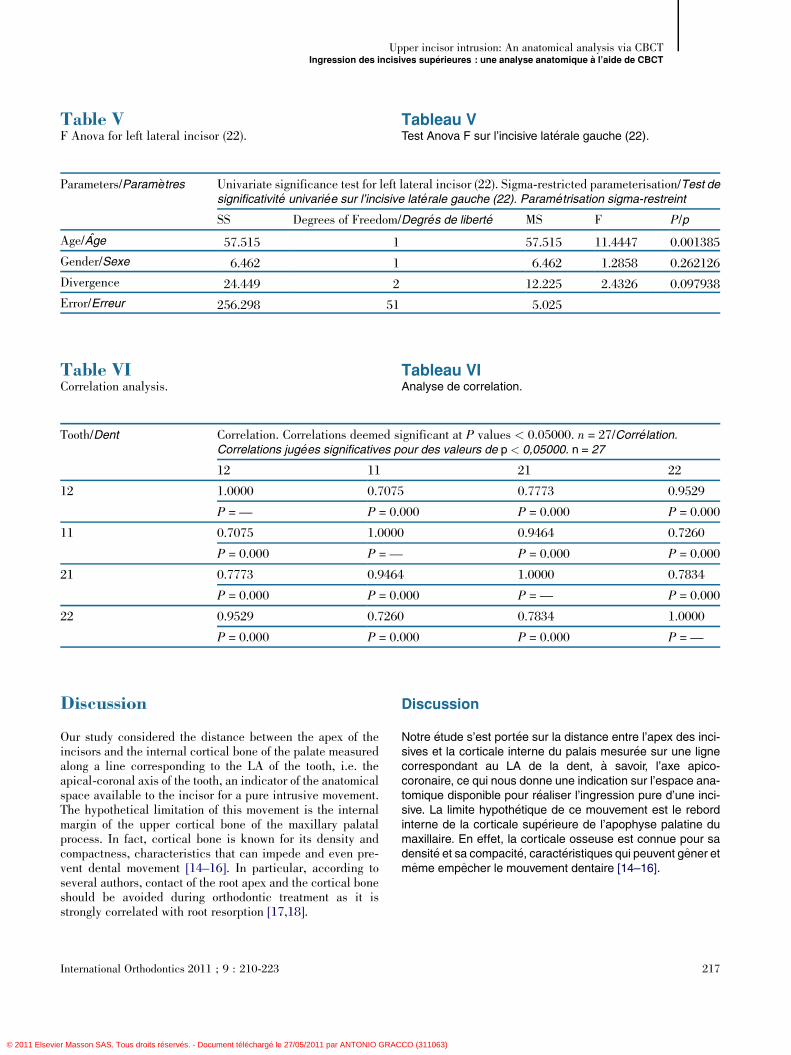
Table VF Anova for left lateral incisor (22).
Tableau VTest Anova F sur l’incisive lat�erale gauche (22).
Parameters/Param�etres Univariate significance test for left lateral incisor (22). Sigma-restricted parameterisation/Test designificativit�e univari�ee sur l’incisive lat�erale gauche (22). Param�etrisation sigma-restreint
SS Degrees of Freedom/Degr�es de libert�e MS F P/p
Age/Age 57.515 1 57.515 11.4447 0.001385
Gender/Sexe 6.462 1 6.462 1.2858 0.262126
Divergence 24.449 2 12.225 2.4326 0.097938
Error/Erreur 256.298 51 5.025
Table VICorrelation analysis.
Tableau VIAnalyse de correlation.
Tooth/Dent Correlation. Correlations deemed significant at P values < 0.05000. n = 27/Corr�elation.Correlations jug�ees significatives pour des valeurs de p < 0,05000. n = 27
12 11 21 22
12 1.0000 0.7075 0.7773 0.9529
P = — P = 0.000 P = 0.000 P = 0.000
11 0.7075 1.0000 0.9464 0.7260
P = 0.000 P = — P = 0.000 P = 0.000
21 0.7773 0.9464 1.0000 0.7834
P = 0.000 P = 0.000 P = — P = 0.000
22 0.9529 0.7260 0.7834 1.0000
P = 0.000 P = 0.000 P = 0.000 P = —
Upper incisor intrusion: An anatomical analysis via CBCTIngression des incisives sup�erieures : une analyse anatomique a l’aide de CBCT
© 2011 Elsevie
Discussion
Our study considered the distance between the apex of theincisors and the internal cortical bone of the palate measuredalong a line corresponding to the LA of the tooth, i.e. theapical-coronal axis of the tooth, an indicator of the anatomicalspace available to the incisor for a pure intrusive movement.The hypothetical limitation of this movement is the internalmargin of the upper cortical bone of the maxillary palatalprocess. In fact, cortical bone is known for its density andcompactness, characteristics that can impede and even pre-vent dental movement [14–16]. In particular, according toseveral authors, contact of the root apex and the cortical boneshould be avoided during orthodontic treatment as it isstrongly correlated with root resorption [17,18].
International Orthodontics 2011 ; 9 : 210-223
r Masson SAS. Tous droits réservés. - Document téléchargé le 27/05/2011 par ANTONIO GRAC
Discussion
Notre �etude s’est port�ee sur la distance entre l’apex des inci-sives et la corticale interne du palais mesur�ee sur une lignecorrespondant au LA de la dent, a savoir, l’axe apico-coronaire, ce qui nous donne une indication sur l’espace ana-tomique disponible pour r�ealiser l’ingression pure d’une inci-sive. La limite hypoth�etique de ce mouvement est le rebordinterne de la corticale sup�erieure de l’apophyse palatine dumaxillaire. En effet, la corticale osseuse est connue pour sadensit�e et sa compacit�e, caract�eristiques qui peuvent gener etmeme empecher le mouvement dentaire [14–16].
217
CO (311063)
Antonio GRACCO et al.
© 2011 Elsevier M
Our study examined only correctly inclined upper incisors inorder that only a ‘pure’ intrusive movement be considered, i.e.that which occurs along the apical-incisal axis of the tooth.The widely accepted reference values considered were thoseproposed by McLaughlin: an angle between dental axis andMP of between 105� and 115�.
The results of this study on untreated patients seem to confirmthe existence, on average, of a distance sufficient for ‘pure’intrusion of the incisors (fig. 4). In fact, the mean of the valuescalculated for each tooth was found to be greater than that themaximum clinical intrusion reported in a recent review of theliterature [11].
In analysis of each age group, measurement of distance forachievable intrusion in Group 2, that is patients of over20 years of age, was found to be significantly higher for threeout of the four teeth considered. Furthermore, an analogoustendency, albeit not statistically significant, was also noted forthe right central incisor. This finding may be explained andsupported by data from the literature regarding modification ofthe upper anterior alveolar height [19–21].
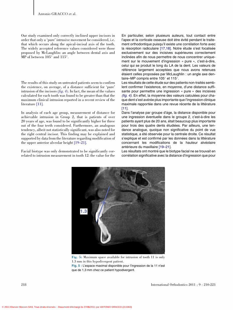
Facial biotype was only demonstrated to be significantly cor-related to intrusion measurement in tooth 12: the value for the
[(Fig._5)TD$FIG]
Fig. 5: Maximum space available1.3 mm in this hypodivergent patienFig. 5 : L’espace maximal disponible
que de 1,3 mm chez ce patient hypo
218
asson SAS. Tous droits réservés. - Document téléchargé le 27/05/2011 par ANTONIO GRACCO
En particulier, selon plusieurs auteurs, tout contact entrel’apex et la corticale osseuse doit etre �evit�e pendant le traite-ment orthodontique puisqu’il existe une corr�elation forte avecla r�esorption radiculaire [17,18]. Notre �etude s’est focalis�eeexclusivement sur des incisives sup�erieures correctementinclin�ees afin de nous permettre de nous concentrer unique-ment sur le mouvement d’ingression « pure », c’est-a-dire,celui qui se produit le long du LA de la dent. Les valeurs der�ef�erence largement accept�ees que nous avons retenues�etaient celles propos�ees par McLaughlin : un angle axe den-taire–MP compris entre 105� et 115�.Les r�esultats de cette �etude sur des patients non-trait�es semb-lent confirmer l’existence, en moyenne, d’une distance suffi-sante pour permettre une ingression « pure » des incisives(fig. 4). En effet, la moyenne des valeurs calcul�ees pour cha-que dent s’est av�er�ee plus importante que l’ingression cliniquemaximale rapport�ee dans une revue r�ecente de la litt�erature[11].Dans l’analyse par groupe d’age, la distance disponible pourune ingression �eventuelle dans le groupe 2, c’est-a-dire lespatients ayant plus de 20 ans, �etait beaucoup plus importantepour trois des quatre dents �etudi�ees. Par ailleurs, une ten-dance analogue, quoique non significative du point de vuestatistique, a �et�e observ�ee pour la centrale droite. Ce r�esultats’explique et est confirm�e par les donn�ees dans la litt�eratureconcernant les modifications de la hauteur alv�eolaireant�erieure du maxillaire [19–21].Les r�esultats ont montr�e que le biotype facial ne se trouvait encorr�elation significative avec la distance d’ingression que pour
for intrusion of tooth 11 is onlyt.pour l’ingression de la 11 n’est
divergent.
International Orthodontics 2011 ; 9 : 210-223
(311063)
Upper incisor intrusion: An anatomical analysis via CBCTIngression des incisives sup�erieures : une analyse anatomique a l’aide de CBCT
© 2011 Elsevie
hypodivergent group was significantly lower with respect tothat of the hyperdivergent subjects (fig. 5). The other incisorsanalysed showed the same tendency, despite the fact that theirvalues were not statistically significant; it is plausible thatthese values could acquire statistical significance in a largersample. The correlation between divergence and intrusion
[(Fig._6)TD$FIG]
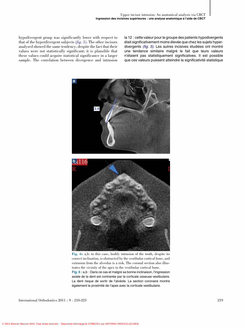
Fig. 6: a,b: in this case, bodily incorrect inclination, is obstructed byextrusion from the alveolus is a risktrates the vicinity of the apex to theFig. 6 : a,b : Dans ce cas et malgr�e s
axiale de la dent est contrari�ee par l
La dent risque de sortir de l’alv�eol
�egalement la proximit�e de l’apex ave
International Orthodontics 2011 ; 9 : 210-223
r Masson SAS. Tous droits réservés. - Document téléchargé le 27/05/2011 par ANTONIO GRAC
la 12 : cette valeur pour le groupe des patients hypodivergents�etait significativementmoins �elev�ee que chez les sujets hyper-divergents (fig. 5). Les autres incisives �etudi�ees ont montr�eune tendance similaire malgr�e le fait que leurs valeursn’�etaient pas statistiquement significatives. Il est possibleque ces valeurs puissent atteindre la significativit�e statistique
trusion of the tooth, despite itsthe vestibular cortical bone, and. The coronal section also illus-vestibular cortical bone.a bonne inclinaison, l’ingression
a corticale osseuse vestibulaire.
e. La section coronaire montre
c la corticale vestibulaire.
219
CO (311063)
Antonio GRACCO et al.
© 2011 Elsevier M
measurement may be explained by the hypothesis that inhyperdivergent subjects, with a prevalently vertical skeletalgrowth and consequent tendency to open-bite, alveolar growthacts to compensate for skeletal disharmony and contribute tomaintenance of incisal contact. Hence, alveolar growth seemsto be correlated to a descent of the incisors, which, if unaf-fected by other factors (dysfunction, bad habits, sagittal skel-etal discrepancies, etc.), occurs to maintain a correct inclina-tion of the tooth with respect to the basal bone. No genderdifference was noted in any of the four incisors.
Analysis of the correlations performed on 27 patients in whomthe values were calculated for all four teeth showed that thefour incisors of each patient are related in a proportionalmanner. It is therefore logical to expect the high values ofone incisor to correspond to high values of the other three.Moreover, the directly proportional relationship was moreaccentuated if pairs of analogous teeth are considered.
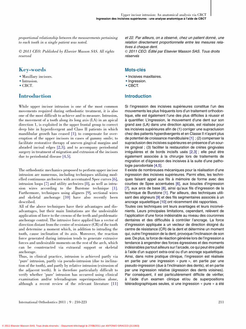
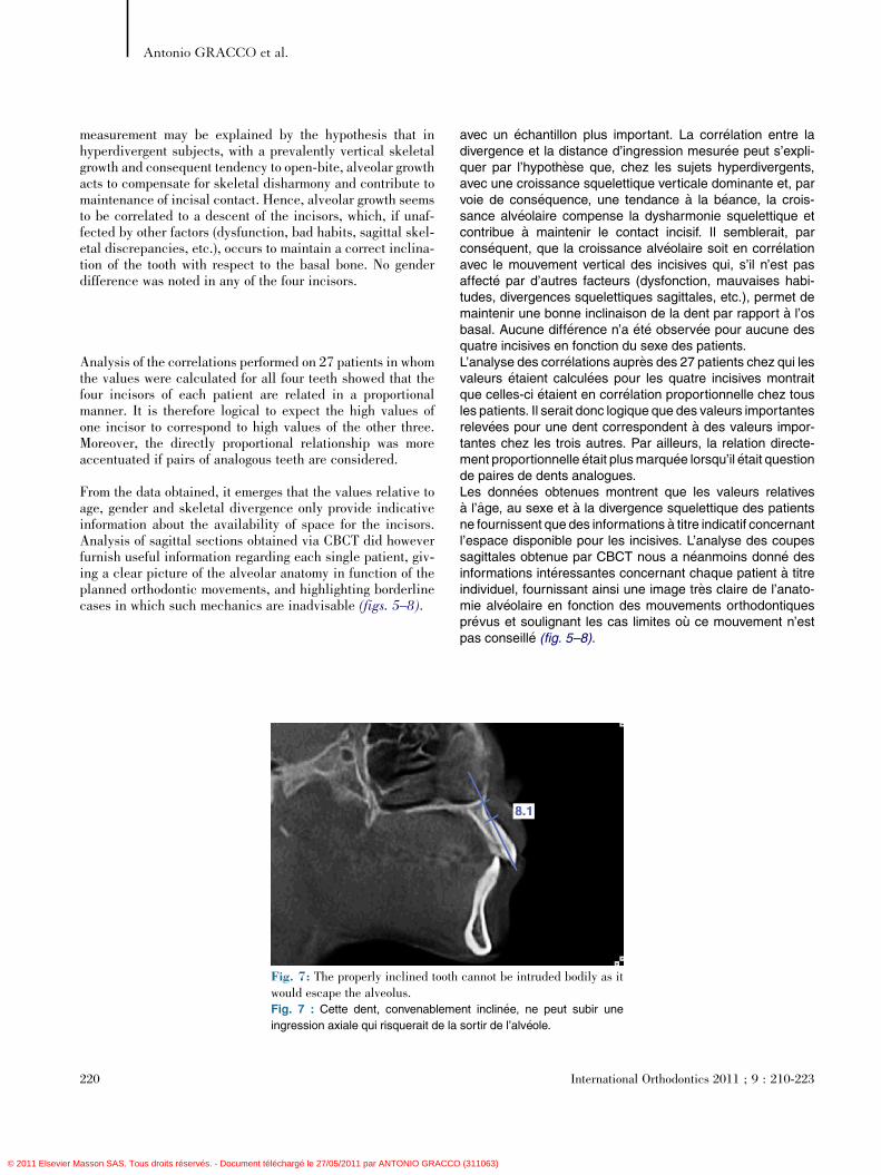
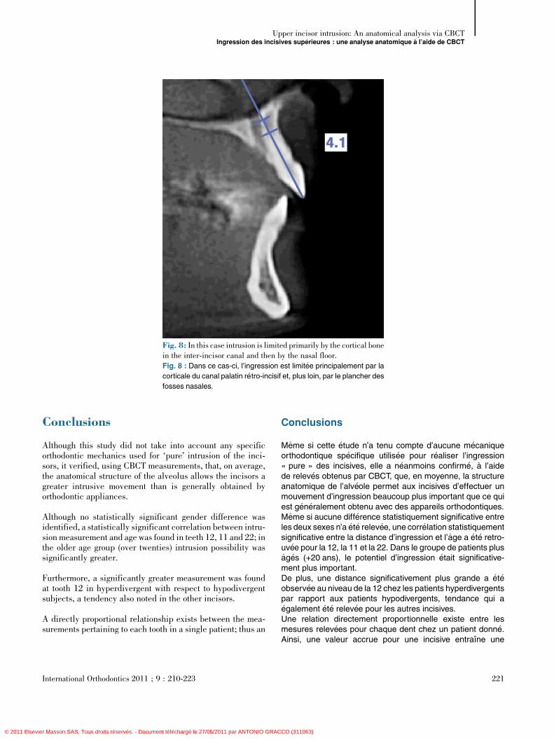
From the data obtained, it emerges that the values relative toage, gender and skeletal divergence only provide indicativeinformation about the availability of space for the incisors.Analysis of sagittal sections obtained via CBCT did howeverfurnish useful information regarding each single patient, giv-ing a clear picture of the alveolar anatomy in function of theplanned orthodontic movements, and highlighting borderlinecases in which such mechanics are inadvisable (figs. 5–8).
[(Fig._7)TD$FIG]
Fig. 7: The properly inclined toothwould escape the alveolus.Fig. 7 : Cette dent, convenableme
ingression axiale qui risquerait de la
220
asson SAS. Tous droits réservés. - Document téléchargé le 27/05/2011 par ANTONIO GRACCO
avec un �echantillon plus important. La corr�elation entre ladivergence et la distance d’ingression mesur�ee peut s’expli-quer par l’hypoth�ese que, chez les sujets hyperdivergents,avec une croissance squelettique verticale dominante et, parvoie de cons�equence, une tendance a la b�eance, la crois-sance alv�eolaire compense la dysharmonie squelettique etcontribue a maintenir le contact incisif. Il semblerait, parcons�equent, que la croissance alv�eolaire soit en corr�elationavec le mouvement vertical des incisives qui, s’il n’est pasaffect�e par d’autres facteurs (dysfonction, mauvaises habi-tudes, divergences squelettiques sagittales, etc.), permet demaintenir une bonne inclinaison de la dent par rapport a l’osbasal. Aucune diff�erence n’a �et�e observ�ee pour aucune desquatre incisives en fonction du sexe des patients.L’analyse des corr�elations aupr�es des 27 patients chez qui lesvaleurs �etaient calcul�ees pour les quatre incisives montraitque celles-ci �etaient en corr�elation proportionnelle chez tousles patients. Il serait donc logique que des valeurs importantesrelev�ees pour une dent correspondent a des valeurs impor-tantes chez les trois autres. Par ailleurs, la relation directe-ment proportionnelle �etait plusmarqu�ee lorsqu’il �etait questionde paires de dents analogues.Les donn�ees obtenues montrent que les valeurs relativesa l’age, au sexe et a la divergence squelettique des patientsne fournissent que des informations a titre indicatif concernantl’espace disponible pour les incisives. L’analyse des coupessagittales obtenue par CBCT nous a n�eanmoins donn�e desinformations int�eressantes concernant chaque patient a titreindividuel, fournissant ainsi une image tr�es claire de l’anato-mie alv�eolaire en fonction des mouvements orthodontiquespr�evus et soulignant les cas limites ou ce mouvement n’estpas conseill�e (fig. 5–8).
cannot be intruded bodily as it
nt inclin�ee, ne peut subir une
sortir de l’alv�eole.
International Orthodontics 2011 ; 9 : 210-223
(311063)
[(Fig._8)TD$FIG]
Fig. 8: In this case intrusion is limited primarily by the cortical bonein the inter-incisor canal and then by the nasal floor.Fig. 8 : Dans ce cas-ci, l’ingression est limit�ee principalement par la
corticale du canal palatin r�etro-incisif et, plus loin, par le plancher des
fosses nasales.
Upper incisor intrusion: An anatomical analysis via CBCTIngression des incisives sup�erieures : une analyse anatomique a l’aide de CBCT
© 2011 Elsevie
Conclusions
Although this study did not take into account any specificorthodontic mechanics used for ‘pure’ intrusion of the inci-sors, it verified, using CBCT measurements, that, on average,the anatomical structure of the alveolus allows the incisors agreater intrusive movement than is generally obtained byorthodontic appliances.
Although no statistically significant gender difference wasidentified, a statistically significant correlation between intru-sion measurement and age was found in teeth 12, 11 and 22; inthe older age group (over twenties) intrusion possibility wassignificantly greater.
Furthermore, a significantly greater measurement was foundat tooth 12 in hyperdivergent with respect to hypodivergentsubjects, a tendency also noted in the other incisors.
A directly proportional relationship exists between the mea-surements pertaining to each tooth in a single patient; thus an
International Orthodontics 2011 ; 9 : 210-223
r Masson SAS. Tous droits réservés. - Document téléchargé le 27/05/2011 par ANTONIO GRAC
Conclusions
Meme si cette �etude n’a tenu compte d’aucune m�ecaniqueorthodontique sp�ecifique utilis�ee pour r�ealiser l’ingression« pure » des incisives, elle a n�eanmoins confirm�e, a l’aidede relev�es obtenus par CBCT, que, en moyenne, la structureanatomique de l’alv�eole permet aux incisives d’effectuer unmouvement d’ingression beaucoup plus important que ce quiest g�en�eralement obtenu avec des appareils orthodontiques.Meme si aucune diff�erence statistiquement significative entreles deux sexes n’a �et�e relev�ee, une corr�elation statistiquementsignificative entre la distance d’ingression et l’age a �et�e retro-uv�ee pour la 12, la 11 et la 22. Dans le groupe de patients plusag�es (+20 ans), le potentiel d’ingression �etait significative-ment plus important.De plus, une distance significativement plus grande a �et�eobserv�ee au niveau de la 12 chez les patients hyperdivergentspar rapport aux patients hypodivergents, tendance qui a�egalement �et�e relev�ee pour les autres incisives.Une relation directement proportionnelle existe entre lesmesures relev�ees pour chaque dent chez un patient donn�e.Ainsi, une valeur accrue pour une incisive entraıne une
221
CO (311063)
Antonio GRACCO et al.
© 2011 Elsevier M
increased value for one tooth comports a proportional increasein the same value for the other incisors.The data obtained from CBCTs and their subsequentre-elaboration via NewTom 3G software permits precise visu-alisation of the alveolar anatomy surrounding each tooth: it istherefore possible to quantify the possible intrusion and toidentify anatomical obstructions to this movement.
Disclosure of interest
The authors declare that they have no conflicts of interestconcerning this article.
References/R�ef�erences
1. Burstone CR. Deep overbite2. Kokich VG. Esthetics and
Contin Educ Dent 1997;18(13. Bellamy LJ, Kokich VG, We
facilitate restoration: the tecDent Assoc 2008;139(6):725
4. Melsen B, Agerbaek N, Markbone loss. Am J Orthod Den
5. Corrente G, Abundo R, Reinfrabony defects in patientsstudy. J Periodontol 2003;74
6. Mulligan TF. Common sense7. Hilgers J. L’essenza dell’orto8. Ricketts RM. Bioprogressiv
Orthod 1976;70(4):359–97.9. Park JH, Kim TW. Deep-bite
Clin Orthod 2009;43(3):15210. Polat-Ozsoy O, Arman-Ozci
Eur J Orthod 2009;31(4):41211. Ng J, Major PW, Heo G, Flo
treatment: a systematic rev2005;128(2):212–9.
12. Ludlow JB, Gubler M, Cevidfication: cone-beam computOrthod Dentofacial Orthop 2
13. Baumgaertel S, Palomo JM,computed tomography denta(1):19-25 [discussion 25–8].
14. Nanda A. Biomechanics in c15. Handelman CS. The anterior
its influence on the occurre[discussion 109–10. Erratum
16. Ten Hoeve A, Mulie RM. Thcortex as studied with lamin
222
asson SAS. Tous droits réservés. - Document téléchargé le 27/05/2011 par ANTONIO GRACCO
augmentation proportionnelle de la meme valeur pour lesautres incisives.Les donn�ees obtenues par CBCT et leur traitement ult�erieuravec le logiciel NewTom 3G permettent une visualisationpr�ecise de l’anatomie alv�eolaire autour de chaque dent. Parcons�equent, il est possible de quantifier le potentiel d’ingres-sion et d’identifier les obstacles anatomiques s’opposant a cemouvement.
D�eclaration d’int�erets
Les auteurs d�eclarent ne pas avoir de conflits d’int�erets enrelation avec cet article.
correction by intrusion. Am J Orthod 1977;72:1-22.vertical tooth position: orthodontic possibilities. Compend2):1225–31 [quiz 1232].issman JA. Using orthodontic intrusion of abraded incisors tohnique’s effects on alveolar bone level and root length. J Am–33.enstam G. Intrusion of incisors in adult patients with marginaltofacial Orthop 1989;96(3):232–41.S, Cardaropoli D, Cardaropoli G. Orthodontic movement intowith advanced periodontal disease: a clinical and radiological(8):1104–9.mechanics. 3. J Clin Orthod 1979;13(11):762–6.donzia pratica. Quaderno SIRIO num. 22. Milan; 1995 .e therapy as an answer to orthodontic needs. Part II. Am J
correction using a clear aligner and intramaxillary elastics. J–7 [quiz 183].rpici A, Veziroglu F. Miniscrews for upper incisor intrusion.–6 [Epub 2009 Mar 16].res-Mir C. True incisor intrusion attained during orthodonticiew and meta-analysis. Am J Orthod Dentofacial Orthop
anes L, Mol A. Precision of cephalometric landmark identi-ed tomography vs conventional cephalometric views. Am J009;136(3):312.e1-312.e10 [discussion 312–3].Palomo L, Hans MG. Reliability and accuracy of cone-beaml measurements. Am J Orthod Dentofacial Orthop 2009;136
linical orthodontics Saunders. Elsevier, Philadelphia 1997.alveolus: its importance in limiting orthodontic treatment andnce of iatrogenic sequelae. Angle Orthod 1996;66(2):95-109in: Angle Orthod].e effect of antero-postero incisor repositioning on the palatalagraphy. J Clin Orthod 1976;10(11):804–22.
International Orthodontics 2011 ; 9 : 210-223
(311063)
17. Horiuchi A, Hotokezaka H, Kobayashi K. Correlation between cortical plate proximity andapical root resorption. Am J Orthod Dentofacial Orthop 1998;114(3):311–8.
18. Kaley J, Phillips C. Factors related to root resorption in edgewise practice. Angle Orthod1991;61(2):125–32.
19. Sarn€as KV, Solow B. Early adult changes in the skeletal and soft-tissue profile. Eur J Orthod1980;2(1):1-12.
20. Tallgren A, Solow B. Age differences in adult dentoalveolar heights. Eur J Orthod1991;13:149–56.
21. Forsberg CM, Eliasson S, Westergren H. Face height and tooth eruption in adults—a 20-year follow-up investigation. Eur J Orthod 1991;13:249–54.
Upper incisor intrusion: An anatomical analysis via CBCTIngression des incisives sup�erieures : une analyse anatomique a l’aide de CBCT
© 2011 Elsevie
International Orthodontics 2011 ; 9 : 210-223
r Masson SAS. Tous droits réservés. - Document téléchargé le 27/05/2011 par ANTONIO GRAC
223
CO (311063)