Embed Size (px)
Citation preview
+
UPDATES IN WOUND
MANAGEMENT
Lisa M. Arello, MS, ANP-BC
Plastic Surgery
+
No Conflict of Interest
No Commercial Support
+Overview
Wound classification and types of wounds
Phases of healing
Factors affecting healing
Current treatment modalities
Topical Products/ Choosing a Dressing
Negative Pressure Options
Extracellular Matrix
Oxygen Therapies
Pulsed Irrigation
Electromagnetic/ Ultrasound Therapies
Growth Factors/Future Study
+Wound Classification
Why is this important?
Drives treatment plan and expected outcomes
Estimating the length of time to heal
Preventing and treating factors affecting the natural timely and
orderly progression of wound healing
A wound is a result of the disruption of the normal skin structure,
skin function and skin architecture. A chronic wound does not
does not progress through the normal stages of healing.
Atiyeh, BS. Et al. Management of acute ad chronic open wounds: the importance of moist
environment in optimal wound healing. Current Phamr. Biotechnology 2002,3:179.
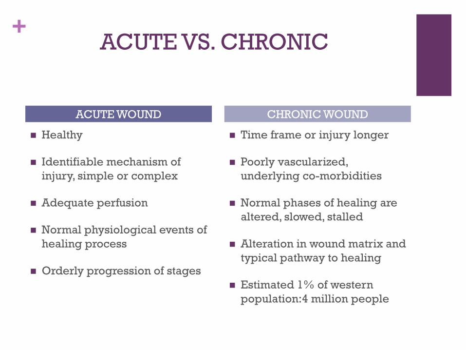
+ACUTE VS. CHRONIC
Healthy
Identifiable mechanism of
injury, simple or complex
Adequate perfusion
Normal physiological events of
healing process
Orderly progression of stages
Time frame or injury longer
Poorly vascularized,
underlying co-morbidities
Normal phases of healing are
altered, slowed, stalled
Alteration in wound matrix and
typical pathway to healing
Estimated 1% of western
population:4 million people
ACUTE WOUND CHRONIC WOUND
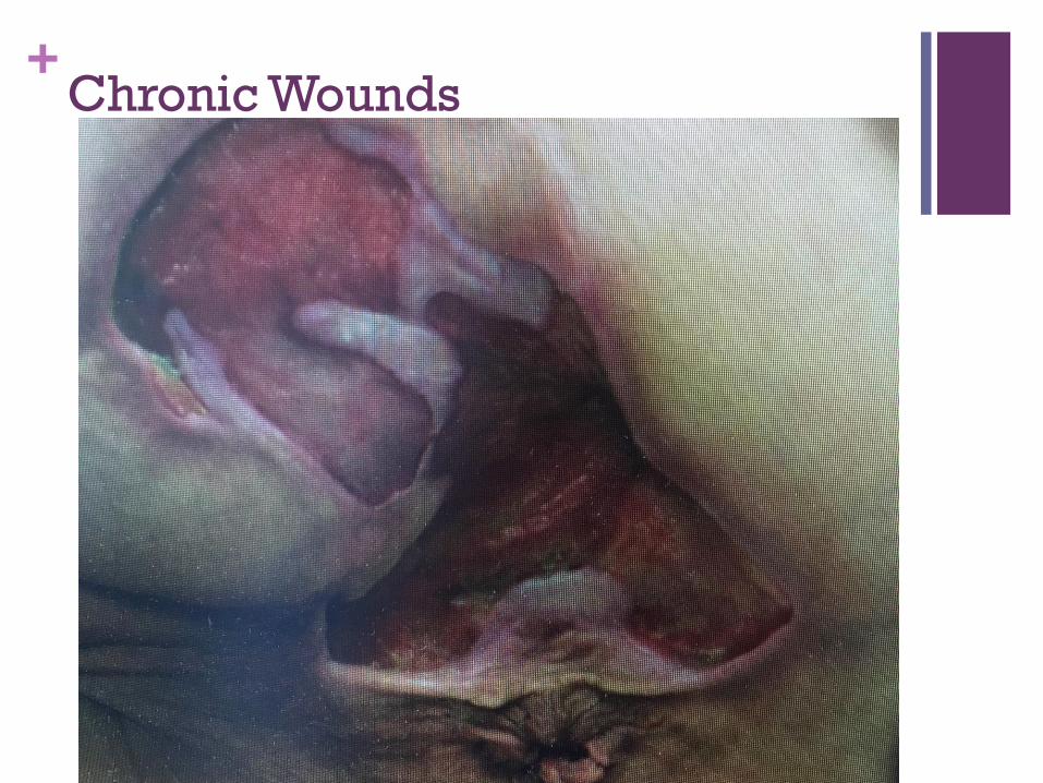
+Chronic Wounds
+Chronic Wounds
Excessive and and aggressive levels of pro-inflammatory cytokines and proteases -> ECM degradation and chronic inflammatory phase of healing
Proliferative phase delay results in delayed granulation and epithelium, which means lack of healing
Excessive bioburden develops, stimulating a chronic inflammatory response, increase neutrophlis and macrophages and therefore, proteases
Bio-film replaces normal planktonic bacteria , have greater antibiotic resistance, are not detected on standard wound cultures
Estimates 60% of chronic wounds have biofilms
Lintzeris, D. etal. Effect of a new Purified Collagen matrix with PCMP on Recalcitrant Wounds of Various Etiologies, Case Study. Vol. 3, #3, 3/18.
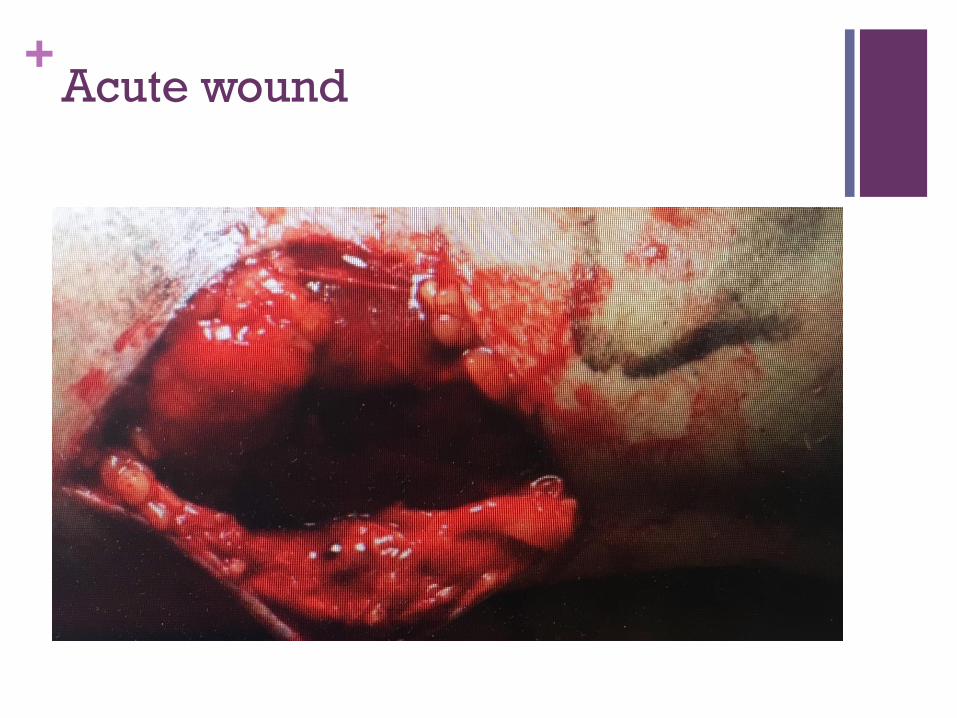
+Acute wound
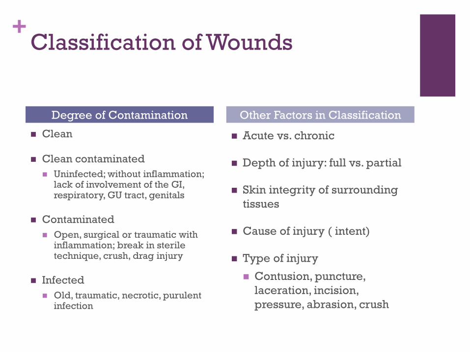
+Classification of Wounds
Clean
Clean contaminated
Uninfected; without inflammation; lack of involvement of the GI, respiratory, GU tract, genitals
Contaminated
Open, surgical or traumatic with inflammation; break in sterile technique, crush, drag injury
Infected
Old, traumatic, necrotic, purulent infection
Acute vs. chronic
Depth of injury: full vs. partial
Skin integrity of surrounding
tissues
Cause of injury ( intent)
Type of injury
Contusion, puncture,
laceration, incision,
pressure, abrasion, crush
Degree of Contamination Other Factors in Classification
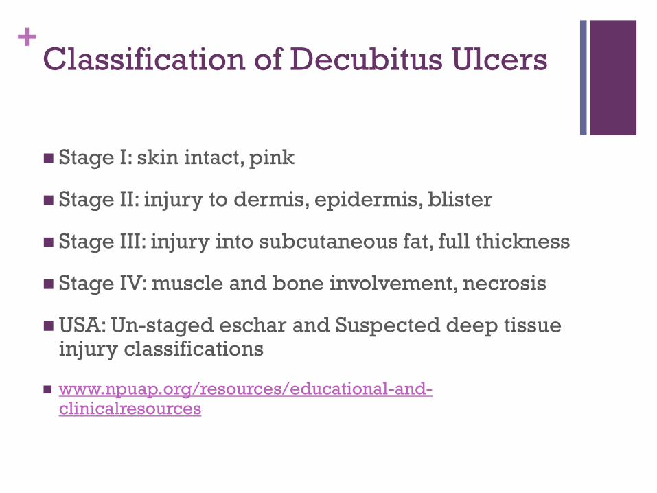
+Classification of Decubitus Ulcers
Stage I: skin intact, pink
Stage II: injury to dermis, epidermis, blister
Stage III: injury into subcutaneous fat, full thickness
Stage IV: muscle and bone involvement, necrosis
USA: Un-staged eschar and Suspected deep tissue injury classifications
www.npuap.org/resources/educational-and-clinicalresources
+
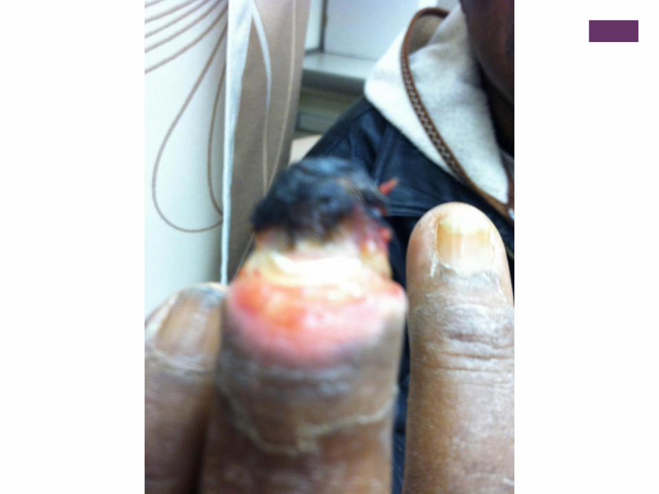
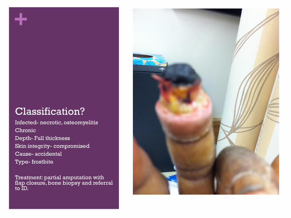
Classification?Infected- necrotic, osteomyelitis
Chronic
Depth- Full thickness
Skin integrity- compromised
Cause- accidental
Type- frostbite
Treatment: partial amputation with flap closure, bone biopsy and referral to ID.
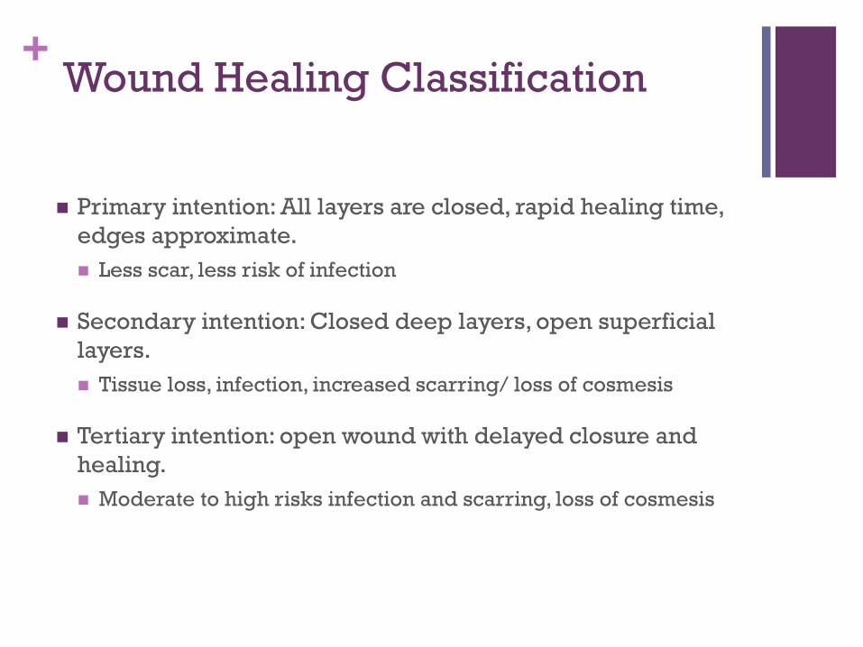
+Wound Healing Classification
Primary intention: All layers are closed, rapid healing time,
edges approximate.
Less scar, less risk of infection
Secondary intention: Closed deep layers, open superficial
layers.
Tissue loss, infection, increased scarring/ loss of cosmesis
Tertiary intention: open wound with delayed closure and
healing.
Moderate to high risks infection and scarring, loss of cosmesis
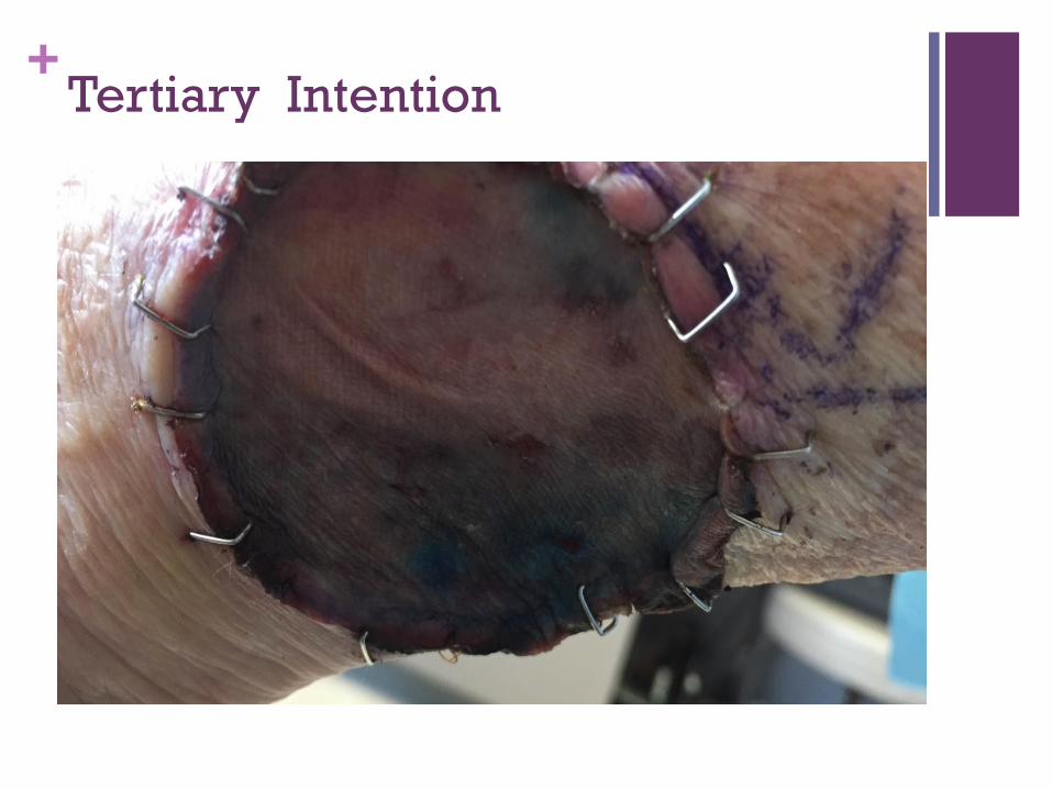
+Tertiary Intention
+Phases of Wound Healing
I. Inflammatory: Hemostasis and inflammation
Begins immediately and lasts up to 3 days, coagulation cascade,
vasoconstriction, platelets aggregate, fibrin clots.
Neutrophils attack bacteria
Macrophages are transformed for cell repair and stimulation of
fibroblast division, collagen synthesis and angiogenesis
Key components of this phase include increased vascular
permeability and cellular recruitment, including secretion of
vimentin, as structural protein for wound healing.
Chronic wounds typically arrested in this stage due to abnormal
production of MMPs impairing function of cytokines to digest
bacteria and necrotic tissue. Armstonrg, etal. www. Uptodate.com, 2016
+Phase II: Proliferative
3 days to 6 weeks
Macrophages stimulate migration of fibroblasts into the
wound for secretion of collagen type III and elastin
Collagen needed for tensile strength
Formation of granulation tissue to replace fibrin/fibronectin
Extracellular matrix deposition and wound contraction
begins
Process impaired by biofilm and bacteria that promote
inflammation and impair epithelialization. www.uptodate.com 2016
+Phase III: Maturation
Week 3 up to year for scar remodeling
Fibroblasts continue to synthesize collagen and
proteoglycan for the wound bed to increase tensile strength.
Collagen III converts to collagen I which increases tensile
strength up to 80% by week 6.
Collagen cross-linking, remodeling, wound contraction and
re-pigmentation.
Fan, W., et al. Current Advances in Modern Wound Healing. Wounds UK, 2010, Vol. 6, No. 3,
22-36.
www.meddean.luc.edu/lumen/meded/surgery 2008 and www.uptodate.com 2016.
+Factors Affecting Wound Healing
Acute vs. Chronic
Infection
edema
Impaired circulation
Impaired mobility
Impaired nutrition
Infection
Smoking
Meddean, 2008.
Diabetes
Obesity
Chronic lung disease
Previous infection
Liver or kidney disease
Immunosepression
Drugs affecting inflammatory
or clotting response
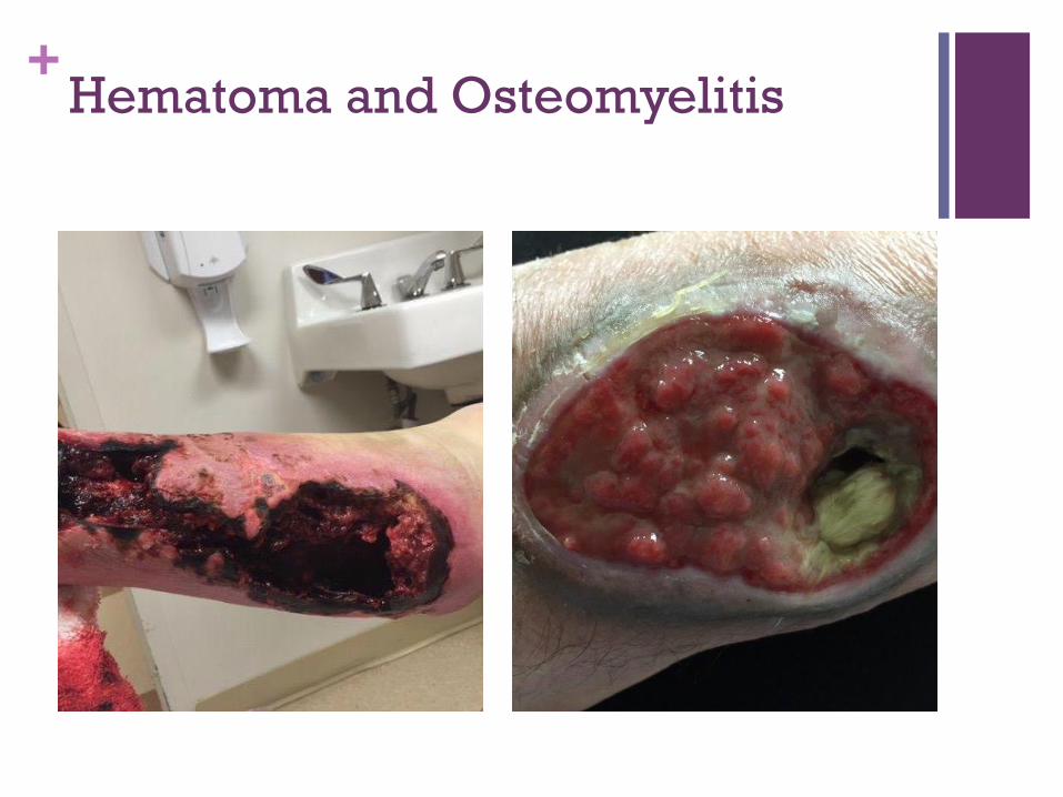
+Hematoma and Osteomyelitis
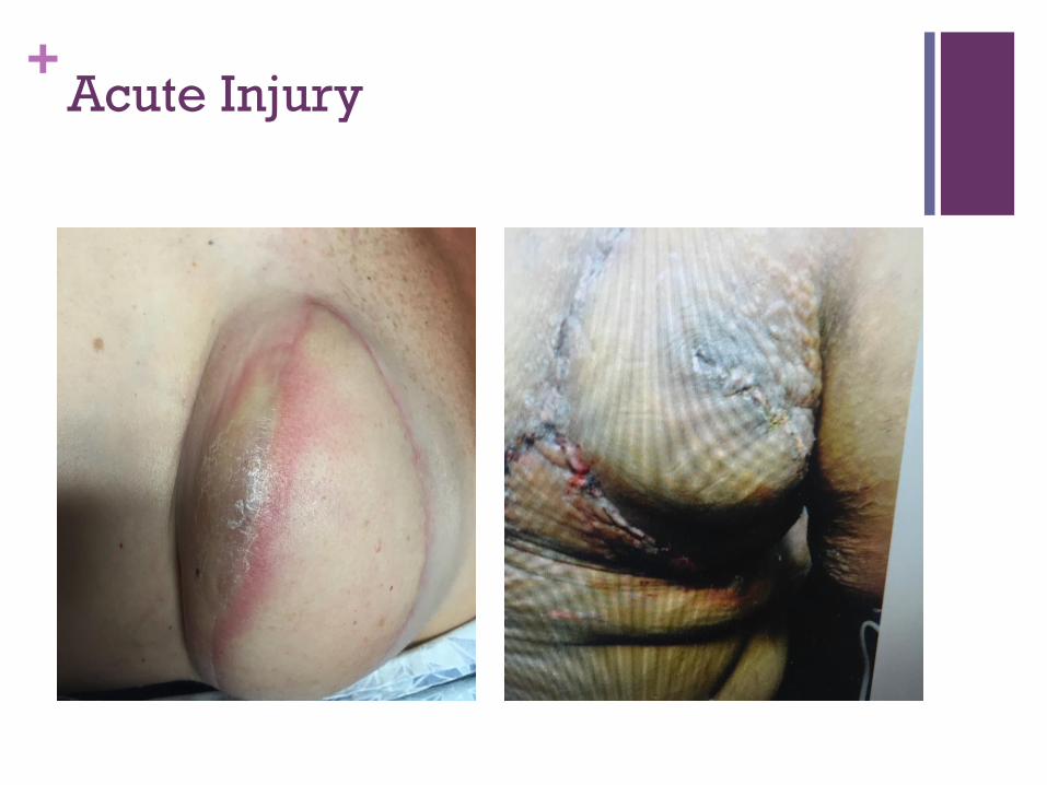
+Acute Injury
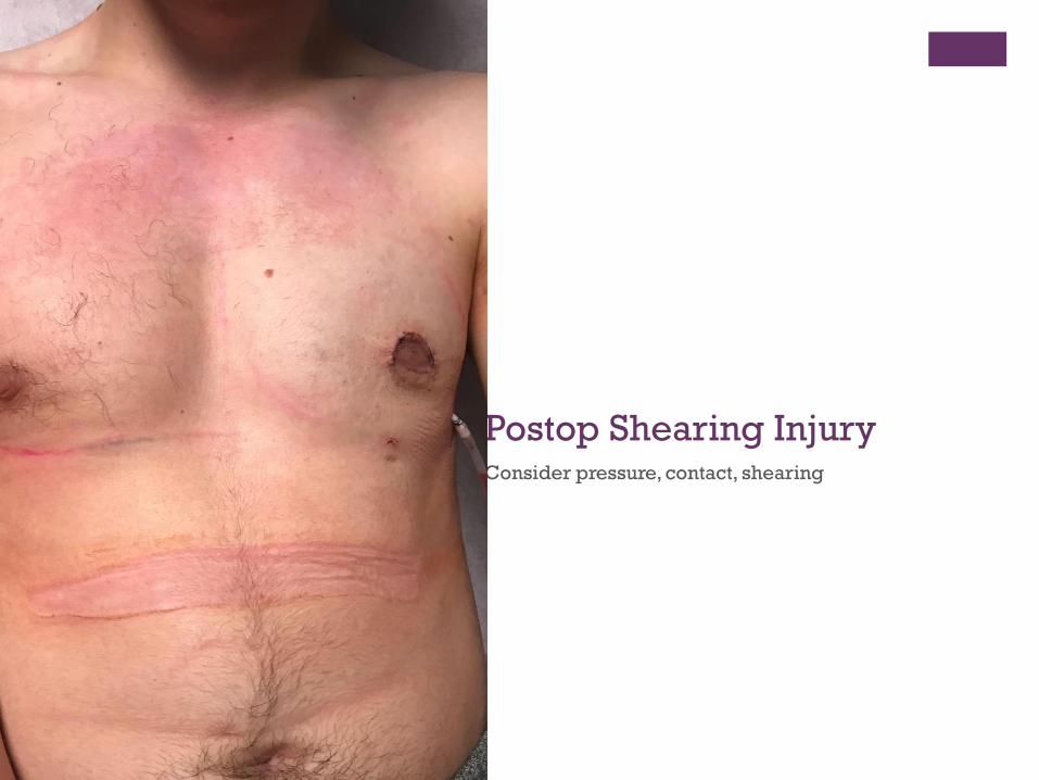
+ Postop Shearing InjuryConsider pressure, contact, shearing
+
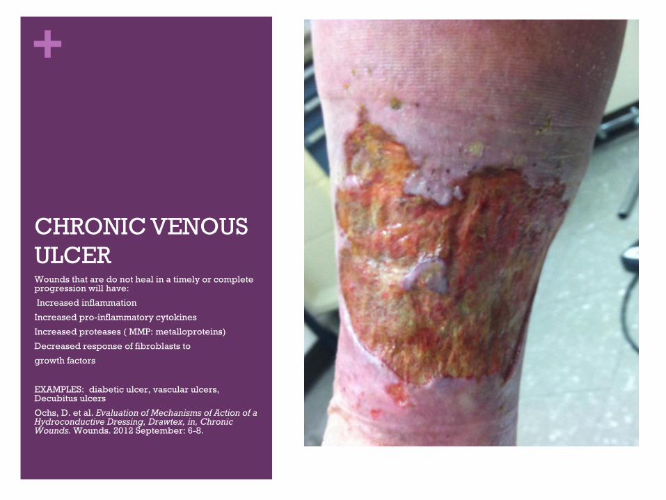
CHRONIC VENOUS
ULCERWounds that are do not heal in a timely or complete progression will have:
Increased inflammation
Increased pro-inflammatory cytokines
Increased proteases ( MMP: metalloproteins)
Decreased response of fibroblasts to
growth factors
EXAMPLES: diabetic ulcer, vascular ulcers, Decubitus ulcers
Ochs, D. et al. Evaluation of Mechanisms of Action of a Hydroconductive Dressing, Drawtex, in, Chronic Wounds. Wounds. 2012 September: 6-8.
+Goals of Wound Healing
Repair tissue in a timely manner
Restore function and anatomy
Prevent infection
Minimize inflammation and edema
Minimize pain
Best possible aesthetic outcome
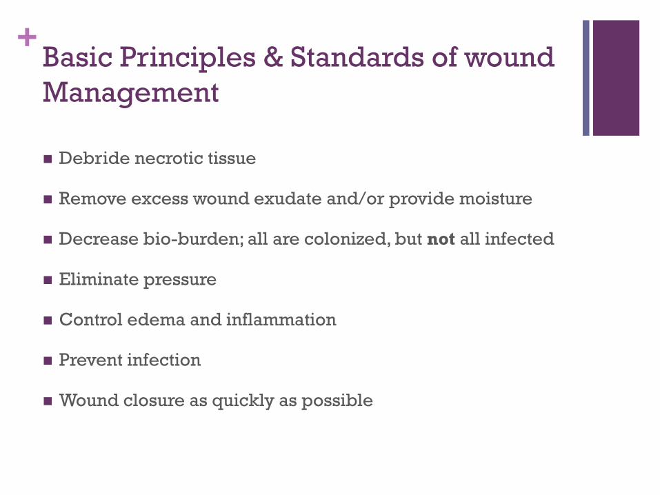
+Basic Principles & Standards of wound
Management
Debride necrotic tissue
Remove excess wound exudate and/or provide moisture
Decrease bio-burden; all are colonized, but not all infected
Eliminate pressure
Control edema and inflammation
Prevent infection
Wound closure as quickly as possible
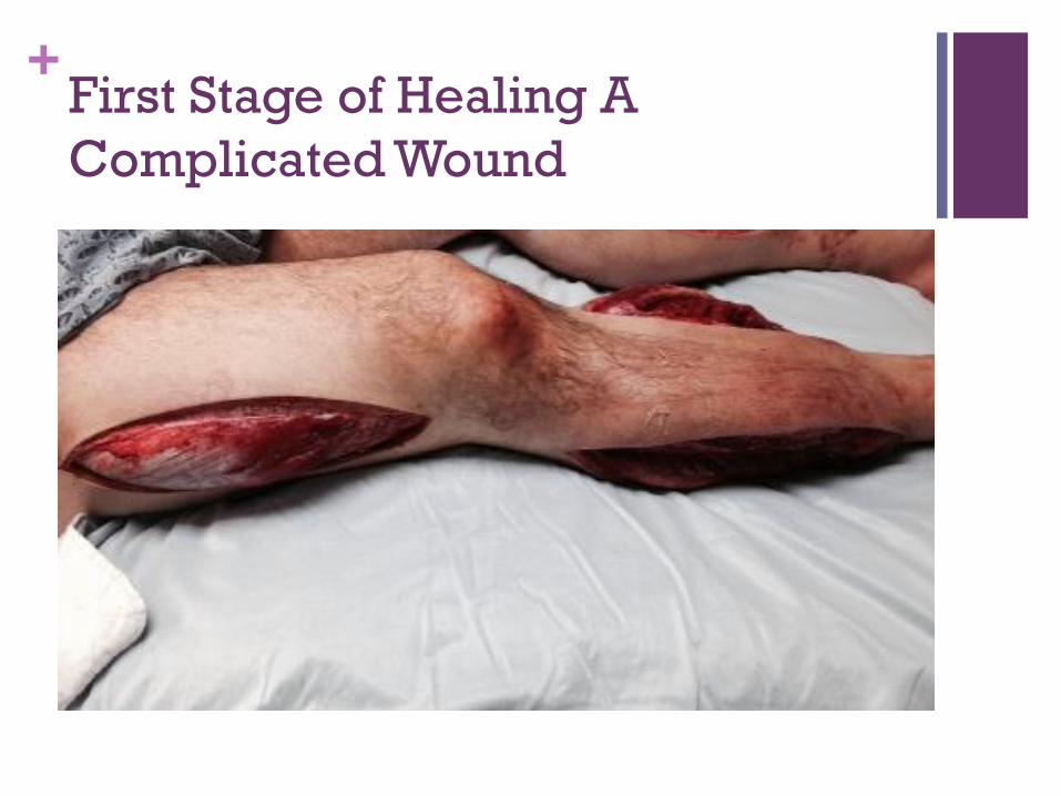
+First Stage of Healing A
Complicated Wound
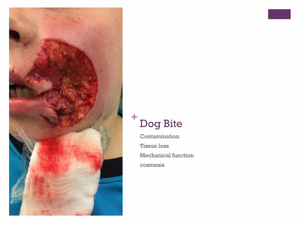
+ Dog BiteContamination
Tissue loss
Mechanical function
cosmesis
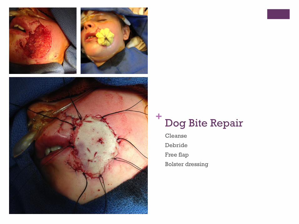
+ Dog Bite RepairCleanse
Debride
Free flap
Bolster dressing
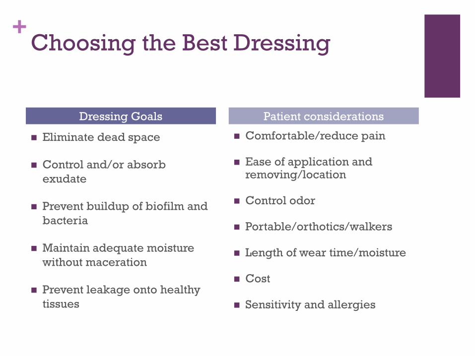
+Choosing the Best Dressing
Eliminate dead space
Control and/or absorb
exudate
Prevent buildup of biofilm and
bacteria
Maintain adequate moisture
without maceration
Prevent leakage onto healthy
tissues
Comfortable/reduce pain
Ease of application and removing/location
Control odor
Portable/orthotics/walkers
Length of wear time/moisture
Cost
Sensitivity and allergies
Dressing Goals Patient considerations
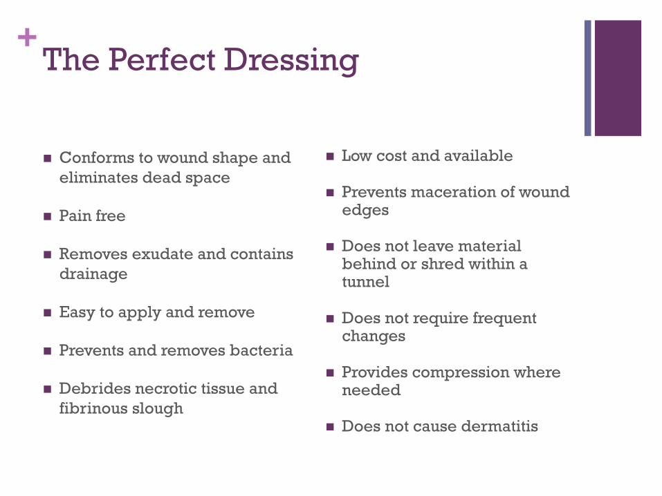
+The Perfect Dressing
Conforms to wound shape and
eliminates dead space
Pain free
Removes exudate and contains
drainage
Easy to apply and remove
Prevents and removes bacteria
Debrides necrotic tissue and
fibrinous slough
Low cost and available
Prevents maceration of wound edges
Does not leave material behind or shred within a tunnel
Does not require frequent changes
Provides compression where needed
Does not cause dermatitis
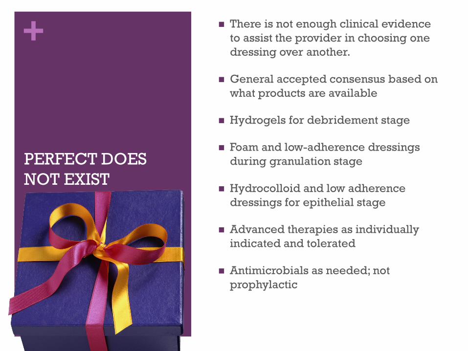
+
PERFECT DOES
NOT EXIST
There is not enough clinical evidence
to assist the provider in choosing one
dressing over another.
General accepted consensus based on
what products are available
Hydrogels for debridement stage
Foam and low-adherence dressings
during granulation stage
Hydrocolloid and low adherence
dressings for epithelial stage
Advanced therapies as individually
indicated and tolerated
Antimicrobials as needed; not
prophylactic
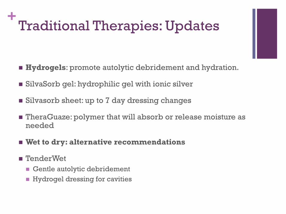
+Traditional Therapies: Updates
Hydrogels: promote autolytic debridement and hydration.
SilvaSorb gel: hydrophilic gel with ionic silver
Silvasorb sheet: up to 7 day dressing changes
TheraGuaze: polymer that will absorb or release moisture as needed
Wet to dry: alternative recommendations
TenderWet
Gentle autolytic debridement
Hydrogel dressing for cavities
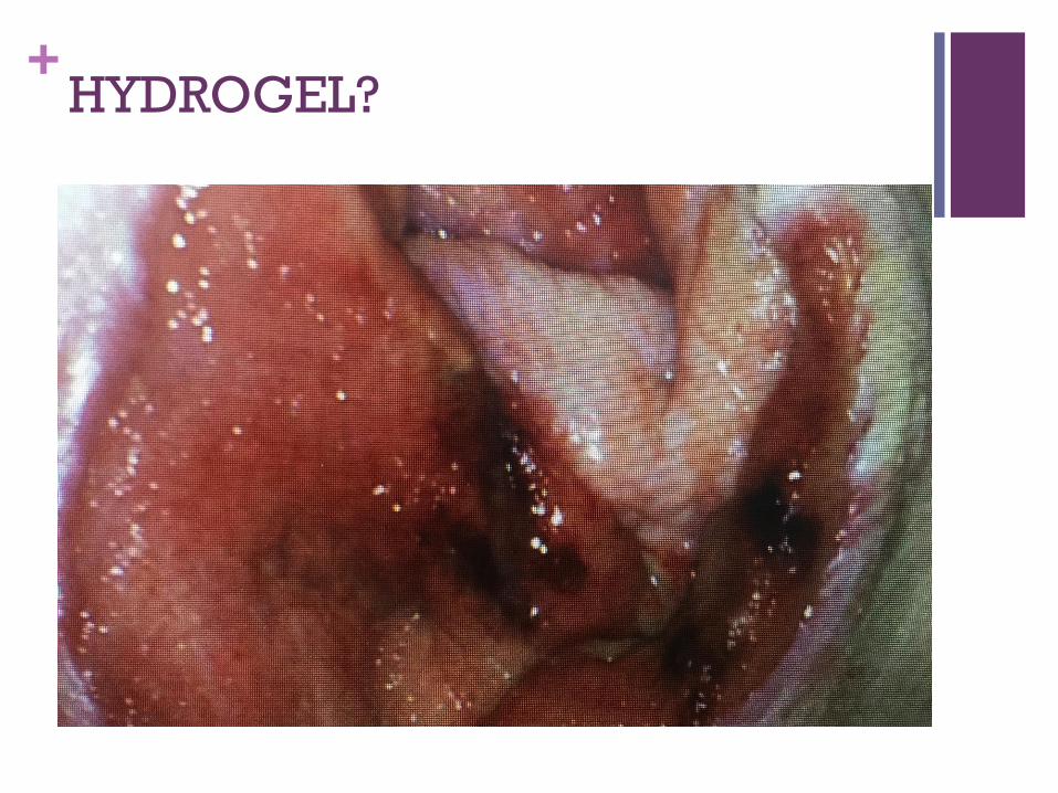
+HYDROGEL?
+Traditional Therapies: Updates
Collagenase: dressing, ointment or powder
Santyl: 250 units/gram collagenase in petrolatum, enzymatic debridement
Promogran Matrix has 55% collagen, 45% ORC and can be used in exudating wounds
Promogran Prisma: 1% ORC added content for antimicrobial action.
Absorbs exudate
Binds to and inactivates MMPs which prevent wound healing ( biofilm)
Fibracol Plus: 90% collagenase and 10% alginate
Option for draining wound
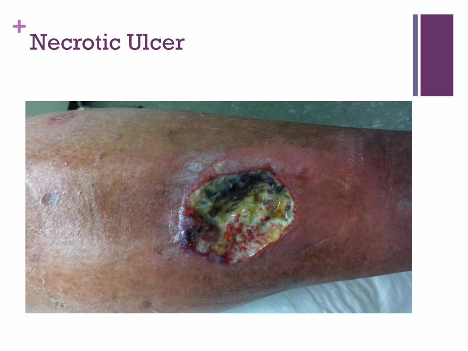
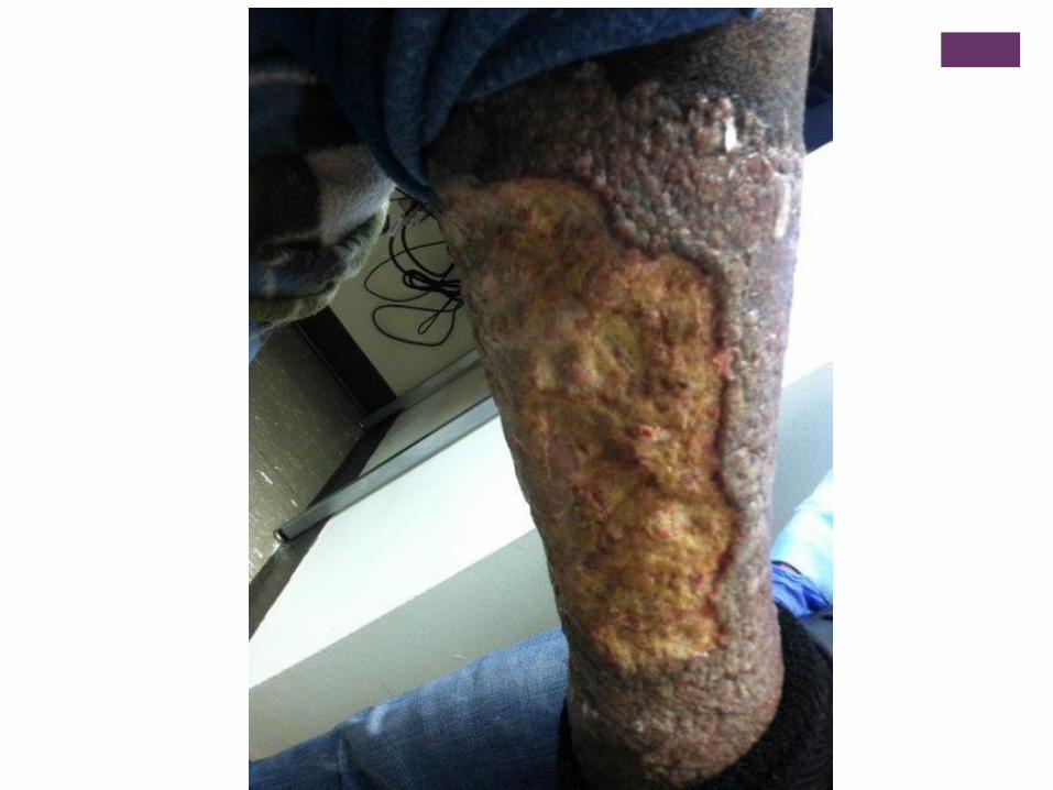
+Necrotic Ulcer
+Collagenase Continued
Triple Helix by MPM Medical: 100% bovine collagen
Biodegradable in wound
Comes in powder form or sheets
Endoform: 90% collagen and 10% intact ECM components
that provide a broad spectrum reduction in MMPs in chronic
wounds.
Daily or Weekly application.
Not a prior authorization item, considered a dressing
+Autolytic Debridement
Resurgence of honey for autolytic debridement, re-hydration
and antimicrobial actions
High concentrations of sugar and high osmolarity result in
broad spectrum antimicrobial activity www.uptodate.com 2016
Careful for sensitivity reactions
Gel, strips, pads
Insufficient data to make scientific recommendations
+Autolytic Debridement Products
PMHB : polyhexamethylene biguande
Tielle foam
Hydrofera blue: ready or classic
PuraPly
Cadexomer idodine
Iodosorb
Iodoflex
+Traditional Therapies: Updates
Alginates: Thicker with better absorption
Reinforced, available with silver, rope for tunnels, not water
soluble
Foam dressings with silicone adhesive; more site conforming
options
Aquacel surgical hydrofiber dressing with flexible hydrocolloid
adhesive dressing; polyurethane film is waterproof with a
viral/bacterial barrier
Silvercel
Maxsorb
Restore
PolyMem
+Foam Dressings
Multiple brands and shapes
With and without silver
Two layer construction with an absorbing hydrophilic layer
and hydrophobic layer to prevent leakage and bacterial
entering the wound.
Silicone adhesive is skin friendly
Multiple day wear
Some pressure relief
+HydroActive Dressing
Newest synthetic dressings
Combines properties of a gel and a foam dressing
Selectively absorbs excess water/drainage and leaves
behind the needed growth factors and proteins to heal a
wound.
Can combine with enzymatic debriding agents
HydroTac
PermaFoam
Sorbact
Duoderm hydroactive dressing
+HydroConductive Dressing
Drawtex: 2011 on the market.
Non-adherent LevaFiber technology
Combines 2 types of absorbent, cross-action structures that facilitate the movement of large volumes of drainage and wound debris through the dressing. Couch KS. Discovering hydroconductive Dressings. Ostomy wound management, 2012; 58(4): 2-3.
Results: decrease bacterial level and nutrients for biofilm production, decrease MMPs and cytokines, facilitate wound bed preparation, minimize exudate
+Hydroconductive Dressing
Can be used in place of NPWT with skin grafts
Pilonidal sinus tracts
Burns
May double the layers for heavy drainage
Consider non-adherent dressing underneath superficial, but
draining wounds
+Diabetic Foot Ulcers:
Considerations
Neuropathy
Charcot foot or other deformity
Vascular status
Smoking
Edema
Offloading; contact casting
osteomyelitis
+Wagner Classification for DFU
Grade 0: no ulcer, but high risk
Grade 1: ulcer is full thickness of skin
Grade 2: Ulcer extends to ligaments, tendon
Grade 3: Cellulitis and/or abscess, osteomyelitis
Grade 4: gangrene locally
Grade 5: extensive gangrene of foot
+DFU Principles
Pressure relief
Control drainage, alginate
Control edema
Debridement, serial if needed
Re-vascularization
Improvements should be noted consistently or else surgical
intervention is warranted, don’t wait
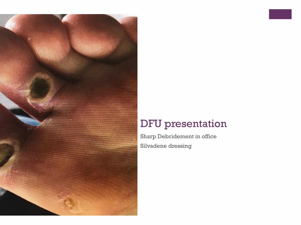
+ DFU presentationSharp Debridement in office
Silvadene dressing
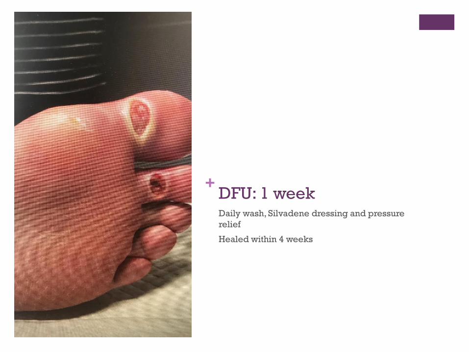
+ DFU: 1 weekDaily wash, Silvadene dressing and pressure
relief
Healed within 4 weeks
+Advanced Wound Therapies
+Extracellular Wound Matrix
Human extracellular matrix (ECM) is the structural complex
that surrounds cells and binds to tissue. In chronic wounds, the
body’s naturally occurring ECM is failing. In healthy skin, ECM
makes up the key components of the basement membrane that
anchors and replenishes epidermal cells, guides, stimulates
cell proliferation and migration to assist in modulation of
cellular response. Macneil S. What Role Does the ECM Service In Skin Grafting and
Wound healing? Burns. 1994; 20(supplement): S67-70.
+ECM and Chronic Wounds
In chronic wounds, fibroblasts are unresponsive to growth
factors and other signals. These wounds lack the integrin
receptor for fibronectin binding and keratinocyte migration. Davin-Haraway,G. WWW.hpmcommunications.com
ECM triggers neovascularization and recruits cells that
differentiate into site specific tissues. When ECM allograft is
absorbed, it leaves functional tissue which becomes scar
tissue.
Le Chemiant, J and Fiel, C. Porcine Urinary Bladder Matrix: A Retrospective Study and
Establishment of Protocol. Journal of Wound Care, volume 21, no. 10, October 2012.
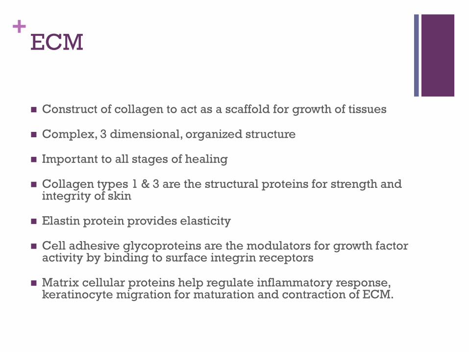
+ECM
Construct of collagen to act as a scaffold for growth of tissues
Complex, 3 dimensional, organized structure
Important to all stages of healing
Collagen types 1 & 3 are the structural proteins for strength and integrity of skin
Elastin protein provides elasticity
Cell adhesive glycoproteins are the modulators for growth factor activity by binding to surface integrin receptors
Matrix cellular proteins help regulate inflammatory response, keratinocyte migration for maturation and contraction of ECM.
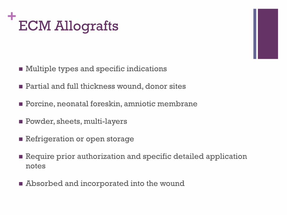
+ECM Allografts
Multiple types and specific indications
Partial and full thickness wound, donor sites
Porcine, neonatal foreskin, amniotic membrane
Powder, sheets, multi-layers
Refrigeration or open storage
Require prior authorization and specific detailed application
notes
Absorbed and incorporated into the wound
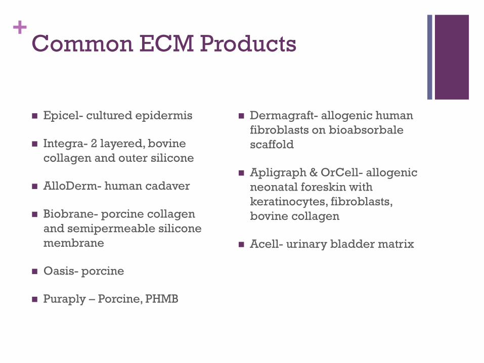
+Common ECM Products
Epicel- cultured epidermis
Integra- 2 layered, bovine
collagen and outer silicone
AlloDerm- human cadaver
Biobrane- porcine collagen
and semipermeable silicone
membrane
Oasis- porcine
Puraply – Porcine, PHMB
Dermagraft- allogenic human
fibroblasts on bioabsorbale
scaffold
Apligraph & OrCell- allogenic
neonatal foreskin with
keratinocytes, fibroblasts,
bovine collagen
Acell- urinary bladder matrix
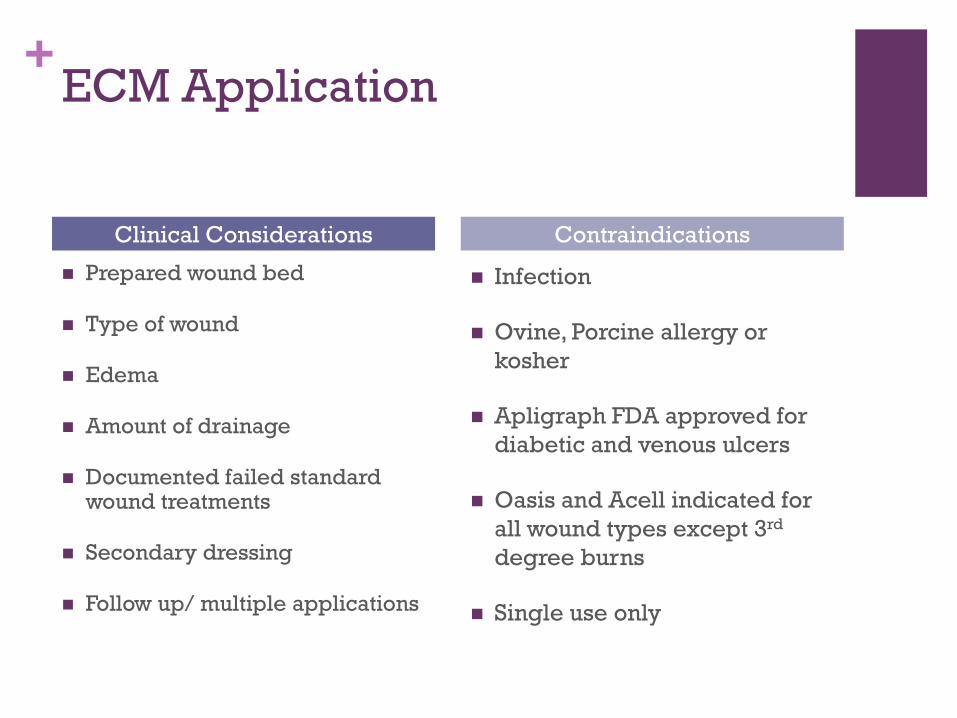
+ECM Application
Prepared wound bed
Type of wound
Edema
Amount of drainage
Documented failed standard wound treatments
Secondary dressing
Follow up/ multiple applications
Infection
Ovine, Porcine allergy or
kosher
Apligraph FDA approved for
diabetic and venous ulcers
Oasis and Acell indicated for
all wound types except 3rd
degree burns
Single use only
Clinical Considerations Contraindications
+ECM: Considerations for Treatment
Multiple, weekly applications
Control edema and drainage
Use of NPWT if needed
Control bio-burden and infection
Pressure relief and ambulation
Odor
Skilled dressing changes
Endpoint: wound closure, lack of progress
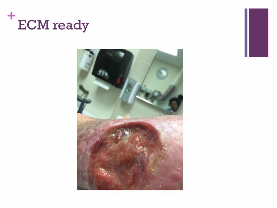
+ECM ready
+Negative pressure Therapy
Vacuum assisted wound closure
Continuous or intermittent -50 to-175mmhg sub-atmospheric pressure applied evenly to the wound surface
Used in tunnels, skin grafts post-op
Disposable, smaller models: PICO, SNAP
KCI: VERIFLOW, ACTIVAC AND VAC ULTA 4: TeleHealth
Fluid installation adjunct treatment
Fluid removal and negative pressure result in a reduction in healing in time with space compression and drawing edges together
Incisional NPWT…sutureless closure on the horizon
+Negative Pressure Therapy
Indications:
Slow, stagnant wounds that fail
conservative treatments
Heavy exudate
Tunneling
Require size reduction for
surgical closure
Skin grafts, 1 or 2 stage
Contraindications:
Malignancy
Untreated osteomyelitis
Necrotic tissue or eschar
Exposed vasculature, nerves,
organs, anastomotic sites,
unexplored fistulae
+NPWT Precautions
Friable, bleeding tissue
Exposed tendon, delicate fascia or ligaments
Enteric fistulae require special precautions
Bony fragments, infection, vascular anastomoses, spinal cord
injury
Henderson, V, etal. NPWT Made Easy in Everyday Practice.
Wounds International 2010; 1(15);
http://www.woundsinternational.com
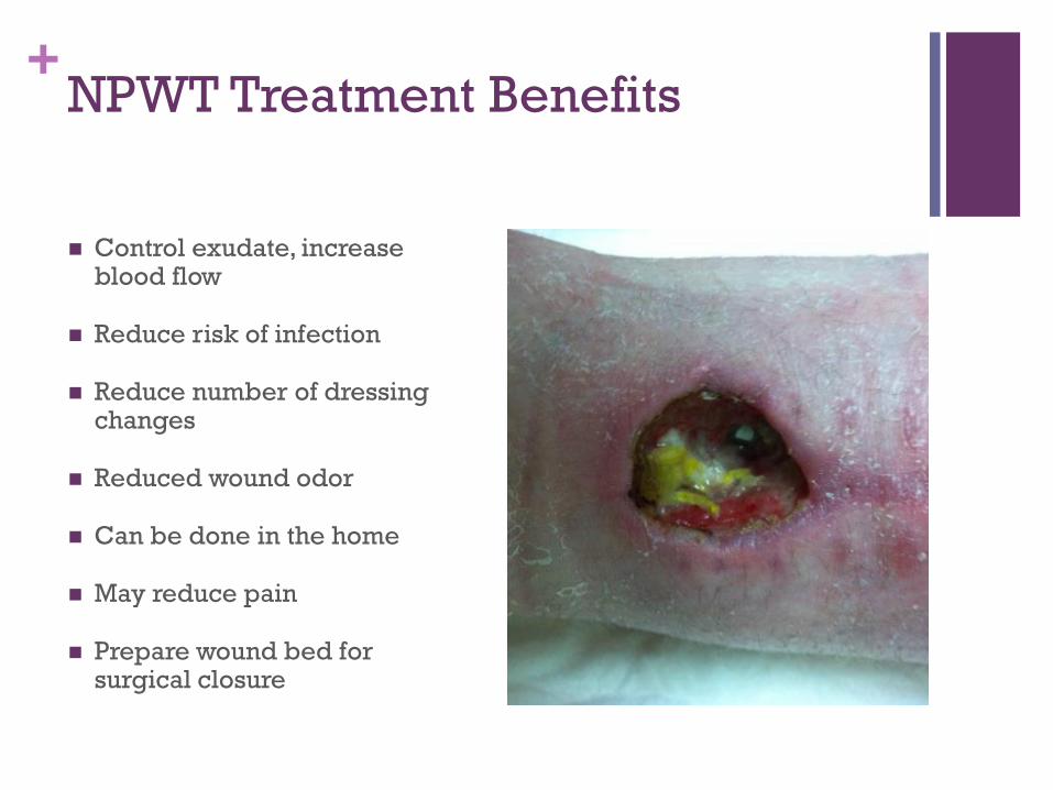
+NPWT Treatment Benefits
Control exudate, increase blood flow
Reduce risk of infection
Reduce number of dressing changes
Reduced wound odor
Can be done in the home
May reduce pain
Prepare wound bed for surgical closure
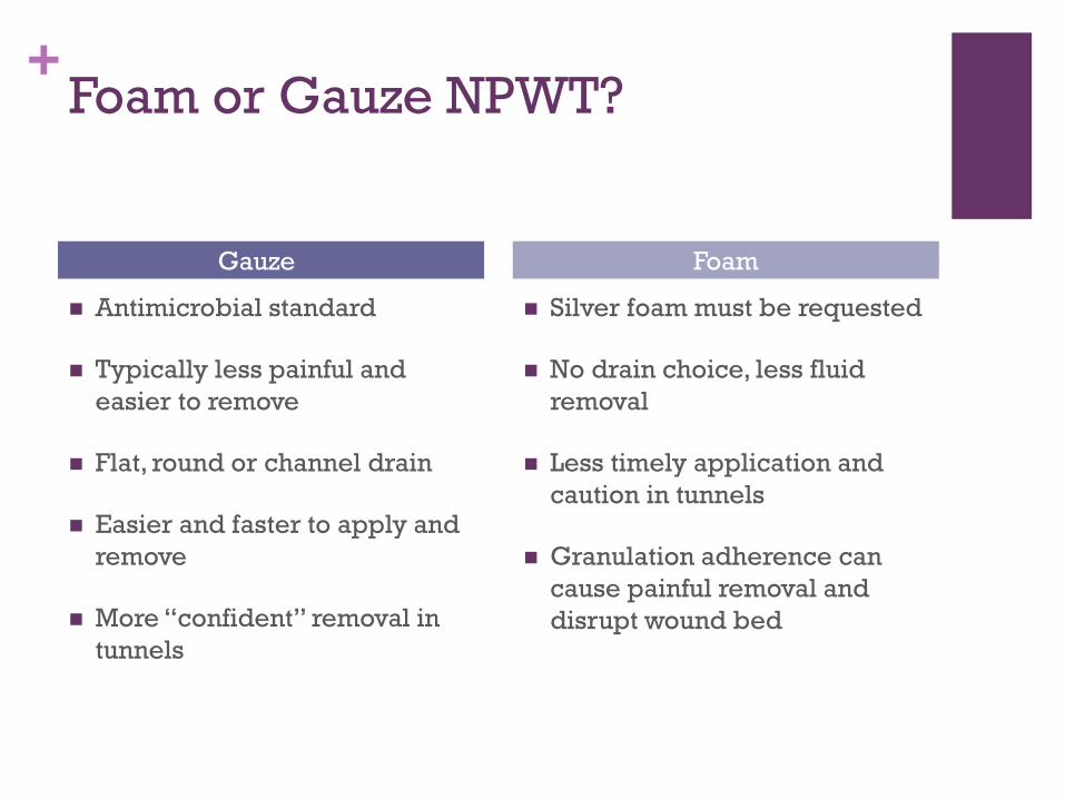
+Foam or Gauze NPWT?
Antimicrobial standard
Typically less painful and
easier to remove
Flat, round or channel drain
Easier and faster to apply and
remove
More “confident” removal in
tunnels
Silver foam must be requested
No drain choice, less fluid
removal
Less timely application and
caution in tunnels
Granulation adherence can
cause painful removal and
disrupt wound bed
Gauze Foam
+NPWT Treatment Considerations
Wound location; bridge
Tubing placement and pressure settings
Wound bed preparation
Size and number of wounds, frequency of changes
Patient expectations, mobility, cognitive and sensory
function, social environment and lifestyle
Installation: NS, 10-20 minutes then 2-6 hours NPWT
@125mmhg. Kim, et al. Negative Pressure Wound Therapy with Installation: Review of Evidence and
Recommendations, Suppl. Ostomy Wound Management, 2015.
+
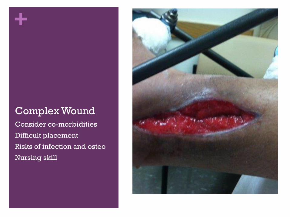
Complex Wound
Consider co-morbidities
Difficult placement
Risks of infection and osteo
Nursing skill
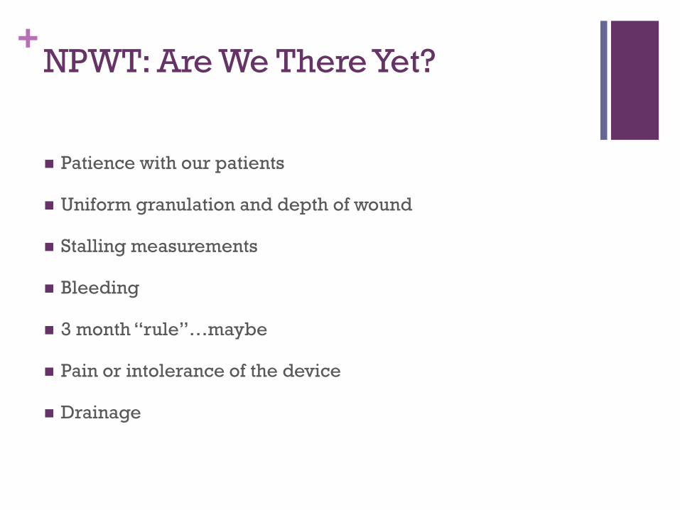
+NPWT: Are We There Yet?
Patience with our patients
Uniform granulation and depth of wound
Stalling measurements
Bleeding
3 month “rule”…maybe
Pain or intolerance of the device
Drainage
+
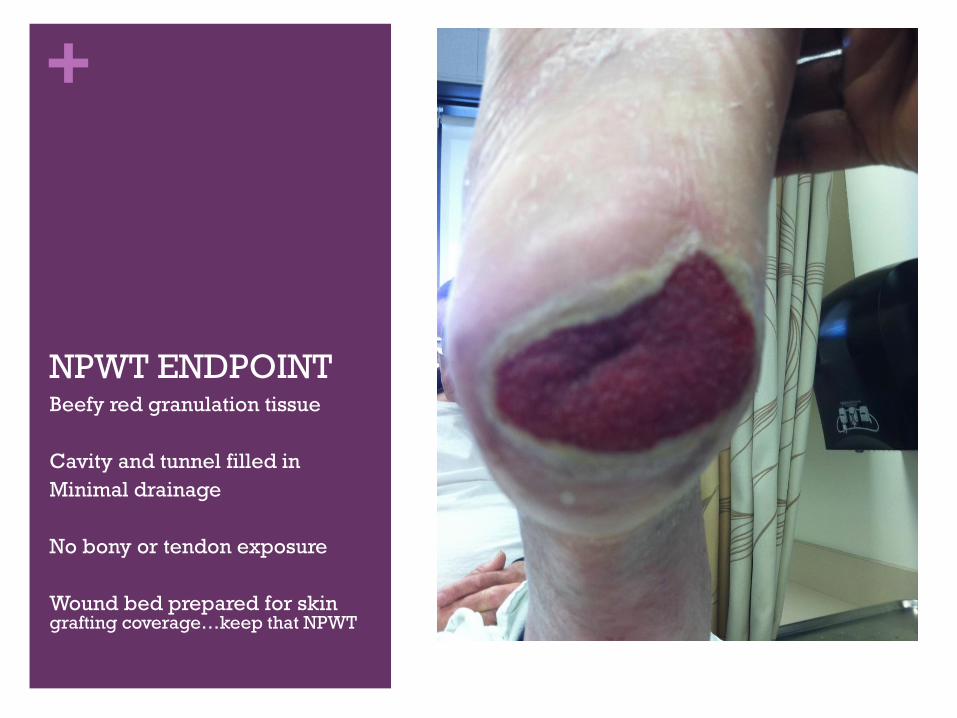
NPWT ENDPOINTBeefy red granulation tissue
Cavity and tunnel filled in
Minimal drainage
No bony or tendon exposure
Wound bed prepared for skin grafting coverage…keep that NPWT
+
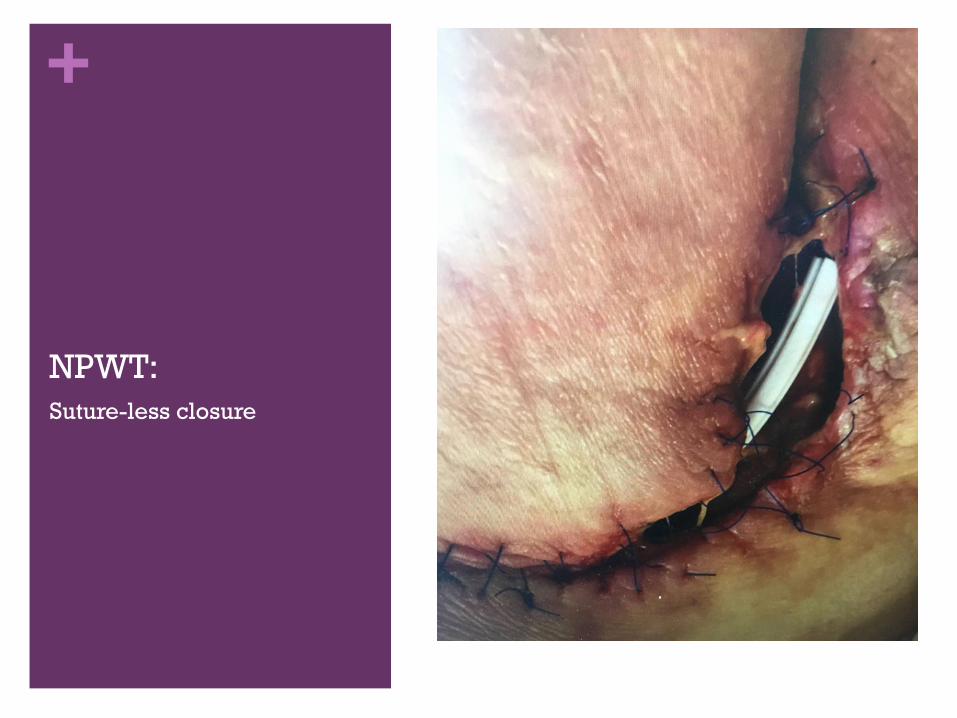
NPWT:
Suture-less closure
+
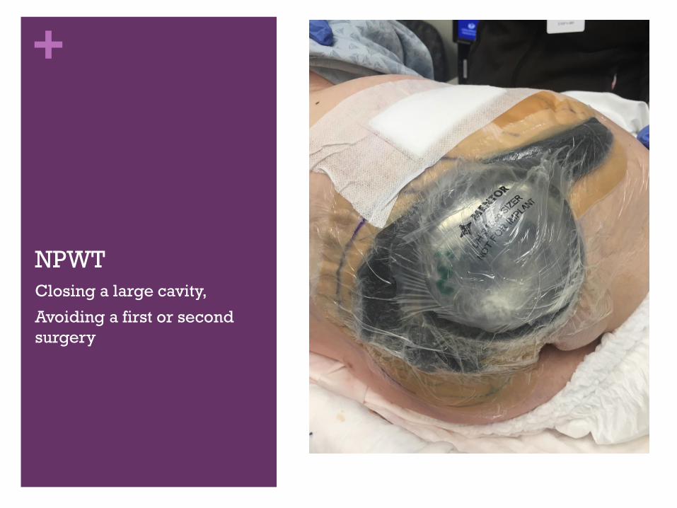
NPWT
Closing a large cavity,
Avoiding a first or second
surgery
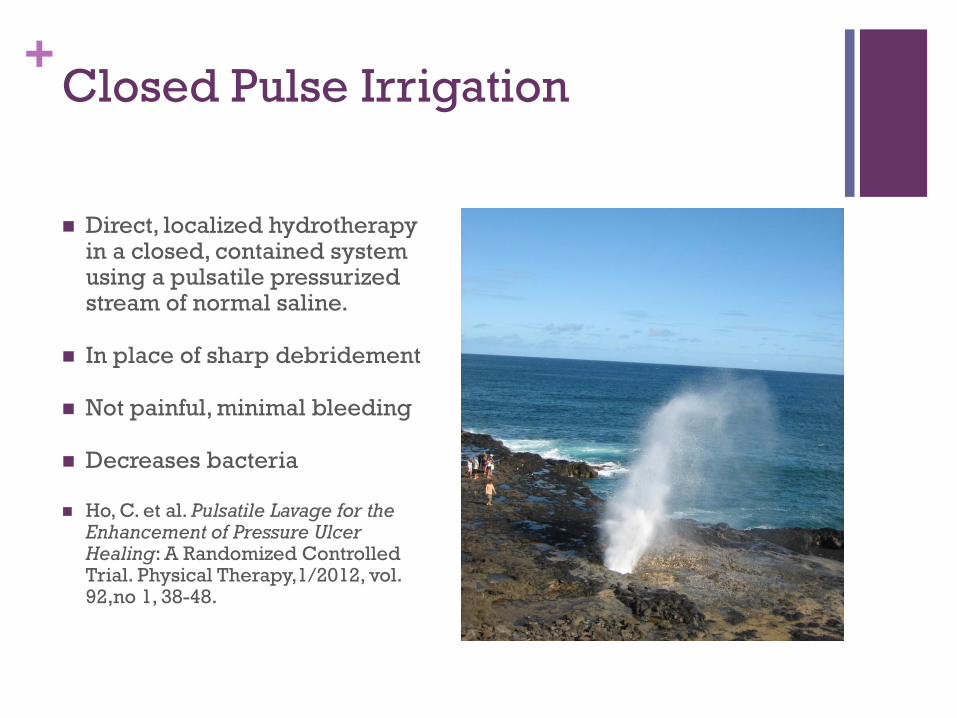
+Closed Pulse Irrigation
Direct, localized hydrotherapy in a closed, contained system using a pulsatile pressurized stream of normal saline.
In place of sharp debridement
Not painful, minimal bleeding
Decreases bacteria
Ho, C. et al. Pulsatile Lavage for the Enhancement of Pressure Ulcer Healing: A Randomized Controlled Trial. Physical Therapy,1/2012, vol. 92,no 1, 38-48.
+Benefits of Pulsed Irrigation
Can be done daily, decreases pain, accelerates healing
Performed by nurses in the clinic, home or rehab setting
Use in conjunction with NPWT
Use in wound tunnels with special tips, 8-15 PSI
CPT codes 97597 ( up to 20 sq. cm) and 97598 used for
reimbursement by providers
Must use closed system to contain aerosols and infectious
disease risks: MDRO outbreak @ John’s Hopkins
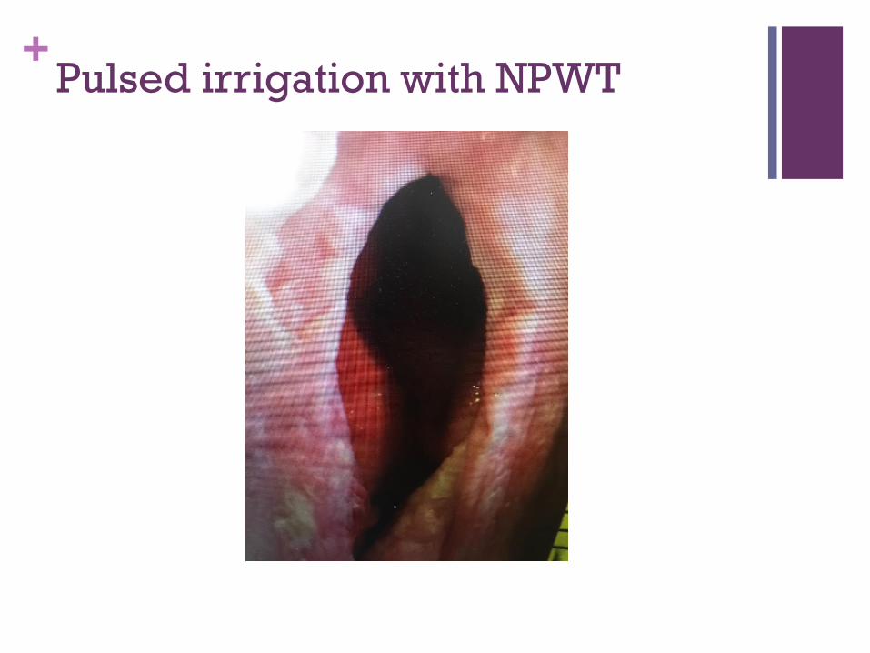
+Pulsed irrigation with NPWT
+Biologically Active Wound Stimulation
Therapies
Electrical Stimulation
Bio-electric Dressing
Ultrasound Assisted Wound Therapy
Mist,
Electromagnetic Therapy, Diathermy
Modalities to reverse the current of injury; loss of the 40-80mV negative charge of the epidermis relative to the deeper tissues that carry a positive charge, when a full thickness injury occurs.
Moore, K. Electrical Stimulation of Chronic Wounds. Journal of Community Nursing, January 2007, volume 21, issue 1: 18-22.
+Basic Concepts of E-Stimulation
Loss of intact skin=change in charge gradient between the
skin and the deeper tissues
A micro-current will flow from the area of the intact skin into
the wound; voltage peaks and decreases as the wound heals.
In chronic wounds, the current flow is defective and healing
stalls.
E-Stim re-applies this current to stimulate healing via direct,
alternating or pulsed currents
+Bio-Electric Dressing
Woven polyester fabric surface with a matrix of bio-
compatible silver and zinc dots, 1 or 2mm
A secondary moist dressing is placed to maintain moisture
level and promote a micro-current of 0.6 to 0.7 volts
Human epithelial cells have an increased level of FGF-2, a
mediator that increases fibroblast proliferation needed for
wound healing through cellular membrane depolarization
that activates voltage dependent calcium channels.
Harding, A. etal. Efficacy of Bio-Electric Dressing in Healing Deep,
Partial Thickness Wounds Using a Porcine Model. Ostomy Wound
Management. 9/2012; 58(9):50-55.
+Electrical Stimulation
Treatment
Pressure ulcers, leg ulcers
High or low voltage
45 minutes 3-5 days per week
Angiogenesis is enhanced by
improving capillary dermal
formation
Effect on Healing
Inhibit bacterial growth and
disrupts biofilm
Increases keratinocyte
migration
Increases fibroblast protein
synthesis, collagen production
and organization of collagen
fibers
+Biologically Stimulating Irrigation
Techniques
Combine pressurized water irrigation with ultrasound
Portable for clinic use
Multiple tips for varying pressure and area treated
Minimal collateral tissue damage
No nerve damage if done properly
Consider storage, sterilizing, reimbursement
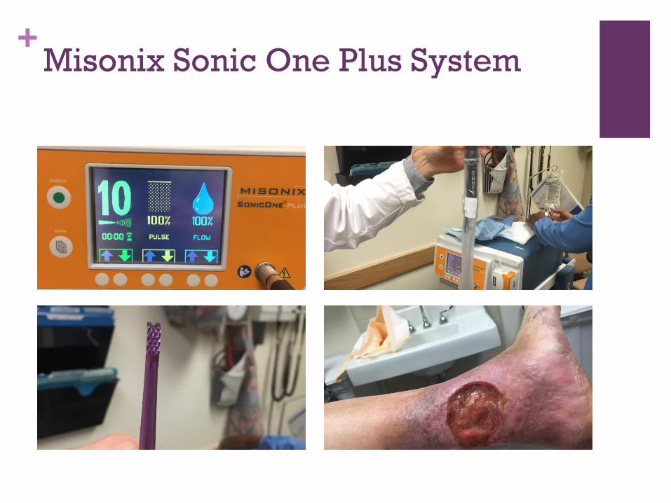
+Misonix Sonic One Plus System
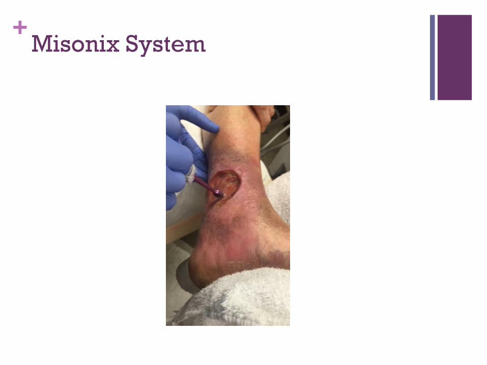
+Misonix System
+
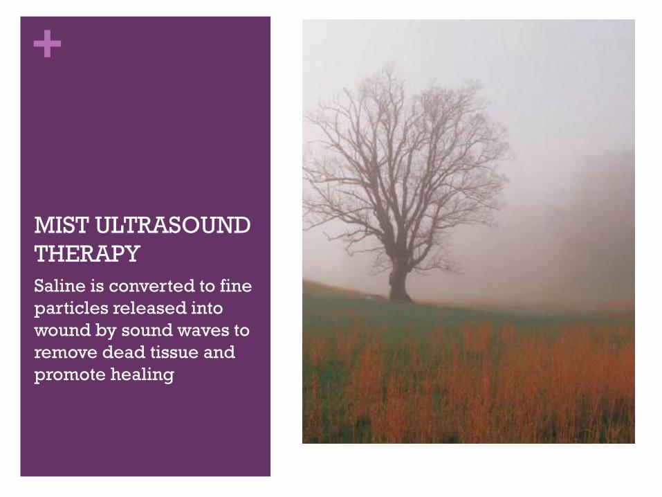
MIST ULTRASOUND
THERAPY
Saline is converted to fine
particles released into
wound by sound waves to
remove dead tissue and
promote healing
+MIST ULTRASOUND
Painless, non-contact, low
frequency ultrasound
Delivered through a saline
mist that acts as a conduit from
the US to the treatment site
Stimulates cells in and below
the wound bed to accelerate
the healing process by
reducing bacteria and biofilm,
decreases MMP-9 and
sustained inflammation
Increases vasodilation and
angiogenesis leading to
release of growth factors and
collagen deposition.
Used in wounds related to
vascular insufficiency, trauma,
ischemia, neuropathy and
chronic wounds
Limited evidence, more
studies needed
Keltie, K. etal, www.alliqua.com,
www.nchi.nlm.nih.gov/pubmed/190
18197
+MIST THERAPY
CONTRAINDICATIONS
Malignancy
Pregnancy
Pacemaker or other electronic implanted devices
Acute gangrene
Acute ischemia
+Ultrasound Assisted Wound Healing
First appeared in the literature in 1949
Therapeutic ultrasound delivers energy in the form of sound waves from mechanical vibrations; similar to diagnostic imaging waves
Primary effects related to inflammatory and repair phases of muscle and soft tissue healing.
Low frequency, provides non-thermal effects of cavitation and acoustic streaming
The shock waves will liquefy necrotic tissue, wound debris and biofilm without injury to healthy tissue with greater tensile strength.
With or without water irrigation
+Ultrasound Assisted Wound Therapy
Wound Indications
Local infection, not systemic
Vascular disease
Pressure ulcers
Diabetic foot and LE ulcers
Wounds needing debridement
Contraindications
Systemic infection
Advancing cellulitis
Joint replacement or local
hardware
Implanted electronic devices
within the treatment field
www.todayswoundclinic.com
+Electromagnetic Therapy: Sub-thermal
PSWT
Pulsed shortwaves produce electromagnetic fields believed to enhance healing; electric or magnetic field
Most research has been done with magnetic field effects
Pulsed wave energy is absorbed in wet, ionic, less dense tissues: muscle and nerves
Diminishes inflammation and increases repair of musculoskeletal and soft tissues through increased membrane transportation that restores ionic balance with energy absorbed
Can be high or radio electromagnetic short wave therapy, but most literature focuses on short wave magnetic
+PSWT
Pulse duration are short resulting in less “on” energy time
than “off” time, resulting in mean power delivery as relatively
low, but up to 150-200 watts of peak power.
Control to vary the mean power delivered and the pulsing
parameters
Most evidence indicates that the combination of pulse
repetition rate and pulse duration is the critical parameter
for energy delivered for tissue heating
Cells in the inflammatory phase have reduced cell
membrane potential, ionic imbalance, altered osmotic action
http://www.linkedin.com/pub/tim-watson/34/351/666
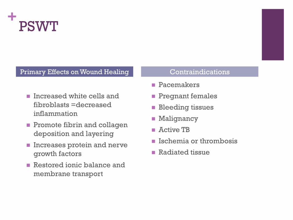
+PSWT
Increased white cells and
fibroblasts =decreased
inflammation
Promote fibrin and collagen
deposition and layering
Increases protein and nerve
growth factors
Restored ionic balance and
membrane transport
Pacemakers
Pregnant females
Bleeding tissues
Malignancy
Active TB
Ischemia or thrombosis
Radiated tissue
Primary Effects on Wound Healing Contraindications
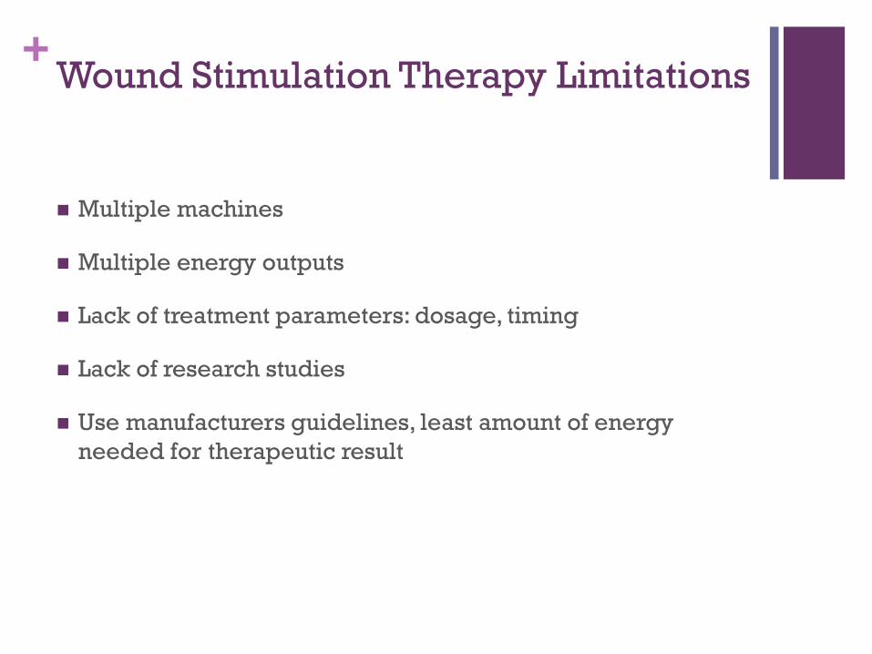
+Wound Stimulation Therapy Limitations
Multiple machines
Multiple energy outputs
Lack of treatment parameters: dosage, timing
Lack of research studies
Use manufacturers guidelines, least amount of energy
needed for therapeutic result
+Oxygen Therapies
Transdermal/continuous diffusion oxygen (CDO), topical
hyperbaric (THO) , hyperbaric oxygen (HBOT)
Therapeutic and technologically differences
HBOT oldest and most accepted form
CDO newest approach with growing evidence, less reported
risks and side effects, due to lower flow rates and lack of
pressure.
www.uptodate.com2016, principles of wound management.
+Hyperbaric Oxygen
Since 1600’s, systemic application 5-7 days per week for 90
minutes in a chamber, intermittent
Adjunct to wound healing therapies
100% O2 delivered under increased atmospheric pressure
greater than 1 atmosphere, up to 3
Goal: raise the O2 levels within the wound bed to correct
hypoxia in chronic wounds
New standards developing for acute burns, surgical flaps and
grafts
+Hyperbaric Oxygen: Therapeutic
Effects
Reverse local tissue hypoxia
Increase stimulation of collagen synthesis
Improved rate of bacterial killing
Diminish inflammatory signals
Issue: Few controlled trials, different wound types and
outcome parameters, but overall, demonstrated improvement
in healing
Eskes, A. et al. Hyperbaric Oxygen Therapy: Solution for Difficult to
Heal Acute Wounds? Systematic Review. World Journal of
Surgery(2011) 35:535-542
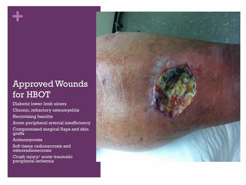
+
Approved Wounds
for HBOTDiabetic lower limb ulcers
Chronic, refractory osteomyelitis
Necrotizing fasciitis
Acute peripheral arterial insufficiency
Compromised surgical flaps and skin grafts
Actinomycosis
Soft tissue radionecrosis and osteoradionecrosis
Crush injury/ acute traumatic peripheral ischemia
+Complications of Hyperbaric
Therapy
Middle ear trauma and effusions
Sinus barotrauma (sneeze)
Reversible myopia
Pulmonary toxicity
seizures
+Topical Hyperbaric Oxygen
Since 1960’s
Affected area in a boot, bag or extremity chamber which is
sealed and filled with O2 at high flow rate for O2 rich
environment
5-7 days per week for 90 minutes to 4 hours, intermittent
Low pressures and therefore lower risk of side effects
Requires an open wound surface, not used for necrotic or
sinus tracts
Noted to increase angiogenesis and wound closure rates
+Continuous Diffusion of Oxygen
Therapy
Newest class
Provide continuous O2 delivery at lowest flow rates
Portable, smaller, increase access, lower cost
Also called low-flow or transcutaneous O2, , continuous
7 days per week, 24 hours, 3-12 ml/hr O2 delivery
Not applicable in wounds with eschar or sinus tracts
Improves granulation tissue, increased collagen and
epitheliazation
+Topical Growth Factors
Becaplermin ( Regranex): platelet derived growth factor gel that promotes cellular angiogenesis to improve wound healing.
Approved for diabetic foot ulcers and chronic wounds
Black box warning for malignancy, noted to be with increased use
GM-CSF: granulocyte-macrophage colony stimulating factor is and intradermal injection to promote healing in chronic leg ulcers. Being studied in chronic wounds.
Epidermal Growth Factor: studies on treating chronic venous ulcers
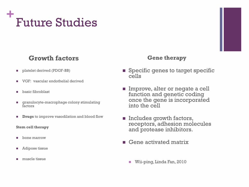
+Future Studies
Growth factors
platelet derived (PDGF-BB)
VGF: vascular endothelial derived
basic fibroblast
granulocyte-macrophage colony stimulating factors
Drugs to improve vasodilation and blood flow
Stem cell therapy
bone marrow
Adipose tissue
muscle tissue
Gene therapy
Specific genes to target specific cells
Improve, alter or negate a cell function and genetic coding once the gene is incorporated into the cell
Includes growth factors, receptors, adhesion molecules and protease inhibitors.
Gene activated matrix
Wii-ping, Linda Fan, 2010
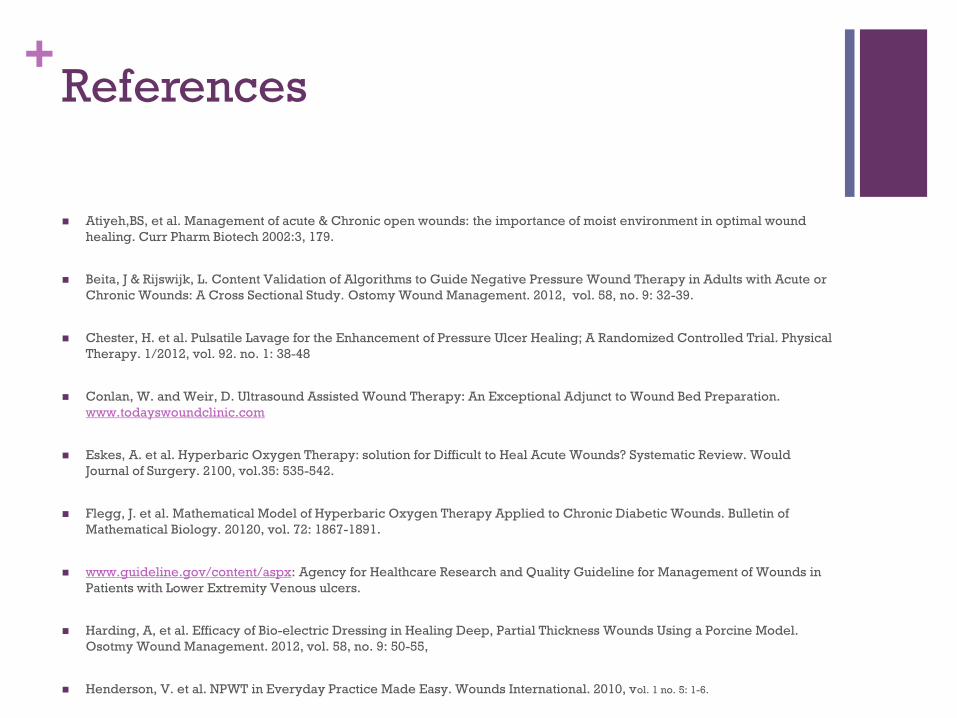
+References
Atiyeh,BS, et al. Management of acute & Chronic open wounds: the importance of moist environment in optimal wound
healing. Curr Pharm Biotech 2002:3, 179.
Beita, J & Rijswijk, L. Content Validation of Algorithms to Guide Negative Pressure Wound Therapy in Adults with Acute or
Chronic Wounds: A Cross Sectional Study. Ostomy Wound Management. 2012, vol. 58, no. 9: 32-39.
Chester, H. et al. Pulsatile Lavage for the Enhancement of Pressure Ulcer Healing; A Randomized Controlled Trial. Physical
Therapy. 1/2012, vol. 92. no. 1: 38-48
Conlan, W. and Weir, D. Ultrasound Assisted Wound Therapy: An Exceptional Adjunct to Wound Bed Preparation.
www.todayswoundclinic.com
Eskes, A. et al. Hyperbaric Oxygen Therapy: solution for Difficult to Heal Acute Wounds? Systematic Review. Would
Journal of Surgery. 2100, vol.35: 535-542.
Flegg, J. et al. Mathematical Model of Hyperbaric Oxygen Therapy Applied to Chronic Diabetic Wounds. Bulletin of
Mathematical Biology. 20120, vol. 72: 1867-1891.
www.guideline.gov/content/aspx: Agency for Healthcare Research and Quality Guideline for Management of Wounds in
Patients with Lower Extremity Venous ulcers.
Harding, A, et al. Efficacy of Bio-electric Dressing in Healing Deep, Partial Thickness Wounds Using a Porcine Model.
Osotmy Wound Management. 2012, vol. 58, no. 9: 50-55,
Henderson, V. et al. NPWT in Everyday Practice Made Easy. Wounds International. 2010, vol. 1 no. 5: 1-6.
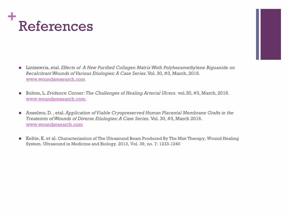
+References
Lintzewris, etal. Effects of A New Purified Collagen Matrix Woth Polyhexamethylene Biguanide on
Recalcitrant Wounds of Various Etiologies: A Case Series. Vol. 30, #3, March, 2016.
www.woundsresearch.com
Bolton, L. Evidence Corner: The Challenges of Healing Arterial Ulcers. vol.30, #3, March, 2016.
www.woundsresearch.com.
Anselmo, D. , etal. Application of Viable Cryopreserved Human Placental Membrane Grafts in the
Treatemtn of Wounds of Diverse Etiologies: A Case Series. Vol. 30, #3, March 2016.
www.woundsrsearch.com
Keltie, K. et al. Characterization of The Ultrasound Beam Produced By The Mist Therapy, Wound Healing
System. Ultrasound in Medicine and Biology. 2013, Vol. 39, no. 7: 1233-1240
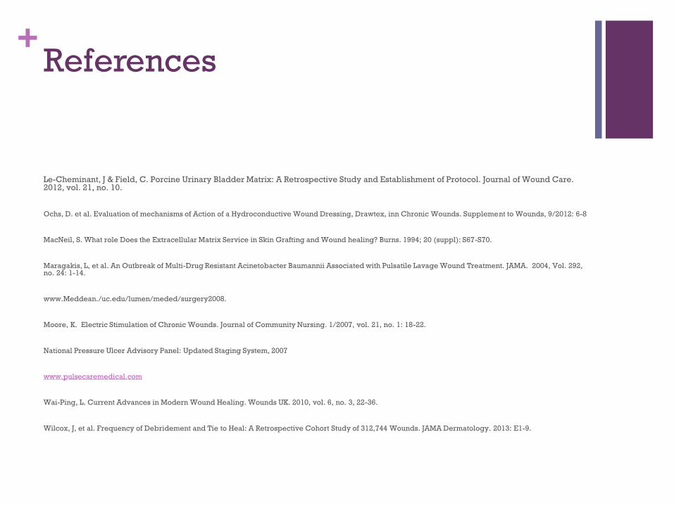
+References
Le-Cheminant, J & Field, C. Porcine Urinary Bladder Matrix: A Retrospective Study and Establishment of Protocol. Journal of Wound Care. 2012, vol. 21, no. 10.
Ochs, D. et al. Evaluation of mechanisms of Action of a Hydroconductive Wound Dressing, Drawtex, inn Chronic Wounds. Supplement to Wounds, 9/2012: 6-8
MacNeil, S. What role Does the Extracellular Matrix Service in Skin Grafting and Wound healing? Burns. 1994; 20 (suppl): S67-S70.
Maragakis, L, et al. An Outbreak of Multi-Drug Resistant Acinetobacter Baumannii Associated with Pulsatile Lavage Wound Treatment. JAMA. 2004, Vol. 292, no. 24: 1-14.
www.Meddean./uc.edu/lumen/meded/surgery2008.
Moore, K. Electric Stimulation of Chronic Wounds. Journal of Community Nursing. 1/2007, vol. 21, no. 1: 18-22.
National Pressure Ulcer Advisory Panel: Updated Staging System, 2007
www.pulsecaremedical.com
Wai-Ping, L. Current Advances in Modern Wound Healing. Wounds UK. 2010, vol. 6, no. 3, 22-36.
Wilcox, J, et al. Frequency of Debridement and Tie to Heal: A Retrospective Cohort Study of 312,744 Wounds. JAMA Dermatology. 2013: E1-9.
+References
Howard, M. Et al. Oxygen and Wound Care: A Review of Current Treatment Modalities and Future
Direction. Wound Repair and Regeneration, vol 21, issue 4, July-August 2013, 503-511.
International Best Practice Guidelines. Wound Management in Diabetic Foot Ulcers. Wounds
International, 013.
Kim P. et al. Negative Pressure Wound Therapy with Instillation: Review of Evidence and
Recommendations. Suppl. Ostomy Wound Management,2016.
Marasco, P. Closed Pulse Irrigation: a Better Pulse Lavage Delivery System for Wound Debridement
and Biofilm Management. Today’s Wound Clinic. November-December 2015.
Armstrong, D. et al. Wound Healing and Risk Factors for Non-healing. www.uptodate.com
Gestring, M. Negative Pressure Wound Therapy. www.uptodate.com.
Mechem, c. and Manaker, s. Hyperbaric Oxygen Therapy. www.uptodate.com
Watson, Timothy. Pulsed Shorthwave Therapy in www.linedin.com/pub/tim-watson/34/351/666.