Embed Size (px)
Citation preview

Updated Classification of Renal cell carcinoma
Suchin Worawichawong, M.D., FRCPath (Thailand) Department of Pathology, Ramathibodi Hospital, Mahidol University
Incidence: Global: 338,000 new cases, 130,000 deaths annually (4/100,000) Czech Republic: male 24.1/100,000, female 10.5/100,000 United States: 64,000 new cases and almost 14,000 deaths (15.3/100,000) European Union: 84,000 new cases and 35,000 deaths China: 2.8/100,000
Semin Intervent Radiol. 2014 Mar; 31(1): 3–8. Eur Urol. 2015 Mar;67(3):519-30.
CA Cancer J Clin. 2017 Jan;67(1):7-30.
Eur J Cancer. 2013 Apr;49(6):1374-403.
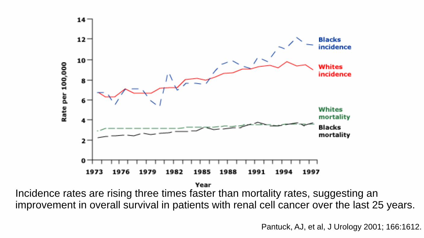
Incidence rates are rising three times faster than mortality rates, suggesting an improvement in overall survival in patients with renal cell cancer over the last 25 years.
Pantuck, AJ, et al, J Urology 2001; 166:1612.
The five-year survival rate of patients with kidney cancer 34 % in 1954 62 % in 1996 73 % in 2005 to 2011
This improved survival and case-fatality rate is mostly due to earlier detection of these tumors at smaller sizes (ie, <4 cm) and curative surgical treatment.
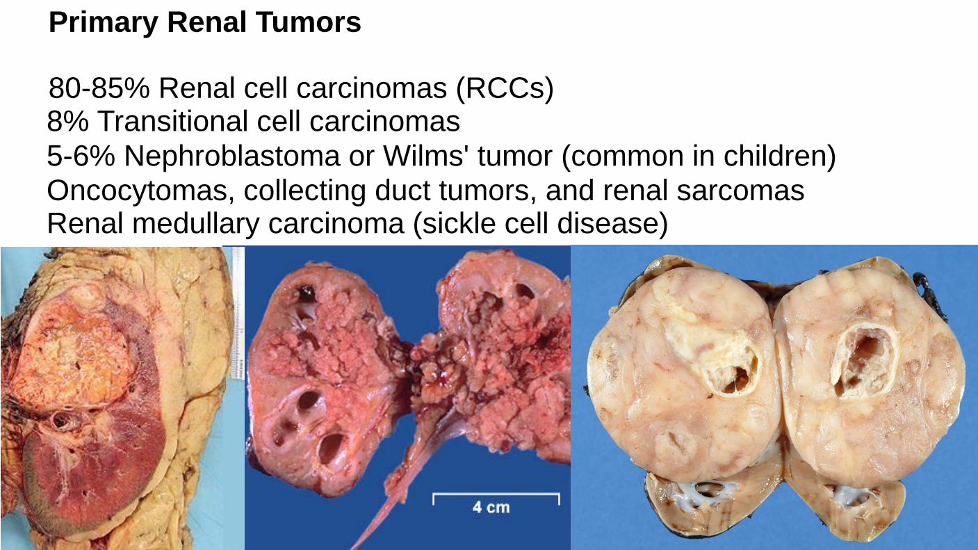
Primary Renal Tumors 80-85% Renal cell carcinomas (RCCs) 8% Transitional cell carcinomas 5-6% Nephroblastoma or Wilms' tumor (common in children) Oncocytomas, collecting duct tumors, and renal sarcomas Renal medullary carcinoma (sickle cell disease)
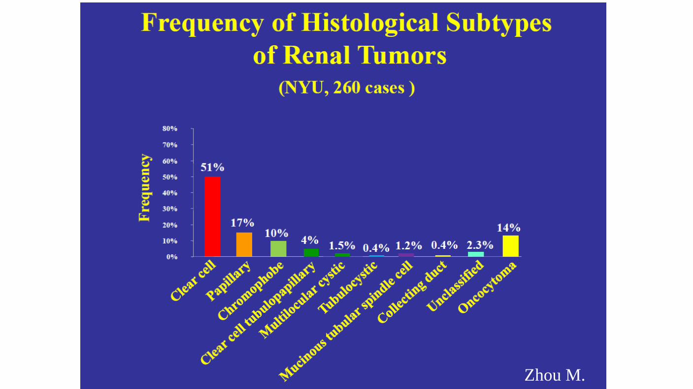
Zhou M.

RISK FACTORS Cigarette smoking: The relative risks for RCC for all smokers, current smokers, and former smokers were 1.31, 1.36, and 1.16, respectively. (24 meta-analysis studies) Hypertension: The underlying biological explanations linking HT to RCC remain unknown. Obesity: A prospective analysis of over 300,000 participants in the National Institutes of Health and American Association for Retired Persons (NIH-AARP) Diet and Health Study. The relative risk (RR) of RCC increased progressively with BMI.
Occupational exposure: Toxic compounds, such as cadmium, asbestos, and petroleum by-products associated with an increased risk of RCC. In one international multicenter study of over 1700 patients with RCCs and 2300 controls, an increased risk of cancer was observed in Asbestos (RR 1.4, 95% CI 1.1-1.8), Cadmium (RR 2.0, 95% CI 1.0-3.9), Gasoline (RR 1.6, 95% CI 1.2-2.0). Increased exposure to such carcinogens may be associated with mutations in genes associated with the pathogenesis of RCC, such as the von Hippel-Lindau (VHL) tumor suppressor gene.
Analgesics: Prolonged ingestion of analgesic combinations, particularly compounds containing phenacetin (acetaminophen) and aspirin, -> CRF -> increased risk for renal pelvic and urothelial tumors. Heavy use of aspirin, NSAIDS, and acetaminophen -> increased risk of RCC.
The largest prospective studies: 77,525 women followed >16 years and 49,043 men followed > 20 years. The regular use of aspirin or acetaminophen was not associated with the development of RCC . The routine use of nonaspirin NSAIDs was associated with a greater risk of RCC (hazard ratio [HR] 1.51, 95% CI 1.12-2.04), which increased with more frequent use and longer period of use. Data from 1217 RCC cases and 1235 controls in the US Kidney Cancer Study, and 98,807 participants in the US Prostate, Lung, Colorectal and Ovarian Cancer Screening Trial (PLCO) found that non-prescription acetaminophen use increased the risk of developing RCC.
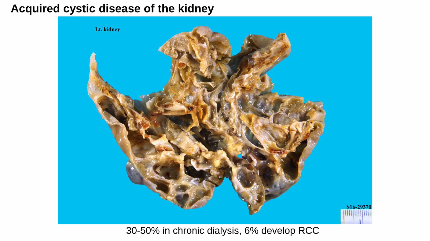
Acquired cystic disease of the kidney
30-50% in chronic dialysis, 6% develop RCC
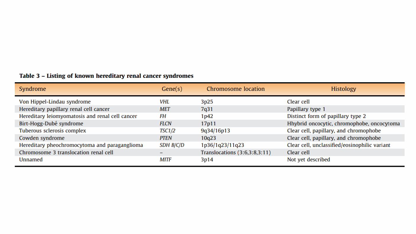
Genetic factors: Most RCCs are sporadic. Several syndromes associated with RCC have been described. Factors favor a hereditary contribution without a clear genetic disease include First degree relatives with a tumor, Onset before the age of 40, Bilateral or multifocal disease. Other individuals with a clear genetic contribution have abnormalities on chromosome 3. Inherited polycystic disease may have an increased risk of RCC. One cohort study in a Chinese population: the risk of RCC was increased in patients with inherited polycystic kidney disease and no history of renal disease compared to a matched control group (adjusted HR 2.5, 95% CI 1.3-4.7)
Cytotoxic chemotherapy: The use of cytotoxic chemotherapy in childhood for malignancies, autoimmune disorders, or bone marrow transplant conditioning has been associated with the subsequent development of translocation RCC. Chronic hepatitis C infection: An epidemiologic study of over 67,000 patients found that chronic infection with hepatitis C virus was associated with a significantly increased risk of RCC after correcting for age, ethnicity, gender, and the presence of chronic kidney disease (HR 1.77, 95% CI 2.05-2.98). Sickle cell disease: Patients with sickle cell trait and sickle cell disease are at risk for renal medullary carcinoma. Kidney stones: A history of kidney stones may be associated with both RCC and transitional cell carcinoma of the upper urinary tract. In a meta-analysis from almost 63,000 patients with kidney stones, the risk ratio of developing RCC was 1.96 (95% CI 1.24-2.49), and the increased risk appeared to be largely limited to men. The risk ratio for transitional cell carcinoma was 2.14 (95% CI 1.35-3.40).
OTHER FACTORS THAT MODIFY RISK Diabetes mellitus: associated with a modest increase in the risk of renal cell carcinoma (RCC) in some studies but not in others. This may be mediated through an increase in the incidence of hypertension. Polycystic kidney disease: It does not increased frequency compared with the general population. The tumors are more often bilateral at presentation (12 versus 1 to 4 percent in sporadic RCC in the general population), multicentric (28 versus 6 percent), and sarcomatoid (33 versus 1 to 5 percent) Alcohol: Associated with a protective effect on the risk of RCC in both men and women. The protective effect of alcohol on the risk of RCC was shown in a 2012 meta-analysis of 20 studies. Other factors: Dietary factors such as the intake of nitrite from processed meat sources, reproductive factors (eg, increasing number of pregnancies), prior radiation therapy (RT). For women, the use of oral contraceptives may reduce risk.
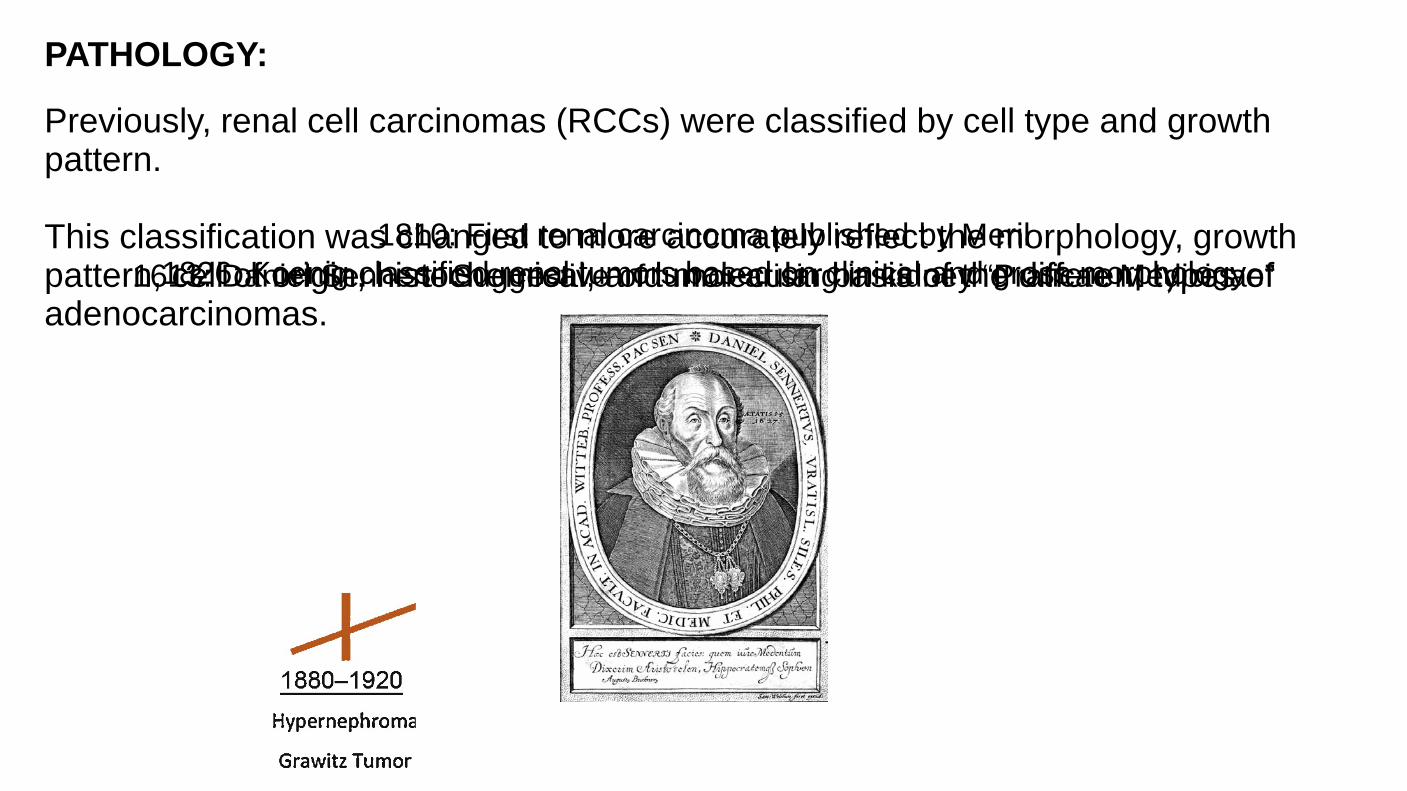
Previously, renal cell carcinomas (RCCs) were classified by cell type and growth pattern. This classification was changed to more accurately reflect the morphology, growth pattern, cell of origin, histochemical, and molecular basis of the different types of adenocarcinomas.
WHO classification 2004
PATHOLOGY:
1613: Daniel Sennert- Suggestive of tumor arising in kidney “Praticae Medicinae” 1810: First renal carcinoma published by Meril
1826: Koenig classified renal tumors based on clinical and gross morphology
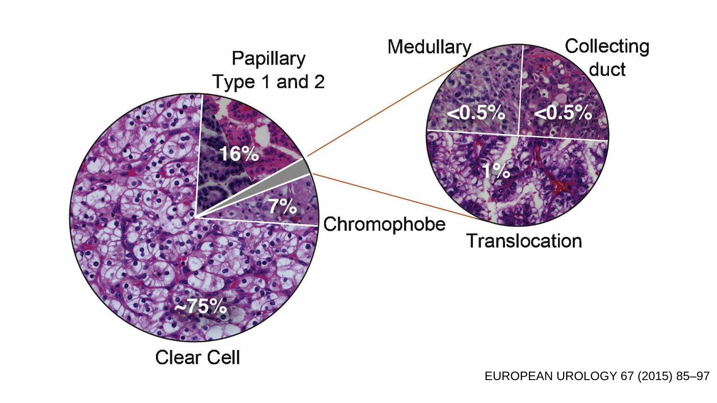
EUROPEAN UROLOGY 67 (2015) 85–97
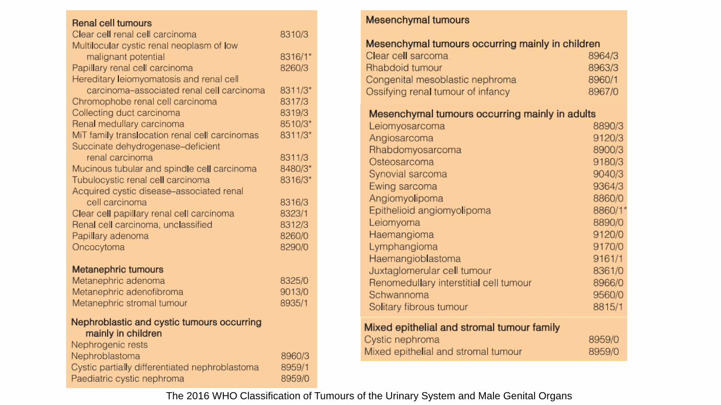
The 2016 WHO Classification of Tumours of the Urinary System and Male Genital Organs

New diagnostic entities Clear cell papillary RCC Hereditary leiomyomatosis and RCC- associated RCC Succinate dehydrogenase–deficient RCC Tubulocystic RCC Acquired cystic disease–associated RCC Evolving RCC classification, such as transcription elongation factor B subunit 1 (TCEB1)– mutated RCC/RCC with angioleiomyoma-like stroma/ RCC with leiomyomatous stroma, RCC associated with anaplastic lymphoma receptor tyrosine kinase (ALK) gene rearrangement, thyroid-like follicular RCC, and RCC in neuroblastoma survivors.
Arch Pathol Lab Med—Vol 140, October 2016

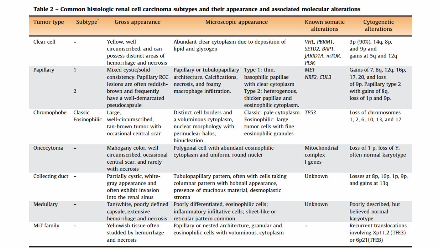
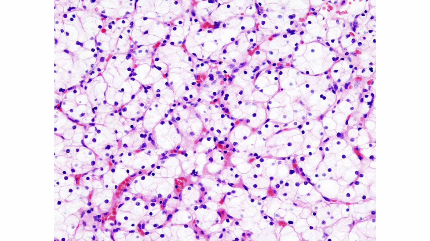
Clear cell carcinomas: Typically have a deletion of 3p. Arise from the proximal tubule. Macroscopically: solid or cystic. In addition to occurring in sporadic disease, clear cell carcinomas are specifically associated with von Hippel-Lindau disease.
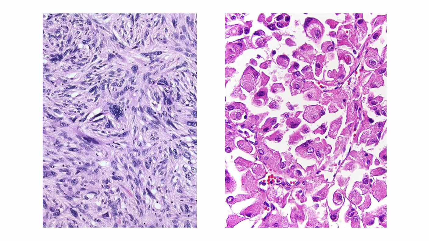
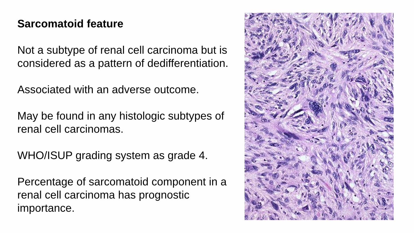
Sarcomatoid feature Not a subtype of renal cell carcinoma but is considered as a pattern of dedifferentiation. Associated with an adverse outcome. May be found in any histologic subtypes of renal cell carcinomas. WHO/ISUP grading system as grade 4. Percentage of sarcomatoid component in a renal cell carcinoma has prognostic importance.
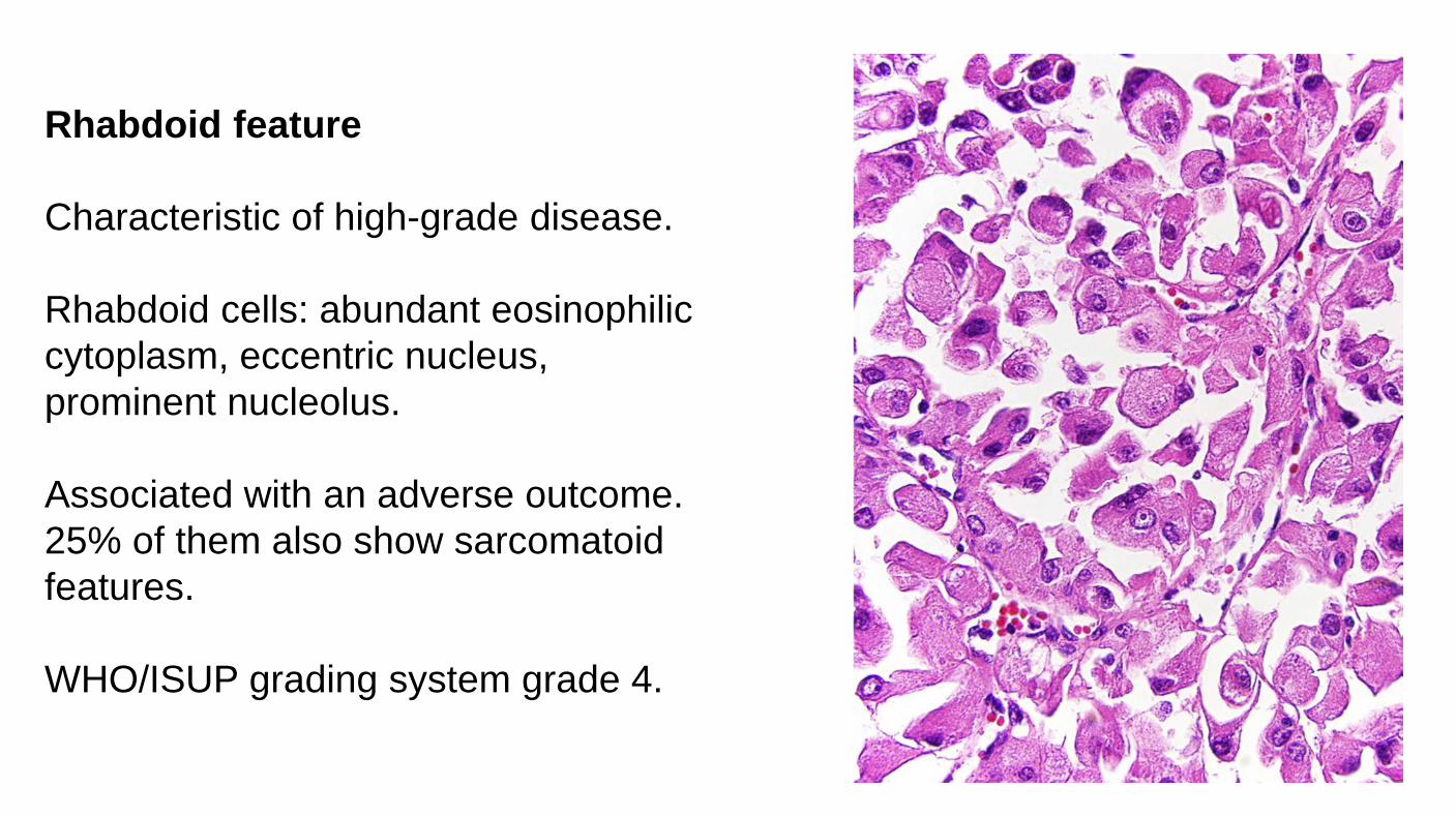
Rhabdoid feature Characteristic of high-grade disease. Rhabdoid cells: abundant eosinophilic cytoplasm, eccentric nucleus, prominent nucleolus. Associated with an adverse outcome. 25% of them also show sarcomatoid features. WHO/ISUP grading system grade 4.

Histologic Grade The WHO/ISUP grading system has supplanted the Fuhrman system Validated for both clear cell and papillary renal cell carcinoma. Chromophobe renal cell carcinoma not be graded. Not applicable Grade X - Cannot be assessed Grade 1 - Nucleoli absent or inconspicuous and basophilic at 400x. Grade 2 - Nucleoli conspicuous and eosinophilic at 400x, visible but not prominent at 100x Grade 3 - Nucleoli conspicuous and eosinophilic at 100x Grade 4 - Extreme nuclear pleomorphism and/or multinuclear giant cells and/or rhabdoid and/or sarcomatoid differentiation Grade should be assigned based on the single high-power field showing the greatest degree of pleomorphism.
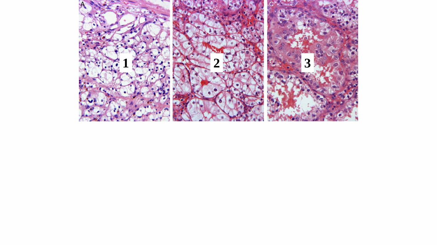
1 2 3
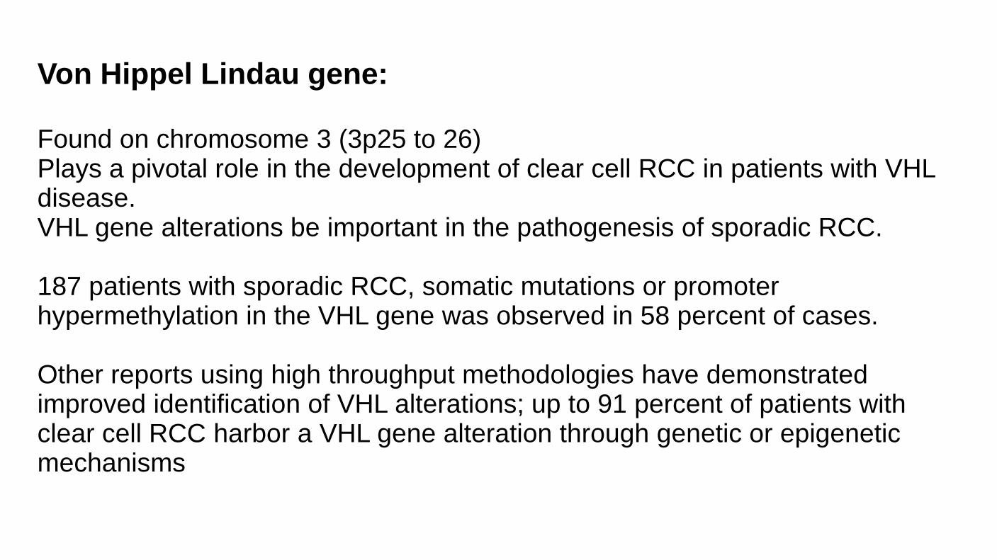
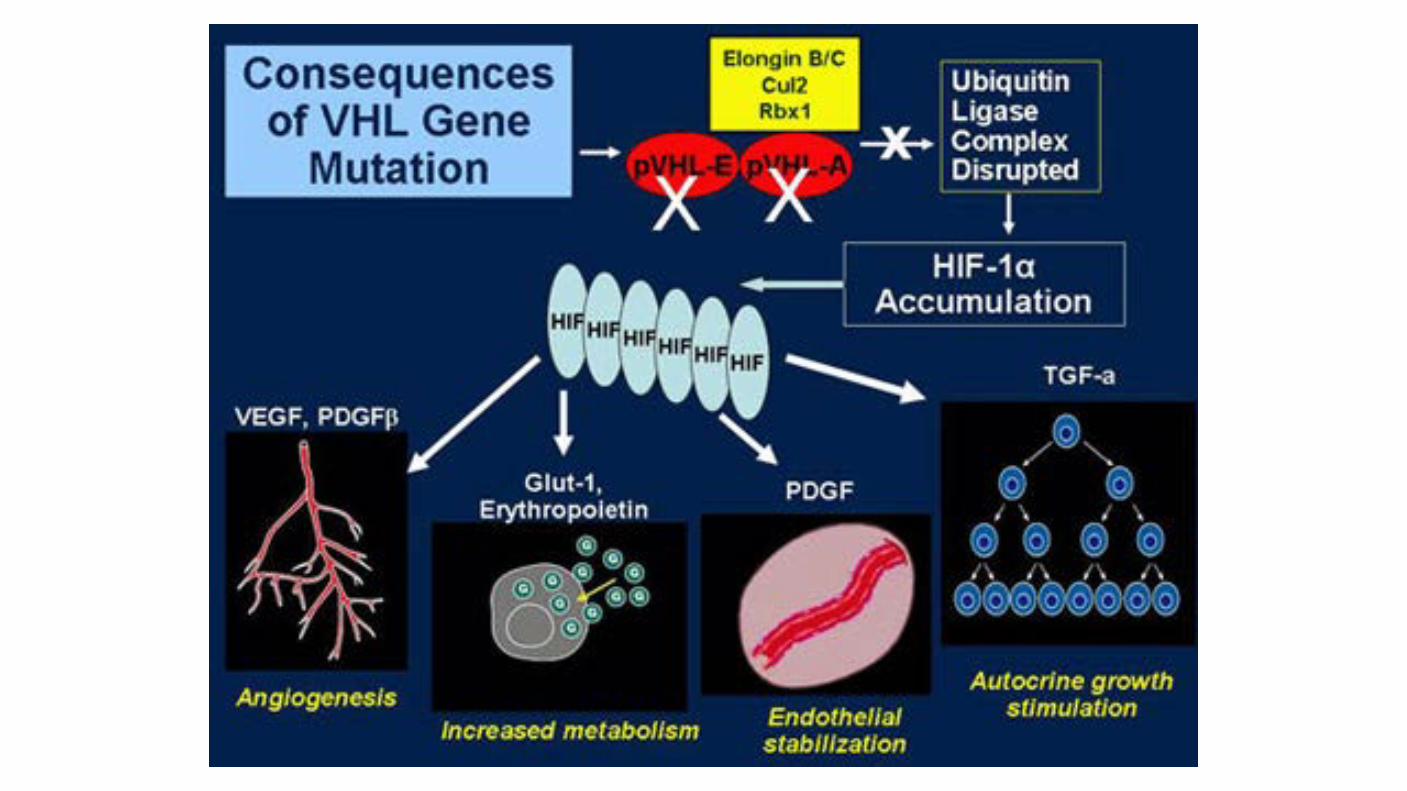
Von Hippel Lindau gene: Found on chromosome 3 (3p25 to 26) Plays a pivotal role in the development of clear cell RCC in patients with VHL disease. VHL gene alterations be important in the pathogenesis of sporadic RCC. 187 patients with sporadic RCC, somatic mutations or promoter hypermethylation in the VHL gene was observed in 58 percent of cases. Other reports using high throughput methodologies have demonstrated improved identification of VHL alterations; up to 91 percent of patients with clear cell RCC harbor a VHL gene alteration through genetic or epigenetic mechanisms
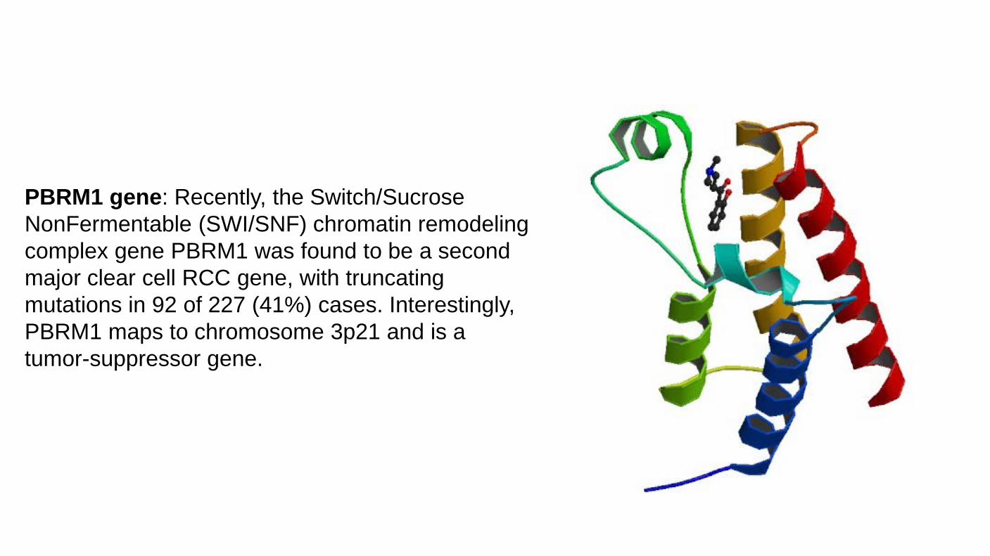
PBRM1 gene: Recently, the Switch/Sucrose NonFermentable (SWI/SNF) chromatin remodeling complex gene PBRM1 was found to be a second major clear cell RCC gene, with truncating mutations in 92 of 227 (41%) cases. Interestingly, PBRM1 maps to chromosome 3p21 and is a tumor-suppressor gene.
BAP1 gene: BRCA1 associated protein-1 (BAP1), located at 3p, is mutated in 15 percent of clear cell RCC. encodes a nuclear deubiquitinase. It is part of the large ubiquitin-mediated proteolysis pathway (UMPP). BAP1-mutant tumors are more likely to be aggressive and display adverse pathologic features, leading to worse survival.
Combined loss of BAP1 and PBRM1 in a few RCCs was associated with rhabdoid features
Nature Genetics 44, 751–759 (2012)
Inactivation of histone-modifying genes: Inactivating mutations in two genes encoding enzymes involved in histone including SET domain containing protein 2 (SETD2) Jumonji AT-rich interactive domain 1C (JARID1C). Chromatic modification machinery may be important to the pathogenesis of RCC. Ubiquitin-mediated proteolysis pathway (UMPP): UMPP is an important pathway for protein degradation through the proteasome. Alterations in UMPP result in similar functional consequences (ie, hypoxia) as VHL inactivation. In one study, UMPP was the most frequently altered pathway in clear cell RCC. Of note, the VHL and BAP1 genes are members of this pathway.
Abnormalities in cellular division: Development of RCCs involve abnormalities in genes that control cell division include Ras family genes and the p53 tumor suppressor gene. Mutations in the p53 gene are identified infrequently in RCCs, Overexpression of p53 protein is detected in approximately one-half of tumors and associated with more aggressive behavior and a worse prognosis. Series of 175 patients, in which the ten-year disease-specific survival was lower for patients whose tumors stained for p53 (48% versus 78%, compared with those not overexpressing p53).

Genetic alterations in clear cell carcinoma:
Genome-wide analysis and gene expression profiles on 90 RCC tumors. Common genetic abnormalities in sporadic von-Hippel Lindau (VHL)-null RCC include the following: - Loss of 3p (94 %), which contains several genes associated with RCC, including the VHL, BRCA1 associated protein-1 (BAP-1), and protein polybromo-1 (PBRM1) genes - Gain of 5q (69 %) - Monosomy or partial loss of 14q (42 %) - 7q gain (20 %) - 8p deletion (32 %) - 9p loss (29 %)
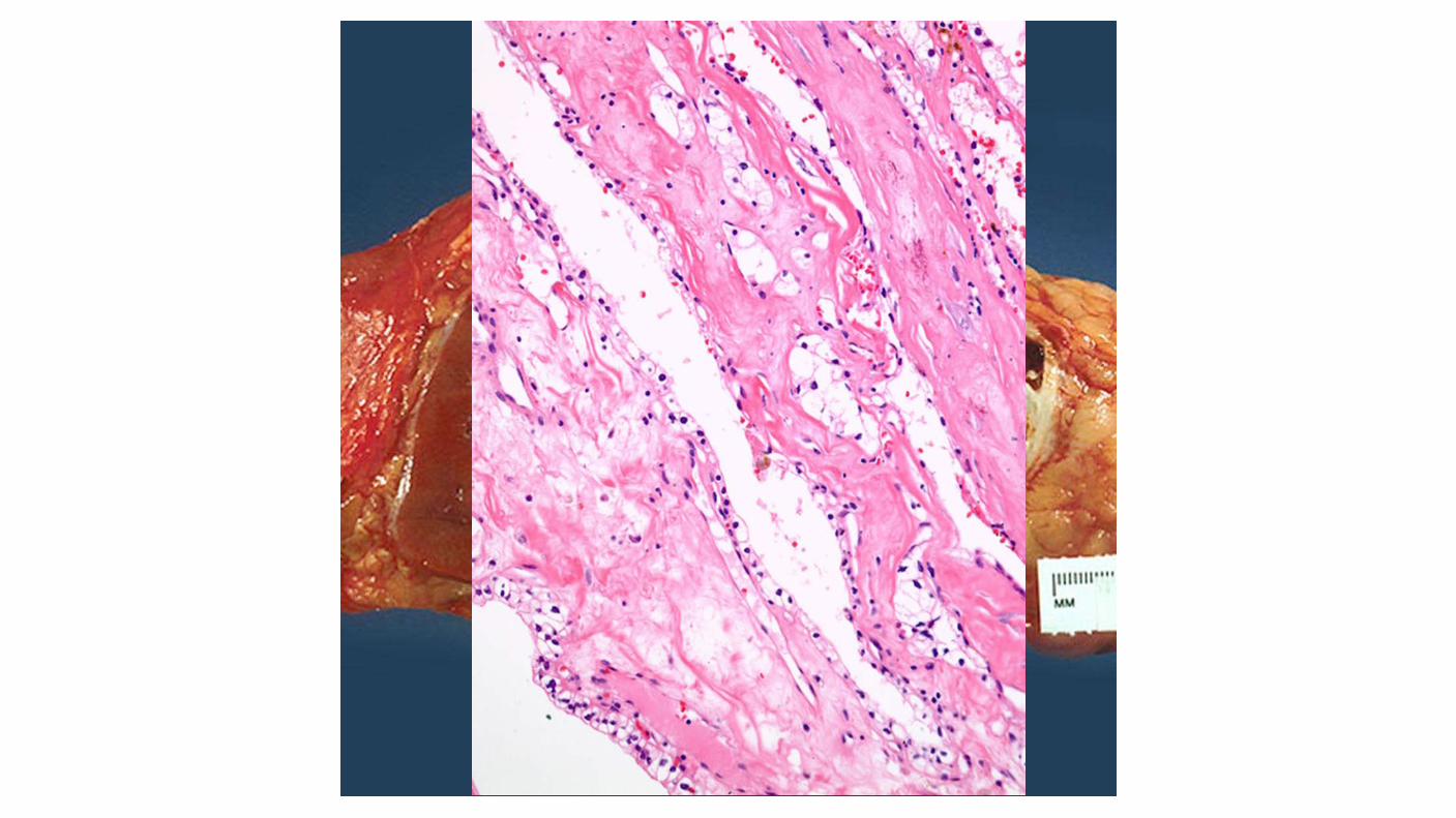
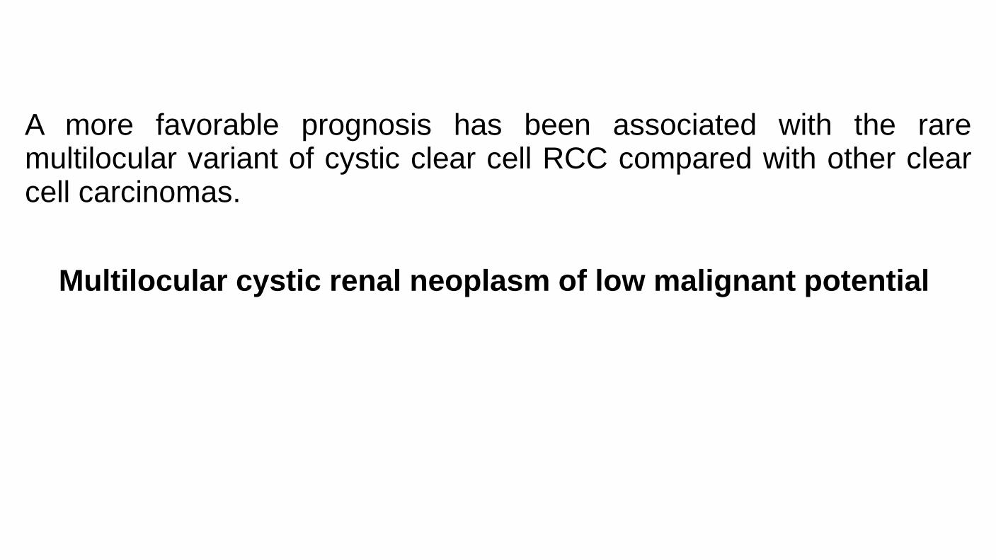
A more favorable prognosis has been associated with the rare multilocular variant of cystic clear cell RCC compared with other clear cell carcinomas.
Multilocular cystic renal neoplasm of low malignant potential
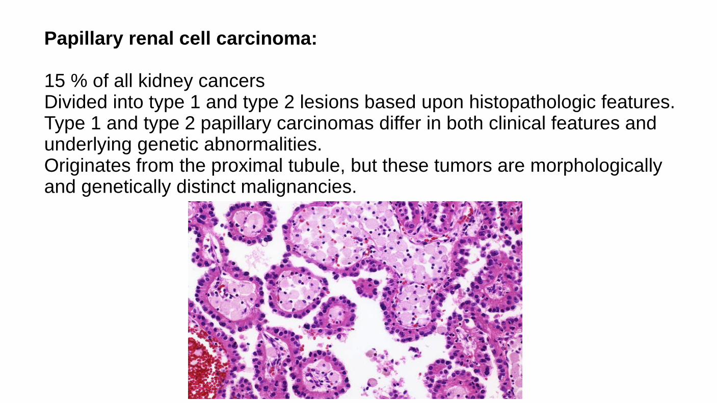
Papillary renal cell carcinoma: 15 % of all kidney cancers Divided into type 1 and type 2 lesions based upon histopathologic features. Type 1 and type 2 papillary carcinomas differ in both clinical features and underlying genetic abnormalities. Originates from the proximal tubule, but these tumors are morphologically and genetically distinct malignancies.

Type 1 papillary RCC: Typically presents with stage I or II disease. Relatively favorable prognosis. Occur in patients with hereditary papillary RCC, the majority of these are sporadic. In the hereditary form, activating germline mutations are seen in MET. In nonhereditary form, somatic mutations in MET have been identified in 10-20%of cases. Altered MET status or increased chromosome 7 copy number was identified in 81% of type 1 papillary RCCs.
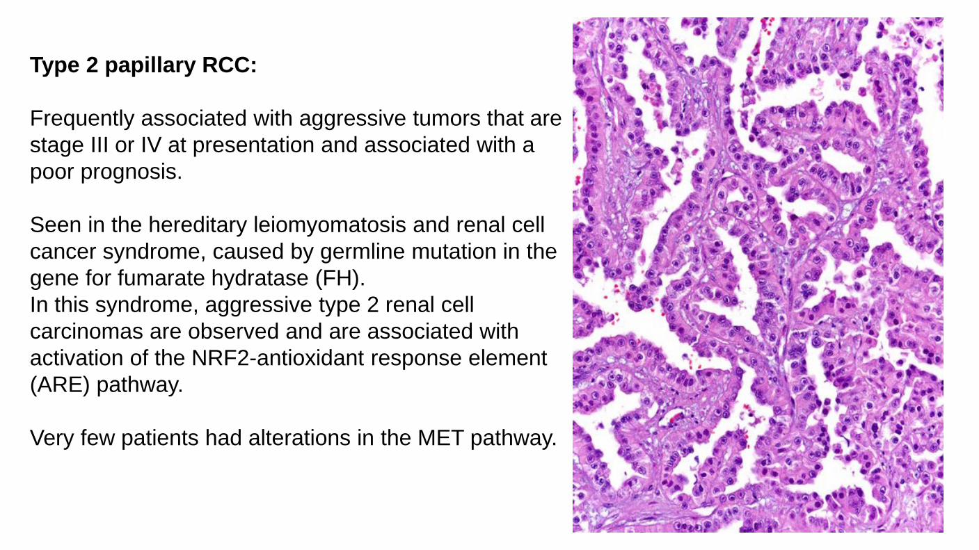
Type 2 papillary RCC: Frequently associated with aggressive tumors that are stage III or IV at presentation and associated with a poor prognosis. Seen in the hereditary leiomyomatosis and renal cell cancer syndrome, caused by germline mutation in the gene for fumarate hydratase (FH). In this syndrome, aggressive type 2 renal cell carcinomas are observed and are associated with activation of the NRF2-antioxidant response element (ARE) pathway. Very few patients had alterations in the MET pathway.
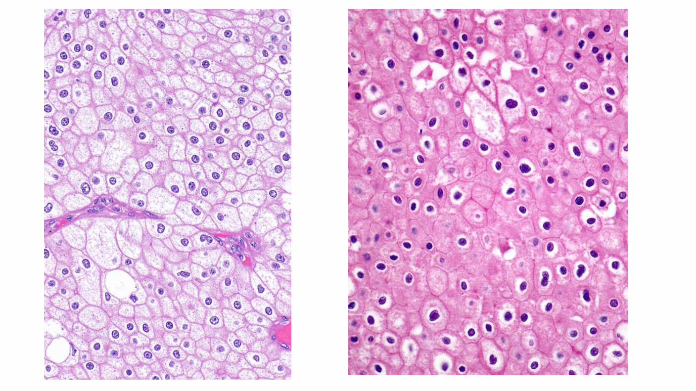
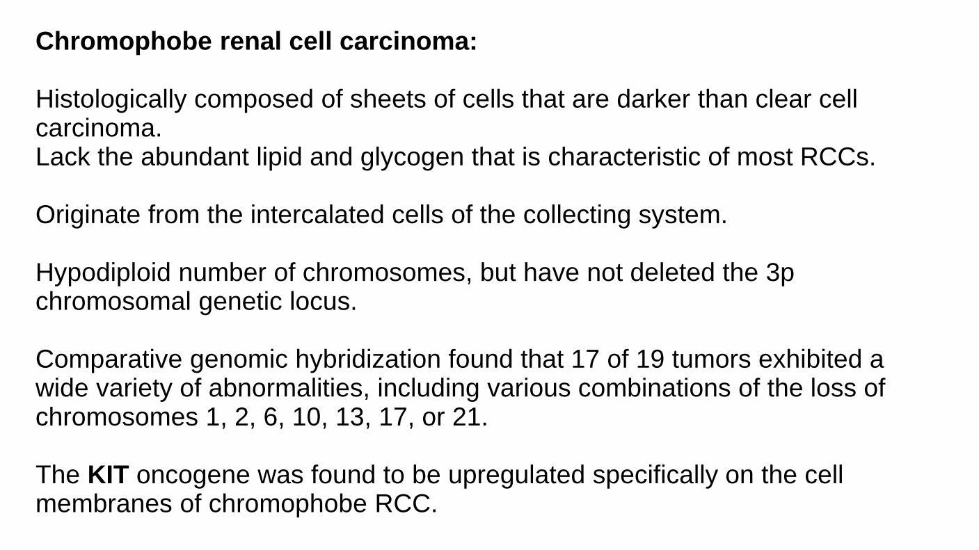
Chromophobe renal cell carcinoma: Histologically composed of sheets of cells that are darker than clear cell carcinoma. Lack the abundant lipid and glycogen that is characteristic of most RCCs. Originate from the intercalated cells of the collecting system. Hypodiploid number of chromosomes, but have not deleted the 3p chromosomal genetic locus. Comparative genomic hybridization found that 17 of 19 tumors exhibited a wide variety of abnormalities, including various combinations of the loss of chromosomes 1, 2, 6, 10, 13, 17, or 21. The KIT oncogene was found to be upregulated specifically on the cell membranes of chromophobe RCC.
Lower risk of disease progression and death compared with clear cell carcinomas, although this is likely due to the fact that patients present at a lower stage. 66 chromophobe RCCs were analyzed. Mitochondrial DNA and gene expression analysis suggested mitochondrial function as an important component of the disease biology. Genomic rearrangements showed recurrent structural breakpoints within the telomerase reverse transcriptase (TERT) promoter region, which correlates with highly elevated TERT expression.
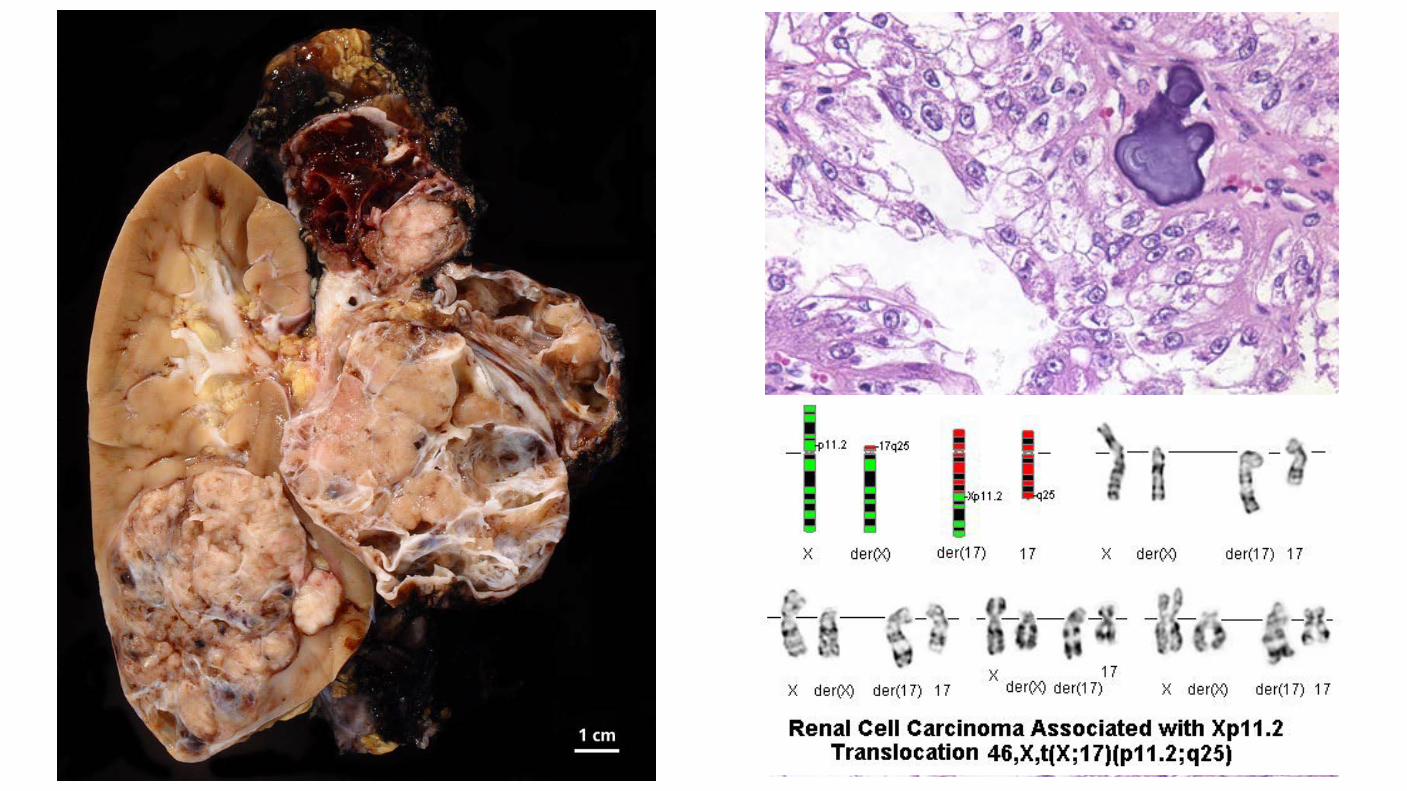
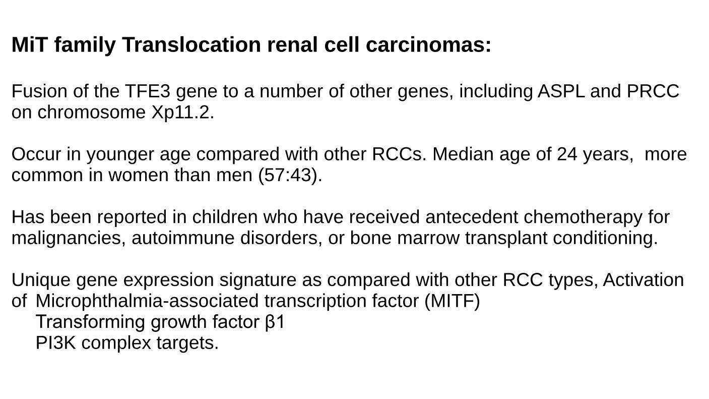
MiT family Translocation renal cell carcinomas: Fusion of the TFE3 gene to a number of other genes, including ASPL and PRCC on chromosome Xp11.2. Occur in younger age compared with other RCCs. Median age of 24 years, more common in women than men (57:43). Has been reported in children who have received antecedent chemotherapy for malignancies, autoimmune disorders, or bone marrow transplant conditioning. Unique gene expression signature as compared with other RCC types, Activation of Microphthalmia-associated transcription factor (MITF) Transforming growth factor β1 PI3K complex targets.
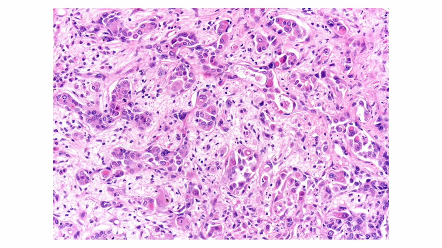
Collecting duct (Bellini's duct) Carcinoma: Rare (SEER 160 VS 33,000 ccRCC) Tend to occur in younger patients and are frequently aggressive. More frequent in black patients. Commonly present with gross hematuria. Presented with advanced (T3/T4) or metastatic disease. Collecting duct tumors have not been associated with a consistent pattern of genetic abnormalities. Biologically, these tumors more closely resemble transitional cell than RCCs. One report of 17 cases assessed by comprehensive genomic profiling showed NF2 and CDKN2A alterations in 29 and 12 percent, respectively.
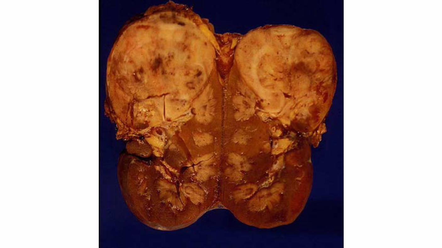
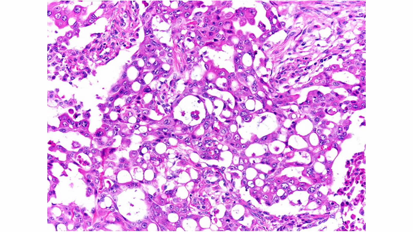
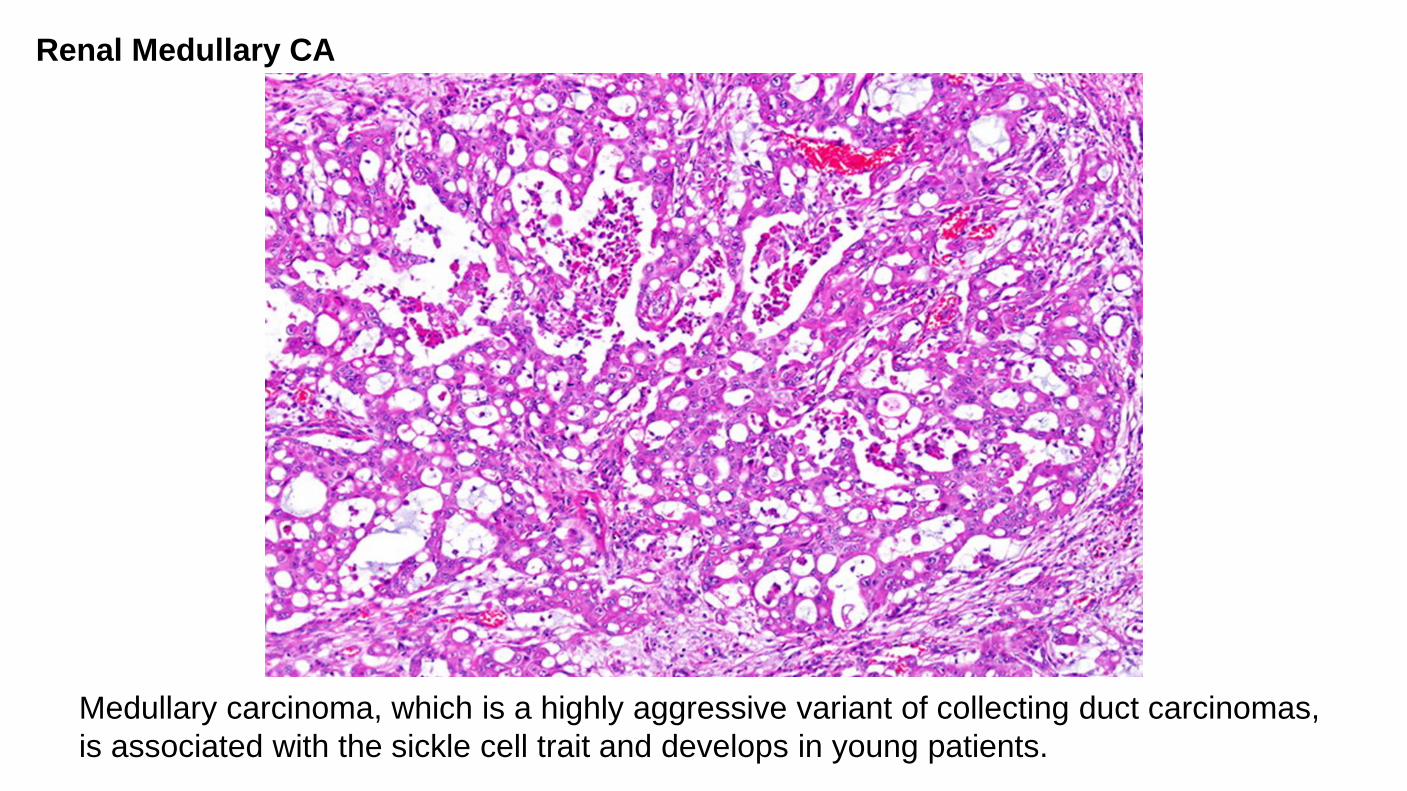
Medullary carcinoma, which is a highly aggressive variant of collecting duct carcinomas, is associated with the sickle cell trait and develops in young patients.
Renal Medullary CA
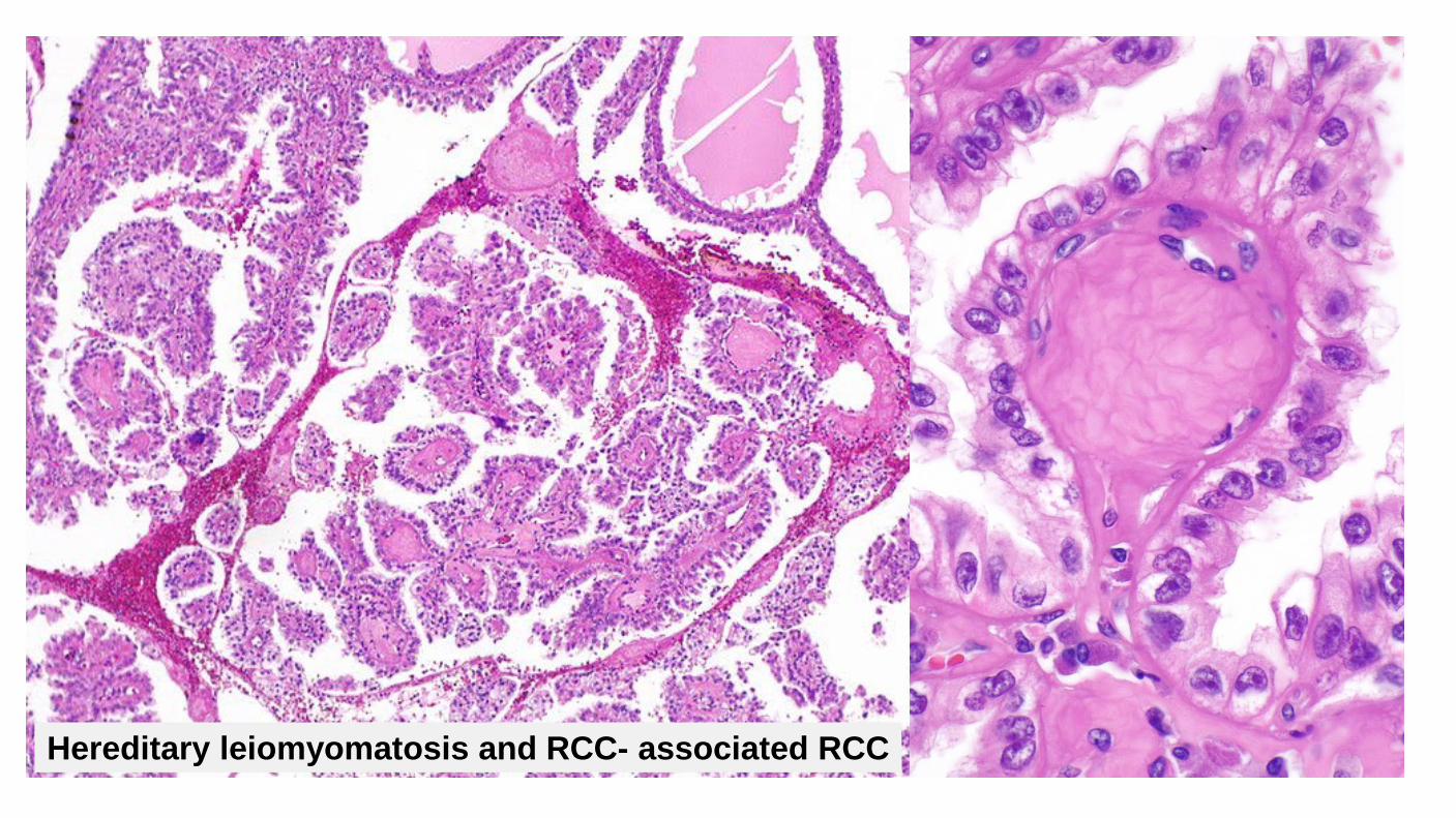
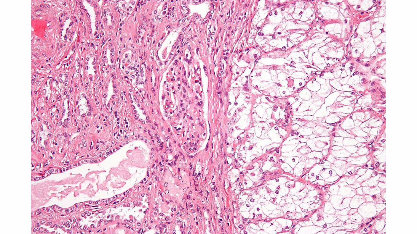
Hereditary leiomyomatosis and RCC- associated RCC
Clinical Uncommon Predominantly younger patients, mean 36 years May present with multiple cutaneous and uterine leiomyomas (85%) Cutaneous lesion most common on arms and thorax Uterine leiomyomas have nuclear features similar to renal tumors Caused by germline mutation in fumarate hydratase gene (recommend genetic counseling) Unlike other hereditary tumors, these may be unilateral and single Poor prognosis Early widespread dissemination common Often at least pT3 Often lymph node metastases Metastases reported even with small tumors
Hereditary leiomyomatosis and RCC- associated RCC
Diagnostic Criteria Large nuclei with prominent, eosinophilic, inclusion-like nucleoli with perinucleolar clearing May only be present focally Papillary architecture most common Foamy histiocytes uncommon May show tubular, tubulopapillary tubulocystic, solid, and mixed architecture May show collecting duct-like morphology with tubules, solid nests or individual cells infiltrating through desmoplastic stroma May show sarcomatoid growth Large cells with abundant eosinophilic cytoplasm Focal areas may have amphophilic or clear cytoplasm Loss of fumarate hydratase (FH) expression
Hereditary leiomyomatosis and RCC- associated RCC
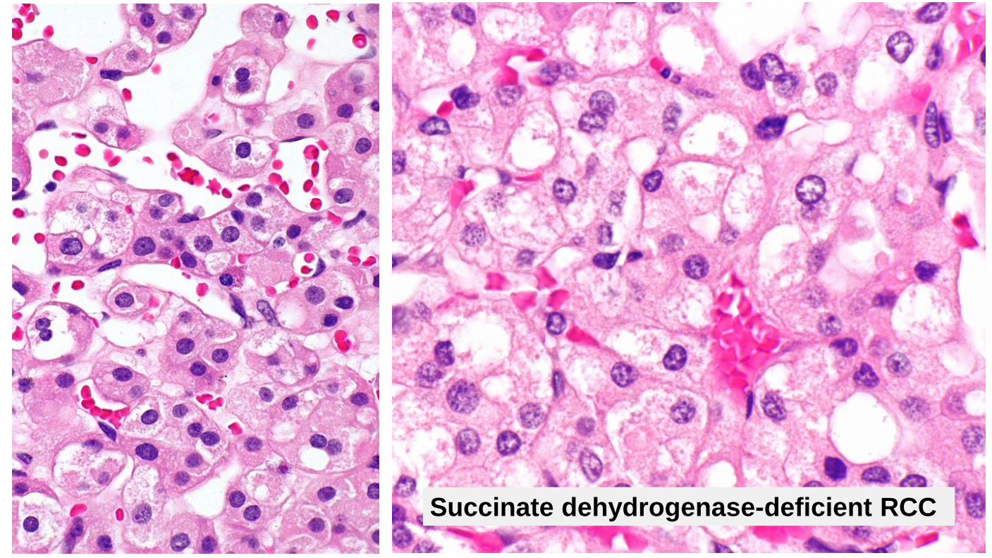
Succinate dehydrogenase-deficient RCC
Clinical Very rare (0.05 to 0.2% of all renal carcinomas) Classically presents in young adults (mean age 38) Slight male predominance Strong hereditary link Great majority of patients have germline mutations in SDHB, SDHC, SDHA, or SDHD associated with an autosomal dominant tumor syndrome characterized by SDH-deficient renal cell carcinoma, paraganglioma/pheochromocytoma, or gastrointestinal stromal tumors All patients with SDH-deficient tumors should be offered genetic testing Patients need long-term surveillance for other SDH-deficient neoplasms May be multifocal/bilateral Relatively good prognosis (metastatic rate of ~10%) Outcome less favorable with dedifferentiation and necrosis
Succinate dehydrogenase–deficient RCC
Diagnostic Criteria Requirement: Loss of immunohistochemical staining for SDHB Loss of staining with SDHB signifies a mutation in either SDHA, SDHB, SDHC, or SDHD Eosinophilic cells with flocculent cytoplasm Cytoplasmic vacuoles or flocculent inclusions Neuroendocrine-like nuclei
Succinate dehydrogenase–deficient RCC
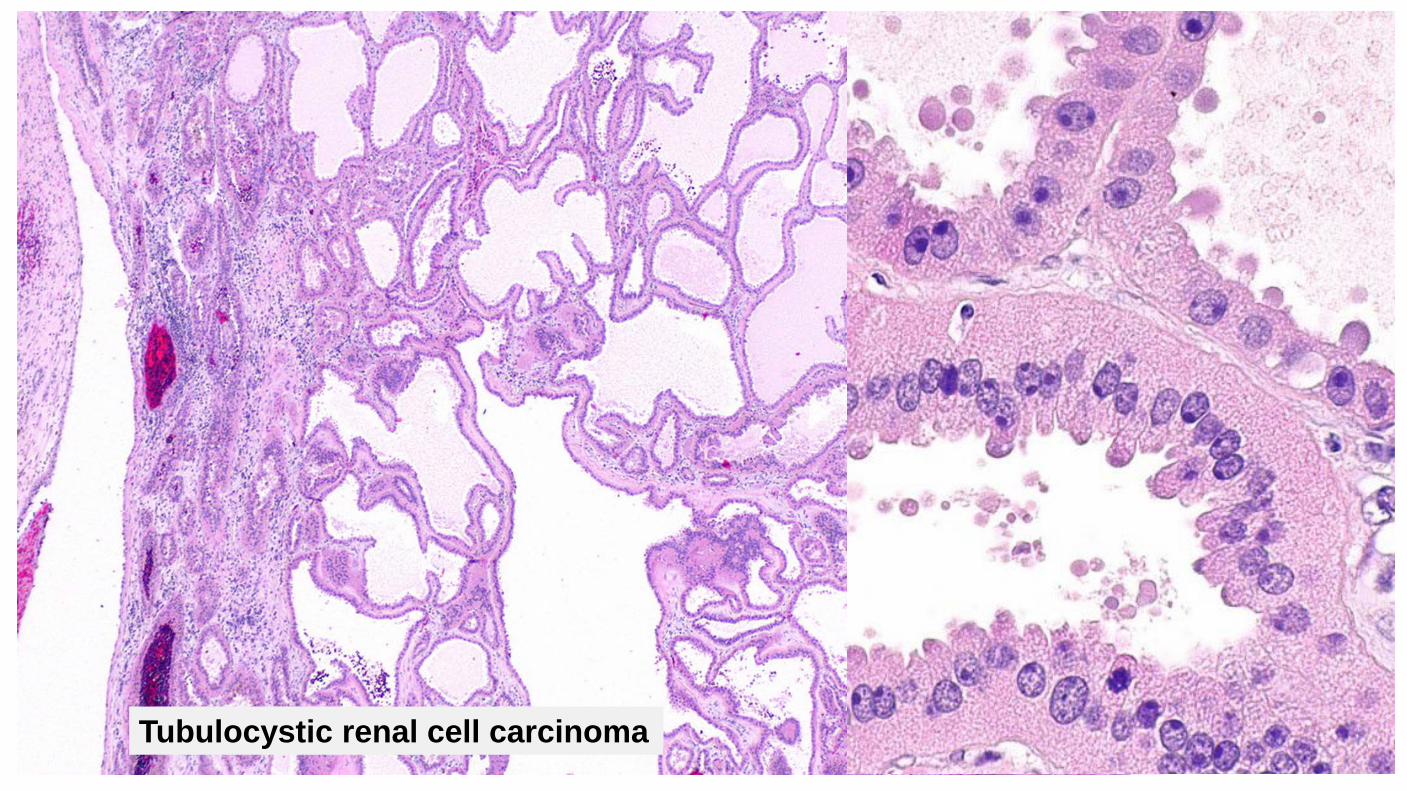
Tubulocystic renal cell carcinoma
Tubulocystic renal cell carcinoma
Clinical Mean age about 60 (30-94) M:F = 7:1 Behavior uncertain 2 of 31 cases reported with metastases
Diagnostic criteria Mixture of closely packed tubules and micro/macro cysts of variable sizes with low grade nuclear features Tubules and cysts are lined by single layer of cuboidal or columnar cells with abundant eosinophilic cytoplasm, uniform nuclei with distinct nucleoli; often have hobnail appearance Overall low grade nuclear features Cysts are closely spaced with variable intervening fibrotic stroma 40% coexist with papillary renal cell carcinoma Minimal mitotic activity, no atypia, no desmoplasia
Tubulocystic renal cell carcinoma
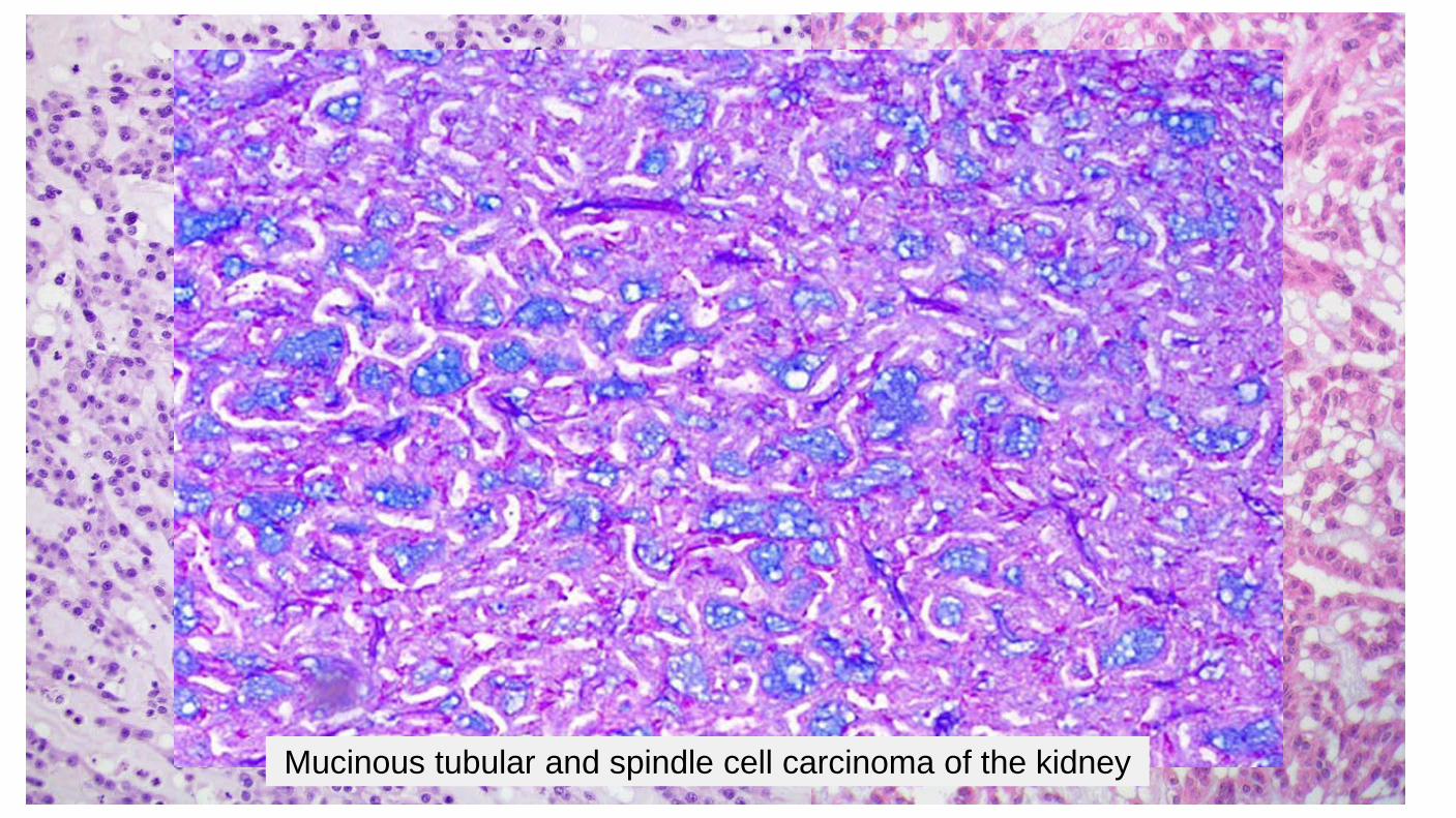
Mucinous tubular and spindle cell carcinoma of the kidney

Diagnostic Criteria Composed of elongate tubules, variably packed Spindle cell population merges with and may represent densely packed collapsed tubules Mucinous stroma separates the tubules Epithelial and spindle cells are cytologically bland Occasional findings Collections of foamy macrophages Lymphocytic infiltrate Psammoma bodies Solid foci without detectable stroma Clear cell foci Sarcomatoid change may be seen in rare cases MTSCC may show morphologic and immunophenotypic overlap with papillary renal cell carcinoma
Mucinous Tubular and Spindle Cell Carcinoma of the Kidney
Clinical < 1% of all renal neoplasms Median age 58 years (range 13 - 81) Female:male ratio: 4:1 Usually indolent behavior Recurrences are uncommon Metastases are rare, but have been reported with both low grade histology and high grade transformation Some occur in association with nephrolithiasis
Mucinous Tubular and Spindle Cell Carcinoma of the Kidney
Clear Cell Papillary Renal Cell Carcinoma
Clinical features Low stage, indolent behavior, no metastases reported 1% - 3% of all renal cell neoplasms, no sex predilection, mean 60 years (18 – 88) Initially reported in patients with end stage renal disease, but most cases reported subsequently are sporadic, in normal kidneys
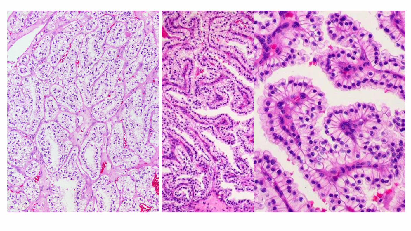
Diagnostic Criteria Most tumors exhibit mixtures of tubular, cystic, acinar and papillary patterns All patterns lined by a single layer of cuboidal to low columnar cells Nuclei typically uniformly separated from base of cell by clear cytoplasm (pseudoendometrial) Low grade and stage, WHO/ISUP grade 1 or 2, 95% pT1a, No vascular or renal sinus invasion Frequent prominent partial to complete capsule Immunophenotype CK7 strong +ve, CA9 basal or cup-like, CD10 –ve to focal, Racemase -ve Small solid foci of clear cells reminiscent of conventional ccRCC may be seen May be seen in sporadic patients and in those with end stage renal disease
Clear Cell Papillary Renal Cell Carcinoma

Arch Pathol Lab Med-Vol 141, April 2017
Acquired Cystic Disease–Associated Renal Cell Carcinoma
Acquired Cystic Disease–Associated Renal Cell Carcinoma
Arises in individuals with acquired cystic kidney disease (ACKD) in the setting of ESRD.
Predominantly male sex.
Occur 10 to 20 years after dialysis.
Occurs in 35% of long term dialysis patients; of these, 6% develop RCC
Risk of developing RCC in ACKD is increased by more than 100X (Adv Anat Pathol. 2003;10(3):135-59)
Histopathology
Cribriform / microcystic / sieve-like architecture
Abundant granular eosinophilic cytoplasm with prominent nucleoli
Intratumoral calcium oxalate crystals are very common but not necessary for diagnosis
May be nodules arising from cyst walls or masses separated from cysts
Sometimes prominent clear cell cytology
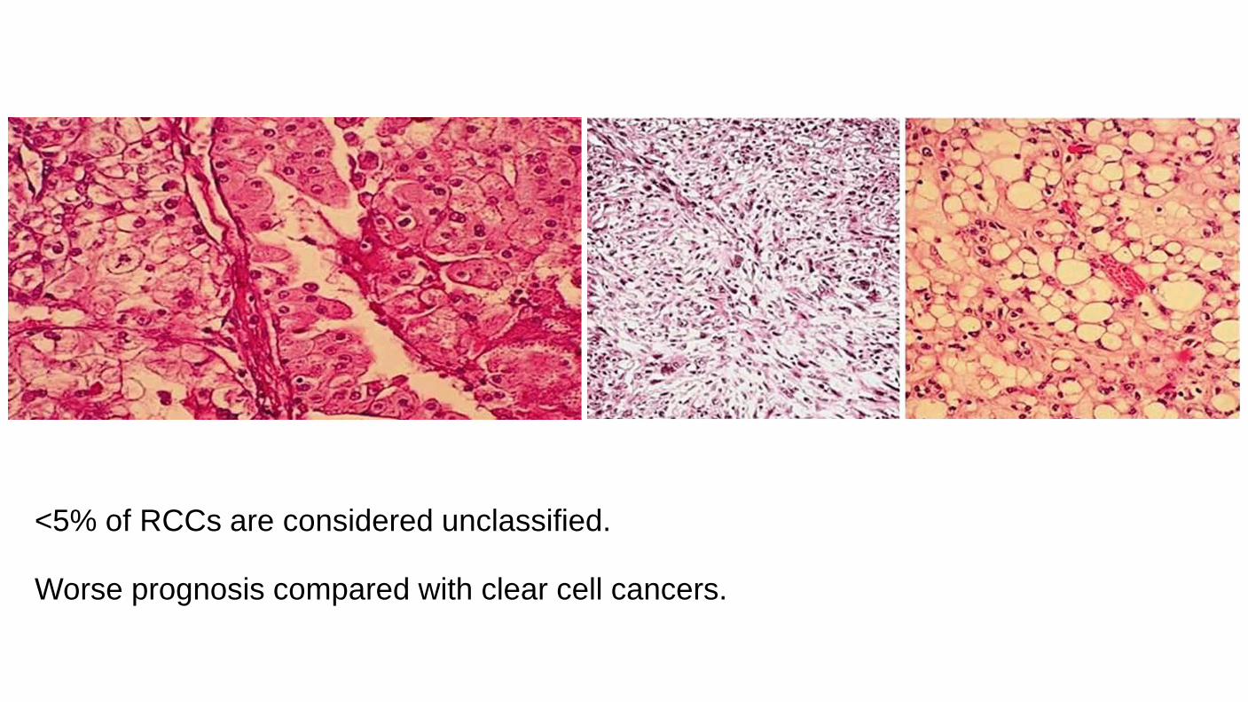
<5% of RCCs are considered unclassified. Worse prognosis compared with clear cell cancers.

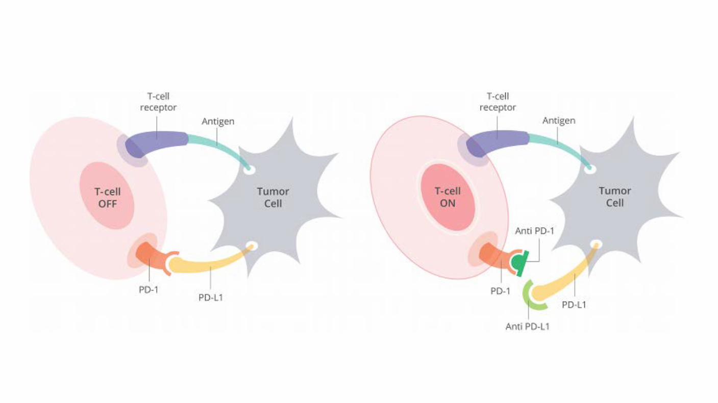
PD-1 is a 288-amino acid cell-surface protein. PD-1 binds two ligands, PD-L1 and PD-L2, which negatively regulate the immune response. Expression of PD-L1 (B7-H1) on tumor cells leads to the inhibition of the T cell-mediated immune response against cancer, thereby enabling tumor progression and metastasis
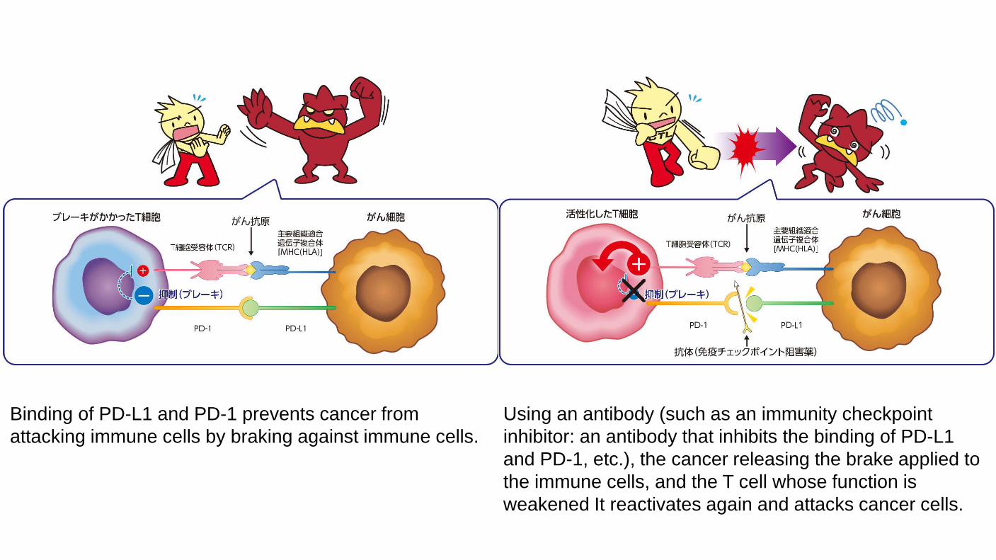
Binding of PD-L1 and PD-1 prevents cancer from attacking immune cells by braking against immune cells.
Using an antibody (such as an immunity checkpoint inhibitor: an antibody that inhibits the binding of PD-L1 and PD-1, etc.), the cancer releasing the brake applied to the immune cells, and the T cell whose function is weakened It reactivates again and attacks cancer cells.

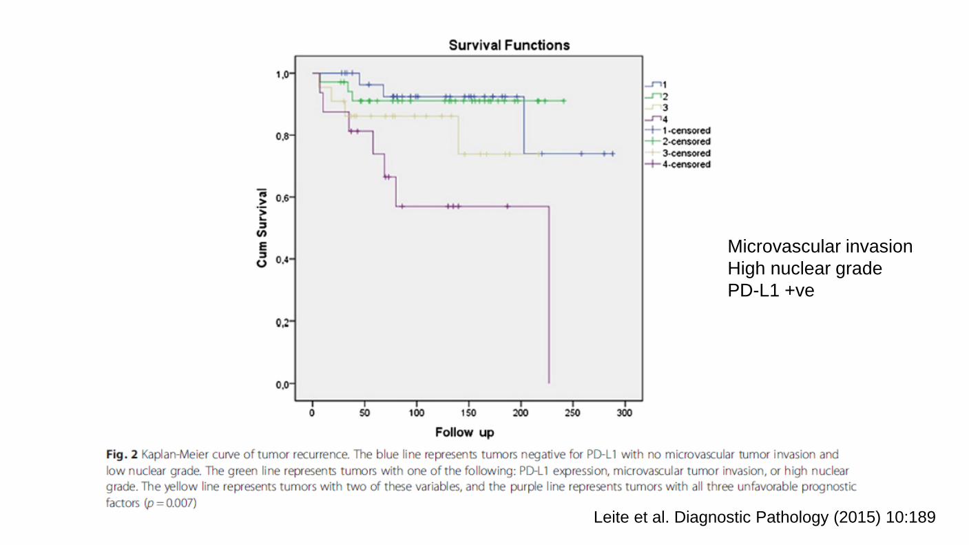
Leite et al. Diagnostic Pathology (2015) 10:189
Leite et al. Diagnostic Pathology (2015) 10:189
Microvascular invasion High nuclear grade PD-L1 +ve
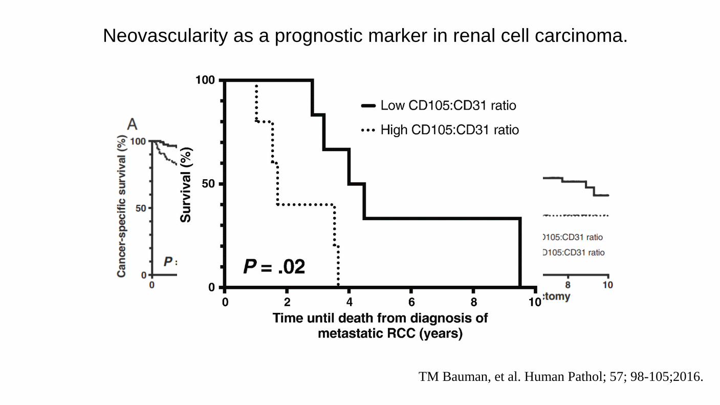
TM Bauman, et al. Human Pathol; 57; 98-105;2016.
Neovascularity as a prognostic marker in renal cell carcinoma.
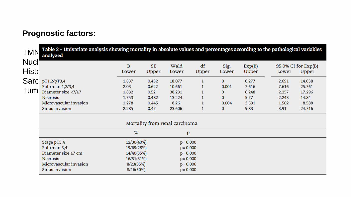
Prognostic factors: TMN staging Nuclear grade Histologic Classification Sarcomatoid/rhabdoid component Tumor necrosis
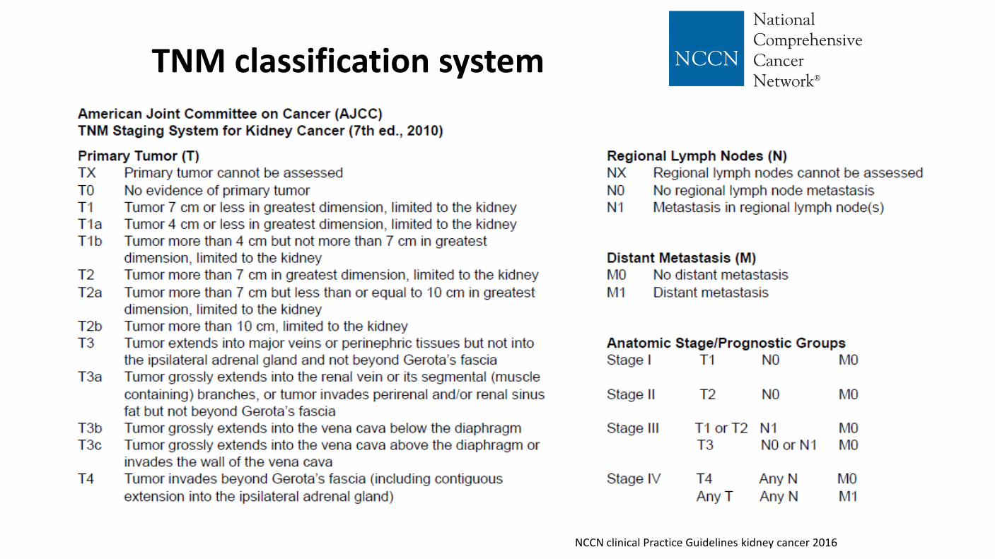
TNM classification system
NCCN clinical Practice Guidelines kidney cancer 2016
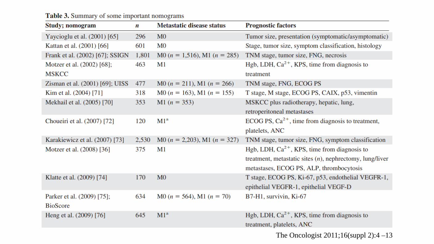
The Oncologist 2011;16(suppl 2):4 –13
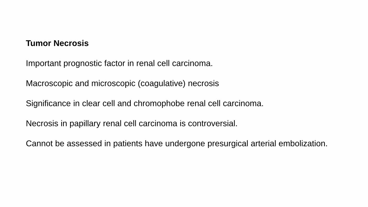
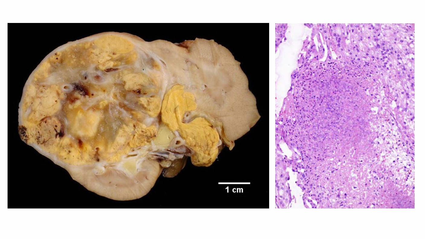
Tumor Necrosis Important prognostic factor in renal cell carcinoma. Macroscopic and microscopic (coagulative) necrosis Significance in clear cell and chromophobe renal cell carcinoma. Necrosis in papillary renal cell carcinoma is controversial. Cannot be assessed in patients have undergone presurgical arterial embolization.
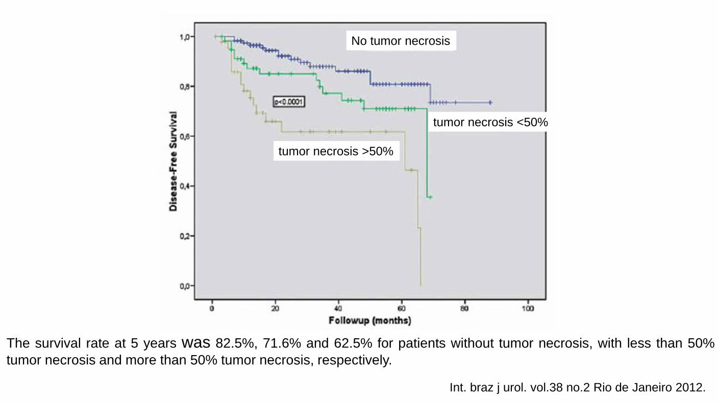
The survival rate at 5 years was 82.5%, 71.6% and 62.5% for patients without tumor necrosis, with less than 50% tumor necrosis and more than 50% tumor necrosis, respectively.
Int. braz j urol. vol.38 no.2 Rio de Janeiro 2012.
No tumor necrosis
tumor necrosis <50%
tumor necrosis >50%