Embed Size (px)
Citation preview
E18 October 2009 • Volume 13, Number 5 • Clinical Journal of Oncology Nursing
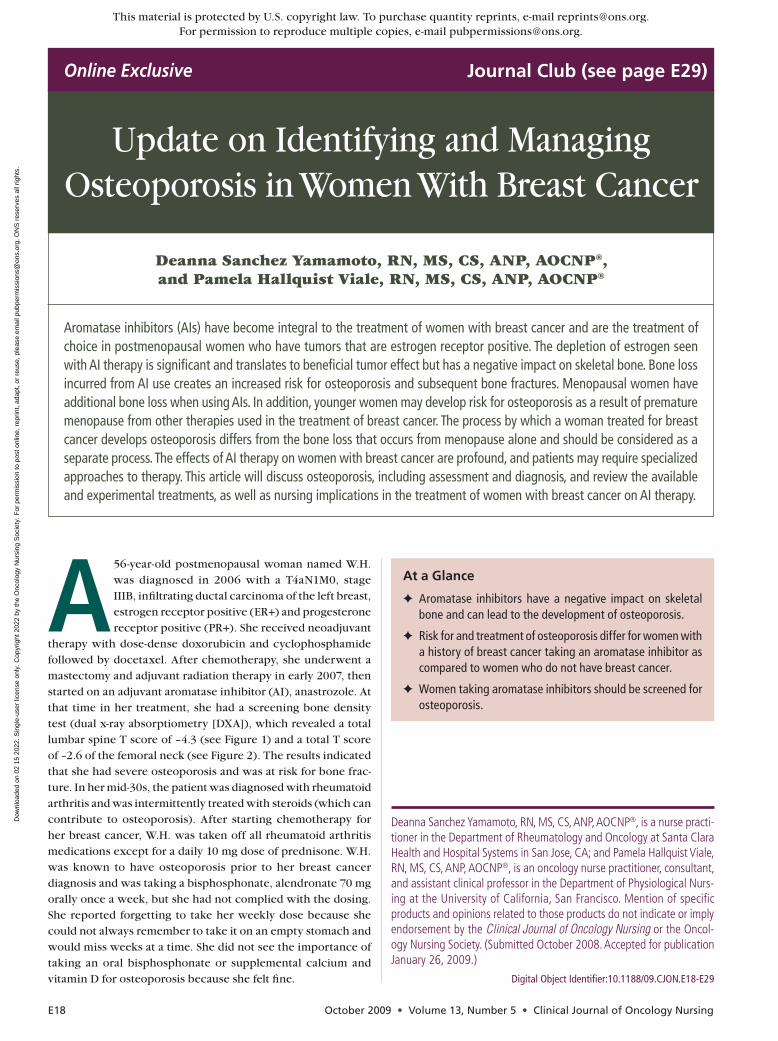
A 56-year-old postmenopausal woman named W.H. was diagnosed in 2006 with a T4aN1M0, stage IIIB, infiltrating ductal carcinoma of the left breast, estrogen receptor positive (ER+) and progesterone receptor positive (PR+). She received neoadjuvant
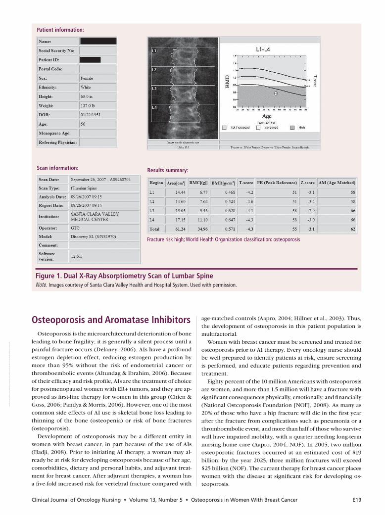
therapy with dose-dense doxorubicin and cyclophosphamide followed by docetaxel. After chemotherapy, she underwent a mastectomy and adjuvant radiation therapy in early 2007, then started on an adjuvant aromatase inhibitor (AI), anastrozole. At that time in her treatment, she had a screening bone density test (dual x-ray absorptiometry [DXA]), which revealed a total lumbar spine T score of –4.3 (see Figure 1) and a total T score of –2.6 of the femoral neck (see Figure 2). The results indicated that she had severe osteoporosis and was at risk for bone frac-ture. In her mid-30s, the patient was diagnosed with rheumatoid arthritis and was intermittently treated with steroids (which can contribute to osteoporosis). After starting chemotherapy for her breast cancer, W.H. was taken off all rheumatoid arthritis medications except for a daily 10 mg dose of prednisone. W.H. was known to have osteoporosis prior to her breast cancer diagnosis and was taking a bisphosphonate, alendronate 70 mg orally once a week, but she had not complied with the dosing. She reported forgetting to take her weekly dose because she could not always remember to take it on an empty stomach and would miss weeks at a time. She did not see the importance of taking an oral bisphosphonate or supplemental calcium and vitamin D for osteoporosis because she felt fine.
At a Glance
F Aromatase inhibitors have a negative impact on skeletal bone and can lead to the development of osteoporosis.
F Risk for and treatment of osteoporosis differ for women with a history of breast cancer taking an aromatase inhibitor as compared to women who do not have breast cancer.
F Women taking aromatase inhibitors should be screened for osteoporosis.
Deanna Sanchez Yamamoto, RN, MS, CS, ANP, AOCNP®, is a nurse practi-tioner in the Department of Rheumatology and Oncology at Santa Clara Health and Hospital Systems in San Jose, CA; and Pamela Hallquist Viale, RN, MS, CS, ANP, AOCNP®, is an oncology nurse practitioner, consultant, and assistant clinical professor in the Department of Physiological Nurs-ing at the University of California, San Francisco. Mention of specific products and opinions related to those products do not indicate or imply endorsement by the Clinical Journal of Oncology Nursing or the Oncol-ogy Nursing Society. (Submitted October 2008. Accepted for publication January 26, 2009.)
Digital Object Identifier:10.1188/09.CJON.E18-E29
Deanna Sanchez Yamamoto, RN, MS, CS, ANP, AOCNP®, and Pamela Hallquist Viale, RN, MS, CS, ANP, AOCNP®
Aromatase inhibitors (AIs) have become integral to the treatment of women with breast cancer and are the treatment of choice in postmenopausal women who have tumors that are estrogen receptor positive. The depletion of estrogen seen with AI therapy is significant and translates to beneficial tumor effect but has a negative impact on skeletal bone. Bone loss incurred from AI use creates an increased risk for osteoporosis and subsequent bone fractures. Menopausal women have additional bone loss when using AIs. In addition, younger women may develop risk for osteoporosis as a result of premature menopause from other therapies used in the treatment of breast cancer. The process by which a woman treated for breast cancer develops osteoporosis differs from the bone loss that occurs from menopause alone and should be considered as a separate process. The effects of AI therapy on women with breast cancer are profound, and patients may require specialized approaches to therapy. This article will discuss osteoporosis, including assessment and diagnosis, and review the available and experimental treatments, as well as nursing implications in the treatment of women with breast cancer on AI therapy.
Update on Identifying and Managing Osteoporosis in Women With Breast Cancer
Online Exclusive Journal Club (see page E29)
This material is protected by U.S. copyright law. To purchase quantity reprints, e-mail [email protected]. For permission to reproduce multiple copies, e-mail [email protected].
Dow
nloa
ded
on 0
2 15
202
2. S
ingl
e-us
er li
cens
e on
ly. C
opyr
ight
202
2 by
the
Onc
olog
y N
ursi
ng S
ocie
ty. F
or p
erm
issi
on to
pos
t onl
ine,
rep
rint,
adap
t, or
reu
se, p
leas
e em
ail p
ubpe
rmis
sion
s@on
s.or
g. O
NS
res
erve
s al
l rig
hts.
Clinical Journal of Oncology Nursing • Volume 13, Number 5 • Osteoporosis in Women With Breast Cancer E19
Osteoporosis and Aromatase InhibitorsOsteoporosis is the microarchitectural deterioration of bone
leading to bone fragility; it is generally a silent process until a painful fracture occurs (Delaney, 2006). AIs have a profound estrogen depletion effect, reducing estrogen production by more than 95% without the risk of endometrial cancer or thromboembolic events (Altundag & Ibrahim, 2006). Because of their efficacy and risk profile, AIs are the treatment of choice for postmenopausal women with ER+ tumors, and they are ap-proved as first-line therapy for women in this group (Chien & Goss, 2006; Pandya & Morris, 2006). However, one of the most common side effects of AI use is skeletal bone loss leading to thinning of the bone (osteopenia) or risk of bone fractures (osteoporosis).
Development of osteoporosis may be a different entity in women with breast cancer, in part because of the use of AIs (Hadji, 2008). Prior to initiating AI therapy, a woman may al-ready be at risk for developing osteoporosis because of her age, comorbidities, dietary and personal habits, and adjuvant treat-ment for breast cancer. After adjuvant therapies, a woman has a five-fold increased risk for vertebral fracture compared with
age-matched controls (Aapro, 2004; Hillner et al., 2003). Thus, the development of osteoporosis in this patient population is multifactorial.
Women with breast cancer must be screened and treated for osteoporosis prior to AI therapy. Every oncology nurse should be well prepared to identify patients at risk, ensure screening is performed, and educate patients regarding prevention and treatment.
Eighty percent of the 10 million Americans with osteoporosis are women, and more than 1.5 million will have a fracture with significant consequences physically, emotionally, and financially (National Osteoporosis Foundation [NOF], 2008). As many as 20% of those who have a hip fracture will die in the first year after the fracture from complications such as pneumonia or a thromboembolic event, and more than half of those who survive will have impaired mobility, with a quarter needing long-term nursing home care (Aapro, 2004; NOF). In 2005, two million osteoporotic fractures occurred at an estimated cost of $19 billion; by the year 2025, three million fractures will exceed $25 billion (NOF). The current therapy for breast cancer places women with the disease at significant risk for developing os-teoporosis.
Figure 1. Dual X-Ray Absorptiometry Scan of Lumbar SpineNote. Images courtesy of Santa Clara Valley Health and Hospital System. Used with permission.
Patient information:
Scan information: Results summary:
Fracture risk high; World Health Organization classification: osteoporosis
Dow
nloa
ded
on 0
2 15
202
2. S
ingl
e-us
er li
cens
e on
ly. C
opyr
ight
202
2 by
the
Onc
olog
y N
ursi
ng S
ocie
ty. F
or p
erm
issi
on to
pos
t onl
ine,
rep
rint,
adap
t, or
reu
se, p
leas
e em
ail p
ubpe
rmis
sion
s@on
s.or
g. O
NS
res
erve
s al
l rig
hts.
E20 October 2009 • Volume 13, Number 5 • Clinical Journal of Oncology Nursing
Pathogenesis of Osteoporosis in Women With Breast CancerThe Effects of Estrogen on Bone Remodeling
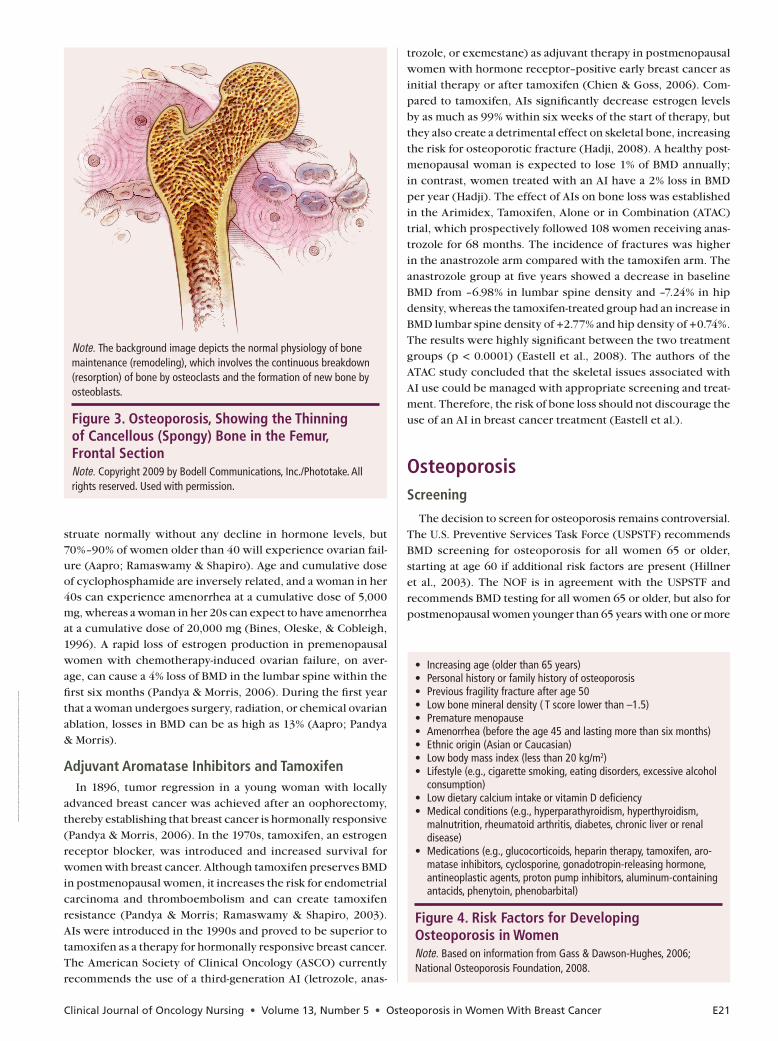
The skeleton is remodeled continually through simultaneous resorption of bone and formation of new bone. Osteoclasts remove bone, whereas osteoblasts are responsible for laying down new bone, and a balance between the two maintains normal bone mass and peak bone mineral density (BMD) in the third decade of life (Chien & Goss, 2006; Delaney, 2006). However, the process is altered at menopause, and a rapid loss of bone occurs two to three years before the cessation of men-ses, continuing for five years after menopause as the result of estrogen depletion (Delaney). Although not a clearly understood mechanism, estrogen deficiency influences the dynamics regu-lating osteoclast and osteoblast function. Osteoblasts produce two proteins, receptor activator nuclear factor-kappa B ligand (RANKL) and osteoprotogerin (OPG), involved in regulating bone remolding (Coetzee & Kruger, 2004). The ratios between the two proteins in the bone marrow activate osteoclasts while controlling the rate of bone resorption and bone mass. If recep-tor sites on osteoclast precursor cells bind with RANKL, it will
stimulate the production of osteoclasts, but if the receptor site receives OPG, it will inhibit osteoclast formation (Coetzee & Kruger). Estrogens stimulate osteoblasts to produce OPG; how-ever, in an estrogen depletion state, a down-regulation of OPG expression occurs, causing increased osteoclastic bone resorp-tion, which leads to decreased bone mass (Coetzee & Kruger; Delaney; Ramaswamy & Shapiro, 2003) (see Figure 3).
Treatment-Induced Ovarian FailurePostmenopausal women are at risk for osteoporosis, and
adjuvant therapies for breast cancer can induce further decline in BMD. Cancer therapies also can cause ovarian failure in younger women, leading to bone loss, putting them at risk for osteoporosis. Hence, in either scenario, a woman with breast cancer is at risk for having an osteoporotic fracture should she develop osteoporosis regardless of age. Alkylating agents, par-ticularly cyclophosphamide, can cause physiologic changes in the ovaries such as decreased number or fibrosis of secondary follicles. The total cumulative dose of cyclophosphamide and the age of the woman are the major causes for ovarian failure and can hasten the onset of menopause by as much as 10 years (Aapro, 2004; Ramaswamy & Shapiro, 2003). Women younger than 30 who receive cyclophosphamide may continue to men-
Figure 2. Dual X-Ray Absorptiometry Scan of HipNote. Images courtesy of Santa Clara Valley Health and Hospital System. Used with permission.
Scan information:
Patient information:
Fracture risk high; World Health Organization classification: osteoporosis
Results summary:
Dow
nloa
ded
on 0
2 15
202
2. S
ingl
e-us
er li
cens
e on
ly. C
opyr
ight
202
2 by
the
Onc
olog
y N
ursi
ng S
ocie
ty. F
or p
erm
issi
on to
pos
t onl
ine,
rep
rint,
adap
t, or
reu
se, p
leas
e em
ail p
ubpe
rmis
sion
s@on
s.or
g. O
NS
res
erve
s al
l rig
hts.
Clinical Journal of Oncology Nursing • Volume 13, Number 5 • Osteoporosis in Women With Breast Cancer E21
struate normally without any decline in hormone levels, but 70%–90% of women older than 40 will experience ovarian fail-ure (Aapro; Ramaswamy & Shapiro). Age and cumulative dose of cyclophosphamide are inversely related, and a woman in her 40s can experience amenorrhea at a cumulative dose of 5,000 mg, whereas a woman in her 20s can expect to have amenorrhea at a cumulative dose of 20,000 mg (Bines, Oleske, & Cobleigh, 1996). A rapid loss of estrogen production in premenopausal women with chemotherapy-induced ovarian failure, on aver-age, can cause a 4% loss of BMD in the lumbar spine within the first six months (Pandya & Morris, 2006). During the first year that a woman undergoes surgery, radiation, or chemical ovarian ablation, losses in BMD can be as high as 13% (Aapro; Pandya & Morris).
Adjuvant Aromatase Inhibitors and Tamoxifen In 1896, tumor regression in a young woman with locally
advanced breast cancer was achieved after an oophorectomy, thereby establishing that breast cancer is hormonally responsive (Pandya & Morris, 2006). In the 1970s, tamoxifen, an estrogen receptor blocker, was introduced and increased survival for women with breast cancer. Although tamoxifen preserves BMD in postmenopausal women, it increases the risk for endometrial carcinoma and thromboembolism and can create tamoxifen resistance (Pandya & Morris; Ramaswamy & Shapiro, 2003). AIs were introduced in the 1990s and proved to be superior to tamoxifen as a therapy for hormonally responsive breast cancer. The American Society of Clinical Oncology (ASCO) currently recommends the use of a third-generation AI (letrozole, anas-
trozole, or exemestane) as adjuvant therapy in postmenopausal women with hormone receptor–positive early breast cancer as initial therapy or after tamoxifen (Chien & Goss, 2006). Com-pared to tamoxifen, AIs significantly decrease estrogen levels by as much as 99% within six weeks of the start of therapy, but they also create a detrimental effect on skeletal bone, increasing the risk for osteoporotic fracture (Hadji, 2008). A healthy post-menopausal woman is expected to lose 1% of BMD annually; in contrast, women treated with an AI have a 2% loss in BMD per year (Hadji). The effect of AIs on bone loss was established in the Arimidex, Tamoxifen, Alone or in Combination (ATAC) trial, which prospectively followed 108 women receiving anas-trozole for 68 months. The incidence of fractures was higher in the anastrozole arm compared with the tamoxifen arm. The anastrozole group at five years showed a decrease in baseline BMD from –6.98% in lumbar spine density and –7.24% in hip density, whereas the tamoxifen-treated group had an increase in BMD lumbar spine density of +2.77% and hip density of +0.74%. The results were highly significant between the two treatment groups (p < 0.0001) (Eastell et al., 2008). The authors of the ATAC study concluded that the skeletal issues associated with AI use could be managed with appropriate screening and treat-ment. Therefore, the risk of bone loss should not discourage the use of an AI in breast cancer treatment (Eastell et al.).
OsteoporosisScreening
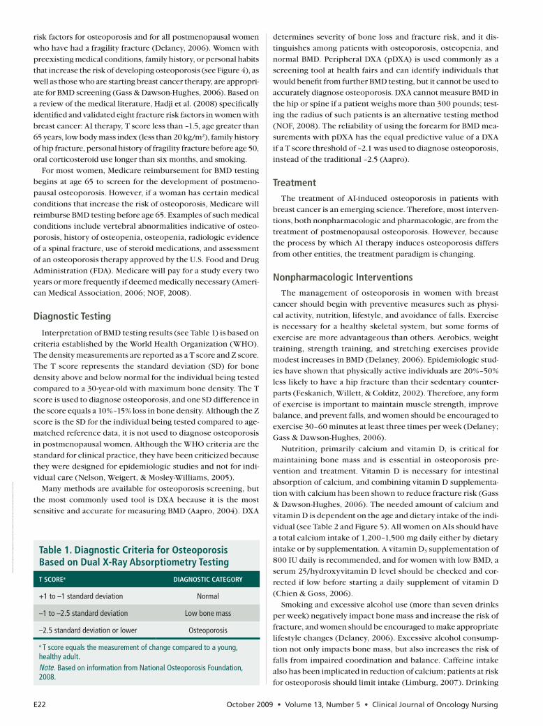
The decision to screen for osteoporosis remains controversial. The U.S. Preventive Services Task Force (USPSTF) recommends BMD screening for osteoporosis for all women 65 or older, starting at age 60 if additional risk factors are present (Hillner et al., 2003). The NOF is in agreement with the USPSTF and recommends BMD testing for all women 65 or older, but also for postmenopausal women younger than 65 years with one or more
• Increasingage(olderthan65years)• Personalhistoryorfamilyhistoryofosteoporosis• Previousfragilityfractureafterage50• Lowbonemineraldensity(Tscorelowerthan–1.5)• Prematuremenopause• Amenorrhea(beforetheage45andlastingmorethansixmonths)• Ethnicorigin(AsianorCaucasian)• Lowbodymassindex(lessthan20kg/m2)• Lifestyle(e.g.,cigarettesmoking,eatingdisorders,excessivealcohol
consumption)• LowdietarycalciumintakeorvitaminDdeficiency• Medicalconditions(e.g.,hyperparathyroidism,hyperthyroidism,
malnutrition, rheumatoid arthritis, diabetes, chronic liver or renal disease)
• Medications(e.g.,glucocorticoids,heparintherapy,tamoxifen,aro-matase inhibitors, cyclosporine, gonadotropin-releasing hormone, antineoplastic agents, proton pump inhibitors, aluminum-containing antacids, phenytoin, phenobarbital)
Figure 4. Risk Factors for Developing Osteoporosis in WomenNote. BasedoninformationfromGass&Dawson-Hughes,2006; National Osteoporosis Foundation, 2008.
Figure 3. Osteoporosis, Showing the Thinning of Cancellous (Spongy) Bone in the Femur, Frontal SectionNote. Copyright2009byBodellCommunications,Inc./Phototake.Allrights reserved. Used with permission.
Note. The background image depicts the normal physiology of bone maintenance (remodeling), which involves the continuous breakdown (resorption) of bone by osteoclasts and the formation of new bone by osteoblasts.
Dow
nloa
ded
on 0
2 15
202
2. S
ingl
e-us
er li
cens
e on
ly. C
opyr
ight
202
2 by
the
Onc
olog
y N
ursi
ng S
ocie
ty. F
or p
erm
issi
on to
pos
t onl
ine,
rep
rint,
adap
t, or
reu
se, p
leas
e em
ail p
ubpe
rmis
sion
s@on
s.or
g. O
NS
res
erve
s al
l rig
hts.
E22 October 2009 • Volume 13, Number 5 • Clinical Journal of Oncology Nursing
risk factors for osteoporosis and for all postmenopausal women who have had a fragility fracture (Delaney, 2006). Women with preexisting medical conditions, family history, or personal habits that increase the risk of developing osteoporosis (see Figure 4), as well as those who are starting breast cancer therapy, are appropri-ate for BMD screening (Gass & Dawson-Hughes, 2006). Based on a review of the medical literature, Hadji et al. (2008) specifically identified and validated eight fracture risk factors in women with breast cancer: AI therapy, T score less than –1.5, age greater than 65 years, low body mass index (less than 20 kg/m2), family history of hip fracture, personal history of fragility fracture before age 50, oral corticosteroid use longer than six months, and smoking.
For most women, Medicare reimbursement for BMD testing begins at age 65 to screen for the development of postmeno-pausal osteoporosis. However, if a woman has certain medical conditions that increase the risk of osteoporosis, Medicare will reimburse BMD testing before age 65. Examples of such medical conditions include vertebral abnormalities indicative of osteo-porosis, history of osteopenia, osteopenia, radiologic evidence of a spinal fracture, use of steroid medications, and assessment of an osteoporosis therapy approved by the U.S. Food and Drug Administration (FDA). Medicare will pay for a study every two years or more frequently if deemed medically necessary (Ameri-can Medical Association, 2006; NOF, 2008).
Diagnostic Testing
Interpretation of BMD testing results (see Table 1) is based on criteria established by the World Health Organization (WHO). The density measurements are reported as a T score and Z score. The T score represents the standard deviation (SD) for bone density above and below normal for the individual being tested compared to a 30-year-old with maximum bone density. The T score is used to diagnose osteoporosis, and one SD difference in the score equals a 10%–15% loss in bone density. Although the Z score is the SD for the individual being tested compared to age-matched reference data, it is not used to diagnose osteoporosis in postmenopausal women. Although the WHO criteria are the standard for clinical practice, they have been criticized because they were designed for epidemiologic studies and not for indi-vidual care (Nelson, Weigert, & Mosley-Williams, 2005).
Many methods are available for osteoporosis screening, but the most commonly used tool is DXA because it is the most sensitive and accurate for measuring BMD (Aapro, 2004). DXA
determines severity of bone loss and fracture risk, and it dis-tinguishes among patients with osteoporosis, osteopenia, and normal BMD. Peripheral DXA (pDXA) is used commonly as a screening tool at health fairs and can identify individuals that would benefit from further BMD testing, but it cannot be used to accurately diagnose osteoporosis. DXA cannot measure BMD in the hip or spine if a patient weighs more than 300 pounds; test-ing the radius of such patients is an alternative testing method (NOF, 2008). The reliability of using the forearm for BMD mea-surements with pDXA has the equal predictive value of a DXA if a T score threshold of –2.1 was used to diagnose osteoporosis, instead of the traditional –2.5 (Aapro).
TreatmentThe treatment of AI-induced osteoporosis in patients with
breast cancer is an emerging science. Therefore, most interven-tions, both nonpharmacologic and pharmacologic, are from the treatment of postmenopausal osteoporosis. However, because the process by which AI therapy induces osteoporosis differs from other entities, the treatment paradigm is changing.
Nonpharmacologic InterventionsThe management of osteoporosis in women with breast
cancer should begin with preventive measures such as physi-cal activity, nutrition, lifestyle, and avoidance of falls. Exercise is necessary for a healthy skeletal system, but some forms of exercise are more advantageous than others. Aerobics, weight training, strength training, and stretching exercises provide modest increases in BMD (Delaney, 2006). Epidemiologic stud-ies have shown that physically active individuals are 20%–50% less likely to have a hip fracture than their sedentary counter-parts (Feskanich, Willett, & Colditz, 2002). Therefore, any form of exercise is important to maintain muscle strength, improve balance, and prevent falls, and women should be encouraged to exercise 30–60 minutes at least three times per week (Delaney; Gass & Dawson-Hughes, 2006).
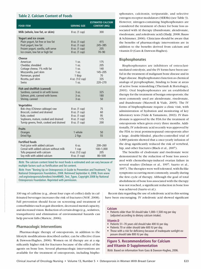
Nutrition, primarily calcium and vitamin D, is critical for maintaining bone mass and is essential in osteoporosis pre-vention and treatment. Vitamin D is necessary for intestinal absorption of calcium, and combining vitamin D supplementa-tion with calcium has been shown to reduce fracture risk (Gass & Dawson-Hughes, 2006). The needed amount of calcium and vitamin D is dependent on the age and dietary intake of the indi-vidual (see Table 2 and Figure 5). All women on AIs should have a total calcium intake of 1,200–1,500 mg daily either by dietary intake or by supplementation. A vitamin D3 supplementation of 800 IU daily is recommended, and for women with low BMD, a serum 25/hydroxyvitamin D level should be checked and cor-rected if low before starting a daily supplement of vitamin D (Chien & Goss, 2006).
Smoking and excessive alcohol use (more than seven drinks per week) negatively impact bone mass and increase the risk of fracture, and women should be encouraged to make appropriate lifestyle changes (Delaney, 2006). Excessive alcohol consump-tion not only impacts bone mass, but also increases the risk of falls from impaired coordination and balance. Caffeine intake also has been implicated in reduction of calcium; patients at risk for osteoporosis should limit intake (Limburg, 2007). Drinking
Table 1. Diagnostic Criteria for Osteoporosis Based on Dual X-Ray Absorptiometry Testing
T SCOREa DIAgNOSTIC CATEgORy
+1to–1standarddeviation Normal
–1to–2.5standarddeviation Lowbonemass
–2.5standarddeviationorlower Osteoporosis
a T score equals the measurement of change compared to a young, healthy adult.Note. Based on information from National Osteoporosis Foundation, 2008.
Dow
nloa
ded
on 0
2 15
202
2. S
ingl
e-us
er li
cens
e on
ly. C
opyr
ight
202
2 by
the
Onc
olog
y N
ursi
ng S
ocie
ty. F
or p
erm
issi
on to
pos
t onl
ine,
rep
rint,
adap
t, or
reu
se, p
leas
e em
ail p
ubpe
rmis
sion
s@on
s.or
g. O
NS
res
erve
s al
l rig
hts.
Clinical Journal of Oncology Nursing • Volume 13, Number 5 • Osteoporosis in Women With Breast Cancer E23
330 mg of caffeine (e.g., about four cups of coffee) daily in caf-feinated beverages increases the risk of fractures (NOF, 2008). Fall prevention should focus on screening and treatment of comorbidities such as gait disorders, decreased mental capacity, and decreased vision. Reduction of certain drugs (e.g., sedatives, tranquilizers) and elimination of environmental hazards can help prevent falls (Maricic, 2006).
Pharmacologic Interventions Pharmacologic therapy of osteoporosis, in addition to the
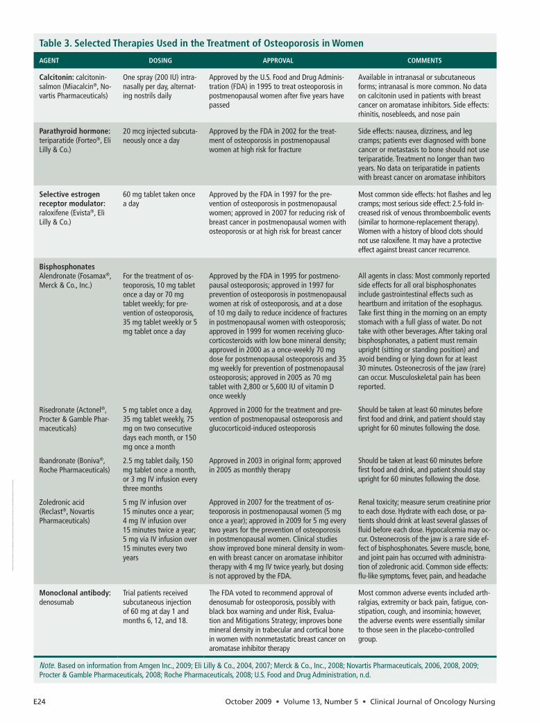
lifestyle modifications described earlier, can be effective (Gass & Dawson-Hughes, 2006). Women on AI therapy are at a sig-nificantly higher risk for fractures because of the effect of the agents on bone loss. Several pharmacologic interventions are available for the treatment of osteoporosis, including bispho-
sphonates, calcitonin, teriparatide, and selective estrogen receptor modulators (SERMs) (see Table 3). However, nitrogen-containing bisphosphonates are considered the treatment of choice for bone loss as-sociated with AI therapy (ibandronate, alendronate, risedronate, and zoledronic acid) (Hadji, 2008; Bauss & Schimmer, 2006). Clinicians should be aware that the benefits of pharmacologic interventions are in addition to the benefits derived from calcium and vitamin D (Gass & Dawson-Hughes).
Bisphosphonates
Bisphosphonates are inhibitors of osteoclast- mediated osteolysis, and the IV forms have been use-ful in the treatment of malignant bone disease and in Paget disease. Bisphosphonates function as chemical analogs of pyrophosphate, binding to bone at areas of active bone remodeling (Theriault & Hortobagyi, 2001). Oral bisphosphonates are an established therapy for the treatment of benign osteoporosis; the most commonly used are alendronate, risedronate, and ibandronate (Maxwell & Viale, 2005). The IV forms of bisphosphonate require a clinic visit, with administration of hydration and monitoring of key laboratory tests (Viale & Yamamoto, 2003). IV iban-dronate is approved by the FDA for the treatment of osteoporosis when given every three months. Addi-tionally, IV zoledronic acid recently was approved by the FDA to treat postmenopausal osteoporosis after a large, double-blinded, placebo-controlled trial of 3,889 patients showed that a once-yearly infusion of the drug significantly reduced the risk of vertebral, hip, and other fractures (Black et al., 2007).
The benefits of clodronate and risedronate were demonstrated by the reduction of bone loss associ-ated with chemotherapy-induced ovarian failure in several studies (Delmas et al., 1997; Saarto et al., 1997). The therapies were well tolerated, with flu-like symptoms occurring most commonly, usually during the first cycle of therapy. Although the goal of total abolishment of bone loss associated with the therapy was not reached, a significant reduction in bone loss was achieved (Saarto et al.).
Recent data regarding the use of zoledronic acid in this setting have been encouraging. IV zoledronic acid showed significant
Table 2. Calcium Content of Foods
FOOD ITEm
SERvINg SIzEESTImATED CALCIum
CONTENT (mg)
milk (whole, low fat, or skim) 8 oz. (1 cup) 300
yogurt and ice cream Plainyogurt,fatfreeorlowfat Fruit yogurt, low fat Frozen yogurt, vanilla, soft serve Ice cream, low fat or high fat
8 oz. (1 cup)8 oz. (1 cup)8 oz. (1 cup)8 oz. (1 cup)
415245–385
20570–90
Cheese American Cheddar, shredded Cottage cheese, 1% milk fat Mozzarella, part skim Parmesan,grated Ricotta, part skim Swiss
1 oz.1 oz.1 oz.1 oz.
1 tbsp4oz.(1/2cup)
1 oz.
175205140
145–20570335
220–270
Fish and shellfish (canned) Sardines, canned in oil with bones Salmon, pink, canned with bones Shrimp, canned
3 oz.3 oz.3 oz.
32518050
vegetables Bok choy (Chinese cabbage) raw Broccoli, cooked and drained Kale, cooked Soybeans, mature, cooked and drained Turnip greens, fresh, cooked and drained
8 oz. (1 cup)8 oz. (1 cup)8 oz. (1 cup)8 oz. (1 cup)8 oz. (1 cup)
756095175200
Fruits Oranges Dried figs
1 whole2 figs
5055
Fortified foods Fruit juice with added calcium Cereal with added calcium without milk Tofu prepared with calcium Soy milk with added calcium
6oz.1 cup
4oz.(1/2cup)8 oz. (1 cup)
200–260100–1,000
20580–500
Note. The calcium content listed for most foods is estimated and can vary because of multiple factors such as fortification and fat content.Note. From“BoningUponOsteoporosis:AGuidetoPreventionandTreatment,”byNationalOsteoporosisFoundation,2008.RetrievedSeptember4,2008,fromwww .nof.org/osteoporosis/bmdtest.htm#BMD_Test_Types. Copyright 2008 by National Osteoporosis Foundation. Reprinted with permission.
Figure 5. Recommendations for Calcium and vitamin D SupplementationNote. BasedoninformationfromGass&Dawson-Hughes,2006.
Calcium• Patientsolderthan50shouldtake1,000–1,500mgperday (adjusted according to dietary calcium intake).
vitamin D• Patients51–70yearsoldshouldtake400IUperday.• Patients70oroldershouldtake600IUperday.• Thosewithariskfordeficiencybecauseofinadequatesunlightex-
posure should take 800 IU per day.
Dow
nloa
ded
on 0
2 15
202
2. S
ingl
e-us
er li
cens
e on
ly. C
opyr
ight
202
2 by
the
Onc
olog
y N
ursi
ng S
ocie
ty. F
or p
erm
issi
on to
pos
t onl
ine,
rep
rint,
adap
t, or
reu
se, p
leas
e em
ail p
ubpe
rmis
sion
s@on
s.or
g. O
NS
res
erve
s al
l rig
hts.
E24 October 2009 • Volume 13, Number 5 • Clinical Journal of Oncology Nursing
Table 3. Selected Therapies used in the Treatment of Osteoporosis in Women
AgENT DOSINg APPROvAL COmmENTS
Calcitonin: calcitonin-salmon (Miacalcin®, No-vartisPharmaceuticals)
One spray (200 IU) intra-nasally per day, alternat-ing nostrils daily
Approved by the U.S. Food and Drug Adminis-tration(FDA)in1995totreatosteoporosisinpostmenopausal women after five years have passed
Available in intranasal or subcutaneous forms; intranasal is more common. No data on calcitonin used in patients with breast cancer on aromatase inhibitors. Side effects: rhinitis, nosebleeds, and nose pain
Parathyroid hormone: teriparatide (Forteo®, Eli Lilly&Co.)
20 mcg injected subcuta-neously once a day
Approved by the FDA in 2002 for the treat-ment of osteoporosis in postmenopausal women at high risk for fracture
Side effects: nausea, dizziness, and leg cramps; patients ever diagnosed with bone cancer or metastasis to bone should not use teriparatide. Treatment no longer than two years. No data on teriparatide in patients with breast cancer on aromatase inhibitors
Selective estrogen receptor modulator: raloxifene (Evista®, Eli Lilly&Co.)
60mgtablettakenoncea day
Approved by the FDA in 1997 for the pre-vention of osteoporosis in postmenopausal women; approved in 2007 for reducing risk of breast cancer in postmenopausal women with osteoporosis or at high risk for breast cancer
Most common side effects: hot flashes and leg cramps;mostserioussideeffect:2.5-foldin-creased risk of venous thromboembolic events (similar to hormone-replacement therapy). Women with a history of blood clots should not use raloxifene. It may have a protective effect against breast cancer recurrence.
Bisphosphonates Alendronate (Fosamax®, Merck & Co., Inc.)
Risedronate (Actonel®, Procter&GamblePhar-maceuticals)
Ibandronate (Boniva®, RochePharmaceuticals)
Zoledronic acid (Reclast®, Novartis Pharmaceuticals)
For the treatment of os-teoporosis, 10 mg tablet once a day or 70 mg tablet weekly; for pre-vention of osteoporosis, 35mgtabletweeklyor5mg tablet once a day
5mgtabletonceaday,35mgtabletweekly,75mg on two consecutive dayseachmonth,or150mg once a month
2.5mgtabletdaily,150mg tablet once a month, or 3 mg IV infusion every three months
5mgIVinfusionover 15minutesonceayear;4mgIVinfusionover 15minutestwiceayear;5mgviaIVinfusionover15minuteseverytwoyears
ApprovedbytheFDAin1995forpostmeno-pausal osteoporosis; approved in 1997 for prevention of osteoporosis in postmenopausal women at risk of osteoporosis, and at a dose of 10 mg daily to reduce incidence of fractures in postmenopausal women with osteoporosis; approved in 1999 for women receiving gluco-corticosteroids with low bone mineral density; approved in 2000 as a once-weekly 70 mg doseforpostmenopausalosteoporosisand35mg weekly for prevention of postmenopausal osteoporosis;approvedin2005as70mgtabletwith2,800or5,600IUofvitaminDonce weekly
Approved in 2000 for the treatment and pre-vention of postmenopausal osteoporosis and glucocorticoid-induced osteoporosis
Approved in 2003 in original form; approved in2005asmonthlytherapy
Approved in 2007 for the treatment of os-teoporosisinpostmenopausalwomen(5mgonceayear);approvedin2009for5mgeverytwo years for the prevention of osteoporosis in postmenopausal women. Clinical studies show improved bone mineral density in wom-en with breast cancer on aromatase inhibitor therapywith4mgIVtwiceyearly,butdosingis not approved by the FDA.
All agents in class: Most commonly reported side effects for all oral bisphosphonates include gastrointestinal effects such as heartburn and irritation of the esophagus. Take first thing in the morning on an empty stomach with a full glass of water. Do not take with other beverages. After taking oral bisphosphonates, a patient must remain upright (sitting or standing position) and avoid bending or lying down for at least 30 minutes. Osteonecrosis of the jaw (rare) can occur. Musculoskeletal pain has been reported.
Shouldbetakenatleast60minutesbeforefirst food and drink, and patient should stay uprightfor60minutesfollowingthedose.
Shouldbetakenatleast60minutesbeforefirst food and drink, and patient should stay uprightfor60minutesfollowingthedose.
Renal toxicity; measure serum creatinine prior to each dose. Hydrate with each dose, or pa-tients should drink at least several glasses of fluid before each dose. Hypocalcemia may oc-cur. Osteonecrosis of the jaw is a rare side ef-fect of bisphosphonates. Severe muscle, bone, and joint pain has occurred with administra-tion of zoledronic acid. Common side effects: flu-like symptoms, fever, pain, and headache
monoclonal antibody: denosumab
Trial patients received subcutaneous injection of60mgatday1andmonths6,12,and18.
The FDA voted to recommend approval of denosumab for osteoporosis, possibly with black box warning and under Risk, Evalua-tion and Mitigations Strategy; improves bone mineral density in trabecular and cortical bone in women with nonmetastatic breast cancer on aromatase inhibitor therapy
Most common adverse events included arth-ralgias, extremity or back pain, fatigue, con-stipation, cough, and insominia; however, the adverse events were essentially similar to those seen in the placebo-controlled group.
Note. BasedoninformationfromAmgenInc.,2009;EliLilly&Co.,2004,2007;Merck&Co.,Inc.,2008;NovartisPharmaceuticals,2006,2008,2009;Procter&GamblePharmaceuticals,2008;RochePharmaceuticals,2008;U.S.FoodandDrugAdministration,n.d.
Dow
nloa
ded
on 0
2 15
202
2. S
ingl
e-us
er li
cens
e on
ly. C
opyr
ight
202
2 by
the
Onc
olog
y N
ursi
ng S
ocie
ty. F
or p
erm
issi
on to
pos
t onl
ine,
rep
rint,
adap
t, or
reu
se, p
leas
e em
ail p
ubpe
rmis
sion
s@on
s.or
g. O
NS
res
erve
s al
l rig
hts.
Clinical Journal of Oncology Nursing • Volume 13, Number 5 • Osteoporosis in Women With Breast Cancer E25
benefits in a study of premenopausal women receiving goserelin plus anastrozole or tamoxifen (Gnant et al., 2002). For the first 172 patients who completed one year of therapy, treatment with zoledronic acid (4 mg every six months over 15 minutes) signifi-cantly preserved BMD in lumbar spine and trochanter (L1-L4 with p < 0.0001 and p < 0.002, respectively). Gnant et al. published results in 2007 of phase III data in which the combination of a bis-phosphonate with an AI effectively prevented cancer treatment–induced bone loss in young, premenopausal women. The study patients were randomly assigned to receive either goserelin plus tamoxifen or goserelin plus anastrozole for three years plus or minus zoledronic acid. In a BMD subprotocol, patients under-went serial BMD measurements at baseline and 6, 12, 24, and 36 months. A total of 401 patients were included in the subprotocol, and the results showed that zoledronic acid effectively inhibited bone loss. No patient experienced a fracture in the study (possi-bly reflecting the younger age of the study participants); no cases of osteonecrosis of the jaw or renal dysfunction were noted.
Currently, the Zoledronic Acid-Letrozole Adjuvant Synergy Trial in North America and a parallel trial in Europe are study-ing zoledronic acid (4 mg every six months) in postmenopausal women receiving letrozole for early-stage hormone receptor–positive breast cancer. The purpose of the studies is to deter-mine the benefit of the therapy when started immediately or delayed (after a patient experiences an asymptomatic fracture or develops severe osteopenia or clinical fracture) (Brufsky et al., 2007, 2008). The interim analysis (including data from 1,667 patients) suggests that zoledronic acid therapy increases BMD of the lumbar spine in postmenopausal women more ef-fectively when given early versus delayed. The results of the studies show that patients with early-stage breast cancer who receive zoledronic acid (4 mg via IV every six months) have
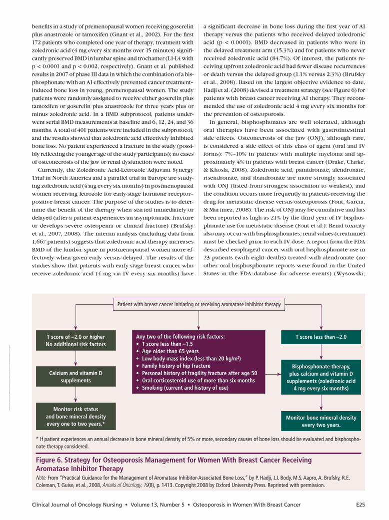
a significant decrease in bone loss during the first year of AI therapy versus the patients who received delayed zoledronic acid (p < 0.0001). BMD decreased in patients who were in the delayed treatment arm (15.3%) and for patients who never received zoledronic acid (84.7%). Of interest, the patients re-ceiving upfront zoledronic acid had fewer disease recurrences or death versus the delayed group (1.1% versus 2.3%) (Brufsky et al., 2008). Based on the largest objective evidence to date, Hadji et al. (2008) devised a treatment strategy (see Figure 6) for patients with breast cancer receiving AI therapy. They recom-mended the use of zoledronic acid 4 mg every six months for the prevention of osteoporosis.
In general, bisphosphonates are well tolerated, although oral therapies have been associated with gastrointestinal side effects. Osteonecrosis of the jaw (ONJ), although rare, is considered a side effect of this class of agent (oral and IV forms): 7%–10% in patients with multiple myeloma and ap-proximately 4% in patients with breast cancer (Drake, Clarke, & Khosla, 2008). Zoledronic acid, pamidronate, alendronate, risendronate, and ibandronate are more strongly associated with ONJ (listed from strongest association to weakest), and the condition occurs more frequently in patients receiving the drug for metastatic disease versus osteoporosis (Font, Garcia, & Martinez, 2008). The risk of ONJ may be cumulative and has been reported as high as 21% by the third year of IV bisphos-phonate use for metastatic disease (Font et al.). Renal toxicity also may occur with bisphosphonates; renal values (creatinine) must be checked prior to each IV dose. A report from the FDA described esophageal cancer with oral bisphosphonate use in 23 patients (with eight deaths) treated with alendronate (no other oral bisphosphonate reports were found in the United States in the FDA database for adverse events) (Wysowski,
*Ifpatientexperiencesanannualdecreaseinbonemineraldensityof5%ormore,secondarycausesofbonelossshouldbeevaluatedandbisphospho-nate therapy considered.
Figure 6. Strategy for Osteoporosis management for Women With Breast Cancer Receiving Aromatase Inhibitor TherapyNote. From“PracticalGuidancefortheManagementofAromataseInhibitor-AssociatedBoneLoss,”byP.Hadji,J.J.Body,M.S.Aapro,A.Brufsky,R.E.Coleman, T. Guise, et al., 2008, Annals of Oncology, 19(8),p.1413.Copyright2008byOxfordUniversityPress.Reprintedwithpermission.
Patientwithbreastcancerinitiatingorreceivingaromataseinhibitortherapy
Any two of the following risk factors:• Tscorelessthan–1.5• Ageolderthan65years• Lowbodymassindex(lessthan20kg/m2)• Familyhistoryofhipfracture• Personalhistoryoffragilityfractureafterage50• Oralcorticosteroiduseofmorethansixmonths• Smoking(currentandhistoryofuse)
Tscoreof–2.0orhigherNo additional risk factors
Tscorelessthan–2.0
Calcium and vitamin D supplements
monitor risk status and bone mineral density every one to two years.*
Bisphosphonate therapy, plus calcium and vitamin D
supplements (zoledronic acid 4 mg every six months)
monitor bone mineral density every two years.
Dow
nloa
ded
on 0
2 15
202
2. S
ingl
e-us
er li
cens
e on
ly. C
opyr
ight
202
2 by
the
Onc
olog
y N
ursi
ng S
ocie
ty. F
or p
erm
issi
on to
pos
t onl
ine,
rep
rint,
adap
t, or
reu
se, p
leas
e em
ail p
ubpe
rmis
sion
s@on
s.or
g. O
NS
res
erve
s al
l rig
hts.
E26 October 2009 • Volume 13, Number 5 • Clinical Journal of Oncology Nursing
2008). The median time to diagnosis of esophageal cancer was 2.1 years; although risk factors were not available for all pa-tients, one patient was determined to have Barrett esophagus (a precursor of esophageal adenocarcinoma). Additional reports of esophageal cancer have surfaced in European and Japanese patients receiving alendronate and other oral bisphosphonate drugs, and the author of the report cautioned that future studies should include oral bisphosphonates and their risk for esopha-geal cancer (Wysowski).
Additional Therapies
Additional therapies have efficacy in preventing fractures in patients with osteoporosis; however, their role in reducing fractures in patients with breast cancer on AI therapy has not been established. A recent systematic review of calcitonin, given intranasally, found that more than one randomized trial or meta-analysis confirmed that it did prevent vertebral fractures when compared with placebo (MacLean et al., 2008). Reduc-tion in the rate of bone turnover is the mechanism of action, resulting in the maintenance of the trabecular architecture of the bone, which preserves bone strength and quality (Mehta, Malootian, & Gilligan, 2003). In addition, calcitonin has a di-rect analgesic effect on bone (Mehta et al.). Teriparatide is a recombinant formulation of the first 34 N-terminal amino acids of parathyroid hormone and is given subcutaneously. It can in-crease bone mass and improve the microstructure of the bone (Gass & Dawson-Hughes, 2006). A study by Neer et al. (2001) showed that treatment of postmenopausal osteoporosis with parathyroid hormone decreased the risk of vertebral and non-vertebral fractures, while increasing the BMD of the vertebral and femoral areas, and it was well tolerated. This agent caused an increase in the incidence of osteosarcoma in male and female rats during clinical trials. It is contraindicated in individuals who are at increased risk for osteosarcoma and should not be used for patients who have received radiation to bone or those with skeletal metastases (Mincey & Tan, 2004). Additionally, if a patient is at risk of developing skeletal metastases, the drug should be used with caution.
The SERM raloxifene was found to maintain BMD and reduce spinal fractures in women at risk for osteoporosis with the same cardiac protective effect (reduced LDL cholesterol) as tamoxifen (Jordan, 2007). Raloxifene also was shown to be as effective as tamoxifen in the reduction of the risk of invasive breast cancer with fewer side effects, although the risk of noninvasive breast cancer was higher but not significantly so (Vogel et al., 2006). The FDA has approved raloxifene as the only SERM for the prevention and treatment of osteoporosis; side effects include risk of deep vein thrombosis and pulmonary embolism (Gass & Dawson-Hughes, 2006). Although estrogen therapy has been shown to increase bone mass and reduce the risk of fracture in low-risk postmenopausal women, it is not recommended for women with breast cancer.
A randomized trial of denosumab in patients receiving adjuvant AIs for nonmetastatic breast cancer was published (Ellis et al., 2008). The trial included 252 patients (of whom 81% completed the 24-month study), and the women received supplemental calcium, vitamin D, and either placebo or subcu-taneous denosumab. Denosumab is a fully human monoclonal
antibody with a novel mechanism of action; it binds significantly to RANKL but does not affect tumor necrosis factor ligands (Ellis et al.). The antibody inhibits osteoclast formation, function, and survival by its effect on RANKL. The women were required to have evidence of low bone mass (osteopenia) but not osteoporo-sis, and the primary end point was the percentage change from baseline at month 12 in the lumbar spine BMD (Ellis et al.). The measurements of BMD in the lumbar spine increased by 5.5% at 12 months and by 7.6% at 24 months, with the increases seen as early as one month after initiation of treatment. The adverse events were similar between study groups, and the researchers concluded that twice-yearly administration of denosumab in-creased BMD significantly over 24 months (Ellis et al.). The FDA advisory committee reviewing denosumab voted to recommend approval of the agent in some patients with prostate cancer and as a therapy for osteopororis; however, indications for bone loss in breast cancer and hormone ablation therapy in men with prostate cancer were not approved. Concerns regarding infec-tions and cancers in patients receiving the agent in clinical trials versus placebo led to the decision (Pollack, 2009).
Nursing InterventionsOncology nurses have a role in osteoporosis prevention and
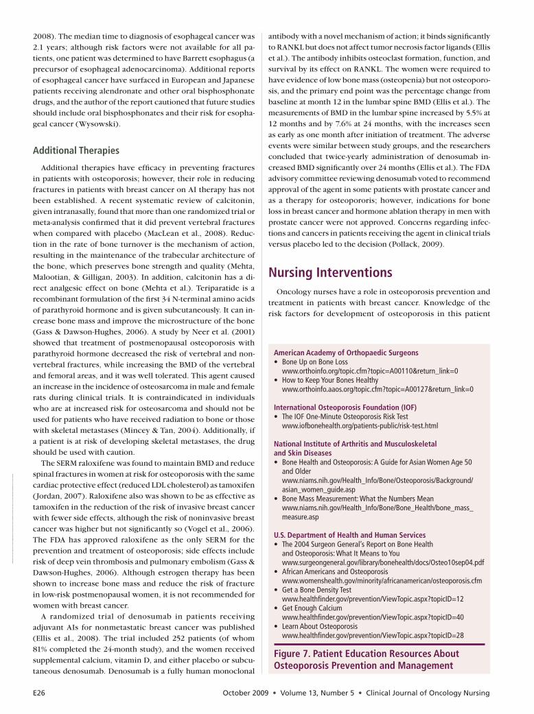
treatment in patients with breast cancer. Knowledge of the risk factors for development of osteoporosis in this patient
Figure 7. Patient Education Resources About Osteoporosis Prevention and management
American Academy of Orthopaedic Surgeons• BoneUponBoneLoss www.orthoinfo.org/topic.cfm?topic=A00110&return_link=0• HowtoKeepYourBonesHealthy www.orthoinfo.aaos.org/topic.cfm?topic=A00127&return_link=0
International Osteoporosis Foundation (IOF)• TheIOFOne-MinuteOsteoporosisRiskTest www.iofbonehealth.org/patients-public/risk-test.html
National Institute of Arthritis and musculoskeletal and Skin Diseases• BoneHealthandOsteoporosis:AGuideforAsianWomenAge50 and Older www.niams.nih.gov/Health_Info/Bone/Osteoporosis/Background/ asian_women_guide.asp• BoneMassMeasurement:WhattheNumbersMean www.niams.nih.gov/Health_Info/Bone/Bone_Health/bone_mass_ measure.asp
u.S. Department of Health and Human Services• The2004SurgeonGeneral’sReportonBoneHealth andOsteoporosis:WhatItMeanstoYou www.surgeongeneral.gov/library/bonehealth/docs/Osteo10sep04.pdf• AfricanAmericansandOsteoporosis www.womenshealth.gov/minority/africanamerican/osteoporosis.cfm• GetaBoneDensityTest www.healthfinder.gov/prevention/ViewTopic.aspx?topicID=12• GetEnoughCalcium www.healthfinder.gov/prevention/ViewTopic.aspx?topicID=40• LearnAboutOsteoporosis www.healthfinder.gov/prevention/ViewTopic.aspx?topicID=28
Dow
nloa
ded
on 0
2 15
202
2. S
ingl
e-us
er li
cens
e on
ly. C
opyr
ight
202
2 by
the
Onc
olog
y N
ursi
ng S
ocie
ty. F
or p
erm
issi
on to
pos
t onl
ine,
rep
rint,
adap
t, or
reu
se, p
leas
e em
ail p
ubpe
rmis
sion
s@on
s.or
g. O
NS
res
erve
s al
l rig
hts.
Clinical Journal of Oncology Nursing • Volume 13, Number 5 • Osteoporosis in Women With Breast Cancer E27
population will ensure that timely DXA screening and educa-tion are performed. Educating patients about physical activity, nutrition, lifestyle, fall prevention, and management of osteo-porosis treatment is important to overall success in preventing osteoporotic fractures (see Figure 7). Oncology nurses should assess all patients with osteoporosis for potential falls, such as a history of falls, fainting, muscle weakness, dizziness or balance problems, impaired vision, and use of sedatives or narcotics. They also should inquire about the safety of patients’ homes for possible environmental dangers such as poor lighting and trip-ping hazards (Delaney, 2006; Gass & Dawson-Hughes, 2006). Nutritional supplementation with calcium and vitamin D is the foundation of osteoporosis treatment, and every oncology nurse should be prepared to address it with their patients. Be-cause bisphosphonates are very poorly absorbed from the gas-trointestinal tract (approximately 1%–2% of the dose), patients must follow specific instructions to ensure proper absorption (Grey & Reid, 2006). Patients should be cautioned regarding the need to take their pills with a full glass of water and to avoid food and beverages for at least 30 minutes after a morning dose to improve absorption. Patients also must stay upright for 30 minutes after administration (ibandronate requires a 60-minute period); these measures are designed to reduce the risk of gas-trointestinal side effects (Gass & Dawson-Hughes).
Adherence to prescribed therapies is a significant issue for patients receiving treatment for chronic conditions (Miaskows-ki, Shockney, & Chlebowski, 2008). Multiple studies have shown nonadherence to oral therapies as high as 30%–60% of the time (Barber, 2002; Haynes, McDonald, & Garg, 2002). Nonadherence can affect the efficacy of standard chemotherapy agents, as well as supportive care treatments such as those for osteoporosis and bone metastasis (Papaioannou et al., 2003; Partridge, Avorn, Wang, & Winer, 2002). In one study, adher-ence to bisphosphonate therapy affected patient outcomes with significantly few fractures noted for the adherent group (Siris et al., 2006). The study cohort included 35,537 women, and an association was noted between compliance with refills of bisphosphonate therapy and a decrease in the reduction of fracture risk. Another study of postmenopausal women who were prescribed weekly versus daily bisphosphonate therapy showed that the weekly group had significantly better adher-ence and persistence than the daily group; however, adher-ence and persistence rates for both groups were suboptimal (Cramer, Amonkar, Hebborn, & Altman, 2005).
Oncology nurses can assist with adherence by questioning patients regarding their medication use at follow-up visits and querying them about side effects and adverse events (Mi-askowski et al., 2008). Reinforcement of the importance of maintaining scheduled visits for IV medication is important as well. Education about expected side effects is critical to help pa-tients maintain therapy and understand possible events related to treatments. Emphasizing reportable side effects also is impor-tant, as patients may stop therapy because of such occurrences. Oncology nurses can triage phone calls and assist patients with side-effect management, while reinforcing the significance of therapies, nutrition, vitamin D and calcium supplementation, and exercise. Nurses are key in partnering with patients for optimal results during therapy and can guide them to appropri-ate support groups or organizations for additional information
(Miaskowski et al.). Additionally, many of the medications used in the treatment of osteoporosis carry significant costs, whether given orally or via IV. Oncology nurses should be educated regarding current guidelines, communicate the need for treat-ment to insurance companies when appropriate, and advocate for patient assistance programs if needed.
Case StudyW.H. was at risk for future bone loss because she was on AI
therapy and because of her history of noncompliance with her weekly oral bisphosphonate. Therefore, she was started on IV therapy with zoledronic acid 4 mg twice a year. She also was started on 1,000 mg of calcium and 800 IU of vitamin D daily. W.H. was advised about exercise and personal habits to help minimize further bone loss. She received education regarding her fracture risk and the mechanism of action for her therapy. W.H. also received telephone numbers for advice calls. Despite W.H.’s age of 56, a yearly DXA was planned, given the severity of her osteoporosis, to monitor her response to treatment.
ConclusionWomen with breast cancer are at risk for osteoporosis and
should be screened, monitored, and treated to prevent fractures. Advancing age and additional risk factors, including breast can-cer treatments, place some women at risk for bone loss and life-changing or life-threatening osteoporotic fractures. Treatment with AIs may require a different approach to therapy for osteo-porosis. Many treatment options are available for osteoporosis, and the future offers promising new drug therapies.
The authors take full responsibility for the content of this ar-ticle. Yamamoto is a stock holder in Amgen Inc. Viale is a speaker and member of the advisory board for Bristol-Myers Squibb, IMER, Meniscus, and Novartis AG, and a speaker for Amgen Inc. and Merck & Co., Inc. The content of this article has been reviewed by independent peer reviewers to ensure that it is balanced, objec-tive, and free from commercial bias.
Author Contact: Deanna Sanchez Yamamoto, RN, MS, CS, ANP, AOCNP®, can be reached at [email protected], with copy to editor at [email protected].
References
Aapro, M.S. (2004). Long-term implications of bone loss in breast
cancer. Breast, 13(Suppl. 1), 29–37.
Altundag, K., & Ibrahim, N.K. (2006). Aromatase inhibitors in
breast cancer: An overview. Oncologist, 11(6), 553–562.
American Medical Association. (2006). An overview of current
Medicare reimbursement for bone density study procedures for
2006. Retrieved September 4, 2008, from http://www.gehealth
care.com/usen/community/reimbursement/docs/BoneDensity
Advisory.pdf
Amgen Inc. (2009). Amgen issues statement on outcomes of advi-
sory committee for reproductive health drugs (ACRHD) meeting.
Retrieved September 3, 2009, from http://www.amgen.com/
media/media_pr_detail.jsp?releaseID=1320684
Dow
nloa
ded
on 0
2 15
202
2. S
ingl
e-us
er li
cens
e on
ly. C
opyr
ight
202
2 by
the
Onc
olog
y N
ursi
ng S
ocie
ty. F
or p
erm
issi
on to
pos
t onl
ine,
rep
rint,
adap
t, or
reu
se, p
leas
e em
ail p
ubpe
rmis
sion
s@on
s.or
g. O
NS
res
erve
s al
l rig
hts.
E28 October 2009 • Volume 13, Number 5 • Clinical Journal of Oncology Nursing
Barber, N. (2002). Should we consider non-compliance a medical
error? Quality and Safety in Health Care, 11(1), 81–84.
Bauss, F., & Schimmer, R.C. (2006). Ibandronate: The first once-
monthly oral bisphosphonate for treatment of postmenopausal
osteoporosis. Therapeutics and Clinical Risk Management,
2(1), 3–18.
Bines, J., Oleske, D.M., & Cobleigh, M.A. (1996). Ovarian function in
premenopausal women treated with adjuvant chemotherapy for
breast cancer. Journal of Clinical Oncology, 14(5), 1718–1729.
Black, D.M., Delmas, P.D., Eastell, R., Reid, I.R., Boonen, S., Cauley,
J.A., et al. (2007). Once-yearly zoledronic acid for treatment of
postmenopausal osteoporosis. New England Journal of Medi-
cine, 356(18), 1809–1822.
Brufsky, A., Bundred, N., Coleman, R., Lambert-Falls, R., Mena, R.,
Hadji, P., et al. (2008). Integrated analysis of zoledronic acid for
prevention of aromatase inhibitor-associated bone loss in post-
menopausal women with early breast cancer receiving adjuvant
letrozole. Oncologist, 13(5), 503–514.
Brufsky, A., Harker, W.G., Beck, J.T., Carroll, R., Tan-Chiu, E., Seidler,
C., et al. (2007). Zoledronic acid inhibits adjuvant letrozole-
induced bone loss in postmenopausal women with early breast
cancer. Journal of Clinical Oncology, 25(7), 829–836.
Chien, A.J., & Goss, P.E. (2006). Aromatase inhibitors and bone
health in women with breast cancer. Journal of Clinical Oncol-
ogy, 24(33), 5305–5312.
Coetzee, M., & Kruger, M.C. (2004). Osteoprotegerin-receptor acti-
vator of nuclear factor-kB ligand ratio: A new approach to osteo-
porosis treatment? Southern Medical Journal, 97(5), 506–511.
Cramer, J.A., Amonkar, M.M., Hebborn, A., & Altman, R. (2005).
Compliance and persistence with bisphosphonate dosing regi-
mens among women with postmenopausal osteoporosis. Current
Medical Research and Opinion, 21(9), 1453–1460.
Delaney, M.F. (2006). Strategies for the prevention and treatment of
osteoporosis during early postmenopause. American Journal of
Obstetrics and Gynecology, 194(2, Suppl.), S12–S23.
Delmas, P.D., Balena, R., Confravreux, E., Hardouin, C., Hardy, P., &
Bremond, A. (1997). Bisphosphonate risedronate prevents bone
loss in women with artificial menopause due to chemotherapy of
breast cancer: A double-blind, placebo-controlled study. Journal
of Clinical Oncology, 15(3), 955–962.
Drake, M.T., Clarke, B.L., & Khosla, S. (2008). Bisphosphonates:
Mechanism of action and role in clinical practice. Mayo Clinic
Proceedings, 83(9), 1032–1045.
Eastell, R., Adams, J.E., Coleman, R.E., Howell, A., Hannon, R.A.,
Cuzick, J., et al. (2008). Effect of anastrozole on bone mineral
density: 5-year results from the Anastrozole, Tamoxifen, Alone
or in Combination Trial 18233230. Journal of Clinical Oncology,
26(7), 1051–1057.
Eli Lilly & Company. (2004). Forteo® (teriparatide rDNA origin)
[Package insert]. Indianapolis, IN: Author.
Eli Lilly & Company. (2007). Evista® (raloxifene) [Package insert].
Indianapolis, IN: Author.
Ellis, G.K., Bone, H.G., Chlebowski, R., Paul, D., Spadafora, S.,
Smith, J., et al. (2008). Randomized trial of denosumab in pa-
tients receiving adjuvant aromatase inhibitors for nonmetastatic
breast cancer. Journal of Clinical Oncology, 26(30). Retrieved
September 6, 2008, from http://jco.ascopubs.org/cgi/reprint/
JCO.2008.16.3832v1
Feskanich, D., Willett, W., & Colditz, G. (2002). Walking and leisure-
time activity and risk of hip fracture in postmenopausal women.
JAMA, 288(18), 2300–2306.
Font, R.G., Garcia, M.L.M., & Martinez, J.M.O. (2008). Osteo-
chemonecrosis of the jaws due to bisphosphonate treatments. Me-
dicina Oral, Patologia Oral y Cirugia Bucal, 13(5), E318–E324.
Retrieved August 31, 2009, from http://www.medicinaoral.com/
medoralfree01/v13i5/medoralv13i5p318.pdf
Gass, M., & Dawson-Hughes, B. (2006). Preventing osteoporosis-
related fractures: An overview. American Journal of Medicine,
119(4, Suppl. 1), S3–S11.
Gnant, M., Hausmaninger, H., Samonigg, H., Mlineritsch, B.,
Taucher, S., Luschin-Ebengreuth, G., et al. (2002, December
11–14). Changes in bone mineral density caused by anastrozole
or tamoxifen in combination with goserelin (± zoledronate) as
adjuvant treatment for hormone receptor-positive premenopausal
breast cancer: Results of a randomized multicenter trial. Abstract
presented at the 25th Annual San Antonio Breast Cancer Sympo-
sium, San Antonio, TX.
Gnant, M.F.X., Mlineritsch, B., Luschin-Ebengreuth, G., Grampp,
S., Kaessmann, H., Schmid, M., et al. (2007). Zoledronic acid
prevents cancer treatment-induced bone loss in premenopausal
women receiving adjuvant endocrine therapy for hormone-
responsive breast cancer: A report from the Austrian Breast and
Colorectal Cancer Study Group. Journal of Clinical Oncology,
25(22), 820–828.
Grey, A., & Reid, I.R. (2006). Differences between the bisphospho-
nates for the prevention and treatment of osteoporosis. Thera-
peutics and Clinical Risk Management, 2(1), 77–86.
Hadji, P. (2008). Aromatase inhibitor-associated bone loss in breast
cancer patients is distinct from postmenopausal osteoporosis.
Critical Reviews in Oncology/Hematology, 69(1), 73–82.
Hadji, P., Body, J.J., Aapro, M.S., Brufsky, A., Coleman, R.E., Guise,
T., et al. (2008). Practical guidance for the management of
aromatase inhibitor-associated bone loss. Annals of Oncology,
19(8), 1407–1416.
Haynes, R.B., McDonald, H.P., & Garg, A.X. (2002). Helping pa-
tients follow prescribed treatment: Clinical applications. JAMA,
288(22), 2880–2883.
Hillner, B.E., Ingle, J.N., Chlebowski, R.T., Gralow, J., Yee, G.C.,
Janjan, N.A., et al. (2003). American Society of Clinical Oncology
2003 update on the role of bisphosphonates and bone health is-
sues in women with breast cancer. Journal of Clinical Oncology,
21(21), 4024–4057.
Jordan, V.C. (2007). SERMs: Meeting the promise of multifunctional
medicines. Journal of the National Cancer Institute, 99(5),
350–356.
Limburg, C.E. (2007). Screening, prevention, detection, and treat-
ment of cancer therapy-induced bone loss in patients with breast
cancer. Oncology Nursing Forum, 34(1), 55–63.
MacLean, C., Newberry, S., Maglione, M., McMahon, M., Ranganath,
V., Suttorp, M., et al. (2008). Systematic review: Comparative ef-
fectiveness of treatments to prevent fractures in men and women
with low bone density or osteoporosis. Annals of Internal Medi-
cine, 148(3), 197–213.
Maricic, M.J. (2006). Osteoporosis. In S. Bartlett (Ed.), Clinical care
in the rheumatic diseases (3rd ed., pp. 135–140). Atlanta, GA:
American College of Rheumatology.
Maxwell, C., & Viale, P.H. (2005). Cancer treatment-induced bone
loss in patients with breast or prostate cancer. Oncology Nursing
Forum, 32(3), 589–603.
Mehta, N.M., Malootian, A., & Gilligan, J.P. (2003). Calcitonin for
osteoporosis and bone pain. Current Pharmaceutical Design,
9(32), 2659–2676.
Dow
nloa
ded
on 0
2 15
202
2. S
ingl
e-us
er li
cens
e on
ly. C
opyr
ight
202
2 by
the
Onc
olog
y N
ursi
ng S
ocie
ty. F
or p
erm
issi
on to
pos
t onl
ine,
rep
rint,
adap
t, or
reu
se, p
leas
e em
ail p
ubpe
rmis
sion
s@on
s.or
g. O
NS
res
erve
s al
l rig
hts.
Clinical Journal of Oncology Nursing • Volume 13, Number 5 • Osteoporosis in Women With Breast Cancer E29
This article has been identified as appropriate for a journal club. When you read this article, think about how you would change yourcurrentpracticeregardingosteoporosisinyourpatients.SeetheEvidence-BasedPracticecolumnintheFebruary2009Clinical Journal of Oncology Nursing(Vol.13,No.1,pp.109–112)onhowtoimplementandparticipateinjournalclubs.Photocopyingof this article for discussion purposes is permitted.
1. What is the clinical practice question the authors are trying to answer?2. Is the purpose of the article described clearly?3. Is the literature review comprehensive, and are major concepts identified and defined?4. Aretheclinicalrecommendationssupportedbyevidence?Whatarethey?5. Howdotheclinicalrecommendationscomparetoyourcurrentpractice?6. Whatpracticechangerecommendationswillyoumakebasedontheevidencepresentedinthisarticle?7. What patient education materials are available on this topic?
Journal Club Discussion Questions
Merck & Co., Inc. (2008). Fosamax® (alendronate sodium) [Pack-
age insert]. Whitehouse Station, NJ: Author.
Miaskowski, C., Shockney, L., & Chlebowski, R.T. (2008). Adherence
to oral endocrine therapy for breast cancer: A nursing perspec-
tive. Clinical Journal of Oncology Nursing, 12(2), 213–221.
Mincey, B.A., & Tan, W.W. (2004). The management of bone loss
in patients with breast or prostate cancer. Supportive Cancer
Therapy, 1(3), 150–156.
National Osteoporosis Foundation. (2008). Types of bone density
tests. Retrieved September 4, 2008, from http://www.nof.org/
osteoporosis/bmdtest.htm#BMD_Test_Types
Neer, R.M., Arnaud, C.D., Zanchetta, J.R., Prince, R., Gaich, G.A.,
Reginster, J.Y., et al. (2001). Effect of parathyroid hormone (1-
34) on fractures and bone mineral density in postmenopausal
women with osteoporosis. New England Journal of Medicine,
344(19), 1434–1441.
Nelson, D.A., Weigert, J.M., & Mosley-Williams, A.D. (2005). Mea-
surement of bone mineral density: DXA and QCT. In M. Maricic
& O.S. Gluck (Eds.), Bone disease in rheumatology (pp. 35–44).
Philadelphia: Lippincott Williams and Wilkins.
Novartis Pharmaceuticals. (2006). Miacalcin® (calcitonin-salmon)
[Package insert]. East Hanover, NJ: Author.
Novartis Pharmaceuticals. (2008). Reclast® (zoledronic acid)
[Package insert]. East Hanover, NJ: Author.
Novartis Pharmaceuticals. (2009). FDA approves Reclast® to prevent
osteoporosis in postmenopausal women with convenient less
frequent dosing. Retrieved September 9, 2009, from http://www
.nibr.novartis.com/newsroom/media-releases/en/2009/1318842
.shtml
Pandya, N., & Morris, G.J. (2006). Toxicity of aromatase inhibitors.
Seminars in Oncology, 33(6), 688–695.
Papaioannou, A., Ioannidis, G., Adachi, J.D., Sebaldt, R.J., Ferko,
N., Puglia, M., et al. (2003). Adherence to bisphosphonates
and hormone replacement therapy in a tertiary care setting of
patients in the CANDOO database. Osteoporosis International,
14(10), 808–813.
Partridge, A.H., Avorn, J., Wang, P.S., & Winer, E.P. (2002). Adher-
ence to therapy with oral antineoplastic agents. Journal of the
National Cancer Institute, 94(9), 652–661.
Pollack, A. (2009, August 13). A partial approval for Amgen’s osteo-
porosis drug. The New York Times, B2.
Procter & Gamble Pharmaceuticals. (2008). Actonel® (risedronate
sodium) [Package insert]. Cincinnati, OH: Author.
Ramaswamy, B., & Shapiro, C.L. (2003). Osteopenia and osteo-
porosis in women with breast cancer. Seminars in Oncology,
30(6), 763–775.
Roche Pharmaceuticals. (2008). Boniva® (ibandronate) [Package
insert]. Nutley, NJ: Author.
Saarto, T., Blomqvist, C., Valimaki, M., Makela, P., Sarna, S., &
Elomaa, I. (1997). Chemical castration induced by adjuvant cy-
clophosphamide, methotrexate, and fluorouracil chemotherapy
causes rapid bone loss that is reduced by clodronate: A random-
ized study in premenopausal breast cancer patients. Journal of
Clinical Oncology, 15(4), 1341–1347.
Siris, E.S., Harris, S.T., Rosen, C.J., Barr, C.E., Arvesen, J.N., Abbott,
T.A., et al. (2006). Adherence to bisphosphonate therapy and
fracture rates in osteoporotic women: Relationship to vertebral
and nonvertebral fractures from 2 US claims databases. Mayo
Clinic Proceedings, 81(8), 1013–1022.
Theriault, R.L., & Hortobagyi, G.N. (2001). The evolving role of
bisphosphonates. Seminars in Oncology, 28(3), 284–290.
U.S. Food and Drug Administration. (n.d.). FDA approved drug prod-
ucts. Retrieved September 9, 2009, from http://www.accessdata
.fda.gov/scripts/cder/drugsatfda/index.cfm?fuseaction=Search
.Label_ApprovalHistory#apphist
Viale, P.H., & Yamamoto, D.S. (2003). Bisphosphonates: Expanded
roles in the treatment of patients with cancer. Clinical Journal
of Oncology Nursing, 7(4), 393–401.
Vogel, V.G., Costantino, J.P., Wickerham, D.L., Cronin, W.M., Cec-
chini, R.S., Atkins, J.N., et al. (2006). Effects of tamoxifen vs
raloxifene on the risk of developing invasive breast cancer and
other disease outcomes: The NSABP Study of Tamoxifen and
Raloxifene (STAR) P-2 Trial. JAMA, 295(23), 2727–2741.
Wysowski, D.K. (2008). Report of esophageal cancer with oral
bisphosphonate use [Letter to the editor]. New England Journal
of Medicine, 360(1), 89–90.
Receive free continuing nursing education credit for reading this article and taking a brief quiz online. To access the test for this and other ar-ticles, visit http://evaluationcenter.ons.org. After entering your Oncology Nursing Society profile username and password, select CNE Listing from the left-hand tabs. Scroll down to Clinical Journal of Oncology Nursing and choose the test(s) you would like to take.
Dow
nloa
ded
on 0
2 15
202
2. S
ingl
e-us
er li
cens
e on
ly. C
opyr
ight
202
2 by
the
Onc
olog
y N
ursi
ng S
ocie
ty. F
or p
erm
issi
on to
pos
t onl
ine,
rep
rint,
adap
t, or
reu
se, p
leas
e em
ail p
ubpe
rmis
sion
s@on
s.or
g. O
NS
res
erve
s al
l rig
hts.