Embed Size (px)
Citation preview

5/22/2015
1
Update in Vasculitis Advances In Internal Medicine 2015
Jonathan Graf, MD
Professor of Clinical Medicine, UCSF
Division of Rheumatology, SFGH
Black Hole Vasculitis
Rare Rare Poorly understood mystery of universe Poorly understood mysteries of medicine Gravity prevents light from escaping Complexity prevents knowledge from escaping If suspected - refer to astrophysicist Suspected cases referred to rheumatologists

5/22/2015
2
General Principles of Vasculitis
• Not necessarily as rare as one might think
• 2 general themes:
• Anatomic consequences of vascular inflammation
• Systemic consequences of intense cytokine release (think sepsis/infection/malignancy) and systemic inflammation
Anatomy of vasculitis

5/22/2015
3
Anatomy of vasculitis
Anatomic consequences of vascular inflammation
• Large vessels – Limb ischemia, claudication, and stroke
• Medium vessel
– Organ ischemia (kidney, bowel, nerve infarction, skin ulcers)
• Small vessel (capillaries)
– Capillaritis –Diffuse alveolar hemorrhage, palpable purpura, glomerulonephritis
5/22/2015
4
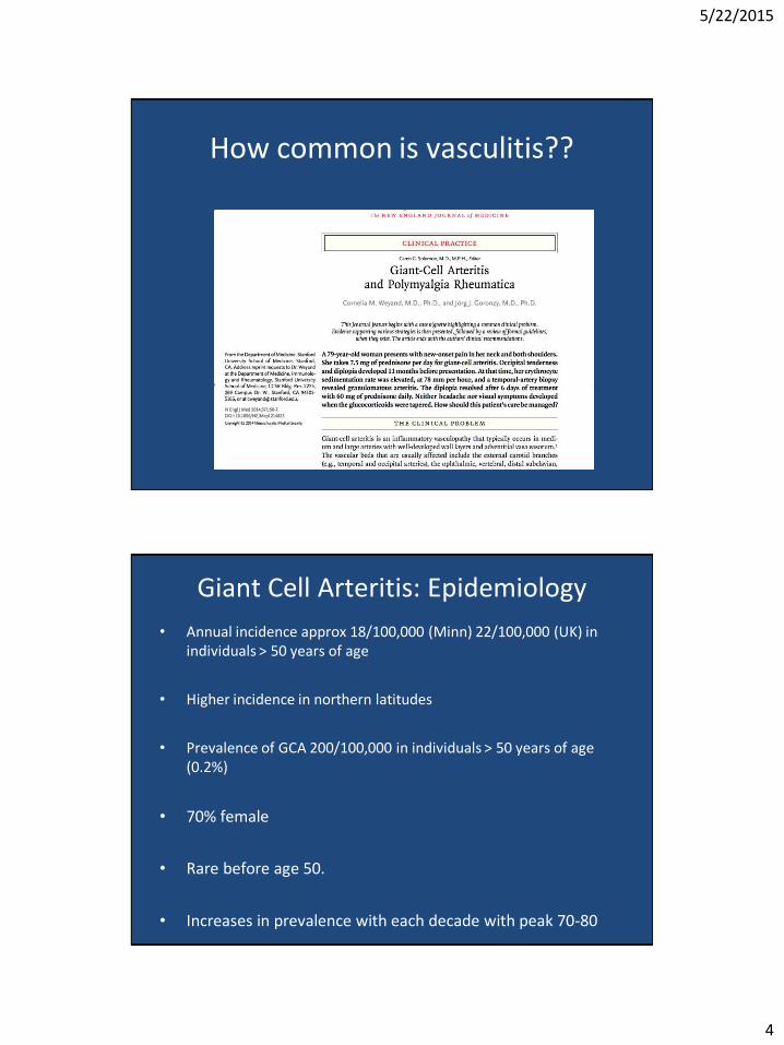
How common is vasculitis??
Giant Cell Arteritis: Epidemiology
• Annual incidence approx 18/100,000 (Minn) 22/100,000 (UK) in individuals > 50 years of age
• Higher incidence in northern latitudes
• Prevalence of GCA 200/100,000 in individuals > 50 years of age (0.2%)
• 70% female
• Rare before age 50.
• Increases in prevalence with each decade with peak 70-80

5/22/2015
5
Giant Cell Arteritis Clinical Manifestations
• Anatomy
• Large Vessel Vasculitis (arteries with internal elastic laminae)
• Most commonly involves extra-cranial vessels (external corotid) but can involve internal corotid and branches
• Inflammation in vessel wall (sometimes but not always with giant cells) leads to intimal and medial proliferation and occlusion of vessel
Giant Cell Arteritis Clinical Manifestations
• Headache (70-80% at one time or another) – Commonly dull, aching, often over the temporal area but
can be anywhere
– Scalp tenderness may be present
• Visual Changes – Present in up to a third of patients
– Blurred vision, diploplia, amaurosis fugax often presage blindness
– Monocular blindness can be abrupt without warning
– Can be permanent

5/22/2015
6
Giant Cell Arteritis Clinical Manifestations
• Jaw Claudication – Most specific symptom for GCA
– Classic presentation is discomfort over masseter muscles with protracted chewing
– This is not pain at temporal mandibular joint
• Constitutional signs are common in this SYSTEMIC disease (lots of pro-inflammatory cytokines)
• Weight loss, malaise • Low grade fever in up to half of patients (Cause of FUO in elderly) • 40-50% develop PMR (may precede, follow, or occur
concomitantly) • Hallmarks of IL-6 driven disease (inflammation and high CRPs)

5/22/2015
7
Retinal Ischemia
Giant Cell Arteritis Work-up
• Establish pre-test probability of GCA using demographics, history, physical exam
• Laboratory Evaluation – ESR and CRP
• >90% patients have an ESR >50; frequently >100
• C-reactive protein may be more sensitive and be elevated in patients with normal ESR

5/22/2015
8
Giant Cell Arteritis: Diagnosis • Temporal artery
biopsy – If elect to pursue
biopsy, initiate prednisone 1 mg/kg/day
– Request 3-5 CM segment of artery.
– Unilateral biopsy is >90% sensitive
– 2 weeks of empiric prednisone does not significantly affect the sensitivity.
GCA: Treatment • Treat with large, long-term corticosteroids (1 mg/kg) and with
expectation of long-term therapy (and morbidity)
• No proven steroid-sparing regimen, but baby ASA usually given as adjuvant therapy to reduce thrombotic complications
• Majority of patients will experience a durable remission but a substantial minority (40%) will relapse
• Relapse can be usually be treated with increases of 10-20% prednisone dosage and are rarely associated with ischemic complications
• Persistent elevations in inflammatory markers (ESR/CRP) and more rapid tapers of corticosteroids associated with higher risk of relapse
5/22/2015
9
Advances in approach to GCA
• Improvements in diagnosis (imaging)
• Better understanding the clinical spectrum of the disease
• Advances in therapy coming soon…..
Diagnosing GCA • Currently – much rests on empiricism
– Practice is to place patients with suspected GCA based upon history/physical exam on high dose prednisone and arrange for a biopsy
– Cutoff can be as low as 10% pre-test clinical suspicion to trigger above algorithm given potential morbidity of disease
• Biopsy is invasive and difficult to diagnose
– Often segmental (skip lesions can be missed) – Negative biopsy raises problems about continuing long
term morbid therapy

5/22/2015
10
GCA Diagnosis: Ultrasound
• In the right hands, classic ultrasound findings of GCA include a periluminal “halo sign” of hypoechoic edema in the vessel wall
• Also can see stenoses and occlusion
• Operator dependent and not reliably reproduced
GCA: Large Vessel Involvement • Large vessel involvement is more common than
once thought
• 25% of patients have large vessel arteritis (often can be symptomatic)
• When great vessel dz is suspected, MRI/MRA or CTA are reliable diagnostic tools for visualizing intramural edema (inflammation), thickening, stenoses, aneurisms
• FDG-PET/CT might be more sensitive: can detect inflammation in vessel wall in over 50% of GCA pts.
• Use of FDG-PET/CT to quantify inflammation in GCA is not standardized and can be nonspecific (atherosclerosis also can look “inflammatory”)
– Cases of FUO or in suspected disease with negative TA biopsy

5/22/2015
11
A 78-y-old woman presented with 6 wk of fever, night sweats, and weight loss.
Zohar Keidar et al. J Nucl Med 2008;49:1980-1985
GCA and mortality
• Traditional wisdom: equivocal if GCA increases mortality
• Increasing recognition of long term complications associated with GCA
• Aortic aneurisms: higher rate of rupture and dissection
• Atherosclerotic CV dz

5/22/2015
12
0-2 yrs 2-10 yrs >10 yrs
All cause mortality MRR 1.17
(1.01, 1.36)
MRR 0.96
(0.88, 1.05)
MRR 1.22
(1.05,1.41)
Circulatory System MRR 1.32 ND MMR 1.47
Coronary artery
disease
MMR 1.39 ND ND
Aortic Aneurism MMR 3.69 ND ND
1787 Patients with Histologically confirmed GCA
GCA: Future Therapies
• Long term corticosteroid exposure associated with morbidity
• Search for steroid-sparing agents generally underwhelming
• Methotrexate
• Azathioprine
• Infliximab and other anti-TNF therapies
5/22/2015
13
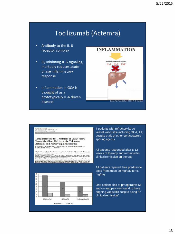
Tocilizumab (Actemra)
• Antibody to the IL-6 receptor complex
• By inhibiting IL-6 signaling, markedly reduces acute phase inflammatory response
• Inflammation in GCA is thought of as a prototypically IL-6 driven disease
Arthritis Care & Research Volume 64, Issue 11, pages 1720-1729, 27 OCT 2012 DOI: 10.1002/acr.21750 http://onlinelibrary.wiley.com/doi/10.1002/acr.21750/full#fig2
7 patients with refractory large
vessel vasculitis (including GCA, TA)
despite trials of other corticosteroid
sparing agents
All patients responded after 8-12
weeks of therapy and remained in
clinical remission on therapy
All patients tapered their prednsone
dose from mean 20 mg/day to <6
mg/day
One patient died of preoperative MI
and on autopsy was found to have
ongoing vasculitis despite being “in
clinical remission”
5/22/2015
14
Giant Cell Arteritis: Summary • Common form of a systemic vasculitis that increases in
prevalence with age and latitude
• Diagnosis continues to rest on clinical suspicion and histopathologic confirmation
• New imaging techniques may be beneficial in specific cases (FUO, TA bx neg, or suspected extra-cranial involvement)
• Treatment continues to rely on long-term susbstntial doses of corticosteroids
– Hope that preliminary data an ongoing large clinical trials will usher in age of biologic (anti-IL6) therapy
Case
• 36 year old female is admitted to the hospital with hemoptysis, respiratory distress, and acute kidney injury. She is taking no medications, is married, and has no children.
• Her exam is significant for hypoxemia, and hypertension and her workup includes CXR with bilateral pulmonary nodules and infiltrates and an elevated creatinine with hematuria and dysmorphic RBC’s. Her urine tox screen is negative and C-ANCA and Proteinase-3 antibodies are positive. Kidney biopsy reveals a pauci-immune necrotizing glomerulonephritis.

5/22/2015
15
Chest CT: Multiple Pulmonary Nodules And Ground Glass Opacities
Necrotizing Glomerulonephritis
Question
• This patient’s diagnosis is most consistent with:
A. Wegener’s Granulomatosis
B. Microscopic polyangiitis
C. Systemic Lupus Erythematosus
D. None of the Above

5/22/2015
16
Apologies!!! A Renamed Disease!
• This patient’s diagnosis is most consistent with:
A. Wegener’s Granulomatosis
B. Microscopic polyangiitis
C. Systemic Lupus Erythematosus
D. None of the Above
Granulomatosis with Polyangiitis (GPA)
• Friedrich Wegener: German pathologist credited with describing the disease (died in 1990)
• Wegener’s past ties to nazi party
(1932) and work near Jewish Ghetto of Lodz have become more clearly understood in recent years
• 2011: Led to renaming of WG as GPA
by major medical organizations including the ACR
• This patient does have this disease!!

5/22/2015
17
ANCA Associated Vasculitis: Quick Facts
• Wegener’s Granulomatosis: Renamed Granulomatosis with Polyangiitis 2010 (GPA)
– Clinical • Sinus – chronic sxs, necrotizing disease
• Lungs – nodules, cavities, alveolar hemorrhage
• Kidneys – pauci-immune glomerulonephritis; normal complements
– c-ANCA: anti-proteinase-3 abs are highly sensitive
ANCA Associated Vasculitis
• Microscopic polyangiitis – Clinical
• Skin – palpable purpura, ulcers • Lungs – Diffuse alveolar hemorrhage • Kidneys – glomerulonephritis • Neuro – mononeuritis multiplex
• p-ANCA – anti-myeloperoxidase abs • MPA – 75% sensitive • Can also be abnormal with Churg-Strauss (EGPA)

5/22/2015
18
Granulomatosis with polyangiitis therapy
• Until 1970’s nearly universal fatal disease
• 80% 3 yr mortality improved only slightly with corticosteroids (mean survival 12.5 months)
• Introduction of cyclophosphamide turned fatal disease into treatable disease
• Oral cyclophosphamide (Cyc) (2mg/kg/day) + corticosteroids (far more potent regimen that IV)
• Corticosteroid taper 6-9 months. Cyc continued full dose for at least 1 year AFTER remission and then tapered
What’s worse: Disease or treatment?
• Long term effects of Cyc therapy: – Infections – Sterility – Post treatment malignancies (hematologic, bladder)
• Clinical trials changed practice habits have evolved – shorter courses of induction therapy – use of less toxic DMARDs for longer term maintenance
of remission – Use of less toxic DMARDs for treatment of “limited
disease” (limited to upper airways) • Methotrexate • Azathioprine

5/22/2015
19
Case continued
The patient is initially treated with high dose pulses of IV corticosteroids and begins to improve. However when cytotoxic therapy with oral cyclophosphamide is recommended, she expresses concern over her risks of becoming infertile, and she strongly desires to have a child in the next few years.
Question
Which of the following statements is not correct?
A. Risk of premature menopause resulting from cytoxan is dose and age-dependent
B. Corticosteroids alone are not sufficient therapy to treat
this disease
C. Cytotoxic therapy should not be delayed for several months while the patient undergoes egg harvesting and cryopreservation
D. Oral Cyclophosphamide is the only therapy approved to
treat GPA

5/22/2015
20
Question
Which of the following statements is not correct?
A. Risk of premature menopause resulting from cytoxan is dose and age-dependent
B. Corticosteroids alone are not sufficient therapy to treat
this disease
C. Cytotoxic therapy should not be delayed for several months while the patient undergoes egg harvesting and cryopreservation
D. Oral Cyclophosphamide is the only therapy approved to
treat GPA
Rituximab versus Cyclophosphamide for ANCA-Associated Vasculitis

5/22/2015
21
Rituximab, B-cells, and ANCA
• Antineutrophil cytoplasmic antibodies are possibly implicated in pathogenesis or propagation/potentiation of ANCA-vasculitis
• Rituximab is a selective B-cell depleting antibody (anti-CD20)
• Possibility to remove ANCA by eliminating B-cells that would
replace short-lived ANCA producing plasma cells
• Other possible mechanisms by which B-cells might be implicated in ANCA vasculitis
RAVE Study Stone et al. NEJM 2010;363:221-32
• Multicenter, randomized, double dummy , double blinded placebo controlled non-inferiority trial
• Oral Cytoxan (gold standard) vs. Rituximab
• Both regimens given with corticosteroids
• Included patients with both GPA and MPA (microscopic
polyagiitis) as well as patients with relapsing disease
• Primary outcome: remission at 6 months free of glucocorticoid use (won’t get into specifics of how remission defined).

5/22/2015
22
RAVE: 6 month Summary • 197 patients enrolled
• 64% patients in rituximab arm reached endpoint vs. 53% in
cytoxan arm (p<0.001) – Comparable non-inferiority for GPA and MPA – Comparable non-inferiority for alveolar hemorrage or major renal
disease
• Ritux more effective for relapsing patients to achieve
primary endpoint (67%) vs. cytoxan (42%) (P=0.01)
• No difference in Adverse Events (surprising) – Our patient was concerned about fertility, not infections!
• FDA approval for rituximab to treat both GPA and MPA in 4/2011
RAVE: 18 months outcomes Encompassing induction and maintenance therapy
• SINGLE course of rituximab compared to oral Cyc + maintenance azathioprine up to 18 months
• Remission Rates 12 mo. 18 mo.
Ritux 48%* 39%*
Cyc + AZA 39%* 33%* • p<0.001 for non-inferiority
• Superiority for patients with previous relapse at 12 months but not 18 months (B-cells have reconstituted)

5/22/2015
23
AAV: Maintenance regimens: Comparing less toxic maintenance regimens
• MTX and AZA equally effective with similar adverse event rate
Pagnoux et al. N Engl J Med 2008;359:2790-803.
Hiemstra et al. JAMA. 2010;304(21):2381-2388
AZA SUPERIOR to MMF (Cellcept) in
maintaining remission (HR 1.69 p=0.03)
with similar adverse event rate
AAV: Role of Rituximab in maintenance Guillevin et al. N Engl J Med 2014;371:1771-80.
• 115 patients (GPA, MPA) in remission after Cyc + corticosteroids
• Randomly assigned to rituximab (0,6,12,18 months) and AZA (2 mg/kg tapered to 1 mg/kg for 22 months)
• NOT blinded
• Patients followed to month 28

5/22/2015
24
Rituximab: Maintenance for ANCA Vasculitis Guillevin et al. N Engl J Med 2014;371:1771-80.
Month 28
relapses(n)
Month 28
relapses (%)
Hazzard
Ratio
Rituximab 3/57 5% 1
Azathioprine 17/58 29% 6.61
p=0.002
ANCA Associated Vasculitis Summary
• Rituximab non-inferior to oral cyclophosphamide in inducing remission for ANCA associated vasculitis but also doesn’t appear to necessarily offer safety advantage
• Rituximab likely superior for patients with relapsing disease
• Methotrexate and azathioprine reasonable alternatives to treat upper airway “limited disease” in GPA
• Rituximab appears to be superior therapy for maintaining remission of AAV > 2 years after remission, although azathioprine is acceptable alternative maintenance regimen

5/22/2015
25
Case
• 46 year old female admitted to to the hospital with painful lesions on both of her legs and ears. Lesions on legs began six months earlier and progressed from small papules to large, necrotic purpura and ulcerations. No other apparent organ involvement other than skin. Her past medical history is notable only for hypertension and her PE other than her skin is unremarkable. Her family history is unremarkable and her social history notable for occasional recreational drug and alcohol use. She only takes lisinopril and hydrazine for hypertension and has no medication allergies.
Skin Rash

5/22/2015
26
Pertinent Laboratory Results
• WBC was 1.8 and her ANC (800) and ALC (900)
were low.
• ESR and CRP were elevated (48 and 44)
• HBV Sag -, HCV +, LFTs normal
• Serum creatinine, urinalysis, blood cultures,
complements, and cryoglobulins normal
Further Diagnostic Workup
• ANA positive (1:640 diffuse pattern)
• Anti-dsDNA was 1:40, but other ANA sub-serologies were negative
• pANCA positive > 1:20,480 – Anti-MPO: low positive
– Anti-PR3: low positive

5/22/2015
27
“Innumerable fibrin thrombi within nearly every superficial
dermal vessel and a variable dense neutrophilic infiltrate
surrounding some affected vessels”
Question
In this patient’s case, what is the most likely
cause of this patient’s symptoms?
A. Systemic lupus erythematosus
B. Granulomatosis with polyangiitis
C. Microscopic polyangiitis
D. Cocaine adulterant

5/22/2015
28
Question
In this patient’s case, what is the most likely
cause of this patient’s symptoms?
A. Systemic lupus erythematosus
B. Granulomatosis with polyangiitis
C. Microscopic polyangiitis
D. Cocaine adulterant
Advisory: Cocaine mixed with
Levamisole
• 70 percent of cocaine tested by DEA 7/09 positive for levamisole
• Immuno-modulating medication to treat colon cancer, nephrotic syndrome, RA
• No longer used in humans because of
toxicity that includes neutropenia
• 20 cases (2 fatal) agranulocytosis in Seattle
and Calgary thought due to levamisole adultered cocaine
• Most of aforementioned cases had
borderline or frank neutropenia

5/22/2015
29
Increasing adulteration of cocaine with
levamisole
Casale J, Corbeil E, and Hays P. DEA Resources, Microgram Journal; 6, Jan-Jun 2008
Novel syndrome associated with
levamisole-adulterated cocaine • Retiform purpura and extensive
cutaneous necrosis that affects mostly females 40-50
• Large areas of skin necrosis - not smaller lesions of palpable purport as would be seen in leukocytoclastic vasculitis
• “Classically” involves skin but occasionally can be associated with vasculitis elsewhere (kidney)
• Extensive small vessel thrombosis with/without leukocytoclastic vasculitis
• Frequent cytopenias (neutropenia)

5/22/2015
30
Novel syndrome associated with
levamisole-adulterated cocaine
• Associated with multiple autoantibodies
– “Sky high” titers of pANCA with antibodies to multiple components of neutrophil granules
– Not just antibodies to MPO
• Tends to improve with cessation of levamisole exposure
– Evidence lacking for use of immunosuppressants
Differentiating levamisole toxicity from
idiopathic pANCA vascultitis
Coc-Levamisole Patients Classic Microscopic Polyangiitis
Retiform purpura Palpable purpura
(cheek/ear) (different distribution)
Neutropenia Leukocytosis
Small vessel thrombotic Leukocytoclastic vascultiis vasculopathy > vascultis
“Sky-high” pANCA+ pANCA+ “within reason”
MPO titer lower than pANCA MPO titer correlates with pANCA