Embed Size (px)
Citation preview

Update in Rheumatic Diseases: Scleroderma/Sjögrens/Myositis
Paul F Dellaripa MD
Associate Professor of Medicine
Harvard Medical School
Division of Rheumatology
Brigham and Women’s Hospital
Paul F Dellaripa MD
• University of Connecticut School of Medicine
• Division of Rheumatology BWH
• Associate Professor of Medicine Harvard Medical School
• Clinical investigations: autoimmune lung diseases

Disclosures
Genetech
Bristol Myers
Boerringer Ingelheim
Up to Date

Outline
1. Scleroderma
2. Sjögren’s syndrome
3. Myositis
• Clinical manifestations and diagnosis
• Treatment options

Systemic Sclerosis (SSc/Scleroderma)
• Complex systemic disease that affects the
skin, lung, heart, GI tract, and kidney
• Pathophysiology• Vascular and endothelial dysfunction
• Autoimmunity
• Fibroblastic activation and proliferation

Scleroderma = Hardening of the Skin
• Localized scleroderma
• Morphea
• Linear scleroderma
Systemic sclerosis
• Diffuse
• Limited (CREST)
• Sine scleroderma
• Overlap Syndrome
Katz KA, Derm Online

Limited Systemic Sclerosis: CREST
• Calcinosis
• Raynaud’s
• Esophageal Dysmotility
• Sclerodactyly
• Telangiectasias
ACR Image Bank

Limited vs Diffuse SSc
Limited Diffuse
Skin thickening Face, forearms, lower legs, hands and feet
Face, trunk, entire arms/legs, hands and feet; tendon friction rubs
Raynaud’s phenomenon Progressive disease following onset of RP
Rapid onset following RP: months to 3 years
Autoantibodies Association with anti-centromere Ab (50-60%)
Association with anti-Scl 70 ab (30%)
Internal organ involvement
Late: primarily pulmonary HTN (10-30%), esophageal dysmotility
Early: ILD (75%), myocardial disease, diffuse GI involvement, scleroderma renal crisis (10-15%)
Prognosis 10-yr survival > 70% 10-yr survival 40%

Skin Thickening

Puffy Hands of Early SSc

Sclerodactyly
ACR Image Bank

Mauskopf Facies

Natural History of Skin Tightening in SSc
Medsger TA et al. Rheum Dis N Am 2003

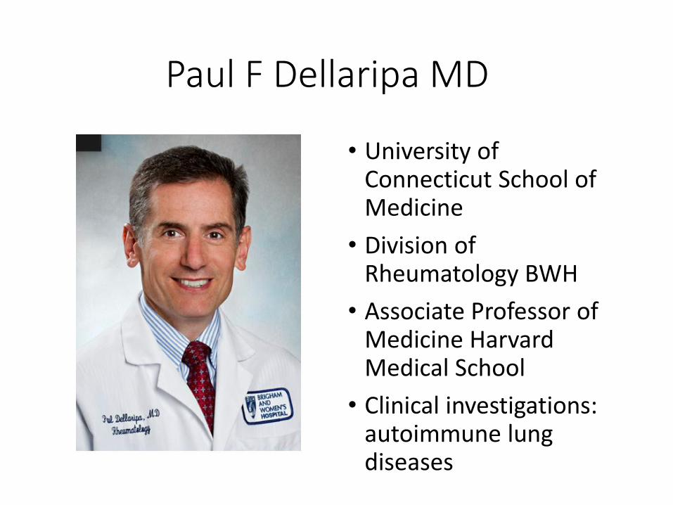
Raynaud’s Phenomenon
• Not an infrequent initial presentation of
CTDs
• Episodic, reversible digital skin color
change
• White to blue to red
• Well-demarcated
• Due to vasospasm
• Usually cold-induced
Raynaud’s Phenomenon

Raynaud’s Phenomenon

• Primary Raynaud’s • Common in young women (<age 30)
• Often have + family history
• ANA mostly negative
• Secondary Raynaud’s• Onset in > age 35
• Digital ulcers, pitting scars in fingers
• Abnormal capillary micrscopy
• Presence of autoantibodies
Raynaud’s Phenomenon

Secondary Raynaud’s Phenomenon
• Connective tissue diseases
• SSc, SLE, MCTD, Undifferentiated CTD,
Sjogren’s syndrome, Dermatomyositis
• Occlusive arterial disease
• Atherosclerosis, Antiphospholipid Syndrome,
Buerger’s disease
• Vascular injury
• Frostbite, vibratory trauma
• Vinyl chloride, bleomycin, amphetamines,
cocaine
• Hyperviscosity/ cold-reacting proteins

Digital Ulcer in SSc

Nailfold Capillaroscopy: Periungual Changes

SSc: Autoantibodies
ANA + in most but not all cases
• Often nucleolar
Anti-centromere
• 50-60% limited SSc
Anti-Scl-70 (anti-topoisomerase ab)
• 30% diffuse SSc
Anti-RNA polymerase III
• Associated with increased risk of renal crisis and increase risk of malignancy
Other antibodies: anti-DNA polymerase, anti-U3-RNP, anti-PM-Scl Th/To

SSc: Internal Organ Involvement
Interstitial lung disease Leading cause of mortality Occurs in 75% diffuse SSc Often within the first 4 years
Pulmonary hypertension More common in limited SSc (10-30%) Usually occurs years into illness
• Concomitant pulmonary hypertension and ILD Extremely challenging clinical scenario
• Increased rate of malignant lung neoplasms

SSc: Internal Organ Involvement
• Renal Disease • 60-80% diffuse SSc in autopsy studies
• Microalbuminuria, HTN, mild Cr rise ~ 50%
• Renal crisis 10-15% (early diffuse SSc)
• Cardiac Involvement• Congestive heart failure, myocardial fibrosis
• Pericarditis (10-20%)
• Arrhythmias
• GI Involvement (90%)• Esophageal hypomotility/ LES incompetence
• Gastroparesis
• Watermelon stomach (vascular ectasia in the antrum)

Interstitial Lung Disease in SSc

SSc: Esophageal Dysmoliity (?Role in ILD)

SSc Treatment: ILD
• Current standard of care• Mycophenolate ( MMF) :preferred agent
• Tocilizumab (IL-6) FDA approved in SSc ILD
• Inhaled treprostinil for PH-ILD (Waxman et al NEJM 2021)
• Anti-fibrotics in IPF: FDA approved 2014 • Pirfenidone approved in IPF
• Nintedanib: now approved in Scleroderma
• Stem cell transplant in selected patients

SSc Treatment: There Has Been Progress!
• Renal: ACE inhibitors
• PAH: Prostacyclin, Endothelin antagonists, Phosphodiesterase inhibitors
• Reflux: PPI high dose, anti reflux surgery
• Raynaud’s: Endothelin antagonists, Prostacyclin, Phosphodiesterase inhibitors, Surgery, Botox
• Lung: MMF, antifibrotics, IL-6 and transplant

Sullivan KM et al., SCOT Study Investigators
January 4, 2018

Key Points in Scleroderma
• ILD remains the leading cause of mortality
• Treatment options have improved
• Raynaud’s that develops > age 35 raises concern for a rheumatic disease
• The presence of concomitant Raynaud’s and GERD should raise suspicion for limited SSc
• Pulmonary HTN is a complication of long standing limited SSc; screening is essential as treatments are available that improve morbidity and possibly mortality
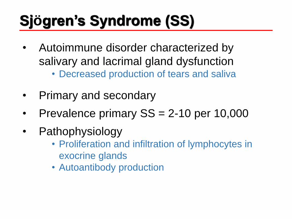
Sjögren’s Syndrome (SS)
• Autoimmune disorder characterized by
salivary and lacrimal gland dysfunction• Decreased production of tears and saliva
• Primary and secondary
• Prevalence primary SS = 2-10 per 10,000
• Pathophysiology• Proliferation and infiltration of lymphocytes in
exocrine glands
• Autoantibody production and the role of B cells in
the pathophysiology of this disease are the
source of ongoing investigation

Sicca Syndrome Manifestations
• Keratoconjunctivitis sicca
• Ocular dryness
• Corneal injury
• Xerostomia
• Oral dryness, dysphagia
• Dental caries, thrush
• Nasal dryness and epistaxis
• Vaginal dryness
• Dyspareunia
• Candidiasis

Classification Criteria for Primary SS
Item Weight/Score
Labial salivary gland with focal lymphocytic sialadenitis and focus score of > 1 foci/4 mm2
3
Anti-SSA/Ro positive 3
Ocular Staining Score >5 in at least 1 eye 1
Schirmer’s test <5 mm/5 min in at least 1 eye 1
Unstimulated whole saliva flow rate <0.1 mL/min
1
Score of >4 needed to establish dx* exclusions: active hepatitis, head/neck xrt, sarcoidosis AIDS,
amyloidosis, IgG4-related diseaseShiboski CH et al., Ann Rheum Dis 2017
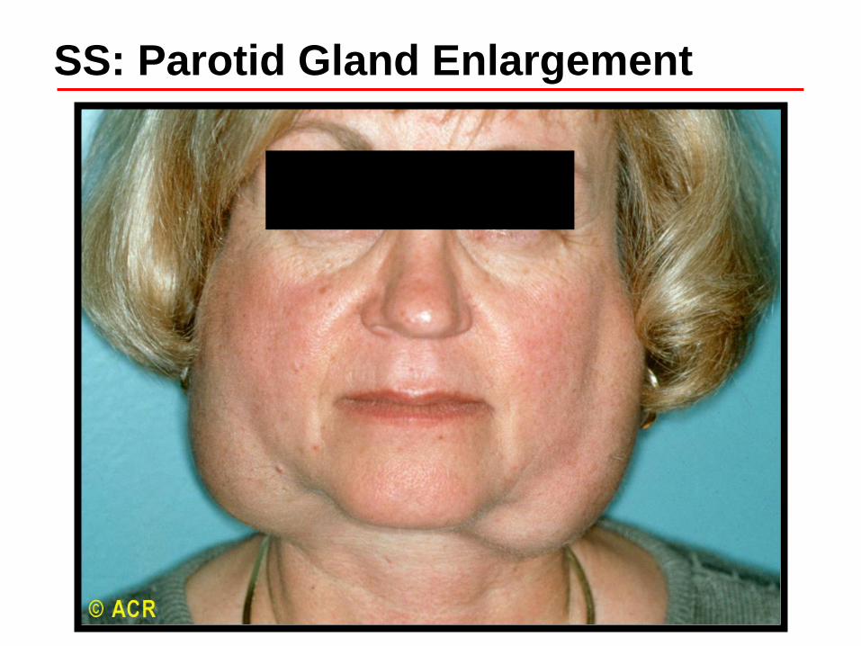
SS: Parotid Gland Enlargement

SS: Corneal Abrasions

SS: Salivary Hypofunction

Primary SS: Extraglandular Disease
• Peripheral neuropathy
• Vasculitis
• Interstitial lung disease (LIP, NSIP)
• Synovitis
• Risk factors for more aggressive disease: +RF, low C4, cryoglobulinemia
• Increased risk of lymphoma

50 yo with Known SS with 8 lb Weight Loss Lung biopsy c/w Marginal Zone Lymphoma

• Connective tissue diseases
• SLE
• RA
• Systemic sclerosis
• Hypothyroidism
• Cryoglobulinemia
• Autoimmune hepatitis
SS: Associated Conditions

• Exocrine gland dysfunction• Xerostomia: oral hygiene and agents to stimulate
salivary secretion (pilocarpine/muscarinic agonist ) • KCS: cellulose products to augment tear replacement
and topical cyclosporine
• Treatment of extraglandular disease is difficult • Trials are ongoing
• Hydroxychloroquine may be useful in those with fatigue and arthralgias
Sjögren’s Syndrome: Treatment

• Higher risk of non-Hodgkins lymphoma in primary Sjögrens (up to 44 fold or 5% lifetime)
• Most cases are secondary to other rheumatic diseases
• Treatment geared towards managing sicca symptoms and promoting oral hygiene
• Treatment of extraglandular disease is challenging
• Patients with low titer ANA, fatigue, and eye or mouth dryness are a significant challenge diagnostically
Key Points About Sjögren’s Syndrome

Inflammatory Myopathies
• Group of autoimmune disorders• Common feature = immune-mediated muscle injury
• Usually present with muscle weakness and
elevated muscle enzymes (CK/aldolase)
• Disorders include• Dermatomyositis (DM)
• Polymyositis (PM)
• Overlap myositis (with another systemic
rheumatic disease)
• Inclusion body myositis (IBM)
• Necrotizing autoimmune myositis (NAM)
• Histopathologic and clinical distinctions
Senecal et al., Arth Rheum 2017

Inflammatory Myopathies: DDx
• Inflammatory myopathies
• Drug-induced myopathies• Steroids
• Statins
• Colchicine
• Hydroxychloroquine
• Alcohol
• Zidovudine
• Infections• Viral
• Toxoplasmosis
• Trichinosis
• Bacterial pyomyositis
• Systemic vasculitis• PAN, GPA, eGPA
• Amyloid myopathy
• Sarcoid myopathy
• Metabolic myopathies• Disorders of carbohydrate
and lipid metabolism
• Hypothyroidism
• Electrolyte disturbances• Hyper/hyponatremia
• Hypokalemia
• Hypophosphatemia
• Hypocalcemia
• Neurologic disorders• Myasthenia gravis
• Motor neuron disease
• Muscular dystrophy

Inclusion Body Myositis v
Idiopathic Inflammatory Myopathy
Inclusion Body Myositis IIM
Sex Male > female Female > male
Age Usually > 50 Common before 50
Onset Slowly progressive Acute or sub-acute
Weakness Distal and asymmetric muscle weakness
Proximal and symmetric
EMG Myopathic and neuropathic changes
Myopathic changes
Muscle biopsy Mononuclear cell infiltrates and vacuoles containing amyloid
Inflammation, fiber necrosis
Response to immunosuppression
Generally poor Generally good

IIM (Photomicrograph)

Inclusion Body Myositis (Photomicrograph)

DM and PM: Clinical Features
• Proximal muscle weakness • > 90% PM patients
• 50-60% DM patients at presentation; skin
features may precede weakness
• Amyopathic DM
• Skin findings• Classic DM
• Not found in PM

DM: Gottron’s Papules

DM: Heliotrope Rash


DM: Poikiloderma (Shawl and V Signs)

DM: Nailbed

DM and PM: Clinical Features
• Interstitial Lung Disease
• Cardiac disease • Myocarditis—frequently subclinical
• 3-4x increased risk MI
• Esophageal disease• Weakness of striated muscle of upper 1/3rd of
esophagus→ aspiration

ILD and Pneumomediastinum

DM and PM: Autoantibodies
~ 80% ANA +
Myositis-specific autoantibodies
Clinical syndrome Prevalence
Antisynthetase antibodies, including anti-Jo-1
Antisynthetase syndrome 20%
Anti-signal recognition particle (SRP)
Severe myopathy, aggressive disease that may be difficult to control
5%
Anti-Mi-2 Acute onset DM, classic skin findings, good prognosis
7-30%
Anti-MDA-5 Rapidly progressive ILD, cutaneous ulceration involving Gottron’s papules, arthritis, alopecia, oral ulcers, amyopathic

Antisynthetase Syndrome
Fever
• Raynaud’s
• Inflammatory
arthritis
• ILD ( can be
severe)
• Mechanics
hands

Treatment Regimens in IIM
Corticosteroids (often with DMARD)
DMARDs• Methotrexate • Calcineurin inhibition (Tacrolimus)• Azathioprine• Mycophenolate
Cyclophosphamide (mostly in ILD)
Rituximab (recent trial efficacy unclear but may be useful in antisynthetase syndrome)
IVIG (refractory cases)
Abatacept (T cell co-stimulatory inhibitor)

• Scleroderma• Recognize clinical patterns• Major morbidity/mortality = lung disease• There are newer treatment options!
• Sjögrens syndrome• Recognize clinical manifestations • Sometimes marked by systemic disease• Higher risk for lymphoma
• Inflammatory myopathy • Recognize clinical patterns• Newer antibodies may help with diagnosis• Major morbidity = lung disease

Board Question #1
53 yo female with 20 year hx of Raynaud’s develops fatigue and dyspnea over the preceding 6 months
On Nifedipine
BP 115/80
Exam notable for prominent P2
Scattered telangictasias on hands and face
PFTs show DLCO 48% predicted (low)
02 sat is 96% rest, 93% with activity (abnormal)
Echo shows mild TR, est RVSP 48 mmHg

What is the appropriate next test for this patient?
• A. CT angiogram of the chest
• B. HRCT of the chest
• C. Pulmonary artery catheterization
• D. Exercise stress test
Board Question #1

• Correct answer: C
• The longstanding history of Raynaud’s raises the question of a CTD and the telangictasias are seen in CREST
• Given the decline in DLCO and echo findings, PAH remains the greatest concern and the patient needs to get a right heart catheterization to confirm the diagnosis.
Board Question #1

• 51 yo man with diffuse cutaneous SSc is admitted with new onset hypertension associated with anemia and thrombocytopenia
• On admission: BP 180/105; skin thickening over face, chest, hands, legs; lungs clear; heart RRR normal S1 S2; 1+ edema in legs
Board Question #2

Board Question #2
• Hgb 9.8 Plt 101K Cr 1.4
• UA 2+ protein, no casts
• Smear: 2+ schistocytes
• Started on captopril 6.25mg every 8hrs
• Captopril escalated to 25 mg every 8hrs
• 3 days later, BP 140/95, Cr now 2.1
• UA 2+ protein

Board Question #2
Which of the following is the most appropriate next step?
• A. Discontinue captopril, begin nifedipine
• B. Continue to increase the captopril
• C. Start plasmapheresis
• D. Angiography to assess for RAS
• E. Order the RNA polymerase III ab

• Correct answer: B
• This patient has systemic sclerosis with diffuse skin disease and is at significant risk for renal crisis which is the case here. Despite the continued increase in creatinine, the ACE inhibitor should be continued.
• The RNA polymerase III ab confers increased risk for renal crisis
Board Question #2

References
Tashkin DP, Elashoff R, Clements PJ et al. for the Scleroderma Lung Study. Cyclophosphamide versus placebo in scleroderma lung disease. N Eng J Med 2006;354:2655-2666
Tashkin DP, Roth M, Clements PJ et al. Mycophenolate in Scleroderma:SLS II. Lancet 2016;4(9):708-719
Distler O, Highland KB, Gahlemann M et al. Nintedanib for scleroderma associated intersitial lung disease N Eng J Med N Engl J Med 2019; 380:2518-2528
Roofeh, D et al Tocilizumab Prevents Progression of Early Systemic Sclerosis-Associated Interstitial Lung Disease Arthritis Rheumatol 2021
Klinger JR, Elliott CG, Leinve DJ et al Therapy for pulmonary arterial hypertension in adults. Chest 2019:155(3):565-586
Carson SE Vivino FB, Parke A et al Treatment guidelines for the rheumatologic manifestations of Sjogren’s syndrome. Arthritis Care Research 2017;69(4):517
A Oddis CV, Aggarwal R . Treatment of Inflammatory myositis. Nature Reviews Rheumatology 2018;14:279-89