Embed Size (px)
Citation preview

Chapter 20. Proteins 20.1 Characteristics of Proteins20.2 Amino Acids: The Building Blocks for Proteins20.3 Chiralityand Amino Acids20.4 Acid-Base Properties of Amino Acids20.5 Cysteine: A Chemically Unique Amino Acid20.6 Peptide Formation20.7 Biochemically Important Small Peptides20.8 General Structural Characteristics of Proteins20.9 Primary Structure of Proteins20.10 Secondary Structure of Proteins20.11 Tertiary Structure of Proteins20.12 Quaternary Structure of Proteins20.13 Fibrous and Globular Proteins20.14 Protein Hydrolysis20.15 Protein Denaturation20.16 Glycoproteins20.17 Lipoproteins
Students should be able to:1. Characterize the structure of a protein using the terms polymer, monomer, amino
acid, peptide bond, amide, and amino acid side chain.2. Describe several types of functions of proteins.3. Identify an α - amino acid.4. Compare the polarity of the side chains of several amino acids and classify the
side chains as hydrophobic, hydrophilic, non-polar, polar, neutral, basic, or acidic.
5. Compare the essential and non-essential amino acids.6. Predict the structure of an amino acid in acidic solution and basic solution.7. Illustrate the acid/base properties of amino acids and describe how the structure
of an amino acid depends on pH.8. Explain the relationship between the zwitterions form of an amino acid and its
isoelectric point (pI).9. Explain how the laboratory technique of electrophoresis is used to characterize
proteins and amino acids.10. Identify a chiral carbon.11. Predict the products of any of the following reactions of amino acids, peptides,
and /or proteins• peptide formation• hydrolysis of a polypeptide• oxidation of cysteine to form a disulfide bond
12. Distinguish between primary, secondary, tertiary, and quaternary structure of proteins.
13. Compare different types of secondary protein structure (alpha helix, etc.).14. Identify the N and C terminals of a peptide chain.15. Describe the types of interactions involved in each level of protein structure
(covalent and non-covalent interactions). 16. Predict the side-chain interactions between two amino acids.
1

17. Draw the structure of a peptide given the structures of amino acid residues.18. Distinguish between a fibrous and a globular protein. Give examples of each.19. Characterize the chemistry of several modes of protein denaturation.20. Give some examples of applications of proteins in everyday life.
20.1 Characteristics of ProteinsIn this chapter we consider proteins which are the third class of the
bioorganic molecules. Various types of proteins in the human body perform extraordinary number of different function essential for maintaining life. A typical human cell contains about 9000 different kinds of proteins, and the whole human body contains about 100,000 different proteins. Proteins are the backbone of enzymes, certain hormones, an some blood components and tissues.
Proteins are the most abundant substances in nearly all cells accounting for about 15% of a cell's overall mass. Proteins contain the elements carbon, hydrogen, oxygen, and nitrogen and few also contain sulfur. The presence of nitrogen in proteins sets them apart from carbohydrates and lipids and imparts specific properties to them. Other elements, such as phosphorus and iron, are also essential constituent of certain specialized proteins. Casein, the main protein of milk, contains phosphorus, an element very important in the diet of infants and children. Hemoglobin, the oxygen-transporting protein of blood, contains iron.Protein Functions Proteins and peptides (small proteins) are essential to the cell. They are most versatile of biomolecules and the most abundant class of biomolecules. In Greek "proteios" means "of the first rank or importance". The 1/2 of dry mass of cell is protein. They are composed of amino acids which are carboxylic acids also containing an amine functional group. They serve two major functions in the cell. Some proteins are enzymes that catalyze most biological reactions in a living organism. Other proteins perform a structural role for the cell - either in the cell wall, the cell membrane or in the cytoplasm.
Enzymes - catalyze biological reactions (alcohol dehydrogenase, glucokinase)
Hormones - signals between cells (insulin, growth hormone) Storage Proteins - store nutrients (ferritin storing iron in the liver) Transport Proteins - transport nutrients through the body (hemoglobin
transport of oxygen) Structural Proteins - form structure of cells ( keratin, elastin, collagen) Protective Proteins - have specific protective function (antibodies bind
to foreign proteins) Contractile Proteins - cell motion (actin and myosin of muscle
contraction) Toxic Proteins - proteins which have adverse consequences (snake
venoms)
2

20.2 Amino Acids: The Building Blocks for Proteins
A protein is a naturally occurring, unbranched polymer in which the monomer units are amino acids. Thus the starting point for a discussion of proteins is an understanding of the structures and chemical properties of amino acids.
Amino acids are primary amines that contain an alpha carbon that is connected to an amino (NH) group, a carboxyl group (COOH), and a variable side group (R). Naturally amino acids in proteins are all alpha amino acids (ie the amino function is on carbon 2 from the carboxyl function. The side group gives each amino acid its distinctive properties and helps to dictate the folding of the protein.
Normal proteins are generally composed of 20 different naturally occuring amino acids.
Common names assigned to the amino acids are currently used.Three letter abbreviations - widely used for naming:
First letter of amino acid name is compulsory and capitalized followed by next two letters not capitalized except in the case of Asparagine (Asn), Glutamine (Gln) and tryptophan (Trp).
One-letter symbols - commonly used for comparing amino acid sequences of proteins:
Usually the first letter of the nameWhen more than one amino acid has the same letter the most abundant amino acid gets the 1st letter.
The amino acids have two shorthand abbreviations. One set of abbreviations consist of a three letter abbreviation. The second set of abbreviations are single letters. Both types of abbreviations are given in the following table:
Name Short Name
Letter Name Side Chain Structure
3

1 Glycine Gly G H
2 Alanine Ala A CH3
3 Valine Val V (CH3)2CH
4 Leucine Leu L (CH3)2CHCH2
5 Isoleucine Ile I CH3CH2CH(CH3)
6 Phenylalanine Phe F PhCH2
7 Tyrosine Tyr Y HOC6H4CH2
8 Tryptophan Trp W Indole-CH2
4

9 Serine Ser S HOCH2
10 Threonine Thr T CH3CH(OH)
11 Methionine Met M CH3SCH2CH2
12 Cysteine Cys C HSCH2
13 Asparagine Asn N H2NCOCH2
14 Glutamine Gln Q H2NCOCH2CH2
15 Proline Pro P -CH2CH2CH2
16 Aspartic acid Asp D HOOCCH2
5

17 Glutamic acid Glu E HOOCCH2CH2
18 Lysine Lys K H2NCH2CH2CH2CH2
19 Arginine Arg R H2NC(=NH)NHCH2CH2CH2
20 Histidine His H Imidazole-CH2
There are 20 common amino acids found in proteins and these amino acids can be classified into 3 groups; polar, non-polar and charged. (S) Small Hydrophilics: Glycine, Alanine, Serine, Threonine, Proline. Side chains in this group do not have ionizable groups, thus they do not contribute to the net charge of proteins (at neutral pH). (H) Basics: Lysine, Arginine, Histidine. Amino acids having additional amino groups in the side chain. They are positively charged at neutral pH. (N)Acids and Amides: Aspartic acid, glutammic acid, asparagine, glutamine. This group includes amino acids having an additional carboxyl groups in the side chain and their amides. Acids are negatively charged at neutral pH. (C)Sulphydril: Cysteine. This group includes only cysteine, having an SH group in the side chain. Cysteine can occur as cystine in which two molecules of cysteines are linked by a disulfide bond. (F) Aromatics: Tyrosine, Phenylalanine, Tryptophan. This group includes amino acids having aromatic rings in the side chain. (V)Small hydrophobic: Leucine, Isoleucine, Valine, Methionine. In the human body some of the amino acids (non-essential amino acids) can
6

be readily synthesized from dietary components and others (essential amino acids) must be obtained from the diet. Non-essential amino acids: Ala, Gly,Pro, Asn, Cys, Glu, Ser, Tyr, Asp, and Glu
Essential amino acids: Arg, His, Lys, Ile, Leu, Met, Phe, Val, Thr, and Trp
20.3 Chirality and Amino AcidsExcept glycene all other 20 amono acids have 0ptical isomers or entiomers show chirality: D-amino acids (NH2 group is found at right). L-amino acids(NH2 group is found at left). NH2 group is found at left. Eucaryotic cells use only "L-amino acids" to make proteins. Some bacteria use D-amino acids.
L-isomer D-isomer
20.4 Acid-Base Properties of Amino AcidsIn pure form amino acids are white crystalline solids. Most amino acids decompose before they melt. They are not very soluble in waterExists as Zwitterion: An ion with + (positive) and – (Nagetive) charges on the same molecule with a net zero charge
Carboxyl groups give-up a proton to get negative charge Amino groups accept a proton to become positive
COO-
R
+H3N H
COO-
CH3
NH3+H
L DZwitterions
Amino acids in solution exist in three different species (zwitterions, positive ion, and negative ion) - Equilibrium shifts with change in pHIsoelectric point (pI) – pH at which the concentration of Zwitterion is maximum -- net charge is zeroDifferent amino acids have different isoelectric pointsIsoelectric Point
7

At isoelectric point - amino acids are not attracted towards an applied electric field because they net zero charge.
20.5 Cysteine: A Chemically Unique Amino AcidCysteine: the only standard amino acid with a sulfhydryl group ( — SH group). The sulfhydryl group imparts cysteine a chemical property unique among the standard amino acids. Cysteine in the presence of mild oxidizing agents dimerizes to form a cystine molecule. Cystine - two cysteine residues linked via a covalent disulfide bond.
20.6 Peptide FormationThe amino acids are linked together by peptide bonds (amide bonds) forming long chains
8
+H3N C
COOH
CH3
+H3N C
COO-
CH3
H2N C
COO-
CH3
H H H
Low pH(net + charge)
High pH(net - charge)Zwitter Ion
(net neutral charge)Neutral pH

Short chains of amino acids are commonly called polypeptides (eg. dipeptide, tripeptide, hexapeptide, etc). Longer chains of amino acids normally called proteins.
20.7 Biochemically Important Small PeptidesMany relatively small peptides are biochemically active in the human body:
HormonesNeurotransmittersAntioxidants
Small Peptide Hormones:Best-known peptide hormones: oxytocin and vasopressin Produced by the pituitary glandnonapeptide (nine amino acid residues) with six of the residues held in the form of a loop by a disulfide bond formed between two cysteine residues
Glutathione (Glu–Cys–Gly) – a tripeptide – is present is in high levels in most cells. Regulator of oxidation–reduction reactions. Glutathione is an antioxidant and protects cellular contents from oxidizing agents such as peroxides and superoxides.Highly reactive forms of oxygen often generated within the cell in response to bacterial invasion
Unusual structural feature – Glu is bonded to Cys through the side-chain carboxyl group.
20.8 General Structural Characteristics of ProteinsProtein Classification Based on Chemical CompositionSimple proteins: A protein in which only amino acid residues are present:
More than one protein subunit may be present but all subunits contain only amino acids
Conjugated protein: A protein that has one or more non-amino acid entities (prosthetic groups) present in its structure:
9

One or more polypeptide chains may be presentNon-amino acid components - may be organic or inorganic - prosthetic groupsLipoproteins contain lipid prosthetic groups Glycoproteins contain carbohydrate groups,Metalloproteins contain a specific metal as prosthetic group
Primary Structure Secondary Structure Tertiary Structure Quaternary
Primary Structure: Primary structure of protein refers to the order in which amino acids are linked together in a proteinEvery protein has its own unique amino acid sequenceFrederick Sanger (1953) sequenced and determined the primary structure for the first protein – Insulin
Protein classification based on shapeThree types of proteins: 1) fibrous,2) globular, and 3) membraneFibrous proteins: protein molecules with elongated shape:
Generally insoluble in water Single type of secondary structure Tend to have simple, regular, linear structures Tend to aggregate together to form macromolecular structures,
e.g., hair, nails, etc.Globular proteins: protein molecules with peptide chains folded into spherical or globular shapes:
Generally water soluble – hydrophobic amino acid residues in the protein coreFunction as enzymes and intracellular signaling molecules
Membrane proteins: associated with cell membranesInsoluble in water – hydrophobic amino acid residues on the surface
Help in transport of molecules across the membrane
20.9 Primary Structure of Proteins
Primary structure is the simple sequence of amino acids in the protein chain. The side group of each amino acid gies its distinctive properties and helps to dictate the folding of the protein. The R group interactions helps to maintain the three-dimensional conformation of a protein.
10

A amino acid.Polymers of amino acids are created by linking an amino group to a caroboxyl group on another amino acid. This is termed a peptide bond as shown below
Peptides and proteins are formed when a ribosome and the rest of the translation machinery link 10 - 10,000 amino acids together in a long polymer. This long chain is termed the primary sequence. The properties of the protein are determined, for the most part, by this primary sequence. In many cases an alteration of any amino acid in the sequence will result in a loss of function for the protein (a mutation). Genetic diseases in humans are often caused by changes in important proteins that causes illness. Sickle cell anemia is caused by a single amino acid change from glutamic acid to valine at position 6 of the hemoglobin protein. Below is the primary sequence of hemoglobin, the oxygen carrying protein found in humans and other mammals.
Hemoglobin amino acid sequence. Only the first 26 amino acids are shown.
20.10 Secondary Structure of Proteins
Chemical Forces Responsible for Protein StructureBasic attractive forces During and after synthesis the primary sequence will associate in a fashion that leads to the most stable, "comfortable" structure for the protein. How a protein folds is largely dictated by the primary sequence of amino acids. Each amino acid in the sequence will associate with other amino acids to conserve the most energy. This structure is stabilized by hydrogen bonds, hydrophobic interactions, ionic interactions, and sulfhydryl linkages.
11

Hydrogen bonds are sharing of electrons between electron starved hydrogen atoms and electron rich (typically oxygen and nitrogen) neighboring atoms in -OH, -NH2 groups. The hydrongen atoms are attracted to the extra electrons and tend to stay in the vicinity of the oxygen or nitrogen. . This is not a covalent bond, but large numbers of them can add significantly to the stability of a protein.
Hydrophobic interactions (water hating) consist of the attraction of non-polar amino acids to one another or aassociation of non-polar side chains of the amino acyl residues. The hydrophobic amino acids can be thought of as oil in water. When you place some oil in a bottle of water, the oil has a tendency to separate from the water and form one large bleb (that is a scientific term). If you shake the bottle, the oil is dispersed, but in a little while it all congregates again. This is hydrophobic interactions at work. The same process is functioning at the molecular level. Hydrophobic amino acids try to hide themselves away from the water on the outside of the protein by all congregating on the inside of the protein. This grouping together helps define the structure of a protein.
Ionic interactions or Salt Bridges are the attraction of opposite charges for one another. Negatively charged amino acid side groups, such as glutamate and aspartate are attracted to positively charged amino acid side groups( -COO- and -NH3+ side chains) such as lysine, arginine, and histidine.
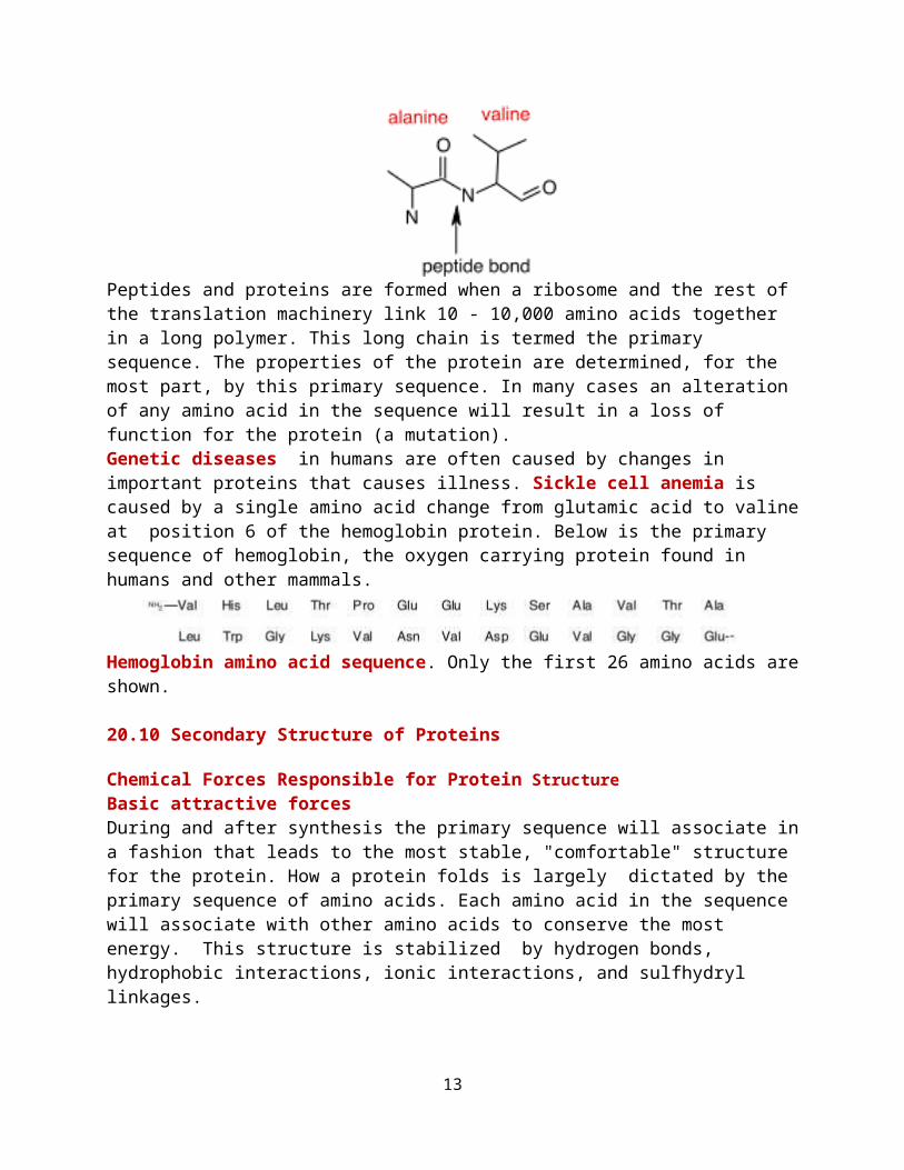
Finally there are sulfhydryl linkages. These are covalent bonds between cysteine groups. Cysteine is a unique amino acid in that it has a sulfur group available for binding to other link to help stabilize a protein as shown below in two views of sulfhydryl linkages Secondary Structure of Proteins
The organization of the amino acid chains in regular patterns or structures.
The chemical structure of a sulhydryl bond A sulfhydryl bond in a peptide
Common Secondary Structures Proteins will often have stretches of amino acids that will associate into two common structures. These are the alpha helix and the beta (pleated)
12
sheet. Formation of these structures is driven by favorable hydrogen bonding and hydrophobic interactions between nearby amino acids in the protein.
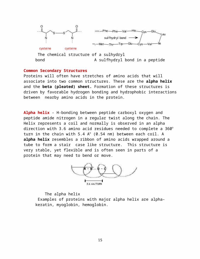
Alpha helix - H-bonding between peptide carboxyl oxygen and peptide amide nitrogen in a regular twist along the chain. The Helix represents a coil and normally is observed in an alpha direction with 3.6 amino acid residues needed to complete a 360o turn in the chain with 5.4 Ao (0.54 nm) between each coil. A alpha helix resembles a ribbon of amino acids wrapped around a tube to form a stair case like structure. This structure is very stable, yet flexible and is often seen in parts of a protein that may need to bend or move.
The alpha helix Examples of proteins with major alpha helix are alpha-keratin, myoglobin, hemoglobin.
Myoglobin Stick Structure Myoglobin Ribbon Model
Beta-Pleated Sheet In the beta sheet, two planes of amino acids will form, lining up in such a fashion so that hydrogen bonds can form between facing amino acids in each sheet. The beta pleated sheet or beta sheet is different than the alpha helix in that far distant amino acids in the protein can come togeher to form this structure. Also, the structure tends to be rigid and less flexible. H bonding between peptide carboxyl oxygen and peptide amide nitrogen between two portions of the chain or two separate peptide chains. Proteins with major beta-pleated sheet secondary structure are generally fibrous, such as silk, but pleated sheet is observed as a significant part of secondary stucture in other proteins.
13

Silk X-Ray Structure Silk Stick Structure
The Triple Helix - Three coiled chains wrapped around each other in a stiff, rod-like structure held tohether by hydrogen bonds along the backbone. Collagens is a triple helix and comprises more than 30% of the total protein in mammals. It is the major protein constituent of skin, tendons, bones, blood vessels and connective tissues.
Collagen Stick Structure Collagen End-On Stick Structure
Structures and functions of the fibrous proteins, like collagen
20.11 Tertiary Structure of Proteins
Tertiary structure of a protein refers to the overall shape of the polypeptide chain caused by folding. 1. Globular Proteins have an overall shape like a ball and are generally soluble in aquous solution. (see Myoglobin and Hemoglobin structures above)
2. Fibrous Proteins generally have rod-like shapes and are not so soluble in water. (See silk and collagen structures above).
14
Hemoglobin Stick Hemoglobin Ribbon (each chain different color)
20.12 Quaternary Structure of ProteinsQuaternary structure of protein refers to the organization among the various peptide chains in a multimeric protein:
Highest level of protein organization Present only in proteins that have 2 or more polypeptide chains
(subunits) Subunits are generally Independent of each other - not covalently
bonded Proteins with quartenary structure are often referred to as oligomeric
proteins Contain even number of subunits
Interactions between more than one chain in a protein. Some proteins are composed as aggregrated structures of multiple numbers of chains associated together in a super structure. For example, myoglobin. Hemoglobin is composed of four separate chains (2 alpha chains and 2 beta chains).
20.13 Fibrous and Globular ProteinsClassification of Proteins Classification generally associated with structure or function of the protein: for example, 1. Simple Proteins - Proteins composed only of amino acyl residues
15

2. Conjugated Proteins - Proteins which have additional functional components other than amino acyl residues (for example he Color moglobin contains the globin protein plus the heme organic molecule associated). 3. Fibrous Proteins - long protein filaments 4. Globular Proteins - generally compact, spherical shapes and very soluble in water.
20.14 Protein HydrolysisHydrolysis of proteins - reverse of peptide bond formation: Results in the generation of an amine and a carboxylic acid functional
groups. Digestion of ingested protein is enzyme-catalyzed hydrolysis Free amino acids produced are absorbed into the bloodstream and
transported to the liver for the synthesis of new proteins. Hydrolysis of cellular proteins and their resynthesis is a continuous
process.
20.15 Protein DenaturationPartial or complete disorganization of protein’s tertiary structureCooking food denatures the protein but does not change protein nutritional valueCoagulation: Precipitation (denaturation of proteins)
Egg white - a concentrated solution of protein albumin - forms a jelly when heated because the albumin is denatured
Cooking: Denatures proteins – Makes it easy for enzymes in our body to hydrolyze/digest proteinKills microorganisms by denaturation of proteinsFever: >104ºF – the critical enzymes of the body start getting denatured
Effect of changes in pH and temperature on protein stucture: denaturation of proteins. Denaturation is loss of secondary, tertiary, and quaternary structures of proteins (usually accompanied by loss of biological function of the protein). Denaturation can be the result of: 1). Heat - weakens side-chain interactions (cooking at temperatures ranging from 50-100o) 2). Mechanical Agitation (foaming) such as beating egg whites 3). Detergents - disrupt hydrophobic interactions of side-chains 4). Organic Solvents - also disrupt hydrophobic interactions of side-chains 5). pH extremes (particularly very acid conditions) - denaturation of proteins in the stomach and curdling of milk when it goes sour are examples. 6). Inorganic Salts - salt ions can disrupt salt bridges in proteins. Heavy metals such as lead, mercury, and silver react with -SH groups and cause precipitates. These metals are not readily cleared from the body, accumulate, and can be very
16

toxic because of this property. 20.16 GlycoproteinsConjugated proteins with carbohydrates linked to them:
Many of plasma membrane proteins are glycoproteins Blood group markers of the ABO system are also glycoproteins Collagen and mmunoglobulins are glycoproteins
Collagen -- glycoprotein Most abundant protein in human body (30% of total body protein) Triple helix structureRich in 4-hydroxyproline (5%) and 5-hydroxylysine (1%) — derivativesSome hydroxylysines are linked to glucose, galactose, and their disaccharides – help in aggregation of collagen fibrils.
ImmunoglobulinGlycoproteins produced as a protective response to the invasion of microorganisms or foreign molecules - antibodies against antigens.Immunoglobulin bonding to an antigen via variable region of an immunoglobulin occurs through hydrophobic interactions, dipole – dipole interactions, and hydrogen bonds.
20.17 LipoproteinsLipoprotein: a conjugated protein that contains lipids in addition to amino acidsMajor function of lipoproteins is to help suspend lipids and transport them through the bloodstreamFour major classes of plasma lipoproteins:Chylomicrons: Transport dietary triacylglycerols from intestine to liver and to adipose tissue.Very-low-density lipoproteins (VLDL): Transport triacylglycerols synthesized in the liver to adipose tissue.Low-density lipoproteins (LDL): Transport cholesterol synthesized in the liver to cells throughout the body.High-density lipoproteins (HDL): Collect excess cholesterol from body tissues and transport it back to the liver for degradation to bile acids.
17

Transport Proteins-HemolglobinThe roles of hemoglobin and myoglobin in oxygen transport and storage. Myoglobin and hemoglobin function by reversibly binding dioxygen at heme sites. Both are globular proteins, that is simply globe-shaped, and over 90% alpha-helical. They are by far the most studied metalloproteins because of their obvious physiological importance and because they could be easily isolated and purified by more traditional chemical methods. At one time or another, everyone has experienced the momentary sensation of having to stop, to "catch one's breath," until enough O2 can be absorbed by the lungs and transported through the blood stream. Imagine what life would be like if we had to rely only on our lungs and the water in our blood to transport oxygen through our bodies.
O2 is only marginally soluble (< 0.0001 M) in blood plasma at physiological pH. If we had to rely on the oxygen that dissolved in blood as our source of oxygen, we would get roughly 1% of the oxygen to which we are accustomed. (Consider what life would be like if the amount of oxygen you received was equivalent to only one breath every 5 min, instead of one breath every 3 s.) The evolution of forms of life even as complex as an earthworm required the development of a mechanism to actively transport oxygen through the system. Our blood stream contains about 150 g/L of the protein known as hemoglobin (Hb), which is so effective as an oxygen-carrier that the concentration of O2 in the blood stream reaches 0.01 M the same concentration as air. Once the Hb-O2 complex reaches the tissue that consumes oxygen, the O2 molecules are transferred to another protein myoglobin (Mb) which transports oxygen through the muscle tissue.
The site at which oxygen binds to both hemoglobin and myoglobin is the heme shown in the figure below.
18

At the center of the heme is an Fe(II) atom. Four of the six coordination sites around this atom are occupied by nitrogen atoms from a planar porphyrin ring. The fifth coordination site is occupied by a nitrogen atom from a histidine side chain on one of the amino acids in the protein. The last coordination site is available to bind an O2 molecule. The heme is therefore the oxygen-carrying portion of the hemoglobin and myoglobin molecules. This raises the question: What is the function of the globular protein or "globin" portion of these molecules?
The structure of myoglobin suggests that the oxygen-carrying heme group is buried inside the protein portion of this molecule, which keeps pairs of hemes group from coming too close together. This is important, because these proteins need to bind O2 reversibly and the Fe(II) heme, by itself, cannot do this. When there is no globin to protect the heme, it reacts with oxygen to form an oxidized Fe(III) atom instead of an Fe(II)-O2 complex.
Hemoglobin consists of four protein chains, each about the size of a myoglobin molecule, which fold to give a structure that looks very similar to myoglobin. Thus, hemoglobin has four separate heme groups that can bind a molecule of O2. Even though the distance between the iron atoms of adjacent hemes in hemoglobin is very large between 250 and 370 nm the act of binding an O2 molecule at one of the four hemes in hemoglobin leads to a significant increase in the affinity for O2 binding at the other hemes.
19

This cooperative interaction between different binding sites makes hemoglobin an unusually good oxygen-transport protein because it enables the molecule to pick up as much oxygen as possible once the partial pressure of this gas reaches a particular threshold level, and then give off as much oxygen as possible when the partial pressure of O2 drops significantly below this threshold level. The hemes are much too far apart to interact directly. But, changes that occur in the structure of the globin that surrounds a heme when it picks up an O2 molecule are mechanically transmitted to the other globins in this protein. These changes carry the signal that facilitates the gain or loss of an O2 molecule by the other hemes.
Drawings of the structures of proteins often convey the impression of a fixed, rigid structure, in which the side-chains of individual amino acid residues are locked into position. Nothing could be further from the truth. The changes that occur in the structure of hemoglobin when oxygen binds to the hemes are so large that crystals of deoxygenated hemoglobin shatter when exposed to oxygen. Further evidence for the flexibility of proteins can be obtained by noting that there is no path in the crystal structures of myoglobin and hemoglobin along which an O2 molecule can travel to reach the heme group. The fact that these proteins reversibly bind oxygen suggests that they must undergo simple changes in their conformation changes that have been called breathing motions. that open up and then close down the pathway along which an O2 molecule travels as it enters the protein. Computer simulations of the motion within proteins suggests that the interior of a protein has a significant "fluidity," with groups moving within the protein by as much as 20 nm.
20

21