Embed Size (px)
Citation preview
metastases alone.All patients had progres-sive neurological deficit and were non-ambulant at the time of surgery. Spinalsurgery was successfully performed with amean operation time of 265 minutes andmean intraoperative bleeding of 1726ml.No surgery-related complication wasobserved. After surgery, all patients re-gained ambulatory function within 2months after surgery. Cytoreductive ne-phrectomy was performed within 1month after spinal surgery for 3 patientswho showed the symptoms of ESCC asthe first presentation. All patients receivedsystemic therapy after surgery, includingcytokine or targeted therapies. Zoledronicacid was used in 4 patients. They wereambulatory with the use of the assistingapparatus, corresponding to Frankel gradeD, just before the terminal stage of thedisease for 4 � 29 months (median 10.5months). Four patients died of disease,but one patient survived for 3 years. Me-dian overall survival time after surgerywas 13 months (4 - 30 months).Conclusions: The direct decompressivesurgery effectively improved ambulatoryfunction in the non-ambulant patientswith ESCC due to metastatic RCC. Al-though it remains undetermined whetherthis surgical intervention will improve theoverall survival, patients with a single siteof cord compression, who are fit for sur-gery may be considered for decompres-sive surgery before radiotherapy.
UP-01.168Incidence of Lymph NodeInvolvement According toPathological T Stage or Tumor Gradein the Urothelial Carcinoma of theUpper Urinary TractTakagi T, Kondo T, Izuka J, Kobayashi H,Hashimoto Y, Tomita E, Yoshida K,Tanabe KDept. of Urology, Tokyo Women’sMedical University, Tokyo, Japan
Introduction and Objectives: Our retro-spective study showed survival benefits oflymphadenectomy in the advanced stageof patients with urothelial carcinoma ofthe upper urinary tract (UCUUT). To iden-tify the incidence of lymph node metasta-sis by malignant potential of the tumorswill help to determine the patients in
whom lymphadenectomy is indicated.Inthe present study, we analyzed the inci-dence of lymph node metastases accord-ing to the pathological T stage or tumorgrade in the patients with UCUUT.Materials and Methods: Until August2010, 279 patients with UCUUT under-went surgical therapy in our departmen-t.All patients were histologically con-firmed to have urothelial carcinoma.Results: Mean age of patients was 68.2 �10.5 years (36 – 90).Follow up period was50.4 � 51.2 months (1 – 231). During thefollow up period, 68 patients were foundto have lymph node metastases at the re-gional sites. Fifty-one patients were diag-nosed to have lymph node metastasis atthe time of surgery. An additional 17 pa-tients newly developed lymph node me-tastases after curative surgery.Accordingto the pathological stage, the incidence oflymphatic metastases was 20% (2/10) inpTis, 0% (0/12) in pT1, 2% (1/51) in pT1,11% (5/46) in pT2, 9% (4/48) in pT3 withinvasion to renal parenchyma in tumors ofrenal pelvis, 39% (15/36) in pT3 with in-vasion to peripelvic fat in tumors of renalpelvis, 41% (18/44) in pT3 with invasionto periureteral fat in tumors of the ureter,and 75% (24/32) in pT4.According to thetumor grade, the incidence was 0% (0/11)in grade 1, 13% (16/120) in grade 2, and35% (52/148) in grade 3.Postoperativesurvival was positively correlated with theincidence of survival.Conclusions: The risk of lymphatic me-tastases in UCUUT amplified as T stage ortumor grade increased. The invasion toadipose tissue around the renal pelvis orthe ureter dramatically increased thechance of lymphatic metastases. The con-trol of lymphatic metastasis in muscle in-vasive disease, especially in the diseaseextending into to the surrounding adiposetissue seems to be strongly associatedwith the improvement of patients’ sur-vival.
UP-01.169Impact of the Tumor EnhancementPattern in Computed Tomography inDetecting Renal Cell Carcinoma fromSmall Renal Masses: Who ReallyRequires Biopsy?Takagi T, Kondo T, Izuka J, Kobayashi H,
Hashimoto Y, Tomita E, Tanabe KDept. of Urology, Tokyo Women’sMedical University, Tokyo, Japan
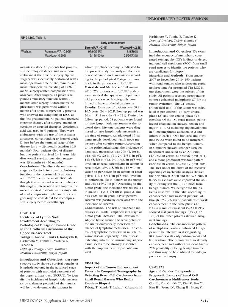
Introduction and Objective: We exam-ined the accuracy of multiphasic com-puted tomography (CT) findings in detect-ing renal cell carcinoma (RCC) from smallrenal masses to identify the patients whoare candidates for biopsy.Materials and Methods: From August2007 to December 2010, 150 patientswith renal tumors who underwent partialnephrectomy for presumed T1a RCC inour department were the subject of thisstudy. All patients received preoperativecontrast-enhanced multiphasic CT for thetumor evaluation. The CT density(Hounsfield unit) of the tumor was calcu-lated at pre-contrast (P), early arterialphase (A) and the venous phase (V).Results: Of the 150 renal masses, patho-logical examination showed benign find-ings in 11 (7%) including angiomyolipomain 4, metanephretic adenoma in 2 andothers in each 1. One hundred and thirty-nine (93%) were found to be malignant.When compared to the benign tumors,RCC tumors showed stronger early en-hancement indicated by an A/P ratio(4.47�2.30 versus 2.15�0.96, p�0.0011)and a more prominent washout pattern(0.86�0.38 versus 1.32�0.73, p�0.0005).The area under the curve of the receiveroperating characteristic analysis showedthe A/P ratio at 2.480 and the V/A ratio at0.995 as a cut-off value improved the abil-ity to distinguish the RCC tumors frombenign tumors. We categorized the pa-tients as shown in the table according toenhancement and washout patterns. Al-though 73% (22/30) of patients with weakenhancement in the early phase (A/P�2.48) and less washout (V/A�0.995)showed malignant findings, 97% (117/120) of the other patients showed malig-nant findings.Conclusions: The enhancement patternof multiphasic contrast enhanced CT ap-pears to be effective in distinguishingRCC tumors with early enhancement andlate washout. The tumors with weak earlyenhancement and without washout have a27% possibility of being benign tumorsand thus may be best advised to undergopreoperative biopsy.
UP-01.171Age and Gender, IndependentPrognostic Factors of Renal CellCarcinomas: A Multicenter StudyCho I1, Yoo C2, Oh C2, Kim S3, Km Y4,Kim H5, Seong D6, Chung H7, Hong S8,
UP-01.169, Table 1.
Early enhancement
Strong(A/P>2.48) Weak(A/P<2.48)Washout Prominent(V/A�0.995) 97/99(98%) 8/9(89%)
Weak(V/A�0.995) 12/12(100%) 22/30(73%)
UNMODERATED POSTER SESSIONS
UROLOGY 78 (Supplement 3A), September 2011 S243


![ASN Kidney Week 2016 – Renal Biopsy: Clinical Correlations · ASN Kidney Week 2016 – Renal Biopsy: Clinical Correlations. ... ( ds) DNA, ANCA, SPEP/UPEP ]. ... ASN Kidney Week](https://img.pdfslide.us/doc/110x75/5aec58b47f8b9a90318e2a7d/asn-kidney-week-2016-renal-biopsy-clinical-correlations-kidney-week-2016-.jpg)