Embed Size (px)
Citation preview
Indian Journal of Medical Case Reports ISSN: 2319–3832(Online)
An Open Access, Online International Journal Available at http://www. cibtech.org/jcr.htm
2015 Vol. 4 (4) October-December, pp. 20-27/Rakesh et al.
Case Report
© Copyright 2014 | Centre for Info Bio Technology (CIBTech) 20
UNUSUAL RECURRENT INTRAMUSCULAR HEMATOMA TURNING
TO BE A SOFT TISSUE SARCOMA
*Rakesh P., Rameshkumar R. and Mohamed Rafi
Department of Radiodiagnosis, Sri Manakula Vinayagar Medical College and Hospital,
Kalitheerthalkuppam, Pondicherry-605107, India
*Author for Correspondence
ABSTRACT
Soft-tissue sarcomas are a heterogeneous group of tumors that arise from tissue of mesenchymal origin
and are characterized by infiltrative local growth. They usually present as an asymptomatic mass
originating in an extremity but can occur anywhere in the body, particularly the trunk, retroperitoneum, or
the head and neck. We are reporting a case of 23 year old female who presented with recurrent swelling
over left groin region, twice in six months. Patient was initially suspected to have intramuscular
hematoma and on subsequent radiological and pathological investigations patient was diagnosed to have
soft tissue sarcoma.
Keywords: Soft Tissue Sarcoma, Intramuscular Hematoma
INTRODUCTION
Sarcomas are a heterogeneous group of rare tumors that arise predominantly from the embryonic
mesoderm.
Soft tissue sarcomas most commonly present as an asymptomatic mass. The various types’ of sarcomas
include bone sarcomas like (osteosarcomas and chondrosarcomas), Ewing’s sarcomas, peripheral
primitive neuroectodermal tumors, and soft tissue sarcomas. Soft tissue sarcomas can occur anywhere in
the body, but most common originate in an extremity (59%), the trunk (19%), the retroperitoneum (15%),
or the head and neck (9%).
More than 50 histological types of soft tissue sarcoma have been identified, but the most common are
malignant fibrous histiocytoma (28%), leiomyosarcoma (12%), liposarcoma (15%), synovial sarcoma
(10%), and malignant peripheral nerve sheath tumors (6%). Rhabdomyosarcoma is the most common soft
tissue sarcoma of childhood (Coindre et al., 2001; Cormier and Pollock, 2003).
CASES
A 23 year old female came with complaints of swelling in the left groin region with dull aching pain since
4 months, swelling preceded the pain.
On examination patients vitals were stable and CVS, RS, CNS was normal and on physical examination a
mass which measures approximately 15 x 10 cm noted which was firm, immobile,tender, andhaving
smooth surface was noted in the left groin.
Patients’ blood routine and urine routine were within normal limits. No history of trauma or bleeding
disorders.
Patient was then referred for USG and was found to have a well defined hypo-echoic lesion with cystic
changes, the lesion arising from the left inguinal region and extending into the pelvis with size measuring
approximately 16 x 11 x 11.6 cm.
No increased vascularity noted within the lesion. There was a mass effect of the lesion on the pelvic
organs in the form of displacement of bladder towards right side.
USG features were suggesting intramuscular hematoma in the left inguinal region with intrapelvic
extension. As USG was suggesting a large intramuscular hematoma patient was further investigated with
CT and MRI.
Both the lower poles of kidney were seen fused anterior to the aorta and diagnosed as horse shoe kidneys.
Rest of the abdominal organs were normal.
Indian Journal of Medical Case Reports ISSN: 2319–3832(Online)
An Open Access, Online International Journal Available at http://www. cibtech.org/jcr.htm
2015 Vol. 4 (4) October-December, pp. 20-27/Rakesh et al.
Case Report
© Copyright 2014 | Centre for Info Bio Technology (CIBTech) 21
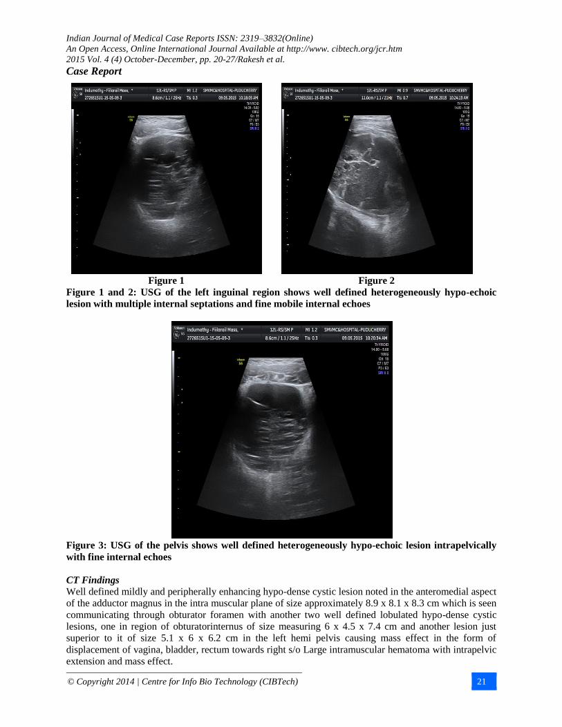
Figure 1 Figure 2
Figure 1 and 2: USG of the left inguinal region shows well defined heterogeneously hypo-echoic
lesion with multiple internal septations and fine mobile internal echoes
Figure 3: USG of the pelvis shows well defined heterogeneously hypo-echoic lesion intrapelvically
with fine internal echoes
CT Findings
Well defined mildly and peripherally enhancing hypo-dense cystic lesion noted in the anteromedial aspect
of the adductor magnus in the intra muscular plane of size approximately 8.9 x 8.1 x 8.3 cm which is seen
communicating through obturator foramen with another two well defined lobulated hypo-dense cystic
lesions, one in region of obturatorinternus of size measuring 6 x 4.5 x 7.4 cm and another lesion just
superior to it of size 5.1 x 6 x 6.2 cm in the left hemi pelvis causing mass effect in the form of
displacement of vagina, bladder, rectum towards right s/o Large intramuscular hematoma with intrapelvic
extension and mass effect.
Indian Journal of Medical Case Reports ISSN: 2319–3832(Online)
An Open Access, Online International Journal Available at http://www. cibtech.org/jcr.htm
2015 Vol. 4 (4) October-December, pp. 20-27/Rakesh et al.
Case Report
© Copyright 2014 | Centre for Info Bio Technology (CIBTech) 22
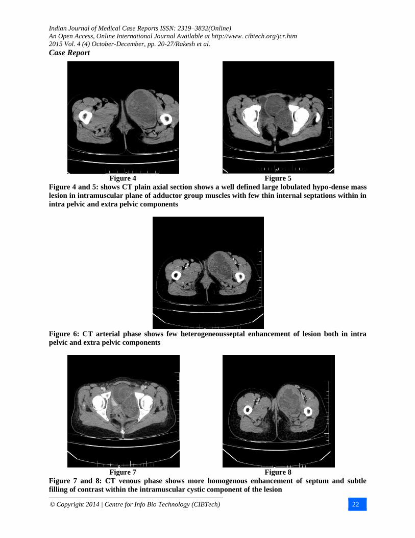
Figure 4 Figure 5
Figure 4 and 5: shows CT plain axial section shows a well defined large lobulated hypo-dense mass
lesion in intramuscular plane of adductor group muscles with few thin internal septations within in
intra pelvic and extra pelvic components
Figure 6: CT arterial phase shows few heterogeneousseptal enhancement of lesion both in intra
pelvic and extra pelvic components
Figure 7 Figure 8
Figure 7 and 8: CT venous phase shows more homogenous enhancement of septum and subtle
filling of contrast within the intramuscular cystic component of the lesion
Indian Journal of Medical Case Reports ISSN: 2319–3832(Online)
An Open Access, Online International Journal Available at http://www. cibtech.org/jcr.htm
2015 Vol. 4 (4) October-December, pp. 20-27/Rakesh et al.
Case Report
© Copyright 2014 | Centre for Info Bio Technology (CIBTech) 23
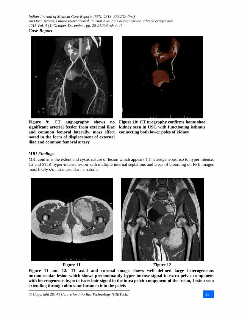
Figure 9: CT angiography shows no
significant arterial feeder from external iliac
and common femoral laterally, mass effect
noted in the form of displacement of external
iliac and common femoral artery
Figure 10: CT urography confirms horse shoe
kidney seen in USG with functioning isthmus
connecting both lower poles of kidney
MRI Findings
MRI confirms the extent and cystic nature of lesion which appears T1 heterogeneous, iso to hyper intense,
T2 and STIR hyper-intense lesion with multiple internal septations and areas of blooming on FFE images
most likely s/o intramuscular hematoma
Figure 11 Figure 12
Figure 11 and 12: T1 axial and coronal image shows well defined large heterogeneous
intramuscular lesion which shows predominantly hyper-intense signal in extra pelvic component
with heterogeneous hypo to iso-echoic signal in the intra pelvic component of the lesion, Lesion seen
extending through obturator foramen into the pelvis
Indian Journal of Medical Case Reports ISSN: 2319–3832(Online)
An Open Access, Online International Journal Available at http://www. cibtech.org/jcr.htm
2015 Vol. 4 (4) October-December, pp. 20-27/Rakesh et al.
Case Report
© Copyright 2014 | Centre for Info Bio Technology (CIBTech) 24
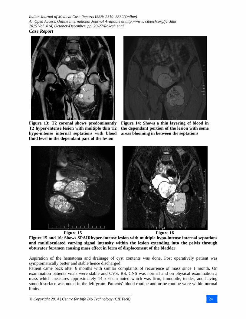
Figure 13: T2 coronal shows predominantly
T2 hyper-intense lesion with multiple thin T2
hypo-intense internal septations with blood
fluid level in the dependant part of the lesion
Figure 14: Shows a thin layering of blood in
the dependant portion of the lesion with some
areas blooming in between the septations
Figure 15 Figure 16
Figure 15 and 16: Shows SPAIRhyper-intense lesion with multiple hypo-intense internal septations
and multiloculated varying signal intensity within the lesion extending into the pelvis through
obturator foramen causing mass effect in form of displacement of the bladder
Aspiration of the hematoma and drainage of cyst contents was done. Post operatively patient was
symptomatically better and stable hence discharged.
Patient came back after 6 months with similar complaints of recurrence of mass since 1 month. On
examination patients vitals were stable and CVS, RS, CNS was normal and on physical examination a
mass which measures approximately 14 x 6 cm noted which was firm, immobile, tender, and having
smooth surface was noted in the left groin. Patients’ blood routine and urine routine were within normal
limits.
Indian Journal of Medical Case Reports ISSN: 2319–3832(Online)
An Open Access, Online International Journal Available at http://www. cibtech.org/jcr.htm
2015 Vol. 4 (4) October-December, pp. 20-27/Rakesh et al.
Case Report
© Copyright 2014 | Centre for Info Bio Technology (CIBTech) 25
Follow Up CT Images
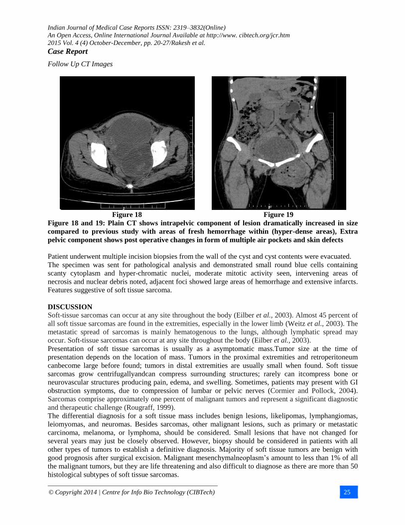
Figure 18 Figure 19
Figure 18 and 19: Plain CT shows intrapelvic component of lesion dramatically increased in size
compared to previous study with areas of fresh hemorrhage within (hyper-dense areas), Extra
pelvic component shows post operative changes in form of multiple air pockets and skin defects
Patient underwent multiple incision biopsies from the wall of the cyst and cyst contents were evacuated.
The specimen was sent for pathological analysis and demonstrated small round blue cells containing
scanty cytoplasm and hyper-chromatic nuclei, moderate mitotic activity seen, intervening areas of
necrosis and nuclear debris noted, adjacent foci showed large areas of hemorrhage and extensive infarcts.
Features suggestive of soft tissue sarcoma.
DISCUSSION
Soft-tissue sarcomas can occur at any site throughout the body (Eilber et al., 2003). Almost 45 percent of
all soft tissue sarcomas are found in the extremities, especially in the lower limb (Weitz et al., 2003). The
metastatic spread of sarcomas is mainly hematogenous to the lungs, although lymphatic spread may
occur. Soft-tissue sarcomas can occur at any site throughout the body (Eilber et al., 2003).
Presentation of soft tissue sarcomas is usually as a asymptomatic mass.Tumor size at the time of
presentation depends on the location of mass. Tumors in the proximal extremities and retroperitoneum
canbecome large before found; tumors in distal extremities are usually small when found. Soft tissue
sarcomas grow centrifugallyandcan compress surrounding structures; rarely can itcompress bone or
neurovascular structures producing pain, edema, and swelling. Sometimes, patients may present with GI
obstruction symptoms, due to compression of lumbar or pelvic nerves (Cormier and Pollock, 2004).
Sarcomas comprise approximately one percent of malignant tumors and represent a significant diagnostic
and therapeutic challenge (Rougraff, 1999).
The differential diagnosis for a soft tissue mass includes benign lesions, likelipomas, lymphangiomas,
leiomyomas, and neuromas. Besides sarcomas, other malignant lesions, such as primary or metastatic
carcinoma, melanoma, or lymphoma, should be considered. Small lesions that have not changed for
several years may just be closely observed. However, biopsy should be considered in patients with all
other types of tumors to establish a definitive diagnosis. Majority of soft tissue tumors are benign with
good prognosis after surgical excision. Malignant mesenchymalneoplasm’s amount to less than 1% of all
the malignant tumors, but they are life threatening and also difficult to diagnose as there are more than 50
histological subtypes of soft tissue sarcomas.
Indian Journal of Medical Case Reports ISSN: 2319–3832(Online)
An Open Access, Online International Journal Available at http://www. cibtech.org/jcr.htm
2015 Vol. 4 (4) October-December, pp. 20-27/Rakesh et al.
Case Report
© Copyright 2014 | Centre for Info Bio Technology (CIBTech) 26
Soft tissue sarcomas may occur anywhere but three fourths occur in the extremities, 10 percent each in the
trunk wall and retroperitoneum. Soft tissue sarcomas become more common with increasing age; the
median age is 65 years. One tenth of the patients have detectable metastases (most common in the lungs)
at diagnosis of the primary tumor. Overall, at least one-third of the patients with soft tissue sarcoma die,
mostly due to lung metastases.
The management of large hematomas in the extremities is difficult when the etiology is not clear.
Hematoma can result from trauma, surgery, or a bleeding disorder, its misdiagnosis can lead to disastrous
results, especially in the presence of malignancy (Krebs et al., 2002; Stafford et al., 2003).
When a patient presents with an expanding, non- traumatic mass mimicking a hematoma, several
differential diagnoses should be considered including aneurysm, any bleeding disorders,
chronicenlarginghematoma and soft tissue sarcoma. An aneurysm can sometimes form an intra or inter
muscular mass with hematoma.
Hematomas can be found in patients with bleeding disorders or deranged coagulation factors. Soft tissue
sarcoma should always be considered in this scenario and patient should be investigated with appropriate
history of trauma, clinical course, and MRI findings (Boyer et al., 1995; Okada et al., 2001; Naito et al.,
2000; Niimi et al., 2006; Gomez and Morcuende, 2004).
Conclusion
When a patient presents with a nonspecific and unusual history of hematoma in the extremities, it is
necessary to consider the possibility of a malignant tumor and investigate the patient’s history of trauma,
clinical course, and MRI. Moreover, prompt biopsies are recommended to facilitate correct diagnosis and
early management.
REFERENCES
Boyer MI, Wang EH and Bell RS (1995). Ruptured deep femoral artery aneurysm simulating a soft-
tissue sarcoma: a case report. Canadian Journal of Surgery 38(1) 92.
Coindre JM, Terrier P, Guillou L, Le Doussal V, Collin F, Ranchère D, Sastre X, Vilain MO,
Bonichon F and N'Guyen Bui B (2001). Predictive value of grade for metastasis development in the
main histologic types of adult soft tissue sarcomas. Cancer 91(10)1914-26.
Cormier JN and Pollock RE (2004). Soft tissue sarcomas. CA: A Cancer Journal for Clinicians 54(2)
94-109.
Eilber FC, Rosen G, Nelson SD, Selch M, Dorey F, Eckardt J and Eilber FR (2003). High-grade
extremity soft tissue sarcomas: factors predictive of local recurrence and its effect on morbidity and
mortality. Annals of Surgery 237(2) 218. Eilber FC, Rosen G, Nelson SD, Selch M, Dorey F, Eckardt J and Eilber FR (2003). High-grade
extremity soft tissue sarcomas: factors predictive of local recurrence and its effect on morbidity and
mortality. Annals of Surgery 237(2) 218.
Enzinger FM and Weiss S (1995). Soft Tissue Tumors, 3rd edition (St Louis: Mosby) 929–64.
Gomez P and Morcuende J (2004). High-grade sarcomas mimicking traumatic intramuscular
hematomas: a report of three cases. The Iowa Orthopaedic Journal 24 106.
Krebs M, Meyer B, Quehenberger P, Turecek P, Hejna M, Sperr W, Lechner K and Pabinger I
(2002). Massive postoperative intramuscular bleeding in acquired von Willebrand's disease. Annals of
Hematology 81(7) 394-6.
Naito N, Ozaki T, Kunisada T, Kawai A, Dan’ura T, Morimoto Y and Inoue H (2000). Synovial
sarcoma with a large hematoma in the inguinal region. Archives of Orthopaedic and Trauma Surgery
120(9) 533-4.
Niimi R, Matsumine A, Kusuzaki K, Okamura A, Matsubara T, Uchida A and Fukutome K (2006). Soft-tissue sarcoma mimicking large haematoma: a report of two cases and review of the literature.
Journal of Orthopaedic Surgery 14(1) 90.
Okada K, Sugiyama T, Kato H and Tani T (2001). Chronic expanding hematoma mimicking soft
tissue neoplasm. Journal of Clinical Oncology 19(11) 2971-2.

Indian Journal of Medical Case Reports ISSN: 2319–3832(Online)
An Open Access, Online International Journal Available at http://www. cibtech.org/jcr.htm
2015 Vol. 4 (4) October-December, pp. 20-27/Rakesh et al.
Case Report
© Copyright 2014 | Centre for Info Bio Technology (CIBTech) 27
Rougraff B (1999). The diagnosis and management of soft tissue sarcomas of the extremities in the adult.
Current Problems in Cancer 23(1) 7-11.
Stafford JM, James TT, Allen AM and Dixon LR (2003). Hemophilic Pseudotumor: Radiologic-
Pathologic Correlation. Radiographics 23(4) 852-6.
Weitz J, Antonescu CR and Brennan MF (2003). Localized extremity soft tissue sarcoma: improved
knowledge with unchanged survival over time. Journal of Clinical Oncology 21(14) 2719-25.