Embed Size (px)
Citation preview

Journal of the Canadian Dental Association
C L I N I C A L P R A C T I C E
April 2001, Vol. 67, No. 4 211
The complexity of the root canal system of maxillarymolars presents a constant challenge, as the dentistmust have a thorough knowledge of root canal
morphology to provide successful endodontic treatment.The form, configuration, and number of root canals
present in maxillary first molars have been discussed formore than half a century.1-12 Differences between studiesmay be attributed to variations in the technique used tostudy the morphology.
• 1925: In a study of 513 maxillary molars, Hess1 foundthat 53% had 4 canals.
• 1927: Okumara2 found that 53% of 299 extracted teethhad 2 canals in the mesiobuccal (MB) root, 2.9% had2 in the distobuccal (DB) root, and 0.3% had 2 in thepalatal root.
• 1969: Weine and others3 studied 208 extracted firstmolars, and found that 51.5% had 2 canals in the MBroot.
• 1972: Pineda and Kuttler4 found that, in 262 maxillaryfirst molars, 60.7% had 2 canals in the MB root,3.6% had 2 in the DB root, and 100% had one canal inthe palatal root.
• 1973: Seidberg and others5 found that 62% of100 extracted first maxillary molars studied in vitro had2 canals in the MB root, but only 33% of 201 maxillary
molars treated with endodontic therapy and studied invivo showed 2 canals.
• 1974: Slowey6 reported that of 103 endodontically treatedmolars 50.4% had 2 MB canals and 49.6% had only one.
• 1974: In a study of 100 decalcified maxillary first molars,Vertucci7 found that a fourth canal occurred in 55%.
• 1974: Pomeranz and Fishelberg8 examined 100 extractedmaxillary first molars and found 69% had 4 canals, with2 located in the MB root.
• 1978: Acosta Vigouroux and Trugeda Bosaans9 showedthat 72% of the 134 maxillary first molars studied hadmore than 3 root canals.
• 1983: Gray10 found a fourth canal in 56% of teethexamined.
• 1993: Thomas and others11 found that 74% of MB rootsshowed a double canal system and over 95% of palataland DB roots contained a single canal.
• 1994: Fogel and others12 reported a double canal systemin the maxillary first molar of 72% of teeth.
What is not generally understood, however, is that theremay be more than 4 canals. Acosta Vigouroux and TrugedaBosaans9 reported 5 root canals in 2.25% of maxillary firstmolars. Gray10 reported 5 canals in 2.4% of the teeth hestudied, with the following distribution: 2 MB, 2 DB, andone palatal canal. Beatty13 reported a case of 5 canals, with3 in the MB area. Harris14 documented a case where the
Unusual Maxillary First Molarwith 2 Palatal Canals Within a Single Root:
A Case Report• Stephen Johal, DMD •
A b s t r a c tA case report is presented regarding a maxillary first molar with 5 canals. The morphology is atypical because it ischaracterized by a single palatal root with 2 canals with separate orifices joining in the apical third. A literaturereview pertaining to the morphology of maxillary first molars is discussed. Modifications to the normal accessopening and examination of the pulpal floor for additional canals are stressed.
MeSH Key Words: molar/abnormalities; root canal therapy; tooth root/abnormalities
© J Can Dent Assoc 2001; 67:211-4This article has been peer reviewed.
Journal of the Canadian Dental Association212 April 2001, Vol. 67, No. 4
Johal
palatal root of a maxillary first molar had 2 separate rootcanals. Cecic and others15 reported a case with 5 canals(2 MB, 1 DB, and 2 palatal), in which the palatal canalsbifurcated at midroot into 2 distinct canals. Martinez-Bernaand Ruiz-Badanelli16 described 3 cases of maxillary firstmolars with 6 canals (3 MB, 2 DB, and 1 palatal). Bondand others17 also presented a 6-canal first molar (2 MB, 2DB, and 2 palatal).
This case report describes a permanent maxillary firstmolar with 5 root canals (2 MB, 1 DB, and 2 palatal), withunusual palatal canal morphology in that 2 canals withseparate orifices join in the apical third.
Case ReportA 42-year-old male patient presented with signs of irre-
versible acute pulpitis. He complained of pain, both spon-taneous and temperature related, on the left side of the facefor several days prior to his appointment. The patient’smedical history was noncontributory. After extensive clini-cal and radiographic examination, the maxillary left firstmolar was prepared for nonsurgical endodontic therapy. Apreoperative radiograph was obtained (Fig. 1). The patientreceived local anesthesia of 2% lidocaine with 1:100,000
epinephrine. A rubber dam was placed, and a conventionalendodontic access opening was made.
In the pulp chamber floor, the 3 principal root canalsystems were identified: MB, DB, and palatal. K-type fileswere used for gross removal of pulp tissue in the 3 maincanals. The pulp chamber floor was then explored to findthe fourth canal in the MB root. After probing with a Hu-Friedy DG 16 endodontic explorer and scraping calcifica-tions with a spoon excavator, a small hemorrhagic point wasnoted in a groove approximately 2 mm from the MBorifice in a palatal direction. At the same time a similarhemorrhagic point was noted near the orifice of the mainpalatal canal. A small amount of dentin that was occludingthe orifice of the second palatal canal was removed. Theconventional triangular access was modified to a trapezoidalshape to improve access to the additional canals. In bothareas there was a “stick” with the endodontic explorer.
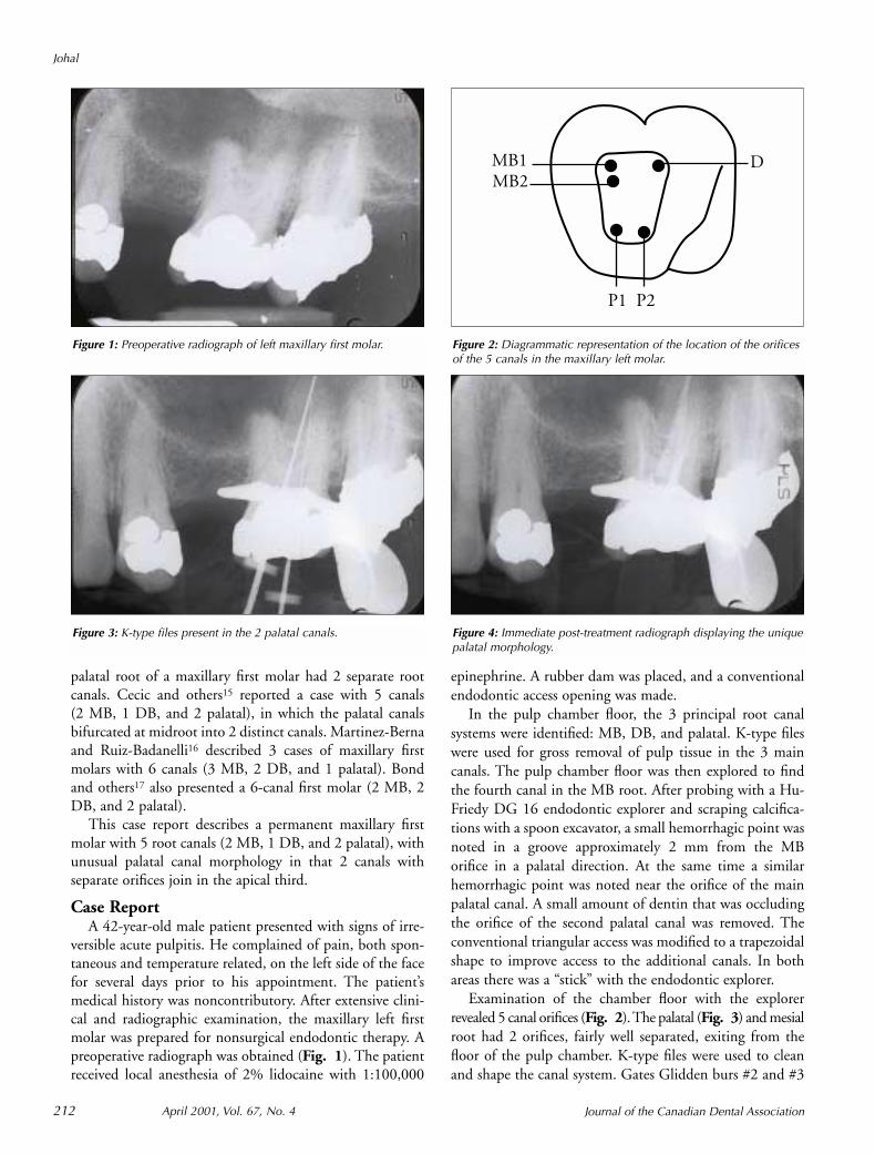
Examination of the chamber floor with the explorerrevealed 5 canal orifices (Fig. 2). The palatal (Fig. 3) and mesialroot had 2 orifices, fairly well separated, exiting from thefloor of the pulp chamber. K-type files were used to cleanand shape the canal system. Gates Glidden burs #2 and #3
Figure 1: Preoperative radiograph of left maxillary first molar.
MB1MB2
D
P1 P2
Figure 2: Diagrammatic representation of the location of the orificesof the 5 canals in the maxillary left molar.
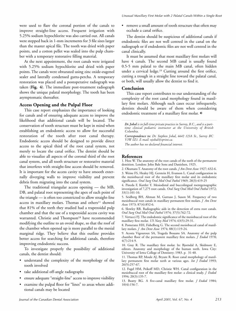
Figure 3: K-type files present in the 2 palatal canals. Figure 4: Immediate post-treatment radiograph displaying the uniquepalatal morphology.
Journal of the Canadian Dental Association April 2001, Vol. 67, No. 4 213
Unusual Maxillary First Molar with 2 Palatal Canals Within a Single Root
were used to flare the coronal portion of the canals toimprove straight-line access. Frequent irrigation with5.25% sodium hypochlorite was also carried out. All canalswere stepped back in 1-mm increments for 3 file sizes largerthan the master apical file. The tooth was dried with paperpoints, and a cotton pellet was sealed into the pulp cham-ber with a temporary restorative filling material.
At the next appointment, the root canals were irrigatedwith 5.25% sodium hypochlorite and dried with paperpoints. The canals were obturated using zinc oxide-eugenolsealer and laterally condensed gutta-percha. A temporaryrestoration was placed and a postoperative radiograph wastaken (Fig. 4). The immediate post-treatment radiographshows the unique palatal morphology. The tooth has beenasymptomatic thereafter.
Access Opening and the Pulpal FloorThis case report emphasizes the importance of looking
for canals and of ensuring adequate access to improve thelikelihood that additional canals will be located. Theconservation of tooth structure must be kept in mind whenestablishing an endodontic access to allow for successfulrestoration of the tooth after root canal therapy.Endodontic access should be designed to provide directaccess to the apical third of the root canal system, notmerely to locate the canal orifice. The dentist should beable to visualize all aspects of the coronal third of the rootcanal system, and all tooth structure or restorative materialthat interferes with straight-line access should be removed.It is important for the access cavity to have smooth exter-nally diverging walls to improve visibility and preventdebris from migrating into the canal system.
The traditional triangular access opening — the MB,DB, and palatal root representing the apex of each point ofthe triangle — is often too constricted to allow straight-lineaccess in maxillary molars. Thomas and others11 showedthat 81% of the teeth they studied had a trapezoidal pulpchamber and that the use of a trapezoidal access cavity waswarranted. Christie and Thompson18 have recommendedmodifying the outline to an ovoid shape, so that the roof ofthe chamber when opened up is more parallel to the mesialmarginal ridge. They believe that this outline providesbetter access for searching for additional canals, thereforeimproving endodontic success.
To investigate properly the possibility of additionalcanals, the dentist should:
• understand the complexity of the morphology of thetooth involved
• take additional off-angle radiographs
• ensure adequate “straight-line” access to improve visibility
• examine the pulpal floor for “lines” to areas where addi-tional canals may be located
• remove a small amount of tooth structure that often mayocclude a canal orifice.
The dentist should be suspicious of additional canals ifendodontic files are not well centred in the canal on theradiograph or if endodontic files are not well centred in thecanal clinically.
It must be assumed that most maxillary first molars willhave 4 canals. The second MB canal is usually found0.5-5 mm palatal to the main MB canal, often hiddenunder a cervical ledge.19 Cutting around the first orifice,cutting a trough in a straight line toward the palatal canal,or both, will usually allow the dentist to find it.
ConclusionThis case report contributes to our understanding of the
complexity of the root canal morphology found in maxil-lary first molars. Although such cases occur infrequently,dentists should be aware of them when consideringendodontic treatment of a maxillary first molar. C
Dr. Johal is in full-time private practice in Surrey, B.C., and is a part-time clinical pediatric instructor at the University of BritishColumbia.
Correspondence to: Dr. Stephen Johal, 6642 123A St., Surrey BCV3W 0Z1. E-mail: [email protected].
The author has no declared financial interest.
References1. Hess W. The anatomy of the root canals of the teeth of the permanentdentition. London: John Bale Sons and Danielsen, 1925.2. Okumara T. Anatomy of the root canals. J Am Dent Assoc 1927; 632-6.3. Weine FS, Healey HJ, Gerstein H, Evanson L. Canal configuration inthe mesiobuccal root of the maxillary first molar and its endodonticsignificance. Oral Surg Oral Med Oral Pathol 1969; 28(3):419-254. Pineda F, Kuttler Y. Mesiodistal and buccolingual roentgenographicinvestigation of 7,275 root canals. Oral Surg Oral Med Oral Pathol 1972;33(1):101-10.5. Seidberg BH, Altman M, Guttuso J, Suson M. Frequency of twomesiobuccal root canals in maxillary permanent first molars. J Am DentAssoc 1973; 87(4):852-6.6. Slowley RR. Radiographic aids in the detection of extra root canals.Oral Surg Oral Med Oral Pathol 1974; 37(5):762-72.7. Vertucci FJ. The endodontic significance of the mesiobuccal root of themaxillary first molar. US Navy Med 1974; 63(5):29-31.8. Pomeranz HH, Fishelberg G. The second mesiobuccal canal of maxil-lary molars. J Am Dent Assoc 1974; 88(1):119-24.9. Acosta Vigouroux SA, Trugeda Bosaans SA. Anatomy of the pulpchamber floor of the permanent maxillary first molars. J Endod 1978;4(7):214-9.10. Gray R. The maxillary first molar. In: Bjorndal A, Skidmore E,editors. Anatomy and morphology of the human teeth. Iowa City:University of Iowa College of Dentistry; 1983. p. 31-40.11. Thomas RP, Moule AJ, Bryant R. Root canal morphology of maxil-lary permanent first molar teeth at various ages. Int J Endod 1993;26(5):257-67.12. Fogel HM, Peikoff MD, Christie WH. Canal configuration in themesiobuccal root of the maxillary first molar: a clinical study. J Endod1994; 20(3):135-7.13. Beatty RG. A five-canal maxillary first molar. J Endod 1984;10(4):156-7.

Journal of the Canadian Dental Association214 April 2001, Vol. 67, No. 4
Johal
14. Harris WE. Unusual root canal anatomy in a maxillary molar.J Endod 1980; 6(5):573-5.15. Cecic P, Hartwell G, Bellizzi R. The multiple root canal system in themaxillary first molar: a case report. J Endod 1982; 8(3):113-5.16. Martinez-Berna A, Ruiz-Badanelli P. Maxillary first molars with sixcanals. J Endod 1983; 9(9):375-81.17. Bond JL, Hartwell G, Portell FR. Maxillary first molar with six canals.J Endod 1988; 14(5):258-60.18. Christie WH, Thompson GK. The importance of endodontic accessin locating maxillary and mandibular molar canals. J Can Dent Assoc1994; 60(6):527-32, 535-6.19. Lovdahl PE, Gutmann JL, Dumsha TC, Hovland EJ. CH 4:Problems in locating and negotiating fine and calcified canals. Problemsolving in endodontics. 2nd ed. St. Louis (MO): Mosby; 1992. p. 50-69.