Embed Size (px)
Citation preview
1130-0108/2016/108/8/513-516Revista española de enfeRmedades digestivas© Copyright 2016. sepd y © ARÁN EDICIONES, S.L.
Rev esp enfeRm dig2016, Vol. 108, N.º 8, pp. 513-516
CASE REPORTS
ABSTRACT
Background: Mycosis fungoides (MF) is a type of T-cell lymphoma with cutaneous involvement. It is a rare disease, of low incidence and usually affects middle-aged men. In most cases only the skin is involved although in advanced stages may present with extra cutaneous involvement including the gastrointestinal tract.
Case report: We report the first case of MF with compromise of duodenal papilla, emphasizing the diagnostic approach and a brief review of the subject.
Discussion: This case report proves the value of the endoscopic studies in patients with lymphoproliferative disorders, because of the impact in the diagnosis and prognosis. Also, this case report is relevant because there is no scientific evidence, as far as we know, of similar cases reported.
Key words: Mycosis fungoides. Lymphoma. T-cell. Cutaneous. Papilla of Vater.
INTRODUCTION
Mycosis fungoides (MF) is a type of T-cell lympho-ma with cutaneous involvement (1). It is a rare disease, of low incidence, and usually affects middle-aged men (2). In most cases, only the skin is involved (3), although in advanced stages it may present with extracutaneous involvement, including the gastrointestinal tract (4,5). We report the first case of MF involving the duodenal papilla, emphasizing the diagnostic approach and a brief review of the subject.
CASE REPORT
This is a 45-years-old man who was diagnosed with cutaneous T lymphoma five years ago and was treated with several lines of chemotherapy, from methotrexate to liposomal doxorubicin. He entered through the emer-
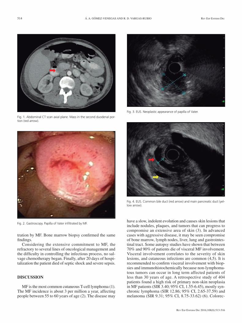
gency room of our hospital complaining of fever, fatigue, weakness for a week and purulent cutaneous lesions in the scalp and eyelids. At physical examination, he presented tachycardia, tachypnea, fever and more than 70% of his body surface was affected by extensive coalescent plaques, nodules and tumors, giving erythrodermic appearance, most with necrotic center and ulcerated. On his scalp there were areas of extensive ulcers draining pus. He received the diagnosis of infected skin lesions by MF and bleph-aroconjunctivitis. He was treated with broad-spectrum antibiotics but patient´s clinical course was torpid, with persistent signs of systemic inflammatory response, thus requiring carbapenem and oxazolidone. Fever persisted after 14 days of antibiotic treatment. A computed tomog-raphy (CT) of the chest and abdomen was performed in search of other sites of infection and as staging of the MF, besides bone marrow biopsy. On CT, some pathological cervical and inguinal lymph nodes were observed and at the level of the second portion of the duodenum there was a mass associated to pneumobilia and a dilated common bile duct (Fig. 1). A duodenoscopy showed a large duodenal papilla, its mucosal surface appearing infiltrated, eroded, friable, easily bleeding. Multiple biopsies were taken (Fig. 2). A fistulous suprapapillary orifice draining bile was also observed. An endoscopic ultrasound (EUS) showed duo-denal papilla infiltrated by a hypoechoic lesion of 26 mm x 24 mm in diameter, heterogeneous, ill-defined borders, with secondary infiltration of the pancreatic head without vascular invasion or regional lymph nodes. The bile duct and main pancreatic duct were dilated with diameters of 8 and 5 mm respectively (Figs. 3 and 4). Histopathology showed duodenal mucosa infiltrated by atypical lymphoid cells. Immunohistochemistry revealed dense infiltration of T cells positive for CD3 and CD5, CD7 focal loss of dominance and CD4 cell proliferation rate of 15% plus few CD20 positive B cells intermingled, suggestive of infil-
Received: 30/04/2015Accepted: 31/05/2015
Correspondence: Álvaro Andrés Gómez-Venegas. Services of Gastroentero-logy and Digestive Endoscopy. Hospital Universitario San Ignacio. Bogotá, Colombiae-mail: [email protected]
Gómez-Venegas AA, Vargas-Rubio RD. Unusual involvement in mycosis fun-goides: duodenal papilla. Rev Esp Enferm Dig 2016;108(8):513-516.
DOI: 10.17235/reed.2015.3831/2015
Unusual involvement in mycosis fungoides: duodenal papillaÁlvaro Andrés Gómez-Venegas and Rómulo Darío Vargas-Rubio
Services of Gastroenterology and Digestive Endoscopy. Hospital Universitario San Ignacio. Bogotá, Colombia
514 Á. A. GÓMEZ-VENEGAS AND R. D. VARGAS-RUBIO Rev esp enfeRm Dig
Rev esp enfeRm Dig 2016;108(8):513-516
tration by MF. Bone marrow biopsy confirmed the same findings.
Considering the extensive commitment to MF, the refractory to several lines of oncological management and the difficulty in controlling the infectious process, no sal-vage chemotherapy began. Finally, after 20 days of hospi-talization the patient died of septic shock and severe sepsis.
DISCUSSION
MF is the most common cutaneous T-cell lymphoma (1). The MF incidence is about 3 per million a year, affecting people between 55 to 60 years of age (2). The disease may
have a slow, indolent evolution and causes skin lesions that include nodules, plaques, and tumors that can progress to compromise an extensive area of skin (3). In advanced cases with aggressive disease, it may be seen compromise of bone marrow, lymph nodes, liver, lung and gastrointes-tinal tract. Some autopsy studies have shown that between 70% and 90% of patients die of visceral MF involvement. Visceral involvement correlates to the severity of skin lesions, and cutaneous infections are common (4,5). It is recommended to confirm visceral involvement with biop-sies and immunohistochemically because non-lymphoma-tous tumors can occur in long term affected patients of less than 30 years of age. A retrospective study of 404 patients found a high risk of primary non-skin neoplasia in MF patients (SIR 3.40; 95% CI, 1.55-6.45), mostly syn-chronic lymphoma (SIR 12.86; 95% CI, 2.65-37.59) and melanoma (SIR 9.31; 95% CI, 8.75-33.62) (6). Colorec-
Fig. 2. Gastroscopy. Papilla of Vater infiltrated by MF.
Fig. 3. EUS. Neoplastic appearance of papilla of Vater.
Fig. 4. EUS. Common bile duct (red arrow) and main pancreatic duct (yel-low arrow).
Fig. 1. Abdominal CT scan axial plane. Mass in the second duodenal por-tion (red arrow).
2016, Vol. 108, N.º 8 UNUSUAL INVOLVEMENT IN MYCOSIS FUNGOIDES: DUODENAL PAPILLA 515
Rev esp enfeRm Dig 2016;108(8):513-516
tal, liver, biliary tract, lung and urinary tumors have been also reported (7). MF patients’ survival depends on disease stage. In early stages it equals general population survival, but in extracutaneous involvement it can be as short as 1 to 2 years. Dismal prognosis factors also include older patients, refractoriness to successive treatment protocols, lymph node involvement and presence of Sézary cells in histology (7,8).
MF gastrointestinal involvement can be primary or spreading from cutaneous disease. Primary forms are quite rare and patients can present diarrhea, perforation, bleed-ing, dysphagia and intestinal obstruction (9-12). In cases of cutaneous spreading to digestive tract, involvement often occurs in the stomach, small bowel and pancreas (13-15). Arai et al. (13) reported an autopsy study of 107 patients and found pancreas involvement in 20% of cases. Madsen et al. (14) reported a 39 year old patient with refractory MF that presented biliary obstruction due to MF involve-ment of biliary tract. Gottlieb et al. (15) also reported a patient with biliary MF obstruction due to a pancreatic MF. On EUS they found a hypoechoic infiltrative lesion in the head of the pancreas, without vascular and lymph node involvement. Fine needle biopsy of the lesion shows lymphoma cells, CD45, CD3 positives, CD5, CD20, SYP, cytokeratines and chromogranin negatives, suggesting T-cell MF lymphoma.
We here report an MF patient with extensive and severe cutaneous disease, with papilla of Vater involvement, with secondary biliary tract dilation; however, it never present-ed jaundice or liver profile alterations, due to the presence of a suprapapillary fistulae. In this patient the diagnosis of MF involvement of papilla of Vater was important to estab-lish a prognosis, supporting the decision of giving only best supportive and compassionate care in a patient refrac-tory to second line treatment protocols. This case report proves the value of the endoscopic studies in patients with lymphoproliferative disorders, because of the impact in the diagnosis and prognosis. Also, this case report is relevant because there is no scientific evidence, as far as we know, of similar cases reported.
REFERENCES
1. Willemze R, Jaffe E, Burg G, et al. WHO-EORTC classification for cutaneous lymphomas. Blood 2005;105:3768-85. DOI: 10.1182/blood-2004-09-3502
2. Morales Suárez-Varela M, Llopis G, Marquina V, et al. Mycosis fungoides: review of epidemiological observations. Dermatology 2000;201:21-8. DOI: 10.1159/000018423
3. Jawed S, Myskowski P, Horwitz S, et al. Primary cutaneous T-cell lymphoma (mycosis fungoides and Sézary syndrome) Part I. Diag-nosis: Clinical and histopathologic features and new molecular and biologic markers. J Am Acad Dermatol 2014;70:205.e1-205.e16. DOI: 10.1016/j.jaad.2013.07.049
4. Long J, Mihm M. Mycosis fungoides with extracutaneous dissemina-tion: A distinct clinicopathologic entity. Cancer 1974;34:1745-55. DOI: 10.1002/1097-0142(197411)34:5<1745::AID-CNCR2820340524> 3.0.CO;2-W
5. Rappaport H, Thomas L. Mycosis fungoides: The pathology of extracutaneous involvement. Cancer 1974;34:1198-229. DOI: 10.1002/1097-0142(197410)34:4<1198::AID-CNCR2820340431> 3.0.CO;2-E
6. Ai WZ, Keegan TH, Press DJ, et al. Outcomes after diagnosis of myco-sis fungoides and Sézary syndrome before 30 years of age: A popula-tion-based study. JAMA Dermatol 2014;150:709-15. DOI: 10.1001/jamadermatol.2013.7747
7. Lindahl L, Fenger-Grøn M, Iversen L. Subsequent cancers, mortality, and causes of death in patients with mycosis fungoides and parapso-riasis: A Danish nationwide, population-based cohort study. J Am Acad Dermatol 2014;71:529-35. DOI: 10.1016/j.jaad.2014.03.044
8. Jawed S, Myskowski P, Horwitz S, et al. Primary cutaneous T-cell lym-phoma (mycosis fungoides and Sézary syndrome) Part II. Prognosis, management, and future directions. J Am Acad Dermatol 2014;70:223.e1-223.e17. DOI: 10.1016/j.jaad.2013.08.033
9. Cohen M, Widerlite L, Schechter G, et al. Gastrointestinal involvement in the Sézary syndrome. Gastroenterology 1977;73:145-9.
10. Camisa C, Goldstein A. Mycosis fungoides: Small-bowel involve-ment complicated by perforation and peritonitis. Arch Dermatol 1981;117:234-7. DOI: 10.1001/archderm.1981.01650040050021
11. Redleaf M, Moran W, Gruber B. Mycosis fungoides involving the cer-vical esophagus. Arch Otolaryngol Head Neck Surg 1993;119:690-3. DOI: 10.1001/archotol.1993.01880180110022
12. Ellis R, Smith C, Goodlad N, et al. Mesenteric vasculitis associated with Sézary syndrome. Gut 1996; 39: 334-5. DOI: 10.1136/gut.39. 2.334
13. Arai E, Katayama I, Ishihara K. Mycosis fungoides and Sézary syn-drome in Japan. Clinicopathologic study of 107 autopsy cases. Pathol Res Pract 1991;187:451-7. DOI: 10.1016/S0344-0338(11)80006-3
14. Madsen J, Tallini G, Glusac E, et al. Biliary tract obstruction secondary to mycosis fungoides: A case report. J Clin Gastroenterol 1999;28:56-60. DOI: 10.1097/00004836-199901000-00015
15. Gottlieb K, Anders K, Kaya H. Obstructive jaundice in a patient with myco-sis fungoides metastatic to the pancreas. EUS findings. JOP 2008;9:719-24.


![Significance of CD30 Expression by Epidermotropic T Cells ... · diagnosis included LyP, lymphomatoid pityriasis lichenoides and “pityriasis lichenoides-like” mycosis fungoides.[7,8]](https://img.pdfslide.us/doc/110x75/60223092b9e61714693c3a28/significance-of-cd30-expression-by-epidermotropic-t-cells-diagnosis-included.jpg)