Embed Size (px)
Citation preview
From the Department of Molecular Medicine and Surgery Karolinska Institutet, Stockholm, Sweden
UNRAVELING GENETIC MECHANISMS IN AUTISM SPECTRUM DISORDERS
Anna Bremer
Stockholm 2011
1

All previously published papers were reproduced with permission from the publisher.
Published by Karolinska Institutet. Printed by Larserics Digital Print AB.
© Anna Bremer, 2011
ISBN 978‐91‐7457‐448‐7
2
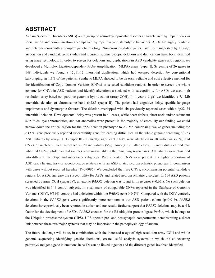
ABSTRACT Autism Spectrum Disorders (ASDs) are a group of neurodevelopmental disorders characterized by impairments in
socialization and communication accompanied by repetitive and stereotypic behaviors. ASDs are highly heritable
and heterogeneous with a complex genetic etiology. Numerous candidate genes have been suggested by linkage,
association and candidate gene studies and recurrent submicroscopic deletions and duplications have been identified
using array technology. In order to screen for deletions and duplications in ASD candidate genes and regions, we
developed a Multiplex Ligation-dependent Probe Amplification (MLPA) assay (paper I). Screening of 26 genes in
148 individuals we found a 15q11-13 interstitial duplication, which had escaped detection by conventional
karyotyping, in 1.3% of the patients. Synthetic MLPA showed to be an easy, reliable and cost-effective method for
the identification of Copy Number Variants (CNVs) in selected candidate regions. In order to screen the whole
genome for CNVs in ASD patients and identify alterations associated with susceptibility for ASDs we used high
resolution array-based comparative genomic hybridization (array-CGH). In 4-year-old girl we identified a 7.1 Mb
interstitial deletion of chromosome band 6p22.3 (paper II). The patient had cognitive delay, specific language
impairments and dysmorphic features. The deletion overlapped with six previously reported cases with a 6p22–24
interstitial deletion. Developmental delay was present in all cases, while heart defects, short neck and/or redundant
skin folds, eye abnormalities, and ear anomalies were present in the majority of cases. By our finding we could
narrow down the critical region for the 6p22 deletion phenotype to 2.2 Mb comprising twelve genes including the
ATXN1 gene previously reported susceptibility gene for learning difficulties. In the whole genome screening of 223
ASD patients by array-CGH (paper III), clinically significant CNVs were identified in 18 individuals (8%) and
CNVs of unclear clinical relevance in 20 individuals (9%). Among the latter cases, 13 individuals carried rare
inherited CNVs, while parental samples were unavailable in the remaining seven cases. All patients were classified
into different phenotype and inheritance subgroups. Rare inherited CNVs were present in a higher proportion of
ASD cases having first- or second-degree relatives with an ASD related neuropsychiatric phenotype in comparison
with cases without reported heredity (P=0.0096). We concluded that rare CNVs, encompassing potential candidate
regions for ASDs, increase the susceptibility for ASDs and related neuropsychiatric disorders. In 514 ASD patients
screened by array-CGH (paper IV), an exonic PARK2 deletion was found in three cases (~0.6%). No such deletion
was identified in 149 control subjects. In a summary of comparable CNVs reported in the Database of Genomic
Variants (DGV), 9/5141 controls had a deletion within the PARK2 gene (~0.2%). Compared with the DGV controls,
deletions in the PARK2 gene were significantly more common in our ASD patient cohort (p=0.019). PARK2
deletions have previously been reported in autism and our results further support that PARK2 deletions may be a risk
factor for the development of ASDs. PARK2 encodes for the E3 ubiquitin-protein ligase Parkin, which belongs to
the Ubiquitin proteasome system (UPS). UPS operate pre- and postsynaptic compartments demonstrating a direct
link between these two major systems that may be important in the pathophysiology of autism.
The future challenge will be to, in combination with the increased usage of high resolution array-CGH and whole
genome sequencing identifying genetic alterations, create useful analysis systems in which the co-occurring
pathways and gene-gene interactions in ASDs can be linked together and the different genes involved identified. 1
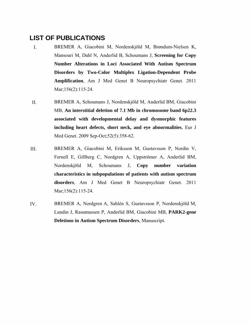
LIST OF PUBLICATIONS I. BREMER A, Giacobini M, Nordenskjöld M, Brøndum-Nielsen K,
Mansouri M, Dahl N, Anderlid B, Schoumans J, Screening for Copy
Number Alterations in Loci Associated With Autism Spectrum
Disorders by Two-Color Multiplex Ligation-Dependent Probe
Amplification, Am J Med Genet B Neuropsychiatr Genet. 2011
Mar;156(2):115-24.
II. BREMER A, Schoumans J, Nordenskjöld M, Anderlid BM, Giacobini
MB, An interstitial deletion of 7.1 Mb in chromosome band 6p22.3
associated with developmental delay and dysmorphic features
including heart defects, short neck, and eye abnormalities, Eur J
Med Genet. 2009 Sep-Oct;52(5):358-62.
III. BREMER A, Giacobini M, Eriksson M, Gustavsson P, Nordin V,
Fernell E, Gillberg C, Nordgren A, Uppströmer A, Anderlid BM,
Nordenskjöld M, Schoumans J, Copy number variation
characteristics in subpopulations of patients with autism spectrum
disorders, Am J Med Genet B Neuropsychiatr Genet. 2011
Mar;156(2):115-24.
IV. BREMER A, Nordgren A, Sahlén S, Gustavsson P, Nordenskjöld M,
Lundin J, Rasumussen P, Anderlid BM, Giacobini MB, PARK2-gene
Deletions in Autism Spectrum Disorders, Manuscript.
2
LIST OF RELATED PUBLICATIONS I. Nordgren A, Corcoran M, Sääf A, BREMER A, Kluin-Nelemans HC,
Schoumans J, Grandér D, Characterisation of hairy cell leukaemia by
tiling resolution array-based comparative genome hybridisation: a series
of 13 cases and review of the literature, Eur J Haematol. 2010 Jan
1;84(1):17-25.
II. Jonsson L, Ljunggren E, BREMER A, Pedersen C, Landén M, Thuresson K,
Giacobini M, Melke J, Mutation screening of melatonin-related genes in
patients with autism spectrum disorders, BMC Med Genomics. 2010 Apr
8;3:10.
III. Wincent J, Bruno DL, van Bon BWM, BREMER A, Stewart H, Bongers
EMHF, Ockeloen CW, Willemsen MH, Keays DA, Baird G, Newbury DF,
Kleefstra T, Marcelis C, Kini U, Stark Z, Savarirayan R, Sheffield LJ,
Zuffardi O, Slater HR, de Vries BB, Knight SJL, Anderlid BM, Schoumans
J, Sixteen new cases contributing to the characterization of patients with
distal 22q11.2 microduplications, Accepted to Mol Syndromol 2010 May
1:246-254.
IV. Bonaglia MC, Giorda R, Beri S, De Agostini C, Novara F, Fichera M, Grillo
L, Galesi O, Vetro A, Ciccone R, Bonati MT, Giglio S, Guerrini R, Osimani
S, Marelli S, Zucca C, Grasso R, Borgatti R, Mani E, Motta C, Molteni M,
Romano C, Greco D, Reitano S, Baroncini A, Lapi E, Cecconi A, Arrigo G,
Patricelli MG, Pantaleoni C, D'Arrigo S, Riva D, Sciacca F, Della
Bernardina B, Zoccante L, Darra F, Termine C, Maserati E, Bigoni S, Priolo
E, Bottani A, Gimelli S, Bena F, Brusco A, Di Gregorio E, Bagnasco I,
Giussani U, Nitsch L, Politi P, Martinez-Frias ML, Martinez-Fernandez ML,
Martinez Guardia N, BREMER A, Anderlid BM, Zuffardi O, Molecular
mechanisms generating and stabilizing terminal 22q13 deletions in 44
subjects with Phelan/McDermid syndrome, PLoS Genet. 2011
Jul;7(7):e1002173.
3
CONTENTS
1 INTRODUCTION ........................................................................................................... 7 1.1 CLINICAL FEATURES .................................................................................................................. 7
1.1.1 Treatment .............................................................................................................................. 8
1.2 GENETICs IN AUTISM SPECTRUM DISORDERS ................................................................... 10
1.3 GENETIC STUDIES IN AUTISM SPECTRUM DISORDERS .................................................... 12
1.3.1 Linkage- Association- and Candidate Gene Studies .......................................................... 12
1.3.1.1 The RELN gene ................................................................................................................. 12
1.3.1.2 The FOXP2 gene .............................................................................................................. 13
1.3.1.3 The Serotonin Transporter, SLC6A4 ................................................................................ 14
1.3.1.4 GABA receptor genes ...................................................................................................... 14
1.3.1.5 Genes encoding cell‐adhesion molecules ........................................................................ 15
1.3.1.6 Genes encoding CAM related proteins ............................................................................ 15
1.3.1.7 The PTEN gene ................................................................................................................ 16
1.3.1.8 Circadian rhythm regulation genes ................................................................................. 16
1.3.2 Chromosomal Studies ......................................................................................................... 17
1.3.2.1 Maternal duplications in chromosome band 15q11‐q13 ................................................ 17
1.3.2.2 Potocki‐Lupski syndrome ................................................................................................. 18
1.3.3 Whole-genome screening studies ....................................................................................... 18
1.3.3.1 Microdeletions and microduplications in chromosome band 15q13.3 ........................... 19
1.3.3.2 Microdeletions‐ and microduplications in chromosome band 16p11.2 .......................... 19
1.3.4 Whole-genome sequencing studies .................................................................................... 20
1.4 MECHANISMS IN CNV FORMATION ........................................................................................ 20
1.5 EPIGENETICS AND ENVIRONMENTAL FACTORS IN ASDs .................................................. 21
2 AIM OF THE THESIS .................................................................................................. 23
3 MATERIAL AND METHODS ...................................................................................... 24 3.1 PATIENT MATERIAL .................................................................................................................. 24
3.2 MULTIPLEX LIGATION-DEPENDENT PROBE AMPLIFICATION ............................................ 25
3.3 ARRAY-BASED COMPARATIVE GENOMIC HYBRIDIZATION (ARRAY-CGH) ....................... 26
3.4 FLUORESCENCE IN SITU HYBRIDIZATION (FISH) ................................................................ 27
4
3.5 DNA SEQUENCING ................................................................................................................... 28
3.6 GENOTYPING USING MICROSATELLITE MARKERS ............................................................. 28
4 RESULTS AND DISCUSSION ................................................................................... 29 4.1 GENETIC ALTERATIONS .......................................................................................................... 29
4.1.1 Alterations identified with MLPA (paper I) ........................................................................... 29
4.1.1.1 15q11‐q13 duplications of maternal origin .................................................................... 29
4.1.1.2 Single nucleotide polymorphism in the RELN gene ......................................................... 30
4.1.2 Alterations identified with array-CGH (paper II, III, IV) ........................................................ 31
4.1.2.1 6p22.3 deletion (paper II) ................................................................................................ 31
4.1.2.2 Microdeletion‐ and microduplication syndromes and recurrent alterations (paper III) . 34
4.1.2.3 Sporadic alterations (paper III) ........................................................................................ 34
4.1.2.4 Genomic alterations of unclear clinical relevance and rare variants (paper III) ............. 35
4.1.2.5 Copy number variations in the PARK2 gene (paper IV) ................................................... 35
4.2 CNV DISTRIBUTION IN DIFFERENT ASD SUB-POPULATIONS (PAPER III) ......................... 38
4.2.1 CNVs and ASD inheritance ................................................................................................. 38
4.2.2 CNVs in different phenotype subgroups ............................................................................. 39
4.2.3 CNV distribution and gender ............................................................................................... 40
5 CONCLUDING REMARKS AND FUTURE PRESPECTIVES ................................... 42
6 ACKNOWLEDGEMENTS ........................................................................................... 46
7 REFERENCES ............................................................................................................ 47
5
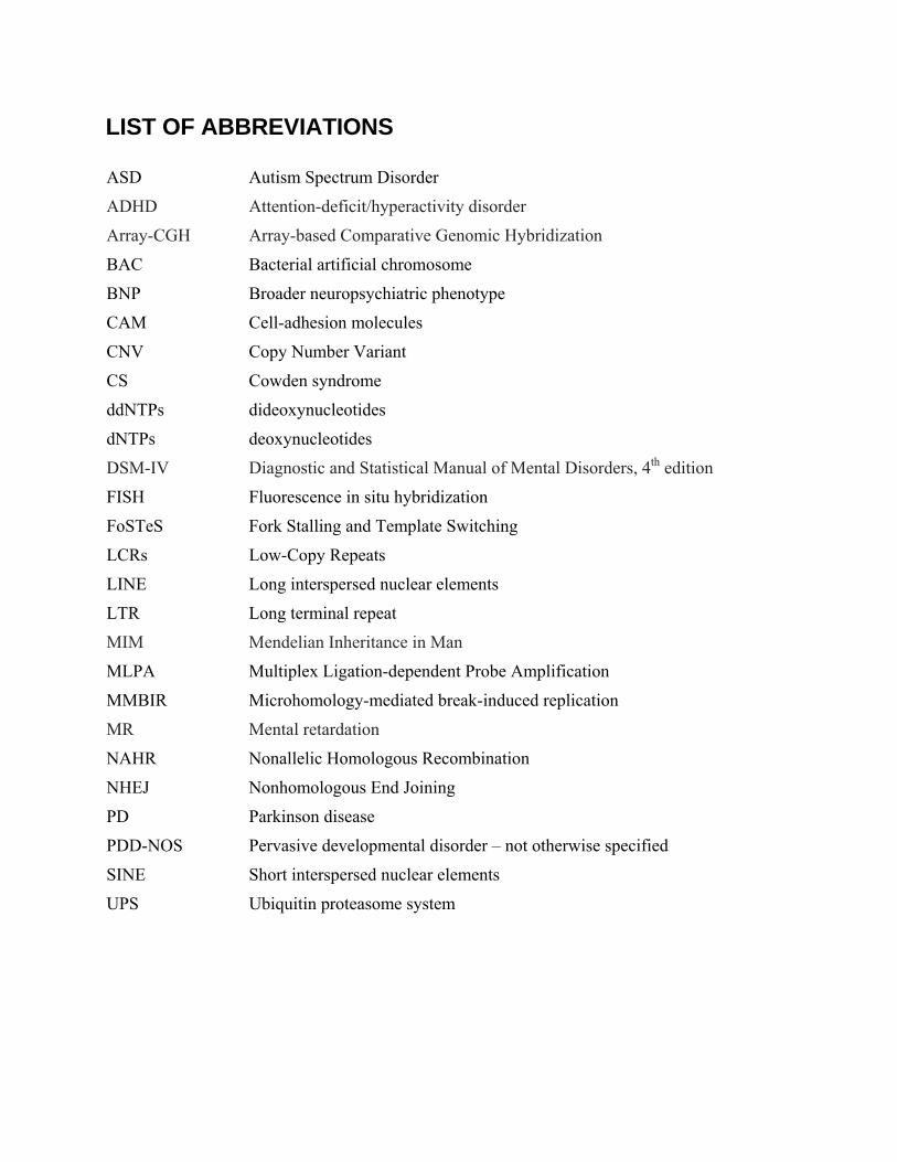
LIST OF ABBREVIATIONS
ASD Autism Spectrum Disorder
ADHD Attention-deficit/hyperactivity disorder
Array-CGH Array-based Comparative Genomic Hybridization
BAC Bacterial artificial chromosome
BNP Broader neuropsychiatric phenotype
CAM Cell-adhesion molecules
CNV Copy Number Variant
CS Cowden syndrome
ddNTPs dideoxynucleotides
dNTPs deoxynucleotides
DSM-IV Diagnostic and Statistical Manual of Mental Disorders, 4th edition
FISH Fluorescence in situ hybridization
FoSTeS Fork Stalling and Template Switching
LCRs Low-Copy Repeats
LINE Long interspersed nuclear elements
LTR Long terminal repeat
MIM Mendelian Inheritance in Man
MLPA Multiplex Ligation-dependent Probe Amplification
MMBIR Microhomology-mediated break-induced replication
MR Mental retardation
NAHR Nonallelic Homologous Recombination
NHEJ Nonhomologous End Joining
PD Parkinson disease
PDD-NOS Pervasive developmental disorder – not otherwise specified
SINE Short interspersed nuclear elements
UPS Ubiquitin proteasome system
6
1 INTRODUCTION Autism Spectrum Disorders (ASDs) [Mendelian Inheritance in Man (MIM) 209850] comprise a
heterogeneous group of disorders including autistic disorder, Aspergers syndrome and pervasive
developmental disorder – not otherwise specified (PDD-NOS). Common for ASDs are reduced
abilities in social- and communicational interaction together with behavioral problems such as
stereotypic and repetitive behaviors as well as specific interests.
Dr. Leo Kanner (1894–1981) described infantile autism already in 1943. He noted that in most of
the cases the altered behaviors were discovered very early in life and suggested that the condition
is inborn and presumably due to a genetic cause (Kanner 1943). Only one year later, pediatrician
Hans Asperger (1906-1980) described children with similar conditions but with much higher
cognitive abilities. Aspergers work remained largely unknown outside German speaking
countries until Lorna Wing brought it to attention almost 40 years later (Wing 1981). Asperger
had noted that the childrens fathers often had similar disabilities as their children. However,
during the 1950s these conditions were described to be of a psychogenic nature and assumed to
be the result of poor parenting (Kanner 1949). The term “refrigerator mother” was coined and
Bruno Bettelheim, among other leading psychologists, championed the notion that autism was
the result of a cold, distant and rejecting mother (Bettelheim 1967). These theories remained
from the 1950s throughout the 1970s. Today, there are convincing data indicating a strong
genetic component in autism. These data together with the lack of convincing evidence for
environmental factors causing autism have lead to an increasing number of genetic studies within
these disorders.
1.1 CLINICAL FEATURES ASDs manifest early in life, often before three years of age. Individuals with autism display
impairments in social interaction encompassing impairments in the use of nonverbal behaviors
such as eye contact, facial expression, body postures, and gestures as well as failure to develop
appropriate peer relationships and lack of social sharing or reciprocity. Patients with autism also
have impairments in communication, such as a delay in, or total lack of, the development of
spoken language. In patients who develop adequate language, there often remains a marked
7
8
impairment in the ability to initiate or sustain a conversation, as well as stereotyped or
idiosyncratic use of language. In addition to the social and communication interaction
impairments, individuals with autism also exhibit restricted, repetitive and stereotyped patterns
of behavior, interests, and activities, including abnormal preoccupation with certain activities and
inflexible adherence to routines or rituals.
ASDs encompass broader phenotypes including Asperger syndrome and pervasive
developmental disorder - not otherwise specified (PDD-NOS). Individuals with Asperger
syndrome do not exhibit delay in language skills and PDD-NOS is a subthreshold condition
where some but not all diagnostic features of autism are displayed. The Diagnostic and Statistical
Manual of Mental Disorders, 4th edition (DSM-IV) (American Psychiatric Association,
Washington, D.C. 1994) specifies the diagnostic criteria for the autism spectrum disorders (Table
1).
A large proportion of individuals with ASDs also have intellectual disabilities, physical/visible
malformations and/or dysmorphic features. The patient group is very heterogeneous with regard
to cognitive abilities and daily life skills.
The prevalence for these disorders is higher than previously believed and is now estimated to be
at least 1% when the whole autism spectrum is included (Baron-Cohen et al. 2009; 2003;
Gillberg and Wing 1999). In a recent comprehensive study of autism prevalence using a total
population sample, an international team of investigators from the U.S., South Korea, and
Canada estimated the prevalence of ASDs in South Korea to be 2.64%, or approximately 1 in 38
children (Kim et al. 2011). There is also a sex bias within the ASDs with a male-female ratio of
4:1, and with an increase in this ratio as the intelligence of the affected individuals increases
(Folstein and Rosen-Sheidley 2001).
1.1.1 Treatment There is today no known cure for autism but medical treatment and educational interventions can
be used to reduce some of the challenges associated with the condition. Pharmacological
treatment can be used to improve specific aspects of the disorders such as aggressive-, self-
destructive-, and overactive behaviors as well as anxiety, depression and sleep disturbances. In
order to enhance communication skills, teach social skills and reduce maladaptive behaviors,
different educational interventions are used (for review see Myers and Johnson 2007).
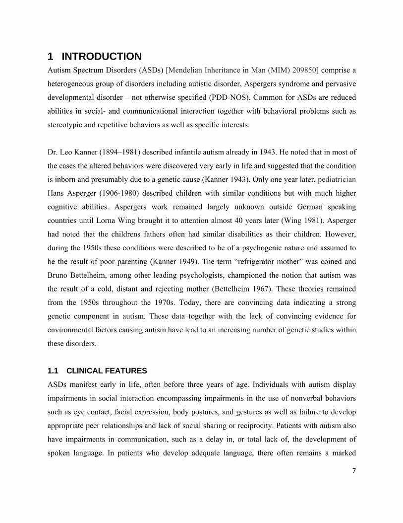
Table 1. Diagnostic criteria for ASDs according to DSM-IV.
Diagnostic criteria for Autistic Syndrome 299.00
(I) A total of six (or more) items from (A), (B), and (C), with at least two from (A), and one each from (B) and (C): (A) qualitative impairment in social interaction, as manifested by at least two of the following: 1. marked impairments in the use of multiple nonverbal behaviors such as eye-to-eye gaze, facial expression, body posture, and gestures to regulate social interaction 2. failure to develop peer relationships appropriate to developmental level 3. a lack of spontaneous seeking to share enjoyment, interests, or achievements with other people, (e.g. by a lack of showing, bringing, or pointing out objects of interest to other people) 4. lack of social or emotional reciprocity ( note: in the description, it gives the following as examples: not actively participating in simple social play or games, preferring solitary activities, or involving others in activities only as tools or "mechanical" aids ) (B) qualitative impairments in communication as manifested by at least one of the following: 1. delay in, or total lack of, the development of spoken language (not accompanied by an attempt to compensate through alternative modes of communication such as gesture or mime) 2. in individuals with adequate speech, marked impairment in the ability to initiate or sustain a conversation with others 3. stereotyped and repetitive use of language or idiosyncratic language 4. lack of varied, spontaneous make-believe play or social imitative play appropriate to developmental level (C) restricted repetitive and stereotyped patterns of behavior, interests and activities, as manifested by at least two of the following: 1. encompassing preoccupation with one or more stereotyped and restricted patterns of interest that is abnormal either in intensity or focus 2. apparently inflexible adherence to specific, nonfunctional routines or rituals 3. stereotyped and repetitive motor mannerisms (e.g. hand or finger flapping or twisting, or complex whole-body movements) 4. persistent preoccupation with parts of objects (II) Delays or abnormal functioning in at least one of the following areas, with onset prior to age 3 years: (A) social interaction (B) language as used in social communication (C) symbolic or imaginative play (III) The disturbance is not better accounted for by Rett's Disorder or Childhood Disintegrative Disorder
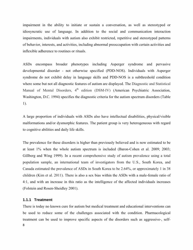
Diagnostic criteria for Asperger Syndrome 299.80
(I) Qualitative impairment in social interaction, as manifested by at least two of the following: (A) marked impairments in the use of multiple nonverbal behaviors such as eye-to-eye gaze, facial expression, body posture, and gestures to regulate social interaction (B) failure to develop peer relationships appropriate to developmental level
9
10
(C) a lack of spontaneous seeking to share enjoyment, interest or achievements with other people, (e.g. by a lack of showing, bringing, or pointing out objects of interest to other people) (D) lack of social or emotional reciprocity (II) Restricted repetitive & stereotyped patterns of behavior, interests and activities, as manifested by at least one of the following: (A) encompassing preoccupation with one or more stereotyped and restricted patterns of interest that is abnormal either in intensity or focus (B) apparently inflexible adherence to specific, nonfunctional routines or rituals (C) stereotyped and repetitive motor mannerisms (e.g. hand or finger flapping or twisting, or complex whole-body movements) (D) persistent preoccupation with parts of objects (III) The disturbance causes clinically significant impairments in social, occupational, or other important areas of functioning. (IV) There is no clinically significant general delay in language (e.g. single words used by age 2 years, communicative phrases used by age 3 years) (V) There is no clinically significant delay in cognitive development or in the development of age-appropriate self help skills, adaptive behavior (other than in social interaction) and curiosity about the environment in childhood. (VI) Criteria are not met for another specific Pervasive Developmental Disorder or Schizophrenia.
Diagnostic criteria for PDD-NOS 299-80
This category should be used when there is a severe and pervasive impairment in the development of reciprocal social interaction or verbal and nonverbal communication skills or when stereotyped behavior, interests, and activities are present but the criteria are not met for a specific pervasive developmental disorder, schizophrenia, schizotypal personality disorder, or avoidant personality disorder. For example, this category includes "atypical autism" presentations that do not meet the criteria for autistic disorder because of late age at onset, atypical symptomatology, or subthreshold symptomatology, or all of these.
1.2 GENETICS IN AUTISM SPECTRUM DISORDERS The highly genetic component in autism was first revealed through twin- and family studies. The
concordance rate in autism has been estimated to approximately 70-90% in monozygotic twins
and between 2-10% in dizygotic twin pairs (Folstein and Rosen-Sheidley 2001), making autism
one of the most genetically influenced disorders of all developmental neuropsychiatric disorders
(Kumar and Christian 2009). During the first years of the autism genetics research era, mainly
linkage, association and candidate gene screening studies were performed in order to identify
genetic regions or genes for the disorders. Numerous interesting loci and genes have been
identified but the support for association has in most cases been weak and it has shown difficult
to obtain consistent results in independent samples (Levy et al. 2009; Pinto et al. 2010b;
Veenstra-Vanderweele et al. 2004) indicating a complex genetic disorder involving multiple
interacting genes as well as epigenetic and environmental effects. The complex genetics of ASDs
is most likely due to the high degree of heterogeneity present within this patient group.
However, a few recurrent aberrations are well-known to cause ASDs. Maternal duplications of
chromosome band 15q11.2-13 are identified in 0.5-3% of ASD cases (Hogart et al. 2008) (see
chapter 1.3.2.1). In addition, autism or autistic features often occur in the single gene disorders
Fragile X syndrome, Tuberous Sclerosis, and Retts syndrome (Gillberg and Coleman 2000;
Zafeiriou et al. 2007). Reversely, these disorders explain around 2%, 0-4% and 0.5% of autism
cases, respectively (Abrahams and Geschwind 2008; Kumar and Christian 2009; Zafeiriou et al.
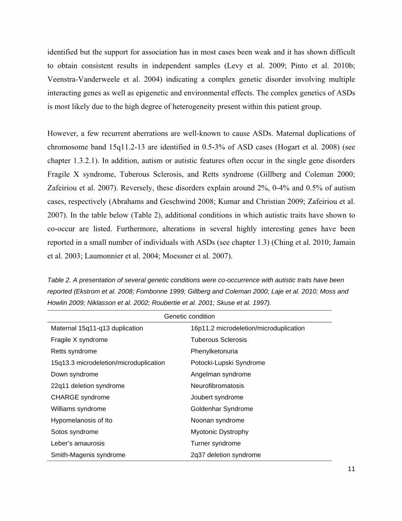
2007). In the table below (Table 2), additional conditions in which autistic traits have shown to
co-occur are listed. Furthermore, alterations in several highly interesting genes have been
reported in a small number of individuals with ASDs (see chapter 1.3) (Ching et al. 2010; Jamain
et al. 2003; Laumonnier et al. 2004; Moessner et al. 2007).
Table 2. A presentation of several genetic conditions were co-occurrence with autistic traits have been
reported (Ekstrom et al. 2008; Fombonne 1999; Gillberg and Coleman 2000; Laje et al. 2010; Moss and
Howlin 2009; Niklasson et al. 2002; Roubertie et al. 2001; Skuse et al. 1997).
Genetic condition
Maternal 15q11-q13 duplication 16p11.2 microdeletion/microduplication
Fragile X syndrome Tuberous Sclerosis
Retts syndrome Phenylketonuria
15q13.3 microdeletion/microduplication Potocki-Lupski Syndrome
Down syndrome Angelman syndrome
22q11 deletion syndrome Neurofibromatosis
CHARGE syndrome Joubert syndrome
Williams syndrome Goldenhar Syndrome
Hypomelanosis of Ito Noonan syndrome
Sotos syndrome Myotonic Dystrophy
Leber’s amaurosis Turner syndrome
Smith-Magenis syndrome 2q37 deletion syndrome
11
12
Until recently, karyotyping has been the standard method for the detection of cytogenetic
aberrations in patients with developmental disorders. The development of whole-genome
screening methodologies for the detection of CNVs, such as Array-based Comparative Genomic
Hybridization (array-CGH), provides a much higher resolution than karyotyping. This has lead to
the identification of novel microdeletion- and microduplication syndromes often associated with
an autism phenotype (Ballif et al. 2007; Miller et al. 2009; Weiss et al. 2008). The constantly
increasing resolution of the arrays has further improved the detection of copy number
abnormalities down to single genes and is likely to provide new advances in the autism genetics
field.
1.3 GENETIC STUDIES IN AUTISM SPECTRUM DISORDERS 1.3.1 Linkage- Association- and Candidate Gene Studies In linkage studies genetic markers are used in family samples in order to investigate if there are
any shared genetic regions within the affected family members that can be linked to the disease.
In association studies, markers are used to compare allele differences or genotype frequencies
between cases and controls. The genetic markers can be distributed throughout the whole
genome or in a specific chromosome of interest. In candidate gene studies, specific genes,
located in linked or associated regions, are selected and screened for alterations in a group of
patients. Genes encoding proteins involved in brain development and function or proteins for
which the level in patients has shown to be disturbed compared to controls are examples of
candidate genes. There have been reports of linkage to almost all chromosomes in ASD. There
are no regions with particularly strong evidence of linkage, but there are a few chromosomal
regions in which the linkage has been consistently replicated (for review see Bacchelli and
Maestrini 2006 and Kumar and Christian 2009). From these studies, several loci and genes have
been suggested to be involved in the etiology of ASDs and below some of the most interesting
genes are presented.
1.3.1.1 The RELN gene
The RELN gene encodes for a large secreted extracellular matrix protein believed to control cell-
cell interactions critical for cell positioning and neuronal migration during brain development.
The RELN gene maps to chromosome band 7q22 which is located in a linkage region 7q22-36
[MIM 209850] reported in multiple genome scans and association studies and RELN has shown
to be a likely candidate gene in this locus (IMGSAC 2001a; b; Serajee et al. 2006; Skaar et al.
2005; Ullmann et al. 2007). Furthermore, mice with deletions of the RELN gene have been
reported to show abnormal positioning of neurons in the cerebral cortex, cerebellum, and
hippocampus regions where alterations have been found in autistic brain (Bailey et al. 1998;
D'Arcangelo et al. 1995). It has also been demonstrated that there are impairments in the Reelin
signaling system in individuals with autism which were shown to have reduced Reelin protein
levels and elevated numbers of Reelin receptors (Fatemi et al. 2005). Although there are many
reports on an association between the RELN gene and autism, there are also several replication
studies in which no association have been identified (Bonora et al. 2003; Krebs et al. 2002; Li et
al. 2004; Zhang et al. 2002).
1.3.1.2 The FOXP2 gene
The FOXP2 gene is located within chromosome band 7q31, within the most susceptible locus of
7q22-36 reported in autism. Initially, FOXP2 was shown to be mutated in patients with language
and speech disorders (Lai et al. 2001). Even though impairments in language and speech are core
features of the autistic phenotype, indicating an involvement in autism of this gene, none of the
above mentioned patients had an ASD. However, the identification of a breakpoint disrupting the
FOXP2 gene in an autistic individual and CNVs in individuals with speech and language
impairments that in addition had an ASD diagnosis confirmed the presumed involvement of
FOXP2 (Feuk et al. 2006; Scherer et al. 2003). FOXP2 encodes for a member of the
forkhead/winged-helix (FOX) family of transcription factor mRNAs. These transcription factors
are known to regulate the expression of a variety of genes. In embryogenesis, FOXP2 has shown
to be strongly expressed in the central nervous system and highly enriched in various brain
structures (Lai et al. 2001). A recent study suggested FOXP2 to act as a regulator in many
networks important for the development of neural connections in the brain (Vernes et al. 2011).
However, several studies have presented a lack of association between FOXP2 and autism and
specific language and speech impairments concluding that FOXP2 unlikely plays a major role in
the onset of these disorders (Gauthier et al. 2003; Newbury et al. 2002). Furthermore, Feuk et al
13
14
(2006) reported a very specific phenotype of language and speech impairments present in
patients with FOXP2 alterations.
1.3.1.3 The Serotonin Transporter, SLC6A4
The serotonin transporter gene SLC6A4 map to chromosome band 17q11, a locus which has been
associated with autism in several studies [MIM 609378]. SLC6A4 has been suggested as a strong
candidate gene for several reasons. The neurotransmitter serotonin has been shown to regulate
brain development and is involved in many psychiatric conditions. It has also been shown that
patients with autism have elevated levels of serotonin in blood. Furthermore, treatment with
serotonin reuptake inhibitors (SSRIs) is sometimes effective for some of the symptoms in autism.
In many studies an association between variants in SLC6A4 and autism has been identified while
other studies have been unable to replicate the association (reviewed by Huang and Santangelo
2008).
1.3.1.4 GABA receptor genes
The gamma-aminobutyric acid (GABA) neurotransmitters are a group of inhibitory
neurotransmitters in the central nervous system which play a major role in regulating neuronal
excitability throughout the nervous system. Multiple lines of evidence indicate that the receptors
of the GABA neurotransmitters may be involved in autism. First, one of the receptor subunit
genes is located on chromosome band 15q11-q13 within the PW/AM syndrome region that is
recurrently duplicated in a proportion of ASD cases. Furthermore, studies have shown the
GABA receptor density to be reduced in the brain in ASD (Blatt et al. 2001; Oblak et al. 2010).
In multiple studies, an association between GABA receptor genes on chromosome 15 and
chromosome 4 have been reported (Buxbaum et al. 2002; Collins et al. 2006; Cook et al. 1998;
Ma et al. 2005). A report of an inversion in chromosome 4p with a breakpoint disrupting the
GABRG1 gene (Vincent et al. 2006). Many reports have been published indicating an association
between autism and the GABA receptor genes, but many reports in which no association has
been identified have been published as well (Curran et al. 2005; Maestrini et al. 1999; Martin et
al. 2000; Salmon et al. 1999).
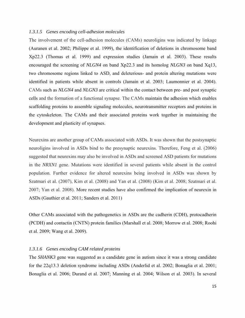
1.3.1.5 Genes encoding cell-adhesion molecules
The involvement of the cell-adhesion molecules (CAMs) neuroligins was indicated by linkage
(Auranen et al. 2002; Philippe et al. 1999), the identification of deletions in chromosome band
Xp22.3 (Thomas et al. 1999) and expression studies (Jamain et al. 2003). These results
encouraged the screening of NLGN4 on band Xp22.3 and its homolog NLGN3 on band Xq13,
two chromosome regions linked to ASD, and deleterious- and protein altering mutations were
identified in patients while absent in controls (Jamain et al. 2003; Laumonnier et al. 2004).
CAMs such as NLGN4 and NLGN3 are critical within the contact between pre- and post synaptic
cells and the formation of a functional synapse. The CAMs maintain the adhesion which enables
scaffolding proteins to assemble signaling molecules, neurotransmitter receptors and proteins in
the cytoskeleton. The CAMs and their associated proteins work together in maintaining the
development and plasticity of synapses.
Neurexins are another group of CAMs associated with ASDs. It was shown that the postsynaptic
neuroligins involved in ASDs bind to the presynaptic neurexins. Therefore, Feng et al. (2006)
suggested that neurexins may also be involved in ASDs and screened ASD patients for mutations
in the NRXN1 gene. Mutations were identified in several patients while absent in the control
population. Further evidence for altered neurexins being involved in ASDs was shown by
Szatmari et al. (2007), Kim et al. (2008) and Yan et al. (2008) (Kim et al. 2008; Szatmari et al.
2007; Yan et al. 2008). More recent studies have also confirmed the implication of neurexin in
ASDs (Gauthier et al. 2011; Sanders et al. 2011)
Other CAMs associated with the pathogenetics in ASDs are the cadherin (CDH), protocadherin
(PCDH) and contactin (CNTN) protein families (Marshall et al. 2008; Morrow et al. 2008; Roohi
et al. 2009; Wang et al. 2009).
1.3.1.6 Genes encoding CAM related proteins
The SHANK3 gene was suggested as a candidate gene in autism since it was a strong candidate
for the 22q13.3 deletion syndrome including ASDs (Anderlid et al. 2002; Bonaglia et al. 2001;
Bonaglia et al. 2006; Durand et al. 2007; Manning et al. 2004; Wilson et al. 2003). In several
15
16
studies, mutations leading to heterozygous deletions of the gene in patients with ASDs have been
identified suggesting that the SHANK3 gene may be one synaptic pathway that can be altered in
ASDs (Durand et al. 2007; Moessner et al. 2007). Shank proteins are scaffolding proteins in the
synapse formation and connect membrane proteins to the actin cytoskeleton and G-protein-
coupled signaling pathways. Shank proteins also play a role in dendritic spine maturation
(Roussignol et al. 2005). Further, SHANK3 is able to bind neuroligins which interact with
neurexins indicating that this network is a strong candidate for being altered in ASDs.
The CNTNAP2 gene, encoding a contactin associated protein that shows structural similarity to
neurexins, has also been suggested to be involved in autism. The gene is located on chromosome
7q35 within the most susceptible locus of 7q22-36 reported in autism. Linkage- and association
studies indicated variations within the CNTNAP2 gene to be associated with ASDs (Alarcon et
al. 2008; Arking et al. 2008). However, it has also been reported that common variants were not
significantly increased in ASD individuals and that alterations in CNTNAP2 only may have a
modest contribution in ASDs (Bakkaloglu et al. 2008).
1.3.1.7 The PTEN gene
The PTEN gene is a tumor suppressor gene localized to chromosome band 10q23. Individuals
with Cowden syndrome (a cancer syndrome) and other related disorders are characterized by
PTEN mutations. Many of these patients have neurobehavioural features including mental
retardation, autism, seizures as well as overgrowth and macrocephaly. Mutations in the PTEN
gene have been found in a subgroup of autism patients recognized by having extreme
macrocephaly (Butler et al. 2005). A remarkable finding concerning the PTEN gene is that
neurological abnormalities in PTEN knockout mice have shown to be reversed by treatment with
rapamycin (Zhou et al. 2009).
1.3.1.8 Circadian rhythm regulation genes
A consistent finding in autism patients is low levels of melatonin (Kulman et al. 2000; Melke et
al. 2008; Tordjman et al. 2005). Melatonin is a hormone secreted by the pineal gland serving as
the signal for darkness in the body. It’s involved in various physiologic functions, including
sleep induction, circadian rhythm regulation, and immune response (Simonneaux and Ribelayga
2003). The cause of decreased levels of melatonin in autism patients has been shown, at least
partly, to be due to low activity of the acetylserotonin O-methyltransferase (ASMT), which is the
last enzyme in the melatonin synthesis (Melke et al. 2008). Detection of various mutations in the
ASMT gene, such as splice site and stop mutations and duplications within the gene or in the
promoter sequence, has been identified and shown to be significantly more common in ASD
patients than in healthy controls (Cai et al. 2008; Melke et al. 2008). In addition, other genes
involved in circadian rhythm regulation and central effects of melatonin, have also been
associated with autism. This indicates that the melatonin signaling pathway and the ASMT gene
may play an important role in the etiology of ASDs (Melke et al. 2008; Nicholas et al. 2007).
1.3.2 Chromosomal Studies By using traditional cytogenetic analysis, chromosomal G-banding techniques and fluorescent in
situ hybridization, chromosomal abnormalities can be identified in 3-7% of ASD cases
(Veenstra-Vanderweele et al. 2004; Vorstman et al. 2006; Xu J 2004). There are several
chromosome syndromes in which ASDs often are present and the majority of these syndromes
are presented in Table I (Chapter 1.2).
1.3.2.1 Maternal duplications in chromosome band 15q11-q13
The most frequent cytogenetic anomaly in ASD is duplications of chromosome band 15q11-
15q13 of maternal origin. Duplications of paternal origin give no or possibly a very mild
phenotype. However, ASDs are not fully penetrant in the 15q11-q13 duplication syndrome but
the majority of cases fulfill the criteria for an ASD diagnosis or show ASD-like behaviors
(Battaglia et al. 2010; Rineer et al. 1998). In ASD cases, maternal duplications of chromosome
band 15q11-13 are identified in 0.5-3%. The phenotypes of patients with a 15q duplication are
highly variable and include hypotonia, hypogonadism, fine motor delays, speech and language
delays, moderate to severe mental retardation, epilepsy, and other behavioral problems (Hogart
et al. 2008).
17
18
1.3.2.2 Potocki-Lupski syndrome
The reciprocal duplication of the deletion syndrome Smith-Magenis on chromosome band
17p11.2 is referred to as the Potocki-Lupski Syndrome. The clinical features of the duplications
are milder than the features present in the deletions and include dysmorphic features,
developmental delay, mental retardation, language impairment, and ASDs (Potocki et al. 2000).
The most likely candidate gene within this region is the dosage sensitive gene RAI1 mainly
responsible for the phenotype outcome in the Smith-Magenis Syndrome (Potocki et al. 2007).
The RAI1 gene is expressed at high levels in neuronal tissues. In both mice and humans,
decreased or increased dosage of RAI1 causes distinct neurobehavioral and craniofacial features
(Carmona-Mora and Walz 2010; Walz et al. 2006).
1.3.3 Whole-genome screening studies The usage of high-resolution whole genome screening methodologies such as array-CGH has
shown that de novo and rare CNVs are significantly more common in individuals with ASDs
than in healthy controls (Bucan et al. 2009; Levy et al. 2011; Marshall et al. 2008; Pinto et al.
2010a; Sebat et al. 2007). Many of these variants are unique and include many different genes
making it difficult to sort out what genes and pathways in fact are involved in the development
of ASDs. The future challenge will be to create useful analysis systems in which the co-
occurring pathways and gene-gene interactions in ASDs can be linked together and the different
genes involved identified. Actually, just recently the first study in which such a method had been
developed and implicated on de novo and rare CNVs identified in a cohort of ASD individuals
was published (Gilman et al. 2011). The results showed support for the hypothesis that autism
primarily is due to malfunctions within the synaptic and neuronal connectivity. However, several
CNVs including only one or a few genes have been identified in the majority of the whole
genome screening studies previously published (Bucan et al. 2009; Glessn er et al. 2009; Levy et
al. 2011; Marshall et al. 2008; Pinto et al. 2010a; Sanders et al. 2011; Szatmari et al. 2007). Most
of these reports support the involvement of genes encoding proteins important for correct
neuronal and synaptic development. Nevertheless, the increased usage of array-CGH has lead to
the identification of novel microdeletion- and microduplication syndromes associated with
ASDs.
1.3.3.1 Microdeletions and microduplications in chromosome band 15q13.3
Through usage of array-CGH, microdeletions and microduplications in chromosome band
15q13.3 were discovered to recurrently occur in ASD patients (Miller et al. 2009; Pagnamenta et
al. 2009; Sharp et al. 2008). This region is located distally to the Prader-Willi/Angelman region.
Patients with the deletion or duplication show phenotypes including minor dysmorphic features,
seizures, cognitive impairments, ASD, language delay, ADHD, anxiety disorder and mood
disorder. ASDs are not always, but commonly represented in this syndrome. The 15q13.3 CNVs
are often inherited and the duplications seem much less penetrant than the deletions (Helbig et al.
2009). Interestingly, many of the patients reported have been adopted and their biological parents
have been reported to show psychiatric conditions including the above mentioned phenotypes as
well as bipolar disorder and schizophrenia (Ben-Shachar et al. 2009). The CHRNA7 gene is one
of the at least six genes located in this region. It encodes the α-subunit of the neuronal nicotinic
receptor, which is a synaptic ion channel protein. The CHRNA7 gene is considered a compelling
candidate gene since it has been associated with epilepsy and broader phenotypes of
neuropsychiatric and neurological disorders (Miller et al. 2009).
1.3.3.2 Microdeletions- and microduplications in chromosome band 16p11.2
Chromosome band 16p11.2 deletions and duplications, identified by array-CGH, have been
associated with mental retardation, ASDs, behavioral problems such as ADHD, seizures, and
schizophrenia (Kumar et al. 2008; McCarthy et al. 2009; Shimojima et al. 2009; Shinawi et al.
2010; Weiss et al. 2008). In ASD patients, these deletions and duplications have shown to be
present in approximately 1% of individuals and the variants are significantly more common in
patients than in healthy controls (Kumar et al. 2008; Weiss et al. 2008). Over twenty genes are
located in this region. Several of these have shown to be expressed in the brain and considered
good candidates for being responsible for the phenotypes present in this syndrome (Kumar et al.
2008; Weiss et al. 2008).
19
20
1.3.4 Whole-genome sequencing studies During recent years, new methodologies in which the whole genome can be sequenced have
been developed. These methods can be used either for sequencing the whole genome, or only the
coding sequences within the whole genome referred to as exome sequencing, or sequencing of
selected candidate regions. In contrast to arrayCGH, which identifies CNVs that most often
include several genes, this approach has greater potential to implicate single genes in ASDs. The
first study of exome sequencing in ASD patients was recently published (O'Roak et al. 2011).
The exome was sequenced in 20 individuals with sporadic ASD and their parents. The authors
found several de novo and protein altering mutations. Four of these were considered potentially
causative and involved the genes FOXP1, GRIN2B, SCN1A and LAMC3. In the FOXP1 mutation
carrier, the authors also observed a rare inherited CNTNAP2 missense variant and suggested a
multi-hit model for disease risk being involved. Such a model has previously been predicted for
ASDs and other conditions and is described elsewhere (see chapter 4.2.1 and the discussion in
paper III) (Girirajan et al. 2010).
1.4 MECHANISMS IN CNV FORMATION This thesis comprises studies mainly exhibiting the involvement of CNVs as alterations in the
genome leading to the development of ASDs. CNVs are structural variations of the genome that
results in the cell having an abnormal number of copies of one or more sections of the DNA.
CNVs are either inherited or caused by de novo mutations. CNVs have like other genetic
mutations been associated with the susceptibility to disease and have in previous studies been
associated with autism, schizophrenia and idiopathic learning disability. There are three
mechanisms that most likely generate the majority of CNV rearrangements in the human
genome. These include Nonallelic Homologous Recombination (NAHR), Nonhomologous End
Joining (NHEJ) and Fork Stalling and Template Switching (FoSTeS) (for review see
Stankiewicz and Lupski 2010 and Zhang et al. 2009).
NAHR is caused by the alignment of and the following crossover between two nonallelic Low-
Copy Repeats (LCRs) of high similarity. NAHR can mediate the formation of deletions and
duplications as well as inversions and translocations. This mechanism has shown to be
responsible for the majority of recurrent reciprocal deletions and duplications.
In contrast to NAHR, NHEJ does not require LCRs for the recombination but may also be
stimulated by genome architecture since the breaks often are located within repetitive elements
such as long interspersed nuclear elements (LINE), short interspersed nuclear elements (SINE)
and long terminal repeat (LTR) retroposons. In NHEJ, double stranded breaks of broken DNA
ends are first detected, then bridged, modified, and finally ligated together. NHEJ often leave a
“molecular scar” in the form of loss or addition of several nucleotides at the DNA end junction.
The NHEJ mechanism mediates nonrecurrent genomic rearrangements.
The FoSTeS mechanism is based on DNA replication error. In this model, the DNA replication
fork stalls and the strand disengage from the original template and anneals to another replication
fork in physical proximity on which the DNA synthesis restarts. Further, a template-switch
model has been proposed called the microhomology-mediated break-induced replication
(MMBIR) model. The identification of a new template in the FoSTeS mechanism has been found
to be utilized by nucleotide microhomology at the new template sequence. FoSTeS/MMBIR
seems to be a major mechanism for generating nonrecurrent CNVs and complex genomic
rearrangements.
1.5 EPIGENETICS AND ENVIRONMENTAL FACTORS IN ASDs Autism spectrum disorders are multifactorial, with many risk factors acting together to produce
the phenotype. The concordance rate of 70-90% in monozygotic twins suggests that the main
cause is genetic but that there may exist other risk factors as well. Both epigenetic and
environmental factors have been suggested to be involved in ASDs. Epigenetic modifications
include DNA methylation, RNA modification, and histone and non-histone protein modifications
(methylation, acetylation, phosphorylation, ubiquitination). Interestingly, several of the linkage
peaks found in ASDs overlap or are in close proximity to regions that are known to be subject to
imprinting (chromosomes15q11–13, 7q21–31.31, 7q32.3–36.3) (Schanen 2006). Furthermore,
the increased susceptibility for ASDs in males than in females has been argued to depend on
21
22
epigenetic effects such as an increased vulnerability for dysregulation of methylation of brain-
expressed genes on the X-chromosome and sex-specific responses to different hormones (Carter
2007; Jones et al. 2008). However, males may also be more vulnerable to minor variations in
ASD susceptibility genes located on the X-chromosome (Noor et al. 2010). Paternal age has also
been shown to act as a risk factor for autism in several studies. De novo germline mutations and
epigenetic alterations have been suggested to act as possible biological mechanisms increasing
with paternal age and thereby increasing risk for ASDs (Hultman et al. 2010). Environmental
factors that have shown to contribute to causing autism are exposure of alcohol or medicine, such
as valproate and thalidomide, during pregnancy (Williams et al. 2001).
2 AIM OF THE THESIS The aim of the thesis was to identify genetic alterations and susceptibility genes involved in the development of ASD in order to gain a better understanding of the underlying genetic mechanisms of as well as improve the genetic diagnostic tools for this group of disorders. Specifically we wanted to address the following questions:
‐ Are CNVs common in ASD candidate genes previously identified with association-, linkage-, candidate gene- and whole-genome screening studies (Paper I and III)?
‐ How common are CNVs in patients with ASD discovered by array based whole genome screening methods and are there phenotypic differences with regard to CNV presence (paper III)?
‐ How common are alterations in potential ASD candidate genes in patients with ASD when compared to healthy controls (paper I and IV)?
‐ Is MLPA a suitable and successful screening method for the discovery of CNVs involving ASD candidate genes and how does it compare to whole genome array based screening (Paper I and III)?
‐ Can we expand the knowledge of lesser known microdeletion syndromes (Paper II and III)?
23
24
3 MATERIAL AND METHODS For detailed descriptions of materials and methods, please see the individual papers.
3.1 PATIENT MATERIAL In all studies, blood samples from ASD patients were collected for DNA extraction at the
Karolinska University Hospital. DNA was isolated from peripheral blood samples using standard
procedures. All patients were tested negative for Fragile-X syndrome and the majority of cases
were investigated by conventional karyotyping (see papers for more information).
In paper I, we received additional DNA samples from our collaborators at the Uppsala
University, Sweden and the John F Kennedy Institute, Denmark.
In paper III, the 223 patients collected at the Karolinska University Hospital were divided into
different subgroups regarding their family history of ASDs and related broader neuropsychiatric
phenotypes (BNPs) and into different subgroups depending on phenotypic expressions. BNPs
included autistic traits and related neuropsychiatric disorders such as attention-
deficit/hyperactivity disorder (ADHD), mental retardation (MR), dyslexia, and/or other speech
and language disorders. The phenotypic subgrouping was made with regard to syndromic
features and cognitive ability. Patients with dysmorphic features and/or growth disorders and/or
malformations were classified as syndromic, and mental retardation was defined as an IQ below
70 (measured by Weschsler scales sometimes in combination with Leiter or Wechsler non-verbal
scale) in conjunction with significant limitations in the adaptive functioning.
‐ There were four different types of family history- or inheritance subgroups. Of the
223 participating ASD individuals, 164 cases were sporadic – they had no relatives
with ASDs or BNPs, 27 cases were familial – they had one or several first degree
relatives with ASDs, 25 cases were BNP-familial – they had one or several first
and/or second degree relatives with BNPs and/or one or several second degree
relatives with ASDs, and the seven remaining cases had an unknown family history
due to adoption in three of the cases and due to lack of information in the four
remaining cases (Table in chapter Results and discussion).
‐ There were four different types of phenotypic subgroups. Of the 223 patients
participating, 25 cases were syndromic with an IQ within the normal range, 45 cases
were syndromic and had MR, 60 cases were nonsyndromic with a normal IQ, and 93
patients were non-syndromic but had MR (Tables in chapter Results and discussion).
3.2 MULTIPLEX LIGATION-DEPENDENT PROBE AMPLIFICATION Multiplex ligation-dependent probe amplification (MLPA) is a method where the copy number
of many loci can be investigated in a single reaction. In MLPA, the two oligonucleotide half-
probes hybridize with the target DNA sequence adjacently permitting ligation between the half-
probes creating one whole probe. The MLPA half-probes are designed in such way that the
length of each ligation product has a unique size between 87 and 130 nucleotides. The ligation
products are amplified in a subsequent PCR amplification and by using fluorescently labeled
primers the PCR-product can be separated and measured by capillary electrophoresis.
Comparison of the relative peak area of each amplification product to a normal control reflects
the relative copy number of the target sequence. MLPA is a method well suitable for screening
many loci in a large group of patients. (Paper I, III and IV)
25
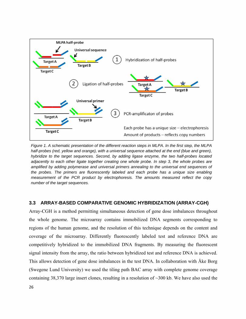
Figure 1. A schematic presentation of the different reaction steps in MLPA. In the first step, the MLPA half-probes (red, yellow and orange), with a universal sequence attached at the end (blue and green), hybridize to the target sequences. Second, by adding ligase enzyme, the two half-probes located adjacently to each other ligate together creating one whole probe. In step 3, the whole probes are amplified by adding polymerase and universal primers annealing to the universal end sequences of the probes. The primers are fluorescently labeled and each probe has a unique size enabling measurement of the PCR product by electrophoresis. The amounts measured reflect the copy number of the target sequences.
3.3 ARRAY-BASED COMPARATIVE GENOMIC HYBRIDIZATION (ARRAY-CGH) Array-CGH is a method permitting simultaneous detection of gene dose imbalances throughout
the whole genome. The microarray contains immobilized DNA segments corresponding to
regions of the human genome, and the resolution of this technique depends on the content and
coverage of the microarray. Differently fluorescently labeled test and reference DNA are
competitively hybridized to the immobilized DNA fragments. By measuring the fluorescent
signal intensity from the array, the ratio between hybridized test and reference DNA is achieved.
This allows detection of gene dose imbalances in the test DNA. In collaboration with Åke Borg
(Swegene Lund University) we used the tiling path BAC array with complete genome coverage
containing 38,370 large insert clones, resulting in a resolution of ~300 kb. We have also used the
26
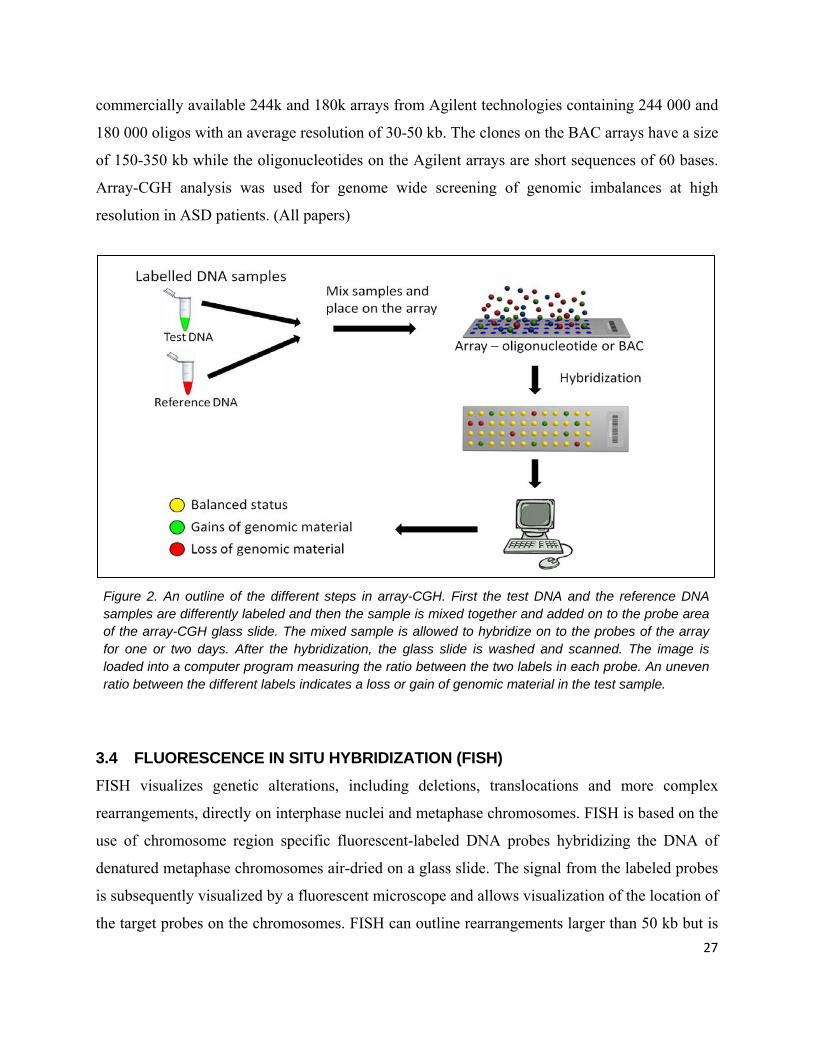
commercially available 244k and 180k arrays from Agilent technologies containing 244 000 and
180 000 oligos with an average resolution of 30-50 kb. The clones on the BAC arrays have a size
of 150-350 kb while the oligonucleotides on the Agilent arrays are short sequences of 60 bases.
Array-CGH analysis was used for genome wide screening of genomic imbalances at high
resolution in ASD patients. (All papers)
Figure 2. An outline of the different steps in array-CGH. First the test DNA and the reference DNA samples are differently labeled and then the sample is mixed together and added on to the probe area of the array-CGH glass slide. The mixed sample is allowed to hybridize on to the probes of the array for one or two days. After the hybridization, the glass slide is washed and scanned. The image is loaded into a computer program measuring the ratio between the two labels in each probe. An uneven ratio between the different labels indicates a loss or gain of genomic material in the test sample.
3.4 FLUORESCENCE IN SITU HYBRIDIZATION (FISH) FISH visualizes genetic alterations, including deletions, translocations and more complex
rearrangements, directly on interphase nuclei and metaphase chromosomes. FISH is based on the
use of chromosome region specific fluorescent-labeled DNA probes hybridizing the DNA of
denatured metaphase chromosomes air-dried on a glass slide. The signal from the labeled probes
is subsequently visualized by a fluorescent microscope and allows visualization of the location of
the target probes on the chromosomes. FISH can outline rearrangements larger than 50 kb but is 27
28
not optimal for the detection of small tandem duplications. In addition, the number of loci
investigated is limited and the method is not suitable for a multiple assay. This technique was
used to confirm chromosome rearrangements detected by array-CGH. (Paper II and III)
3.5 DNA SEQUENCING DNA sequencing analysis is a method that detects sequence alterations such as base substitutions
and small insertions or deletions. In dye-terminator sequencing, dideoxynucleotides (ddNTPs)
labeled with different fluorescent colors, one for each nucleotide type (A,T,G,C), are mixed with
deoxynucleotides (dNTPs), sequencing enzyme polymerase, primer sequence and the double
stranded PCR product of interest. The double stranded PCR product is denatured and hybridized
with the sequencing primer, which allows the polymerase enzyme to incorporate additional
nucleotides. Each time a ddNTP is incorporated, the chemical properties of the ddNTP disallow
further incorporation of nucleotides. The final product contains DNA strands of different length
with a labeled ddNTP at the 3’end. Size separation of the DNA strands is performed with capillary
electrophoresis and fluorescence is detected with and a CCD camera in an automatic DNA
sequencer. The differently labeled nucleotides are presented as peaks of different colors, and
sequence alterations are seen as overlapping peaks in generated chromatograms. (Paper I)
3.6 GENOTYPING USING MICROSATELLITE MARKERS Microsatellites consist of di, tri, or tetra repeats. They are highly polymorphic, dense and spaced
across the whole genome and easy to amplify by PCR by using different fluorescently labeled
primers and allele length PCR products. The PCR-product is separated and measured by
capillary electrophoresis revealing the number of repeats within the different alleles.
Microsatellite markers were used to trace inheritance patterns. (Paper I)
4 RESULTS AND DISCUSSION Two methods for detecting genetic alterations in patients with ASDs have mainly been used in
this thesis; MLPA and array-CGH. By using these methods we have screened cohorts of ASD
patients by two different types of approaches. MLPA for screening selected candidate genes and
regions for CNVs and array-CGH for screening of the whole genome for rare CNVs susceptible
for ASDs.
4.1 GENETIC ALTERATIONS
Table 3. A presentation of the methods used and the different findings identified in each paper.
Method Study Genetic alterations
MLPA Paper I Two cases with a 15q11-q13 duplication
Three cases with a RELN SNPs
Array-CGH Paper II One case with a 6p22.3 deletion
Paper III Eighteen cases with causative CNVs
Seven cases with CNVs of unclear relevance
Thirteen cases with rare inherited CNVs
Paper IV Four cases with a deletion- and two cases with a duplication within the PARK2 gene
4.1.1 Alterations identified with MLPA (paper I)
4.1.1.1 15q11-q13 duplications of maternal origin
29
In the screening study of 26 autism candidate genes (Table I, paper I) by MLPA in 148 ASD
patients, we detected chromosome 15q11.2-13.1 duplications in two cases (1.3%). This finding is
in accordance with the previously estimated frequency of such duplications occurring in
approximately 1-3% of ASD cases. The phenotypes of the two patients were variable. One of the
patients, a young adult male, had autism, neurodevelopmental delay and minor dysmorphic facial
features, while the other patient, a young female, had a diagnosis of Asperger syndrome and no
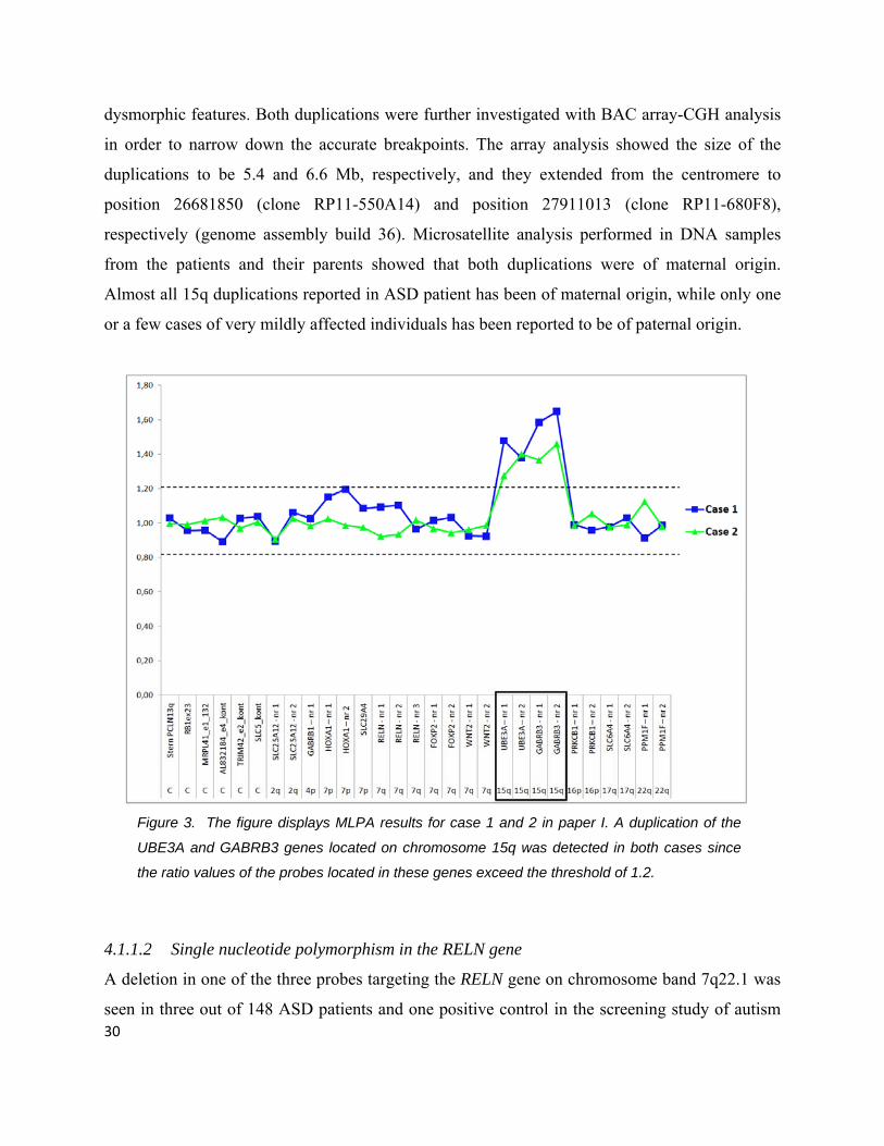
dysmorphic features. Both duplications were further investigated with BAC array-CGH analysis
in order to narrow down the accurate breakpoints. The array analysis showed the size of the
duplications to be 5.4 and 6.6 Mb, respectively, and they extended from the centromere to
position 26681850 (clone RP11-550A14) and position 27911013 (clone RP11-680F8),
respectively (genome assembly build 36). Microsatellite analysis performed in DNA samples
from the patients and their parents showed that both duplications were of maternal origin.
Almost all 15q duplications reported in ASD patient has been of maternal origin, while only one
or a few cases of very mildly affected individuals has been reported to be of paternal origin.
Figure 3. The figure displays MLPA results for case 1 and 2 in paper I. A duplication of the
UBE3A and GABRB3 genes located on chromosome 15q was detected in both cases since
the ratio values of the probes located in these genes exceed the threshold of 1.2.
4.1.1.2 Single nucleotide polymorphism in the RELN gene
A deletion in one of the three probes targeting the RELN gene on chromosome band 7q22.1 was
seen in three out of 148 ASD patients and one positive control in the screening study of autism 30
candidate genes using MLPA (paper I). Direct DNA sequencing analysis revealed a single
nucleotide substitution in the ligation site of the MLPA probe (c.533C>T) resulting in a missense
mutation (p.Ser1719Leu) in all four individuals. This substitution had not been reported as a SNP
in the UCSC Genome Browser (2006) but had previously been reported by Bonora et al. (2003)
who observed this variant in 0.5% of healthy controls. We screened 192 healthy control samples
obtained from blood donors for this variation and identified it in one individual (0.5%). In
addition, the single nucleotide substitution was inherited from a healthy parent in two of the
cases in which parental samples were available indicating that this SNP is likely a rare
polymorphism.
4.1.2 Alterations identified with array-CGH (paper II, III, IV)
4.1.2.1 6p22.3 deletion (paper II)
When we started to screen ASD patients by whole genome BAC array-CGH we identified an
interstitial deletion located on chromosome band 6p22.3 in a 4-year-old girl. The patient was
referred for whole-genome screening due to a general developmental delay and a suspected ASD
diagnosis along with syndromic features including eye abnormalities, short neck, and a
ventricular septum defect. However, after having undergone a complete neuropsychiatric
assessment she didn’t fulfill the criteria for ASD, but was diagnosed with expressive speech
disorder (delay of expressive language development with only a few spoken words).
Searching the literature, interstitial deletions involving the chromosome 6p22.3 region had only
been reported in seven cases. An accurate genotype–phenotype correlation was complicated
since all patients had large deletions of variable sizes and locations, resulting in somewhat
variable phenotypes. However, developmental delay was present in all cases, while heart defects,
short neck and/or redundant skin folds, eye abnormalities, and ear anomalies were present in the
majority of cases (Table 1, paper II).
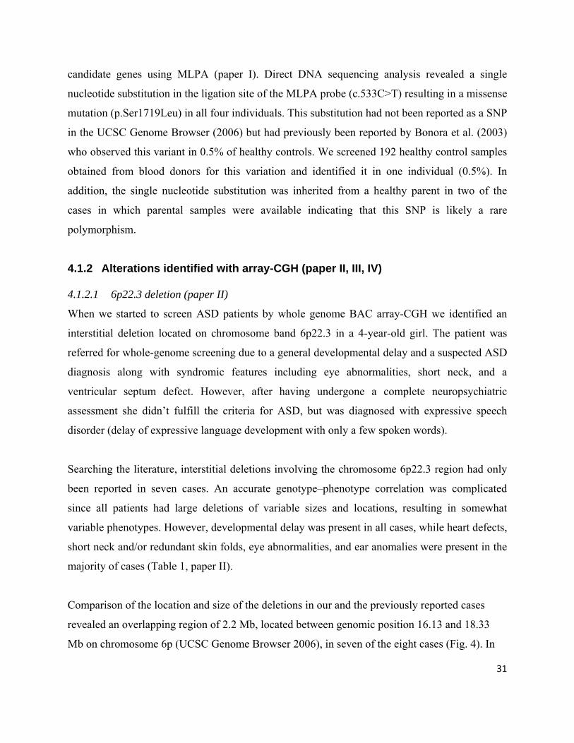
Comparison of the location and size of the deletions in our and the previously reported cases
revealed an overlapping region of 2.2 Mb, located between genomic position 16.13 and 18.33
Mb on chromosome 6p (UCSC Genome Browser 2006), in seven of the eight cases (Fig. 4). In
31
32
the eighth case the deletion breakpoints reported were uncertain due to the low-resolution
technologies used and it was therefore not possible to distinguish whether the deletion really
overlapped with the deletion in our case. However, the overlapping region identified in the seven
remaining cases involved twelve genes; the MYLIP, GMPR, ATXN1, RBM24, CAP2, FAM8A1,
NUP153, KIF13A, KIN13A, NHLRCI, TPMT, and AOF1 gene. The ATXN1 (ataxin-1 protein)
and MYLIP (themyosin regulatory lightchain interacting protein) genes had previously been
reported as likely candidate genes involved in the cognitive delay in patients with a deletion
encompassing chromosome band 6p22. Mice with homozygous deletions in the ATXN1 gene are
phenotypically normal but show learning deficits and the MYLIP gene has been shown to be
expressed in both developing and adult rat brain and it suppresses neurite outgrowth. In addition,
we proposed the CAP2 (adenylyl cyclaseassociated protein 2) gene as a plausible candidate gene.
The CAP2 gene is expressed in brain, heart- and skeletal muscle, and in the skin. The function of
CAP2 is unknown, but it has been shown that the amount of CAP2 is strongly enriched in
developing cardiomyocytes. A heterozygous deletion of this gene could therefore be involved in
both the heart defects as well as the cognitive dysfunctions present in patients with an interstitial
deletion involving chromosome band 6p22.
After publication of the paper, we got information of another patient with a similar deletion (Dr.
Shen, Children's Hospital Central California, USA). The patient, a young girl, had a deletion
comprising almost only the overlapping region presented in our publication. The phenotypic
features of the patient included global developmental delay – predominantly in speech -, an atrial
septal defect, hypotonia and strabismus. We performed an Agilent array-CGH analysis on this
patient in order to accurately distinguish the breakpoints and compare them with the breakpoints
in our patient. The analysis revealed a 4.1 Mb deletion between genomic position 15.08 and
19.17 Mb on chromosome 6p (Fig. In addition, the deletion in our patient was reanalyzed by
Agilent array and showed a 7 Mb deletion between position 16.21 and 23.21 Mb. The
overlapping region of approximately 3 Mb was located between position 16.21 and 19.17 Mb on
chromosome 6 and included almost only the critical region we suggested in our publication. This
finding further confirms that the overlapping region in our publication indeed is a critical region
that includes one or more of the causing genes in the 6p22 deletion syndrome.
USA patientc
Figure 4. a) The array-CGH result of the patient presented in paper II. The CGH plot presents the log2
ratio of all clones located on chromosome 6 detecting a 7.1 Mb deletion on band 6p22.3. (b) A closer
view of chromosome band 6p24.2-p22.1 is displayed along with a schematic representation of the
overlapping deletions in previously reported cases and our case (referred to as present patient). The
overlap of previous cases together with our patient narrows down the overlapping critical region from 4.1
Mb down to a maximum size of 2.2 Mb located on chromosome band 6p22.3. One of the previously
reported cases had to be excluded from the comparison since it was uncertain whether the deletion
really overlapped with our deletion due to the low-resolution technologies used at the time not allowing
accurate mapping*. c) The black bar presents the deletion detected in a patient from USA with a 6p22
deletion phenotype. This finding further confirms that the overlapping region presented in b) indeed is a
critical region including causing genes. 33
34
4.1.2.2 Microdeletion- and microduplication syndromes and recurrent alterations (paper III)
In the screening of 223ASD patients in paper III, 18 (8%) cases were identified with causative
alterations. Twelve (5.4%) of these cases had microdeletion- and microduplication syndromes or
alterations that included regions containing genes in which deletions or duplications have
previously been reported to cause ASDs. These alterations involved chromosome band 2p16.3,
3q27.2q29, 15q13.2q13.3, 16p11.2, 16p13.3, 17p11.2, 17p13.3, 22q11.2, 22q13.3, and Xq28
(Table 1 in paper III).
The alteration in chromosome band 2p16.3 was a partial heterozygous, partial homozygous
deletion including the NRXN1 gene. Deletions in the NRXN1 gene have been reported as causing
genetic alterations recurrently occurring in patients with ASDs (see chapter 1.3.1.5). In our case,
the patient had inherited a heterozygous deletion from each parent which had lead to the partial
heterozygous, partial homozygous deletion including the NRXN1 gene.
One case had a 25 kb de novo deletion involving the SHANK3 gene and the ACR gene. Deletions
in SHANK3 have recurrently been reported to cause neurodevelopmental disorders (see chapter
1.3.1.6). Interestingly, the patient’s father had behavior problems and her half-brother had
neurodevelopmental delay, but none of them had the deletion in the SHANK3 gene. All three
individuals on the other hand had a 50 kb deletion including the ASTN2 gene located on
chromosome band 9q33.1. The ASTN2 gene has also been associated with ASD and is known to
be involved in neuronal development (Glessner et al. 2009). However, the girl was more severely
affected with a more moderate developmental delay compared with her father and brother.
4.1.2.3 Sporadic alterations (paper III)
In the screening of 223ASD patients in paper III, six cases (2.7% of all cases) had deletions and
duplications that were not located within any specific genetic syndrome regions, but within
regions with previously described larger or partly overlapping aberrations. These six aberrations
appeared in chromosome bands 1q25.3q31.1, 3p25.3-pter, 7p22.1, 9q13q21.31, 17p13.2, and
18q22.2-qter. The four aberrations within chromosome 1, 7, 13 and 18 were shown to be de
novo, while one or both parental samples were unavailable for the two remaining cases with
aberrations in chromosome 3 and 17. In the case identified with a duplication in chromosome
band 3p25.3, an unbalanced translocation between chromosome 3 and 13,
46,XX,der(13)t(3;13)(p25.3;qter), was identified by FISH analysis (Table 1 in paper III).
4.1.2.4 Genomic alterations of unclear clinical relevance and rare variants (paper III)
During the array-CGH screening, not only cases with clear causative alterations were identified,
but also seven cases with genomic alterations of unclear relevance in which the parental origin
could not be investigated and thirteen cases with rare but inherited variants that most likely are
benign but possibly could increase the risk for ASDs.
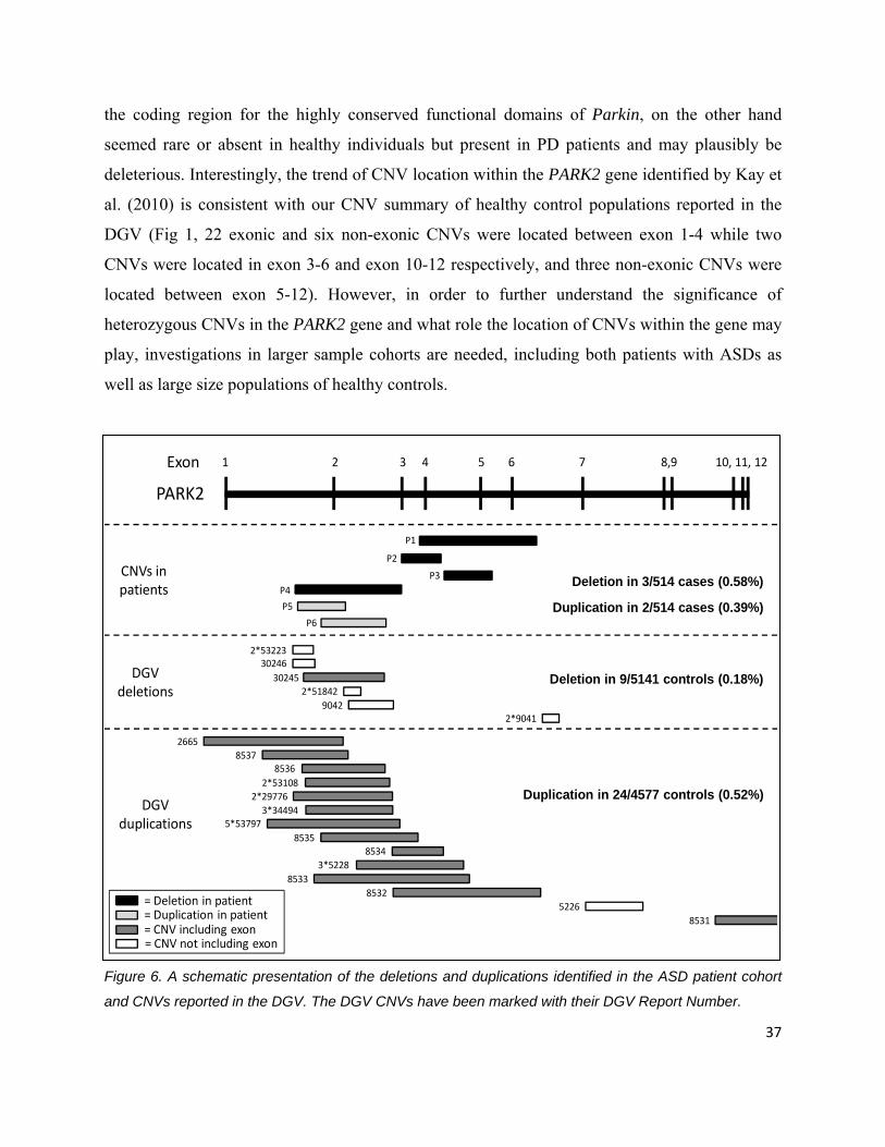
4.1.2.5 Copy number variations in the PARK2 gene (paper IV)
In the first cohort of 160 patients screened in paper II, two of the cases had deletions within the
PARK2 gene. PARK2 gene deletions have been reported as a plausible cause of ASDs. The gene,
located on chromosome band 6q26, encodes for the E3 ubiquitin-protein ligase Parkin, which
belongs to the Ubiquitin proteasome system (UPS) proteins that process proteins for proteasomal
degradation. UPS operate pre- and postsynaptic compartments, such as CAMs and CAM related
proteins, demonstrating a direct link between these two major systems that may be important in
the pathophysiology of autism (Glessner et al. 2009; Lehman 2009). An additional case with a
PARK2 deletion was collected through collaboration with the Sahlgrenska University Hospital,
Gothenburg (Dr Peder Rasmussen and colleagues). In a second ASD cohort of 354 ASD patients
collected from the clinic (Clinical Genetics, Karolinska University Hospital, Solna, Sweden), we
identified one patient with a deletion and two patients with duplications in the PARK2 gene.
Parental samples are being collected for the cases identified in our two ASD cohorts. We are also
screening healthy controls for CNVs in the PARK2 gene by MLPA with probes designed in all
exons of the gene. So far, 149 control samples have been screened and no variation in copy
number has been identified.
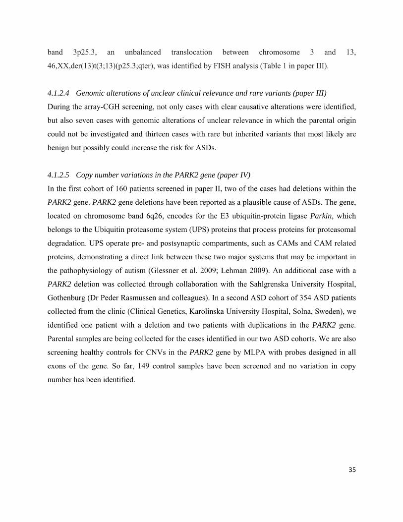
35
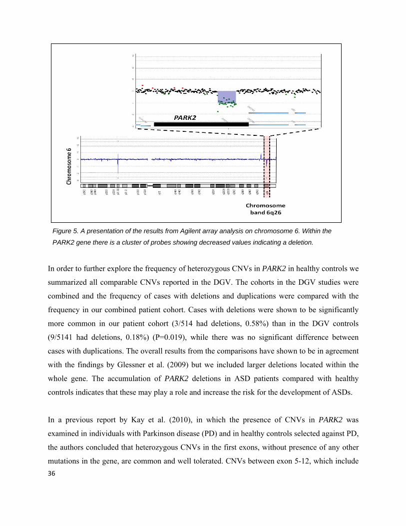
Figure 5. A presentation of the results from Agilent array analysis on chromosome 6. Within the
PARK2 gene there is a cluster of probes showing decreased values indicating a deletion.
In order to further explore the frequency of heterozygous CNVs in PARK2 in healthy controls we
summarized all comparable CNVs reported in the DGV. The cohorts in the DGV studies were
combined and the frequency of cases with deletions and duplications were compared with the
frequency in our combined patient cohort. Cases with deletions were shown to be significantly
more common in our patient cohort (3/514 had deletions, 0.58%) than in the DGV controls
(9/5141 had deletions, 0.18%) (P=0.019), while there was no significant difference between
cases with duplications. The overall results from the comparisons have shown to be in agreement
with the findings by Glessner et al. (2009) but we included larger deletions located within the
whole gene. The accumulation of PARK2 deletions in ASD patients compared with healthy
controls indicates that these may play a role and increase the risk for the development of ASDs.
In a previous report by Kay et al. (2010), in which the presence of CNVs in PARK2 was
examined in individuals with Parkinson disease (PD) and in healthy controls selected against PD,
the authors concluded that heterozygous CNVs in the first exons, without presence of any other
mutations in the gene, are common and well tolerated. CNVs between exon 5-12, which include 36
the coding region for the highly conserved functional domains of Parkin, on the other hand
seemed rare or absent in healthy individuals but present in PD patients and may plausibly be
deleterious. Interestingly, the trend of CNV location within the PARK2 gene identified by Kay et
al. (2010) is consistent with our CNV summary of healthy control populations reported in the
DGV (Fig 1, 22 exonic and six non-exonic CNVs were located between exon 1-4 while two
CNVs were located in exon 3-6 and exon 10-12 respectively, and three non-exonic CNVs were
located between exon 5-12). However, in order to further understand the significance of
heterozygous CNVs in the PARK2 gene and what role the location of CNVs within the gene may
play, investigations in larger sample cohorts are needed, including both patients with ASDs as
well as large size populations of healthy controls.
1 2 3 765 8,9 10, 11, 124Exon
PARK2
P1
P2
P3
9042
30245
2*9041
2*51842
2*53223
DGV deletions
DGV duplications
= CNV including exon= CNV not including exon
= Deletion in patient
CNVs in patients
26658537
8536
2*297762*53108
3*344945*53797
85358534
3*52288533
85325226
8531= Duplication in patient
30246
P4
P6
P5
Deletion in 9/5141 controls (0.18%)
Duplication in 24/4577 controls (0.52%)
Deletion in 3/514 cases (0.58%)
Duplication in 2/514 cases (0.39%)
Figure 6. A schematic presentation of the deletions and duplications identified in the ASD patient cohort
and CNVs reported in the DGV. The DGV CNVs have been marked with their DGV Report Number.
37
38
4.2 CNV DISTRIBUTION IN DIFFERENT ASD SUB-POPULATIONS (PAPER III) In the array-CGH study (paper III), all patients were divided into different subgroups regarding
their family history of ASDs and related BNPs and into different subgroups depending on
phenotypic expressions. The patients were also divided into to two groups depending on gender.
The distribution of CNVs was shown to vary between different subgroups.
4.2.1 CNVs and ASD inheritance Out of the 18 cases identified with a clinically significant alteration, 14 were sporadic (8.5% of
164) and 4 were familial (14.8% of 27) (Table 4). Of the seven cases with CNVs of unclear
significance, four were sporadic (2.4% of 164), one was familial (3.7% of 27), one was BNP-
familial (4% of 25), and one had an unknown inheritance pattern (adoption) (Table 4). Of the 13
cases identified with rare CNVs inherited from a healthy parent, 6 were sporadic (3.7% of 164),
2 were familial (7.4% of 27), and 5 were BNP-familial (20.0% of 25) (Table 4). Separate
comparison of CNV distribution between the three inheritance subgroups (excluding all cases
with another type of CNV than the CNV compared) using two-tailed Fisher exact P-tests (2x2
tables) showed that rare inherited CNVs were significantly more common in BNP-familial cases
(in 5/24 cases) compared with in sporadic cases (in 6/146 cases) (P=0.0096, the 95% confidence
intervals lied between 1.5 and 8.7% and 7.1–42.2% in respective group). It has been suggested
that two or several CNVs could interact in predisposing individuals for neuropsychiatric disorder
(Stankiewicz and Lupski 2010) and furthermore a two-hit model has been reported in which a
second CNV in patients with 16p12.1 deletions lead to a more severe phenotype outcome of the
syndrome (Girirajan et al. 2010). It has also been reported that rare CNVs containing ASD or
BNP associated genes are enriched in ASD cases compared with healthy controls (Bucan et al.
2009). These findings together with our result of a significantly higher presence of rare inherited,
potentially neuropsychiatric disorder related CNVs in BNP-familial cases than in sporadic cases,
may indicate that these types of CNVs, rather than being directly causative, increase
susceptibility for the development of ASDs and ASD related phenotypes. This may also, at least
in part, explain the complex genetics underlying ASDs and the difficulties we face in finding
recurrent genetic causes. Hence, CNVs currently hypothesized to be clinically benign, may play
an important role in the development of ASDs and ASD-related phenotypes as well as in other
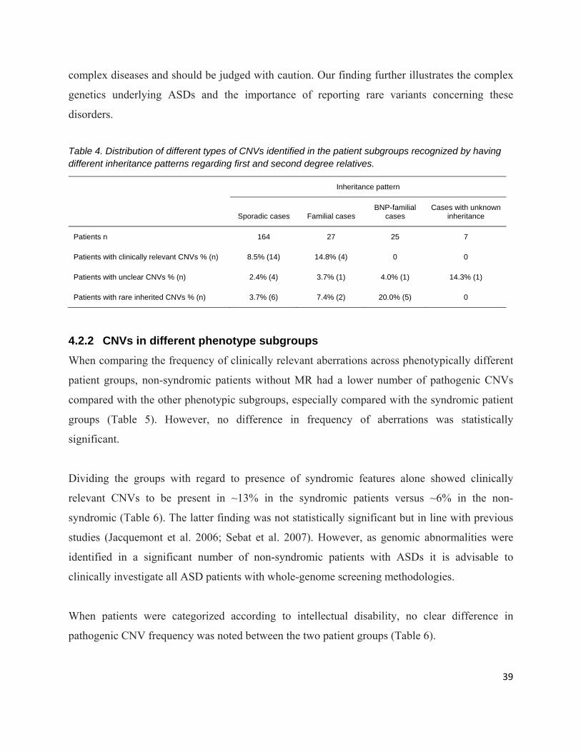
complex diseases and should be judged with caution. Our finding further illustrates the complex
genetics underlying ASDs and the importance of reporting rare variants concerning these
disorders.
Table 4. Distribution of different types of CNVs identified in the patient subgroups recognized by having different inheritance patterns regarding first and second degree relatives.
Inheritance pattern
Sporadic cases Familial cases BNP-familial
cases Cases with unknown
inheritance
Patients n 164 27 25 7
Patients with clinically relevant CNVs % (n) 8.5% (14) 14.8% (4) 0 0
Patients with unclear CNVs % (n) 2.4% (4) 3.7% (1) 4.0% (1) 14.3% (1)
Patients with rare inherited CNVs % (n) 3.7% (6) 7.4% (2) 20.0% (5) 0
4.2.2 CNVs in different phenotype subgroups When comparing the frequency of clinically relevant aberrations across phenotypically different
patient groups, non-syndromic patients without MR had a lower number of pathogenic CNVs
compared with the other phenotypic subgroups, especially compared with the syndromic patient
groups (Table 5). However, no difference in frequency of aberrations was statistically
significant.
Dividing the groups with regard to presence of syndromic features alone showed clinically
relevant CNVs to be present in ~13% in the syndromic patients versus ~6% in the non-
syndromic (Table 6). The latter finding was not statistically significant but in line with previous
studies (Jacquemont et al. 2006; Sebat et al. 2007). However, as genomic abnormalities were
identified in a significant number of non-syndromic patients with ASDs it is advisable to
clinically investigate all ASD patients with whole-genome screening methodologies.
When patients were categorized according to intellectual disability, no clear difference in
pathogenic CNV frequency was noted between the two patient groups (Table 6).
39
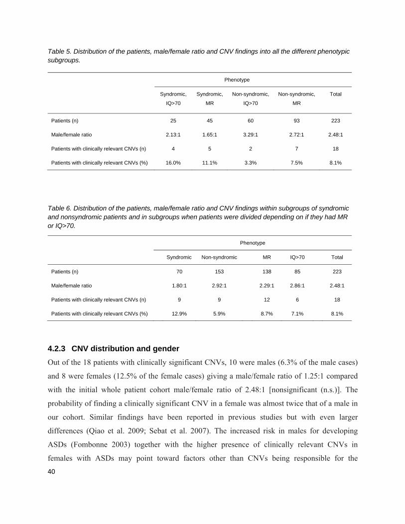
Table 5. Distribution of the patients, male/female ratio and CNV findings into all the different phenotypic subgroups.
Phenotype
Syndromic,
IQ>70
Syndromic,
MR
Non-syndromic,
IQ>70
Non-syndromic,
MR
Total
Patients (n) 25 45 60 93 223
Male/female ratio 2.13:1 1.65:1 3.29:1 2.72:1 2.48:1
Patients with clinically relevant CNVs (n) 4 5 2 7 18
Patients with clinically relevant CNVs (%) 16.0% 11.1% 3.3% 7.5% 8.1%
Table 6. Distribution of the patients, male/female ratio and CNV findings within subgroups of syndromic and nonsyndromic patients and in subgroups when patients were divided depending on if they had MR or IQ>70.
Phenotype
Syndromic Non-syndromic MR IQ>70 Total
Patients (n) 70 153 138 85 223
Male/female ratio 1.80:1 2.92:1 2.29:1 2.86:1 2.48:1
Patients with clinically relevant CNVs (n) 9 9 12 6 18
Patients with clinically relevant CNVs (%) 12.9% 5.9% 8.7% 7.1% 8.1%
4.2.3 CNV distribution and gender Out of the 18 patients with clinically significant CNVs, 10 were males (6.3% of the male cases)
and 8 were females (12.5% of the female cases) giving a male/female ratio of 1.25:1 compared
with the initial whole patient cohort male/female ratio of 2.48:1 [nonsignificant (n.s.)]. The
probability of finding a clinically significant CNV in a female was almost twice that of a male in
our cohort. Similar findings have been reported in previous studies but with even larger
differences (Qiao et al. 2009; Sebat et al. 2007). The increased risk in males for developing
ASDs (Fombonne 2003) together with the higher presence of clinically relevant CNVs in
females with ASDs may point toward factors other than CNVs being responsible for the
40
increased susceptibility for ASDs in males. It has been argued to depend on epigenetic effects
such as an increased vulnerability for dysregulation of methylation of brain-expressed genes on
the X-chromosome and sex-specific responses to different hormones (Carter 2007; Jones et al.
2008). Males may also be more vulnerable to minor variations in ASD susceptibility genes
located on the X-chromosome (Noor et al. 2010).
41
42
5 CONCLUDING REMARKS AND FUTURE PRESPECTIVES The project has lead to the identification of a genetic diagnosis in many patients. Parents to
children with a chronic condition like ASD often experience great grief, not just because of the
condition itself, but also because they may not understand what the condition involves and what
their expectations should be. It has earlier been reported that medical knowledge about a specific
diagnosis affects the parents reactions and enhance their sense of control (Cunningham et al).
Our own experience together with this report infer that getting an explanation of why the child
has been affected is very important and meaningful for the parents and that parents are helped by
receiving a genetic explanation to their child’s disorder. The identification of the genetic cause in
families also gives them the opportunity to an adequate genetic counseling and makes prenatal
diagnosis possible.
MLPA and array-CGH where the main tools for genetic screening used in this thesis. MLPA
showed to be a fast and cost-effective method for screening several selected loci in many patients
simultaneously. At the time of the study, MLPA was a good alternative as a screening tool for
alterations in previously described candidate genes in ASDs, partly because it was much cheaper,
and partly because it could detect smaller alterations than early array-CGH methodologies.
However, there has been a rapid advancement in the development of whole-genome screening
array-CGH methodologies which has lead to a great increase in resolution and a large decrease in
usage expences making the usage of MLPA in these types of studies lose its advantage. Today, a
suitable use for MLPA is confirmation of identified CNVs. This is excellently demonstrated in
one of the related publications (I), where MLPA probes were designed for several alterations
identified by array-CGH and thereby confirmed. Another suitable area for the usage of MLPA is
screening of controls for particular CNVs which have been identified in one or more patients, as
has been done in paper IV. However, during the MLPA screening study, two out of 148 patients
were identified with a duplication of the maternal chromosome band 15q11-15q13, the same
region that is deleted in Prader-Willi- and Angelman syndrome. Such duplications are recurrent
in 0.5-3% of ASD cases which was confirmed in our study.
Trough the identification and publication of the patient with a chromosome band 6p22 deletion a
communication with the parents of our patient and of the girl in California was established. They
are now exchanging experiences with each other, which have led to a great sence of support for
the two families.
In the third study, several genetic alterations were identified including both recurrent aberrations
known to cause ASD, sporadic causative alterations, variants with unclear clinical relevance and
rare inherited variants. All patients were divided into different subgroups depending on their
family history and their phenotype. In previous reports, patients have only been divided into
sporadic and familial cases. We introduced a third inheritance group reffered to as BNP-familial
cases – a group that included patients with relatives that had BNPs. Rare but inherited CNVs
were significantly more common in this group compared with the sporadic inheritance group.
Such CNVs may therefore have a significant influence in increasing the risk for development of
ASDs and related neuropsychiatric disorders. This finding did not only illustrate the genetic
complexity in ASD but also presented new insights into what may be one of the underlying
causes of the genetic complexety. It also reminds us of the importance of reporting rare variants
in these disorders since we in the future may be able to see enrichments of certain CNVs or CNV
patterns that can reveal further genetic causes in ASDs. Concerning the CNV distribution of
causative CNVs in the phenotypic subgroups, we found that they were more common in
syndromic patients than in non-syndromic and in females compared with males. None of these
findings were statistically significant but in line with previous reports. However, causative CNVs
were found in a significant proportion in all phenotypic subgroups and it is therefore
recommended to clinically investigate all ASD patients with whole genome screening
methodologies.
The last study was generated from the results in the whole genome screening study discussed
above. The identfication of two PARK2 gene deletions in paper III lead to the screening of more
patients in which one additional case with a deletion and two cases with a duplication were
identified. Comparison of CNV frequency in the PARK2 gene between our ASD cohort and
DGV controls indicated an enrichment of deletions in ASD patients compared with healthy
43
44
individuals. The association between deletions in the PARK2 gene and ASDs has only been
reported once before by Glessner et al. (2009). Our results confirm the association but we
included larger deletions located within the whole gene. Further studies, in larger patient cohorts
and with matched large size control populations, are needed in order to evaluate the true
significance of PARK2 deletions in ASDs. The results of this particular study is a good example
of and in line with the emerging evidence which begins to identify the biological networks in
which the affected genes operate and in many cases have shown to alter synaptic connectivity
and function.
During the first years of the autism genetics research era, mainly linkage, association and
candidate gene screening studies were performed. Multiple loci and genes have been reported to
be linked or associated with autism but the association has in most cases been weak in it has
shown difficult to obtain consistent results in independent samples. The usage of high-resolution
whole genome screening methodologies has revealed that rare or de novo CNVs are significantly
more common in ASD individuals than in healthy controls. It has also lead to the identification
of novel microdeletion- and microduplication syndromes often associated with an autism
phenotype and to the identification of alterations in genes likely to be involved in ASDs.
Altogether, there are indications for several different pathways and genes to be involved in the
development of ASDs. Neuronal migration and cell growth influenced by for instance PTEN are
strong candidates for being causative in ASDs. Genes involved in excitatory and inhibitory
neurotransmission such as GABA and glutamate receptors are other strong candidates. One of the
most genetically evident tracks is alterations in cell-adhesion molecules (CAMs) and related
proteins working in this pathway system.
The increased resolution of array-CGH together with the development of new technologies such
as whole genome sequencing, in which new genetic alterations already have started to be
reported in ASDs, will most likely lead to the detection of even smaller rare variants affecting
single genes. The future challenge will be to, in combination with the increased usage of high
resolution array-CGH and whole genome sequencing, create useful analysis systems in which the
co-occurring pathways and gene-gene interactions in ASDs can be linked together and the
different genes involved identified.
Furthermore, it has recently been shown that the phenotypes in Fragile X syndrome and Rett’s
syndrome mice models can be altered after birth through administration of metabotophic
glutamate antagonists and reinstatement of MECP2 (Dolen et al. 2007; Guy et al. 2007; Yan et
al. 2005). Similarly, neurological abnormalities in TSC1 and TSC2 knockout mice (tuberous
sclerosis) and PTEN knockout mice (macrocephaly and ASD) can be reversed by treatment with
rapamycin (Ehninger et al. 2008; Zhou et al. 2009). This is highly encouraging implying that the
identification of causative targets in ASDs, as well as in other neuropsychiatric disorders, may
lead to the development of therapeutic strategies.
45
46
6 ACKNOWLEDGEMENTS I would like to express my gratitude to all people that have helped me and been supportive during my time as a PhD student at Karolinska Institutet. A special thanks to:
MaiBritt Giacobini, my main supervisor, for your never ending support and encouragement, for having trust in me and for everything I have learned from you. Thank you for your kindness and for being such a great person that always makes me feel good.
BrittMarie Anderlid, my co-supervisor, for all valuable discussions and comments and for always having a positive spirit.
Magnus Nordenskjöld, my co-supervisor, for your helpful comments and for creating such a positive working atmosphere in the lab.
Sigrid Sahlén, for helping me with my lab work, for your great support and for always taking care of me. I would never have made it without your support and guidance.
Margareta Lagerberg, for helping me with my lab work, for all your support and for always being so kind and encouraging.
Ann-Britt Wikström, for helping me out with all paper works but also for your great support.
Peter Gustavsson, for valuable discussions and comments and for always being optimistic and encouraging.
All friends and co-workers in the lab and at the CMM floor 02, Mårten, Anna, Fredrik, Johanna, Frideborg, Annika, Josephine, Marie, Josefin, Hovsep, Tatjana, Kristina, Eva, Miriam, Monica, Tatja, Aron, Vasse, Agne, Malin, Edward and Tobias, for your help and support and for creating such a nice, positive and welcoming atmosphere.
The lab-ladies, Anna-Lena and Christina, for being helpful and friendly and for all nice after works we have had during these years.
The staff at the Department of Clinical Genetics and at the MMK administration, for always being kind to me and for helping me out whenever I needed.
My dear friends, Sanna, Jenny, Sabina, Cissi, Ullis and Jane, for all your support and for all good and crazy times. Your friendship is invaluable.
Thomas, Mamma, Pappa and Oskar, for listening, being supportive and for always believing in me. I love you!
7 REFERENCES Abrahams BS, Geschwind DH. 2008. Advances in autism genetics: on the threshold of a new
neurobiology. Nat Rev Genet 9(5):341‐55. Alarcon M, Abrahams BS, Stone JL, Duvall JA, Perederiy JV, Bomar JM, Sebat J, Wigler M, Martin CL,
Ledbetter DH and others. 2008. Linkage, association, and gene‐expression analyses identify CNTNAP2 as an autism‐susceptibility gene. Am J Hum Genet 82(1):150‐9.
Anderlid BM, Schoumans J, Anneren G, Tapia‐Paez I, Dumanski J, Blennow E, Nordenskjold M. 2002. FISH‐mapping of a 100‐kb terminal 22q13 deletion. Hum Genet 110(5):439‐43.
Arking DE, Cutler DJ, Brune CW, Teslovich TM, West K, Ikeda M, Rea A, Guy M, Lin S, Cook EH and others. 2008. A common genetic variant in the neurexin superfamily member CNTNAP2 increases familial risk of autism. Am J Hum Genet 82(1):160‐4.
Auranen M, Vanhala R, Varilo T, Ayers K, Kempas E, Ylisaukko‐Oja T, Sinsheimer JS, Peltonen L, Jarvela I. 2002. A genomewide screen for autism‐spectrum disorders: evidence for a major susceptibility locus on chromosome 3q25‐27. Am J Hum Genet 71(4):777‐90.
Bacchelli E, Maestrini E. 2006. Autism spectrum disorders: molecular genetic advances. Am J Med Genet C Semin Med Genet 142C(1):13‐23.
Bailey A, Luthert P, Dean A, Harding B, Janota I, Montgomery M, Rutter M, Lantos P. 1998. A clinicopathological study of autism. Brain 121 ( Pt 5):889‐905.
Bakkaloglu B, O'Roak BJ, Louvi A, Gupta AR, Abelson JF, Morgan TM, Chawarska K, Klin A, Ercan‐Sencicek AG, Stillman AA and others. 2008. Molecular cytogenetic analysis and resequencing of contactin associated protein‐like 2 in autism spectrum disorders. Am J Hum Genet 82(1):165‐73.
Ballif BC, Hornor SA, Jenkins E, Madan‐Khetarpal S, Surti U, Jackson KE, Asamoah A, Brock PL, Gowans GC, Conway RL and others. 2007. Discovery of a previously unrecognized microdeletion syndrome of 16p11.2‐p12.2. Nat Genet 39(9):1071‐3.
Baron‐Cohen S, Scott FJ, Allison C, Williams J, Bolton P, Matthews FE, Brayne C. 2009. Prevalence of autism‐spectrum conditions: UK school‐based population study. Br J Psychiatry 194(6):500‐9.
Battaglia A, Parrini B, Tancredi R. 2010. The behavioral phenotype of the idic(15) syndrome. Am J Med Genet C Semin Med Genet 154C(4):448‐55.
Ben‐Shachar S, Lanpher B, German JR, Qasaymeh M, Potocki L, Nagamani SC, Franco LM, Malphrus A, Bottenfield GW, Spence JE and others. 2009. Microdeletion 15q13.3: a locus with incomplete penetrance for autism, mental retardation, and psychiatric disorders. J Med Genet 46(6):382‐8.
Bettelheim B. 1967. The empty fortress: infantile autism and the birth of the self. . New York: Free Press. Blatt GJ, Fitzgerald CM, Guptill JT, Booker AB, Kemper TL, Bauman ML. 2001. Density and distribution of
hippocampal neurotransmitter receptors in autism: an autoradiographic study. J Autism Dev Disord 31(6):537‐43.
Bonaglia MC, Giorda R, Borgatti R, Felisari G, Gagliardi C, Selicorni A, Zuffardi O. 2001. Disruption of the ProSAP2 gene in a t(12;22)(q24.1;q13.3) is associated with the 22q13.3 deletion syndrome. Am J Hum Genet 69(2):261‐8.
Bonaglia MC, Giorda R, Mani E, Aceti G, Anderlid BM, Baroncini A, Pramparo T, Zuffardi O. 2006. Identification of a recurrent breakpoint within the SHANK3 gene in the 22q13.3 deletion syndrome. J Med Genet 43(10):822‐8.
Bonora E, Beyer KS, Lamb JA, Parr JR, Klauck SM, Benner A, Paolucci M, Abbott A, Ragoussis I, Poustka A and others. 2003. Analysis of reelin as a candidate gene for autism. Mol Psychiatry 8(10):885‐92.
Bucan M, Abrahams BS, Wang K, Glessner JT, Herman EI, Sonnenblick LI, Alvarez Retuerto AI, Imielinski M, Hadley D, Bradfield JP and others. 2009. Genome‐wide analyses of exonic copy number
47
48
variants in a family‐based study point to novel autism susceptibility genes. PLoS Genet 5(6):e1000536.
Butler MG, Dasouki MJ, Zhou XP, Talebizadeh Z, Brown M, Takahashi TN, Miles JH, Wang CH, Stratton R, Pilarski R and others. 2005. Subset of individuals with autism spectrum disorders and extreme macrocephaly associated with germline PTEN tumour suppressor gene mutations. J Med Genet 42(4):318‐21.
Buxbaum JD, Silverman JM, Smith CJ, Greenberg DA, Kilifarski M, Reichert J, Cook EH, Jr., Fang Y, Song CY, Vitale R. 2002. Association between a GABRB3 polymorphism and autism. Mol Psychiatry 7(3):311‐6.
Cai G, Edelmann L, Goldsmith JE, Cohen N, Nakamine A, Reichert JG, Hoffman EJ, Zurawiecki DM, Silverman JM, Hollander E and others. 2008. Multiplex ligation‐dependent probe amplification for genetic screening in autism spectrum disorders: efficient identification of known microduplications and identification of a novel microduplication in ASMT. BMC Med Genomics 1:50.
Carmona‐Mora P, Walz K. 2010. Retinoic Acid Induced 1, RAI1: A Dosage Sensitive Gene Related to Neurobehavioral Alterations Including Autistic Behavior. Curr Genomics 11(8):607‐17.
Carter CS. 2007. Sex differences in oxytocin and vasopressin: implications for autism spectrum disorders? Behav Brain Res 176(1):170‐86.
Ching MS, Shen Y, Tan WH, Jeste SS, Morrow EM, Chen X, Mukaddes NM, Yoo SY, Hanson E, Hundley R and others. 2010. Deletions of NRXN1 (neurexin‐1) predispose to a wide spectrum of developmental disorders. Am J Med Genet B Neuropsychiatr Genet 153B(4):937‐47.
Collins AL, Ma D, Whitehead PL, Martin ER, Wright HH, Abramson RK, Hussman JP, Haines JL, Cuccaro ML, Gilbert JR and others. 2006. Investigation of autism and GABA receptor subunit genes in multiple ethnic groups. Neurogenetics 7(3):167‐74.
Cook EH, Jr., Courchesne RY, Cox NJ, Lord C, Gonen D, Guter SJ, Lincoln A, Nix K, Haas R, Leventhal BL and others. 1998. Linkage‐disequilibrium mapping of autistic disorder, with 15q11‐13 markers. Am J Hum Genet 62(5):1077‐83.
Curran S, Roberts S, Thomas S, Veltman M, Browne J, Medda E, Pickles A, Sham P, Bolton PF. 2005. An association analysis of microsatellite markers across the Prader‐Willi/Angelman critical region on chromosome 15 (q11‐13) and autism spectrum disorder. Am J Med Genet B Neuropsychiatr Genet 137B(1):25‐8.
D'Arcangelo G, Miao GG, Chen SC, Soares HD, Morgan JI, Curran T. 1995. A protein related to extracellular matrix proteins deleted in the mouse mutant reeler. Nature 374(6524):719‐23.
Dolen G, Osterweil E, Rao BS, Smith GB, Auerbach BD, Chattarji S, Bear MF. 2007. Correction of fragile X syndrome in mice. Neuron 56(6):955‐62.
Durand CM, Betancur C, Boeckers TM, Bockmann J, Chaste P, Fauchereau F, Nygren G, Rastam M, Gillberg IC, Anckarsater H and others. 2007. Mutations in the gene encoding the synaptic scaffolding protein SHANK3 are associated with autism spectrum disorders. Nat Genet 39(1):25‐7.
Ehninger D, Han S, Shilyansky C, Zhou Y, Li W, Kwiatkowski DJ, Ramesh V, Silva AJ. 2008. Reversal of learning deficits in a Tsc2+/‐ mouse model of tuberous sclerosis. Nat Med 14(8):843‐8.
Ekstrom AB, Hakenas‐Plate L, Samuelsson L, Tulinius M, Wentz E. 2008. Autism spectrum conditions in myotonic dystrophy type 1: a study on 57 individuals with congenital and childhood forms. Am J Med Genet B Neuropsychiatr Genet 147B(6):918‐26.
Fatemi SH, Snow AV, Stary JM, Araghi‐Niknam M, Reutiman TJ, Lee S, Brooks AI, Pearce DA. 2005. Reelin signaling is impaired in autism. Biol Psychiatry 57(7):777‐87.
Feuk L, Kalervo A, Lipsanen‐Nyman M, Skaug J, Nakabayashi K, Finucane B, Hartung D, Innes M, Kerem B, Nowaczyk MJ and others. 2006. Absence of a paternally inherited FOXP2 gene in developmental verbal dyspraxia. Am J Hum Genet 79(5):965‐72.
Folstein SE, Rosen‐Sheidley B. 2001. Genetics of autism: complex aetiology for a heterogeneous disorder. Nat Rev Genet 2(12):943‐55.
Fombonne E. 1999. The epidemiology of autism: a review. Psychol Med 29(4):769‐86. Fombonne E. 2003. Epidemiological surveys of autism and other pervasive developmental disorders: an
update. J Autism Dev Disord 33(4):365‐82. Gauthier J, Joober R, Mottron L, Laurent S, Fuchs M, De Kimpe V, Rouleau GA. 2003. Mutation screening
of FOXP2 in individuals diagnosed with autistic disorder. Am J Med Genet A 118A(2):172‐5. Gauthier J, Siddiqui TJ, Huashan P, Yokomaku D, Hamdan FF, Champagne N, Lapointe M, Spiegelman D,
Noreau A, Lafreniere RG and others. 2011. Truncating mutations in NRXN2 and NRXN1 in autism spectrum disorders and schizophrenia. Hum Genet.
Gillberg C, Coleman M. 2000. The Biology of the Autistic Syndromes. London, UK: Mac Keith Press, Distributed by Cambridge University Press.
Gillberg C, Wing L. 1999. Autism: not an extremely rare disorder. Acta Psychiatr Scand 99(6):399‐406. Gilman SR, Iossifov I, Levy D, Ronemus M, Wigler M, Vitkup D. 2011. Rare de novo variants associated
with autism implicate a large functional network of genes involved in formation and function of synapses. Neuron 70(5):898‐907.
Girirajan S, Rosenfeld JA, Cooper GM, Antonacci F, Siswara P, Itsara A, Vives L, Walsh T, McCarthy SE, Baker C and others. 2010. A recurrent 16p12.1 microdeletion supports a two‐hit model for severe developmental delay. Nat Genet 42(3):203‐9.
Glessner JT, Wang K, Cai G, Korvatska O, Kim CE, Wood S, Zhang H, Estes A, Brune CW, Bradfield JP and others. 2009. Autism genome‐wide copy number variation reveals ubiquitin and neuronal genes. Nature 459(7246):569‐73.
Guy J, Gan J, Selfridge J, Cobb S, Bird A. 2007. Reversal of neurological defects in a mouse model of Rett syndrome. Science 315(5815):1143‐7.
Helbig I, Mefford HC, Sharp AJ, Guipponi M, Fichera M, Franke A, Muhle H, de Kovel C, Baker C, von Spiczak S and others. 2009. 15q13.3 microdeletions increase risk of idiopathic generalized epilepsy. Nat Genet 41(2):160‐2.
Hogart A, Wu D, LaSalle JM, Schanen NC. 2008. The comorbidity of autism with the genomic disorders of chromosome 15q11.2‐q13. Neurobiol Dis 38(2):181‐91.
Huang CH, Santangelo SL. 2008. Autism and serotonin transporter gene polymorphisms: a systematic review and meta‐analysis. Am J Med Genet B Neuropsychiatr Genet 147B(6):903‐13.
Hultman CM, Sandin S, Levine SZ, Lichtenstein P, Reichenberg A. 2010. Advancing paternal age and risk of autism: new evidence from a population‐based study and a meta‐analysis of epidemiological studies. Mol Psychiatry Epub ahead of print.
IMGSAC. 2001a. Further characterization of the autism susceptibility locus AUTS1 on chromosome 7q. Hum Mol Genet 10(9):973‐82.
IMGSAC. 2001b. A genomewide screen for autism: strong evidence for linkage to chromosomes 2q, 7q, and 16p. Am J Hum Genet 69(3):570‐81.
Jacquemont ML, Sanlaville D, Redon R, Raoul O, Cormier‐Daire V, Lyonnet S, Amiel J, Le Merrer M, Heron D, de Blois MC and others. 2006. Array‐based comparative genomic hybridisation identifies high frequency of cryptic chromosomal rearrangements in patients with syndromic autism spectrum disorders. J Med Genet 43(11):843‐9.
49
50
Jamain S, Quach H, Betancur C, Rastam M, Colineaux C, Gillberg IC, Soderstrom H, Giros B, Leboyer M, Gillberg C and others. 2003. Mutations of the X‐linked genes encoding neuroligins NLGN3 and NLGN4 are associated with autism. Nat Genet 34(1):27‐9.
Jones JR, Skinner C, Friez MJ, Schwartz CE, Stevenson RE. 2008. Hypothesis: dysregulation of methylation of brain‐expressed genes on the X chromosome and autism spectrum disorders. Am J Med Genet A 146A(17):2213‐20.
Kanner L. 1943. Autistic disturbances of affective contact. Nerv Child 2:217‐50. Kanner L. 1949. Problems of nosology and psychodynamics of early infantile autism. Am J
Orthopsychiatry 19(3):416‐26. Kim HG, Kishikawa S, Higgins AW, Seong IS, Donovan DJ, Shen Y, Lally E, Weiss LA, Najm J, Kutsche K and
others. 2008. Disruption of neurexin 1 associated with autism spectrum disorder. Am J Hum Genet 82(1):199‐207.
Kim YS, Leventhal BL, Koh YJ, Fombonne E, Laska E, Lim EC, Cheon KA, Kim SJ, Kim YK, Lee H and others. 2011. Prevalence of Autism Spectrum Disorders in a Total Population Sample. Am J Psychiatry.
Krebs MO, Betancur C, Leroy S, Bourdel MC, Gillberg C, Leboyer M. 2002. Absence of association between a polymorphic GGC repeat in the 5' untranslated region of the reelin gene and autism. Mol Psychiatry 7(7):801‐4.
Kulman G, Lissoni P, Rovelli F, Roselli MG, Brivio F, Sequeri P. 2000. Evidence of pineal endocrine hypofunction in autistic children. Neuro Endocrinol Lett 21(1):31‐34.
Kumar RA, Christian SL. 2009. Genetics of autism spectrum disorders. Curr Neurol Neurosci Rep 9(3):188‐97.
Kumar RA, KaraMohamed S, Sudi J, Conrad DF, Brune C, Badner JA, Gilliam TC, Nowak NJ, Cook EH, Jr., Dobyns WB and others. 2008. Recurrent 16p11.2 microdeletions in autism. Hum Mol Genet 17(4):628‐38.
Lai CS, Fisher SE, Hurst JA, Vargha‐Khadem F, Monaco AP. 2001. A forkhead‐domain gene is mutated in a severe speech and language disorder. Nature 413(6855):519‐23.
Laje G, Morse R, Richter W, Ball J, Pao M, Smith AC. 2010. Autism spectrum features in Smith‐Magenis syndrome. Am J Med Genet C Semin Med Genet 154C(4):456‐62.
Laumonnier F, Bonnet‐Brilhault F, Gomot M, Blanc R, David A, Moizard MP, Raynaud M, Ronce N, Lemonnier E, Calvas P and others. 2004. X‐linked mental retardation and autism are associated with a mutation in the NLGN4 gene, a member of the neuroligin family. Am J Hum Genet 74(3):552‐7.
Lehman NL. 2009. The ubiquitin proteasome system in neuropathology. Acta Neuropathol 118(3):329‐47.
Levy D, Ronemus M, Yamrom B, Lee YH, Leotta A, Kendall J, Marks S, Lakshmi B, Pai D, Ye K and others. 2011. Rare de novo and transmitted copy‐number variation in autistic spectrum disorders. Neuron 70(5):886‐97.
Levy SE, Mandell DS, Schultz RT. 2009. Autism. Lancet 374(9701):1627‐38. Li J, Nguyen L, Gleason C, Lotspeich L, Spiker D, Risch N, Myers RM. 2004. Lack of evidence for an
association between WNT2 and RELN polymorphisms and autism. Am J Med Genet B Neuropsychiatr Genet 126B(1):51‐7.
Ma DQ, Whitehead PL, Menold MM, Martin ER, Ashley‐Koch AE, Mei H, Ritchie MD, Delong GR, Abramson RK, Wright HH and others. 2005. Identification of significant association and gene‐gene interaction of GABA receptor subunit genes in autism. Am J Hum Genet 77(3):377‐88.
Maestrini E, Lai C, Marlow A, Matthews N, Wallace S, Bailey A, Cook EH, Weeks DE, Monaco AP. 1999. Serotonin transporter (5‐HTT) and gamma‐aminobutyric acid receptor subunit beta3 (GABRB3)
gene polymorphisms are not associated with autism in the IMGSA families. The International Molecular Genetic Study of Autism Consortium. Am J Med Genet 88(5):492‐6.
Manning MA, Cassidy SB, Clericuzio C, Cherry AM, Schwartz S, Hudgins L, Enns GM, Hoyme HE. 2004. Terminal 22q deletion syndrome: a newly recognized cause of speech and language disability in the autism spectrum. Pediatrics 114(2):451‐7.
Marshall CR, Noor A, Vincent JB, Lionel AC, Feuk L, Skaug J, Shago M, Moessner R, Pinto D, Ren Y and others. 2008. Structural variation of chromosomes in autism spectrum disorder. Am J Hum Genet 82(2):477‐88.
Martin ER, Menold MM, Wolpert CM, Bass MP, Donnelly SL, Ravan SA, Zimmerman A, Gilbert JR, Vance JM, Maddox LO and others. 2000. Analysis of linkage disequilibrium in gamma‐aminobutyric acid receptor subunit genes in autistic disorder. Am J Med Genet 96(1):43‐8.
McCarthy SE, Makarov V, Kirov G, Addington AM, McClellan J, Yoon S, Perkins DO, Dickel DE, Kusenda M, Krastoshevsky O and others. 2009. Microduplications of 16p11.2 are associated with schizophrenia. Nat Genet 41(11):1223‐7.
Melke J, Goubran Botros H, Chaste P, Betancur C, Nygren G, Anckarsater H, Rastam M, Stahlberg O, Gillberg IC, Delorme R and others. 2008. Abnormal melatonin synthesis in autism spectrum disorders. Mol Psychiatry 13(1):90‐8.
Miller DT, Shen Y, Weiss LA, Korn J, Anselm I, Bridgemohan C, Cox GF, Dickinson H, Gentile J, Harris DJ and others. 2009. Microdeletion/duplication at 15q13.2q13.3 among individuals with features of autism and other neuropsychiatric disorders. J Med Genet 46(4):242‐8.
Moessner R, Marshall CR, Sutcliffe JS, Skaug J, Pinto D, Vincent J, Zwaigenbaum L, Fernandez B, Roberts W, Szatmari P and others. 2007. Contribution of SHANK3 mutations to autism spectrum disorder. Am J Hum Genet 81(6):1289‐97.
Morrow EM, Yoo SY, Flavell SW, Kim TK, Lin Y, Hill RS, Mukaddes NM, Balkhy S, Gascon G, Hashmi A and others. 2008. Identifying autism loci and genes by tracing recent shared ancestry. Science 321(5886):218‐23.
Moss J, Howlin P. 2009. Autism spectrum disorders in genetic syndromes: implications for diagnosis, intervention and understanding the wider autism spectrum disorder population. J Intellect Disabil Res 53(10):852‐73.
Myers SM, Johnson CP. 2007. Management of children with autism spectrum disorders. Pediatrics 120(5):1162‐82.
Newbury DF, Bonora E, Lamb JA, Fisher SE, Lai CS, Baird G, Jannoun L, Slonims V, Stott CM, Merricks MJ and others. 2002. FOXP2 is not a major susceptibility gene for autism or specific language impairment. Am J Hum Genet 70(5):1318‐27.
Nicholas B, Rudrasingham V, Nash S, Kirov G, Owen MJ, Wimpory DC. 2007. Association of Per1 and Npas2 with autistic disorder: support for the clock genes/social timing hypothesis. Mol Psychiatry 12(6):581‐92.
Niklasson L, Rasmussen P, Oskarsdottir S, Gillberg C. 2002. Chromosome 22q11 deletion syndrome (CATCH 22): neuropsychiatric and neuropsychological aspects. Dev Med Child Neurol 44(1):44‐50.
Noor A, Whibley A, Marshall CR, Gianakopoulos PJ, Piton A, Carson AR, Orlic‐Milacic M, Lionel AC, Sato D, Pinto D and others. 2010. Disruption at the PTCHD1 Locus on Xp22.11 in Autism Spectrum Disorder and Intellectual Disability. Sci Transl Med 2(49):49ra68.
O'Roak BJ, Deriziotis P, Lee C, Vives L, Schwartz JJ, Girirajan S, Karakoc E, Mackenzie AP, Ng SB, Baker C and others. 2011. Exome sequencing in sporadic autism spectrum disorders identifies severe de novo mutations. Nat Genet 43(6):585‐9.
51
52
Oblak AL, Gibbs TT, Blatt GJ. 2010. Decreased GABA(B) receptors in the cingulate cortex and fusiform gyrus in autism. J Neurochem 114(5):1414‐23.
Pagnamenta AT, Wing K, Sadighi Akha E, Knight SJ, Bolte S, Schmotzer G, Duketis E, Poustka F, Klauck SM, Poustka A and others. 2009. A 15q13.3 microdeletion segregating with autism. Eur J Hum Genet 17(5):687‐92.
Philippe A, Martinez M, Guilloud‐Bataille M, Gillberg C, Rastam M, Sponheim E, Coleman M, Zappella M, Aschauer H, Van Maldergem L and others. 1999. Genome‐wide scan for autism susceptibility genes. Paris Autism Research International Sibpair Study. Hum Mol Genet 8(5):805‐12.
Pinto D, Pagnamenta AT, Klei L, Anney R, Merico D, Regan R, Conroy J, Magalhaes TR, Correia C, Abrahams BS and others. 2010a. Functional impact of global rare copy number variation in autism spectrum disorders. Nature 466(7304):368‐72.
Pinto D, Pagnamenta AT, Klei L, Anney R, Merico D, Regan R, Conroy J, Magalhaes TR, Correia C, Abrahams BS and others. 2010b. Functional impact of global rare copy number variation in autism spectrum disorders. Nature.
Potocki L, Bi W, Treadwell‐Deering D, Carvalho CM, Eifert A, Friedman EM, Glaze D, Krull K, Lee JA, Lewis RA and others. 2007. Characterization of Potocki‐Lupski syndrome (dup(17)(p11.2p11.2)) and delineation of a dosage‐sensitive critical interval that can convey an autism phenotype. Am J Hum Genet 80(4):633‐49.
Potocki L, Chen KS, Park SS, Osterholm DE, Withers MA, Kimonis V, Summers AM, Meschino WS, Anyane‐Yeboa K, Kashork CD and others. 2000. Molecular mechanism for duplication 17p11.2‐ the homologous recombination reciprocal of the Smith‐Magenis microdeletion. Nat Genet 24(1):84‐7.
Qiao Y, Riendeau N, Koochek M, Liu X, Harvard C, Hildebrand MJ, Holden JJ, Rajcan‐Separovic E, Lewis ME. 2009. Phenomic determinants of genomic variation in autism spectrum disorders. J Med Genet 46(10):680‐8.
Rineer S, Finucane B, Simon EW. 1998. Autistic symptoms among children and young adults with isodicentric chromosome 15. Am J Med Genet 81(5):428‐33.
Roohi J, Montagna C, Tegay DH, Palmer LE, DeVincent C, Pomeroy JC, Christian SL, Nowak N, Hatchwell E. 2009. Disruption of contactin 4 in three subjects with autism spectrum disorder. J Med Genet 46(3):176‐82.
Roubertie A, Semprino M, Chaze AM, Rivier F, Humbertclaude V, Cheminal R, Lefort G, Echenne B. 2001. Neurological presentation of three patients with 22q11 deletion (CATCH 22 syndrome). Brain Dev 23(8):810‐4.
Roussignol G, Ango F, Romorini S, Tu JC, Sala C, Worley PF, Bockaert J, Fagni L. 2005. Shank expression is sufficient to induce functional dendritic spine synapses in aspiny neurons. J Neurosci 25(14):3560‐70.
Salmon B, Hallmayer J, Rogers T, Kalaydjieva L, Petersen PB, Nicholas P, Pingree C, McMahon W, Spiker D, Lotspeich L and others. 1999. Absence of linkage and linkage disequilibrium to chromosome 15q11‐q13 markers in 139 multiplex families with autism. Am J Med Genet 88(5):551‐6.
Sanders SJ, Ercan‐Sencicek AG, Hus V, Luo R, Murtha MT, Moreno‐De‐Luca D, Chu SH, Moreau MP, Gupta AR, Thomson SA and others. 2011. Multiple recurrent de novo CNVs, including duplications of the 7q11.23 Williams syndrome region, are strongly associated with autism. Neuron 70(5):863‐85.
Schanen NC. 2006. Epigenetics of autism spectrum disorders. Hum Mol Genet 15 Spec No 2:R138‐50. Scherer SW, Cheung J, MacDonald JR, Osborne LR, Nakabayashi K, Herbrick JA, Carson AR, Parker‐
Katiraee L, Skaug J, Khaja R and others. 2003. Human chromosome 7: DNA sequence and biology. Science 300(5620):767‐72.
Sebat J, Lakshmi B, Malhotra D, Troge J, Lese‐Martin C, Walsh T, Yamrom B, Yoon S, Krasnitz A, Kendall J and others. 2007. Strong association of de novo copy number mutations with autism. Science 316(5823):445‐9.
Serajee FJ, Zhong H, Mahbubul Huq AH. 2006. Association of Reelin gene polymorphisms with autism. Genomics 87(1):75‐83.
Sharp AJ, Mefford HC, Li K, Baker C, Skinner C, Stevenson RE, Schroer RJ, Novara F, De Gregori M, Ciccone R and others. 2008. A recurrent 15q13.3 microdeletion syndrome associated with mental retardation and seizures. Nat Genet 40(3):322‐8.
Shimojima K, Inoue T, Fujii Y, Ohno K, Yamamoto T. 2009. A familial 593‐kb microdeletion of 16p11.2 associated with mental retardation and hemivertebrae. Eur J Med Genet 52(6):433‐5.
Shinawi M, Liu P, Kang SH, Shen J, Belmont JW, Scott DA, Probst FJ, Craigen WJ, Graham BH, Pursley A and others. 2010. Recurrent reciprocal 16p11.2 rearrangements associated with global developmental delay, behavioural problems, dysmorphism, epilepsy, and abnormal head size. J Med Genet 47(5):332‐41.
Simonneaux V, Ribelayga C. 2003. Generation of the melatonin endocrine message in mammals: a review of the complex regulation of melatonin synthesis by norepinephrine, peptides, and other pineal transmitters. Pharmacol Rev 55(2):325‐95.
Skaar DA, Shao Y, Haines JL, Stenger JE, Jaworski J, Martin ER, DeLong GR, Moore JH, McCauley JL, Sutcliffe JS and others. 2005. Analysis of the RELN gene as a genetic risk factor for autism. Mol Psychiatry 10(6):563‐71.
Skuse DH, James RS, Bishop DV, Coppin B, Dalton P, Aamodt‐Leeper G, Bacarese‐Hamilton M, Creswell C, McGurk R, Jacobs PA. 1997. Evidence from Turner's syndrome of an imprinted X‐linked locus affecting cognitive function. Nature 387(6634):705‐8.
Stankiewicz P, Lupski JR. 2010. Structural variation in the human genome and its role in disease. Annu Rev Med 61:437‐55.
Szatmari P, Paterson AD, Zwaigenbaum L, Roberts W, Brian J, Liu XQ, Vincent JB, Skaug JL, Thompson AP, Senman L and others. 2007. Mapping autism risk loci using genetic linkage and chromosomal rearrangements. Nat Genet 39(3):319‐28.
Thomas NS, Sharp AJ, Browne CE, Skuse D, Hardie C, Dennis NR. 1999. Xp deletions associated with autism in three females. Hum Genet 104(1):43‐8.
Tordjman S, Anderson GM, Pichard N, Charbuy H, Touitou Y. 2005. Nocturnal excretion of 6‐sulphatoxymelatonin in children and adolescents with autistic disorder. Biol Psychiatry 57(2):134‐8.
Ullmann R, Turner G, Kirchhoff M, Chen W, Tonge B, Rosenberg C, Field M, Vianna‐Morgante AM, Christie L, Krepischi‐Santos AC and others. 2007. Array CGH identifies reciprocal 16p13.1 duplications and deletions that predispose to autism and/or mental retardation. Hum Mutat 28(7):674‐82.
Walz K, Paylor R, Yan J, Bi W, Lupski JR. 2006. Rai1 duplication causes physical and behavioral phenotypes in a mouse model of dup(17)(p11.2p11.2). J Clin Invest 116(11):3035‐41.
Wang K, Zhang H, Ma D, Bucan M, Glessner JT, Abrahams BS, Salyakina D, Imielinski M, Bradfield JP, Sleiman PM and others. 2009. Common genetic variants on 5p14.1 associate with autism spectrum disorders. Nature 459(7246):528‐33.
Veenstra‐Vanderweele J, Christian SL, Cook EH, Jr. 2004. Autism as a paradigmatic complex genetic disorder. Annu Rev Genomics Hum Genet 5:379‐405.
Weiss LA, Shen Y, Korn JM, Arking DE, Miller DT, Fossdal R, Saemundsen E, Stefansson H, Ferreira MA, Green T and others. 2008. Association between microdeletion and microduplication at 16p11.2 and autism. N Engl J Med 358(7):667‐75.
53
54
Vernes SC, Oliver PL, Spiteri E, Lockstone HE, Puliyadi R, Taylor JM, Ho J, Mombereau C, Brewer A, Lowy E and others. 2011. Foxp2 regulates gene networks implicated in neurite outgrowth in the developing brain. PLoS Genet 7(7):e1002145.
Williams G, King J, Cunningham M, Stephan M, Kerr B, Hersh JH. 2001. Fetal valproate syndrome and autism: additional evidence of an association. Dev Med Child Neurol 43(3):202‐6.
Wilson HL, Wong AC, Shaw SR, Tse WY, Stapleton GA, Phelan MC, Hu S, Marshall J, McDermid HE. 2003. Molecular characterisation of the 22q13 deletion syndrome supports the role of haploinsufficiency of SHANK3/PROSAP2 in the major neurological symptoms. J Med Genet 40(8):575‐84.
Vincent JB, Horike SI, Choufani S, Paterson AD, Roberts W, Szatmari P, Weksberg R, Fernandez B, Scherer SW. 2006. An inversion inv(4)(p12‐p15.3) in autistic siblings implicates the 4p GABA receptor gene cluster. J Med Genet 43(5):429‐34.
Wing L. 1981. Asperger's syndrome: a clinical account. Psychol Med 11(1):115‐29. Vorstman JA, Staal WG, van Daalen E, van Engeland H, Hochstenbach PF, Franke L. 2006. Identification
of novel autism candidate regions through analysis of reported cytogenetic abnormalities associated with autism. Mol Psychiatry 11(1):1, 18‐28.
Xu J ZL, Szatmari P, Scherer S. 2004. Molecular Cytogenetics of Autism. Curr Genomics 5:1–18. Yan J, Noltner K, Feng J, Li W, Schroer R, Skinner C, Zeng W, Schwartz CE, Sommer SS. 2008. Neurexin
1alpha structural variants associated with autism. Neurosci Lett 438(3):368‐70. Yan QJ, Rammal M, Tranfaglia M, Bauchwitz RP. 2005. Suppression of two major Fragile X Syndrome
mouse model phenotypes by the mGluR5 antagonist MPEP. Neuropharmacology 49(7):1053‐66. Zafeiriou DI, Ververi A, Vargiami E. 2007. Childhood autism and associated comorbidities. Brain Dev
29(5):257‐72. Zhang F, Gu W, Hurles ME, Lupski JR. 2009. Copy number variation in human health, disease, and
evolution. Annu Rev Genomics Hum Genet 10:451‐81. Zhang H, Liu X, Zhang C, Mundo E, Macciardi F, Grayson DR, Guidotti AR, Holden JJ. 2002. Reelin gene
alleles and susceptibility to autism spectrum disorders. Mol Psychiatry 7(9):1012‐7. Zhou J, Blundell J, Ogawa S, Kwon CH, Zhang W, Sinton C, Powell CM, Parada LF. 2009. Pharmacological
inhibition of mTORC1 suppresses anatomical, cellular, and behavioral abnormalities in neural‐specific Pten knock‐out mice. J Neurosci 29(6):1773‐83.