Embed Size (px)
Citation preview

University of ZurichZurich Open Repository and Archive
Winterthurerstr 190
CH-8057 Zurich
httpwwwzorauzhch
Year 2008
A versatile prion replication assay in organotypic brain slices
Falsig J Julius C Margalith I Schwarz P Heppner F L Aguzzi A
Falsig J Julius C Margalith I Schwarz P Heppner F L Aguzzi A (2008) A versatile prion replication assay inorganotypic brain slices Nature Neuroscience 11(1)109-117Postprint available athttpwwwzorauzhch
Posted at the Zurich Open Repository and Archive University of Zurichhttpwwwzorauzhch
Originally published atNature Neuroscience 2008 11(1)109-117
A versatile prion replication assay in organotypic brain slices
Abstract
Methods enabling prion replication ex vivo are important for advancing prion studies However fewsuch technologies exist and many prion strains are not amenable to them Here we describe a prionorganotypic slice culture assay (POSCA) that allows prion amplification and titration ex vivo underconditions that closely resemble intracerebral infection Thirty-five days after contact with prionsmouse cerebellar slices had amplified the abnormal isoform of prion protein PrP(Sc) gt10(5)-fold Thisis quantitatively similar to amplification in vivo but fivefold faster PrP(Sc) accumulated predominantlyin the molecular layer as in infected mice The POSCA detected replication of prion strains fromdisparate sources including bovines and ovines with variable detection efficiency Pharmacogeneticablation of microglia from POSCA slices led to a 15-fold increase in prion titers and PrP(Sc)concentrations over those in microglia-containing slices as well as an increase in susceptibility toinfection This suggests that the extensive microglial activation accompanying prion diseases representsan efficacious defensive reaction
A versatile prion replication assay in organotypic brain slices
Jeppe Falsig Christian Julius Ilan Margalith Petra Schwarz Frank L Heppner
and Adriano Aguzzi1
Institute of Neuropathology University of Zuumlrich Schmelzbergstrasse 12 CH-8091 Zuumlrich
Switzerland
1 correspondence
Adriano Aguzzi Institute of Neuropathology
University of Zuumlrich Schmelzbergstrasse 12
CH-8091 Zuumlrich Switzerland
Tel +41-44-255-2107
E-mail adrianoaguzziuszch
Running title Microglia-mediated prion removal
2
Methods enabling prion replication ex vivo are important for advancing prion science
However few such technologies exist and many prion strains are intractable with
them Here we describe a prion organotypic slice culture assay (POSCA) which allows
for prion amplification and titration ex vivo under conditions that closely resemble
intracerebral infection Thirty-five days after contact with prions cerebellar slices from
wild-type mice had amplified PrPSc gt105-fold This is quantitatively similar to
amplification in vivo but ca 5-fold faster PrPSc accumulated predominantly in the
molecular layer similarly to infected mice The POSCA detected replication of prion
strains from disparate sources including bovines and ovines Pharmacogenetic
ablation of microglia from POSCA slices led to a 15-fold increase in prion titers and
PrPSc concentration as well as a 10-fold increase in susceptibility of slices to
infection This suggests that the extensive microglial activation and proliferation
accompanying prion diseases represent a defensive reaction that significantly
decelerates prion replication
3
Introduction
Transmissible spongiform encephalopathies (TSEs) are fatal neurodegenerative
diseases of the central nervous system1 Their defining trait is transmissibility between
individuals and often across species The infectious agent causing TSEs is the prion which
may be identical to PrPSc a partially protease-resistant isoform of a GPI-linked membrane
glycoprotein termed PrPC Mice devoid of the Prnp gene which encodes PrPC resist prion
infections2
The most commonly used technique for measuring prions consists of inoculating test
materials intracerebrally into susceptible ldquoindicatorrdquo animals Precise determinations require
end-point dilutions with very large numbers of indicator animals The incubation time of prion
infections is negatively correlated to the size of the inoculum allowing for a simplified
incubation-time bioassay which sacrifices precision yet requires much fewer animals3 Since
small amounts of infectivity go along with exceedingly long incubation times these assays
require observation periods spanning the entire natural life of indicator animals This is
impractical and very expensive In addition minimizing the numbers of animals used for
titration would reduce the suffering wrought by experimental prion infections
The introduction of the scrapie cell assay in end-point format (SCEPA) has eliminated
some of the concerns listed above and has allowed for a dramatic acceleration of infectivity
assays4 Here cells are exposed to end-point titrations of prions and are subsequently
passaged several times Prion titers are determined by counting PrPSc-positive tissue culture
wells Although the SCEPA does not require inoculation of animals it is biologically
equivalent to animal bioassays in that it detects actual transfer of prion infectivity from test
materials to susceptible cells However the SCEPA suffers from two major limitations Firstly
the infection of cultured cells reproduces only certain aspects of the in vivo situation The
reactions of the central nervous system (CNS) to prion infection include a concert of highly
diverse cell types such as neurons astrocytes oligodendrocytes and microglia This
diversity cannot be reproduced in the SCEPA which is based on monoclonal highly
homogeneous cells Secondly the spectrum of cell lines that have been identified as
4
susceptible to prion infection in vitro is limited5 This presently restricts the applicability of the
SCEPA to a small subset of mouse-adapted prion strains These two limitations may be
interdependent since the molecular machinery necessary for overcoming strain and species
barriers may require actions by cells distinct from those that replicate prions Be as it may
any infectivity titrations of prion variants that do not efficiently infect cultured cells still require
animal bioassays
Here we have investigated whether ex vivo cultures of tissues derived from wild-type or
from genetically modified mice might help circumventing some of the above limitations We
found that chronically cultured organotypic cerebellar slices efficiently and rapidly amplify
PrPSc after exposure to prions This paradigm allows for dissecting CNS pathologies in a
complex cellular environment morphologically very similar to the intact brain The sensitivity
of the prion organotypic slice culture assay (POSCA) was marginally lower than that of the
SCEPA While the POSCA makes use of animals as the original tissue donors many
genetically identical slices can be produced from each individual mouse Hence genetic
background variation ndash which is likely to account for some of the variability of mouse
bioassays ndash can be controlled by comparing samples prepared from one and the same
individual mouse
Activation of microglia in prion infections is very extensive and precedes significant
neurodegeneration6 7 Microglia are abundantly associated with prion plaques and PrPSc can
sometimes be found within microglia6 8 However it is unclear whether microglial activation
represents a precipitating cofactor or rather a defensive reaction The POSCA is ideally
suited to studying these questions We found that complete microglial ablation from slices
from CD11b-HSVTK transgenic mice led to a dramatic increase in prion titers and PrPSc
deposition These data suggest that microglia plays a significant role in containing prion
loads during the course of prion infections
5
Results
Longevity of mouse cerebellar slices in long-term cultures
As prion infections are extremely slow processes in vivo we reasoned that successful slice
infection would crucially depend on the establishment of organotypic cultures with the
greatest possible longevity Various culturing parameters were tested including the age of
mice used for preparing slices the method of slice preparation culture medium
compositions as well as the volume and frequency of media changing (data not shown) The
best results were consistently achieved when slices were prepared by vibratome sectioning
and were cultured on Millicell-CM polytetrafluoroethylene membranes The full volume of
medium was exchanged three times each week
Slice viability was assessed after 5 weeks of culturing by morphometric quantitation of
cells permeable to propidium iodide (PI) and by DEVDase activity assays These assays
measure the momentary prevalence of dead cells and the substrate turnover rate of
executioner caspases respectively For control slices were treated with staurosporine
which induces generalized apoptotic death of neuronal cultures Staurosporine-induced
damage was very extensive as assessed in situ by counting the PI+ area within whole-
mounted slices Both PI retention and caspase activation reached a plateau within 24 hours
of exposure to staurosporine (Supplemental Fig 1a-c) After 35 days in vitro untreated slices
showed 01plusmn002 of the PI+ cells (or lt01plusmn002 of the total tissue surface area) and
21plusmn06 of the DEVDase activity levels seen in staurosporine-treated slices We conclude
that acceptable slice viability was maintained during a period of up to 5 weeks Morphological
and immunohistochemical analyses with various markers of CNS constituents
(Supplementary Fig 1d-h) supported the latter contention
Establishment of the prion organotypic slice culture assay (POSCA)
The most broadly used assays for assessing the presence of prion infections rely on
the differential susceptibility of PrPC and PrPSc to digestion with proteinase K (PK)9 The
6
relative resistance of PrPSc to PK depends on the prion strain examined as well as the
composition and concentration of non-PrP contaminants within the samples We therefore
determined the minimal conditions under which all PrPC present in slices would be fully
degraded by PK Protein lysates prepared from 5-week old slices were subjected to digestion
with various concentrations of PK and their PrPC content was probed by western blotting
(Supplementary Fig 2) Digestion of 20 μg protein lysate with 25 μgml PK (in a reaction
volume of 20 μl corresponding to 1 mgml total protein) was found to ensure complete
reproducible removal of PrPC These conditions were used in all subsequent experiments
We then inoculated 3-day old pups in vivo with a relatively large inoculum
intracerebellarly (3 μl 1 RML) using a Hamilton syringe Cultures were prepared from
inoculated pups at 9 days post inoculation (dpi) Slices were cultured for 35 days in vitro
harvested and assayed for PrPSc No PK-resistant material was detected in any of 6
inoculated animals (supplementary Fig 3) This outcome is consistent with multiple reports
that prions undergo a long period of eclipse after intracerebral challenge during which no
infectivity can be recovered from the site of inoculation We conclude that prion infection
before explanting the tissues is not an efficient way to achieve prion replication in culture
Next we prepared organotypic slice cultures from 10-day old tga20TK pups that
overexpress PrPC (F1 offspring of homozygous tga20++ males crossed to CD11b-HSVTK
females10) or from Prnpoo pupsBuumleler et al Nature 1992 Groups of 10 slices were
incubated with 1 ml of medium containing 20 mg of ldquoRMLrdquo inoculum which consists of crude
brain homogenate from pooled terminally sick CD-1 mice infected with sixth-passage RML
prions In order to minimize any potential toxicity stemming from cellular debris exposure to
RML inoculum was carried out for 1 h at 4degC in the presence of kynurenic acid a glutamate
receptor antagonist
After 5 weeks in vitro slices were harvested lysed and 20 microg of protein were analyzed
by western blotting (Fig 1a) Detection of PrP was always carried out with monoclonal
antibody POM1Polymenidou Lancet Neurology In order to facilitate comparisons 20 microg
protein from one and the same terminally scrapie-sick C57BL6 mouse brain was loaded
7
onto the first lane of each blot (labeled as ldquoScrdquo) In lysates of tga20TK cultures infected with
RML a characteristic triplet band pattern representing di- mono- and un-glycosylated forms
of PrPSc was observed (marked by arrows) after treatment with 25 or 50 μgml PK (Fig 1a
lanes 3 8 12) RML-exposed slices from Prnpoo mice as well as slices exposed to
uninfected brain homogenate (Oslash) never showed any PK-resistant material (Fig 1a lanes
679-1113-15)
Operating characteristics and performance of the POSCA
We examined the possibility that the PK-resistant PrP signal observed in POSCA slices
might partly represent residual inoculum adhering to slices We therefore performed titration
experiments using varying amounts of inoculum for the infection of slices PrPSc was
unambiguously detected in cultured tga20TK slices that had been exposed to as little as 1 μg
RML homogenate per 10 slices (Fig 1b lane 15) The amount of PrPSc recovered after 5
weeks was ge40 times higher than the total amount of inoculum used to infect the cultures
(supplementary Fig 4) Hence all PrP signals resulted from bona fide slice infection and de
novo PrPSc amplification within slices In RML-treated Prnpoo cultures (n = 5) or in cultures
treated with uninfected brain homogenates (n = 5) no PK-resistant material was observed
even when the slices were exposed to 10 mg of RML inoculum (Fig 1b lane 4-6) indicating
that PrPSc was amplified at least 104-fold in tga20TK slices
Slices infected with small amounts of RML displayed less PrPSc than those that had
been infected with larger amounts (Fig 1b lane 13-15) This variability cannot be explained
by variations in PrPC expression (Supplementary Fig 5a) Instead the correlation between
the size of the inoculum and the recovery of PrPSc suggests that the accumulation of PrPSc
was growing exponentially at the time of analysis This is analogous to the kinetics of prion
replication in brains of prion-infected animals but different from lymphoreticular prion
accumulation which typically reaches a plateau during early pathogenesis
In order to examine PrPSc amplification within slices in more detail a time-course
experiment was performed Pools of 10 slices were inoculated with 100 μg RML (Fig 1c)
8
Already 1 hr post inoculation no residual inoculum was detected (Fig 1c lanes 4-5) and no
indication of prion replication was observed 14 days post inoculation Only after 21 days
could a definite PrPSc signal be observed (Fig 1c lane 10) PrPC expression was similar in all
cultures (Supplementary Fig 5b)
We then examined the impact of host PrPC expression levels onto prion replication We
infected cultures from homozygous tga20TK mice (ie carrying two tga20 transgenic alleles)
heterozygous Prnp+o mice or heterozygous tga20+ mice and compared PrPSc levels after
inoculating the respective slices with 1 mg RML We observed a positive correlation between
host expression of PrPC (tga20TK gt tga20+ gt Prnp+o supplementary Fig 5c) and the amount
of PrPSc observed in the cultures 5 weeks post inoculation (tga20TK gt tga20+ gt Prnp+o Fig
1d) analogously to what was observed upon intracerebral inoculation of miceBuumleler 1994
6256
Amplification of infectivity in organotypic cultures
We then tested whether deposition of PrPSc goes along with prion replication defined as a
measurable increase in prion infectivity Samples derived at 1 7 14 28 and 35 days post-
inoculation (Fig 1c) were used to infect the PK1 subclone4 of N2a neuroblastoma cells
Tga20TK slice culture homogenates showed no infectivity when assayed at lt28 days after
exposure to RML However slices assayed at 28 and 35 days contained gt68 log TCI50
(tissue culture infectivity) unitsg protein of prion infectivity (1 TCI50 unit being the infectious
dose required for infecting 50 of PK1 culture wells) as determined by the method of
Kloehn4 (Fig 1e) No infectivity was detected in RML-exposed Prnpoo slice culture lysates at
any time point We compared the amount of infectivity in POSCA slices to a calibration curve
of RML (Fig 1e) RML contained gt68 log TCI50 unitsg protein (1 g RML homogenate
contained 138 mg protein) similar to the amount of infectivity measured in RML infected
slices Therefore both PrPSc and prions were amplified in slices to levels similar to those
detected in terminally sick mouse brains
9
Sensitivity of POSCA and comparison to other assays
To determine the infectious dose required for infecting 50 of the slice cultures we
performed an endpoint dilution of RML Slices infected with 1 μg RML contains very little
PrPSc (Fig 1b) so 1 and 01 μg RML were transmitted to different groups of 10 slices on
separate days All cultures infected with 1 μg RML showed PK-resistant PrPSc whereas no
PK-resistant material was observed in cultures infected with 01 μg RML (Table 1
supplementary Fig 6 and data not shown) The dose at which 50 of the cultures were
infected (slice culture infectivity SCI50) was determined and 1 ml of 10 RML (100 mg
homogenate) contain gt57 log SCI50 calculated according to the Kaumlrber method11
Accordingly 1 g brain yielded 67plusmn01 log SCI50
In order to compare the sensitivity of the POSCA with that of other bioassays we
titrated dilution series of RML homogenate by endpoint dilution and intracerebral inoculation
into tga20 and CD-1 mice or by SCEPA (Table 1) The tga20 bioassay measured 99plusmn02
log ID50 units and CD-1 mice 93plusmn09 log ID50 unitsg brain homogenate In comparison the
SCEPA showed a sensitivity of 77plusmn05 log ID50g (Table 1)
Prion infection of slices does not induced obvious pathology
We then evaluated the impact of prion infection on slice morphology We assessed the
morphological integrity of slices by standard light microscopy and immunohistochemistry at
1 7 21 and 35 days post inoculation Neurons (calbindin) astrocytes (GFAP) and myelin
(MBP) appeared intact However broadening of cell layers became evident 7-35 days after
culture preparation (Supplementary Fig 1d-f) Isolectin-B4 (IB4) and CD68+
microgliamacrophages increased over time confirming slices underwent progressive
microgliosis (Supplementary Fig 1g-h) However no difference in any of the above
parameters was detected between RML-treated Prnpoo RML-treated tga20TK or tga20TK slice
cultures treated with uninfected brain homogenates at any given time point (Supplementary
Fig 1d-h data not shown)
10
Having validated PI as a suitable cell death marker in our cultures we evaluated PI
retention after 5 weeks of prion infection Surprisingly prion infection did not exert any
significant effect on viability (one-way ANOVA with Bonferronirsquos multiple comparison test
pgt005 n = 12 Fig 2a-b) The locales of PrPSc replicationdeposition were evaluated using
the histoblot technique12 Proteins were transferred from slice cultures to nitrocellulose
membranes by pressing the tissue onto membranes soaked in lysis buffer After protein
transfer membranes were digested with 50 μgml PK for 30 min at 37 degC PrPSc was
detected by immunoblotting (Fig 2c) A strong PrPSc signal was observed in the molecular
layer and in the Purkinje cell layers suggesting significant PrPSc deposition in granule cell
axons in Purkinje neurons and possibly in astrocytes An intermediate signal was observed
in the granule cell layer and a weak signal was present in the white matter (Fig 2c)
The POSCA is applicable to a broad range of prion strains
Theoretically the POSCA should be applicable to a much broader range of prion strains than
the current versions of the SCEPA We tested this prediction by exposing POSCA slices to a
variety of murine-adapted prion strains originally derived from scrapie-infected sheep from
cows infected with Bovine Spongiform Encephalopathy (BSE) or from deer infected with
Chronic Wasting Disease (CWD) (see supplementary Table 1 for details of all strains
utilized) Slices from tga20++ pups were treated with homogenates of the two mouse-adapted
scrapie strains 79A and 139A (100 μg brain homogenate per 10 slices) we have determined
to replicate in PK1 cells (data not shown) Infection of the tissue with either 79A or 139A led
to an abundant accumulation of PrPSc in the tissue 5 weeks post inoculation the extent of
which was comparable to or even higher than that seen in RML-exposed slices (Fig 3a
lane 11-13) Two other much less chronically adapted scrapie isolates 51931 and 51922
(brain homogenates from a terminally scrapie sick sheep from a Colorado flock passaged
twice in tga20 or wt mice respectively a gift by CJ Sigurdson) also replicated in tga20++
slices but showed less abundant PrPSc accumulation after 5 weeks (Fig 3b lane 14-15)
The mouse-adapted BSE strain 301C replicated in tga20++ slices (Fig 3b lane 12) PK1
11
cells are known to be resistant to an infection with the mouse adapted scrapie strain ME74
In slices derived from tga20++ pups infected with ME7 (100 μg) a faint PrPSc signal could be
found 5 weeks post inoculation (Fig 3b lane 13) We infected slices with an increased
amount of ME7 (1 mg) and after 5 weeks a clear PrPSc signal was found in all ME7-treated
tga20++ cultures (Fig 3c lane 13-15) showing that POSCA exhibit a broad specificity
However not all strains tested positive in the POSCA after 5 weeks Four murine-adapted
scrapie strains 87A 87V 22F and 301V failed to infect slice cultures 5 week post
inoculation (100 μg brain homogenate per 10 slices Supplementary Fig 6 and data not
shown) All of these strains exhibited long incubation times after intracerebral inoculation of
30 μl 10 brain homogenate into C57BL6 mice (gt280 days Supplementary table 1) In
comparison all strains that replicate in POSCA showed incubation times between 150-200
days (RML 79A 139A ME7 51931 and 301C Supplementary table 1) The only exception
was 51922 that showed an incubation time of 270 days (Supplementary table 1) Therefore
the capability of various strains to induce disease upon intracerebral inoculation into C57BL6
mice correlates well with their replication in the POSCA
We also tested a strain of mouse-adapted CWD from a Colorado mule-deer flock13 Tga20
mice inoculated intracerebrally with brain homogenate of a terminally sick mule-deer (30 μl
10 homogenate) developed disease 400 days post inoculation upon 1st passage (5002)
228 days post inoculation upon 2nd passage (5005) and 138 days post inoculation upon 4th
passage (5194) Groups of 10 slices were exposed to 100 μg of tga20 brain homogenate
from each of the passages described above However this murine-adapted CWD inoculum
did not appear to replicate in tga20 slices 5 weeks post inoculation (data not shown)
Conditional microglia depletion in organotypic slice cultures
We had previously generated CD11b-HSVTK mice which allow for conditionally ablating
microglia in vivo and in vitro with the cytotoxic prodrug ganciclovir (GCV)10 We prepared
organotypic cerebellar slices derived from 10-day old CD11b-HSVTK mice and added GCV
(5 μgml) to the medium After 14 days GCV treatment led to an almost complete loss of IB4+
12
and CD68+ cells in CD11b-HSVTK+ slices (tk+) but not in CD11b-HSVTK- slices (tk-) (Fig 4a-
b) The few remaining IB4+ cells appeared fragmented or associated to IB4
+ blood vessels
(Fig 4a) Time-lapse microscopy performed on IB4-labelled GCV-treated tk+ slices after 9-14
days of culture revealed that many microglial cells undergo cell death upon engulfment by
neighboring microglia suggesting apoptosis (supplementary Video 1) As cells were
undergoing apoptotic changes other viable neighboring microglia took up the dying cells
These phagocytes in turn underwent apoptosis and were removed by other microglia until
most microglia were gone eventually leaving only few microglia in the tissue
Microglia depletion was quantified from images acquired by confocal laser scanning
microscopy at a depth of 35-40 μm (ie underneath the glial scar) Tk+ slices showed a
reduction in microglia of 97 98 and 99 after 14 28 and 42 days of GCV treatment
respectively (one-way ANOVA with Bonferronirsquos multiple comparison test plt0001 n = 4
Fig 4c) Very few if any microglia were found outside the tissue in treated tk+ slices (Fig 4d)
Quantification of mRNA transcript levels by quantitative RT-PCR showed a 98 reduction in
CD11b expression in tk+ slices treated with GCV for 14 days (plt0001 n = 3) whereas
GFAP MBP and neurofilament heavy-chain transcripts (markers for astrocytes
oligodendrocytes and neurons respectively) were unaffected (one-way ANOVA with
Bonferronirsquos multiple comparison test pgt005) (Fig 4e) Importantly GCV treatment had no
effect on tk- slices (Fig 4e) GFAP protein expression increased over time and multiple
lower-running isoforms could be observed after 7-21 days in culture (Fig 4f) Depletion of
microglia did not affect GFAP expression suggesting that the astrogliosis induced by
preparing and culturing the slices was independent of microglia activation The concentration
of PrPC was unaltered in microglia-depleted slices (Fig 4g)
Lack of lsquobystander killingrsquo after microglia depletion
Phosphorylated GCV released from dying microglia may be taken up by neighboring cells
and cause bystander toxicity The viability of CD11b-HSVTK+ slices treated with GCV for 14
days was assessed with respect to PI incorporation and DEVDase activity Microglia-
13
depleted slice cultures displayed a small but significant increase in PI+ cells from
approximately 1 up to 2 of the total surface area (One-way ANOVA with Bonferronirsquos
multiple comparison test plt0001 n = 12) (Fig 4h) No difference in caspase activity was
observed (n = 3 pools of 4 slices) (Fig 4h)
To evaluate if the increase in cell death could be due to impaired corpse removal we
reconstituted microglia-depleted slices with peritoneal wt macrophages which are resistant
to GCV Macrophages were either placed directly onto the slices or were separated from
them through Millicell-CM membranes Direct reconstitution of slices with wt peritoneal
macrophages (macrophages placed onto slices) partially rescued the GCV-induced increase
in PI+ cells suggesting a decrease in the removal of dead cells within the tissue
(Supplementary Fig 7) We speculated that the increased PI+ cells might represent
dyingdead tk+ microglia To test this we performed dual-labelling of unfixed GCV-treated
CD11b-HSVTK slices with PI and IB4 In GCV-treated slices most PI+ cells were co-stained
with IB4 indicating that the increase in PI+ cells was indeed due to dead microglia (Fig 4i-j)
Phosphorylated GCV released from dying cells cannot only cause bystander cell death
but also cell-cycle arrest14 To sensitively test for adverse GCV-mediated effects we assayed
BrdU incorporation into the DNA of dividing cells GCV treatment of tk+ slices for 14 days
abolished BrdU+CD68+ cells as expected whereas the number of Brdu+CD68- cells (non-
microglia BrdU+ cells characterized by a large nuclei) was unaffected (Fig 4k-l) Also GCV
treatment of tk- slices had no significant effect on BrdU incorporation Therefore depletion of
CD68+ cells is selective and transgene-dependent and does not elicit any significant
bystander effects
Microglia depletion dramatically affects prion replication
To address the contribution of microglia to prion replication we prepared slices from tga20TK
CD11b-HSVTK- (tga20TK-) and tga20TK CD11b-HSVTK+ pups (tga20TK+) Tga20TK+ and
tga20TK- slices infected with RML (100 μg RML ie 27 log SCI50) were split into two pools
one of which was treated with GCV After 30 days similar amounts of PrPSc were observed
14
in untreated tga20TK+ and in both untreated and GCV-treated tga20TK- slices Interestingly
microglia depletion by GCV treatment of tga20TK+ slices resulted in a 5-fold increase in PK-
resistant PrPSc (Fig 5a) Since this increase was independent of PrPC expression
(Supplementary Fig 8a) microglia either directly affected the conversion of PrPC into PrPSc
or ndash more likely ndash affected the half-life of PrPSc by phagocytosing and degrading prions To
test whether the observed increase in PrPSc was associated only with microglial expression
of HSVTK we infected slices prepared from 10-day old GFAP-HSVTK mice (gfapTK) that
allows for conditional ablation of astrocytes15 In contrast to CD11b-HSVTK+ cultures GCV-
treatment of GFAP-HSVTK+ (gfapTK+) cultures had no major impact on PrPSc or PrPC (Fig 5c
Supplementary Fig 8b) Hence increased PrPSc levels are selectively associated with
microglia depletion
To further study whether the observed effect was due to GCV-mediated removal of
microglia we tested whether the enhanced accumulation of PrPSc could be counteracted by
reconstituting the macrophage population After exposure to the RML inoculum (27 log
SCI50) tga20TK+ and PrnpooTK+ were separated into 4 pools each and 3 poolscondition were
treated with GCV to deplete microglia 250000 tga20 or Prnpoo macrophages (labeled as
tga20Ф and PrnpooФ respectively) were added to two GCV-treated cultures and 30 days
post inoculation the tissues were harvested and analyzed Microglia-depleted tissue was
reconstituted at the time of infection with peritoneal macrophages from Prnpoo and tga20++
mice respectively After microglia depletion a clear increase in PrPSc was observed as
expected (Fig 5c) This increase in PrPSc could be reduced upon reconstituting slices with
macrophages irrespectively of whether the macrophages expressed PrPC (Fig 5c) Microglia
depletionreconstitution was verified by analyzing the expression levels of the macrophage
marker Iba1 (Fig 5e) PrPC levels were equal in all samples (Fig 5d) and no differences in
caspase activity (Fig 5g) or GFAP expression (Fig 5f) were observed in either of the prion
infected tissues
Next we used SCEPA to analyze tga20TK+ samples untreated or treated with GCV for
30 days from three independent biological experiments performed on different days
15
Microglia-depleted cultures showed a significant 145plusmn132 fold increase over non-depleted
cultures in their prion titers (one-tailed paired student T-test p = 0474 n = 3)(Fig 5h) which
corresponds very well to the difference observed by western blot analysis of microglia-
depleted samples (a 9plusmn118 fold increase over non-depleted slices n = 6) Despite a high
inter-experimental variance all six independent replicas displayed a clear increase in PrPSc
accumulation after microglia-depletion (2-33 fold) No residual inoculum was observed in
Prnpoo-tk+ culture irrespectively of microglia status (Fig 5h) These results confirm that
microglia regulate both PrPSc and prion formation in a negative fashion
Microglia-depleted tissue accumulates prions to a higher degree begetting the
question whether GCV-treated tk+ slices would be more sensitive to a prion infection We
therefore performed an end-point titration experiment with RML on tga20TK+ slices Tga20TK+
slices were infected with various concentrations of RML and separated into two pools One
pool was left untreated and the other was treated with GCV After 5 weeks in vitro PK-
resistant material was observed in microglia-depleted tissue at a 10-fold higher RML-dilution
(01 μg ie -03 log SCI50) showing that microglia-depleted tissue indeed is more sensitive
to a prion infection (Fig 5i Supplementary Fig 8c)
16
Discussion
Analytical sensitivity of POSCA SCEPA and other methods
Measuring prions is still fraught with difficulties As no certainty has yet been attained
about the precise physical nature of the agent the transmission of serially diluted test
materials to susceptible hosts remains the undisputed gold standard The POSCA strives to
combine the versatility of the mouse bioassay which can identify many different strains of
prions and can take advantage of a large repository of genetically modified mice with the
speed and analytical precision of the SCEPA which is based on continuously growing
neuroblastoma cells4
How does the analytical sensitivity of the POSCA rate against other methods of prion
titration We have evaluated the operating characteristics of the POSCA against the two
most commonly used prion titration assays the SCEPA4 and the mouse endpoint bioassay16
The sensitivity of SCEPA was lt10-fold higher than that of POSCA the limits of detection
being 77 log LD50g brain for SCEPA 99 log LD50g for the tga20 bioassay and gt67 log
LD50g for POSCA Depletion of microglia led to a 10-fold increase in analytical sensitivity
thereby approaching the characteristics of the SCEPA In contrast to SCEPA cultures
POSCA slices are exposed to infectivity for much shorter periods of time and are washed
extensively after inoculation Perhaps sensitivity can be optimized by varying the exposure
and washing steps particularly since residual inoculum does not appear to interfere with
POSCA readings As prion replication may have not have reached a plateau in slices after 35
days sensitivity may conceivably be further enhanced by harvesting slices at later time
points Organotypic murine slice cultures has been reported to be viable in culture for up to
26 weeks17
Some alternative cell-culture based assays have been reported Primary cultures of
neurons and astrocytes18 and neurospheres has been shown to be infectible in vitro19 In a
historical comparison the neurosphere assay measured approximately 5 log LD50g19 In
these culture systems however residual prion inoculum persisted in Prnp-deficient cultures
17
after 24-28 days The latter is a serious hindrance to the adaptation of such systems to prion
titrations
In theory the SCEPA should be adaptable to detecting any existing prion strain by
using appropriate cell lines In practice however only few selected cell lines appear to be
efficiently and chronically infectible with prions and the crucial determinants of infectibility
are unknown Moreover the few suitable cell lines are highly selective in their permissibility
to various prion strains In contrast the transgenic expression of xenogeneic Prnp
minigenes particularly when including domains from different species has significantly
broadened the spectrum of strains measurable by mouse bioassay This broad spectrum of
hosts can be fully exploited by the POSCA as organotypic brain slice cultures can be
prepared from mice of any desired genotype This yields a flexible and robust experimental
system to study prion infection Accordingly we found that not only the chronically mouse-
adapted RML prions but also a variety of sheep scrapie and bovine BSE strains replicate in
slices derived from tga20++ pups The collection of POSCA-compatible strains includes ME7
prions which do not replicate in PK1 cells4
We predict that the versatility of the POSCA will be increased even further by availing
of specific mouse lines susceptible to human bovine ovine and cervid prions This may help
clarifying the molecular basis of strain adaptation20 by enabling a highly controlled
environment and a compressed time frame As a first step towards this goal we have
successfully infected POSCA slices with POSCA-derived inoculum (data not shown)
Compared with its in vivo counterparts the POSCA has a very short incubation time
intracerebral inoculation of tga20 mice with RML prions induces disease after 60-150 days21
yet we detected prion replication after 20 days in tga20TK slices (10-3 RML) Analogously we
observed prion replication in Prnp+o slices after 35 days whereas Prnp+o mice show
incubation times of 225-300 days after intracerebral inoculation2 The rapid replication in
slices suggests the existence of unidentified protective processes that are operative in vivo
but may be disabled in slices
18
Surprisingly we did not observe any pathological changes in POSCA slices as a
consequence of prion infection Although neuronal death and astrogliosis are among the
defining morphological characteristics of prion-infected brains we saw no significant changes
in slice viability and in astrogliosis (as measured by GFAP expression) It is unlikely that any
developing pathology was masked by continuous removal of dead cells by microglia since
there was no difference between microglia-depleted and non-depleted prion-infected slices
(Fig 5g) Instead it is more likely that the considerable distortions resulting from slice
isolation and culture including significant astrogliosis (Fig 4f) microgliosis neuronal death
and formation of glial scars may mask any additional prion-induced pathology Inasmuch as
the relevant neuroectodermal cell types are preserved we do not view these changes as
limiting for the deployment of POSCA
Microglia in prion disease
Microglia activation is an early extensive hallmark of most prion diseases yet it is
unclear whether microglial proliferation is good or bad for the prion-infected brain Many
studies point to potentially deleterious effects of microglia Firstly microglia can release
inflammatory and neurotoxic factors typically associated with neurodegenerative diseases22-
24 Secondly microglial cells are often spatially associated with prion plaques and infectivity
was recovered from microglia purified from terminally sick mice6 8 25 leading to the
conjecture that microglia acts as a carrier of prions within the brain This scenario predicts
that microglia might play a detrimental role at several levels but cannot be easily reconciled
with the observation that peripheral macrophages can reduce prion infectivity in vitro and
splenic PrPSc accumulation in vivo26-28 In addition Prnp++ microglia do not support prion
replication when grafted into Prnpoo brains29
Selective manipulation of microglial cells in vivo is hampered by their similarities to
macrophages Also many microglialmacrophage markers are coexpressed by
hematopoietic stem cells thereby limiting the feasibility of ablation strategies Since
hematopoiesis is irrelevant to brain slices the POSCA is well suited to studying the impact of
19
microglial manipulations onto prion replication We found that microglia can be completely
ablated by treating CD11b-HSVTK+ cerebellar slice cultures10 with GCV without affecting the
viability of other cell populations The administration of GCV appeared to encourage
microgliarsquos instincts time-lapse microscopy (supplementary video) revealed a striking
landscape of widespread suicide fratricide and cannibalism of microglia at the hand of
fellow microglia On the other hand we found that microglia removal drastically unleashes
prion replication in organotypic slices This observation points to a role for microglia in
containment of prion infections Microglia may exert this role by phagocytosing and
degrading prions in situ26-29 possibly explaining the presence of prions in microglia acutely
isolated from scrapie-sick mice25 despite its inability to replicate prions29
There may be eminently practical ramifications to the phenomena described above
One ID50 unit of prions measured upon intracerebral challenge has been reported to be
equivalent to 104-105 PrP molecules30 This is in sharp contrast to many viral diseases in
which 1-10 particles are equivalent to one ID50 and has been taken to cast doubts on the
identity of the prion with PrPSc31 The priolytic activity of microglia uncovered in the present
study may contribute to increasing the minimal amount of PrPSc needed to infect individual
organisms By extension one might speculate that medical conditions and iatrogenic
interventions which affect the function of microglia may heighten the susceptibility to prion
infections
20
Methods
Culture preparation and prion inoculation All mouse experiments were performed
according to Swiss federal regulations The pups used for most experiments were F1
offspring of tga20++ Prnpoo males on a 129SvBL6 background crossed to heterozygous
CD11b-HSVTK females (tg620) on a C57BL6 background10 We refer to the Prnp+o tga20+
HSVTK transgene-negative offspring as tga20TK- and the transgene-positive littermates as
tga20TK+ Prnpoo Prnp+o and Prnpoo tga20+ mice were all on a 129SvBL6 background
CD11b-HSVTK (tg620) and GFAP-HSVTK mice (line 71) were all on a C57BL6 background
and were genotyped as previously described10 32 Rocky Mountain Laboratory strain
passage-6 (RML) was amplified in CD-1 mice ME7 in 129Sv mice 51931 in tga20 mice
and 22F 79A 87A 87V 139V 301V 51922 and 301C (murine-adapted BSE) were all
amplified in C57BL6 mice by intracerebral inoculation of 30 μl 10 brain homogenate
Organotypic cerebellar slice cultures were prepared according to a modified version of a
protocol by Stoppini et al33 In brief the cerebellum was obtained from 9-12 day old pups
Brain tissue was embedded in 5 ml liquid (2 wv) Ultra-low melting point agarose dissolved
in Geyrsquos balanced salt solution (GBSS) (NaCl 8 gl KCl 037 gl Na2HPO4 012 gl
CaCl2middot2H20 022 gl KH2PO4 009 gl MgSO4middot7H20 007 gl MgCl2middot6H20 0210 gl NaHCO3
0227 gl) supplemented with the glutamate receptor antagonist kynurenic acid (1 mM)
(GBSS-K) Liquid agarose (kept at 37 degC using a water bath) was transferred to a small
container the isolated brain tissue was submerged and cooled on ice until the agarose
solidified (10 min) The agarose block was mounted onto a specimen disc and 350 μm thick
slices were cut on a vibratome (VT1000 Leica-microsystems) while submerged in a cooled
reservoir (4 degC) containing GBSS-K Slices were recovered from the agarose and transferred
in a small volume of liquid to a 24-well plate using a Pasteur pipette (using the broad end of
the pipette) Cultures were treated with various concentrations of prion-infected or uninfected
brain homogenates (homogenized in PBS) diluted in 1 ml GBSS-K Tissues were incubated
with brain homogenates as free-floating sections for 1 h at 4 ordmC Slices were washed twice by
21
transferring the slices in a small volume (approximately 05 ml) to a reservoir containing 6 ml
fresh GBSS-K and 5-10 slices were placed on a 6-well Millicell-CM BioporeTM PTFE
membrane insert (Millipore) Residual GBSS was removed and the inserts were transferred
to a cell culture plate and cultured for up to 5 weeks in slice culture medium (50 MEM 25
BME and 25 horse serum supplemented with 065 glucose (wv) penicillinstreptomycin
(1x) and Glutamax (1x) (Invitrogen) Cultures were kept in a standard cell incubator (37 degC
5 CO2 and 95 humidity) and 80 of the culture medium was exchanged three times a
week
For reconstitution experiments peritoneal macrophages were obtained Mice were
euthanized and the abdominal skin was removed without disturbing the peritoneal cavity 5
ml ice-cold PBS was injected into the peritoneal cavity using a 25-gauge needle After
massaging the abdomen macrophages were recovered and cells were counted using 04
Trypan blue OCSCs were reconstituted at the time of preparation at a density of 250000
macrophagesinsert (10 slices6-well insert) and GCV-treatment was initiated immediately
GCV was used at a concentration of 5 μgml a concentration previously shown to be optimal
for microglia depletion in vitro10
Western blot analyses Cultures were washed twice in PBS residual PBS was
removed and the tissue was scraped off the membrane using 10 μl lysis buffer per slice
(05 sodium deoxycholate and 05 Nonidet P-40 in PBS) Tissue was lysed by three
freeze-thaw cycles and triturated using a 200 μl pipette until homogeneous For SCEPA
analysis Slices were harvested by scraping the tissue off the membrane in PBS triturated
using a 29-G insulin syringe followed by 30 sec of sonication Each sample was a pool of 5-
10 slices grown on the same insert yielding approximately 2-350 μg protein in total Protein
concentration was determined using the bicinchoninic acid assay (Pierce) and the tissue was
digested with PK (Roche) in lysis buffer for 30 minutes at 37 degC PK-resistant material was
detected as a standard by digesting 20 μg protein lysate in a reaction volume of 20 μl [1
mgml total protein] with 25 μgml PK This condition was more sensitive as compared to
using higher PK concentrations but still allowed for specific detection of PrPSc PK digestion
22
was stopped by adding loading buffer (NuPAGE Invitrogen) and boiling the samples at 95 degC
for 5 minutes Proteins were separated on a 12 Bis-Tris polyacrylamide gel (NuPAGE
Invitrogen) and blotted onto a nitrocellulose membrane Membranes were blocked with 5
Topblock (Fluka) in tris-buffered saline supplemented with tween (150 mM NaCl 10 mM Tris-
HCl 005 Tween 20 (vv)) and incubated with primary antibodies in 1 Topblock Primary
antibodies used were POM1 mouse anti-PrPC IgG1 (200 ngml) ascites fluid of mouse anti-
Glial Fibrillary Acidic Protein (GFAP) IgG1 (Clone GA5) (13000 Sigma-Aldrich) and goat
anti-human Iba1 (05 μgml Wako) Secondary antibodies used were horseradish
peroxidase-conjugated (HRP) rabbit anti-mouse IgG1 (110000 Zymed) and HRP-
conjugated swine anti-rabbit IgG (260 ngml DAKO) The blots were developed using
SuperSignalreg West Pico Chemiluminescent substrate (Pierce) and detected using the
VersaDoc system (model 3000 Biorad) The molecular weight marker was spiked with
recombinant PrPC yielding a PrP-signal at 23 kDa with a degradation product at 15 kDa 10
μl marker were loaded in the left lane on all blots
Cell death measurements PI incorporation was measured by incubating the slices
with propidium iodide (PI) (10 μgml Molecular Probes) for 15 minutes Images were
acquired using a fluorescent microscope (Axiovert 200M equipped with Axiocam HRm
Zeiss) and images were analyzed using image analysis software analySIScopy vs50 (Olympus
Soft Imaging Solutions GmbH Muumlnster Germany) All images were acquired using the same
exposure times and analyzed using the same software settings within a region of interest
(the total area of tissue present in the image) After recording fluorescent images the tissue
was washed in PBS and lysed in PBS 05 Nonidet-P40 05 DOC and 5 mM 14-
dithiothreitol Caspase-3 Fluorometric Correlate-Assay (Assay designs) was used to
determine caspase-3 DEVDase activity according to the manufacturers instruction In brief
protein lysates were incubated with the fluorogenic caspase-3 substrate Ac-DEVD-7-Amino-
4-methyl coumarin and enzymatic cleavage by caspase-3 was quantified by kinetic
measurements (1 measurement every two minutes for two hours) using a fluorometer The
specific caspase activity was calculated from a standard curve generated using recombinant
23
caspase-3 with a known enzymatic activity and normalized to the protein content One unit of
caspase-3 activity is defined as the amount of enzyme needed to convert one picomol of
substrate per minute at 30 ordmC
Immunocytochemistry Histoblot analysis was performed according to a standard
protocol using 50 μgml PK (30 min 37 degC)12 For immunocytochemistry the tissue was fixed
in 2 PFA on at 4 degC Membrane inserts were washed and incubated for 1 h in blocking
buffer (01 Triton X-100 and 2 goat serum dissolved in PBS) and incubated for 2 days
with primary antibody diluted in blocking buffer Primary antibodies and concentrations used
were ascites fluid of anti-chicken calbindin IgG1 antibody (12000 Swant) rabbit anti-mouse
GFAP polyclonal antibody (11000 DAKO) rat anti-mouse CD68 IgG2a (1 μgml Serotec) or
ascietes fluid of rat anti-bovine MBP IgG2a (1250 Serotec) The primary antibodies were
detected using Alexa-conjugated secondary antibodies (4 μgml Molecular Probes) and
counterstained with Dapi (1 μgml) Microglia were detected in fixed and in live tissue by
staining with IB4 (Griffonia Simplicifolia) (2-4 μgml Molecular Probes) The culture
membranes were removed from the inserts and mounted directly onto a microscope slide
using fluorescent mounting medium (DAKO) Images were acquired in a focal plane 35-40
μm below the tissue surface using a laser-scanning confocal microscope (SP 2 Leica) by
fluorescent microscopy (Axiovert 200M Zeiss) or for live imaging studies by using wide-field
microscope (DM IRBE Leica) equipped with a temperature controller and a CO2 box (37 degC
and 5 CO2) Incorporation of 5-bromodeoxyuridine (BrdU) into the tissue was assessed
after 24 h incubation with 25 μgml BrdU DNA was denatured by treatment with 1N HCl for
1h at 37 degC followed by 2 x 30 min neutralization steps with 01 M Borate (pH 85) and 2
washes in PBS The tissue was subsequently stained with 15 μgml mouse anti-
bromodeoxyuridine IgG1 (clone BU-33 Sigma) and rat anti-mouse CD68 according to normal
procedures Counting was performed manually assisted by image analysis software
analySIScopy vs50
Quantitative PCR Organotypic slice cultures were prepared and incubated as
previously stated Cultures were washed once with PBS and total RNA was extracted using
24
TRIzol reagent (Invitrogen Life Technologies) according to the manufacturerrsquos protocol
Before cDNA synthesis residual genomic DNA was removed using the DNA-free kit (Ambion)
and cDNA was synthesized from 1 μg total RNA with QuantiTect reverse transcription kit
(Qiaqen) using random hexamers according to the manufacturerrsquos protocol Successful
cDNA synthesis and contamination of total RNA with genomic DNA was tested by PCR with
primers specific for Actb Quantitative real-time PCR was performed using the SYBR Green
PCR Master Mix (Applied Biosystems) on an ABI PRISM 7700 Sequence Detector
(PerkinElmer) Fold regulation was calculated relative to untreated transgene-negative slices
(wt) after normalization to the β-actin signal The following primer pairs were used Actb
sense (NM_007393) 5rsquo-GAC GGC CAG GTC ATC ACT AT-3rsquo antisense 5rsquo-ACA TCT GCT
GGA AGG TGG AC-3rsquo Itgam sense (NM_008401) 5rsquo-GAC TCA GTG AGC CCC ATC AT-3rsquo
antisense 5rsquo-AGATCG TCT TGG CAG ATG CT-3rsquo MBP (NM_010777) and Nefh
(NM_010904) were detected using the commercially available QuantiTect primer assay
(Qiagen)
Scrapie cell assay in endpoint format (SCEPA) Prion-susceptible neuroblastoma
cells (subclone N2aPK1) were exposed to brain homogenates in 96-well plates for 3 days
Cells were subsequently split three times 13 every 2 days and three times 110 every 3
days After confluence was reached 25000 cells from each well were filtered onto the
membrane of an ELISPOT plate treated with PK (05 μgml for 90 min at 37 degC) denatured
and individual infected (PrPSc-positive) cells were detected by immunocytochemistry using
alkaline phosphatase-conjugated POM1 mouse anti-PrP antibody and alkaline phosphatase
conjugate substrate kit (Biorad) Serial ten-fold dilutions were performed in cell culture
medium containing healthy mouse brain homogenate Scrapie-susceptible PK1 cells were
then exposed to dilutions of experimental samples ranging from 10-3 to 10-6 or a 10-3
dilution
of healthy mouse brain homogenate (Oslash) or RML Samples were quantified by total
luminescence per well or in end-point format by counting positive wells according to
established methods4
25
Statistical analysis One-way ANOVA with Bonferroni posttest for selected pairs of
columns was used for statistical analysis of experiments involving the comparison of three or
more samples Paired student T-test was used for comparing two samples Results are
displayed as the average of replicas plusmn standard deviation For assay comparisons in Table 1
all values are given as the determined titer plusmn the standard error calculated within the single
experiment (ie not the biological variation)
26
Table 1 Comparison of the sensitivity of four different prion infectivity assays The assay sensitivity of the tga20 and CD1 bioassay SCEPA and POSCA was compared by performing endpoint titrations of the same batch of RML brain homogenate in the various assays The infectivity titers per g of brain were determined according to Kaumlrber11 and expressed as TCI50 units for the SCEPA SCI50 for the POSCA and LD50 for tga20 and CD1 mice Each data point represents the number of infected animals or cultures out by the total number of animals or cultures exposed to the inoculum SE (standard error) The sensitivity of POSCA lags behind that of SCEPA by 1 log but is considerably enhanced by microglial depletion (asterisk)
Dilution SicktotalIncubation time
days plusmn SD SicktotalIncubation time
days plusmn SDSCEPA
(Postotal)POSCA
(Postotalic inoculation
10 -1 44 54 plusmn 1 77 119 plusmn 1 (nd) 55 10 -2 44 59 plusmn 2 77 124 plusmn 1 66 55 10 -3 33 69 plusmn 1 77 126 plusmn 1 66 151510 -4 44 77 plusmn 2 66 132 plusmn 4 1212 55 10 -5 66 86 plusmn 2 55 135 plusmn 3 1212 99 10 -6 66 93 plusmn 2 55 159 plusmn 4 1224 09 (22)10 -7 66 119 plusmn 4 25 167 plusmn16 524 03 10 -8 16 121 gt236 15 202 gt239 024 10 -9 05 gt236 05 gt239
log ID50g plusmn SE 99 plusmn 02 93 plusmn 09 77 plusmn 05 gt67 plusmn 01
tga20 CD1
microglia-depleted slices
ND not determined
27
Figure legends
Figure 1 Viability and prion replication of POSCA slices (a) PrPSc accumulation in slice
cultures Slices from 12-day old tga20TK and Prnpoo mice were exposed to RML-infected (R)
or uninfected brain homogenate (Oslash) cultured for 5 weeks optionally digested with 25 or 50
μgml PK and probed with anti-PrP antibody POM1 In all figures sample labels indicate the
PrPc expression levels as dictated by the genotype of the tissue donors ldquooordquo Prnpoo ldquo+-ldquo
hemizygous ldquo++rdquo wild-type ldquo++rdquo hemizygous for the tga20 allele and Prnpoo ldquo+++rdquo
hemizygous for the tga20 allele and Prnp+o ldquo++++rdquo homozygous for the tga20 allele and
Prnpoo Arrows identify the characteristic glycoforms of PrPSc (b) Sensitivity of the POSCA
tga20TK and Prnpoo slices were inoculated with decadal dilutions (104-1 microg) of RML-infected
or uninfected brain homogenate and cultured for 35 days prior to harvesting Successful
slice infection was brought about by as little as 1 microg homogenate (c) Time course of PrPSc
accumulation in infected slices tga20TK (+) and Prnpoo (-) slices were inoculated with 100 μg
RML-infected or uninfected brain homogenate (Oslash) and harvested at various time points (dpi
days post inoculation) PrPSc was first detectable after 21 days and progressively
accumulated until 35 days without reaching a plateau (d) Prnp gene dosage and PrPSc
accumulation in slices Tga20TK tga20+ Prnp+o or Prnpoo slices were inoculated with 1 mg
RML or Oslash and harvested 35 days post inoculation (b-d) Western blots were performed on
samples digested with PK (+) or without PK (-) (e) Time-dependent buildup of prion titers in
slices RML and slice homogenates from the time course experiment depicted in panel (c)
were diluted by serial 10-fold dilutions in uninfected brain homogenate and prion titers were
determined by SCEPA with PK1 cells Exposure to 10 μg or 1 μg but not 100 ng proteinml
from slices cultured for ge28 days resulted in unambiguous replication of prion infectivity in
PK1 In agreement with the PrPSc determinations no infectivity from any residual inoculum
was detected as early as after 1 day of culture Vigorous prion replication was detected in
PK1 cells exposed to 10 microgml homogenate
28
Figure 2 Localization and impact of prion replication (a) Tga20TK and Prnpoo cultures were
inoculated with 100 μg RML or Oslash 35 days post inoculation PI incorporation was analyzed
Data are presented as the average of 12 slices plusmn SD (b) Representative examples of images
used for PI incorporation (c) Tga20TK and Prnpoo cultures were inoculated with 100 μg RML
or Oslash 35 dpi slice culture homogenates were blotted onto nitrocellulose The membrane was
digested with 50 μgml PK and PrPSc was detected as described in materials and methods
using the POM1 antibody
Figure 3 A broad spectrum of strains is tractable by POSCA (a-b) Cultures were prepared
from 10-day old tga20++ and Prnpoo pups inoculated with various prion strains as indicated
(100 μg homogenate) and harvested after 35 days Oslash noninfectious brain homogenate (a)
Transmission of murine-adapted scrapie strains RML 139A 79A and Oslash (b) Transmission of
murine-adapted BSE 301C murine adapted scrapie strains 51931 51922 ME7 RML and
Oslash For comparative purposes RML-infected slice culture samples were diluted 10-fold in
uninfected brain homogenate prior to PK-digestion to decrease the PrPSc signal on the
western blot (lane 10) Western blotting was performed under less stringent conditions on 60
μg protein digested with 50 μgml PK (+) or 30 μg non-digested protein (-) and detected with
mouse anti-mouse PrPC IgG1 (POM1) PrPSc was found in tga20++ cultures treated with RML
301C 51931 or 51922 showing that the strain replicates in slices Only a very faint signal
was detected in the ME7-treated sample (c) Cultures were prepared from 10-day old
tga20++ and Prnpoo pups inoculated with 01 and 1 mg of RML ME7 or Oslash and cultured for
35 days prior to harvesting the tissue Western blotting was performed as indicated in (b) A
clear PrPSc signal was observed in slices treated with higher amounts of ME7 (1mg)
Figure 4 Microglia depletion in organotypic slice cultures Slices from 10-day old CD11b-
HSVTK+ mice were cultured for 1 14 28 or 42 days (DIV) in the optional presence of GCV
fixed in 2 PFA and stained with (a) IB4-Alexa488 or (b) α-CD68 The quantification of cells
resident within (c) or migrating out of slices (d) revealed a complete loss of IB4+ cells within
29
14 days of GCV treatment (e) CD11b-HSVTK+ (tk+) and CD11b-HSVTK- (tk-) slices were
cultured for 14 days in presence or absence of GCV Whereas the abundance of neuron
(Neurofilament) astrocyte (GFAP) and oligodendrocyte-specific (MBP) transcripts were
unaffected transcription of the microglia-specific CD11b gene was dramatically reduced (n =
3) (f-g) Cultures were prepared from 10-day old tk+ and tk- mice and cultured for different
days in vitro with or without GCV Western blotting was performed on 10 μg protein and
detected with (f) mouse anti-mouse PrPC IgG1 (POM1) or (g) mouse anti-GFAP IgG1 (Clone
GA5) (h-i) Cultures were prepared as in (e) and treated with GCV for 5 weeks including a
positive control of tk- tissue treated with 5 μM staurosporine for 24 h and PI incorporation was
measured (n = 12) Slices were harvested and caspase-3 enzymatic activity was measured
and normalized to protein contents (n = 3 pools of 4 slices) (i-j) Tk+ slices were treated with
GCV for 10 days and living tissue was incubated with IB4 (green) and PI (red) Images were
acquired on a wide-field microscope Open arrows indicate IB4-PI+ cells and closed arrows
indicate IB4+PI+ cells (k-l) Samples were prepared as in (e) except that after 13 DPI (days
post inoculation) BrdU incorporation was performed and the tissue was stained with rat α-
bromodeoxyuridine IgG1 (green) and α-CD68 IgG2a (red) (k) Example of CD68+ (red) and
BrdU+ (green) cells Closed arrows represent CD68+BrdU+ cells and open arrows represent
BrdU+CD68- cells (l) CD68+BrdU+ and CD68-BrdU+ cells were counted (n = 6 each the
average of 5 individual 40X images) Data points represent the averages of n replicas plusmn
standard deviation (SD)
Figure 5 Impact of microglia depletion on prion replication Cultures were prepared from 10-
day old tga20TK or Prnpoo mice either CD11b-HSVTK+ (tga20TK+ or PrnpooTK+) or CD11b-
HSVTK- (tga20TK-) mice (a) After treatment with RML (27 log SCI50) tga20TK- and tga20TK+
slices were separated into 2 pools each and 1 insert out of each pool were treated with GCV
(a) Western blotting of samples digested with PK detected with mouse anti-mouse PrPC IgG1
(POM1) (b) Cultures prepared from 10-day old GFAP-HSVTK- (gfapTK-) GFAP-HSVTK+
(gfapTK+) or Prnpoo mice were treated and analyzed as in (a) (c-h) Macrophage-
30
reconstitution of microglia-depleted tissue (c) Experiment performed as in (a) GCV-
treatment of RML-infected tga20TK+ slices led to a dramatic increase in PrPSc accumulation
(lane 6-7) which was partially rescued by reconstitution with Prnpoo and tga20++
macrophages (lane 8-9) showing that the effects observed by microglia depletion were
macrophage specific (d) Undigested sample detected with POM1 (e) rabbit anti-human Iba1
( indicate an unknown protein cross reacting with the Iba1 antibody as described by the
supplier) or (f) mouse anti-GFAP IgG1 (Clone GA5) (g) Caspase-3 enzymatic activity was
measured and normalized to protein contents Data is presented as the average of 4
biological replicas plusmn standard deviation (SD) (ns = pgt005) (h) Homogenates of tga20TK+ or
PrnpooTK+ slices infected with RML (27 log SCI50) for 5 weeks either in the absence (-GCV)
or presence of GCV (+GCV) were transmitted into the scrapie cell assay in endpoint format
(SCEPA) 3 independent biological replicas of tga20TK+ and single replicas of PrnpooTK+
slices or RML were analyzed in 10-fold dilution steps from 100 μgml ndash 1 ngml using 6-12
replica wells per dilution (300 μlwell) Infected slices prepared from the same animal (pairs
of -GCV and +GCV samples) are represented by identical color codes TCI Tissue culture
infectivity (i) Cultures were prepared from 10-day old tga20TK+ or PrnpooTK+ mice and treated
with RML in 10-fold dilution steps from 17 to -13 log SCI50 Each sample was divided in two
pools One pool was treated with GCV and one pool was left untreated The tissue was
harvested 35 days post inoculation
31
Supplementary Material Online
Supplementary Figure 1 Viability and morphology of organotypic cerebellar tissue cultures
(a) PI retention in whole-mounted cerebellar slices Native slices were mostly PI-negative
even after 35 days of culture whereas staurosporine induced PI retention in a large fraction
of cells Owing to its high cellularity the granule cell layer is prominently stained (b) PI
incorporation (n = 12) and caspase-3 activity in staurosporine-treated slices (3 pools of 4
slices) Untreated samples show less than 2 PI-incorporation and caspase activity of
staurosporine-treated samples showing that the tissue is viable (c) Impact of prion infection
on slice tissue morphological integrity over time Tga20TK and Prnpoo cultures were
inoculated with 100 μg RML or Oslash 1 7 21 or 35 days post inoculation the tissue was fixed in
2 PFA and stained with (c) α-Calbindin IgG1 (d) rat α-MBP IgG2a (e) rabbit α-GFAP
polyclonal antibody (f) IB4-Alexa488 or (g) rat α-CD68 IgG2a Neurons (calbindin) astrocytes
(GFAP) and myelin (MBP) appeared intact However a broadening of the different cell layers
were evident at later time points especially for the purkinje layer (c) Isolectin-B4 (IB4) and
CD68+ microgliamacrophages increased over time confirming that preparing the slices
induces microgliosis (also see Fig 4c-d k-l) No difference was detected between RML-
treated Prnpoo RML-treated tga20TK or mock-treated tga20TK cultures at any given time point
(c-f) data not shown)
Supplementary Figure 2 Determination of optimal PK concentration for PrPSc detection in
slices Slices were cultured for 5 weeks lysates (20 or 80 μg) were blotted after PK digestion
(25-100 μgml) and PrP was detected with antibody POM1 Undigested PrPC was observed
when 80 μg protein lysate was digested with 20 μgml PK (lane 11) but not when 20 μg
protein lysate was digested with 20 μgml PK Therefore 20 μgml PK represents the minimal
PK-concentration needed to fully digest PrPC in tga20TK slices
Supplementary Figure 3 Slice cultures of prion-inoculated mice 3-day old tga20 or Prnpoo
pups were inoculated with 30 μg RML (3 μl 1 brain homogenate) injected into the
32
cerebellum Cerebellar slices were prepared from 12-day old tga20 mice (9 days post
inoculation) and kept 35 days in vitro Samples were analyzed by Western blotting Abundant
PK-resistant material was found in brain homogenates from a terminal scrapie-sick C57BL6
mouse (lane 2-3) No PK-resistant material was observed in slices prepared from RML-
treated tga20 mice (lane 8-15 n=4) or RML-treated Prnpoo mice (lane 4-7) indicating that
the tissue was not prion-infected
Supplementary Figure 4 Sensitivity of western blot detection of PrPSc Tga20TK cultures
were inoculated with 1 μg RML and after 35 DIV PrPSc levels were compared by PrPC
Western blotting to a dilution curve of RML diluted in uninfected brain homogenate The total
amount of protein recovered from the RML-infected slices was 400 μg of which only 20 μg
(5) were loaded onto the blot (lane 10) In the unlikely event that 100 of the inoculum
used to infect the cultures (1 μg) was recovered 5 weeks post inoculation maximally 50 ng
(5) of inoculum could have been loaded onto the blot The PrPSc band intensity for the RML
infected sample (containing at most 50 ng RML) corresponded to the band intensity of a
dilution containing 2000 ng RML (lane 5 compared to lane 10) Since we detect 40 times
more PrPSc than what we initially treated the tissue with we conclude that an amplification of
PrPSc has taken place Legend for the RML-treated tga20TK sample indicates the maximal
amount of input inoculum that theoretically could be recovered
Supplementary Figure 5 Expression of PrPC in POSCA slices (a) non-digested samples
from the experiment depicted in Fig 1b Tga20TK and Prnpoo slices were inoculated with
various concentrations of RML or noninfectious brain homogenate and cultured for 35 days
No differences in PrPC expression were seen in the prion infected tga20TK samples and no
residual inoculum was observed in any of the Prnpoo samples (b) Non-digested samples
from Fig 1c Cultures from tga20TK (+) and Prnpoo (-) pups were inoculated with 100 μg Oslash or
RML and harvested after 1 h or after 7 21 or 35 days (c) Non-digested samples from Fig
33
1d Tga20TK tga20+ Prnp+o or Prnpoo slices were inoculated with 1 mg RML (R) or
uninfected brain homogenate (Oslash) and harvested at 35 dpi
Supplementary Figure 6 Determination of ID50 Tga20TK and Prnpoo cultures were
inoculated with 100 μg of scrapie strain 87V 100 μg 1 μg (n = 4) or 01 μg (n = 4) of RML
and cultured for 35 days in vitro Western blotting was performed on (+) samples digested
with PK or (-) undigested samples PrPSc was observed in all tga20TK samples treated with 1
μg or more RML and in none of the samples treated with 01 μg RML
Supplementary figure 7 Cultures were prepared from 10-day old CD11b-HSVTK+ (tk+) and
CD11b-HSVTK- (tk-) mice and cultured for 14 days with or without GCV Slices were
reconstituted with 250000 macrophages either by adding macrophages directly onto the
tissue (+Ф) or separated by a membrane (sep) A small increase was observed in microglia
depleted tissue and this increase could be partially rescued by reconstituting the tissue with
macrophages No effect was observed when the macrophages were cultured physically
separated from the tissue PI incorporation was measured (One-way ANOVA with
Bonferronirsquos multiple comparison test plt0001 n = 12) Data is presented as the average of
n replicas plusmn standard deviation (SD)
Supplementary Figure 8 Expression of PrPC in POSCA slices (a) Non-digested samples
from Fig 5a Cultures were prepared from 10-day old tga20TK or Prnpoo mice either CD11b-
HSVTK+ (tga20TK+ or PrnpooTK+) or CD11b-HSVTK- (tga20TK-) mice After treatment with RML
(27 log SCI50) tga20TK- and tga20TK+ slices were separated into 2 pools each and 1 insert
out of each pool were treated with GCV No difference were observed in the expression of
PrPC (b) Non-digested samples from Fig 5b Cultures were prepared from 10-day old
GFAP-HSVTK- (gfapTK-) GFAP-HSVTK+ (gfapTK+) or Prnpoo mice and treated as in (a) No
major differences were observed in the expression of PrPC (c) Non-digested samples from
Fig 5i Cultures were prepared from 10-day old tga20TK+ or PrnpooTK+ mice and treated with
34
RML in 10 fold dilution steps from 17 to -13 log SCI50 Each samples was divided in two
pools One pool was treated with GCV and one pool was left untreated The tissue was
harvested 35 days post inoculation No major differences were observed in the expression of
PrPC
Supplementary Video 1 Video microscopy of microglia depletion Slices prepared from
CD11b-HSVTK mice were treated with GCV for 8 days and IB4 was added to the cell culture
medium Images were recorded on a wide-field fluorescent microscope every 30 minutes for
5 days and the video is shown at a rate of 10 framess
Supplementary Table 1 Strain overview (POSCA replication relative to incubation times)
Original inocula were derived from scrapie-affected sheep BSE-affected cows and CWD-
affected deer The number of mouse passages of the inoculum (more than 4 passages (gt4)
are considered fully adapted) and the genotype of the last mouse the inoculum was
passaged in are indicated The average time until terminal disease (plusmn SD) for the mice that
was used to generate the inoculum is indicated Furthermore the approximate intensity of
PrPSc western blot signal 5 weeks post inoculation in tga20 slices treated with 100 μgml
inoculum is indicated No replication is indicated as (-) weak replication as (+) intermediate
replication as (++) and strong replication as (+++)
Acknowledgements
The authors thank Lotty Rietschin and Beat Gaumlhwiler for help establishing organotypic
slice cultures Denis Marino and Audrey Marcel for technical help Christina J Sigurdson and
Markus Glatzel for supplying mouse-adapted prion strains as well as Anne Greet
Bittermann Mathias Houmlchli and Claas Boumlrger for help with acquiring and analyzing images
AA is supported by grants of the EU (TSEUR) the Swiss National Foundation the National
Competence Center on Neural Plasticity and Repair the Stammbach foundation and
35
DEFRA JF is supported by a grant from Zentrum fuumlr Neurowissenschaften Zurich (ZNZ) by
the Desiree and Niels Yde foundation by the Swiss center of transgenesis expertise by
Henny Sophie Clausen og moslashbelarkitekt Axel Clausens Foundation and by the Ivan Nielsens
Foundatio and FLH was supported by a NIH grant (NINDS R01 NS046006)
Author contribution
P Schwartz performed Histoblots and transmissions of RML into tga20 and CD-1 mice and
C Julius performed SCEPAs Ilan Margalith performed all experimental work in Fig 3
Development of POSCA slice culture preparation infections westernblots and all other
experimental work were performed by J Falsig CD11b-HSVTK mice had originally been
generated by FL Heppner This study was conducted under the supervision of A Aguzzi All
authors contributed intellectually with practical or theoretical input
36
References
1 Aguzzi A amp Polymenidou M Mammalian prion biology One century of evolving concepts Cell 116 313-327 (2004)
2 Buumleler HR et al Mice devoid of PrP are resistant to scrapie Cell 73 1339-1347 (1993)
3 Prusiner SB et al Measurement of the scrapie agent using an incubation time interval assay Annals of Neurology 11 353-358 (1982)
4 Klohn PC Stoltze L Flechsig E Enari M amp Weissmann C A quantitative highly sensitive cell-based infectivity assay for mouse scrapie prions Proc Natl Acad Sci U S A 100 11666-11671 (2003)
5 Solassol J Crozet C amp Lehmann S Prion propagation in cultured cells Br Med Bull 66 87-97 (2003)
6 Manuelidis L Fritch W amp Xi YG Evolution of a strain of CJD that induces BSE-like plaques Science 277 94-98 (1997)
7 Williams A Lucassen PJ Ritchie D amp Bruce M PrP deposition microglial activation and neuronal apoptosis in murine scrapie Exp Neurol 144 433-438 (1997)
8 Andreoletti O et al Phenotyping of protein-prion (PrPsc)-accumulating cells in lymphoid and neural tissues of naturally scrapie-affected sheep by double-labeling immunohistochemistry J Histochem Cytochem 50 1357-1370 (2002)
9 Bolton DC McKinley MP amp Prusiner SB Identification of a protein that purifies with the scrapie prion Science 218 1309-1311 (1982)
10 Heppner FL et al Experimental autoimmune encephalomyelitis repressed by microglial paralysis Nat Med 11 146-152 (2005)
11 Kaerber G Beitrag zur kollektiven behandlung pharmakologischer reihenversuche Arch Exp Pathol Pharmakol 162 480ndash483 (1931)
12 Taraboulos A et al Regional mapping of prion proteins in brain Proc Natl Acad Sci U S A 89 7620-7624 (1992)
13 Sigurdson CJ et al Strain fidelity of chronic wasting disease upon murine adaptation J Virol 80 12303-12311 (2006)
14 Rubsam LZ DB Shewach DS Superior cytotoxicity with ganciclovir compared with acyclovir and 1-beta-D-arabinofuranosylthymine in herpes simplex virus-thymidine kinase-expressing cells a novel paradigm for cell killing Cancer Res 58 3873-3882 (1998)
15 Bush TG et al Leukocyte infiltration neuronal degeneration and neurite outgrowth after ablation of scar-forming reactive astrocytes in adult transgenic mice Neuron 23 297-308 (1999)
16 Fischer M et al Prion protein (PrP) with amino-proximal deletions restoring susceptibility of PrP knockout mice to scrapie EMBO J 15 1255-1264 (1996)
17 Gogolla N GI DePaola V Caroni P Preparation of organotypic hippocampal slice cultures for long-term live imaging Nat Protoc 1 1165-1171 (2006)
18 Cronier S LH Peyrin JM Prions can infect primary cultured neurons and astrocytes and promote neuronal cell death Proc Natl Acad Sci USA 101 12271-12276 (2004)
19 Giri RK YR Pitstick R DeArmond SJ Prusiner SB Carlson GA Prion infection of mouse neurospheres Proc Natl Acad Sci USA 103 3875-3880 (2006)
20 Weissmann C The state of the prion Nat Rev Microbiol 2 861-871 (2004) 21 Brandner S et al Normal host prion protein necessary for scrapie-induced
neurotoxicity Nature 379 339-343 (1996) 22 Dandoy-Dron F et al Gene expression in scrapie Cloning of a new scrapie-
responsive gene and the identification of increased levels of seven other mRNA transcripts J Biol Chem 273 7691-7697 (1998)
37
23 Baker CA amp Manuelidis L Unique inflammatory RNA profiles of microglia in Creutzfeldt-Jakob disease Proc Natl Acad Sci U S A 100 675-679 (2003)
24 Baker CA Lu ZY amp Manuelidis L Early induction of interferon-responsive mRNAs in Creutzfeldt-Jakob disease J Neurovirol 10 29-40 (2004)
25 Baker CA Martin D amp Manuelidis L Microglia from Creutzfeldt-Jakob disease-infected brains are infectious and show specific mRNA activation profiles J Virol 76 10905-10913 (2002)
26 Carp RI amp Callahan SM Effect of mouse peritoneal macrophages on scrapie infectivity during extended in vitro incubation Intervirology 17 201-207 (1982)
27 Michel B Tamalet J Bongrand P Gambarelli D amp Gastaut JL [Role of phagocytes in experimental scrapie in hamsters] Rev Neurol Paris 143 526-531 (1987)
28 Beringue V et al Role of spleen macrophages in the clearance of scrapie agent early in pathogenesis J Pathol 190 495-502 (2000)
29 Priller J Prinz M Heikenwalder M Zeller N Schwarz P and Aguzzi A Early and rapid engraftment of bone marrow-derived microglia in scrapie Journal of Neuorscience 26 (2006)
30 McKinley MP Bolton DC amp Prusiner SB A protease-resistant protein is a structural component of the scrapie prion Cell 35 57-62 (1983)
31 Manuelidis L Sklaviadis T amp Manuelidis EE Evidence suggesting that PrP is not the infectious agent in Creutzfeldt-Jakob disease Embo J 6 341-347 (1987)
32 Bush TG et al Fulminant jejuno-ileitis following ablation of enteric glia in adult transgenic mice Cell 93 189-201 (1998)
33 Stoppini L Buchs PA amp Muller D A simple method for organotypic cultures of nervous tissue J Neurosci Methods 37 173-182 (1991)
38
Supplementary Table 1 Synopsis of the various prion strains used for infection of POSCA slices
Strain Mouse genotype (murine passages)
Incubation time days plusmn SD
POSCA replication
51931 tga20 (2) 138 +RML6 CD-1 (gt4) 141 plusmn 8 +++79A C57BL6 (gt4) 159 plusmn 3 +++139A C57BL6 (gt4) 159 plusmn 2 +++ME7 129Sv (gt4) 180 +
51922 C57BL6 (2) 270 +22F C57BL6 (gt4) 282 plusmn 8 -
301V C57BL6 (gt4) 287 plusmn 22 -87A C57BL6 (gt4) 358 plusmn 12 -87V C57BL6 (gt4) 635 plusmn 0 -
BSE 301C C57BL6 184 plusmn 8 ++
5194 tga20 (4) 138 -5005 tga20 (2) 228 -5002 tga20 (1) 400 -
Inoculum was generated from a single mouse Only 3 out of 4 mice came down with scrapie
CW
DSc
rapi
e
No replication (-) weak replication (+) intermediate replication (++) strong replication (+++)

A versatile prion replication assay in organotypic brain slices
Jeppe Falsig Christian Julius Ilan Margalith Petra Schwarz Frank L Heppner
and Adriano Aguzzi1
Institute of Neuropathology University of Zuumlrich Schmelzbergstrasse 12 CH-8091 Zuumlrich
Switzerland
1 correspondence
Adriano Aguzzi Institute of Neuropathology
University of Zuumlrich Schmelzbergstrasse 12
CH-8091 Zuumlrich Switzerland
Tel +41-44-255-2107
E-mail adrianoaguzziuszch
Running title Microglia-mediated prion removal
2
Methods enabling prion replication ex vivo are important for advancing prion science
However few such technologies exist and many prion strains are intractable with
them Here we describe a prion organotypic slice culture assay (POSCA) which allows
for prion amplification and titration ex vivo under conditions that closely resemble
intracerebral infection Thirty-five days after contact with prions cerebellar slices from
wild-type mice had amplified PrPSc gt105-fold This is quantitatively similar to
amplification in vivo but ca 5-fold faster PrPSc accumulated predominantly in the
molecular layer similarly to infected mice The POSCA detected replication of prion
strains from disparate sources including bovines and ovines Pharmacogenetic
ablation of microglia from POSCA slices led to a 15-fold increase in prion titers and
PrPSc concentration as well as a 10-fold increase in susceptibility of slices to
infection This suggests that the extensive microglial activation and proliferation
accompanying prion diseases represent a defensive reaction that significantly
decelerates prion replication
3
Introduction
Transmissible spongiform encephalopathies (TSEs) are fatal neurodegenerative
diseases of the central nervous system1 Their defining trait is transmissibility between
individuals and often across species The infectious agent causing TSEs is the prion which
may be identical to PrPSc a partially protease-resistant isoform of a GPI-linked membrane
glycoprotein termed PrPC Mice devoid of the Prnp gene which encodes PrPC resist prion
infections2
The most commonly used technique for measuring prions consists of inoculating test
materials intracerebrally into susceptible ldquoindicatorrdquo animals Precise determinations require
end-point dilutions with very large numbers of indicator animals The incubation time of prion
infections is negatively correlated to the size of the inoculum allowing for a simplified
incubation-time bioassay which sacrifices precision yet requires much fewer animals3 Since
small amounts of infectivity go along with exceedingly long incubation times these assays
require observation periods spanning the entire natural life of indicator animals This is
impractical and very expensive In addition minimizing the numbers of animals used for
titration would reduce the suffering wrought by experimental prion infections
The introduction of the scrapie cell assay in end-point format (SCEPA) has eliminated
some of the concerns listed above and has allowed for a dramatic acceleration of infectivity
assays4 Here cells are exposed to end-point titrations of prions and are subsequently
passaged several times Prion titers are determined by counting PrPSc-positive tissue culture
wells Although the SCEPA does not require inoculation of animals it is biologically
equivalent to animal bioassays in that it detects actual transfer of prion infectivity from test
materials to susceptible cells However the SCEPA suffers from two major limitations Firstly
the infection of cultured cells reproduces only certain aspects of the in vivo situation The
reactions of the central nervous system (CNS) to prion infection include a concert of highly
diverse cell types such as neurons astrocytes oligodendrocytes and microglia This
diversity cannot be reproduced in the SCEPA which is based on monoclonal highly
homogeneous cells Secondly the spectrum of cell lines that have been identified as
4
susceptible to prion infection in vitro is limited5 This presently restricts the applicability of the
SCEPA to a small subset of mouse-adapted prion strains These two limitations may be
interdependent since the molecular machinery necessary for overcoming strain and species
barriers may require actions by cells distinct from those that replicate prions Be as it may
any infectivity titrations of prion variants that do not efficiently infect cultured cells still require
animal bioassays
Here we have investigated whether ex vivo cultures of tissues derived from wild-type or
from genetically modified mice might help circumventing some of the above limitations We
found that chronically cultured organotypic cerebellar slices efficiently and rapidly amplify
PrPSc after exposure to prions This paradigm allows for dissecting CNS pathologies in a
complex cellular environment morphologically very similar to the intact brain The sensitivity
of the prion organotypic slice culture assay (POSCA) was marginally lower than that of the
SCEPA While the POSCA makes use of animals as the original tissue donors many
genetically identical slices can be produced from each individual mouse Hence genetic
background variation ndash which is likely to account for some of the variability of mouse
bioassays ndash can be controlled by comparing samples prepared from one and the same
individual mouse
Activation of microglia in prion infections is very extensive and precedes significant
neurodegeneration6 7 Microglia are abundantly associated with prion plaques and PrPSc can
sometimes be found within microglia6 8 However it is unclear whether microglial activation
represents a precipitating cofactor or rather a defensive reaction The POSCA is ideally
suited to studying these questions We found that complete microglial ablation from slices
from CD11b-HSVTK transgenic mice led to a dramatic increase in prion titers and PrPSc
deposition These data suggest that microglia plays a significant role in containing prion
loads during the course of prion infections
5
Results
Longevity of mouse cerebellar slices in long-term cultures
As prion infections are extremely slow processes in vivo we reasoned that successful slice
infection would crucially depend on the establishment of organotypic cultures with the
greatest possible longevity Various culturing parameters were tested including the age of
mice used for preparing slices the method of slice preparation culture medium
compositions as well as the volume and frequency of media changing (data not shown) The
best results were consistently achieved when slices were prepared by vibratome sectioning
and were cultured on Millicell-CM polytetrafluoroethylene membranes The full volume of
medium was exchanged three times each week
Slice viability was assessed after 5 weeks of culturing by morphometric quantitation of
cells permeable to propidium iodide (PI) and by DEVDase activity assays These assays
measure the momentary prevalence of dead cells and the substrate turnover rate of
executioner caspases respectively For control slices were treated with staurosporine
which induces generalized apoptotic death of neuronal cultures Staurosporine-induced
damage was very extensive as assessed in situ by counting the PI+ area within whole-
mounted slices Both PI retention and caspase activation reached a plateau within 24 hours
of exposure to staurosporine (Supplemental Fig 1a-c) After 35 days in vitro untreated slices
showed 01plusmn002 of the PI+ cells (or lt01plusmn002 of the total tissue surface area) and
21plusmn06 of the DEVDase activity levels seen in staurosporine-treated slices We conclude
that acceptable slice viability was maintained during a period of up to 5 weeks Morphological
and immunohistochemical analyses with various markers of CNS constituents
(Supplementary Fig 1d-h) supported the latter contention
Establishment of the prion organotypic slice culture assay (POSCA)
The most broadly used assays for assessing the presence of prion infections rely on
the differential susceptibility of PrPC and PrPSc to digestion with proteinase K (PK)9 The
6
relative resistance of PrPSc to PK depends on the prion strain examined as well as the
composition and concentration of non-PrP contaminants within the samples We therefore
determined the minimal conditions under which all PrPC present in slices would be fully
degraded by PK Protein lysates prepared from 5-week old slices were subjected to digestion
with various concentrations of PK and their PrPC content was probed by western blotting
(Supplementary Fig 2) Digestion of 20 μg protein lysate with 25 μgml PK (in a reaction
volume of 20 μl corresponding to 1 mgml total protein) was found to ensure complete
reproducible removal of PrPC These conditions were used in all subsequent experiments
We then inoculated 3-day old pups in vivo with a relatively large inoculum
intracerebellarly (3 μl 1 RML) using a Hamilton syringe Cultures were prepared from
inoculated pups at 9 days post inoculation (dpi) Slices were cultured for 35 days in vitro
harvested and assayed for PrPSc No PK-resistant material was detected in any of 6
inoculated animals (supplementary Fig 3) This outcome is consistent with multiple reports
that prions undergo a long period of eclipse after intracerebral challenge during which no
infectivity can be recovered from the site of inoculation We conclude that prion infection
before explanting the tissues is not an efficient way to achieve prion replication in culture
Next we prepared organotypic slice cultures from 10-day old tga20TK pups that
overexpress PrPC (F1 offspring of homozygous tga20++ males crossed to CD11b-HSVTK
females10) or from Prnpoo pupsBuumleler et al Nature 1992 Groups of 10 slices were
incubated with 1 ml of medium containing 20 mg of ldquoRMLrdquo inoculum which consists of crude
brain homogenate from pooled terminally sick CD-1 mice infected with sixth-passage RML
prions In order to minimize any potential toxicity stemming from cellular debris exposure to
RML inoculum was carried out for 1 h at 4degC in the presence of kynurenic acid a glutamate
receptor antagonist
After 5 weeks in vitro slices were harvested lysed and 20 microg of protein were analyzed
by western blotting (Fig 1a) Detection of PrP was always carried out with monoclonal
antibody POM1Polymenidou Lancet Neurology In order to facilitate comparisons 20 microg
protein from one and the same terminally scrapie-sick C57BL6 mouse brain was loaded
7
onto the first lane of each blot (labeled as ldquoScrdquo) In lysates of tga20TK cultures infected with
RML a characteristic triplet band pattern representing di- mono- and un-glycosylated forms
of PrPSc was observed (marked by arrows) after treatment with 25 or 50 μgml PK (Fig 1a
lanes 3 8 12) RML-exposed slices from Prnpoo mice as well as slices exposed to
uninfected brain homogenate (Oslash) never showed any PK-resistant material (Fig 1a lanes
679-1113-15)
Operating characteristics and performance of the POSCA
We examined the possibility that the PK-resistant PrP signal observed in POSCA slices
might partly represent residual inoculum adhering to slices We therefore performed titration
experiments using varying amounts of inoculum for the infection of slices PrPSc was
unambiguously detected in cultured tga20TK slices that had been exposed to as little as 1 μg
RML homogenate per 10 slices (Fig 1b lane 15) The amount of PrPSc recovered after 5
weeks was ge40 times higher than the total amount of inoculum used to infect the cultures
(supplementary Fig 4) Hence all PrP signals resulted from bona fide slice infection and de
novo PrPSc amplification within slices In RML-treated Prnpoo cultures (n = 5) or in cultures
treated with uninfected brain homogenates (n = 5) no PK-resistant material was observed
even when the slices were exposed to 10 mg of RML inoculum (Fig 1b lane 4-6) indicating
that PrPSc was amplified at least 104-fold in tga20TK slices
Slices infected with small amounts of RML displayed less PrPSc than those that had
been infected with larger amounts (Fig 1b lane 13-15) This variability cannot be explained
by variations in PrPC expression (Supplementary Fig 5a) Instead the correlation between
the size of the inoculum and the recovery of PrPSc suggests that the accumulation of PrPSc
was growing exponentially at the time of analysis This is analogous to the kinetics of prion
replication in brains of prion-infected animals but different from lymphoreticular prion
accumulation which typically reaches a plateau during early pathogenesis
In order to examine PrPSc amplification within slices in more detail a time-course
experiment was performed Pools of 10 slices were inoculated with 100 μg RML (Fig 1c)
8
Already 1 hr post inoculation no residual inoculum was detected (Fig 1c lanes 4-5) and no
indication of prion replication was observed 14 days post inoculation Only after 21 days
could a definite PrPSc signal be observed (Fig 1c lane 10) PrPC expression was similar in all
cultures (Supplementary Fig 5b)
We then examined the impact of host PrPC expression levels onto prion replication We
infected cultures from homozygous tga20TK mice (ie carrying two tga20 transgenic alleles)
heterozygous Prnp+o mice or heterozygous tga20+ mice and compared PrPSc levels after
inoculating the respective slices with 1 mg RML We observed a positive correlation between
host expression of PrPC (tga20TK gt tga20+ gt Prnp+o supplementary Fig 5c) and the amount
of PrPSc observed in the cultures 5 weeks post inoculation (tga20TK gt tga20+ gt Prnp+o Fig
1d) analogously to what was observed upon intracerebral inoculation of miceBuumleler 1994
6256
Amplification of infectivity in organotypic cultures
We then tested whether deposition of PrPSc goes along with prion replication defined as a
measurable increase in prion infectivity Samples derived at 1 7 14 28 and 35 days post-
inoculation (Fig 1c) were used to infect the PK1 subclone4 of N2a neuroblastoma cells
Tga20TK slice culture homogenates showed no infectivity when assayed at lt28 days after
exposure to RML However slices assayed at 28 and 35 days contained gt68 log TCI50
(tissue culture infectivity) unitsg protein of prion infectivity (1 TCI50 unit being the infectious
dose required for infecting 50 of PK1 culture wells) as determined by the method of
Kloehn4 (Fig 1e) No infectivity was detected in RML-exposed Prnpoo slice culture lysates at
any time point We compared the amount of infectivity in POSCA slices to a calibration curve
of RML (Fig 1e) RML contained gt68 log TCI50 unitsg protein (1 g RML homogenate
contained 138 mg protein) similar to the amount of infectivity measured in RML infected
slices Therefore both PrPSc and prions were amplified in slices to levels similar to those
detected in terminally sick mouse brains
9
Sensitivity of POSCA and comparison to other assays
To determine the infectious dose required for infecting 50 of the slice cultures we
performed an endpoint dilution of RML Slices infected with 1 μg RML contains very little
PrPSc (Fig 1b) so 1 and 01 μg RML were transmitted to different groups of 10 slices on
separate days All cultures infected with 1 μg RML showed PK-resistant PrPSc whereas no
PK-resistant material was observed in cultures infected with 01 μg RML (Table 1
supplementary Fig 6 and data not shown) The dose at which 50 of the cultures were
infected (slice culture infectivity SCI50) was determined and 1 ml of 10 RML (100 mg
homogenate) contain gt57 log SCI50 calculated according to the Kaumlrber method11
Accordingly 1 g brain yielded 67plusmn01 log SCI50
In order to compare the sensitivity of the POSCA with that of other bioassays we
titrated dilution series of RML homogenate by endpoint dilution and intracerebral inoculation
into tga20 and CD-1 mice or by SCEPA (Table 1) The tga20 bioassay measured 99plusmn02
log ID50 units and CD-1 mice 93plusmn09 log ID50 unitsg brain homogenate In comparison the
SCEPA showed a sensitivity of 77plusmn05 log ID50g (Table 1)
Prion infection of slices does not induced obvious pathology
We then evaluated the impact of prion infection on slice morphology We assessed the
morphological integrity of slices by standard light microscopy and immunohistochemistry at
1 7 21 and 35 days post inoculation Neurons (calbindin) astrocytes (GFAP) and myelin
(MBP) appeared intact However broadening of cell layers became evident 7-35 days after
culture preparation (Supplementary Fig 1d-f) Isolectin-B4 (IB4) and CD68+
microgliamacrophages increased over time confirming slices underwent progressive
microgliosis (Supplementary Fig 1g-h) However no difference in any of the above
parameters was detected between RML-treated Prnpoo RML-treated tga20TK or tga20TK slice
cultures treated with uninfected brain homogenates at any given time point (Supplementary
Fig 1d-h data not shown)
10
Having validated PI as a suitable cell death marker in our cultures we evaluated PI
retention after 5 weeks of prion infection Surprisingly prion infection did not exert any
significant effect on viability (one-way ANOVA with Bonferronirsquos multiple comparison test
pgt005 n = 12 Fig 2a-b) The locales of PrPSc replicationdeposition were evaluated using
the histoblot technique12 Proteins were transferred from slice cultures to nitrocellulose
membranes by pressing the tissue onto membranes soaked in lysis buffer After protein
transfer membranes were digested with 50 μgml PK for 30 min at 37 degC PrPSc was
detected by immunoblotting (Fig 2c) A strong PrPSc signal was observed in the molecular
layer and in the Purkinje cell layers suggesting significant PrPSc deposition in granule cell
axons in Purkinje neurons and possibly in astrocytes An intermediate signal was observed
in the granule cell layer and a weak signal was present in the white matter (Fig 2c)
The POSCA is applicable to a broad range of prion strains
Theoretically the POSCA should be applicable to a much broader range of prion strains than
the current versions of the SCEPA We tested this prediction by exposing POSCA slices to a
variety of murine-adapted prion strains originally derived from scrapie-infected sheep from
cows infected with Bovine Spongiform Encephalopathy (BSE) or from deer infected with
Chronic Wasting Disease (CWD) (see supplementary Table 1 for details of all strains
utilized) Slices from tga20++ pups were treated with homogenates of the two mouse-adapted
scrapie strains 79A and 139A (100 μg brain homogenate per 10 slices) we have determined
to replicate in PK1 cells (data not shown) Infection of the tissue with either 79A or 139A led
to an abundant accumulation of PrPSc in the tissue 5 weeks post inoculation the extent of
which was comparable to or even higher than that seen in RML-exposed slices (Fig 3a
lane 11-13) Two other much less chronically adapted scrapie isolates 51931 and 51922
(brain homogenates from a terminally scrapie sick sheep from a Colorado flock passaged
twice in tga20 or wt mice respectively a gift by CJ Sigurdson) also replicated in tga20++
slices but showed less abundant PrPSc accumulation after 5 weeks (Fig 3b lane 14-15)
The mouse-adapted BSE strain 301C replicated in tga20++ slices (Fig 3b lane 12) PK1
11
cells are known to be resistant to an infection with the mouse adapted scrapie strain ME74
In slices derived from tga20++ pups infected with ME7 (100 μg) a faint PrPSc signal could be
found 5 weeks post inoculation (Fig 3b lane 13) We infected slices with an increased
amount of ME7 (1 mg) and after 5 weeks a clear PrPSc signal was found in all ME7-treated
tga20++ cultures (Fig 3c lane 13-15) showing that POSCA exhibit a broad specificity
However not all strains tested positive in the POSCA after 5 weeks Four murine-adapted
scrapie strains 87A 87V 22F and 301V failed to infect slice cultures 5 week post
inoculation (100 μg brain homogenate per 10 slices Supplementary Fig 6 and data not
shown) All of these strains exhibited long incubation times after intracerebral inoculation of
30 μl 10 brain homogenate into C57BL6 mice (gt280 days Supplementary table 1) In
comparison all strains that replicate in POSCA showed incubation times between 150-200
days (RML 79A 139A ME7 51931 and 301C Supplementary table 1) The only exception
was 51922 that showed an incubation time of 270 days (Supplementary table 1) Therefore
the capability of various strains to induce disease upon intracerebral inoculation into C57BL6
mice correlates well with their replication in the POSCA
We also tested a strain of mouse-adapted CWD from a Colorado mule-deer flock13 Tga20
mice inoculated intracerebrally with brain homogenate of a terminally sick mule-deer (30 μl
10 homogenate) developed disease 400 days post inoculation upon 1st passage (5002)
228 days post inoculation upon 2nd passage (5005) and 138 days post inoculation upon 4th
passage (5194) Groups of 10 slices were exposed to 100 μg of tga20 brain homogenate
from each of the passages described above However this murine-adapted CWD inoculum
did not appear to replicate in tga20 slices 5 weeks post inoculation (data not shown)
Conditional microglia depletion in organotypic slice cultures
We had previously generated CD11b-HSVTK mice which allow for conditionally ablating
microglia in vivo and in vitro with the cytotoxic prodrug ganciclovir (GCV)10 We prepared
organotypic cerebellar slices derived from 10-day old CD11b-HSVTK mice and added GCV
(5 μgml) to the medium After 14 days GCV treatment led to an almost complete loss of IB4+
12
and CD68+ cells in CD11b-HSVTK+ slices (tk+) but not in CD11b-HSVTK- slices (tk-) (Fig 4a-
b) The few remaining IB4+ cells appeared fragmented or associated to IB4
+ blood vessels
(Fig 4a) Time-lapse microscopy performed on IB4-labelled GCV-treated tk+ slices after 9-14
days of culture revealed that many microglial cells undergo cell death upon engulfment by
neighboring microglia suggesting apoptosis (supplementary Video 1) As cells were
undergoing apoptotic changes other viable neighboring microglia took up the dying cells
These phagocytes in turn underwent apoptosis and were removed by other microglia until
most microglia were gone eventually leaving only few microglia in the tissue
Microglia depletion was quantified from images acquired by confocal laser scanning
microscopy at a depth of 35-40 μm (ie underneath the glial scar) Tk+ slices showed a
reduction in microglia of 97 98 and 99 after 14 28 and 42 days of GCV treatment
respectively (one-way ANOVA with Bonferronirsquos multiple comparison test plt0001 n = 4
Fig 4c) Very few if any microglia were found outside the tissue in treated tk+ slices (Fig 4d)
Quantification of mRNA transcript levels by quantitative RT-PCR showed a 98 reduction in
CD11b expression in tk+ slices treated with GCV for 14 days (plt0001 n = 3) whereas
GFAP MBP and neurofilament heavy-chain transcripts (markers for astrocytes
oligodendrocytes and neurons respectively) were unaffected (one-way ANOVA with
Bonferronirsquos multiple comparison test pgt005) (Fig 4e) Importantly GCV treatment had no
effect on tk- slices (Fig 4e) GFAP protein expression increased over time and multiple
lower-running isoforms could be observed after 7-21 days in culture (Fig 4f) Depletion of
microglia did not affect GFAP expression suggesting that the astrogliosis induced by
preparing and culturing the slices was independent of microglia activation The concentration
of PrPC was unaltered in microglia-depleted slices (Fig 4g)
Lack of lsquobystander killingrsquo after microglia depletion
Phosphorylated GCV released from dying microglia may be taken up by neighboring cells
and cause bystander toxicity The viability of CD11b-HSVTK+ slices treated with GCV for 14
days was assessed with respect to PI incorporation and DEVDase activity Microglia-
13
depleted slice cultures displayed a small but significant increase in PI+ cells from
approximately 1 up to 2 of the total surface area (One-way ANOVA with Bonferronirsquos
multiple comparison test plt0001 n = 12) (Fig 4h) No difference in caspase activity was
observed (n = 3 pools of 4 slices) (Fig 4h)
To evaluate if the increase in cell death could be due to impaired corpse removal we
reconstituted microglia-depleted slices with peritoneal wt macrophages which are resistant
to GCV Macrophages were either placed directly onto the slices or were separated from
them through Millicell-CM membranes Direct reconstitution of slices with wt peritoneal
macrophages (macrophages placed onto slices) partially rescued the GCV-induced increase
in PI+ cells suggesting a decrease in the removal of dead cells within the tissue
(Supplementary Fig 7) We speculated that the increased PI+ cells might represent
dyingdead tk+ microglia To test this we performed dual-labelling of unfixed GCV-treated
CD11b-HSVTK slices with PI and IB4 In GCV-treated slices most PI+ cells were co-stained
with IB4 indicating that the increase in PI+ cells was indeed due to dead microglia (Fig 4i-j)
Phosphorylated GCV released from dying cells cannot only cause bystander cell death
but also cell-cycle arrest14 To sensitively test for adverse GCV-mediated effects we assayed
BrdU incorporation into the DNA of dividing cells GCV treatment of tk+ slices for 14 days
abolished BrdU+CD68+ cells as expected whereas the number of Brdu+CD68- cells (non-
microglia BrdU+ cells characterized by a large nuclei) was unaffected (Fig 4k-l) Also GCV
treatment of tk- slices had no significant effect on BrdU incorporation Therefore depletion of
CD68+ cells is selective and transgene-dependent and does not elicit any significant
bystander effects
Microglia depletion dramatically affects prion replication
To address the contribution of microglia to prion replication we prepared slices from tga20TK
CD11b-HSVTK- (tga20TK-) and tga20TK CD11b-HSVTK+ pups (tga20TK+) Tga20TK+ and
tga20TK- slices infected with RML (100 μg RML ie 27 log SCI50) were split into two pools
one of which was treated with GCV After 30 days similar amounts of PrPSc were observed
14
in untreated tga20TK+ and in both untreated and GCV-treated tga20TK- slices Interestingly
microglia depletion by GCV treatment of tga20TK+ slices resulted in a 5-fold increase in PK-
resistant PrPSc (Fig 5a) Since this increase was independent of PrPC expression
(Supplementary Fig 8a) microglia either directly affected the conversion of PrPC into PrPSc
or ndash more likely ndash affected the half-life of PrPSc by phagocytosing and degrading prions To
test whether the observed increase in PrPSc was associated only with microglial expression
of HSVTK we infected slices prepared from 10-day old GFAP-HSVTK mice (gfapTK) that
allows for conditional ablation of astrocytes15 In contrast to CD11b-HSVTK+ cultures GCV-
treatment of GFAP-HSVTK+ (gfapTK+) cultures had no major impact on PrPSc or PrPC (Fig 5c
Supplementary Fig 8b) Hence increased PrPSc levels are selectively associated with
microglia depletion
To further study whether the observed effect was due to GCV-mediated removal of
microglia we tested whether the enhanced accumulation of PrPSc could be counteracted by
reconstituting the macrophage population After exposure to the RML inoculum (27 log
SCI50) tga20TK+ and PrnpooTK+ were separated into 4 pools each and 3 poolscondition were
treated with GCV to deplete microglia 250000 tga20 or Prnpoo macrophages (labeled as
tga20Ф and PrnpooФ respectively) were added to two GCV-treated cultures and 30 days
post inoculation the tissues were harvested and analyzed Microglia-depleted tissue was
reconstituted at the time of infection with peritoneal macrophages from Prnpoo and tga20++
mice respectively After microglia depletion a clear increase in PrPSc was observed as
expected (Fig 5c) This increase in PrPSc could be reduced upon reconstituting slices with
macrophages irrespectively of whether the macrophages expressed PrPC (Fig 5c) Microglia
depletionreconstitution was verified by analyzing the expression levels of the macrophage
marker Iba1 (Fig 5e) PrPC levels were equal in all samples (Fig 5d) and no differences in
caspase activity (Fig 5g) or GFAP expression (Fig 5f) were observed in either of the prion
infected tissues
Next we used SCEPA to analyze tga20TK+ samples untreated or treated with GCV for
30 days from three independent biological experiments performed on different days
15
Microglia-depleted cultures showed a significant 145plusmn132 fold increase over non-depleted
cultures in their prion titers (one-tailed paired student T-test p = 0474 n = 3)(Fig 5h) which
corresponds very well to the difference observed by western blot analysis of microglia-
depleted samples (a 9plusmn118 fold increase over non-depleted slices n = 6) Despite a high
inter-experimental variance all six independent replicas displayed a clear increase in PrPSc
accumulation after microglia-depletion (2-33 fold) No residual inoculum was observed in
Prnpoo-tk+ culture irrespectively of microglia status (Fig 5h) These results confirm that
microglia regulate both PrPSc and prion formation in a negative fashion
Microglia-depleted tissue accumulates prions to a higher degree begetting the
question whether GCV-treated tk+ slices would be more sensitive to a prion infection We
therefore performed an end-point titration experiment with RML on tga20TK+ slices Tga20TK+
slices were infected with various concentrations of RML and separated into two pools One
pool was left untreated and the other was treated with GCV After 5 weeks in vitro PK-
resistant material was observed in microglia-depleted tissue at a 10-fold higher RML-dilution
(01 μg ie -03 log SCI50) showing that microglia-depleted tissue indeed is more sensitive
to a prion infection (Fig 5i Supplementary Fig 8c)
16
Discussion
Analytical sensitivity of POSCA SCEPA and other methods
Measuring prions is still fraught with difficulties As no certainty has yet been attained
about the precise physical nature of the agent the transmission of serially diluted test
materials to susceptible hosts remains the undisputed gold standard The POSCA strives to
combine the versatility of the mouse bioassay which can identify many different strains of
prions and can take advantage of a large repository of genetically modified mice with the
speed and analytical precision of the SCEPA which is based on continuously growing
neuroblastoma cells4
How does the analytical sensitivity of the POSCA rate against other methods of prion
titration We have evaluated the operating characteristics of the POSCA against the two
most commonly used prion titration assays the SCEPA4 and the mouse endpoint bioassay16
The sensitivity of SCEPA was lt10-fold higher than that of POSCA the limits of detection
being 77 log LD50g brain for SCEPA 99 log LD50g for the tga20 bioassay and gt67 log
LD50g for POSCA Depletion of microglia led to a 10-fold increase in analytical sensitivity
thereby approaching the characteristics of the SCEPA In contrast to SCEPA cultures
POSCA slices are exposed to infectivity for much shorter periods of time and are washed
extensively after inoculation Perhaps sensitivity can be optimized by varying the exposure
and washing steps particularly since residual inoculum does not appear to interfere with
POSCA readings As prion replication may have not have reached a plateau in slices after 35
days sensitivity may conceivably be further enhanced by harvesting slices at later time
points Organotypic murine slice cultures has been reported to be viable in culture for up to
26 weeks17
Some alternative cell-culture based assays have been reported Primary cultures of
neurons and astrocytes18 and neurospheres has been shown to be infectible in vitro19 In a
historical comparison the neurosphere assay measured approximately 5 log LD50g19 In
these culture systems however residual prion inoculum persisted in Prnp-deficient cultures
17
after 24-28 days The latter is a serious hindrance to the adaptation of such systems to prion
titrations
In theory the SCEPA should be adaptable to detecting any existing prion strain by
using appropriate cell lines In practice however only few selected cell lines appear to be
efficiently and chronically infectible with prions and the crucial determinants of infectibility
are unknown Moreover the few suitable cell lines are highly selective in their permissibility
to various prion strains In contrast the transgenic expression of xenogeneic Prnp
minigenes particularly when including domains from different species has significantly
broadened the spectrum of strains measurable by mouse bioassay This broad spectrum of
hosts can be fully exploited by the POSCA as organotypic brain slice cultures can be
prepared from mice of any desired genotype This yields a flexible and robust experimental
system to study prion infection Accordingly we found that not only the chronically mouse-
adapted RML prions but also a variety of sheep scrapie and bovine BSE strains replicate in
slices derived from tga20++ pups The collection of POSCA-compatible strains includes ME7
prions which do not replicate in PK1 cells4
We predict that the versatility of the POSCA will be increased even further by availing
of specific mouse lines susceptible to human bovine ovine and cervid prions This may help
clarifying the molecular basis of strain adaptation20 by enabling a highly controlled
environment and a compressed time frame As a first step towards this goal we have
successfully infected POSCA slices with POSCA-derived inoculum (data not shown)
Compared with its in vivo counterparts the POSCA has a very short incubation time
intracerebral inoculation of tga20 mice with RML prions induces disease after 60-150 days21
yet we detected prion replication after 20 days in tga20TK slices (10-3 RML) Analogously we
observed prion replication in Prnp+o slices after 35 days whereas Prnp+o mice show
incubation times of 225-300 days after intracerebral inoculation2 The rapid replication in
slices suggests the existence of unidentified protective processes that are operative in vivo
but may be disabled in slices
18
Surprisingly we did not observe any pathological changes in POSCA slices as a
consequence of prion infection Although neuronal death and astrogliosis are among the
defining morphological characteristics of prion-infected brains we saw no significant changes
in slice viability and in astrogliosis (as measured by GFAP expression) It is unlikely that any
developing pathology was masked by continuous removal of dead cells by microglia since
there was no difference between microglia-depleted and non-depleted prion-infected slices
(Fig 5g) Instead it is more likely that the considerable distortions resulting from slice
isolation and culture including significant astrogliosis (Fig 4f) microgliosis neuronal death
and formation of glial scars may mask any additional prion-induced pathology Inasmuch as
the relevant neuroectodermal cell types are preserved we do not view these changes as
limiting for the deployment of POSCA
Microglia in prion disease
Microglia activation is an early extensive hallmark of most prion diseases yet it is
unclear whether microglial proliferation is good or bad for the prion-infected brain Many
studies point to potentially deleterious effects of microglia Firstly microglia can release
inflammatory and neurotoxic factors typically associated with neurodegenerative diseases22-
24 Secondly microglial cells are often spatially associated with prion plaques and infectivity
was recovered from microglia purified from terminally sick mice6 8 25 leading to the
conjecture that microglia acts as a carrier of prions within the brain This scenario predicts
that microglia might play a detrimental role at several levels but cannot be easily reconciled
with the observation that peripheral macrophages can reduce prion infectivity in vitro and
splenic PrPSc accumulation in vivo26-28 In addition Prnp++ microglia do not support prion
replication when grafted into Prnpoo brains29
Selective manipulation of microglial cells in vivo is hampered by their similarities to
macrophages Also many microglialmacrophage markers are coexpressed by
hematopoietic stem cells thereby limiting the feasibility of ablation strategies Since
hematopoiesis is irrelevant to brain slices the POSCA is well suited to studying the impact of
19
microglial manipulations onto prion replication We found that microglia can be completely
ablated by treating CD11b-HSVTK+ cerebellar slice cultures10 with GCV without affecting the
viability of other cell populations The administration of GCV appeared to encourage
microgliarsquos instincts time-lapse microscopy (supplementary video) revealed a striking
landscape of widespread suicide fratricide and cannibalism of microglia at the hand of
fellow microglia On the other hand we found that microglia removal drastically unleashes
prion replication in organotypic slices This observation points to a role for microglia in
containment of prion infections Microglia may exert this role by phagocytosing and
degrading prions in situ26-29 possibly explaining the presence of prions in microglia acutely
isolated from scrapie-sick mice25 despite its inability to replicate prions29
There may be eminently practical ramifications to the phenomena described above
One ID50 unit of prions measured upon intracerebral challenge has been reported to be
equivalent to 104-105 PrP molecules30 This is in sharp contrast to many viral diseases in
which 1-10 particles are equivalent to one ID50 and has been taken to cast doubts on the
identity of the prion with PrPSc31 The priolytic activity of microglia uncovered in the present
study may contribute to increasing the minimal amount of PrPSc needed to infect individual
organisms By extension one might speculate that medical conditions and iatrogenic
interventions which affect the function of microglia may heighten the susceptibility to prion
infections
20
Methods
Culture preparation and prion inoculation All mouse experiments were performed
according to Swiss federal regulations The pups used for most experiments were F1
offspring of tga20++ Prnpoo males on a 129SvBL6 background crossed to heterozygous
CD11b-HSVTK females (tg620) on a C57BL6 background10 We refer to the Prnp+o tga20+
HSVTK transgene-negative offspring as tga20TK- and the transgene-positive littermates as
tga20TK+ Prnpoo Prnp+o and Prnpoo tga20+ mice were all on a 129SvBL6 background
CD11b-HSVTK (tg620) and GFAP-HSVTK mice (line 71) were all on a C57BL6 background
and were genotyped as previously described10 32 Rocky Mountain Laboratory strain
passage-6 (RML) was amplified in CD-1 mice ME7 in 129Sv mice 51931 in tga20 mice
and 22F 79A 87A 87V 139V 301V 51922 and 301C (murine-adapted BSE) were all
amplified in C57BL6 mice by intracerebral inoculation of 30 μl 10 brain homogenate
Organotypic cerebellar slice cultures were prepared according to a modified version of a
protocol by Stoppini et al33 In brief the cerebellum was obtained from 9-12 day old pups
Brain tissue was embedded in 5 ml liquid (2 wv) Ultra-low melting point agarose dissolved
in Geyrsquos balanced salt solution (GBSS) (NaCl 8 gl KCl 037 gl Na2HPO4 012 gl
CaCl2middot2H20 022 gl KH2PO4 009 gl MgSO4middot7H20 007 gl MgCl2middot6H20 0210 gl NaHCO3
0227 gl) supplemented with the glutamate receptor antagonist kynurenic acid (1 mM)
(GBSS-K) Liquid agarose (kept at 37 degC using a water bath) was transferred to a small
container the isolated brain tissue was submerged and cooled on ice until the agarose
solidified (10 min) The agarose block was mounted onto a specimen disc and 350 μm thick
slices were cut on a vibratome (VT1000 Leica-microsystems) while submerged in a cooled
reservoir (4 degC) containing GBSS-K Slices were recovered from the agarose and transferred
in a small volume of liquid to a 24-well plate using a Pasteur pipette (using the broad end of
the pipette) Cultures were treated with various concentrations of prion-infected or uninfected
brain homogenates (homogenized in PBS) diluted in 1 ml GBSS-K Tissues were incubated
with brain homogenates as free-floating sections for 1 h at 4 ordmC Slices were washed twice by
21
transferring the slices in a small volume (approximately 05 ml) to a reservoir containing 6 ml
fresh GBSS-K and 5-10 slices were placed on a 6-well Millicell-CM BioporeTM PTFE
membrane insert (Millipore) Residual GBSS was removed and the inserts were transferred
to a cell culture plate and cultured for up to 5 weeks in slice culture medium (50 MEM 25
BME and 25 horse serum supplemented with 065 glucose (wv) penicillinstreptomycin
(1x) and Glutamax (1x) (Invitrogen) Cultures were kept in a standard cell incubator (37 degC
5 CO2 and 95 humidity) and 80 of the culture medium was exchanged three times a
week
For reconstitution experiments peritoneal macrophages were obtained Mice were
euthanized and the abdominal skin was removed without disturbing the peritoneal cavity 5
ml ice-cold PBS was injected into the peritoneal cavity using a 25-gauge needle After
massaging the abdomen macrophages were recovered and cells were counted using 04
Trypan blue OCSCs were reconstituted at the time of preparation at a density of 250000
macrophagesinsert (10 slices6-well insert) and GCV-treatment was initiated immediately
GCV was used at a concentration of 5 μgml a concentration previously shown to be optimal
for microglia depletion in vitro10
Western blot analyses Cultures were washed twice in PBS residual PBS was
removed and the tissue was scraped off the membrane using 10 μl lysis buffer per slice
(05 sodium deoxycholate and 05 Nonidet P-40 in PBS) Tissue was lysed by three
freeze-thaw cycles and triturated using a 200 μl pipette until homogeneous For SCEPA
analysis Slices were harvested by scraping the tissue off the membrane in PBS triturated
using a 29-G insulin syringe followed by 30 sec of sonication Each sample was a pool of 5-
10 slices grown on the same insert yielding approximately 2-350 μg protein in total Protein
concentration was determined using the bicinchoninic acid assay (Pierce) and the tissue was
digested with PK (Roche) in lysis buffer for 30 minutes at 37 degC PK-resistant material was
detected as a standard by digesting 20 μg protein lysate in a reaction volume of 20 μl [1
mgml total protein] with 25 μgml PK This condition was more sensitive as compared to
using higher PK concentrations but still allowed for specific detection of PrPSc PK digestion
22
was stopped by adding loading buffer (NuPAGE Invitrogen) and boiling the samples at 95 degC
for 5 minutes Proteins were separated on a 12 Bis-Tris polyacrylamide gel (NuPAGE
Invitrogen) and blotted onto a nitrocellulose membrane Membranes were blocked with 5
Topblock (Fluka) in tris-buffered saline supplemented with tween (150 mM NaCl 10 mM Tris-
HCl 005 Tween 20 (vv)) and incubated with primary antibodies in 1 Topblock Primary
antibodies used were POM1 mouse anti-PrPC IgG1 (200 ngml) ascites fluid of mouse anti-
Glial Fibrillary Acidic Protein (GFAP) IgG1 (Clone GA5) (13000 Sigma-Aldrich) and goat
anti-human Iba1 (05 μgml Wako) Secondary antibodies used were horseradish
peroxidase-conjugated (HRP) rabbit anti-mouse IgG1 (110000 Zymed) and HRP-
conjugated swine anti-rabbit IgG (260 ngml DAKO) The blots were developed using
SuperSignalreg West Pico Chemiluminescent substrate (Pierce) and detected using the
VersaDoc system (model 3000 Biorad) The molecular weight marker was spiked with
recombinant PrPC yielding a PrP-signal at 23 kDa with a degradation product at 15 kDa 10
μl marker were loaded in the left lane on all blots
Cell death measurements PI incorporation was measured by incubating the slices
with propidium iodide (PI) (10 μgml Molecular Probes) for 15 minutes Images were
acquired using a fluorescent microscope (Axiovert 200M equipped with Axiocam HRm
Zeiss) and images were analyzed using image analysis software analySIScopy vs50 (Olympus
Soft Imaging Solutions GmbH Muumlnster Germany) All images were acquired using the same
exposure times and analyzed using the same software settings within a region of interest
(the total area of tissue present in the image) After recording fluorescent images the tissue
was washed in PBS and lysed in PBS 05 Nonidet-P40 05 DOC and 5 mM 14-
dithiothreitol Caspase-3 Fluorometric Correlate-Assay (Assay designs) was used to
determine caspase-3 DEVDase activity according to the manufacturers instruction In brief
protein lysates were incubated with the fluorogenic caspase-3 substrate Ac-DEVD-7-Amino-
4-methyl coumarin and enzymatic cleavage by caspase-3 was quantified by kinetic
measurements (1 measurement every two minutes for two hours) using a fluorometer The
specific caspase activity was calculated from a standard curve generated using recombinant
23
caspase-3 with a known enzymatic activity and normalized to the protein content One unit of
caspase-3 activity is defined as the amount of enzyme needed to convert one picomol of
substrate per minute at 30 ordmC
Immunocytochemistry Histoblot analysis was performed according to a standard
protocol using 50 μgml PK (30 min 37 degC)12 For immunocytochemistry the tissue was fixed
in 2 PFA on at 4 degC Membrane inserts were washed and incubated for 1 h in blocking
buffer (01 Triton X-100 and 2 goat serum dissolved in PBS) and incubated for 2 days
with primary antibody diluted in blocking buffer Primary antibodies and concentrations used
were ascites fluid of anti-chicken calbindin IgG1 antibody (12000 Swant) rabbit anti-mouse
GFAP polyclonal antibody (11000 DAKO) rat anti-mouse CD68 IgG2a (1 μgml Serotec) or
ascietes fluid of rat anti-bovine MBP IgG2a (1250 Serotec) The primary antibodies were
detected using Alexa-conjugated secondary antibodies (4 μgml Molecular Probes) and
counterstained with Dapi (1 μgml) Microglia were detected in fixed and in live tissue by
staining with IB4 (Griffonia Simplicifolia) (2-4 μgml Molecular Probes) The culture
membranes were removed from the inserts and mounted directly onto a microscope slide
using fluorescent mounting medium (DAKO) Images were acquired in a focal plane 35-40
μm below the tissue surface using a laser-scanning confocal microscope (SP 2 Leica) by
fluorescent microscopy (Axiovert 200M Zeiss) or for live imaging studies by using wide-field
microscope (DM IRBE Leica) equipped with a temperature controller and a CO2 box (37 degC
and 5 CO2) Incorporation of 5-bromodeoxyuridine (BrdU) into the tissue was assessed
after 24 h incubation with 25 μgml BrdU DNA was denatured by treatment with 1N HCl for
1h at 37 degC followed by 2 x 30 min neutralization steps with 01 M Borate (pH 85) and 2
washes in PBS The tissue was subsequently stained with 15 μgml mouse anti-
bromodeoxyuridine IgG1 (clone BU-33 Sigma) and rat anti-mouse CD68 according to normal
procedures Counting was performed manually assisted by image analysis software
analySIScopy vs50
Quantitative PCR Organotypic slice cultures were prepared and incubated as
previously stated Cultures were washed once with PBS and total RNA was extracted using
24
TRIzol reagent (Invitrogen Life Technologies) according to the manufacturerrsquos protocol
Before cDNA synthesis residual genomic DNA was removed using the DNA-free kit (Ambion)
and cDNA was synthesized from 1 μg total RNA with QuantiTect reverse transcription kit
(Qiaqen) using random hexamers according to the manufacturerrsquos protocol Successful
cDNA synthesis and contamination of total RNA with genomic DNA was tested by PCR with
primers specific for Actb Quantitative real-time PCR was performed using the SYBR Green
PCR Master Mix (Applied Biosystems) on an ABI PRISM 7700 Sequence Detector
(PerkinElmer) Fold regulation was calculated relative to untreated transgene-negative slices
(wt) after normalization to the β-actin signal The following primer pairs were used Actb
sense (NM_007393) 5rsquo-GAC GGC CAG GTC ATC ACT AT-3rsquo antisense 5rsquo-ACA TCT GCT
GGA AGG TGG AC-3rsquo Itgam sense (NM_008401) 5rsquo-GAC TCA GTG AGC CCC ATC AT-3rsquo
antisense 5rsquo-AGATCG TCT TGG CAG ATG CT-3rsquo MBP (NM_010777) and Nefh
(NM_010904) were detected using the commercially available QuantiTect primer assay
(Qiagen)
Scrapie cell assay in endpoint format (SCEPA) Prion-susceptible neuroblastoma
cells (subclone N2aPK1) were exposed to brain homogenates in 96-well plates for 3 days
Cells were subsequently split three times 13 every 2 days and three times 110 every 3
days After confluence was reached 25000 cells from each well were filtered onto the
membrane of an ELISPOT plate treated with PK (05 μgml for 90 min at 37 degC) denatured
and individual infected (PrPSc-positive) cells were detected by immunocytochemistry using
alkaline phosphatase-conjugated POM1 mouse anti-PrP antibody and alkaline phosphatase
conjugate substrate kit (Biorad) Serial ten-fold dilutions were performed in cell culture
medium containing healthy mouse brain homogenate Scrapie-susceptible PK1 cells were
then exposed to dilutions of experimental samples ranging from 10-3 to 10-6 or a 10-3
dilution
of healthy mouse brain homogenate (Oslash) or RML Samples were quantified by total
luminescence per well or in end-point format by counting positive wells according to
established methods4
25
Statistical analysis One-way ANOVA with Bonferroni posttest for selected pairs of
columns was used for statistical analysis of experiments involving the comparison of three or
more samples Paired student T-test was used for comparing two samples Results are
displayed as the average of replicas plusmn standard deviation For assay comparisons in Table 1
all values are given as the determined titer plusmn the standard error calculated within the single
experiment (ie not the biological variation)
26
Table 1 Comparison of the sensitivity of four different prion infectivity assays The assay sensitivity of the tga20 and CD1 bioassay SCEPA and POSCA was compared by performing endpoint titrations of the same batch of RML brain homogenate in the various assays The infectivity titers per g of brain were determined according to Kaumlrber11 and expressed as TCI50 units for the SCEPA SCI50 for the POSCA and LD50 for tga20 and CD1 mice Each data point represents the number of infected animals or cultures out by the total number of animals or cultures exposed to the inoculum SE (standard error) The sensitivity of POSCA lags behind that of SCEPA by 1 log but is considerably enhanced by microglial depletion (asterisk)
Dilution SicktotalIncubation time
days plusmn SD SicktotalIncubation time
days plusmn SDSCEPA
(Postotal)POSCA
(Postotalic inoculation
10 -1 44 54 plusmn 1 77 119 plusmn 1 (nd) 55 10 -2 44 59 plusmn 2 77 124 plusmn 1 66 55 10 -3 33 69 plusmn 1 77 126 plusmn 1 66 151510 -4 44 77 plusmn 2 66 132 plusmn 4 1212 55 10 -5 66 86 plusmn 2 55 135 plusmn 3 1212 99 10 -6 66 93 plusmn 2 55 159 plusmn 4 1224 09 (22)10 -7 66 119 plusmn 4 25 167 plusmn16 524 03 10 -8 16 121 gt236 15 202 gt239 024 10 -9 05 gt236 05 gt239
log ID50g plusmn SE 99 plusmn 02 93 plusmn 09 77 plusmn 05 gt67 plusmn 01
tga20 CD1
microglia-depleted slices
ND not determined
27
Figure legends
Figure 1 Viability and prion replication of POSCA slices (a) PrPSc accumulation in slice
cultures Slices from 12-day old tga20TK and Prnpoo mice were exposed to RML-infected (R)
or uninfected brain homogenate (Oslash) cultured for 5 weeks optionally digested with 25 or 50
μgml PK and probed with anti-PrP antibody POM1 In all figures sample labels indicate the
PrPc expression levels as dictated by the genotype of the tissue donors ldquooordquo Prnpoo ldquo+-ldquo
hemizygous ldquo++rdquo wild-type ldquo++rdquo hemizygous for the tga20 allele and Prnpoo ldquo+++rdquo
hemizygous for the tga20 allele and Prnp+o ldquo++++rdquo homozygous for the tga20 allele and
Prnpoo Arrows identify the characteristic glycoforms of PrPSc (b) Sensitivity of the POSCA
tga20TK and Prnpoo slices were inoculated with decadal dilutions (104-1 microg) of RML-infected
or uninfected brain homogenate and cultured for 35 days prior to harvesting Successful
slice infection was brought about by as little as 1 microg homogenate (c) Time course of PrPSc
accumulation in infected slices tga20TK (+) and Prnpoo (-) slices were inoculated with 100 μg
RML-infected or uninfected brain homogenate (Oslash) and harvested at various time points (dpi
days post inoculation) PrPSc was first detectable after 21 days and progressively
accumulated until 35 days without reaching a plateau (d) Prnp gene dosage and PrPSc
accumulation in slices Tga20TK tga20+ Prnp+o or Prnpoo slices were inoculated with 1 mg
RML or Oslash and harvested 35 days post inoculation (b-d) Western blots were performed on
samples digested with PK (+) or without PK (-) (e) Time-dependent buildup of prion titers in
slices RML and slice homogenates from the time course experiment depicted in panel (c)
were diluted by serial 10-fold dilutions in uninfected brain homogenate and prion titers were
determined by SCEPA with PK1 cells Exposure to 10 μg or 1 μg but not 100 ng proteinml
from slices cultured for ge28 days resulted in unambiguous replication of prion infectivity in
PK1 In agreement with the PrPSc determinations no infectivity from any residual inoculum
was detected as early as after 1 day of culture Vigorous prion replication was detected in
PK1 cells exposed to 10 microgml homogenate
28
Figure 2 Localization and impact of prion replication (a) Tga20TK and Prnpoo cultures were
inoculated with 100 μg RML or Oslash 35 days post inoculation PI incorporation was analyzed
Data are presented as the average of 12 slices plusmn SD (b) Representative examples of images
used for PI incorporation (c) Tga20TK and Prnpoo cultures were inoculated with 100 μg RML
or Oslash 35 dpi slice culture homogenates were blotted onto nitrocellulose The membrane was
digested with 50 μgml PK and PrPSc was detected as described in materials and methods
using the POM1 antibody
Figure 3 A broad spectrum of strains is tractable by POSCA (a-b) Cultures were prepared
from 10-day old tga20++ and Prnpoo pups inoculated with various prion strains as indicated
(100 μg homogenate) and harvested after 35 days Oslash noninfectious brain homogenate (a)
Transmission of murine-adapted scrapie strains RML 139A 79A and Oslash (b) Transmission of
murine-adapted BSE 301C murine adapted scrapie strains 51931 51922 ME7 RML and
Oslash For comparative purposes RML-infected slice culture samples were diluted 10-fold in
uninfected brain homogenate prior to PK-digestion to decrease the PrPSc signal on the
western blot (lane 10) Western blotting was performed under less stringent conditions on 60
μg protein digested with 50 μgml PK (+) or 30 μg non-digested protein (-) and detected with
mouse anti-mouse PrPC IgG1 (POM1) PrPSc was found in tga20++ cultures treated with RML
301C 51931 or 51922 showing that the strain replicates in slices Only a very faint signal
was detected in the ME7-treated sample (c) Cultures were prepared from 10-day old
tga20++ and Prnpoo pups inoculated with 01 and 1 mg of RML ME7 or Oslash and cultured for
35 days prior to harvesting the tissue Western blotting was performed as indicated in (b) A
clear PrPSc signal was observed in slices treated with higher amounts of ME7 (1mg)
Figure 4 Microglia depletion in organotypic slice cultures Slices from 10-day old CD11b-
HSVTK+ mice were cultured for 1 14 28 or 42 days (DIV) in the optional presence of GCV
fixed in 2 PFA and stained with (a) IB4-Alexa488 or (b) α-CD68 The quantification of cells
resident within (c) or migrating out of slices (d) revealed a complete loss of IB4+ cells within
29
14 days of GCV treatment (e) CD11b-HSVTK+ (tk+) and CD11b-HSVTK- (tk-) slices were
cultured for 14 days in presence or absence of GCV Whereas the abundance of neuron
(Neurofilament) astrocyte (GFAP) and oligodendrocyte-specific (MBP) transcripts were
unaffected transcription of the microglia-specific CD11b gene was dramatically reduced (n =
3) (f-g) Cultures were prepared from 10-day old tk+ and tk- mice and cultured for different
days in vitro with or without GCV Western blotting was performed on 10 μg protein and
detected with (f) mouse anti-mouse PrPC IgG1 (POM1) or (g) mouse anti-GFAP IgG1 (Clone
GA5) (h-i) Cultures were prepared as in (e) and treated with GCV for 5 weeks including a
positive control of tk- tissue treated with 5 μM staurosporine for 24 h and PI incorporation was
measured (n = 12) Slices were harvested and caspase-3 enzymatic activity was measured
and normalized to protein contents (n = 3 pools of 4 slices) (i-j) Tk+ slices were treated with
GCV for 10 days and living tissue was incubated with IB4 (green) and PI (red) Images were
acquired on a wide-field microscope Open arrows indicate IB4-PI+ cells and closed arrows
indicate IB4+PI+ cells (k-l) Samples were prepared as in (e) except that after 13 DPI (days
post inoculation) BrdU incorporation was performed and the tissue was stained with rat α-
bromodeoxyuridine IgG1 (green) and α-CD68 IgG2a (red) (k) Example of CD68+ (red) and
BrdU+ (green) cells Closed arrows represent CD68+BrdU+ cells and open arrows represent
BrdU+CD68- cells (l) CD68+BrdU+ and CD68-BrdU+ cells were counted (n = 6 each the
average of 5 individual 40X images) Data points represent the averages of n replicas plusmn
standard deviation (SD)
Figure 5 Impact of microglia depletion on prion replication Cultures were prepared from 10-
day old tga20TK or Prnpoo mice either CD11b-HSVTK+ (tga20TK+ or PrnpooTK+) or CD11b-
HSVTK- (tga20TK-) mice (a) After treatment with RML (27 log SCI50) tga20TK- and tga20TK+
slices were separated into 2 pools each and 1 insert out of each pool were treated with GCV
(a) Western blotting of samples digested with PK detected with mouse anti-mouse PrPC IgG1
(POM1) (b) Cultures prepared from 10-day old GFAP-HSVTK- (gfapTK-) GFAP-HSVTK+
(gfapTK+) or Prnpoo mice were treated and analyzed as in (a) (c-h) Macrophage-
30
reconstitution of microglia-depleted tissue (c) Experiment performed as in (a) GCV-
treatment of RML-infected tga20TK+ slices led to a dramatic increase in PrPSc accumulation
(lane 6-7) which was partially rescued by reconstitution with Prnpoo and tga20++
macrophages (lane 8-9) showing that the effects observed by microglia depletion were
macrophage specific (d) Undigested sample detected with POM1 (e) rabbit anti-human Iba1
( indicate an unknown protein cross reacting with the Iba1 antibody as described by the
supplier) or (f) mouse anti-GFAP IgG1 (Clone GA5) (g) Caspase-3 enzymatic activity was
measured and normalized to protein contents Data is presented as the average of 4
biological replicas plusmn standard deviation (SD) (ns = pgt005) (h) Homogenates of tga20TK+ or
PrnpooTK+ slices infected with RML (27 log SCI50) for 5 weeks either in the absence (-GCV)
or presence of GCV (+GCV) were transmitted into the scrapie cell assay in endpoint format
(SCEPA) 3 independent biological replicas of tga20TK+ and single replicas of PrnpooTK+
slices or RML were analyzed in 10-fold dilution steps from 100 μgml ndash 1 ngml using 6-12
replica wells per dilution (300 μlwell) Infected slices prepared from the same animal (pairs
of -GCV and +GCV samples) are represented by identical color codes TCI Tissue culture
infectivity (i) Cultures were prepared from 10-day old tga20TK+ or PrnpooTK+ mice and treated
with RML in 10-fold dilution steps from 17 to -13 log SCI50 Each sample was divided in two
pools One pool was treated with GCV and one pool was left untreated The tissue was
harvested 35 days post inoculation
31
Supplementary Material Online
Supplementary Figure 1 Viability and morphology of organotypic cerebellar tissue cultures
(a) PI retention in whole-mounted cerebellar slices Native slices were mostly PI-negative
even after 35 days of culture whereas staurosporine induced PI retention in a large fraction
of cells Owing to its high cellularity the granule cell layer is prominently stained (b) PI
incorporation (n = 12) and caspase-3 activity in staurosporine-treated slices (3 pools of 4
slices) Untreated samples show less than 2 PI-incorporation and caspase activity of
staurosporine-treated samples showing that the tissue is viable (c) Impact of prion infection
on slice tissue morphological integrity over time Tga20TK and Prnpoo cultures were
inoculated with 100 μg RML or Oslash 1 7 21 or 35 days post inoculation the tissue was fixed in
2 PFA and stained with (c) α-Calbindin IgG1 (d) rat α-MBP IgG2a (e) rabbit α-GFAP
polyclonal antibody (f) IB4-Alexa488 or (g) rat α-CD68 IgG2a Neurons (calbindin) astrocytes
(GFAP) and myelin (MBP) appeared intact However a broadening of the different cell layers
were evident at later time points especially for the purkinje layer (c) Isolectin-B4 (IB4) and
CD68+ microgliamacrophages increased over time confirming that preparing the slices
induces microgliosis (also see Fig 4c-d k-l) No difference was detected between RML-
treated Prnpoo RML-treated tga20TK or mock-treated tga20TK cultures at any given time point
(c-f) data not shown)
Supplementary Figure 2 Determination of optimal PK concentration for PrPSc detection in
slices Slices were cultured for 5 weeks lysates (20 or 80 μg) were blotted after PK digestion
(25-100 μgml) and PrP was detected with antibody POM1 Undigested PrPC was observed
when 80 μg protein lysate was digested with 20 μgml PK (lane 11) but not when 20 μg
protein lysate was digested with 20 μgml PK Therefore 20 μgml PK represents the minimal
PK-concentration needed to fully digest PrPC in tga20TK slices
Supplementary Figure 3 Slice cultures of prion-inoculated mice 3-day old tga20 or Prnpoo
pups were inoculated with 30 μg RML (3 μl 1 brain homogenate) injected into the
32
cerebellum Cerebellar slices were prepared from 12-day old tga20 mice (9 days post
inoculation) and kept 35 days in vitro Samples were analyzed by Western blotting Abundant
PK-resistant material was found in brain homogenates from a terminal scrapie-sick C57BL6
mouse (lane 2-3) No PK-resistant material was observed in slices prepared from RML-
treated tga20 mice (lane 8-15 n=4) or RML-treated Prnpoo mice (lane 4-7) indicating that
the tissue was not prion-infected
Supplementary Figure 4 Sensitivity of western blot detection of PrPSc Tga20TK cultures
were inoculated with 1 μg RML and after 35 DIV PrPSc levels were compared by PrPC
Western blotting to a dilution curve of RML diluted in uninfected brain homogenate The total
amount of protein recovered from the RML-infected slices was 400 μg of which only 20 μg
(5) were loaded onto the blot (lane 10) In the unlikely event that 100 of the inoculum
used to infect the cultures (1 μg) was recovered 5 weeks post inoculation maximally 50 ng
(5) of inoculum could have been loaded onto the blot The PrPSc band intensity for the RML
infected sample (containing at most 50 ng RML) corresponded to the band intensity of a
dilution containing 2000 ng RML (lane 5 compared to lane 10) Since we detect 40 times
more PrPSc than what we initially treated the tissue with we conclude that an amplification of
PrPSc has taken place Legend for the RML-treated tga20TK sample indicates the maximal
amount of input inoculum that theoretically could be recovered
Supplementary Figure 5 Expression of PrPC in POSCA slices (a) non-digested samples
from the experiment depicted in Fig 1b Tga20TK and Prnpoo slices were inoculated with
various concentrations of RML or noninfectious brain homogenate and cultured for 35 days
No differences in PrPC expression were seen in the prion infected tga20TK samples and no
residual inoculum was observed in any of the Prnpoo samples (b) Non-digested samples
from Fig 1c Cultures from tga20TK (+) and Prnpoo (-) pups were inoculated with 100 μg Oslash or
RML and harvested after 1 h or after 7 21 or 35 days (c) Non-digested samples from Fig
33
1d Tga20TK tga20+ Prnp+o or Prnpoo slices were inoculated with 1 mg RML (R) or
uninfected brain homogenate (Oslash) and harvested at 35 dpi
Supplementary Figure 6 Determination of ID50 Tga20TK and Prnpoo cultures were
inoculated with 100 μg of scrapie strain 87V 100 μg 1 μg (n = 4) or 01 μg (n = 4) of RML
and cultured for 35 days in vitro Western blotting was performed on (+) samples digested
with PK or (-) undigested samples PrPSc was observed in all tga20TK samples treated with 1
μg or more RML and in none of the samples treated with 01 μg RML
Supplementary figure 7 Cultures were prepared from 10-day old CD11b-HSVTK+ (tk+) and
CD11b-HSVTK- (tk-) mice and cultured for 14 days with or without GCV Slices were
reconstituted with 250000 macrophages either by adding macrophages directly onto the
tissue (+Ф) or separated by a membrane (sep) A small increase was observed in microglia
depleted tissue and this increase could be partially rescued by reconstituting the tissue with
macrophages No effect was observed when the macrophages were cultured physically
separated from the tissue PI incorporation was measured (One-way ANOVA with
Bonferronirsquos multiple comparison test plt0001 n = 12) Data is presented as the average of
n replicas plusmn standard deviation (SD)
Supplementary Figure 8 Expression of PrPC in POSCA slices (a) Non-digested samples
from Fig 5a Cultures were prepared from 10-day old tga20TK or Prnpoo mice either CD11b-
HSVTK+ (tga20TK+ or PrnpooTK+) or CD11b-HSVTK- (tga20TK-) mice After treatment with RML
(27 log SCI50) tga20TK- and tga20TK+ slices were separated into 2 pools each and 1 insert
out of each pool were treated with GCV No difference were observed in the expression of
PrPC (b) Non-digested samples from Fig 5b Cultures were prepared from 10-day old
GFAP-HSVTK- (gfapTK-) GFAP-HSVTK+ (gfapTK+) or Prnpoo mice and treated as in (a) No
major differences were observed in the expression of PrPC (c) Non-digested samples from
Fig 5i Cultures were prepared from 10-day old tga20TK+ or PrnpooTK+ mice and treated with
34
RML in 10 fold dilution steps from 17 to -13 log SCI50 Each samples was divided in two
pools One pool was treated with GCV and one pool was left untreated The tissue was
harvested 35 days post inoculation No major differences were observed in the expression of
PrPC
Supplementary Video 1 Video microscopy of microglia depletion Slices prepared from
CD11b-HSVTK mice were treated with GCV for 8 days and IB4 was added to the cell culture
medium Images were recorded on a wide-field fluorescent microscope every 30 minutes for
5 days and the video is shown at a rate of 10 framess
Supplementary Table 1 Strain overview (POSCA replication relative to incubation times)
Original inocula were derived from scrapie-affected sheep BSE-affected cows and CWD-
affected deer The number of mouse passages of the inoculum (more than 4 passages (gt4)
are considered fully adapted) and the genotype of the last mouse the inoculum was
passaged in are indicated The average time until terminal disease (plusmn SD) for the mice that
was used to generate the inoculum is indicated Furthermore the approximate intensity of
PrPSc western blot signal 5 weeks post inoculation in tga20 slices treated with 100 μgml
inoculum is indicated No replication is indicated as (-) weak replication as (+) intermediate
replication as (++) and strong replication as (+++)
Acknowledgements
The authors thank Lotty Rietschin and Beat Gaumlhwiler for help establishing organotypic
slice cultures Denis Marino and Audrey Marcel for technical help Christina J Sigurdson and
Markus Glatzel for supplying mouse-adapted prion strains as well as Anne Greet
Bittermann Mathias Houmlchli and Claas Boumlrger for help with acquiring and analyzing images
AA is supported by grants of the EU (TSEUR) the Swiss National Foundation the National
Competence Center on Neural Plasticity and Repair the Stammbach foundation and
35
DEFRA JF is supported by a grant from Zentrum fuumlr Neurowissenschaften Zurich (ZNZ) by
the Desiree and Niels Yde foundation by the Swiss center of transgenesis expertise by
Henny Sophie Clausen og moslashbelarkitekt Axel Clausens Foundation and by the Ivan Nielsens
Foundatio and FLH was supported by a NIH grant (NINDS R01 NS046006)
Author contribution
P Schwartz performed Histoblots and transmissions of RML into tga20 and CD-1 mice and
C Julius performed SCEPAs Ilan Margalith performed all experimental work in Fig 3
Development of POSCA slice culture preparation infections westernblots and all other
experimental work were performed by J Falsig CD11b-HSVTK mice had originally been
generated by FL Heppner This study was conducted under the supervision of A Aguzzi All
authors contributed intellectually with practical or theoretical input
36
References
1 Aguzzi A amp Polymenidou M Mammalian prion biology One century of evolving concepts Cell 116 313-327 (2004)
2 Buumleler HR et al Mice devoid of PrP are resistant to scrapie Cell 73 1339-1347 (1993)
3 Prusiner SB et al Measurement of the scrapie agent using an incubation time interval assay Annals of Neurology 11 353-358 (1982)
4 Klohn PC Stoltze L Flechsig E Enari M amp Weissmann C A quantitative highly sensitive cell-based infectivity assay for mouse scrapie prions Proc Natl Acad Sci U S A 100 11666-11671 (2003)
5 Solassol J Crozet C amp Lehmann S Prion propagation in cultured cells Br Med Bull 66 87-97 (2003)
6 Manuelidis L Fritch W amp Xi YG Evolution of a strain of CJD that induces BSE-like plaques Science 277 94-98 (1997)
7 Williams A Lucassen PJ Ritchie D amp Bruce M PrP deposition microglial activation and neuronal apoptosis in murine scrapie Exp Neurol 144 433-438 (1997)
8 Andreoletti O et al Phenotyping of protein-prion (PrPsc)-accumulating cells in lymphoid and neural tissues of naturally scrapie-affected sheep by double-labeling immunohistochemistry J Histochem Cytochem 50 1357-1370 (2002)
9 Bolton DC McKinley MP amp Prusiner SB Identification of a protein that purifies with the scrapie prion Science 218 1309-1311 (1982)
10 Heppner FL et al Experimental autoimmune encephalomyelitis repressed by microglial paralysis Nat Med 11 146-152 (2005)
11 Kaerber G Beitrag zur kollektiven behandlung pharmakologischer reihenversuche Arch Exp Pathol Pharmakol 162 480ndash483 (1931)
12 Taraboulos A et al Regional mapping of prion proteins in brain Proc Natl Acad Sci U S A 89 7620-7624 (1992)
13 Sigurdson CJ et al Strain fidelity of chronic wasting disease upon murine adaptation J Virol 80 12303-12311 (2006)
14 Rubsam LZ DB Shewach DS Superior cytotoxicity with ganciclovir compared with acyclovir and 1-beta-D-arabinofuranosylthymine in herpes simplex virus-thymidine kinase-expressing cells a novel paradigm for cell killing Cancer Res 58 3873-3882 (1998)
15 Bush TG et al Leukocyte infiltration neuronal degeneration and neurite outgrowth after ablation of scar-forming reactive astrocytes in adult transgenic mice Neuron 23 297-308 (1999)
16 Fischer M et al Prion protein (PrP) with amino-proximal deletions restoring susceptibility of PrP knockout mice to scrapie EMBO J 15 1255-1264 (1996)
17 Gogolla N GI DePaola V Caroni P Preparation of organotypic hippocampal slice cultures for long-term live imaging Nat Protoc 1 1165-1171 (2006)
18 Cronier S LH Peyrin JM Prions can infect primary cultured neurons and astrocytes and promote neuronal cell death Proc Natl Acad Sci USA 101 12271-12276 (2004)
19 Giri RK YR Pitstick R DeArmond SJ Prusiner SB Carlson GA Prion infection of mouse neurospheres Proc Natl Acad Sci USA 103 3875-3880 (2006)
20 Weissmann C The state of the prion Nat Rev Microbiol 2 861-871 (2004) 21 Brandner S et al Normal host prion protein necessary for scrapie-induced
neurotoxicity Nature 379 339-343 (1996) 22 Dandoy-Dron F et al Gene expression in scrapie Cloning of a new scrapie-
responsive gene and the identification of increased levels of seven other mRNA transcripts J Biol Chem 273 7691-7697 (1998)
37
23 Baker CA amp Manuelidis L Unique inflammatory RNA profiles of microglia in Creutzfeldt-Jakob disease Proc Natl Acad Sci U S A 100 675-679 (2003)
24 Baker CA Lu ZY amp Manuelidis L Early induction of interferon-responsive mRNAs in Creutzfeldt-Jakob disease J Neurovirol 10 29-40 (2004)
25 Baker CA Martin D amp Manuelidis L Microglia from Creutzfeldt-Jakob disease-infected brains are infectious and show specific mRNA activation profiles J Virol 76 10905-10913 (2002)
26 Carp RI amp Callahan SM Effect of mouse peritoneal macrophages on scrapie infectivity during extended in vitro incubation Intervirology 17 201-207 (1982)
27 Michel B Tamalet J Bongrand P Gambarelli D amp Gastaut JL [Role of phagocytes in experimental scrapie in hamsters] Rev Neurol Paris 143 526-531 (1987)
28 Beringue V et al Role of spleen macrophages in the clearance of scrapie agent early in pathogenesis J Pathol 190 495-502 (2000)
29 Priller J Prinz M Heikenwalder M Zeller N Schwarz P and Aguzzi A Early and rapid engraftment of bone marrow-derived microglia in scrapie Journal of Neuorscience 26 (2006)
30 McKinley MP Bolton DC amp Prusiner SB A protease-resistant protein is a structural component of the scrapie prion Cell 35 57-62 (1983)
31 Manuelidis L Sklaviadis T amp Manuelidis EE Evidence suggesting that PrP is not the infectious agent in Creutzfeldt-Jakob disease Embo J 6 341-347 (1987)
32 Bush TG et al Fulminant jejuno-ileitis following ablation of enteric glia in adult transgenic mice Cell 93 189-201 (1998)
33 Stoppini L Buchs PA amp Muller D A simple method for organotypic cultures of nervous tissue J Neurosci Methods 37 173-182 (1991)
38
Supplementary Table 1 Synopsis of the various prion strains used for infection of POSCA slices
Strain Mouse genotype (murine passages)
Incubation time days plusmn SD
POSCA replication
51931 tga20 (2) 138 +RML6 CD-1 (gt4) 141 plusmn 8 +++79A C57BL6 (gt4) 159 plusmn 3 +++139A C57BL6 (gt4) 159 plusmn 2 +++ME7 129Sv (gt4) 180 +
51922 C57BL6 (2) 270 +22F C57BL6 (gt4) 282 plusmn 8 -
301V C57BL6 (gt4) 287 plusmn 22 -87A C57BL6 (gt4) 358 plusmn 12 -87V C57BL6 (gt4) 635 plusmn 0 -
BSE 301C C57BL6 184 plusmn 8 ++
5194 tga20 (4) 138 -5005 tga20 (2) 228 -5002 tga20 (1) 400 -
Inoculum was generated from a single mouse Only 3 out of 4 mice came down with scrapie
CW
DSc
rapi
e
No replication (-) weak replication (+) intermediate replication (++) strong replication (+++)
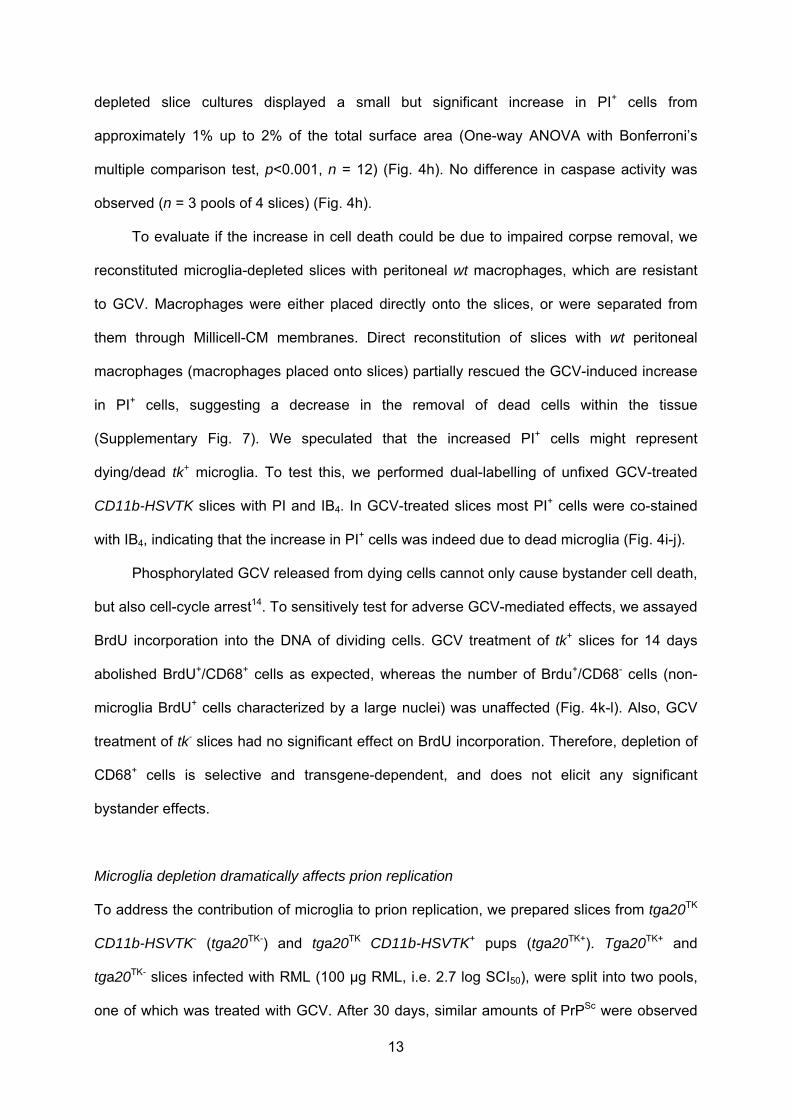