Embed Size (px)
Citation preview
University of Udine PhD course in Clinical Sciences and Technologies (XXVIII cycle)
Department of Medical and Biological Sciences
To investigate in vitro the pathogenic mechanism of anti-
PS/PT antibodies to better define their role in the diagnosis
of APS syndrome
Supervisor: PhD student:
Prof. Francesco Curcio Adriana Cifù
Assistant supervisor:
Dott. Martina Fabris
_______________________________________________________
Year Of The Final Exam: 2017

Sommario 1. ABSTRACT ............................................................................................. 1
2. ANTIPHOSPHOLIPID SYNDROME (APS) ..................................................... 3
2.1 HISTORICAL BACKGROUND ................................................................ 3
2.2 CLASSIFICATION CRITERIA AND DIAGNOSIS ......................................... 4
2.3 HAEMOSTASIS .................................................................................. 7
2.3.1 CELL-BASED MODEL OF HAEMOSTASIS ......................................... 8
2.4 ANTIGENIC TARGETS OF ANTIPHOSPHOLIPID AUTOANTIBODIES (aPL) . 10
2.4.1 β2-GLYCOPROTEIN I .................................................................. 10
2.4.2 PROTHROMBIN ........................................................................ 12
2.5 APS AND THROMBOSIS ................................................................... 13
2.5.1 THE “TWO-HIT” HYPOTHESIS .................................................... 13
2.5.1.1 CELLULAR COMPONENTS ....................................................... 13
2.5.1.2 HUMORAL FACTORS .............................................................. 15
2.5.1.3 INFLAMMATION .................................................................... 16
2.6 APS AND PREGNANCY ..................................................................... 17
2.6.1 aPL AND INFLAMMASOMES ...................................................... 19
2.7 RISK FACTORS OTHER THAN aPL IN APS PATIENTS .............................. 20
2.7.1 PLASMATIC PLATELET-ACTIVATING FACTOR ACETYLHYDROLASE
ACTIVITY (PAF-AH)............................................................................ 21
3. AIM OF THE STUDY .............................................................................. 23
4. MATERIAL AND METHODS .................................................................... 23
4.1 PATIENTS ....................................................................................... 23
4.2 ANTIBODY DETERMINATION AND ANTIGENIC SPECIFICITY .................. 24
4.2.1 LA .......................................................................................... 24
4.2.2 aCL and aβ2GpI ....................................................................... 25
4.2.3 aPS/PT .................................................................................... 26
4.3 MEASUREMENT OF PAF-AH ACTIVITY ........................................... 26
4.4 ISOLATION OF IgG ....................................................................... 27
4.5 CELL CULTURE ................................................................................ 29
4.5.1 ISOLATION OF MONOCYTES ...................................................... 29
4.5.2 HUVEC .................................................................................... 29
4.6 PROCOAGULANT CELL TREATMENT .................................................. 30
4.7 RNA ISOLATION AND REAL-TIME PCR ............................................... 31
4.8 MEASUREMENT OF NITRIC OXIDE (NO) PRODUCTION ........................ 32
4.9 QUANTIFICATION OF CYTOKINES AND CHEMOKINES .......................... 33
4.10 STATISTICAL ANALYSIS ................................................................... 34
5. RESULTS .............................................................................................. 35
5.1 IgG PURIFICATION .......................................................................... 35
5.2 SETTING OF PROCOAGULANT TREATMENT ....................................... 36
5.3 EFFECT OF IgG FROM BLOOD DONORS' SERUM ON MONOCYTES AND
HUVECs .............................................................................................. 36
5.4 EFFECT OF IgG FROM APS PATIENTS’ SERUM ON MONOCYTES ............ 38
5.5 TF EXPRESSION IN HUVECs TREATED WITH IgG OBTAINED FROM APS
PATIENTS’ SERUM ................................................................................ 41
5.6 THE EFFECT OF IgG OBTAINED FROM APS PATIENTS’ SERUM ON NITRIC
OXIDE (NO) PRODUCTION IN HUVECs .................................................... 42
5.7 SOLUBLE FACTOR RELEASED BY HUVECs AFTER PROCOAGULANT
TREATMENT: PRELIMINARY DATA .......................................................... 42
5.8 PAF-AH .......................................................................................... 44
5.8.1 PAF-AH PLASMATIC ACTIVITY IN PATIENTS AND CONTROLS:
CORRELATION WITH LIPOD METABOLIC MARKERS .............................. 44
5.8.2 PAF-AH PLASMATIC ACTIVITY IN PATIENTS DISCLOSING DISTINCT
PATTERN OF aPL POSITIVITY .............................................................. 45
6. DISCUSSION ........................................................................................ 47
7. CONCLUSIONS ..................................................................................... 52
8. PUBLISHED ABSTRACTS ........................................................................ 53
9. PUBLICATIONS ..................................................................................... 56
10. REFERENCES ...................................................................................... 57
1
1. ABSTRACT
Antiphospholipid syndrome (APS) is an autoimmune disorder characterized by
vascular thrombosis (venous or arterial) and/or adverse obstetric outcomes
accompanied by persistent and elevated levels of antiphospholipid (aPL)
antibodies. According to the 2006 revised international classification criteria,
the presence of one among anti-beta2 glycoprotein I (aβ2GPI) IgG or IgM, anti-
cardiolipin (aCL) IgG or IgM and the lupus anticoagulant (LA) is indicated for a
definite diagnosis of APS. However, not infrequently, none of the “criteria”
antibodies can be demonstrated. Only recently the so-called “seronegative
APS” was definitely recognized as a distinctive setting, or better re-defined by
the demonstration of new classes of aPL antibodies, such as the
autoantibodies directed against prothrombin (aPT and aPS/PT). In the next
future, these autoantibodies, particularly aPS/PT, could become additional
serological classification criteria for APS especially to recognized patients
negative for classical aPL. The combination of aβ2GPI, aPS/PT and LA
demonstrates the best diagnostic accuracy for APS and aPS/PT were recently
recommended as a surrogate of LA when specific inhibitors and/or analytical
variables may affect its interpretation. Despite these recommendations, very
few clinical laboratories include aPS/PT in routine analyzes so far. Moreover,
no definite recommendations are available to guide the therapeutic approach
in patients positive only for aPS/PT antibodies. To clarify their role in APS
diagnosis and treatment, a better comprehension of its pathogenic
mechanisms is needed. Thus, the principal aim of this thesis is to investigate
the pathogenic mechanism underlying the thrombotic manifestations
associated to the presence of aPS/PT. To address this issue, the biological
effects sustained in vitro by aPS/PT were compared to those sustained by
aβ2GpI, the most studied and recognized player in APS, by developing an
experimental model able to investigate the thrombotic effect of these
autoantibodies on monocytes and endothelial cells. Beside this principal
study, to improve the risk management of APS patients, the plasmatic activity
2
of the PAF-AH (Platelet Activating Factor Acetylhydrolase) was investigated as
a new potential prognostic biomarker. PAF-AH is a specific marker of vascular
inflammation dependent to common lipid metabolism markers (i.e. LDL) which
is involved in the atherosclerotic plaque instability.
Obtained data on the TF mRNA expression and Nitric Oxide production
(colorimetric assay), confirmed that aPS/PT and aβ2GpI exert similar pro-
thrombotic effects on monocytes and endothelial cells. On the contrary, the
different effect of aβ2GpI and aPS/PT on mRNA expression of IL1β and NLRP3,
and the different impact on PAF-AH activity (colorimetric assay), may suggest
that these classes of antibodies probably activate different metabolic
pathways. Moreover, plasmatic PAF-AH activity in patients with positive aPL
antibodies appeared to be independent to common lipid metabolism markers
(i.e. LDL). Based on these results, PAF-AH plasmatic activity may represent a
new prognostic biomarker also in the context of aPL antibodies, to identify
patients at major risk and favouring more tailored therapeutic interventions.
Further prospective studies on selected patients are ongoing.
3
2. ANTIPHOSPHOLIPID SYNDROME (APS)
Antiphospholipid Syndrome (APS) is a systemic autoimmune disease
characterized by vascular thrombosis (venous or arterial) and/or adverse
obstetric outcomes, accompanied by persistent and elevated levels of
antiphospholipid (aPL) antibodies, namely lupus anticoagulant (LA),
anticardiolipin antibodies (aCL) or anti-β2 glycoprotein I antibodies (aβ2GpI)
(Harper, 2011; Gomez-Puerta, 2014).
2.1 HISTORICAL BACKGROUND
The antiphospholipid antibody story begins in 1906 when Wasserman
developed a serological test for syphilis (Arachchillage, 2014). The Wasserman
reagin test was attributed to antibody reactivity against antigens derived from
Treponema Pallidum, the causative organism of this infection (Hanly, 2003). In
1941 Pangborn demonstrated that isolated cardiolipin from bovine heart, was
the antigenic component of reagin test. The use of purified cardiolipin
together with lecithin and cholesterol formed the basis for more efficient tests
as the Venereal Disease Research Laboratory (VDRL) microflocculation assay. In
1952 Moore and Mohr identified two circumstances in which biological false
positive for syphilis test could occurs:
transient positivity during acute viral infection or after vaccination;
persistent positivity (>6 months) associated with autoimmune
disorders like Systemic Lupus Erythematosus (SLE), Rheumatoid
Arthritis (RA) and Sjogren’s Syndrome (SjS).
At same time Conley and Hartmann (1951) wrote a case report of two patients
with SLE, biological false positive for syphilis and with a “peculiar hemorrhagic
disorder” (prolongation of prothrombin time). This was the initial description
of “Lupus Anticoagulant” (LA) (Arachchillage, 2014; Hanly, 2003).
LA is a biological paradox: in vivo causes thrombotic effects, but in vitro there
is a prolongation of a phospholipid dependent coagulation test that is not due
to a specific inhibitor of coagulation factor (Watson, 2012).
4
In the 60’s, the association of LA phenomenon with thrombosis, recurrent
fetal losses and thrombocytopenia was observed (Gomez-Puerta, 2014).
The development of more sensitive assay for anticardiolipin antibody (such as
enzyme-linked immunosorbent assay -ELISA-), facilitated clinical and
epidemiological studies and description of the APS (Hanly, 2003).
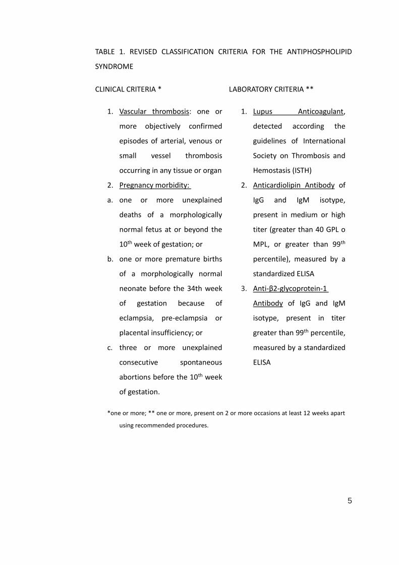
2.2 CLASSIFICATION CRITERIA AND DIAGNOSIS
The international criteria for the classification of patients with definite APS
were defined in 1998 (the so-called “Sapporo criteria”) and then revised in
2004 during the 11° International Congress on aPL in Sydney. APS is diagnosed
if at least one of clinical criteria and one of laboratory criteria are both present
(Table 1) (Wilson, 1999; Miyakis,2006).
However, there are different features associated with APS, but not included in
the revised criteria (Table 2) (Miyakis,2006). Only recently APS patients so-
called “seronegative APS” (negative for LA, aCL and aβ2GpI) was definitely
recognized as a distinctive setting, or better re-defined by the demonstration
of new classes of aPL antibodies, such as the autoantibodies directed against
prothrombin (aPT and aPS/PT): in the next future these autoantibodies,
particularly aPS/PT, could become additional serological classification criteria
for APS especially to recognized patients negative for classical aPL (Sciascia and
Khamashta, 2014).
5
TABLE 1. REVISED CLASSIFICATION CRITERIA FOR THE ANTIPHOSPHOLIPID
SYNDROME
CLINICAL CRITERIA * LABORATORY CRITERIA **
1. Vascular thrombosis: one or
more objectively confirmed
episodes of arterial, venous or
small vessel thrombosis
occurring in any tissue or organ
2. Pregnancy morbidity:
a. one or more unexplained
deaths of a morphologically
normal fetus at or beyond the
10th week of gestation; or
b. one or more premature births
of a morphologically normal
neonate before the 34th week
of gestation because of
eclampsia, pre-eclampsia or
placental insufficiency; or
c. three or more unexplained
consecutive spontaneous
abortions before the 10th week
of gestation.
1. Lupus Anticoagulant,
detected according the
guidelines of International
Society on Thrombosis and
Hemostasis (ISTH)
2. Anticardiolipin Antibody of
IgG and IgM isotype,
present in medium or high
titer (greater than 40 GPL o
MPL, or greater than 99th
percentile), measured by a
standardized ELISA
3. Anti-β2-glycoprotein-1
Antibody of IgG and IgM
isotype, present in titer
greater than 99th percentile,
measured by a standardized
ELISA
*one or more; ** one or more, present on 2 or more occasions at least 12 weeks apart
using recommended procedures.
6
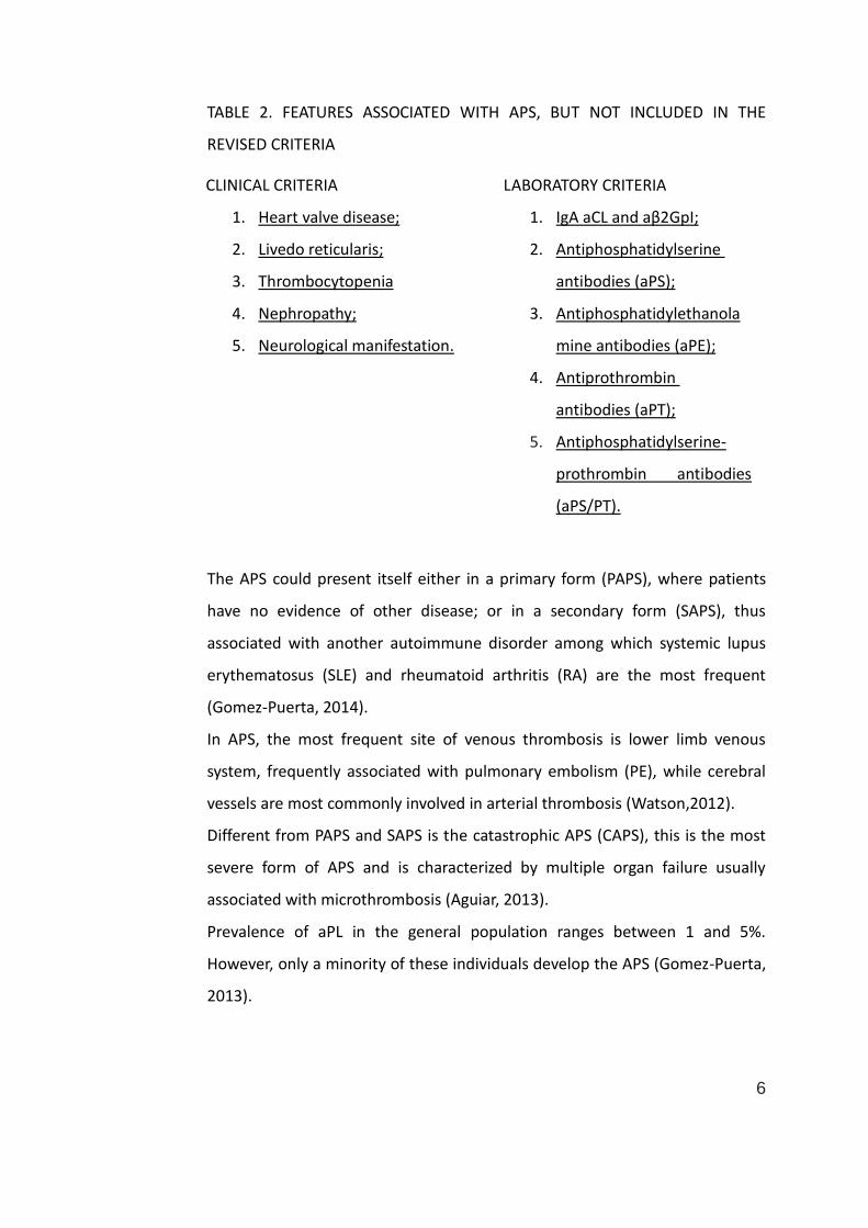
TABLE 2. FEATURES ASSOCIATED WITH APS, BUT NOT INCLUDED IN THE
REVISED CRITERIA
CLINICAL CRITERIA
1. Heart valve disease;
2. Livedo reticularis;
3. Thrombocytopenia
4. Nephropathy;
5. Neurological manifestation.
LABORATORY CRITERIA
1. IgA aCL and aβ2GpI;
2. Antiphosphatidylserine
antibodies (aPS);
3. Antiphosphatidylethanola
mine antibodies (aPE);
4. Antiprothrombin
antibodies (aPT);
5. Antiphosphatidylserine-
prothrombin antibodies
(aPS/PT).
The APS could present itself either in a primary form (PAPS), where patients
have no evidence of other disease; or in a secondary form (SAPS), thus
associated with another autoimmune disorder among which systemic lupus
erythematosus (SLE) and rheumatoid arthritis (RA) are the most frequent
(Gomez-Puerta, 2014).
In APS, the most frequent site of venous thrombosis is lower limb venous
system, frequently associated with pulmonary embolism (PE), while cerebral
vessels are most commonly involved in arterial thrombosis (Watson,2012).
Different from PAPS and SAPS is the catastrophic APS (CAPS), this is the most
severe form of APS and is characterized by multiple organ failure usually
associated with microthrombosis (Aguiar, 2013).
Prevalence of aPL in the general population ranges between 1 and 5%.
However, only a minority of these individuals develop the APS (Gomez-Puerta,
2013).
7
2.3 HAEMOSTASIS
Haemostasis is a tightly regulated homeostatic mechanism that ensures the
maintenance of blood flow under physiological conditions, but also permits
rapid, localized coagulation in the event of tissue damage (Allford, 2007;
Norris, 2003; Panteleev, 2015). A delicate balance exists between four major
components: vascular endothelium, platelets, the coagulation pathway and
fibrinolysis (Allford, 2007).
The traditional concept of coagulation was based on two main pathways that
were mutually exclusive and of equal importance: the intrinsic (or contact)
pathway and the extrinsic (or tissue factor) pathway (Allford, 2007; Norris,
2003). This model (figure 1) described the coagulation as a “cascade” of
reactions involving activation of several clotting factors resulting in the
production of a large amount of thrombin and subsequent formation of a
fibrin clot (Hoffman,2003). However, this cascade paradigm was useful in vitro
for diagnostic purposes, but failed to explain in vivo phenomena (Allford,
2007). In 90’s Mann proposed a cell-based model of haemostasis in which was
emphasized the interaction of clotting factors with specific surfaces, explaining
the unresolved in vivo phenomena (Mann, 1991; Hoffman, 2003).
8
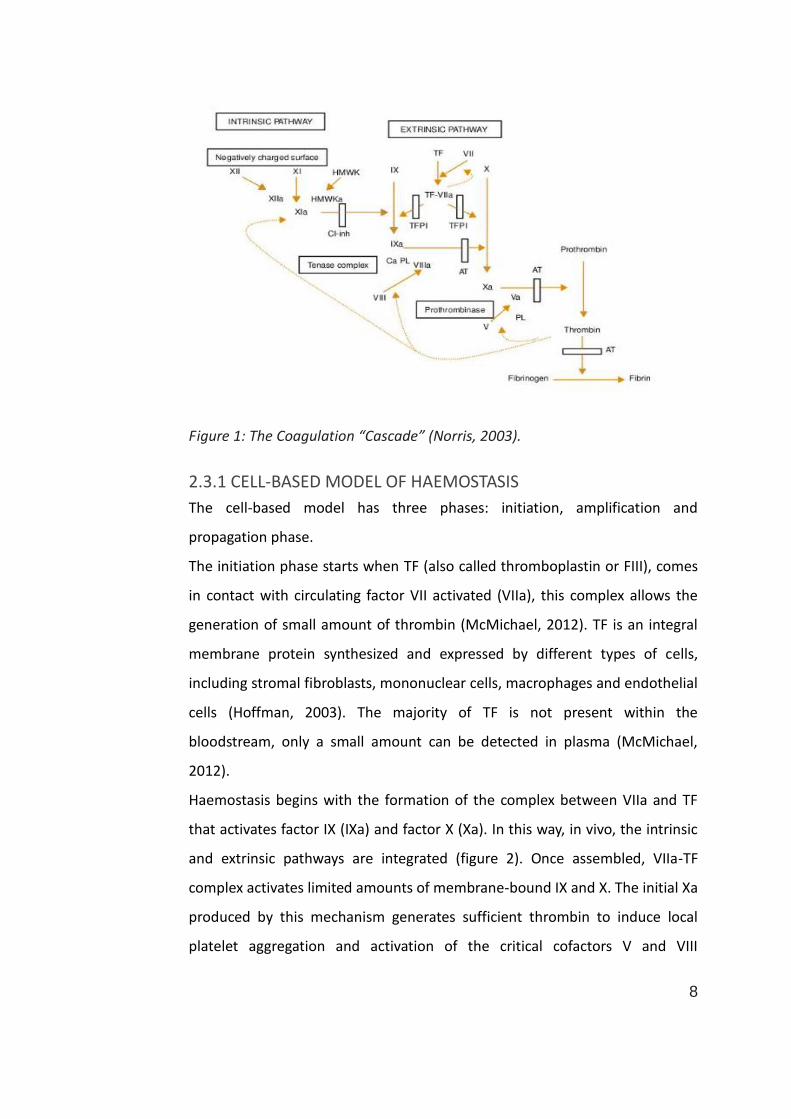
Figure 1: The Coagulation “Cascade” (Norris, 2003).
2.3.1 CELL-BASED MODEL OF HAEMOSTASIS
The cell-based model has three phases: initiation, amplification and
propagation phase.
The initiation phase starts when TF (also called thromboplastin or FIII), comes
in contact with circulating factor VII activated (VIIa), this complex allows the
generation of small amount of thrombin (McMichael, 2012). TF is an integral
membrane protein synthesized and expressed by different types of cells,
including stromal fibroblasts, mononuclear cells, macrophages and endothelial
cells (Hoffman, 2003). The majority of TF is not present within the
bloodstream, only a small amount can be detected in plasma (McMichael,
2012).
Haemostasis begins with the formation of the complex between VIIa and TF
that activates factor IX (IXa) and factor X (Xa). In this way, in vivo, the intrinsic
and extrinsic pathways are integrated (figure 2). Once assembled, VIIa-TF
complex activates limited amounts of membrane-bound IX and X. The initial Xa
produced by this mechanism generates sufficient thrombin to induce local
platelet aggregation and activation of the critical cofactors V and VIII
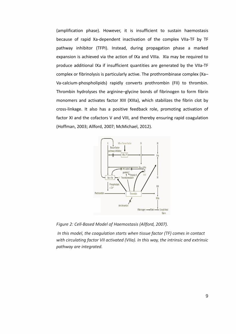
9
(amplification phase). However, it is insufficient to sustain haemostasis
because of rapid Xa-dependent inactivation of the complex VIIa-TF by TF
pathway inhibitor (TFPI). Instead, during propagation phase a marked
expansion is achieved via the action of IXa and VIIIa. XIa may be required to
produce additional IXa if insufficient quantities are generated by the VIIa-TF
complex or fibrinolysis is particularly active. The prothrombinase complex (Xa–
Va-calcium-phospholipids) rapidly converts prothrombin (FII) to thrombin.
Thrombin hydrolyses the arginine–glycine bonds of fibrinogen to form fibrin
monomers and activates factor XIII (XIIIa), which stabilizes the fibrin clot by
cross-linkage. It also has a positive feedback role, promoting activation of
factor XI and the cofactors V and VIII, and thereby ensuring rapid coagulation
(Hoffman, 2003; Allford, 2007; McMichael, 2012).
Figure 2: Cell-Based Model of Haemostasis (Allford, 2007).
In this model, the coagulation starts when tissue factor (TF) comes in contact
with circulating factor VII activated (VIIa). In this way, the intrinsic and extrinsic
pathway are integrated.
10
2.4 ANTIGENIC TARGETS OF ANTIPHOSPHOLIPID AUTOANTIBODIES (aPL)
The term “Antiphospholipid Syndrome” is used to connect the clinical
manifestations to the presence of aPL (Amengual, 2003).
Initially it was thought that these autoantibodies were directed against anionic
phospholipids, but in the last decade, different groups of investigators have
been demonstrated that aPL are part of a family of autoantibodies against
phospholipid-binding plasma proteins or phospholipid-protein complexes.
Several antigenic targets have been identified among which high and low
molecular weight kininogens, protein C, annexin V and protein S. However, the
most common and best characterized target for aPL are β2-Glycoprotein I
(β2GpI) and prothrombin (PT). All these antigenic targets are involved in
coagulation system, giving an explication of high incidence of thrombotic
events in patients with APS (Amengual, 2003).
2.4.1 β2-GLYCOPROTEIN I
The main antigenic target for aPL is β2GpI, also known as apolipoprotein H. In
90’s, it has been demonstrated that aCL associated with APS, were not
directed against cardiolipin alone, in fact they require a cofactor that is a
plasmatic protein: β2GpI (Amengual, 2003).
In human, this protein is synthesized by different cells: hepatocytes,
endothelial, and trophoblast cells. β2GpI circulates in blood at high
concentration: the mean serum level is about 200 μg/ml (Miyakis,2004;
Mahler, 2012). β2GpI is a 50-KDa anionic phospholipid- binding glycoprotein
that belongs to the CCP superfamily (complement control protein).
The CCP domain functions as a protein-protein interaction module in many
different proteins. β2GpI is organized in five CCP domains: the first four
domains have regular, conserved sequences, while the fifth domain is aberrant
and has additional amino acids (multiple lysine). This amino acid strain creates
a positively charged domain that is responsible for the binding to the anionic
phospholipids. The crystal structure shows that the phospholipid-binding site
11
is located at the bottom side of domain V and predicts that the potential
binding site for aβ2GpI is located in domain I (Groot, 2011; Miyakis,2004).
β2GpI can exist in plasma in two different conformations: closed/circular or
open/hockey-stick like conformations. As shown in figure 3, binding of β2GpI
to anionic surfaces results in a conformational change: the conversion from
closed to open conformation leads to expose the antibody-binding site, that is
not accessible to autoantibodies in the closed conformation (Groot, 2011;
Harper,2011).
About its physiological role, not many other information are available, but it is
supposed that β2GpI plays an important role in biology, since it shares high
homology with different mammalian species (Miyakis, 2004). The homology
with other proteins involved in innate immunity suggests that β2GpI could
play a role in host defense against bacteria (Groot, 2011).
Multi-centric studies have found a strong association between aβ2GpI
antibodies and history of thrombosis (Mahler, 2012). Moreover recent studies
have shown that aβ2GpI antibodies associated with major risk of thrombosis,
bind the domain I of the β2GpI, that is exposed in the open conformation
(Harper, 2011), as already demonstrated by Andreoli et. al, who have
demonstrated that only autoantibodies directed against domain I of β2GpI are
associated with increased risk of thrombosis, while a significant lower risk of
thrombosis has been found in case of aβ2GpI antibodies targeting other
domains of β2GpI (Harper, 2011; Andreoli,2010).
12
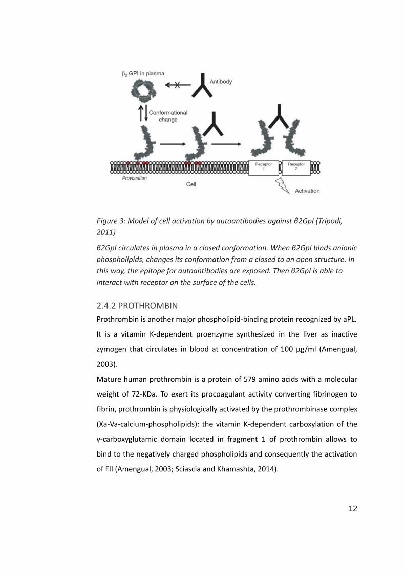
Figure 3: Model of cell activation by autoantibodies against β2GpI (Tripodi,
2011)
β2GpI circulates in plasma in a closed conformation. When β2GpI binds anionic
phospholipids, changes its conformation from a closed to an open structure. In
this way, the epitope for autoantibodies are exposed. Then β2GpI is able to
interact with receptor on the surface of the cells.
2.4.2 PROTHROMBIN
Prothrombin is another major phospholipid-binding protein recognized by aPL.
It is a vitamin K-dependent proenzyme synthesized in the liver as inactive
zymogen that circulates in blood at concentration of 100 μg/ml (Amengual,
2003).
Mature human prothrombin is a protein of 579 amino acids with a molecular
weight of 72-KDa. To exert its procoagulant activity converting fibrinogen to
fibrin, prothrombin is physiologically activated by the prothrombinase complex
(Xa-Va-calcium-phospholipids): the vitamin K-dependent carboxylation of the
γ-carboxyglutamic domain located in fragment 1 of prothrombin allows to
bind to the negatively charged phospholipids and consequently the activation
of FII (Amengual, 2003; Sciascia and Khamashta, 2014).
13
2.5 APS AND THROMBOSIS
A strong association between aPL and thrombosis has been demonstrated, but
nevertheless the pathogenic role of aPL in the development of thrombosis
should be clarified (Gomez-Puerta, 2014).
2.5.1 THE “TWO-HIT” HYPOTHESIS
In APS, the key elements involved in pathogenic mechanism of thrombosis are:
cellular component of vessels;
humoral components that regulate haemostasis (coagulation factor,
natural anticoagulants and fibrinolytic system);
inflammation (inflammatory cells, soluble inflammatory mediators and
infectious agents) (Willis, 2015).
The complex interaction of these elements, lead to proinflammatory and
prothrombotic state, for this reason, it is referred to as the “two-hit”
hypothesis. Theory postulates that even though the persistence of elevated
levels of aPL is a necessary condition, the occurrence of APS is seemingly
triggered by an additional “second hit”, such as trauma or infection (Willis,
2015; Brandt, 2013).
2.5.1.1 CELLULAR COMPONENTS
The aPL are directed against anionic phospholipids, therefore several studies
have investigated the interaction of β2GpI with cellular membranes and cells.
Both in vitro and in vivo studies have shown that the complex β2GpI-aβ2GpI
can bind and activate many different cells such as endothelial cells (ECs),
monocytes and platelets (Tripodi, 2011).
Activation of endothelial cells by aPL is a major thrombogenic mechanism. The
specific binding of aβ2GpI to EC up-regulates the cell-surface expression of cell
adhesion molecules (CAMs): intracellular adhesion molecule-1 (ICAM-1),
vascular cell adhesion molecule-1 (VCAM-1) and E-selectin, promoting
leukocyte adhesion (Willis, 2015; Brandt, 2013).
14
Furthermore, activated EC up-regulate the expression of TF (the key initiator of
the coagulation pathway), microparticle formation, fibrinolysis inhibitor PAI-1
and inflammatory cytokines/chemokines such as IL-6, monocyte chemotactic
protein-1 (MCP-1), fractalkine (Meroni, 2001). On the other side activation of
endothelium leads to decrease expression of thrombomodulin (Meroni, 2001;
Rikarni, 2015). These finding suggest that the complex β2GpI-aβ2GpI can
induce an endothelial activation either directly or by cytokine autocrine loop
(Meroni, 2001). Another mechanism involved in thrombus formation is
vasoconstriction: endothelium regulates vessel tone through endothelin-1
peptide, the most potent endothelium-derived contracting factor (Meroni,
2001); supporting this idea, Atsumi et al, reported that plasma level of
endothelin-1 peptide significantly correlated with history of thrombosis in APS
patient (Atsumi, 1998). All these mechanisms lead to a
procoagulant/proinflammatory phenotype that increases the risk of
thrombotic occlusions (Brandt, 2013).
The monocytes are another player in the development of thrombosis in APS
patients, indeed exposed to aPL, monocytes up-regulate expression of TF via
p38 MAPK pathway, resulting in nuclear factor-kB (NF-kB) activation (Willis,
2015; Brandt, 2013). TF expression is associated with increased plasma levels
of vascular endothelial growth factor (VEGF) and cell surface expression of
both VEGF and the Flt-1 tyrosine kinase receptor. Stimulation of Flt-1 tyrosine
kinase receptor by VEGF results in TF mRNA and protein expression.
Furthermore, monocytes derived from APS patients or normal monocytes
exposed to aPL, show an increased expression of protease-activated receptor 1
(PAR-1) and PAR-2. This is very significant because that PAR-1 and PAR-2
mediate several effects of thrombin such as up-regulation of proinflammatory
cytokines (IL-6, IL-8, MCP-1) (Harper, 2011; Willis, 2015).
In vivo, platelets are central to arterial thrombus formation, indeed in APS
patients, platelet activation is increased. Plentiful evidence from
epidemiological and mechanistic studies indicates that aPL activate platelets,
15
resulting in increased expression of thromboxane B2 (TXB2), fibrinogen
receptor glycoprotein IIb/IIIa (GPIIb/IIIa) and consequently platelet
aggregation (Harper, 2011; Willis, 2015). Urbanus et al. demonstrated that
plasma β2GpI does not bind to platelets, whereas β2GpI in complex with
aβ2GpI does bind to platelets (Urbanus, 2008). The platelet receptors involved
in the interaction of β2GpI/aβ2GpI complex are apolipoprotein E receptor 2
(ApoER2) and von Willebrand factor receptor glycoprotein Ibα (GPIbα)
(Harper, 2011; Urbanus, 2008). Therefore β2GpI/aβ2GpI complex can bind to
ApoER2 and/or GPIbα and this binding mediates the activation of platelets and
the induction of thromboxane A2 synthesis (Urbanus, 2008).
Finally, activated platelets secrete platelet factor 4 (PF4), a member of the CXC
chemokine family with multiple prothrombotic effects (inhibition of
inactivation of thrombin by antithrombin, potentiation of platelet aggregation
and accelerating cleavage of activated protein C) and also and antigenic target
in APS (Harper, 2011; Giannakopoulos, 2013).
The activation of all these cell types by aPL coupled with the release of several
proinflammatory mediators has linked to the development of thrombosis in
APS animal models and in same case in human APS patients (Willis, 2015).
2.5.1.2 HUMORAL FACTORS
aPL act at various levels of the coagulation cascade leading to uncontrolled
fibrin formation and impaired thrombus resolution (Willis, 2015).
Direct activation of prothrombin binding to the surface of ECs, has been
demonstrated in APS patients and has been attributed to anti-prothrombin
antibodies (aPT) with LA activity; this activation induces TF expression and
thrombosis (Willis, 2015; Amengual, 2003). Furthermore, aPL bind directly to
antithrombin III (ATIII) resulting in reduced inactivation of FIXa and FXa (Willis,
2015; Harper, 2011).
aPL interfere with the fibrinolytic system: the action of β2GpI/aβ2GpI complex
on endothelium leads to decrease thrombomodulin expression (TM) and tp
16
increase of plasminogen activator inhibitor (PAI-1), this might be one of the
causes of thrombophilic diathesis in APS (Meroni, 2001; Rikarni ,2015).
Modulation of activated protein C (APC), a phospholipid-dependent major
antithrombotic pathway, has been found in APS patients who have increased
resistance to APC resulting in greater thrombin generation overtime (Meroni,
2001; Harper, 2011; Willis, 2015).
Finally, it has been hypothesized an involvement of annexin 5 (A5): normally
A5 binds to phosphatidylserine surfaces of ECs, forming a shield that inhibits
the formation of procoagulant complex; in a model of the pathogenesis of the
antiphospholipid syndrome, aβ2GpI that bind to the domain 1 of the β2GpI
can disrupt the A5 antithrombotic shield present on the endothelial cells
(Giannakopoulos, 2013; Tripodi, 2015).
Overall, the resistance of activated coagulation factors to inactivation and the
reduced activity of natural anticoagulant and fibrinolytic agents, potentiate
unchecked fibrin formation and the thrombogenic state (Willis, 2015; Tripodi,
2011).
2.5.1.3 INFLAMMATION
In addition to activation of the coagulation pathway, APS is characterized by
proinflammatory changes. Indeed, inflammation acts as a key trigger event for
the thrombotic manifestation of APS and it is very important for changes in
antigen conformation and immune cell activity, critical elements in aPL
ontogeny (Harper, 2011; Willis, 2015).
Several studies have demonstrated that APS patients are characterized by
increased oxidative stress: paraoxonase activity (a glycoprotein that prevents
oxidation of low-density lipoprotein-LDL- cholesterol) is significantly decreased
in these patients, whereas 8-epi-prostaglandin F2∝, a biomarker of lipid
peroxidation, is upregulated (Giannakopoulos, 2013).
In APS patients, oxidative stress has an effect on β2GpI, in fact APS patients
frequently show high levels of oxidized β2GpI. Oxidative stress acts on β2GpI
at different levels:
17
increases β2GpI production through gene promoter up-regulation via
nuclear factor kappa B (NFkB);
increases the immunogenicity of β2GpI through post-translational
modification;
induces conformational changes exposing hidden epitopes of β2GpI,
important for aPL production (Willis, 2015)
It has been proposed that disturbance of redox balance in patients with APS
could constitute the “first hit” which allows the formation of β2GpI/aβ2GpI
complex on ECs (Giannakopoulos, 2013).
Furthermore, oxidative stress can up-regulate annexin II (A2) expression, an
endothelial receptor that mediates the binding of β2GpI to ECs (Ma, 2000),
and, in a murine model of thrombosis, induces platelet aggregation, EC
stimulation and von Willebrand factor expression (Nishimura, 2011).
Patients with APS have decreased levels of plasma nitrite, as compared with
controls. This suggests an abnormal activity of endothelial nitric oxide
synthase (e-NOS). Endothelium-derived nitric oxide is fundamental for normal
function of endothelium and a reduced expression of e-NOS results in
superoxide and peroxynitrite production (Giannakopoulos, 2013).
Activation of the complement cascade also contributes to the pathogenic
effects of aPL; in particular, the anaphylatoxins C3a and C5a induce the
inflammatory vascular phenotype of APS and are necessary players connecting
EC, monocytes, and platelet activation by aPL and the thrombotic
manifestation (Willis, 2015).
2.6 APS AND PREGNANCY
The risk for adverse pregnancy outcomes, such as recurrent miscarriage, fetal
demise, placental insufficiency, preeclampsia and intrauterine growth
restriction (IUGR) in women with APS is greatest from the 10th week of
gestation onward (Hanly,2003; Mulla, 2013).
18
There is also evidence that these women have an increased risk of giving birth
to a premature infant because of pregnancy-associated hypertension and
utero-placental insufficiency (Hanly, 2003).
Unlike the systemic APS that is a prothrombotic and proinlammatory disease,
obstetric APS (OAPS) is primarily a proinflammatory syndrome (Mulla, 2013).
Indeed, the first hypothesis that OAPS was due to an intraplacental thrombosis
with consequently alteration of maternal-fetal blood exchanges, has not been
confirmed by histological studies (Khamashta, 2016).
Different groups of investigators have been postulated two mechanisms for
aPL-induced pregnancy morbidity: defective placentation and inflammation
(Khamashta, 2016).
In the placenta, aβ2GpI can react with both sides, maternal and fetal (Simone,
2000). This ability induces direct placental damage with different mechanisms:
inhibiting trophoblast differentiation and syncytialization;
inducing trophoblast apoptosis;
impairing trophoblast invasiveness;
affecting trophoblast expression of adhesion molecules that regulate
its adhesion to and invasion of the maternal tissue;
inhibiting production of angiogenic factor by trophoblasts (Khamashta,
2016; Tong, 2014).
Moreover, has been proposed as an additional mechanism for preeclampsia
due to the internalization of aPL by trophoblasts with the subsequent
acceleration of cell death and release of debris that can activate maternal
endothelial cells (Khamashta, 2016).
It has been proved that inflammation has an important role in OAPS. This idea
is based on:
the histological demonstration of complement deposition, neutrophil
infiltration, and tumor necrosis factor α (TNFα) secretion in decidual
tissue;
19
the observation that complement deficiency in animal models or
complement inhibition in vivo are protective against obstetrical
complications;
the evidence of a protective effect of heparin linked to its anti-
complement activity;
the observation in in vitro studies that aPL can induce
trophoblasts to produce interleukin-1 β (IL1β) by activation of
inflammasome (Mulla, 2013; Khamashta, 2016; Müller-Calleja, 2015).
2.6.1 aPL AND INFLAMMASOMES
Inflammasomes (NLR) are large soluble cytoplasmatic complexes that are
capable of activating the cystein protease caspase-1 in response to a wide
range of stimuli including microbial and self-molecules. The activation of
inflammasome leading to the processing and activation of pro-IL1β and pro-
IL18 through caspase-1 (Chen, 2009). Inflammasome includes several
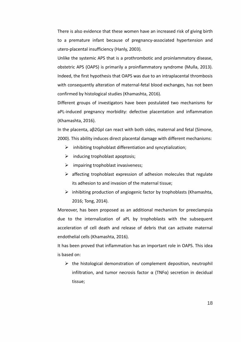
members: NLRP1, NLRP3, NLRP6, NLRP7, NLRP12, NLRC4 and NAIP proteins
(Barbè, 2014). The most well characterized are NLRP1 and NLRP3 (figure 4)
and different studies on OAPS, have associated the production of IL1β with the
activation of NLRP3 (Mulla, 2013; Khamashta, 2016; Müller-Calleja, 2015).
20
Figure 4: Mechanism of Inflammasome.
Three different Inflammasome (NLRC4, NLRP3 and NLRP1) activate caspase-1
in response to several stimuli such as microbial component or crystal; in this
way, pro-IL1β and pro-IL18 are processed
2.7 RISK FACTORS OTHER THAN aPL IN APS PATIENTS
Recently, the role of vascular risk factors in the development of clinical events
in patients with APS has been established (Khamashta, 2016). The presence of
multiple risk factors such as hypertension, smoking, hypercholesterolemia, or
estrogen use may increase the occurrence of thrombosis in patients with aPL
(Erkan, 2002).
SLE is a risk factor for thrombosis per se: in patients with SLE there are a
higher-than-expected incidence of vascular events, which are not completely
explained by traditional vascular risk factors (Esdaile, 2001). The combination
of SLE and aPL positivity has been shown to increase the risk of thrombosis.
Indeed, in SLE patients with aPL positivity, the annual risk of first thrombosis is
higher than in healthy aPL positive subjects without other cardiovascular risk
(4% vs < 1%) (Khamashta, 2016).
21
Thrombotic risk assessment should be considered also in patients with
primary APS, such as women with a history of pregnancy morbidity due to
aPLs (OAPS). In fact, these patients have a higher thrombotic event rate than
healthy women (3.3 vs 0-0.5/100patients-years) (Lefevre, 2011).
Moreover, there are many aPL carriers that never develop APS, only few cases
will develop thrombosis or obstetrical manifestations and only a very small
group will develop CAPS. In this scenario, it could be of great advantage to
make a risk stratification of thrombotic/obstetric events in such patients.
To date, three score model have been proposed for risk stratification: the first
two scores are focused on the aPL profile, while the third, the Global APS
Score (GAPSS) included other variables such as autoimmune profile or
cardiovascular risk factor. This model seems to be the better one (Khamashta,
2016).
To help clinicians in patient management, in addition to GAPSS, it would be
useful to identify a new specific plasmatic biomarker, independent from the
other classic risk factors for thrombosis.
2.7.1 PLASMATIC PLATELET-ACTIVATING FACTOR ACETYLHYDROLASE ACTIVITY (PAF-AH)
Platelet activating factor acetylhydrolase activity (PAF-AH) is a Ca2+-
independent A2 phospholipase, also known as lipoprotein-associated
phospholipase A2 (Lp-PLA2). The plasmatic PAF-AH is constitutively active and
circulates bound to LDL, HDL and other lipoproteins. PAF-AH hydrolyzes the
ester bond at the sn-2 position of phospholipids, such as PAF and PAF
mimetics, that are early mediators of inflammation (McIntyre, 2008). PAF
activates a variety of cells of the innate immune system promoting migration,
adhesion and inflammatory effects. Thus, PAF-AH while inactivating PAF, is
considered an important factor to prevent an exaggerated inflammatory
response and to protect cells from uncontrolled oxidative damage (Rosenson,
2012). Several studies have shown an association between high levels of PAF-
AH activity and the severity of cardiovascular diseases and identified PAF-AH

22
as a marker of vascular inflammation involved in the atherosclerotic plaque
instability (Davidson, 2008; Maiolino, 2012). To date, there are not study on
PAF-AH activity and APS.
23
3. AIM OF THE STUDY
Numbers of recent papers underlined the important role of aPS/PT. In
particular, the combination of aβ2GPI, aPS/PT and LA demonstrates the best
diagnostic accuracy for APS and aPS/PT were recently recommended as a
surrogate of LA when specific inhibitors and/or analytical variables may affect
its interpretation (Bertolaccini, 2011). Despite these recommendations, very
few clinical laboratories include aPS/PT in routine analyses so far. Moreover,
no definite recommendations are available to guide the therapeutic approach
in patients positive only for aPS/PT antibodies. To clarify their role in APS
diagnosis and treatment, a better comprehension of its pathogenic
mechanisms is needed. Thus, the principal aim of this thesis is to investigate
the pathogenic mechanism underlying the thrombotic manifestations
associated to the presence of anti-phosphatidylserine-prothrombin
antibodies. To address this issue, since aβ2GpI antibodies represent the most
studied and recognized player in APS, I decided to compare the biological
effects sustained in vitro by aPS/PT to those sustained by aβ2GpI, by
developing an experimental model able to investigate the thrombotic effect.
Beside this principal study, to better assess the atherosclerotic risk in APS
population and improve the risk management of these patients in the follow-
up, I will investigate a new potential prognostic biomarker, such as the
plasmatic activity of the PAF-AH (Platelet Activating Factor
Acetylhydrolase), that is a specific marker of vascular inflammation involved in
the atherosclerotic plaque instability.
4. MATERIAL AND METHODS
4.1 PATIENTS
For in vitro experiments, total IgG were purified from six selected patients, in
particular three positive only for aβ2GpI IgG and three positive only for aPS/PT
24
IgG, all were LA positive. Patients positive for aβ2GpI IgG all recognized also
the domain I. As controls, total IgG were purified from five blood donors (BD),
who were tested negative for LA, aβ2GpI and aPS/PT antibodies.
The plasmatic PAF-AH activity was evaluated in a series of 167 consecutive
unselected patients (124 females and 69 males; mean age: 51±16 years)
screened for the presence of aPL at the Laboratory of Immunopathology of the
University Hospital of Udine in a routinely context of thrombotic events, risk of
thrombosis or obstetric complications. Patients were compared to 77 blood
donors (BDs; 39 females and 38 males; mean age: 39±13 years) enrolled at the
Transfusion Unit of the same Hospital.
All patients and controls gave their informed consent to these studies
according to the Declaration of Helsinki and to the Italian legislation
(Authorization of the Privacy Guarantor No. 9, 12 December 2013).
4.2 ANTIBODY DETERMINATION AND ANTIGENIC SPECIFICITY
4.2.1 LA
Plasma samples were tested for the presence of LA at the Laboratory of
Haemostasis of the University Hospital of Udine, according to the
recommended criteria from the ISTH Subcommittee on Lupus Anticoagulant-
Phospholipid-dependent antibodies.
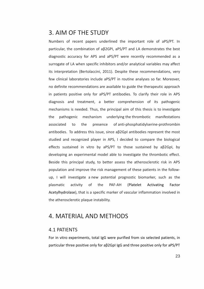
These criteria require a three-step procedure that is summarized in figure 5
(Tripodi,2011).
25
Figure 5: Flowchart for the Laboratory Detection of Lupus Anticoagulants
(Tripodi, 2011).
* the presence of heparin is ruled out by a normal thrombin clotting time; §
PNP, pooled normal plasma; PL, phospholipids.
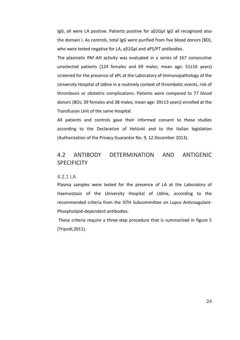
4.2.2 aCL and aβ2GpI
Anti-cardiolipin (aCL) IgG/IgM and anti-β2GpI (aβ2GpI) IgG/IgM antibodies
(figure 6) were detected by commercial methods (CLIA, Zenit RA, Menarini
Diagnostic; cutoff IgG 10, IgM 20). Anti-β2GpI IgG antibodies specifically
directed against domain I were detected by CLIA using the Inova Diagnostic Kit
(Bioflash; cutoff 20).
26
Figure 6: CLIA System for detection of aCL IgG/IgM and aβ2GpI IgG/IgM
(Menarini Diagnostic)
The assay is based on a two-step indirect chemiluminescent method that
generates quantitative results. This particular technique uses autoantigen-
coated magnetic particles as slid phase and an antibody labeled with dimethyl
acridinium ester (DMAE) as detection marker.
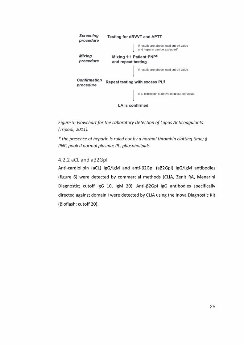
4.2.3 aPS/PT
The aPS/PT IgG and IgM antibodies, in serum samples, were analyzed by ELISA
(figure 7) using the Quanta Lite aPS/PT IgG/IgM ELISA kit (Inova Diagnostic Inc,
San Diego, CA; cutoff IgG 40 AU/ml, IgM 30 AU/ml).
Figure 7: ELISA System for the detection of aPS/PT (Sciascia and Khamashta,
2014). Antibodies are able to bind prothrombin (PT) when it is exposed to
immobilized anionic phospholipids.
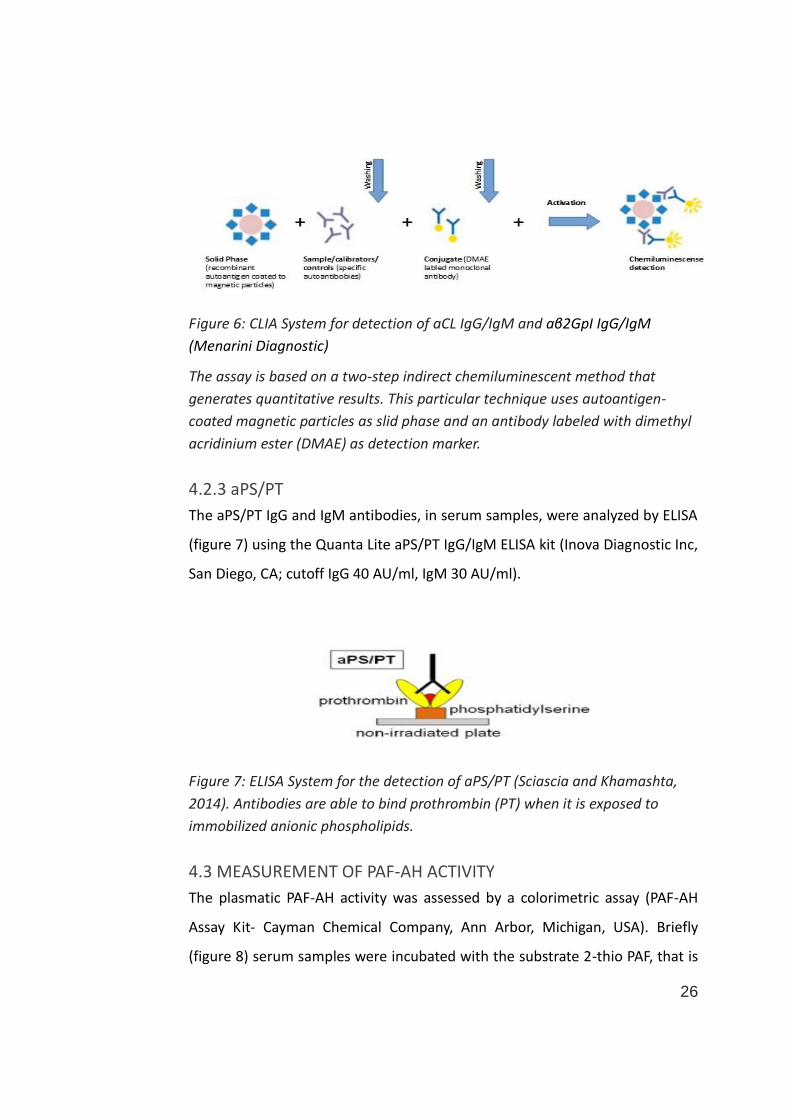
4.3 MEASUREMENT OF PAF-AH ACTIVITY
The plasmatic PAF-AH activity was assessed by a colorimetric assay (PAF-AH
Assay Kit- Cayman Chemical Company, Ann Arbor, Michigan, USA). Briefly
(figure 8) serum samples were incubated with the substrate 2-thio PAF, that is
27
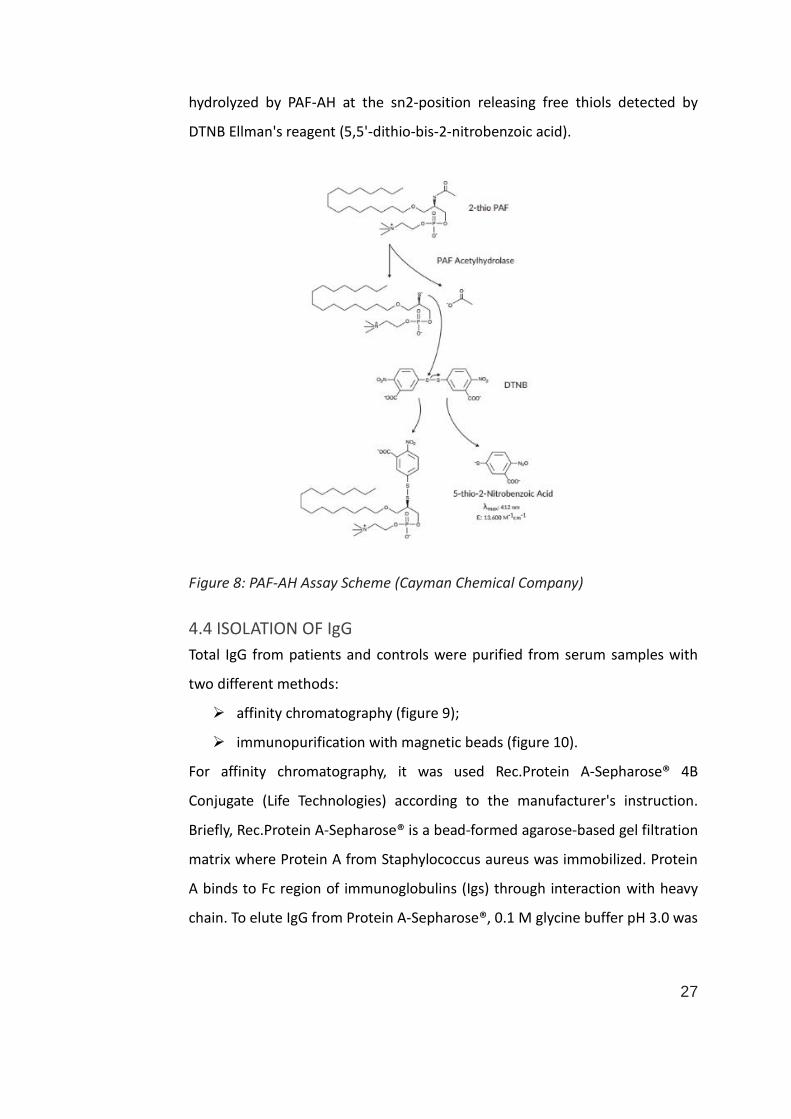
hydrolyzed by PAF-AH at the sn2-position releasing free thiols detected by
DTNB Ellman's reagent (5,5'-dithio-bis-2-nitrobenzoic acid).
Figure 8: PAF-AH Assay Scheme (Cayman Chemical Company)
4.4 ISOLATION OF IgG
Total IgG from patients and controls were purified from serum samples with
two different methods:
affinity chromatography (figure 9);
immunopurification with magnetic beads (figure 10).
For affinity chromatography, it was used Rec.Protein A-Sepharose® 4B
Conjugate (Life Technologies) according to the manufacturer's instruction.
Briefly, Rec.Protein A-Sepharose® is a bead-formed agarose-based gel filtration
matrix where Protein A from Staphylococcus aureus was immobilized. Protein
A binds to Fc region of immunoglobulins (Igs) through interaction with heavy
chain. To elute IgG from Protein A-Sepharose®, 0.1 M glycine buffer pH 3.0 was
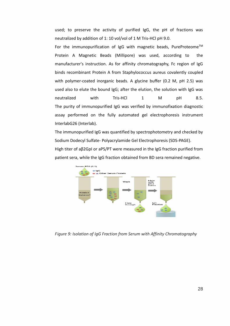
28
used; to preserve the activity of purified IgG, the pH of fractions was
neutralized by addition of 1: 10 vol/vol of 1 M Tris-HCl pH 9.0.
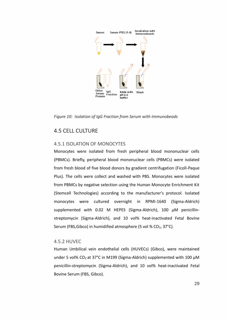
For the immunopurification of IgG with magnetic beads, PureProteomeTM
Protein A Magnetic Beads (Millipore) was used, according to the
manufacturer's instruction. As for affinity chromatography, Fc region of IgG
binds recombinant Protein A from Staphylococcus aureus covalently coupled
with polymer-coated inorganic beads. A glycine buffer (0.2 M, pH 2.5) was
used also to elute the bound IgG; after the elution, the solution with IgG was
neutralized with Tris-HCl 1 M pH 8.5.
The purity of immunopurified IgG was verified by immunofixation diagnostic
assay performed on the fully automated gel electrophoresis instrument
InterlabG26 (Interlab).
The immunopurified IgG was quantified by spectrophotometry and checked by
Sodium Dodecyl Sulfate- Polyacrylamide Gel Electrophoresis (SDS-PAGE).
High titer of aβ2GpI or aPS/PT were measured in the IgG fraction purified from
patient sera, while the IgG fraction obtained from BD sera remained negative.
Figure 9: Isolation of IgG Fraction from Serum with Affinity Chromatography
29
Figure 10: Isolation of IgG Fraction from Serum with Immunobeads
4.5 CELL CULTURE
4.5.1 ISOLATION OF MONOCYTES
Monocytes were isolated from fresh peripheral blood mononuclear cells
(PBMCs). Briefly, peripheral blood mononuclear cells (PBMCs) were isolated
from fresh blood of five blood donors by gradient centrifugation (Ficoll-Paque
Plus). The cells were collect and washed with PBS. Monocytes were isolated
from PBMCs by negative selection using the Human Monocyte Enrichment Kit
(Stemcell Technologies) according to the manufacturer's protocol. Isolated
monocytes were cultured overnight in RPMI-1640 (Sigma-Aldrich)
supplemented with 0.02 M HEPES (Sigma-Aldrich), 100 μM penicillin-
streptomycin (Sigma-Aldrich), and 10 vol% heat-inactivated Fetal Bovine
Serum (FBS,Gibco) in humidified atmosphere (5 vol % CO2, 37°C).
4.5.2 HUVEC
Human Umbilical vein endothelial cells (HUVECs) (Gibco), were maintained
under 5 vol% CO2 at 37°C in M199 (Sigma-Aldrich) supplemented with 100 μM
penicillin-streptomycin (Sigma-Aldrich), and 10 vol% heat-inactivated Fetal
Bovine Serum (FBS, Gibco).
30
4.6 PROCOAGULANT CELL TREATMENT
To test prothrombotic effect of the fraction of IgG aPL positive, cells were
treated as shown in table 3 for 4, 16 and 24 hours (Oku, 2013; Raschi, 2014).
Monocytes and HUVECs were treated for 4 h for mRNA analysis and for 8, 16
and 24 h for cytokine and chemokine expression.
TABLE 3. PROCOAGULANT TREATMENT FOR MONOCYTES AND HUVECs
UN*
***
LPS BD aβ2GpI aPS/PT
Ca2+ (2.5mM) * ✓ ✓ ✓ ✓
PT ** ✓ ✓ ✓ ✓
LPS (1ng/ml) *** ✓ ✓ ✓ ✓
IgG BD (500μg/ml) ✓
IgG aβ2GpI (500μg/ml) ✓
IgG aPS/PT (500μg/ml) ✓
*This concentration of Ca2+ was sufficient to facilitate the binding of PT to
phosphatidylserine
**PT (prothrombin) were added to monocytes at a concentration of 10μg/ml
and to HUVECs at a concentration of 15μg/ml.
***LPS (lipopolysaccharide) were added to pre-activate cells and to mimic the
“second-hit”
****UN unstimulated cells
31
4.7 RNA ISOLATION AND REAL-TIME PCR
Total RNA was extracted from the cells using ReliaPrepTM RNA Cell Miniprep
System according to the manufacturer’s protocol and stored at -80°C until use.
RNA quantification was determined with NanoDrop ND-1000
Spectrophotometer (NanoDrop Technologies Inc, Wilmington, Del). The purity
of the RNA samples was evaluated with the optical density 260:280 and
260:230 ratio and with denaturing agarose gel electrophoresis and ethidium
bromide staining.
Complementary DNA (cDNA) was generated using the iScriptTM Select cDNA
Synthesis Kit (Bio-Rad Laboratories) according to the random primer protocol
provided by the manufacturer.
In order to evaluate mRNA relative expression of TF, IL1β, NLRP1 and NLRP3,
real-time PCR was performed using SsoAdvance universal SYBR green
supermix (Bio-Rad Laboratories) and a LightCycler 480 (Roche Diagnostics Ltd)
according to the manufacturer’s instructions. The primers used are shown in
table 4. The result of mRNA expression was analyzed by measuring threshold
cycle and the value was normalized with GAPDH using ΔΔct method.

32
TABLE 4: PRIMER SEQUENCES* USED FOR TF, IL1β, NLRP1 AND NLRP3 GENE EXPRESSION
ANALYSES
Genes Analysed Forward (sequence 5'-3') Reverse (sequence 5'-3)
TF TGTTCAAATAAGCACTAAGTCAGGAGAT TCGTCGGTGAGGTCACACTCT
IL1β TGCCCGTCTTCCTGGGAGGG GGCTGGGGATTGGCCCTGAA
NLRP1 GACCTGGCCTCTGTGCTTAG AGTCCCCAAAGGCTTCGTAT
NLRP3 CTGTGTGTGGGACTGGAAGCAC GCAGCTCTGCTGTTTCAGCAC
GAPDH AGTATGACAACAGCCTCAAG TCTAGACGGCAGGTCAGGTCCAC
*Primers were projected to target two consecutive exons of gene in order to prevent the
amplification of any contaminating genomic DNA
4.8 MEASUREMENT OF NITRIC OXIDE (NO) PRODUCTION
After stimulation of HUVECs for 16 as described previously, supernatants were
aspirated, centrifuged at 15500 g (5 min, 4°C), and stored at -80°C until
quantification of NO production.
The measurement of NO levels was performed using a colorimetric assay
(NITRATE/NITRITE COLORIMETRIC Assay Kit- Cayman Chemical Company, Ann
Arbor, Michigan, USA) according to the manufacturer’s instruction. Briefly, NO
is scavenged rapidly (t1/2), the final products (NOx) in vivo are nitrite (NO2-) and
nitrate (NO3-), thus the best index of total NO production is the sum of nitrite
and nitrate. This assay is a two-step process (figure 11). The first step provides
a conversion of nitrate to nitrite through the nitrate reductase. Griess Reagent
is added during second step, converting nitrite in a deep purple azo-
compound. Concentrations were calculated by comparing absorption of
samples and a standard curve.
33
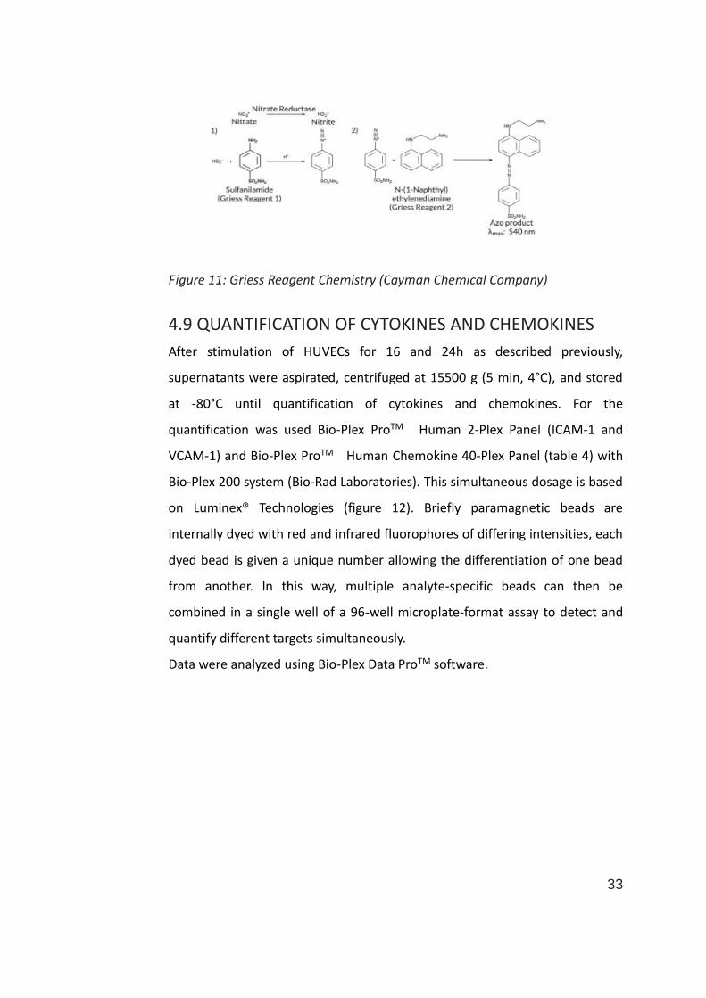
Figure 11: Griess Reagent Chemistry (Cayman Chemical Company)
4.9 QUANTIFICATION OF CYTOKINES AND CHEMOKINES
After stimulation of HUVECs for 16 and 24h as described previously,
supernatants were aspirated, centrifuged at 15500 g (5 min, 4°C), and stored
at -80°C until quantification of cytokines and chemokines. For the
quantification was used Bio-Plex ProTM Human 2-Plex Panel (ICAM-1 and
VCAM-1) and Bio-Plex ProTM Human Chemokine 40-Plex Panel (table 4) with
Bio-Plex 200 system (Bio-Rad Laboratories). This simultaneous dosage is based
on Luminex® Technologies (figure 12). Briefly paramagnetic beads are
internally dyed with red and infrared fluorophores of differing intensities, each
dyed bead is given a unique number allowing the differentiation of one bead
from another. In this way, multiple analyte-specific beads can then be
combined in a single well of a 96-well microplate-format assay to detect and
quantify different targets simultaneously.
Data were analyzed using Bio-Plex Data ProTM software.
34
Figure 12: Bio-Plex sandwich immunoassay
Table 4 Bio-Plex ProTM Human Chemokine 40-Plex Panel
CCL21 CXCL1 IL16 CCL3
CXCL13 CXCL2 CXCL10 CCL15
CCL27 CCL1 CXCL11 CCL20
CXCL5 IFNϒ CCL2 CCL19
CCL11 IL1β CCL8 CCL23
CCL24 IL2 CCL7 CXCL16
CCL26 IL4 CCL13 CXCL12
CX3CL1 IL6 CCL22 CCL17
CXCL6 IL8 MIF CCL25
GM-CSF IL10 CXCL9 TNFα
4.10 STATISTICAL ANALYSIS
Quantitative variables were expressed as mean ± standard deviation (SD) and
checked for normality distribution by the Shapiro Wilk test. To compare
biomarker serum levels between patient and control series, either Mann-
Whitney or unpaired t-test was used when appropriate. Correlation analysis
were performed using the Pearson's or the Spearman's rank correlation
coefficient, when appropriate. Statistical analyses were performed with
GraphPad Prism software. P values less than 0.05 were considered as
significant.
35
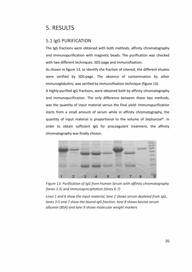
5. RESULTS
5.1 IgG PURIFICATION
The IgG fractions were obtained with both methods, affinity chromatography
and immunopurification with magnetic beads. The purification was checked
with two different techniques: SDS-page and immunofixation.
As shown in figure 13, to identify the fraction of interest, the different eluates
were verified by SDS-page. The absence of contamination by other
immunoglobulins, was verified by immunofixation technique (figure 14).
A highly-purified IgG fractions, were obtained both by affinity chromatography
and immunopurification. The only difference between these two methods,
was the quantity of input material versus the final yield: immunopurification
starts from a small amount of serum while in affinity chromatography, the
quantity of input material is proportional to the volume of Sepharose®. In
order to obtain sufficient IgG for procoagulant treatment, the affinity
chromatography was finally chosen.
Figure 13: Purification of IgG from Human Serum with affinity chromatography
(lanes 1-5) and immunoprecipitation (lanes 6-7)
Lines 1 and 6 show the input material, lane 2 shows serum depleted from IgG,
lanes 3-5 and 7 show the bound IgG fraction, lane 8 shows bovine serum
albumin (BSA) and lane 9 shows molecular weight markers
36
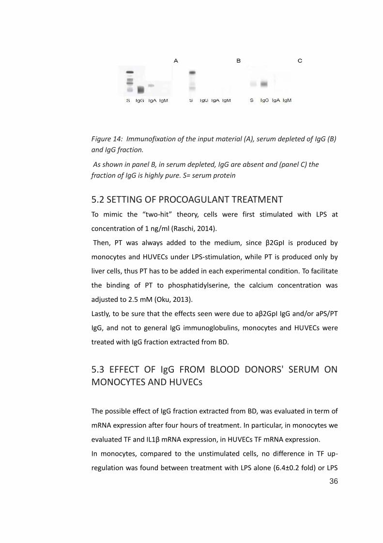
Figure 14: Immunofixation of the input material (A), serum depleted of IgG (B)
and IgG fraction.
As shown in panel B, in serum depleted, IgG are absent and (panel C) the
fraction of IgG is highly pure. S= serum protein
5.2 SETTING OF PROCOAGULANT TREATMENT
To mimic the “two-hit” theory, cells were first stimulated with LPS at
concentration of 1 ng/ml (Raschi, 2014).
Then, PT was always added to the medium, since β2GpI is produced by
monocytes and HUVECs under LPS-stimulation, while PT is produced only by
liver cells, thus PT has to be added in each experimental condition. To facilitate
the binding of PT to phosphatidylserine, the calcium concentration was
adjusted to 2.5 mM (Oku, 2013).
Lastly, to be sure that the effects seen were due to aβ2GpI IgG and/or aPS/PT
IgG, and not to general IgG immunoglobulins, monocytes and HUVECs were
treated with IgG fraction extracted from BD.
5.3 EFFECT OF IgG FROM BLOOD DONORS' SERUM ON MONOCYTES AND HUVECs
The possible effect of IgG fraction extracted from BD, was evaluated in term of
mRNA expression after four hours of treatment. In particular, in monocytes we
evaluated TF and IL1β mRNA expression, in HUVECs TF mRNA expression.
In monocytes, compared to the unstimulated cells, no difference in TF up-
regulation was found between treatment with LPS alone (6.4±0.2 fold) or LPS
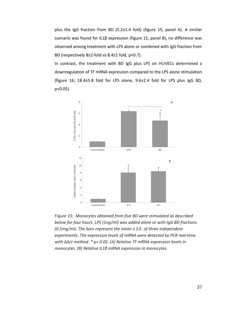
37
plus the IgG fraction from BD (5.2±1.4 fold) (figure 15, panel A). A similar
scenario was found for IL1β expression (figure 15, panel B), no difference was
observed among treatment with LPS alone or combined with IgG fraction from
BD (respectively 8±2-fold vs 8.4±1 fold, p=0.7).
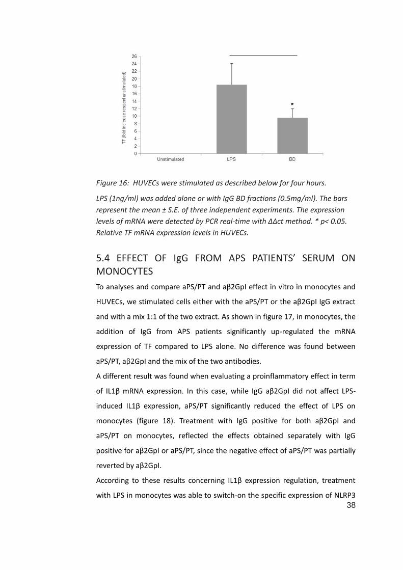
In contrast, the treatment with BD IgG plus LPS on HUVECs determined a
downregulation of TF mRNA expression compared to the LPS alone stimulation
(figure 16; 18.4±5.8 fold for LPS alone, 9.6±2.4 fold for LPS plus IgG BD,
p<0.05).
Figure 15: Monocytes obtained from five BD were stimulated as described
below for four hours. LPS (1ng/ml) was added alone or with IgG BD fractions
(0.5mg/ml). The bars represent the mean ± S.E. of three independent
experiments. The expression levels of mRNA were detected by PCR real-time
with ΔΔct method. * p< 0.05. (A) Relative TF mRNA expression levels in
monocytes. (B) Relative IL1β mRNA expression in monocytes.
38
Figure 16: HUVECs were stimulated as described below for four hours.
LPS (1ng/ml) was added alone or with IgG BD fractions (0.5mg/ml). The bars
represent the mean ± S.E. of three independent experiments. The expression
levels of mRNA were detected by PCR real-time with ΔΔct method. * p< 0.05.
Relative TF mRNA expression levels in HUVECs.
5.4 EFFECT OF IgG FROM APS PATIENTS’ SERUM ON MONOCYTES
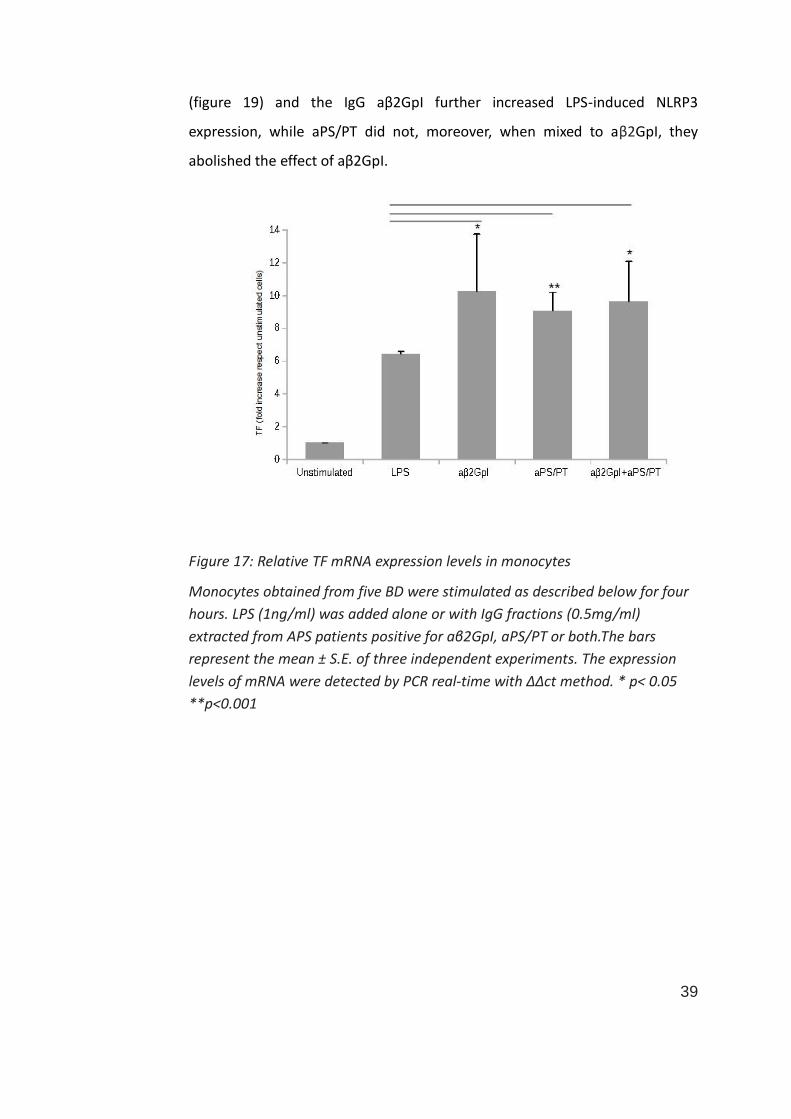
To analyses and compare aPS/PT and aβ2GpI effect in vitro in monocytes and
HUVECs, we stimulated cells either with the aPS/PT or the aβ2GpI IgG extract
and with a mix 1:1 of the two extract. As shown in figure 17, in monocytes, the
addition of IgG from APS patients significantly up-regulated the mRNA
expression of TF compared to LPS alone. No difference was found between
aPS/PT, aβ2GpI and the mix of the two antibodies.
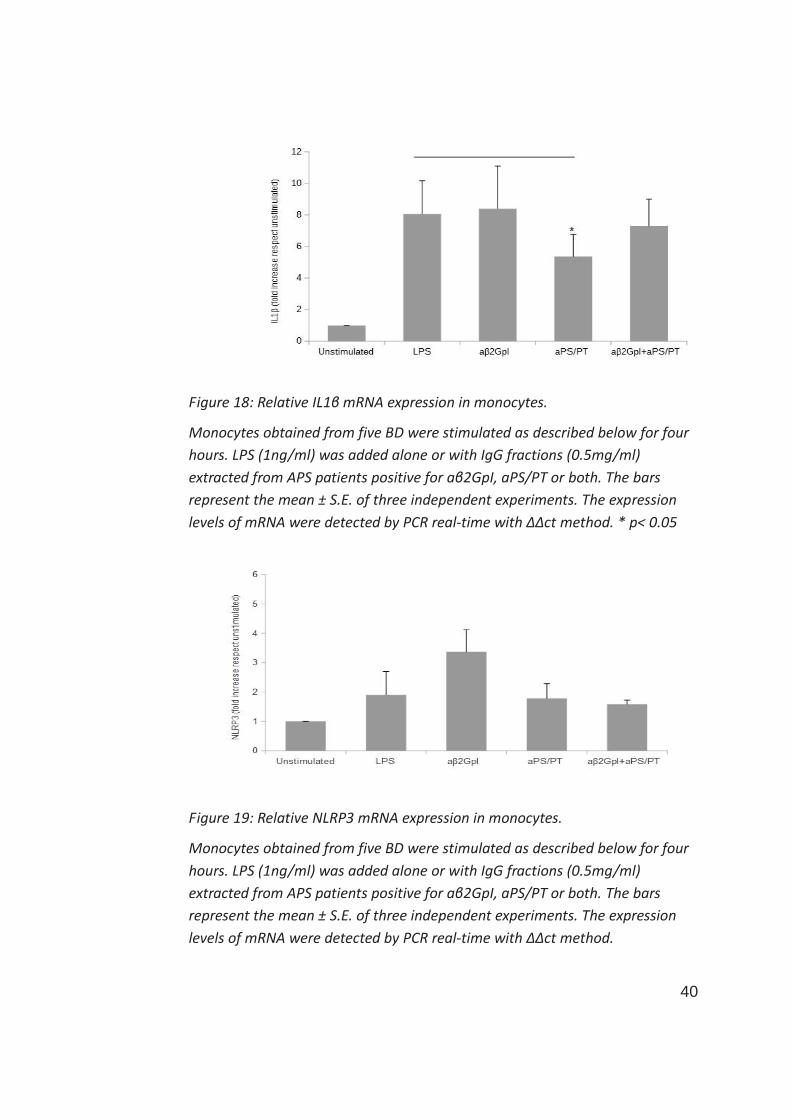
A different result was found when evaluating a proinflammatory effect in term
of IL1β mRNA expression. In this case, while IgG aβ2GpI did not affect LPS-
induced IL1β expression, aPS/PT significantly reduced the effect of LPS on
monocytes (figure 18). Treatment with IgG positive for both aβ2GpI and
aPS/PT on monocytes, reflected the effects obtained separately with IgG
positive for aβ2GpI or aPS/PT, since the negative effect of aPS/PT was partially
reverted by aβ2GpI.
According to these results concerning IL1β expression regulation, treatment
with LPS in monocytes was able to switch-on the specific expression of NLRP3
39
(figure 19) and the IgG aβ2GpI further increased LPS-induced NLRP3
expression, while aPS/PT did not, moreover, when mixed to aβ2GpI, they
abolished the effect of aβ2GpI.
Figure 17: Relative TF mRNA expression levels in monocytes
Monocytes obtained from five BD were stimulated as described below for four
hours. LPS (1ng/ml) was added alone or with IgG fractions (0.5mg/ml)
extracted from APS patients positive for aβ2GpI, aPS/PT or both.The bars
represent the mean ± S.E. of three independent experiments. The expression
levels of mRNA were detected by PCR real-time with ΔΔct method. * p< 0.05
**p<0.001
40
Figure 18: Relative IL1β mRNA expression in monocytes.
Monocytes obtained from five BD were stimulated as described below for four
hours. LPS (1ng/ml) was added alone or with IgG fractions (0.5mg/ml)
extracted from APS patients positive for aβ2GpI, aPS/PT or both. The bars
represent the mean ± S.E. of three independent experiments. The expression
levels of mRNA were detected by PCR real-time with ΔΔct method. * p< 0.05
Figure 19: Relative NLRP3 mRNA expression in monocytes.
Monocytes obtained from five BD were stimulated as described below for four
hours. LPS (1ng/ml) was added alone or with IgG fractions (0.5mg/ml)
extracted from APS patients positive for aβ2GpI, aPS/PT or both. The bars
represent the mean ± S.E. of three independent experiments. The expression
levels of mRNA were detected by PCR real-time with ΔΔct method.
41
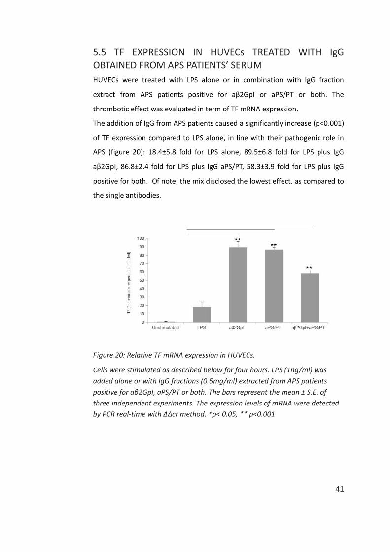
5.5 TF EXPRESSION IN HUVECs TREATED WITH IgG OBTAINED FROM APS PATIENTS’ SERUM
HUVECs were treated with LPS alone or in combination with IgG fraction
extract from APS patients positive for aβ2GpI or aPS/PT or both. The
thrombotic effect was evaluated in term of TF mRNA expression.
The addition of IgG from APS patients caused a significantly increase (p<0.001)
of TF expression compared to LPS alone, in line with their pathogenic role in
APS (figure 20): 18.4±5.8 fold for LPS alone, 89.5±6.8 fold for LPS plus IgG
aβ2GpI, 86.8±2.4 fold for LPS plus IgG aPS/PT, 58.3±3.9 fold for LPS plus IgG
positive for both. Of note, the mix disclosed the lowest effect, as compared to
the single antibodies.
Figure 20: Relative TF mRNA expression in HUVECs.
Cells were stimulated as described below for four hours. LPS (1ng/ml) was
added alone or with IgG fractions (0.5mg/ml) extracted from APS patients
positive for aβ2GpI, aPS/PT or both. The bars represent the mean ± S.E. of
three independent experiments. The expression levels of mRNA were detected
by PCR real-time with ΔΔct method. *p< 0.05, ** p<0.001
42
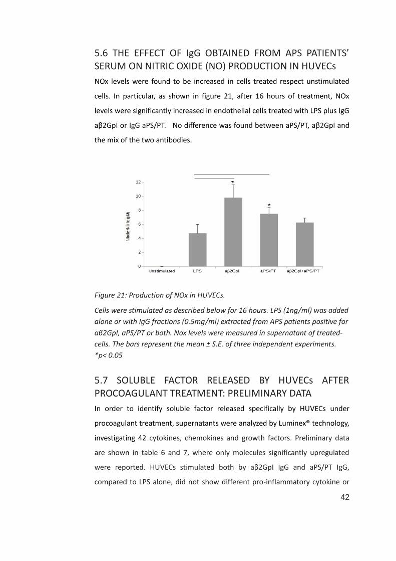
5.6 THE EFFECT OF IgG OBTAINED FROM APS PATIENTS’ SERUM ON NITRIC OXIDE (NO) PRODUCTION IN HUVECs
NOx levels were found to be increased in cells treated respect unstimulated
cells. In particular, as shown in figure 21, after 16 hours of treatment, NOx
levels were significantly increased in endothelial cells treated with LPS plus IgG
aβ2GpI or IgG aPS/PT. No difference was found between aPS/PT, aβ2GpI and
the mix of the two antibodies.
Figure 21: Production of NOx in HUVECs.
Cells were stimulated as described below for 16 hours. LPS (1ng/ml) was added
alone or with IgG fractions (0.5mg/ml) extracted from APS patients positive for
aβ2GpI, aPS/PT or both. Nox levels were measured in supernatant of treated-
cells. The bars represent the mean ± S.E. of three independent experiments.
*p< 0.05
5.7 SOLUBLE FACTOR RELEASED BY HUVECs AFTER PROCOAGULANT TREATMENT: PRELIMINARY DATA
In order to identify soluble factor released specifically by HUVECs under
procoagulant treatment, supernatants were analyzed by Luminex® technology,
investigating 42 cytokines, chemokines and growth factors. Preliminary data
are shown in table 6 and 7, where only molecules significantly upregulated
were reported. HUVECs stimulated both by aβ2GpI IgG and aPS/PT IgG,
compared to LPS alone, did not show different pro-inflammatory cytokine or
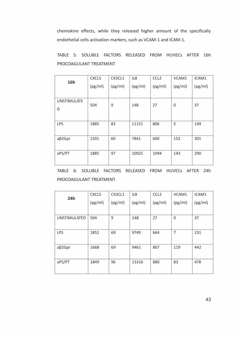
43
chemokine effects, while they released higher amount of the specifically
endothelial cells activation markers, such as VCAM-1 and ICAM-1.
TABLE 5: SOLUBLE FACTORS RELEASED FROM HUVECs AFTER 16h
PROCOAGULANT TREATMENT
16h CXCL5
(pg/ml)
CX3CL1
(pg/ml)
IL8
(pg/ml)
CCL2
(pg/ml)
VCAM1
(pg/ml)
ICAM1
(pg/ml)
UNSTIMULATE
D 504 9 148 27 0 37
LPS 1885 83 11151 806 5 149
aβ2GpI 1501 60 7841 600 152 301
aPS/PT 1885 97 10925 1094 143 290
TABLE 6: SOLUBLE FACTORS RELEASED FROM HUVECs AFTER 24h
PROCOAGULANT TREATMENT
24h CXCL5
(pg/ml)
CX3CL1
(pg/ml)
IL8
(pg/ml)
CCL2
(pg/ml)
VCAM1
(pg/ml)
ICAM1
(pg/ml)
UNSTIMULATED 504 9 148 27 0 37
LPS 1851 69 9749 664 7 231
aβ2GpI 1668 69 9461 867 119 442
aPS/PT 1849 96 13316 880 83 478
44
5.8 PAF-AH
5.8.1 PAF-AH PLASMATIC ACTIVITY IN PATIENTS AND CONTROLS: CORRELATION WITH LIPOD METABOLIC MARKERS
PAF-AH plasmatic activity in BDs disclosed a mean value of 15.6±4
nmol/min/ml (range 5.9 – 28.4). As expected (Maiolino, 2012), a significant
correlation was found between PAF-AH activity and total cholesterol (r=0.25;
p=0.032) with direct strong correlation with Low Density Lipoprotein (LDL)
(r=0.46, p<0.0001) and significant inverse correlation with High Density
Lipoprotein (HDL) (r= -0.45, p<0.0001). Instead, no correlation was found with
age. 116 /167 patients undergoing aPL investigation, showed at least one
positive aPL among LAC, aCL, aβ2GpI or aPS/PT antibodies, while 51/167
resulted all negative. PAF-AH activity was clearly more elevated in the overall
patients (19.8±5.5 nmol/min/ml) than in BDs (p<0.0001), but no difference
was found between aPL positive and aPL negative patients (19.9±5.8 versus
19.6±4.7 nmol/min/ml; figure 22). The analysis on total cholesterol shown that
levels of cholesterol did not differ significantly between BDs and the patients.
In particular, no difference was observed between BDs and aPL positive
patients (188±38 mg/dl versus 198±42 mg/dl; p=0.10) and between aPL
positive and aPL negative patients (206±52 mg/dl; p=0.47). However, LDL
serum levels were higher in aPL negative patients than in BDs (127±42 mg/dl
vs 104±35 mg/dl; p=0.0073) as well as in aPL positive patients (109±35 mg/dl;
p=0.032 vs aPL negative; p=ns vs BDs). The significant correlation between
PAF-AH activity and cholesterol, LDL and HDL serum levels persisted in aPL
positive patients (r=0.21, p=0.041; r=0.23, p=0.024 and r= -0.31, p=0.0027
respectively), while in aPL negative patients this correlation was evident only
for LDL (r=0.29, p=0.14; r=0.25, p=0.0027 and r= -0.25, p=0.21 respectively).
45
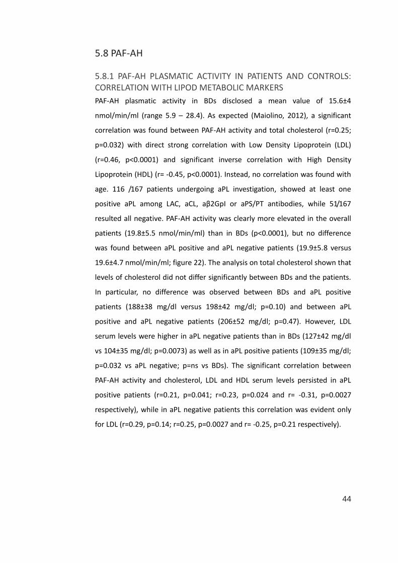
Figure 22: PAF-AH Plasmatic Activity in Patients and Controls. PAF-AH
plasmatic activity was clearly more elevated in the all patients (19.8±5.5
nmol/min/ml) than in BDs (p<0.0001), but no difference occurred between aPL
positive and aPL negative patients (19.9±5.8 versus 19.6±4.7 nmol/min/ml;
p=ns). LA positive patients disclosed higher PAF-AH than LA negative (22.1±6.4
versus 19.5±4.1 nmol/min/ml; p=0.0032)
5.8.2 PAF-AH PLASMATIC ACTIVITY IN PATIENTS DISCLOSING DISTINCT PATTERN OF aPL POSITIVITY
Dividing aPL positive patients based on LA assay, PAF-AH activity was higher in
LA positive patients than LA negative patients, as shown in figure 22 (22.1±6.4
versus 19.5±4.1 nmol/min/ml; p=0.0032). Of note, total cholesterol levels did
not differ between LA positive and LA negative patients (202±39 mg/dl versus
201±34 mg/dl; p=ns), as well as LDL (113±39 mg/dl versus 108±26 mg/dl;
p=ns) and HDL serum levels (60±21 mg/dl versus 63±21 mg/dl; p=ns).
Moreover, LA positive patients disclosed higher PAF-AH than aPL negative
patients (p=0.03), with again no difference as regard to HDL (62±24 mg/dl in
aPL-negative; p=ns) and LDL (127±42 mg/dl in aPL-negative; p=ns). As
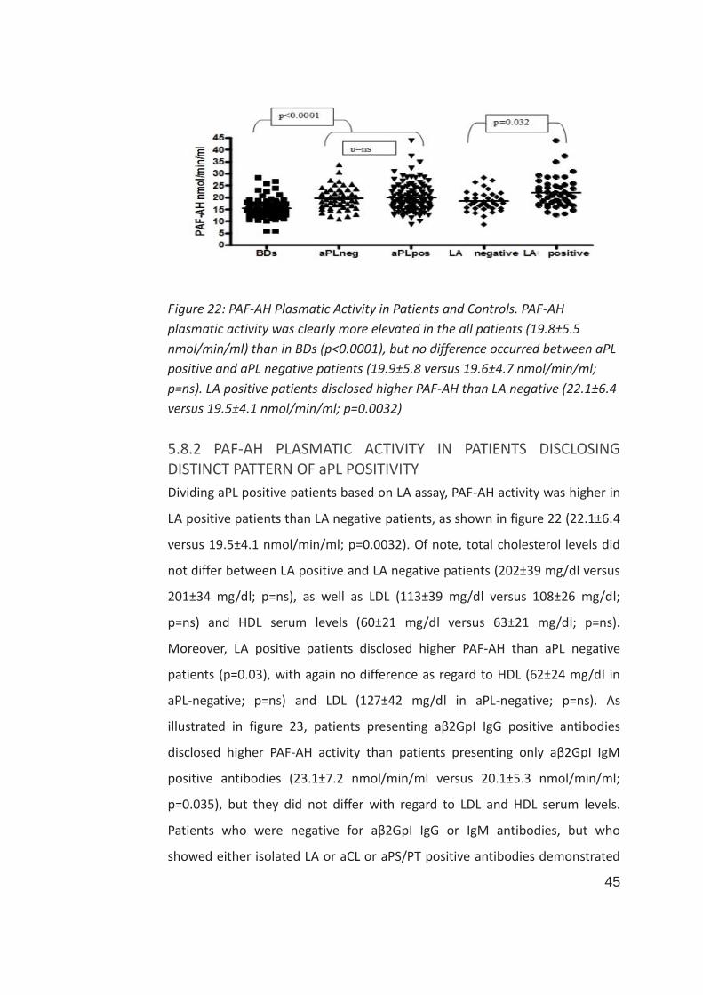
illustrated in figure 23, patients presenting aβ2GpI IgG positive antibodies
disclosed higher PAF-AH activity than patients presenting only aβ2GpI IgM
positive antibodies (23.1±7.2 nmol/min/ml versus 20.1±5.3 nmol/min/ml;
p=0.035), but they did not differ with regard to LDL and HDL serum levels.
Patients who were negative for aβ2GpI IgG or IgM antibodies, but who
showed either isolated LA or aCL or aPS/PT positive antibodies demonstrated
46
significantly lower PAF-AH activities, that appeared comparable to those
measured in BDs (figure 17; 16.9±3.8 nmol/min/ml; p=ns versus BDs; p=0.003
versus aβ2GpI IgM positive). Total cholesterol, LDL and HDL serum levels in the
latter subgroup of patients did not differ from those measured in patients with
aβ2GpI IgM positive or IgG positive antibodies. Overall, aPS/PT IgG positive
patients disclosed PAF-AH activity close to that of aPS/PT IgM positive patients
(17.3±3 nmol/min/ml versus 16.1±3.9 nmol/min/ml; p=ns). Finally, patients
disclosing aβ2GpI IgG positive antibodies together with aPS/PT IgG positive
antibodies tended to show higher PAF-AH activity than patients disclosing only
aβ2GpI IgG positive antibodies (23.4±7 nmol/min/ml versus 21±4.7; p=ns).
Figure 23: PAF-AH plasmatic activity in patients with distinct aPL specificity.
Patients presenting aβ2GpI IgG + antibodies disclosed higher PAF-AH plasmatic
activity than patients presenting only aβ2GpI IgM+ antibodies (23.1±7.2
nmol/min/ml versus 20.1±5.3 nmol/min/ml; p=0.035). Patients negative for
aβ2GpI IgG or IgM antibodies, showing either isolated LA or aCL or aPS/PT
positive antibodies demonstrated significantly lower PAF-AH activity (16.9±3.8
nmol/min/ml; p=0.003 versus a aβ2GpI IgM+)
47
6. DISCUSSION
At present, the laboratory diagnosis of APS is frequently complicated: among
APS patients, there is a subgroup defined “seronegative” that shows clinical
criteria but results negative for all “criteria” aPL (LA, aCL and aβ2GpI isotype
IgG and/or IgM). To improve APS laboratory diagnosis, it has been proposed
several autoantibodies that are directed against other plasma proteins from
the coagulation cascade (i.e. PS-PT complex), or interfere with the
anticoagulant activity of A5 (Khamashta, 2016).
aPS/PT antibodies have been proposed as potential new biomarkers for
thrombosis and/or pregnancy morbidity in the setting of APS. The introduction
of the aPS/PT IgM and IgG antibodies among the routinely investigated aPL
antibodies, leads to an improvement in APS laboratory diagnostic
performance, as shown in a recent observational study performed in our
laboratory (Fabris, 2014). Moreover, given their elevated correlation with LA
activity, aPS/PT could help when immunological deficits or anticoagulant
therapy avoid a correct LA interpretation. However, their pathogenic
mechanism is still substantially undefined.
To date, aPS/PT antibodies are not included among “criteria” aPL. To better
correlate the presence of aPS/PT and APS clinical manifestation, an in vitro
study was carried out to compare the prothrombotic effect of these
autoantibodies versus the criteria antibodies, such as the aβ2GpI. Several
studies have already demonstrated the prothrombotic effect of aβ2GpI IgG on
monocytes and endothelial cells (Rikarni, 2015). Indeed, aβ2GpI induce TF
expression on these cells. On the other side, there are few information about
the effect of aPS/PT (Oku, 2013) and no direct comparison between aβ2GpI
and aPS/PT.
In this study, for the first time, a pro-coagulant treatment for the
contemporary analysis of the effect of aβ2GpI IgG and aPS/PT IgG on
48
monocytes and ECs was employed. Compared to the stimulation by the IgG
fraction obtained by BD, either the IgG fractions by aβ2GpI positive and
aPS/PT positive patients determined a significant activation of monocytes and
HUVECs, showing a procoagulant phenotype. While the effect was similar in
term of TF mRNA expression and release of specific endothelial activation
factors (NO, ICAM, VCAM), a different effect was noticed in terms of IL-1b and
NLRP3 expression, since aPS/PT antibodies seem to have an opposite effect
compared to aβ2GpI. Further studies are needed to clarify these results.
Endothelial cells are actively involved into the inflammatory process with
specific adaptive response that includes formation of reactive oxygen species
(ROS) with upregulation of nitric oxide (NO) production (Assis, 2002; Laurindo,
1994). Furthermore, endothelial dysfunction plays an important role in
atherosclerotic disease (Pasaoglu, 2014). The upregulation of NO production in
response to both aβ2GpI and aPS/PT antibodies indicate that they both induce
an adaptatively response on endothelial cells.
Several studies demonstrated that PAF-AH is a cardiovascular risk marker
independent respect the traditional risk factors for CV. Its increased expression
was correlated with the vulnerability of atherosclerotic plaques. Therefore, in
order to assess the CV risk, PAF-AH dosage has been proposed to ensure a
better stratification of at risk populations, (Corson, 2008). To date, PAF-AH has
never been investigated in the context of APS patients, or, even less, in
patients at risk to develop an overt APS (i.e. asymptomatic carriers of aPL
antibodies).
This study was conducted on patients routinely screened for APS,
demonstrating a significant association between the presence of aPL
antibodies (LA and aβ2GpI IgG in particular) and PAF-AH activity upregulation
in plasma.
Atherosclerosis is definitively recognized as a chronic inflammatory response
due to the accumulation of lipoproteins in the walls of arteries (Libby, 2016).
PAF-AH is manly associated with LDL and it is predominantly express in the
49
necrotic centre of atherosclerotic plaques and in the macrophage-rich areas
releasing pro-inflammatory mediators, such as lysophospholipids and oxidized
fatty acids (Rosenson, 2012).
In addition to the presence of LA, several different targets of aPL could be
determined by many analytical methods, with frequent discordant results,
that could make the laboratory diagnosis of APS extremely complicated. The
main role of the aβ2GpI antibodies, especially those specifically targeting
domain I (Giannakopoulos, 2013), is widely accepted and present results seem
to further confirm their importance with regard to CV risk stratification, since
PAF-AH appeared particularly elevated in aβ2GpI positive patients and more so
in those displaying LA activity and carrying the IgG isotype. This particular
association may be explained by the fact that IgG aβ2GpI antibodies are able
to recognize the stable complex between oxLDL and β2GpI, thus facilitating
macrophage-derived foam cell formation in patients with APS (Zhang, 2014).
The immune-pathological mechanisms sustained by oxLDL/β2GpI complexes
are not yet fully understood, but TLR4 was recently shown to be involved
(Zhang, 2014). TLR4 could be the key player linking PAF-AH up-regulation to
aβ2GpI IgG antibodies in APS, as evidenced by a mouse model of preterm
delivery which demonstrated that PAF effects and signalling depend upon
TLR4 stimulation (Agrawal, 2014).
Lp-PLA2 activity proved to be markedly reduced in vivo when the enzyme is
bound to HDL (Rosenson, 2012), and this is in line with our observation that
aβ2GpI IgG+ patients disclosed higher PAF-AH and lesser HDL than BDs. This is
not true for other subgroups of patients, such as aPL-negative patients or
those presenting only isolated LAC or aCL or aPS/PT antibodies. Compared to
these patients, PAF-AH plasmatic activity up-regulation in aβ2GpI IgG+ cases
appeared to be at least partially disconnected from the lipoprotein levels and
specifically linked to the presence of such aPL antibodies.
Therefore, PAF-AH up-regulation arose as a specific thrombotic risk marker in
patients carrying aβ2GpI antibodies and is not generally associated with other
50
aPL antibodies possibly implicated in APS manifestations, but further studies
are needed to confirm this observation.
Unfortunately, in two large randomized clinical trials, an inhibitor of PAF-AH
(darapladib) (O’Donoghue, 2014; Wallentin, 2016) failed to reduce the risk of
major coronary events as compared to placebo. In addition, it was associated
with significantly higher rates of drug discontinuation and adverse effects.
These results suggested that PAF-AH may be a biomarker of vascular
inflammation, rather than a causal pathway of CV diseases (Wallentin, 2016).
Therefore, high PAF-AH activity could reflect a response to pro-inflammatory
stress characteristic both of atherosclerosis and APS (Marsthe, 2014).
The leading cause of death in primary and secondary APS patients are
cardiovascular events due to accelerated atherosclerosis, which often
progresses more rapidly, compared with the general population (Silva, 2014).
Some key pro-inflammatory proteins correlate with APS clinical manifestations
(Becarevic, 2016) and common radiological markers of
subclinical atherosclerosis and CV risk were often reported in such patients
(Ambrosino, 2014). However, to date, besides the presence of aPL itself, no
serological biomarkers specifically associated with aPL-related pathogenic
mechanisms have been identified as useful to improve the classification of CV
risk in aPL+ patients with and without overt APS clinical manifestations.
In this scenario, present findings on PAF-AH assume a relevant place, possibly
representing a reliable and affordable biomarker useful to identify patients at
higher risk in which to take a more cautious therapeutic attitude in the follow-
up.
Moreover, studying PAF-AH metabolic pathway may help to better explain the
pathogenesis of APS and to improve management and interpretation of aPL-
related issues, from the analytical result, to the final therapeutic decision.
Even if we and others recently demonstrated an important role of aPS/PT
antibodies in the laboratory diagnosis of APS (Fabris, 2014; Amengual, 2016),
the therapeutic management of patients characterized by the presence of

51
isolated aPS/PT remains an open issue. Patients with isolated aPS/PT
antibodies disclosed lower (BDs-like) PAF-AH as compared to patients with
positive aβ2GpI antibodies. Nevertheless, aPS/PT antibodies, may exert their
distinct pathogenic role through pathways in which PAF-AH is not involved, as
shown previously as regard to IL1b and NLRP3 expression on monocytes
treated with such antibodies compared to aβ2GpI.
52
7. CONCLUSIONS
The introduction of aPS/PT antibodies in the diagnostic process of APS is
highly recommended, since they disclosed diagnostic laboratory performances
at least equal to the aCL and aβ2GpI antibodies and a high correlation with LA
activity, such that they can be a viable alternative.
Data obtained by our in vitro study, even if preliminary, confirmed that aPS/PT
exert similar pro-thrombotic effects on monocytes and HUVECs as compared
to the aβ2GpI antibodies. The different effect of aβ2GpI and aPS/PT on
expression of IL1β and NLRP3, and the different impact on PAF-AH production,
may suggest that these classes of antibodies, while disclosing similar pro-
thrombotic effects, probably activate different metabolic pathways. Further
studies are needed to better clarify these issues.
Anyway, the prognostic information conveyed by plasmatic PAF-AH activity in
patients with positive aPL antibodies appeared to be independent to that of
common lipid metabolism markers (i.e. LDL), as previously reported by other
authors in the context of patients with major coronary events (Maiolino, 2012)
and, based on present results, PAF-AH plasmatic activity may represent a new
prognostic biomarker also in the context of aPL antibodies, to identify patients
at major risk and favouring more tailored therapeutic interventions. Further
prospective studies on selected patients are ongoing.
53
8. PUBLISHED ABSTRACTS
Dubsky de Wittenau G, Radovic S, Pascolo R, Cesca F, Cifù A, Curcio F, Lonigro
R.De-novo deletion or uniparental disomy as SMA determining cause: a
case report. European society of human genetics. Milan (Italy), 2014
CifùA, Zago S, Poz A, Giacomello R, Tonutti E, Curcio F, Fabris M. Anti-
phosphatidylserine/ prothrombin Autoantibodies Significantly Improve
the Laboratory Diagnostic Process Of Anti-Phospholipids Antibody
Syndrome. Secondo congresso dell'area di patologia e diagnostica di
laboratorio. Palermo (Italy), 2014
Fabris M, Giacomello R, Cifù A, Tanzi N, Curcio F, Tonutti E. Phosphatidylserine-
dependent antiprothrombin antibodies in clinical practice. SIPMET
meeting the young scientists -Alba (Italy), 2015
CifùA, Poz A, Tonutti E, Giacomello R, Pistis C, Curcio F, Fabris M. Studio pilota:
PAF (platelet activating factor) -acetilidrolasi quale nuovo possibile
marker di rischio trombotico nei pazienti affetti da sindrome da
anticorpi anti-fosfolipidi. Primo congresso nazionale della società
italiana di patologia clinica e medicina di laboratorio - Roma (Italy), 2015
Tonelli V, Pistis C, Cifù A, Tonutti E, Vescini F, Curcio F, Grimaldi F, Fabris
M.Elevati livelli di B-Lymphocyte Stimulator (BLyS) nei pazienti con
54
tumore neuroendocrino identificano una malattia più severa e non
responsiva alla terapia. Primo congresso nazionale della società italiana
di patologia clinica e medicina di laboratorio - Roma (Italy), 2015
Cifù A, Fabris M, Giacomello R, Pistis C, Tonutti E, Curcio F. The plasmatic
Platelet-Activating Factor acetylhydrolase activity: a possible risk biomarker in
patients with anti-phospholipid syndrome.
10th international congress of autoimmunity - Leipzig (Germany),2016
Fabris M, Tanzi N, Giacomello R, Cifù A, Poz A, Curcio F, Tonutti E. Anti-
phospholipid antibodies testing by ELISA versus CLIA: a comparative
Italian study. 10th international congress of autoimmunity - Leipzig
(Germany),2016
Cifù A, Sanvilli N, Tanzi N, Pistis C, Fabris M, Fontana DE, Giacomello R, Curcio
F, Gigli G, Janes F. Asymmetrical Dimethylarginine (ADMA) In
Asympomatic Cerebral Small Vessel Disease. The American Journal of
Pathology- Supplement, Volume 186 (2016) S6
Domenis R, Zanutel R, Cifù A, Pistis C, Di Benedetto P, Causero A, Pozzi M,
Bassini F, Fabris M, Niazi KR, Curcio F. Characterization of the
Proinflammatory Profile of Synovial Fluid-Derived Exosomes of Patients
55
with Gonarthrosis. The American Journal of Pathology- Supplement,
Volume 186 (2016) S23
Fabris M, Cifù A, Pistis C, Giacomello R, Siega-Ducaton M, Tonutti E, Curcio F.
Elevated Plasma Platelet-Activating Factor Acetylhydrolase Activity is
Significantly Associated with High Risk Anti-Phospholipid Antibody
Antigenic Specificities. The American Journal of Pathology- Supplement,
Volume 186 (2016) S24
Cifù A, Pistis C, Domenis R, Curcio F, Fabris M. Comparison between Anti-
β2Glycoprotein I Antibody and Anti-Phosphatidylserine/Prothrombin
Antibody Biological Effects on Peripheral Blood Monocytes. The
American Journal of Pathology- Supplement, Volume 186 (2016) S27
Cifù A, Pistis C, Fabris M, Domenis R, Zanello A, Gandolfo S, De Vita S, Curcio F.
Characterization of Parotid Gland Epithelial Cell Primary Cultures from
Patients Affected by Sjogren’s Syndrome. The American Journal of
Pathology- Supplement, Volume 186 (2016) S31
Pistis C, Domenis R, Moretti M, Vicario A, Cifù A, Di Benedetto P, Causero A,
Pozzi M, Bassini F, Fabris M, Niazi KR, Curcio F. The immunomodulatory
properties of adipose mesenchymal stem cells are strengthened by
treatment with osteoarthritic synovial fluid. PISA 2016 Breakthroughs in
56
biology: from underlying pathogenesis to translational medicine-
Houston (Texas, USA),2016
9. PUBLICATIONS
Fabris M,Cifù A, Pistis C, Siega-Ducaton M, Fontana DE, Giacomello R, Tonutti
E, Curcio F.Exploring the Plasmatic Platelet-Activating Factor
Acetylhydrolase Activity in Patients with Anti-phospholipid Antibodies.
Autoimmunity Highlights – ahead of print
Domenis R, Zanutel R, Caponnetto F, Toffoletto B, Cifù A, Pistis C, Di Benedetto
P, Causero A, Pozzi M, Bassini F, Fabris M, Niazi KR, Soon-Shiong P, Curcio
F. Characterization of the proinflammatory profile of synovial fluid-
derived exosomes of patients with osteoarthritis. Mediators of
Inflammation - under review
57
10. REFERENCES
Agrawal V, Jaiswal MK, Ilievski V, Beaman KD, Jilling T, Hirsch E. Platelet-
Activating Factor: a Role in Preterm Delivery and an Essential Interaction
with Toll-Like Receptor Signaling in Mice. Biology of Reproduction
2014;91(5):119.
Aguiar CL, Erkan D. Catastrophic antiphospholipid syndrome: how to diagnose
a rare but highly fatal disease. Therapeutic Advances in Musculoskeletal
Disease 2013;5(6):305–14.
Allford SL, Machin S. Haemostasis. Surgery (Oxford) 2007;25(6):241–4.
Ambrosino P, Lupoli R, Minno AD, Iervolino S, Peluso R, Minno MNDD. Markers
of cardiovascular risk in patients with antiphospholipid syndrome: A
meta-analysis of literature studies. Annals of Medicine 2014;46(8):693–
702.
Amengual O, Forastiero R, Sugiura-Ogasawara M, et al. Evaluation of
phosphatidylserine-dependent antiprothrombin antibody testing for the
diagnosis of antiphospholipid syndrome: results of an international
multicentre study. Lupus 2017;26(3):266–76.
Amengual O, Atsumi T, Koike T. Specificities, properties, and clinical
significance of antiprothrombin antibodies. Arthritis & Rheumatism
2003;48(4):886–95.
Andreoli L, Nalli C, Motta M, et al. Anti- 2-glycoprotein I IgG antibodies from 1-
year-old healthy children born to mothers with systemic autoimmune
58
diseases preferentially target domain 4/5: might it be the reason for their
'innocent' profile? Annals of the Rheumatic Diseases 2010;70(2):380–3.
Arachchillage DJ, Greaves M. The chequered history of the antiphospholipid
syndrome. British Journal of Haematology 2014;165(5):609–17.
Assis MCD, Plotkowski MC, Fierro IM, Barja-Fidalgo C, Freitas MSD. Expression
of inducible nitric oxide synthase in human umbilical vein endothelial
cells during primary culture. Nitric Oxide 2002;7(4):254–61.
Atsumi T, Khamashta MA, Haworth RS, et al. Arterial disease and thrombosis in
the antiphospholipid syndrome: A pathogenic role for endothelin 1.
Arthritis & Rheumatism 1998;41(5):800–7.
Barbé F, Douglas T, Saleh M. Advances in Nod-like receptors (NLR) biology.
Cytokine & Growth Factor Reviews 2014;25(6):681–97.
Bertolaccini M, Amengual O, Atsumi T, et al. ‘Non-criteria’ aPL tests: report of
a task force and preconference workshop at the 13th International
Congress on Antiphospholipid Antibodies, Galveston, TX, USA, April
2010. Lupus 2011;20(2):191–205.
Bećarević M, Ignjatović S. Proinflammatory proteins in female and male
patients with primary antiphospholipid syndrome: preliminary data.
Clinical Rheumatology 2016;35(10):2477–83.
Brandt KJ, Kruithof EK, Moerloose PD. Receptors involved in cell activation by
antiphospholipid antibodies. Thrombosis Research 2013;132(4):408–13.
Chen G, Pedra JH. The Inflammasome in Host Defense. Sensors 2009;10(1):97–
111.
59
Corson MA, Jones PH, Davidson MH. Review of the Evidence for the Clinical
Utility of Lipoprotein-Associated Phospholipase A2 as a Cardiovascular
Risk Marker. The American Journal of Cardiology 2008;101(12).
Davidson MH, Corson MA, Alberts MJ, et al. Consensus Panel
Recommendation for Incorporating Lipoprotein-Associated
Phospholipase A2 Testing into Cardiovascular Disease Risk Assessment
Guidelines. The American Journal of Cardiology 2008;101(12).
Erkan D. A cross-sectional study of clinical thrombotic risk factors and
preventive treatments in antiphospholipid syndrome. Rheumatology
2002;41(8):924–9.
Esdaile JM, Abrahamowicz M, Grodzicky T, et al. Traditional Framingham risk
factors fail to fully account for accelerated atherosclerosis in systemic
lupus erythematosus. Arthritis & Rheumatism 2001;44(10):2331–7.
Fabris M, Giacomello R, Poz A, et al. The introduction of anti-
phosphatidylserine/prothrombin autoantibodies in the laboratory
diagnostic process of anti-phospholipid antibody syndrome: 6 months of
observation. Autoimmunity Highlights 2014;5(2):63–7.
Giannakopoulos B, Krilis SA. The Pathogenesis of the Antiphospholipid
Syndrome. New England Journal of Medicine 2013;368(11):1033–44.
Groot PGD, Meijers JCM. β2-Glycoprotein I: evolution, structure and function.
Journal of Thrombosis and Haemostasis 2011;9(7):1275–84.
Gómez-Puerta JA, Cervera R. Diagnosis and classification of the
antiphospholipid syndrome. Journal of Autoimmunity 2014;48-49:20–5.
60
Hanly JG. Antiphospholipid Syndrome: an overview. Canadian Medical
Association Journal 2003; 168(13): 1675-1682
Harper BE, Willis R, Pierangeli SS. Pathophysiological mechanisms in
antiphospholipid syndrome. International Journal of Clinical
Rheumatology 2011;6(2):157–71.
Hoffman M. A cell-based model of coagulation and the role of factor VIIa.
Blood Reviews 2003;17.
Khamashta M, Taraborelli M, Sciascia S, Tincani A. Antiphospholipid syndrome.
Best Practice & Research Clinical Rheumatology 2016;30(1):133–
48.
Laurindo FR, De APM, Barbeiro HV, et al. Vascular free radical release. Ex vivo
and in vivo evidence for a flow- dependent endothelial mechanism.
Circulation Research 1994;74(4):700–9.
Lefèvre G, Lambert M, Bacri J-L, et al. Thrombotic events during long-term
follow-up of obstetric antiphospholipid syndrome patients. Lupus
2011;20(8):861–5.
Libby P, Bornfeldt KE, Tall AR. Atherosclerosis. Circulation Research
2016;118(4):531–4.
Ma K. High Affinity Binding of beta 2-Glycoprotein I to Human Endothelial Cells
Is Mediated by Annexin II. Journal of Biological Chemistry
2000;275(20):15541–8.
Mahler M, Norman GL, Meroni PL, Khamashta M. Autoantibodies to domain 1
of beta 2 glycoprotein 1: A promising candidate biomarker for risk
61
management in antiphospholipid syndrome. Autoimmunity Reviews
2012;12(2):313–7.
Maiolino G, Pedon L, Cesari M, et al. Lipoprotein-Associated Phospholipase A2
Activity Predicts Cardiovascular Events in High Risk Coronary Artery
Disease Patients. PLoS ONE 2012;7(10).
Maiolino G. Lipoprotein-associated phospholipase A2 prognostic role in
atherosclerotic complications. World Journal of Cardiology
2015;7(10):609.
Mann KG, Bovill EG, Krishnaswamy S. Surface-Dependent Reactions in the
Propagation Phase of Blood Coagulation. Annals of the New York
Academy of Sciences 1991;614(1):63–75.
Marathe GK, Pandit C, Lakshmikanth CL, Chaithra VH, Jacob SP, D'souza CJM.
To hydrolyze or not to hydrolyze: the dilemma of platelet-activating
factor acetylhydrolase. The Journal of Lipid Research 2014;55(9):1847–
54.
Mcintyre TM, Prescott SM, Stafforini DM. The emerging roles of PAF
acetylhydrolase. The Journal of Lipid Research 2008;50(Supplement).
Mcmichael M. New Models of Hemostasis.Topics in Companion Animal
Medicine 2012;27(2):40–5.
Meroni PL, Riboldi P. Pathogenic mechanisms mediating antiphospholipid
syndrome. Current Opinion in Rheumatology 2001;13(5):377–82.
Miyakis S, Lockshin MD, Atsumi T, et al. International consensus statement on
an update of the classification criteria for definite antiphospholipid
62
syndrome (APS). Journal of Thrombosis and Haemostasis 2006;4(2):295–
306.
Miyakis S, Giannakopoulos B, Krilis SA. Beta 2 glycoprotein I-function in health
and disease. Thrombosis Research 2004;114(5-6):335–46.
Mulla MJ, Salmon JE, Chamley LW, et al. A Role for Uric Acid and the Nalp3
Inflammasome in Antiphospholipid Antibody-Induced IL-1β Production
by Human First Trimester Trophoblast. PLoS ONE 2013;8(6).
Müller-Calleja N, Köhler A, Siebald B, et al. Cofactor-independent
antiphospholipid antibodies activate the NLRP3-inflammasome via
endosomal NADPH-oxidase: implications for the antiphospholipid
syndrome. Thrombosis and Haemostasis 2015;113(5):1071–83.
Nishimura S, Manabe I, Nagasaki M, et al. In vivo imaging visualizes discoid
platelet aggregations without endothelium disruption and implicates
contribution of inflammatory cytokine and integrin signaling. Blood
2011;119(8).
Norris LA. Blood coagulation. Best Practice & Research Clinical Obstetrics
& Gynaecology 2003;17(3):369–83.
Oku K, Amengual O, Zigon P, Horita T, Yasuda S, Atsumi T. Essential role of the
p38 mitogen-activated protein kinase pathway in tissue factor gene
expression mediated by the phosphatidylserine-dependent
antiprothrombin antibody. Rheumatology 2013;52(10):1775–84.
63
O’Donoghue ML, Braunwald E, White HD, et al. Effect of Darapladib on Major
Coronary Events After an Acute Coronary Syndrome. Jama
2014;312(10):1006.
Panteleev MA, Dashkevich NM, Ataullakhanov FI. Hemostasis and thrombosis
beyond biochemistry: roles of geometry, flow and diffusion. Thrombosis
Research 2015;136(4):699–711.
Pasaoglu OT, Turkozkan N, Ark M, Polat B, Agilli M, Yaman H. The Effect of
Taurine on the Relationship Between NO, ADMA and Homocysteine in
Endotoxin-Mediated Inflammation in HUVEC Cultures. Inflammation
2014;37(5):1439–43.
Raschi E, Chighizola CB, Grossi C, et al. β2-glycoprotein I, lipopolysaccharide
and endothelial TLR4: Three players in the two-hit theory for anti-
phospholipid-mediated thrombosis. Journal of Autoimmunity 2014;
55:42–50.
Rikarni, Dharma R, Tambunan KL, Isbagyo H, Dewi BE, Acang N, Setiabudy R,
Aman AK. Prothrombotic effect of Anti-beta-2 Glycoprotein-1 Antibodies
on the expression of Tissue Factor, Thrombomodulin, and Plasminogen
Activator Inhibitor-1 in Endothelial Cells. Acta Med Indones.
2015;47(1):31-7
Rosenson RS, Hurt-Camejo E. Phospholipase A2 enzymes and the risk of
atherosclerosis. European Heart Journal 2012;33(23):2899–909.
64
Sciascia S, Bertolaccini M. Antibodies to phosphatidylserine/prothrombin
complex and the antiphospholipid syndrome. Lupus 2014;23(12):1309–
12.
Sciascia S, Khamashta MA, Bertolaccini ML. New Tests to Detect
Antiphospholipid Antibodies: Antiprothrombin (aPT) and Anti-
Phosphatidylserine/Prothrombin (aPS/PT) Antibodies. Current
Rheumatology Reports 2014;16(5).
Silva FFD, Levy RA, Carvalho JFD. Cardiovascular Risk Factors in the
Antiphospholipid Syndrome. Journal of Immunology Research 2014;
2014:1–6.
Simone ND, Meroni PL, Papa ND, et al. Antiphospholipid antibodies affect
trophoblast gonadotropin secretion and invasiveness by binding directly
and through adhered β2-glycoprotein I. Arthritis & Rheumatism
2000;43(1):140–50.
Tripodi A, Groot PGD, Pengo V. Antiphospholipid syndrome: laboratory
detection, mechanisms of action and treatment. Journal of Internal
Medicine 2011;270(2):110–22.
Urbanus RT, Pennings MTT, Derksen RHWM, Groot PGD. Platelet activation by
dimeric2-glycoproteinI requires signaling via both glycoproteinIb and
apolipoproteinE receptor2. Journal of Thrombosis and Haemostasis
2008;6(8):1405–12.
Wallentin L, Held C, Armstrong PW, et al. Lipoprotein‐Associated
Phospholipase A 2 Activity Is a Marker of Risk But Not a Useful Target for
65
Treatment in Patients With Stable Coronary Heart Disease. Journal of
the American Heart Association 2016;5(6).
Watson H, Davidson S, Keeling D. Guidelines on the diagnosis and
management of heparin-induced thrombocytopenia: second edition.
British Journal of Haematology 2012;
Willis R, Gonzalez EB, Brasier AR. The Journey of Antiphospholipid Antibodies
From Cellular Activation to Antiphospholipid Syndrome. Current
Rheumatology Reports 2015;17(3).
Wilson WA, Gharavi AE, Koike T, et al. International consensus statement on
preliminary classification criteria for definite antiphospholipid
syndrome: Report of an International workshop. Arthritis &
Rheumatism 1999;42(7):1309–11.
Zhang X, Xie Y, Zhou H, et al. Involvement of TLR4 in Oxidized LDL/β2GPI/Anti-
β2GPI-Induced Transformation of Macrophages to Foam Cells. Journal of
Atherosclerosis and Thrombosis 2014;21(11):1140–51