Embed Size (px)
Citation preview
Volume 2 I Summer 2013
• EpicardialAblation EndsVentricular Fibrillationin BrugadaSyndrome page4
• Cardiacstenting ‘bythewrist’ improvesoutcomes page6
• Linkingvascular abnormalitiesand Alzheimer’sdisease page8
University of Rochester Medical Center
Heart and Vascular Center News
heart.urmc.edu
Continued on page 2
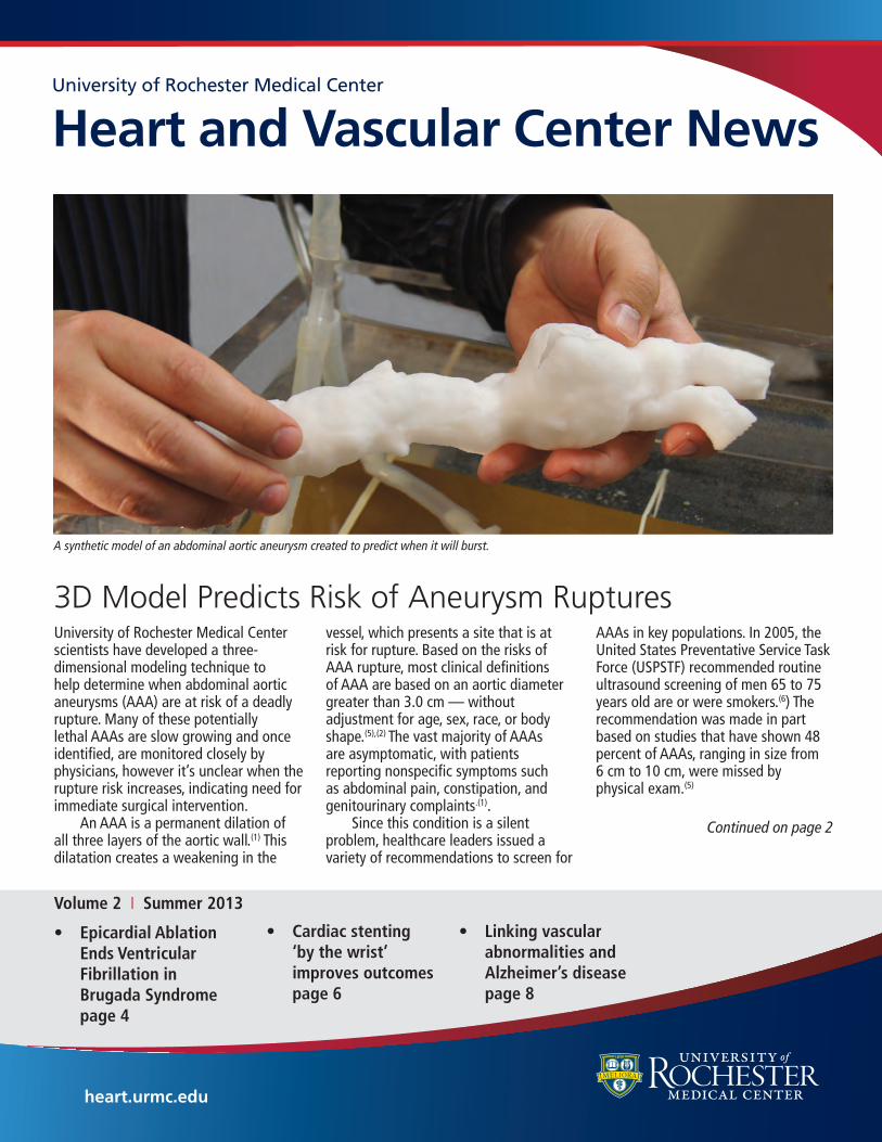
University of Rochester Medical Center scientists have developed a three-dimensional modeling technique to help determine when abdominal aortic aneurysms (AAA) are at risk of a deadly rupture. Many of these potentially lethal AAAs are slow growing and once identified, are monitored closely by physicians, however it’s unclear when the rupture risk increases, indicating need for immediate surgical intervention. An AAA is a permanent dilation of all three layers of the aortic wall.(1) This dilatation creates a weakening in the
vessel, which presents a site that is at risk for rupture. Based on the risks of AAA rupture, most clinical definitions of AAA are based on an aortic diameter greater than 3.0 cm — without adjustment for age, sex, race, or body shape.(5),(2) The vast majority of AAAs are asymptomatic, with patients reporting nonspecific symptoms such as abdominal pain, constipation, and genitourinary complaints.(1). Since this condition is a silent problem, healthcare leaders issued a variety of recommendations to screen for
AAAs in key populations. In 2005, the United States Preventative Service Task Force (USPSTF) recommended routine ultrasound screening of men 65 to 75 years old are or were smokers.(6) The recommendation was made in part based on studies that have shown 48 percent of AAAs, ranging in size from 6 cm to 10 cm, were missed by physical exam.(5)
3D Model Predicts Risk of Aneurysm Ruptures
A synthetic model of an abdominal aortic aneurysm created to predict when it will burst.
The Centers for Medicare and Medicaid expanded the USPTF recommendations, encouraging screening of women aged 65 to 75 who have a family history of AAA.(3) And, the Society for Vascular Surgery (SVS) most recently recommended screening men with a family history of AAA at 55, all others at 65, and women at 65 if they’ve smoked or have a family history of AAA. AAA incident in men aged 65 to 79 is 5 percent to 10 percent,(7) and in women 65 to 79 is 0.7 percent to 3 percent. (3), (8), (9) Estimates also suggest that a third of all AAA will eventually rupture and more than 80 percent will be fatal.(5)
Traditionally, the greatest risk factor associated with rupture has been diameter. This is based on an engineering principle known as the “aw of Laplace, which states that wall tension is directly proportional to radius and intra-luminal pressure. The Laplace relationship is clinically supported, as annual risk-of-rupture rates versus maxim aortic size is: • .3percentforAAAlessthan 4 cm; • 1.5percentto13percentfor 4 cm to 4.9 cm; • 6.5percentto40percentbetween 5 cm and 5.9 cm; and, • 40percentfrom7cmto10cm (4), (3).
Smaller AAAs grow an average of .2 cm to .5 cm per year, with an average five years between diagnosis at 3cm and symptoms of rupture at 6 cm. In the majority of studies used to determine risk, women were underrepresented and thus the determination of risk is largely underestimated. AAA at any size for women carries a 2-to-4.5-fold greater risk of rupture.(4) However, AAA diameter is not a consistent measure of rupture potential, as many patients rupture and die with AAA smaller than 5 cm and other patients survive and present with AAA 6 cm or greater. Autopsy studies have shown that 10 percent to 24 percent of all ruptured AAAs have a maximum diameter less than 5.5 cm. (10, 11) The lack of a clear method to assess risk has
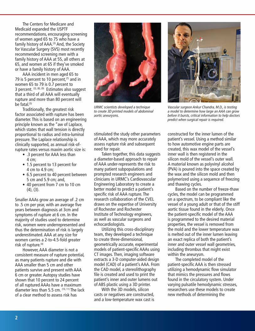
stimulated the study other parameters of AAA, which may more accurately assess rupture risk and subsequent need for repair. Taken together, this data suggests a diameter-based approach to repair of AAA under-represents the risk to many patient subpopulations and prompted research engineers and clinicians in URMC’s Cardiovascular Engineering Laboratory to create a better model to predict a patient’s specific risk of AAA rupture. The research collaboration of the CVEL draws on the expertise of University of Rochester and Rochester Institute of Technology engineers, as well as vascular surgeons and echocardiologists. Utilizing this cross-disciplinary team, they developed a technique to create three-dimensional, geometrically accurate, experimental models of patient-specific AAAs using CT images. Then, imaging software extracts a 3-D computer-aided design model (CAD) of a patient’s AAA. From the CAD model, a stereolithography file is created and used to print the patient’s inner and outer lumens out of ABS plastic using a 3D printer. With the 3D models, silicon casts or negatives are constructed, and a low-temperature wax cast is
constructed for the inner lumen of the patient’s vessel. Using a method similar to how automotive engine parts are created, this wax model of the vessel’s inner wall is then registered in the silicon mold of the vessel’s outer wall. A material known as polyvinyl alcohol (PVA) is poured into the space created by the wax and the silicon mold and then polymerized using a sequence of freezing and thawing cycles. Based on the number of freeze-thaw cycles, the model can be programmed on a spectrum, to be compliant like the vessel of a young adult or that of the stiff aortic tissue found in the elderly. Once the patient-specific model of the AAA is programmed to the desired material properties, the vessel is removed from the mold and the lower temperature wax is melted out of the inner lumen leaving an exact replica of both the patient’s inner and outer vessel wall geometries, including thrombus that might exist within the aneurysm. The completed model of the patient-specific AAA is then stressed utilizing a hemodynamic flow simulator that mimics the pressures and flows found in the circulatory system. Under varying pulsatile hemodynamic stresses, researchers use these models to create new methods of determining the
URMC scientists developed a technique to create 3D printed models of abdominal aortic aneurysms.
Vascular surgeon Ankur Chandra, M.D., is testing a model to determine how large an AAA can grow before it bursts, critical information to help doctors predict when surgical repair is required.
2
heart.urmc.edu
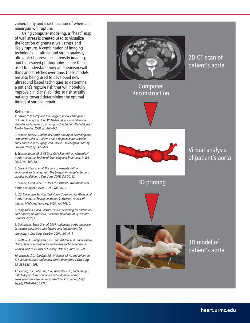
vulnerability and exact location of where an aneurysm will rupture. Using computer modeling, a “heat” map of wall stress is created used to visualize the location of greatest wall stress and likely rupture. A combination of imaging techniques — ultrasound strain analysis, ultraviolet fluorescence intensity imaging, and high-speed photography — are then used to understand how an aneurysm wall thins and stretches over time. These models are also being used to developed new ultrasound based techniques to determine a patient’s rupture risk that will hopefully improve clinicians’ abilities to risk stratify patients toward determining the optimal timing of surgical repair.
References:1. Baxter, B. Timothy and MacTaggart, Jason. Pathogenesis of Aortic Aneurysms. John W. Hallett, et al. Comprehensive Vascular and Endovascular Surgery. 2nd Edition. Philadelphia : Mosby Elsevier, 2009, pp. 465-472.
2. Lederle, Frank A. Abdominal Aortic Aneurysm Screening and Evaluation. John W. Hallett, et al. Comprehensive Vascular and Endovascular Surgery. 2nd Edition. Philadelphia : Mosby Elsevier, 2009, pp. 473-479.
3. Schermerhorn, M. A 66-Year-Old Man With an Abdominal Aortic Aneurysm: Review of Screening and Treatment. JAMA. 2009, Vol. 302, 18.
4. Chaikof, Elliot L, et al. The care of patients with an abdominal aortic aneurysm: The Society for Vascular Surgery practice guidelines. J Vasc Surg. 2009, Vol. 50, 8S.
5. Lederle, F and Simel, D. Does This Patient Have Abdominal
Aortic Aneurysm? JAMA. 1999, Vol. 281, 1.
6. U.S. Preventive Services Task Force. Screening for Abdominal Aortic Aneurysm: Recommendation Statement. Annals of
Internal Medicine. February, 2005, Vol. 142, 5.
7. Leng, Gillian C and Cosford, Paul A. Screening for abdominal aortic aneurysm (Review). Cochrane Database of Systematic Reviews. 2010, 1.
8. DeRubertis, Brian G, et al. 2007 Abdominal aortic aneurysm in women prevalence, risk factors, and implications for
screening. J Vasc Surg. October, 2007, Vol. 46, 4.
9. Scott, R. A., Bridgewater, S. G. and Ashton, H. A. Randomized clinical trial of screening for abdominal aortic aneurysm in
women. British Journal of Surgery. October, 2002, Vol. 89.
10. Nicholls, S.C., Gardner, J.B., Meissner, M.H., and Johansen, K. Rupture in small abdominal aortic aneurysms. J Vasc Surg,
28, 884-888, 1998.
11. Darling, R.C., Messina, C.R., Brewster, D.C., and Ottinger, L.W. Autopsy study of unoperated abdominal aortic aneurysms. The case for early resection. Circulation, 56(3 Suppl), II161-II164, 1977.
2D CT scan of patient’s aorta
Virtual analysis of patient’s aorta
3D model of patient’s aorta
Computer Reconstruction
3D printing
heart.urmc.edu
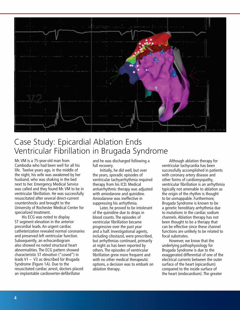
Mr. VM is a 75-year-old man from Cambodia who had been well for all his life. Twelve years ago, in the middle of the night, his wife was awakened by her husband, who was shaking in the bed next to her. Emergency Medical Service was called and they found Mr. VM to be in ventricular fibrillation. He was successfully resuscitated after several direct-current countershocks and brought to the University of Rochester Medical Center for specialized treatment. His ECG was noted to display ST segment elevation in the anterior precordial leads. An urgent cardiac catheterization revealed normal coronaries and preserved left ventricular function. Subsequently, an echocardiogram also showed no noted structural heart abnormalities. The ECG pattern showed characteristic ST elevation (“coved”) in leads V1 – V3 as described for Brugada Syndrome (Figure 1A). Due to the resuscitated cardiac arrest, doctors placed an implantable cardioverter-defibrillator
and he was discharged following a full recovery. Initially, he did well, but over the years, sporadic episodes of ventricular tachyarrhythmia required therapy from his ICD. Medical antiarrhythmic therapy was adjusted with amiodarone and quinidine. Amiodarone was ineffective in suppressing his arrhythmia. Later, he proved to be intolerant of the quinidine due to drops in blood counts. The episodes of ventricular fibrillation became progressive over the past year and a half. Investigational agents, including cilostazol, were prescribed, but arrhythmias continued, primarily at night as has been reported by others. The episodes of ventricular fibrillation grew more frequent and with no other medical therapeutic options, a decision was to embark on ablation therapy.
Case Study: Epicardial Ablation Ends Ventricular Fibrillation in Brugada Syndrome
Although ablation therapy for ventricular tachycardia has been successfully accomplished in patients with coronary artery disease and other forms of cardiomyopathy, ventricular fibrillation is an arrhythmia typically not amenable to ablation as the origin of the rhythm is thought to be unmappable. Furthermore, Brugada Syndrome is known to be a genetic hereditary arrhythmia due to mutations in the cardiac sodium channels. Ablation therapy has not been thought to be a therapy that can be effective since these channel functions are unlikely to be related to focal substrates. However, we know that the underlying pathophysiology for Brugada Syndrome is due to the exaggerated differential of one of the electrical currents between the outer surface of the heart (epicardium) compared to the inside surface of the heart (endocardium). The greater
4
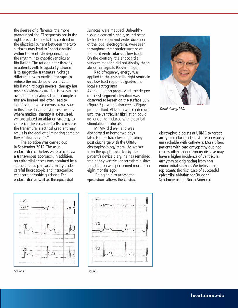
Figure 1 Figure 2
David Huang, M.D.
the degree of difference, the more pronounced the ST segments are in the right precordial leads. This contrast in the electrical current between the two surfaces may lead in “short circuits” within the ventricle degenerating the rhythm into chaotic ventricular fibrillation. The rationale for therapy in patients with Brugada Syndrome is to target the transmural voltage differential with medical therapy, to reduce the incidence of ventricular fibrillation, though medical therapy has never considered curative. However the available medications that accomplish this are limited and often lead to significant adverse events as we saw in this case. In circumstances like this where medical therapy is exhausted, we postulated an ablation strategy to cauterize the epicardial cells to reduce the transmural electrical gradient may result in the goal of eliminating some of these “short circuits.” The ablation was carried out in September 2012. The usual endocardial catheters were placed via a transvenous approach. In addition, an epicardial access was obtained by a subcutaneous pericardial entry under careful fluoroscopic and intracardiac echocardiographic guidance. The endocardial as well as the epicardial
surfaces were mapped. Unhealthy tissue electrical signals, as indicated by fractionation and wider duration of the local electrograms, were seen throughout the anterior surface of the right ventricular outflow tract. On the contrary, the endocardial surfaces mapped did not display these abnormal signals (Cover image). Radiofrequency energy was applied to the epicardial right ventricle outflow tract region as guided the local electrograms. As the ablation progressed, the degree of the ST segment elevation was observed to lessen on the surface ECG (Figure 2 post-ablation versus Figure 1 pre-ablation). Ablation was carried out until the ventricular fibrillation could no longer be induced with electrical stimulation protocols. Mr. VM did well and was discharged to home two days later. He has had close monitoring post discharge with the URMC electrophysiology team. As we see from the graph recorded by our patient’s device diary, he has remained free of any ventricular arrhythmia since the ablation was performed more than eight months ago. Being able to access the epicardium allows the cardiac
electrophysiologists at URMC to target arrhythmia foci and substrate previously unreachable with catheters. More often, patients with cardiomyopathy due not causes other than coronary disease may have a higher incidence of ventricular arrhythmias originating from non-endocardial sources. We believe this represents the first case of successful epicardial ablation for Brugada Syndrome in the North America.
heart.urmc.edu
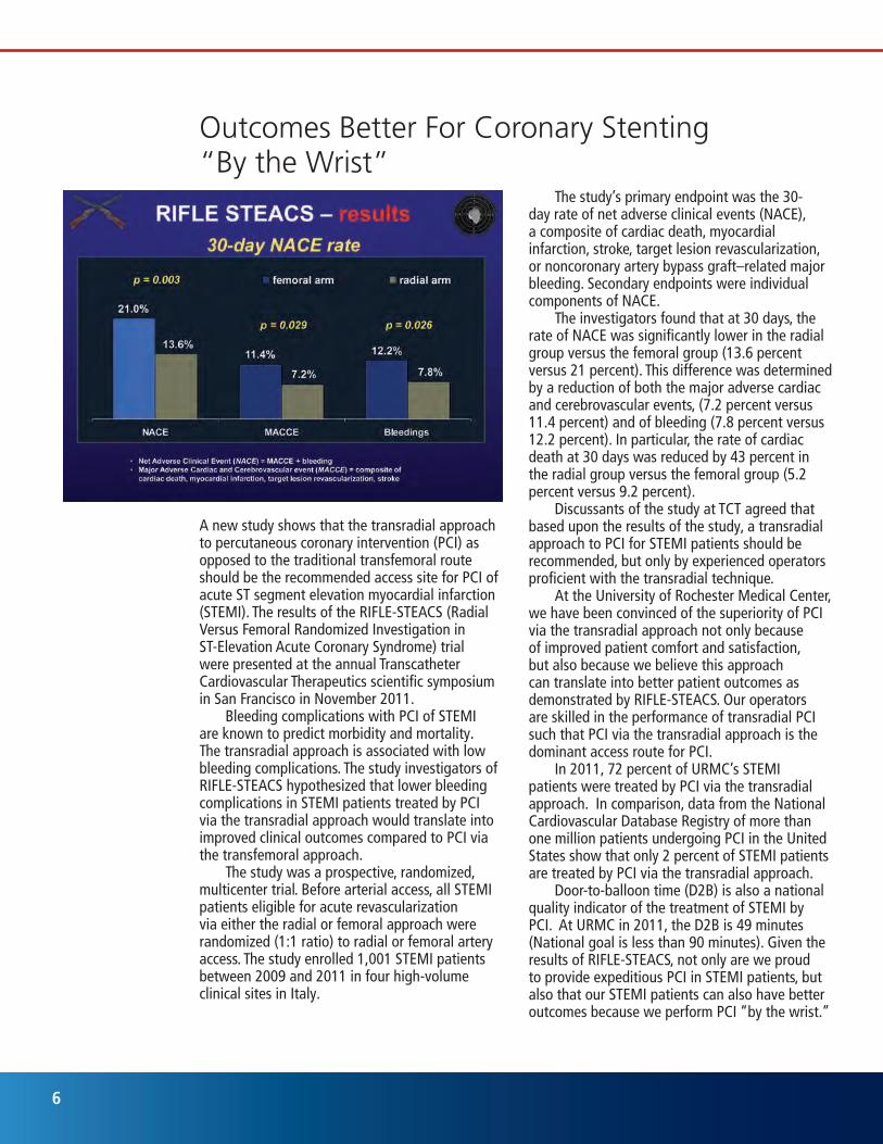
A new study shows that the transradial approach to percutaneous coronary intervention (PCI) as opposed to the traditional transfemoral route should be the recommended access site for PCI of acute ST segment elevation myocardial infarction (STEMI). The results of the RIFLE-STEACS (Radial Versus Femoral Randomized Investigation in ST-Elevation Acute Coronary Syndrome) trial were presented at the annual Transcatheter Cardiovascular Therapeutics scientific symposium in San Francisco in November 2011. Bleeding complications with PCI of STEMI are known to predict morbidity and mortality. The transradial approach is associated with low bleeding complications. The study investigators of RIFLE-STEACS hypothesized that lower bleeding complications in STEMI patients treated by PCI via the transradial approach would translate into improved clinical outcomes compared to PCI via the transfemoral approach. The study was a prospective, randomized, multicenter trial. Before arterial access, all STEMI patients eligible for acute revascularization via either the radial or femoral approach were randomized (1:1 ratio) to radial or femoral artery access. The study enrolled 1,001 STEMI patients between 2009 and 2011 in four high-volume clinical sites in Italy.
The study’s primary endpoint was the 30-day rate of net adverse clinical events (NACE), a composite of cardiac death, myocardial infarction, stroke, target lesion revascularization, or noncoronary artery bypass graft–related major bleeding. Secondary endpoints were individual components of NACE. The investigators found that at 30 days, the rate of NACE was significantly lower in the radial group versus the femoral group (13.6 percent versus 21 percent). This difference was determined by a reduction of both the major adverse cardiac and cerebrovascular events, (7.2 percent versus 11.4 percent) and of bleeding (7.8 percent versus 12.2 percent). In particular, the rate of cardiac death at 30 days was reduced by 43 percent in the radial group versus the femoral group (5.2 percent versus 9.2 percent). Discussants of the study at TCT agreed that based upon the results of the study, a transradial approach to PCI for STEMI patients should be recommended, but only by experienced operators proficient with the transradial technique. At the University of Rochester Medical Center, we have been convinced of the superiority of PCI via the transradial approach not only because of improved patient comfort and satisfaction, but also because we believe this approach can translate into better patient outcomes as demonstrated by RIFLE-STEACS. Our operators are skilled in the performance of transradial PCI such that PCI via the transradial approach is the dominant access route for PCI. In 2011, 72 percent of URMC’s STEMI patients were treated by PCI via the transradial approach. In comparison, data from the National Cardiovascular Database Registry of more than one million patients undergoing PCI in the United States show that only 2 percent of STEMI patients are treated by PCI via the transradial approach. Door-to-balloon time (D2B) is also a national quality indicator of the treatment of STEMI by PCI. At URMC in 2011, the D2B is 49 minutes (National goal is less than 90 minutes). Given the results of RIFLE-STEACS, not only are we proud to provide expeditious PCI in STEMI patients, but also that our STEMI patients can also have better outcomes because we perform PCI “by the wrist.”
Outcomes Better For Coronary Stenting “By the Wrist”
6
Resistant hypertension is defined as blood pressure above goal despite three anti-hypertensive drugs. Ideal blood pressure is less than 140/90 mm Hg for most patients. (Patients with diabetes or chronic kidney disease should have blood pressure less than 130/80 mm Hg). However, some hypertensive patients do not achieve their target blood pressure — even when they take maximally tolerated doses of three appropriate antihypertensive drugs including a diuretic. Although the exact prevalence is unknown, studies indicate 20 percent to 30 percent of hypertensive patients have treatment-resistant hypertension. In these patients, many face co-morbidities, such as obesity, diabetes, and chronic kidney disease. Even with application of existing pharmacotherapies, resistant hypertension is expected to increase in coming decades due to the aging of the population, an increased burden of cardiovascular disease, and obesity/metabolic syndrome. If left untreated, long-term hypertension contributes to organ damage and mortality.
Many hypertensive patients can successfully reach their blood pressure goal with relatively minor changes in their medication regimen:• Intensificationofdiuretictherapy, such as use of chlorthalidone rather than hydrochlorothiazide, can provide great success. • Thelongeractionoftorsemide may be more favorable than furosemide in patients with renal insufficiency that warrants the use of a loop diuretic. • Spironolactoneoreplerenone can be particularly effective as add-on drugs in patients with resistant hypertension and require appropriate vigilance of serum potassium.• AppropriatelydosedACEs,ARBs, and CCBs can also be of great use and beta blockers still have a role in many patients, but not usually as first-line. However, newer therapies are needed. URMC is one of a few centers testing a newer, smaller version of an implantable carotid sinus stimulator
for blood pressure control in these patients. The device is implanted below the clavicle, and a single lead runs up to the carotid sinus providing continuous electrical stimulation that, in European trials, has resulted in a 26 point blood pressure drop. This device should be available by mid-summer for use in clinical trials. Renal nerve ablation is also a promising therapy. Via an arterial catheter, the nerves surrounding the renal arteries are ablated, resulting is decreased central sympathetic activity and lower blood pressure. It is likely that this therapy will be available at URMC in the fall. The Joint National Commission on the Prevention, Detection, Evaluation and Treatment of Blood Pressure is expected to issue new evidence-based treatment guidelines later this year. Additionally, the American Heart Association recently published a scientific statement entitled “Beyond Medications and Diet: Alternative Approaches to Lowering Blood Pressure.” This can be downloaded from the AHA website.
Advancing Resistant Hypertension Therapies
Charles J. Lowenstein, M.D., Chief of Cardiology
Heart and Vascular Center Leaders To refer a patient:(585) 275-2877
To get more information, email: [email protected]
George L. Hicks, M.D., Chief of Cardiac Surgery
Joseph M. Delehanty, M.D., Director, Heart and Vascular Center
heart.urmc.eduheart.urmc.edu
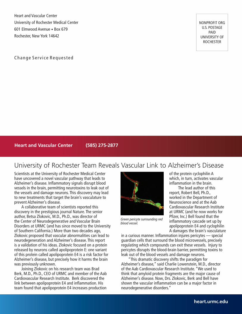
Scientists at the University of Rochester Medical Center have uncovered a novel vascular pathway that leads to Alzheimer’s disease. Inflammatory signals disrupt blood vessels in the brain, permitting neurotoxins to leak out of the vessels and damage neurons. This discovery may lead to new treatments that target the brain’s vasculature to prevent Alzheimer’s disease. A collaborative team of scientists reported this discovery in the prestigious journal Nature. The senior author, Betsa Zlokovic, M.D., Ph.D., was director of the Center of Neurodegenerative and Vascular Brain Disorders at URMC (and has since moved to the University of Southern California.) More than two decades ago, Zlokovic proposed that vascular abnormalities can lead to neurodegeneration and Alzheimer’s disease. This report is a validation of his ideas. Zlokovic focused on a protein released by neurons called apolipoprotein E: one variant of this protein called apolipoprotein E4 is a risk factor for Alzheimer’s disease, but precisely how it harms the brain was previously unknown. Joining Zlokovic on his research team was Brad Berk, M.D., Ph.D., CEO of URMC and member of the Aab Cardiovascular Research Institute. Berk discovered the link between apolipoprotein E4 and inflammation. His team found that apolipoprotein E4 increases production
University of Rochester Team Reveals Vascular Link to Alzheimer’s Diseaseof the protein cyclophilin A which, in turn, activates vascular inflammation in the brain. The lead author of this report, Robert Bell, Ph.D., worked in the Department of Neuroscience and at the Aab Cardiovascular Research Institute at URMC (and he now works for Pfizer, Inc.) Bell found that the inflammatory cascade set up by apolipoprotein E4 and cyclophilin A damages the brain’s vasculature
in a curious manner. Inflammation injures pericytes — special guardian cells that surround the blood microvessels, precisely regulating which compounds can exit these vessels. Injury to pericytes disrupts the blood-brain barrier, permitting toxins to leak out of the blood vessels and damage neurons. “This dramatic discovery shifts the paradigm for Alzheimer’s disease,” said Charlie Lowenstein, M.D., director of the Aab Cardiovascular Research Institute. “We used to think that amyloid protein fragments are the major cause of Alzheimer’s disease. Now, Drs. Zlokovic, Berk and Bell have shown the vascular inflammation can be a major factor in neurodegenerative disorders.”
NONPROFIT ORGU.S. POSTAGE
PAIDUNIVERSITY OF
ROCHESTER
Heart and Vascular Center
University of Rochester Medical Center
601 Elmwood Avenue • Box 679
Rochester, New York 14642
C h a n g e S e r v i c e R e q u e s t e d
heart.urmc.edu
Green pericyte surrounding red blood vessel.
Heart and Vascular Center (585) 275-2877
heart.urmc.edu