Embed Size (px)
Citation preview

University of Groningen
Structural organization of the needle complex of the type III secretion apparatus of ShigellaflexneriSani, Musa; Allaoui, Abdelmounaaïm; Fusetti, Fabrizia; Oostergetel, Gert T.; Keegstra, Wilko;Boekema, Egbert J.Published in:Micron
DOI:10.1016/j.micron.2006.04.007
IMPORTANT NOTE: You are advised to consult the publisher's version (publisher's PDF) if you wish to cite fromit. Please check the document version below.
Document VersionPublisher's PDF, also known as Version of record
Publication date:2007
Link to publication in University of Groningen/UMCG research database
Citation for published version (APA):Sani, M., Allaoui, A., Fusetti, F., Oostergetel, G. T., Keegstra, W., & Boekema, E. J. (2007). Structuralorganization of the needle complex of the type III secretion apparatus of Shigella flexneri. Micron, 38(3),291 - 301. https://doi.org/10.1016/j.micron.2006.04.007
CopyrightOther than for strictly personal use, it is not permitted to download or to forward/distribute the text or part of it without the consent of theauthor(s) and/or copyright holder(s), unless the work is under an open content license (like Creative Commons).
The publication may also be distributed here under the terms of Article 25fa of the Dutch Copyright Act, indicated by the “Taverne” license.More information can be found on the University of Groningen website: https://www.rug.nl/library/open-access/self-archiving-pure/taverne-amendment.
Take-down policyIf you believe that this document breaches copyright please contact us providing details, and we will remove access to the work immediatelyand investigate your claim.
Downloaded from the University of Groningen/UMCG research database (Pure): http://www.rug.nl/research/portal. For technical reasons thenumber of authors shown on this cover page is limited to 10 maximum.

www.elsevier.com/locate/micron
Micron 38 (2007) 291–301
Structural organization of the needle complex of the
type III secretion apparatus of Shigella flexneri
Musa Sani a, Abdelmounaaım Allaoui b, Fabrizia Fusetti c, Gert T. Oostergetel a,Wilko Keegstra a, Egbert J. Boekema a,*
a Biophysical Chemistry, Groningen Biomolecular Sciences and Biotechnology Institute,
University of Groningen, Nijenborgh 4, 9747 AG Groningen, The Netherlandsb Laboratoire de Bacteriologie Moleculaire, Faculte de Medecine, Universite Libre deBruxelles,
808 Route de Lennik, CP 614 b, B-1070 Bruxelles, Belgiumc Biochemistry, Groningen Biomolecular Sciences and Biotechnology Institute, University of Groningen,
Nijenborgh 4, 9747 AG Groningen, The Netherlands
Received 28 February 2006; received in revised form 11 April 2006; accepted 11 April 2006
Abstract
The secretion apparatus known as the needle complex (NC) from the bacterium Shigella flexneri was studied by single particle electron
microscopy. The isolated intact NC appears in projection to be composed of a basal body consisting of seven rings and a protruding needle
appendage. A comparison of averaged projections of the intact NC and its fragments revealed the organization of the NC into several major
subcomplexes. One of these lacks an inner membrane ring of the basal body but still presents the needle appendage attached to four upper rings.
The position of the needle appendage within these rings is variable, suggesting that the dissociated component is necessary for stabilizing the
needle appendage. Averaged images of the subcomplex lacking the inner membrane basal rings show a thicker extension at the base of the needle
appendage, called the socket. This socket was also found to be present in images of the basal body fragment isolated from mutants lacking the mxiH
and mxiI genes. This suggests that the socket is not composed of MxiH and MxiI subunits, which form the needle appendage. A symmetry analysis
of the basal body top view projections indicated that a peripheral protein component of the inner membrane ring is present in a ring with 24 copies,
in contrast to the Salmonella typhimurium NC. A model is presented in which the NC is only associated to the outer- and inner-membranes with its
first and seventh ring, respectively.
# 2006 Elsevier Ltd. All rights reserved.
Keywords: Shigella flexneri; Type III secretion system; Needle complex; Basal body; Electron microscopy
1. Introduction
Pathogenic bacteria have evolved a variety of delivery
systems to translocate their proteins (some of them virulent in
nature) to their host and/or surrounding environment. One such
system, termed the type III secretion system (TTS), is used by
pathogenic Gram-negative bacteria to cause a variety of
diseases in animals and plants. The TTS is a supramolecular
system used as a molecular syringe to inject proteins directly
into host cells (Hueck, 1998). Characteristic features of this
system are (i) the absence of a typical, cleavable, sec-dependent
signal sequence in secreted substrates; (ii) the requirement of
accessory proteins for secretion; (iii) the transit of proteins
* Corresponding author. Tel.: +31 50 3634225; fax: +31 50 3634800.
E-mail address: [email protected] (E.J. Boekema).
0968-4328/$ – see front matter # 2006 Elsevier Ltd. All rights reserved.
doi:10.1016/j.micron.2006.04.007
through both the inner and outer bacterial membranes; and (iv)
the requirement of activating signals to initiate secretion. The
TTS apparatus is a complex multi-subunit membrane-bound
structure consisting of more than 20 proteins (Cornelis and Van
Gijsegem, 2000). Approximately eight components of this
apparatus are homologous to components of the flagellar
assembly apparatus, indicating a common ancestry (Hueck,
1998). The bacterial effector molecules that transit through the
secretion apparatus and their resulting effects on eukaryotic
cells vary among different pathogens. Whereas Shigella and
Salmonella deliver their effectors to facilitate their uptake by
non-professional phagocytes, Yersinia spp. and enteropatho-
genic Echerischia coli deliver theirs to inhibit uptake by
macrophages (Hueck, 1998).
Bacteria of the Shigella group are the causative agent of
bacillary dysentery in humans (LaBrec et al., 1994). Bacteria

M. Sani et al. / Micron 38 (2007) 291–301292
are transmitted via the faecal-oral route and cause disease by
invading the colonic epithelium, which results in tissue
destruction and inflammation (LaBrec et al., 1994). Genes
required for bacterial entry into host cells are clustered on a
30 kb region of the 220 kb virulence plasmid (Buchrieser et al.,
2000) and encode a TTS apparatus and effectors that induce
internalization of bacteria. The TTS apparatus (�50 copies per
cell) is assembled at 37 8C before any host contact (Tamano
et al., 2000).
Recent studies indicate that the needle complex (NC) of
Shigella and Salmonella spp. share structural similarities, with
a base embedded in the bacterial membrane and an axial
structure protruding from the bacterial surface to form a needle-
like rod (Tamano et al., 2000; Blocker et al., 1999; Kubori et al.,
1998). Electron microscopy studies on the NC from S. flexneri
and Salmonella spp. revealed that the NC is composed of a
basal body which consists of pairs of upper and lower rings
(Tamano et al., 2000; Kubori et al., 1998) and a tubular needle
appendage of about 450–500 A in length which protrudes from
the basal body. The latter has a diameter of �70 A enclosing a
central channel of about 25 A and is composed of MxiH and
MxiI in S. flexneri and of PrgI and PrgJ in Salmonella, of which
MxiI and PrgJ are indicated to be minor essential components
(Tamano et al., 2000; Blocker et al., 2001; Kimbrough and
Miller, 2000; Kubori et al., 2000). The 3D structure of the
needle appendage of the NC of Shigella was solved at a
resolution of 16 A and resembles the helical architecture seen in
the flagellar hook and filament (Cordes et al., 2003). It is
interesting to note that a complete atomic model of the
flagellum obtained by cryo-electron microscopy and image
analysis (Yonekura et al., 2003) shows that flagellin subunits
are held together by hydrophobic interactions, and that the
central channel of the flagellum is lined with polar residues.
The base structure of the NC is composed of three major
components, MxiD, MxiG, and MxiJ in S. flexneri and InvG,
PrgH, and PrgK in S. typhimurium and resembles the flagellar
basal body (Tamano et al., 2000; Blocker et al., 1999; Kubori
et al., 1998). These three major components of the base structure
all have cleavable N-terminal sec-dependent export signals
(Allaoui et al., 1992, 1993). While MxiG and MxiJ form most of
the basal part (Blocker et al., 1999) MxiD has been proposed to
form the upper ring doublet of the NC (Blocker et al., 1999).
Detailed structural analysis of the basal part of the NC from
Salmonella typhimurium has revealed that the basal part (which
henceforth will be referred to as the basal body due to its
structural similarity to the flagellar basal body) possesses a 20- or
21-fold symmetry in the inner ring (Marlovits et al., 2004).
In the present study, we have used electron microscopy and
single particle analysis to get further insight into the structural
assembly of the NC of Shigella flexneri. Purification of the NC
was achieved by gel filtration and CsCl gradient centrifugation.
This procedure yielded, in addition to many intact NCs, several
intermediate subcomplexes, which were also analyzed. Addi-
tional information was obtained by a structural analysis of the
basal body from mutants lacking specific subunits. Based on
these data, the division of the NC into several distinct
substructures is discussed.
2. Materials and methods
2.1. Bacterial strains and growth media
The S. flexneri ipaC strain SF621, a derivative of the wild-
type strain M90T, serotype 5 (Menard et al., 1993), was a kind
gift from Dr. C. Parsot (Pasteur Institute, Paris). This strain
produces normal NC, but is non-invasive, due to the absence of
the IpaC translocator. Other mutant strains used in this study are
SB116 (relevant genotype mxiH�) and SB125 (relevant
genotype mxiI�) (Blocker et al., 1999). Bacteria were grown
in tryptic casein soy broth (Sigma) supplemented with
kanamycin (30 mg ml�1) at 37 8C.
2.2. Purification of NC and dissociation experiments
The needle complexes of S. flexneri were extensively
purified from the envelope fractions by adaptation of the
method described by Tamano et al. (2000). In brief, to allow
spheroblast formation bacteria were suspended in 20% sucrose
supplemented with 2 mM EDTA and 0.5 mg/ml lysozyme
(Sigma). Cells were lysed with 0.1% Triton X 100 at a total
protein to detergent ratio of 5:1 (w/w), followed by DNA
digestion with 4 mM MgCl2 and 80 mg/ml DNAse (Sigma).
The membrane fraction was separated by ultracentrifugation at
110,000 � g for 1 h. The resulting pellet was resuspended in
TET buffer (10 mM Tris–HCl, pH 8.0, 1 mM EDTA, 0.1%
Triton X-100) and loaded onto a Superdex 200 gel filtration
column (Amersham Biosciences) at a flow rate of 0.4 ml/min.
Fractions containing the NC were collected and concentrated
by centrifugation at 80,000 � g for 45 min, and subsequently
resuspended in TET buffer. Column fractionated samples were
further purified by 40% (w/v) CsCl density gradient
centrifugation in a SW41 rotor at 37,000 � g for 14 h at
20 8C. To isolate NC components from mxiH and mxiI mutants,
harvested cells were first cross-linked as previously described
(Schuch and Maurelli, 2001). Membrane fractions were
prepared and solubilized as described above for the ipaC
mutant. The preparation was then loaded onto a Mono Q HR 5/5
column (Amersham Biosciences), which had been pre-
equilibrated with five column volumes of TET buffer. Bound
proteins were eluted with a linear gradient of 0–1 M NaCl, pH
8.0. Fractions were pooled and centrifuged at 250,000 � g for
1 h. The pellets were resuspended in TET buffer and analyzed
by SDS/PAGE. Proteins were either transferred to a nitro-
cellulose membrane and immuno-blotted as previously
described (Magdalena et al., 2002) or prepared for matrix-
assisted laser desorption/ionization tandem mass spectrometry
(MS/MS) analysis (MALDI-MS/MS).
2.3. MALDI-MS/MS analysis
Purified particles obtained from the IpaC, MxiH and MxiI
deficient mutants were separated on a 15% (160 mm � 160
mm � 1.0 mm) tricine SDS-polyacrylamide gel and stained
with colloidal Coomassie Blue (Serva). Each lane was system-
atically cut and excised gel fragments were washed for 1 h with

M. Sani et al. / Micron 38 (2007) 291–301 293
25 mM ammonium bicarbonate, 50% HPLC grade acetonitrile
and further dehydrated in pure acetonitrile. Hundred nano-
grams trypsin (Sequencing grade modified, Promega) in 25 mM
ammonium bicarbonate (pH 7.8) were added to each gel
fragment. Digestion was carried out overnight at 37 8C. The
resulting tryptic peptides were extracted twice with 0.1 ml of
60% (v/v) acetonitrile/1% (v/v) trifluoroacetic acid in an
ultrasonic bath for 15 min. Extracts were pooled, lyophilized
and the peptides dissolved in 10 ml 0.1% (v/v) trifluoroacetic
acid. Peptide samples (0.5 ml) were mixed on the MALDI
target with 0.5 ml of alpha-cyano-4-hydroxycinnamic acid, in
50% (v/v) acetonitrile/0.1% (v/v) trifluoroacetic acid, and left
to dry. MALDI spectra were recorded on a MALDI TOF/TOF
mass spectrometer 4700 Proteomics Analyzer (Applied
Biosystem, Framingham, MA, USA). The most abundant
peptides were subjected to MS/MS fragmentation. Data were
analyzed using GPS Explorer software (Applied Biosystem).
MSDB and Swiss-Prot database were searched with the
MASCOT software (Matrix Science, London, UK). A tolerance
of 0.15 Da for both the MS and MS/MS analyses, and one
missed cleavage site were set as fixed parameters.
2.4. Electron microscopy and image analysis
Samples of purified complex were negatively stained using
the droplet method with 2% uranyl acetate on glow discharged
carbon-coated copper grids. Electron microscopy was per-
formed on a Philips CM20FEG equipped with a field emission
gun operated at 200 kV. The ‘‘GRACE’’ system for semi-
automated specimen selection and data acquisition (Oostergetel
et al., 1998) was used to record 2048 � 2048 pixel images at
66,850� calibrated magnification with a Gatan 4000 SP 4K
slow-scan CCD camera. The step size (after binning the
images) was 30 mm, corresponding to a pixel size of 4.5 A at
the specimen level. A total of 10,000 particle projections were
collected interactively from 3500 images. Single particle
analysis was performed as previously described (Harauz et al.,
1988) with IMAGIC and Groningen Image Processing
(‘‘GRIP’’) software packages (W. Keegstra, unpublished data)
on a PC cluster. Briefly, the images were first treated to
normalize the variance and band-pass filtered to remove
unwanted high and low frequencies of the images during the
analysis. Projections were subjected to rotational and transla-
tional alignments with correlation methods (Harauz et al.,
1988; Van Heel et al., 1992). The aligned projections were
treated with multivariate statistical analysis (MSA) in
combination with hierarchical classification (Van Heel et al.,
1992). During classification, 15–20% of the images was
rejected. Finally, sums of projections belonging to the various
classes were made by adding the original images without
imposed band-pass filter. The best 60–80% of the class
members was taken for the final sums using the correlation
coefficient of the alignment procedure as the quality criterion.
The resolution of the class averages was calculated by Fourier
ring correlation using the 3s criterion (Van Heel, 1984). In
addition, two subsets were independently processed in parallel
by reference-free alignment procedures (Penczek et al., 1992)
for comparison. The resolution of the class averages was
estimated from the latter by Fourier ring correlation using the
3s criterion (Van Heel, 1984).
To precisely localize the positions of substructures within
the NC, difference mapping of averaged projections was
performed. In this approach, normalized class sum images
representing side-view projections of the NC and subcomplexes
were subtracted from one another. Positive differences appear
in white, negative differences in black and insignificant
differences in gray. Rotational symmetry analysis of top views
was performed according to Kocsis et al. (1995). In brief, 95 top
views of the basal body from a mutant preparation were
centered to a reference obtained by averaging the data set after
translational alignments. By comparing the rotational power
spectrum of centered particles with background images picked
from individual micrographs, signals that are marginally above
the background will build up to a statistically significant level to
give the rotational symmetry.
3. Results
3.1. Purification of NC and electron microscopy
To investigate the structure of the needle complex (NC),
single particles of the complex were isolated from three
different mutants of S. flexneri. Intact NCs were purified from
SF621 ipaC. This strain produces normal NCs, but is non-
invasive, due to the absence of the IpaC translocator (Menard
et al., 1993). Solubilized membrane fractions were first loaded
onto a size exclusion column to separate the NC components
from large membrane fragments (Fig. 1A). Aliquots containing
NC components (checked by EM) were pooled and concen-
trated and further fractionated by 30% (w/v) CsCl density
gradient centrifugation. In this way, a relative high number of
pure intact complexes could be obtained (first lane of Fig. 1B).
Resolved proteins in Fig. 1B were subjected to in-gel trypsin
digestion followed by MALDI-MS/MS analysis. The results
clearly show the presence of MxiD, MxiG, MxiJ and MxiM
subunits in all three samples, while MxiH is only detected in the
NC components expressed by the ipaC mutant (Fig. 1B). MxiM
is observed to co-purify with all NC components isolated from
all the investigated strains and thus indicates that it associates
with the isolated complex. Neither in Shigella (Tamano et al.,
2000; Blocker et al., 1999) nor in Salmonella (Marlovits et al.,
2004) has this subunit been detected in previous isolations of
this complex. Similar sample compositions were detected by
MS analysis irrespective of whether the particles were isolated
by CsCl gradient centrifugation or by size-exclusion chroma-
tography (data not shown), and thus rules out that the observed
components could be due to artifacts. The fraction from the ipaC
mutant of Fig. 1B showed, besides the relatively homogeneous
monodisperse NC particles, also particles of NC-like substruc-
tures. Apparently, the purification step resulted in a limited
dissociation of the NC. This dissociation is corroborated by Fig. 2
where three types of subcomplexes are visible, which were
tentatively classified as: (i) NCs minus the needle appendage
denoted as BB (for basal body); (ii) NCs lacking the inner

M. Sani et al. / Micron 38 (2007) 291–301294
Fig. 1. (A) Gel chromatography of solubilized needle complexes. The fraction
containing the highest numbers of NCs (as checked by EM), used for further
purification with density gradient centrifugation, is indicated. (B) Tricine SDS-
PAGE of purified NC and its subcomplexes S. flexneri mutant strains. Samples
from the three different strains (IpaC�, MxiH�, MxiI�) were separated on a
15% gel stained with colloidal Coomassie Blue stain (upper part) or blotted with
antibodies specific for MxiJ and MxiH (lower part).
Fig. 2. An electron micrograph of negatively stained purified NC particles
isolated from an IpaC� strain after the CsCl gradient step. Some complete NCs
have been indicated (NC). Three specific fragments are dominant: first, so-
called basal bodies which are NCs lacking the needle appendage (BB); second,
NCs lacking the inner membrane rings (NR) and third, dissected outer mem-
brane rings (OMR).
membrane rings denoted as NR (for needle plus rings) and (iii) an
outer membrane ring subcomplex, denoted as OMR. The
population of all three subcomplexes was relatively homo-
geneous. No continuum of intermediate subcomplexes was
found, suggesting that the dissociation of the NCs is stepwise.
We also investigated NC components from two S. flexneri
mutants deficient either in MxiH or MxiI. These proteins are
proposed to be the sole components of the needle appendage of
the NC (Tamano et al., 2000; Blocker et al., 1999; Kubori et al.,
1998). NCs lacking the needle appendage were purified from
both MxiH and MxiI deficient strains. Samples purified from
both mutant strains were analyzed by MALDI-MS/MS. MxiD,
MxiG, MxiJ and MxiM were identified (Fig. 1B), but not MxiH
and MxiI. This is consistent with previous observations
(Blocker et al., 1999). Western immuno-blotting confirmed
the presence of MxiH (Fig. 1C) and MxiI (data not shown) only
in the ipaC mutant. In contrast, MxiH could not be detected in
mxiI mutant preparations; conversely, MxiI was also not
detected in mxiH mutant preparations (data not shown), a
finding consistent with earlier observations (Sukhan et al.,
2003).
3.2. Image analysis
Single particle image analysis was performed by selecting all
well-preserved projections from non-overlapping NC particles
and subcomplexes from a large set of electron microscopy
images. Over 10,000 particles were picked from the ipaC mutant
strain, of which 66% were intact NCs, while the remaining
projections were assigned to the OMR subcomplex (17%), the
NR (9%), and basal body (8%), respectively. With the exception
of the OMR projections, which were assumed to represent a top
view orientation, the NC particles and the remaining sub-
complexes all oriented on the carbon film in a side view position
approximately perpendicular to the long axis of the complex. In
an initial alignment step, one projection was selected arbitrarily

M. Sani et al. / Micron 38 (2007) 291–301 295
Fig. 3. Image analysis of the whole NC and four specific subcomplexes of the ipaC mutant. A set of 8250 projections represents the most abundant projections of five
different types of (sub)complexes that were independently processed after sorting out by an initial classification. The final projections represent averages of the best
classes with (A) a sum of 1500 needle complex particles showing strong mirror-symmetry along the vertical axis within the basal body part. White arrows indicate
prominent spikes between the upper two rings of the basal body part; (B) subset of 63 needle complex particles without strong mirror symmetry, as can be seen from
the upper ring of the basal body and protrusions, of which only the one at the right of the basal body is visible; (C) 337 basal body particles; (D) 343 NR particles; (E)
243 NR particles and (F) 450 OMR particles. (G) Result of classification (2 classes of 1536 projections each) of needle appendages from needle complex particles,
which were aligned after masking the basal body moiety. The space bar indicates 250 A.
from the set of images and used as a first reference. After the first
alignment relative to this noisy image, a new reference was
generated by summation of the images with the highest
correlation coefficient related to the first reference. The aligned
data set was treated by MSA and partitioned into 113 classes. It
was subsequently reclassified into five major classes representing
the different particle types. These classes were further partitioned
into several classes by independent MSA and hierarchical
classification procedures to determine inter-class difference. The
quality of these average classes was further improved by iterative
alignments in a multi-reference alignment scheme. Fig. 3 shows
the final averaged projections of the five particle types. The
reproducibility of the features in the average was assessed by
independently analyzing subsets of projections obtained from
different preparations by a reference free alignment procedure
(Penczek et al., 1992). Specific features visible in these particle
types are discussed below.
3.3. Needle complex
A total of 4158 whole NC particles, composed of a needle
appendage protruding from a basal part composed of several
rings was analyzed. Alignment was carried out with respect to the
basal part of the complex since the overall length of the needle
appendage is somewhat variable. The classification of this data
set showed only one dominant view (Fig. 3A). In this view the
basal part with a height of 310 A is clearly resolved. The
resolution of this component in the final average projection was
estimated to be 16 A using the 3s criterion (Van Heel, 1987) and
19 A using the 0.5s criterion. Characteristic features of this
projection are protrusions attached to the periphery of the OMR
doublet (see arrows in Fig. 3A). This density protrudes about
25 A from the ring doublet and is located right within the OMR. It
is surprising that only one type of dominant projection was found
for the whole NC particles. We only found a very limited number
of projections that deviated from this dominant view of Fig. 3A if
the aligned set was decomposed into about 100 classes (Fig. 3B).
It shows an average image with some deviations around the upper
two rings of the basal part. Moreover, only the protrusion on the
right side is clearly visible.
The needle appendage of the NC is a straight hollow tube
(indicated by a stain penetrated line along its axis) which is
500 A long on average. The overall size and shape of the NC are
in agreement with previous investigations (Tamano et al., 2000;
Blocker et al., 1999; Kubori et al., 1998; Cordes et al., 2003).
However, details in the needle portion appeared to have been
smeared out during averaging procedures. Significant features
of the needle portion could be resolved when projections were
aligned and classified after masking the basal part. A
decomposition into two classes, each of 1536 projections is
shown in Fig. 3G.
3.4. The basal body
A subset of 701 side views of basal body (BB) particles
isolated from the ipaC mutant strain was processed and one
dominant projection was obtained (Fig. 3C). Similar BBs could
also be obtained from the MxiH and MxiI deficient strains.
Actually this was the only intact components that could be
purified from these mutants. The micrographs of EM samples
prepared from both mxiH and mxiI mutants, however, showed
low numbers of such particles per image. The number of particles
increased slightly when a cross-linking step was included in the
isolation procedure. A total of 500 particles could be picked from
more than 2000 images. Average projections of side views of
these mutants show a basal body lacking the needle appendage
(Fig. 4A and B), identical to the substructure co-purified from the
IpaC mutant (Fig. 4C). A striking feature of all three basal body
projections is the presence of a central oval-shaped ‘‘bulge’’ in
the lower half of the particle. This feature was described as a

M. Sani et al. / Micron 38 (2007) 291–301296
Fig. 4. (A–C) Averages of side view projections of the basal body isolated from mutants of MxiH, MxiI and ipaC, respectively. The ‘‘socket’’ has been indicated in the
frame of (C), (the same as frame Fig. 3C), by a green overlay. (D–E) Averages of top view projections of basal body of mutants of mxiH and mxiI, in which the particles
are tilted slightly out of plane. (F) The top view of the basal body of the mxiH mutant, with 24-fold symmetry imposed. Lower half: symmerty analysis of the image of
frame D. The n-fold type of rotational imposed on frame D has been indicated. The sacle bar in A–F indicates 250 A.
M. Sani et al. / Micron 38 (2007) 291–301 297
socket in a 17 A 3D reconstruction of the type III secretion
complex from S. typhimurium (Marlovits et al., 2004) and hence
this name will be kept up. In addition to the observation of side
views of basal bodies lacking the needle appendage, we also
observed top view projections. Examination of the average top
view from the mxiH and mxiI mutants show small densities
arranged in an inner ring of 90 A and an outer ring of 246 A
(Fig. 4D and E). It is reasonable to infer that these densities
correspond to individual subunits that arrange to form these
annular substructures. A symmetry analysis of the basal body top
views of the mxiH mutant was performed. By comparing 2–30-
fold imposed rotational symmetries it is apparent that the outer
ring of densities has a clear 24-fold rotational symmetry, as well
as the lower 2-, 3-, 6-, 8- and 12-fold symmetries and that all other
imposed types of rotational symmetries did not enhance the
image features (Fig. 4, lower half). Therefore, the 24-fold
rotational symmetry was imposed in Fig. 4F. No such periodicity
is observed for the inner ring suggesting that the inner ring has a
different symmetry; which is possibly 15-fold (Fig. 4, lower
half). The view of the mxiI mutant is too far tilted out of the
horizontal plane to determine its rotational symmetry.
The resolution attained for all three basal body side view
averages is 19 A. The lower part of the basal body has an overall
dimensions of 310 A (height) � 242 A (diameter), while the
upper rings which might be inserted in the outer membrane and
the peptidoglycan layer have a height of 81 A and a diameter of
158 A. Immediately below the outer membrane ring (OMR)
doublet are three minor rings with diameters of 122, 135 and
161 A. At the base is an inner membrane ring with a diameter of
240 A and a height of 58 A. Comparison of the basal body
fragments from the ipaC mutant with those isolated from mxiH
and mxiI mutants shows no discernible differences suggesting
that the basal body which co-purified with intact NC could be
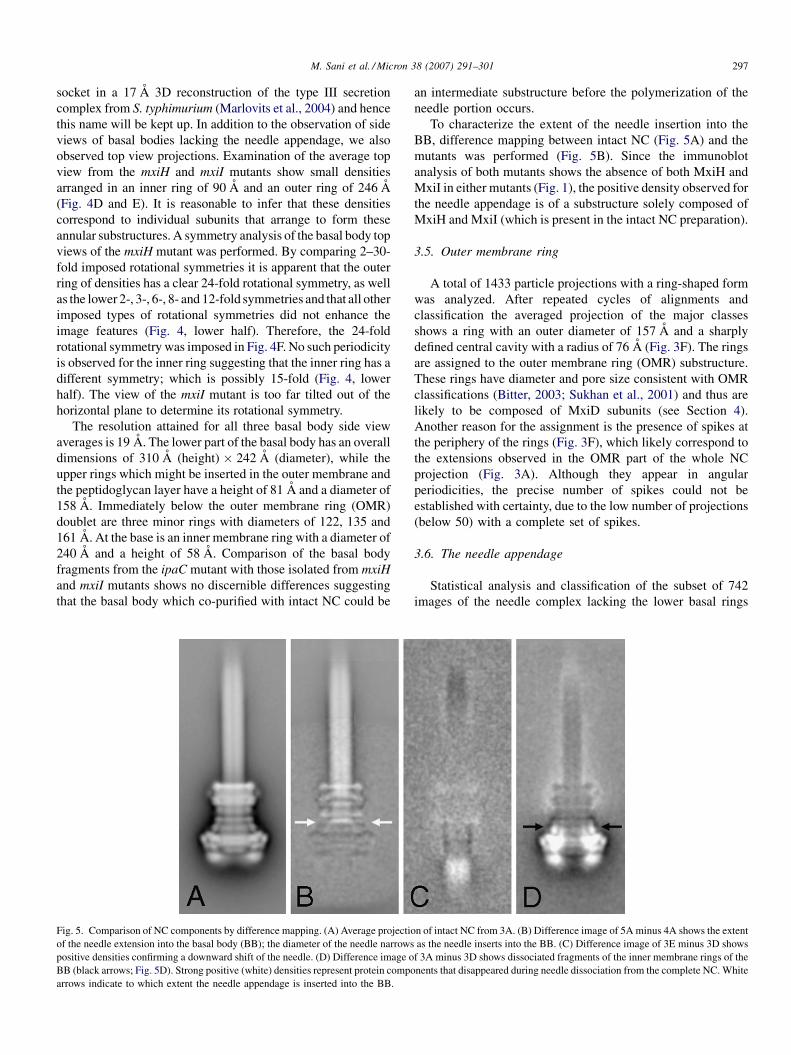
Fig. 5. Comparison of NC components by difference mapping. (A) Average projectio
of the needle extension into the basal body (BB); the diameter of the needle narrows
positive densities confirming a downward shift of the needle. (D) Difference image o
BB (black arrows; Fig. 5D). Strong positive (white) densities represent protein compo
arrows indicate to which extent the needle appendage is inserted into the BB.
an intermediate substructure before the polymerization of the
needle portion occurs.
To characterize the extent of the needle insertion into the
BB, difference mapping between intact NC (Fig. 5A) and the
mutants was performed (Fig. 5B). Since the immunoblot
analysis of both mutants shows the absence of both MxiH and
MxiI in either mutants (Fig. 1), the positive density observed for
the needle appendage is of a substructure solely composed of
MxiH and MxiI (which is present in the intact NC preparation).
3.5. Outer membrane ring
A total of 1433 particle projections with a ring-shaped form
was analyzed. After repeated cycles of alignments and
classification the averaged projection of the major classes
shows a ring with an outer diameter of 157 A and a sharply
defined central cavity with a radius of 76 A (Fig. 3F). The rings
are assigned to the outer membrane ring (OMR) substructure.
These rings have diameter and pore size consistent with OMR
classifications (Bitter, 2003; Sukhan et al., 2001) and thus are
likely to be composed of MxiD subunits (see Section 4).
Another reason for the assignment is the presence of spikes at
the periphery of the rings (Fig. 3F), which likely correspond to
the extensions observed in the OMR part of the whole NC
projection (Fig. 3A). Although they appear in angular
periodicities, the precise number of spikes could not be
established with certainty, due to the low number of projections
(below 50) with a complete set of spikes.
3.6. The needle appendage
Statistical analysis and classification of the subset of 742
images of the needle complex lacking the lower basal rings
n of intact NC from 3A. (B) Difference image of 5A minus 4A shows the extent
as the needle inserts into the BB. (C) Difference image of 3E minus 3D shows
f 3A minus 3D shows dissociated fragments of the inner membrane rings of the
nents that disappeared during needle dissociation from the complete NC. White
M. Sani et al. / Micron 38 (2007) 291–301298
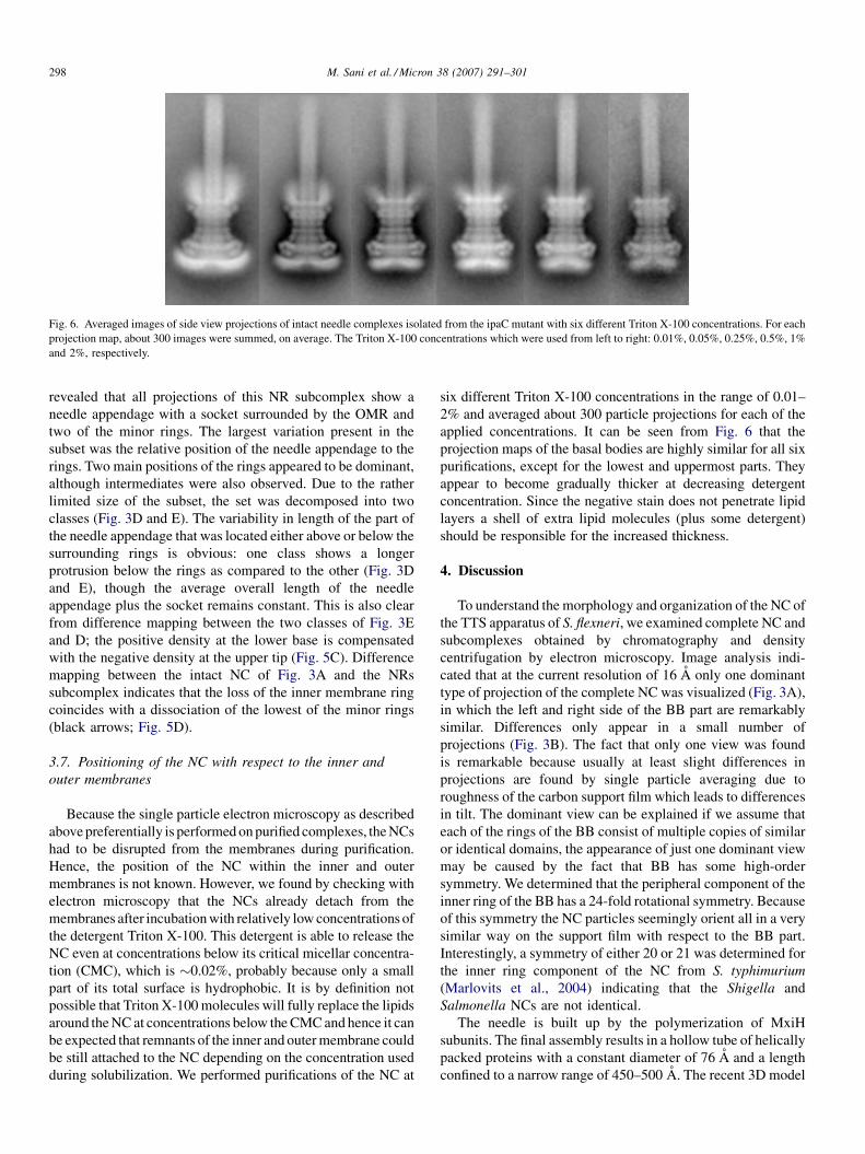
Fig. 6. Averaged images of side view projections of intact needle complexes isolated from the ipaC mutant with six different Triton X-100 concentrations. For each
projection map, about 300 images were summed, on average. The Triton X-100 concentrations which were used from left to right: 0.01%, 0.05%, 0.25%, 0.5%, 1%
and 2%, respectively.
revealed that all projections of this NR subcomplex show a
needle appendage with a socket surrounded by the OMR and
two of the minor rings. The largest variation present in the
subset was the relative position of the needle appendage to the
rings. Two main positions of the rings appeared to be dominant,
although intermediates were also observed. Due to the rather
limited size of the subset, the set was decomposed into two
classes (Fig. 3D and E). The variability in length of the part of
the needle appendage that was located either above or below the
surrounding rings is obvious: one class shows a longer
protrusion below the rings as compared to the other (Fig. 3D
and E), though the average overall length of the needle
appendage plus the socket remains constant. This is also clear
from difference mapping between the two classes of Fig. 3E
and D; the positive density at the lower base is compensated
with the negative density at the upper tip (Fig. 5C). Difference
mapping between the intact NC of Fig. 3A and the NRs
subcomplex indicates that the loss of the inner membrane ring
coincides with a dissociation of the lowest of the minor rings
(black arrows; Fig. 5D).
3.7. Positioning of the NC with respect to the inner and
outer membranes
Because the single particle electron microscopy as described
above preferentially is performed on purified complexes, the NCs
had to be disrupted from the membranes during purification.
Hence, the position of the NC within the inner and outer
membranes is not known. However, we found by checking with
electron microscopy that the NCs already detach from the
membranes after incubation with relatively low concentrations of
the detergent Triton X-100. This detergent is able to release the
NC even at concentrations below its critical micellar concentra-
tion (CMC), which is �0.02%, probably because only a small
part of its total surface is hydrophobic. It is by definition not
possible that Triton X-100 molecules will fully replace the lipids
around the NC at concentrations below the CMC and hence it can
be expected that remnants of the inner and outer membrane could
be still attached to the NC depending on the concentration used
during solubilization. We performed purifications of the NC at
six different Triton X-100 concentrations in the range of 0.01–
2% and averaged about 300 particle projections for each of the
applied concentrations. It can be seen from Fig. 6 that the
projection maps of the basal bodies are highly similar for all six
purifications, except for the lowest and uppermost parts. They
appear to become gradually thicker at decreasing detergent
concentration. Since the negative stain does not penetrate lipid
layers a shell of extra lipid molecules (plus some detergent)
should be responsible for the increased thickness.
4. Discussion
To understand the morphology and organization of the NC of
the TTS apparatus of S. flexneri, we examined complete NC and
subcomplexes obtained by chromatography and density
centrifugation by electron microscopy. Image analysis indi-
cated that at the current resolution of 16 A only one dominant
type of projection of the complete NC was visualized (Fig. 3A),
in which the left and right side of the BB part are remarkably
similar. Differences only appear in a small number of
projections (Fig. 3B). The fact that only one view was found
is remarkable because usually at least slight differences in
projections are found by single particle averaging due to
roughness of the carbon support film which leads to differences
in tilt. The dominant view can be explained if we assume that
each of the rings of the BB consist of multiple copies of similar
or identical domains, the appearance of just one dominant view
may be caused by the fact that BB has some high-order
symmetry. We determined that the peripheral component of the
inner ring of the BB has a 24-fold rotational symmetry. Because
of this symmetry the NC particles seemingly orient all in a very
similar way on the support film with respect to the BB part.
Interestingly, a symmetry of either 20 or 21 was determined for
the inner ring component of the NC from S. typhimurium
(Marlovits et al., 2004) indicating that the Shigella and
Salmonella NCs are not identical.
The needle is built up by the polymerization of MxiH
subunits. The final assembly results in a hollow tube of helically
packed proteins with a constant diameter of 76 A and a length
confined to a narrow range of 450–500 A. The recent 3D model

M. Sani et al. / Micron 38 (2007) 291–301 299
of the helical arrangement of the MxiH needle appendage
(Cordes et al., 2003) shows about 5.6 subunits in one turn, with
a helical pitch of 24 A. The dominant projection of the whole
NC does not show any detail in the needle portion (Fig. 3A).
This was also the case when the aligned data set (aligned on the
basal body) was classified only on the needle portion with
decomposition into 100 classes. When the basal part is masked
off and alignment performed with respect to the needle portion,
features similar to helical turns as presented in the 3D model of
Cordes et al. (2003) can be discerned (Fig. 3G). This may
indicate that the needle does not have a fixed rotational
orientation towards the basal body or a different symmetry.
Analysis of NR subcomplexes missing the inner membrane
basal rings (IMR), shows a needle appendage with the OMR
and two minor lower rings attached to it (Fig. 3D and E).
Classification indicated a variable position of the needle
appendage within the rings (Fig. 5C). This indicates that the
needle appendage is not rigidly fixed to the OMR but is flexible
in the absence of the IMR. Apparently the latter ring has a
function in keeping the needle appendage in proper position.
Because of its helical architecture, the needle diameter should
be constant over its full length. However, the averaged
projections of the NR subcomplexes, which are missing the
major inner membrane rings (Fig. 3D and E), show a slight
widening of the basal end of the needle. This widening is also
obvious in many of the individual projections and is referred to
as the socket (see Section 3).
Analysis of the basal body subcomplex indicates that this
structure is composed of the same number of rings as in the
complete NC. The averaged projection of the BB subcomplex
in Fig. 4 shows a striking resemblance of these subcomplexes to
the flagellar basal body (Francis et al., 1994; Sosinsky et al.,
1992; Thomas et al., 2001). All three BBs contain the socket
which directly confirms that this density is not part of the needle
appendage. Comparison between BBs co-purified with intact
NC and BB of mxiH and mxiI mutants show no discernible
difference. This suggests that the BB isolated in intact
preparation is an intermediate substructure of the NC. Although
the basal bodies of the mxiH and mxiI mutants showed similar
sockets as observed in the ipaC mutant, these mutants are not
able to assemble a complete NC with a needle appendage. Since
this needle appendage is composed of MxiH and MxiI, the
socket should have a different protein composition.
The numerous ring-shaped projections (Fig. 3F) found in the
data set represent a NC subcomplex which can only be assigned
to the outer membrane ring (OMR) subcomplex, according to
their diameter of 157 A. The OMR substructure is probably
composed mostly of MxiD, which is a member of the secretin
superfamily (Allaoui et al., 1993). These proteins organize into
multimeric ring-shaped structures. The stability of these
structures is influenced by another protein, MxiM (Schuch
and Maurelli, 1999). MxiM is a lipoprotein that belongs to a
group of proteins called secretin pilots (Allaoui et al., 1992;
Schuch and Maurelli, 1999). These proteins protect secretins
from proteolysis in the periplasm and promote their insertion
into the outer membrane by using their lipid extension to anchor
this otherwise hydrophilic protein in the outer membrane
(Schuch and Maurelli, 1999; Hardie et al., 1996). The average
image of the OMR subcomplex suggests the presence of about
10 spikes (Fig. 3F), although the exact number of spikes could
not be established due to the lack of a sufficient number of
intact particles. These spikes are quite similar to those observed
for the PulD–PulS complex (Nouwen et al., 1999), which
suggests that they correspond to MxiM, the proposed functional
equivalent of PulS. This is in agreement with previous studies
that showed that MxiM and MxiD interact directly within the
outer membrane envelope and co-purify together when co-
expressed in E. coli. Similar spikes are also visible at the
periphery of the averaged projection of complete NC (arrows,
Fig. 3A). They protrude about 25 A from the ring doublet and
were not clearly resolved in previous studies (Blocker et al.,
1999; Marlovits et al., 2004).
A comparison of subcomplexes of the NC could shed some
light on the complicated composition of the type III needle
complex, because the integrity of a particular substructure
reflects the interaction of the individual components that make
up that subcomplex. The present EM data on the subcomplexes
can also be tentatively interpreted in the light of available
biochemical data and a recent 3D model for the NC complex of
S. typhimurium (Marlovits et al., 2004). We propose a model for
the structural organization of the S. flexneri NC with seven
protein rings in Fig. 7. The model presents for the first time the
position of the inner and outer membrane, which appears to be
almost exclusively associated with the first and last rings of the
NC. In our model the NC is composed of seven rings of
proteins, of which six are clearly visible in the projection maps
of the intact NC. The upper four plus rings (in red) are present in
the NR subcomplex of Fig. 3D and E and consist of two outer
membrane rings and two minor rings, although actually only
the upper of the two outer membrane rings is really associated
to the outer membrane. In this subcomplex remnants of a fifth
(minor) ring (in purple) are at the lower end of this complex.
Hence, we consider the upper four rings to have strong
interaction with each other. The upper two rings correspond to
the OMR and probably contain the MxiD secretin. The spikes
(orange) that protrude from the OMR would correspond to the
MxiM secretin protein. This hypothesis is supported by the
recent crystal structure of MxiM (Lario et al., 2005) that shows
it as a conically shaped structure with dimensions of
40 A � 30 A � 30 A. These spikes were not visible in the
model of Salmonella (Marlovits et al., 2004). The third and
fourth ring might correspond to either another portion of MxiD,
possibly the N-terminal portion (Bitter, 2003) or MxiJ (Schuch
and Maurelli, 2001).
According to the EM data the NC has a wider sixth ring
which is embedded in the inner membrane (in blue). It should
contain MxiJ, because it has a strongly hydrophobic region near
its C-terminal extremity, which is proposed to span the inner
membrane (Seydel et al., 1999; Yamaguchi et al., 1998; Yip
et al., 2005). A recent crystal map of EscJ, which is a MxiJ
homolog in enteropathogenic E. coli that lacks the C-terminal
extremity (Yip et al., 2005), indicates that this protein could
oligomerize to form a ring with 24 copies that is anchored to the
inner membrane. MxiG has a strong hydrophobic central region

M. Sani et al. / Micron 38 (2007) 291–301300
Fig. 7. Model for the position of components of the needle complex from S.
flexneri. Components are color labeled as follows: the needle portion composed
of MxiH and MxiI (yellow), the outer membrane ring (OMR) substructure
mostly composed of MxiD and its periplasmic extension plus two additional
minor rings (red) and MxiM spikes (orange). The MxiD periplasmic portion
reaches towards and interacts with another minor ring assigned to MxiJ
(purple). The two lower rings (blue) form the inner membrane rings (IMR)
and appear to have a larger diameter than the upper four rings. The blue and
purple rings thus facilitate space for the socket (green) which is attached to the
lower end of the needle appendage. The inner and outer membranes have been
indicated in purple. The symmetry of a peripheral IMR protein component was
established to be 24; the symmetry of the components of the upper rings is not
clear.
that is presumably inserted within the inner membrane as well
(Allaoui et al., 1995) and thus should be a component of the
inner membrane rings (IMR). Accordingly, its N-terminal
domain should be located in the cytoplasm and its C-terminal
domain in the periplasm, where it could interact with MxiD
and/or MxiM (Blocker et al., 1999). The NC ends in our
projection maps into two fuzzy globular masses of about
70 A � 50 A, which could represent a discontinuous ring
(Fig. 3A). However, the 3D reconstruction from the Salmonella
NC suggests that this is a very symmetrical continuous ring as
well (Marlovits et al., 2004), bringing up the total number of
rings up to 7.
Acknowledgements
We thank Dr. C. Parsot from the Pasteur Institute Paris and
Dr. B.W. Dijkstra and Dr. C. Hamiaux from the Biophysical
Chemistry Department of the University of Groningen for
helpful discussions. We further thank Mrs. N. Bolaky for
technical support.
References
Allaoui, A., Sansonetti, P.J., Parsot, C., 1992. MxiJ, a lipoprotein involved in
secretion of Shigella Ipa Invasins, is homologous to Yscj. a secretion factor
of the Yersinia Yop proteins. J. Bacteriol. 174, 7661–7669.
Allaoui, A., Sansonetti, P.J., Parsot, C., 1993. MxiD, an outer-membrane protein
necessary for the secretion of the Shigella Flexneri Ipa Invasins. Mol.
Microbiol. 7, 9–68.
Allaoui, A., Sansonetti, P.J., Menard, R., Barzu, S., Mounier, J., Phalipon, A.,
Parsot, C., 1995. MxiG, a membrane-protein required for secretion of
Shigella Spp. Ipa Invasins—involvement in entry into epithelial-cells and
in intercellular dissemination. Mol. Microbiol. 17, 461–470.
Bitter, W., 2003. Secretins of Pseudomonas aeruginosa: large holes in the outer
membrane. Arch. Microbiol. 179, 307–314.
Blocker, A., Gounon, P., Larquet, E., Niebuhr, K., Cabiaux, V., Parsot, C.,
Sansonetti, P., 1999. The tripartite type III secreton of Shigella flexneri
inserts IpaB and IpaC into host membranes. J. Cell Biol. 147, 683–693.
Blocker, A., Jouihri, N., Larquet, E., Gounon, P., Ebel, F., Parsot, C., Sansonetti,
P., Allaoui, A., 2001. Structure and composition of the Shigella flexneri
’needle complex’, a part of its type III secreton. Mol. Microbiol. 39, 652–
663.
Buchrieser, C., Glaser, P., Rusniok, C., Nedjari, H., d’Hauteville, H., Kunst, F.,
Sansonetti, P., Parsot, C., 2000. The virulence plasmid pWR100 and the
repertoire of proteins secreted by the type III secretion apparatus of Shigella
flexneri. Mol. Microbiol. 38, 760–771.
Cordes, F.S., Komoriya, K., Larquet, E., Yang, S.X., Egelman, E.H., Blocker,
A., Lea, S.M., 2003. Helical structure of the needle of the type III secretion
system of Shigella flexneri. J. Biol. Chem. 278, 17103–17107.
Cornelis, G.R., Van Gijsegem, F., 2000. Assembly and function of type III
secretory systems. Annu. Rev. Microbiol. 54, 735–774.
Francis, N.R., Sosinsky, G.E., Thomas, D., DeRosier, D.J., 1994. Isolation,
characterization and structure of bacterial flagellar motors containing the
switch complex. J. Mol. Biol. 235, 1261–1270.
Harauz, G., Boekema, E., van Heel, M., 1988. Statistical image analysis of
electron micrographs of ribosomal subunits. Methods Enzymol. 164, 35–49.
Hardie, K.R., Seydel, A., Guilvout, I., Pugsley, A.P., 1996. The secretin specific,
chaperone-like protein of the general secretory path-way: separation of
proteolytic protection and piloting functions. Mol. Microbiol. 22, 967–976.
Hueck, C.J., 1998. Type III protein secretion systems in bacterial pathogens of
animals and plants. Microbiol. Mol. Biol. Rev. 62, 379–433.
Kimbrough, T.G., Miller, S.I., 2000. Contribution of Salmonella typhimurium
type III secretion components to needle complex formation. Proc. Natl.
Acad. Sci. U.S.A. 97, 11008–11013.
Kocsis, E., Cerritelli, M.E., Trus, B.L., Cheng, N., Steven, A.C., 1995. Improved
methods for determination of rotational symmetries in macromolecules.
Ultramicroscopy 60, 219–228.
Kubori, T., Matsushima, Y., Nakamura, D., Uralil, J., Lara-Tejero, M., Sukhan,
A., Galan, J.E., Aizawa, S., 1998. Supramolecular structure of the Sal-
monella typhimurium type III protein secretion system. Science 280, 602–
605.
Kubori, T., Sukhan, A., Aizawa, S.I., Galan, J.E., 2000. Molecular character-
ization and assembly of the needle complex of the Salmonella typhimurium
type III protein secretion system. Proc. Natl. Acad. Sci. U.S.A. 97, 10225–
10230.
LaBrec, E.H., Schneider, H., Magnani, T.J., Formal, S.B., 1994. Epithelial cell
penetration as an essential step in the pathogenesis of bacillary dysentery. J.
Bacteriol. 43, 1503–1518.
Lario, P.I., Pfuetzner, R.A., Frey, E.A., Creagh, L., Haynes, C., Maurelli, A.T.,
Strynadka, N.C., 2005. Structure and biochemical analysis of a secretin pilot
protein. EMBO J. 24, 1111–1121.
Magdalena, J., Hachani, A., Chamekh, M., Jouihri, N., Gounon, P., Blocker, A.,
Allaoui, A., 2002. Spa32 regulates a switch in substrate specificity of the
type III secreton of Shigella flexneri from needle components to Ipa
proteins. J. Bacteriol. 184, 3433–3441.

M
M
N
O
P
S
S
S
S
M. Sani et al. / Micron 38 (2007) 291–301 301
arlovits, T.C., Kubori, T., Sukhan, A., Thomas, D.R., Galan, J.E., Unger,
V.M., 2004. Structural insights into the assembly of the Type III secretion
system needle complex. Science 306, 1040–1042.
enard, R., Sansonetti, P.J., Parsot, C., 1993. Nonpolar mutagenesis of the Ipa
genes defines IpaB, IpaC, and IpaD as effectors of Shigella flexneri entry
into epithelial cells. J. Bacteriol. 175, 5899–5906.
ouwen, N., Ranson, N., Saibil, H., Wolpensinger, B., Engel, A., Ghazi, A.,
Pugsley, A.P., 1999. Secretin PulD: association with pilot PulS, structure,
and ion-conducting channel formation. Proc. Natl. Acad. Sci. 96, 8173–
8177.
ostergetel, G.T., Keegstra, W., Brisson, A., 1998. Automation of specimen
selection and data acquisition for protein electron crystallography. Ultra-
microscopy 74, 47–59.
enczek, P., Radermacher, M., Frank, J., 1992. Three-dimensional reconstruc-
tion of single particles embedded in ice. Ultramicroscopy 40, 33–53.
chuch, R., Maurelli, A.T., 1999. The Mxi-Spa type III secretory pathway of
Shigella flexneri requires an outer membrane lipoprotein, MxiM, for invasin
translocation. Infect. Immun. 67, 1982–1991.
chuch, R., Maurelli, A.T., 2001. MxiM and MxiJ, base elements of the
Mxi Spa type III secretion system of Shigella, interact with and stabilize
the MxiD secretin in the cell envelope. J. Bacteriol. 183, 6991–
6998.
eydel, A., Gounon, P., Pugsley, A.P., 1999. Testing the +2 rule’ for lipoprotein
sorting in the Escherichia coli cell envelope with a new genetic selection.
Mol. Microbiol. 34, 810–821.
osinsky, G.E., Francis, N.R., Stallmeyer, M.J., DeRosier, D.J., 1992. Sub-
structure of the flagellar basal body of Salmonella typhimurium. J. Mol.
Biol. 223, 171–184.
Sukhan, A., Kubori, T., Wilson, J., Galan, J.E., 2001. Genetic analysis of
assembly of the Salmonella enterica serovar typhimurium type III secretion-
associated needle complex. J. Bacteriol. 183, 1159–1167.
Sukhan, A., Kubori, T., Galan, J.E., 2003. Synthesis and localization of the
Salmonella SPI-1 type III secretion needle complex proteins PrgI and PrgJ.
J. Bacteriol. 185, 3480–3483.
Tamano, K., Aizawa, S., Katayama, E., Nonaka, T., Imajoh-Ohmi, S., Kuwae,
A., Nagai, S., Sasakawa, C., 2000. Supramolecular structure of the Shigella
type III secretion machinery: the needle part is changeable in length and
essential for delivery of effectors. EMBO J. 19, 3876–3887.
Thomas, D., Morgan, D.G., DeRosier, D.J., 2001. Structures of bacterial
flagellar motors from two FliF-FliG gene fusion mutants. J. Bacteriol.
183, 6404–6412.
Van Heel, M., 1984. Multivariate statistical classification of noisy images (ran-
domly oriented biological macromolecules). Ultramicroscopy 13, 165–183.
Van Heel, M., 1987. Similarity measures between images. Ultramicroscopy 21,
95–100.
Van Heel, M., Schatz, M., Orlova, E., 1992. Correlation functions revisited.
Ultramicroscopy 46, 307–316.
Yamaguchi, K., Yu, F., Inouye, M., 1998. A single amino acid determinant of the
membrane localization of lipoproteins in E. coli. Cell 53, 423–432.
Yip, C.K., Kimbrough, T.G., Felise, H.B., Vuckovic, M., Thomas, N.A.,
Pfuetzner, R.A., Frey, E.A., Finlay, B.B., Miller, S.I., Strynadka, N.C.J.,
2005. Structural characterization of the molecular platform for type III
secretion system assembly. Nature 435, 702–707.
Yonekura, K., Maki-Yonekura, S., Namba, K., 2003. Complete atomic model of
the bacterial flagellar filament by electron cryomicroscopy. Nature 424,
643–650.


![Chloroplast Translation: Structural and Functional Organization, … · REVIEW Chloroplast Translation: Structural and Functional Organization, Operational Control, and Regulation[OPEN]](https://img.pdfslide.us/doc/110x75/5f19b2e855f99a62222957f6/chloroplast-translation-structural-and-functional-organization-review-chloroplast.jpg)