Embed Size (px)
Citation preview
University of Groningen
Rational use of Jatropha curcas L. in food and medicineInsanu, Muhamad
IMPORTANT NOTE: You are advised to consult the publisher's version (publisher's PDF) if you wish to cite fromit. Please check the document version below.
Document VersionPublisher's PDF, also known as Version of record
Publication date:2014
Link to publication in University of Groningen/UMCG research database
Citation for published version (APA):Insanu, M. (2014). Rational use of Jatropha curcas L. in food and medicine: from toxicity problems to safeapplications. [S.n.].
CopyrightOther than for strictly personal use, it is not permitted to download or to forward/distribute the text or part of it without the consent of theauthor(s) and/or copyright holder(s), unless the work is under an open content license (like Creative Commons).
The publication may also be distributed here under the terms of Article 25fa of the Dutch Copyright Act, indicated by the “Taverne” license.More information can be found on the University of Groningen website: https://www.rug.nl/library/open-access/self-archiving-pure/taverne-amendment.
Take-down policyIf you believe that this document breaches copyright please contact us providing details, and we will remove access to the work immediatelyand investigate your claim.
Downloaded from the University of Groningen/UMCG research database (Pure): http://www.rug.nl/research/portal. For technical reasons thenumber of authors shown on this cover page is limited to 10 maximum.
Download date: 14-12-2021

Rational Use of Jatropha curcas L.
in Food and Medicine
From Toxicity Problems to Safe Applications
Muhamad Insanu
The research described in this thesis was conducted at the Department
of Pharmaceutical Biology (Groningen Research Institute of Pharmacy,
University of Groningen, The Netherlands) according to the
requirements of the Graduate School of Science (Faculty of
Mathematics and Natural Sciences, University of Groningen, The
Netherlands).
This work was supported by D1 Oil, Koninklijke Nederlandse
Akademie van Wetenschappen, Selective Programme Indonesia
Netherland, Agentschap NL, Knowledge Transfer Partnerships and the
Netherlands Ministry of Economic Affairs, grant SOM083006.
© Copyright 2014 Muhamad Insanu
ISBN: 978-90-367-7315-7 (printed version)
ISBN: 978-90-367-7314-0 (electronic version)
Printing: Off Page, Amsterdam, The Netherlands
Rational Use of Jatropha curcas L. in Food
and Medicine
From Toxicity Problems to Safe Applications
Proefschrift
ter verkrijging van de graad van doctor aan de
Rijksuniversiteit Groningen
op gezag van de
rector magnificus prof. dr. E. Sterken
en volgens besluit van het College voor Promoties
De openbare verdediging zal plaatsvinden op
vrijdag 24 oktober 2014 om 16.15 uur
door
Muhamad Insanu
geboren op 10 februari 1982
te Bandung, Indonesië

Promotores Prof. dr. O. Kayser
Prof. dr. W.J. Quax
Beoordelingscommissie Prof. dr. J.P.M. Sanders
Prof. dr. ir. H.J. Heeres
Prof. dr. G.M.M. Groothuis
Table of contents Chapter 1 Introduction and scope of thesis 7
Chapter 2 Rational use of Jatropha curcas L.
in food and medicine: from toxicity problems to
safe applications
17
Chapter 3 Development of tandem mass spectrometry for
dehydroxy phorbol ester and phorbol myristate
acetate analysis
55
Chapter 4 Curcacycline a and b – new pharmacological
insights to an old drug
67
Chapter 5 Validation of detoxification process for
Jatropha curcas L. kernel meal for use as
animal feed
100
Chapter 6 Discussion, Summary and Perspective 119
Appendix Nederlandse en Indonesische samenvatting 127
List of Publications 135
Acknowledgements 136


CHAPTER 1
Introduction and scope of thesis
Chapter 1
8
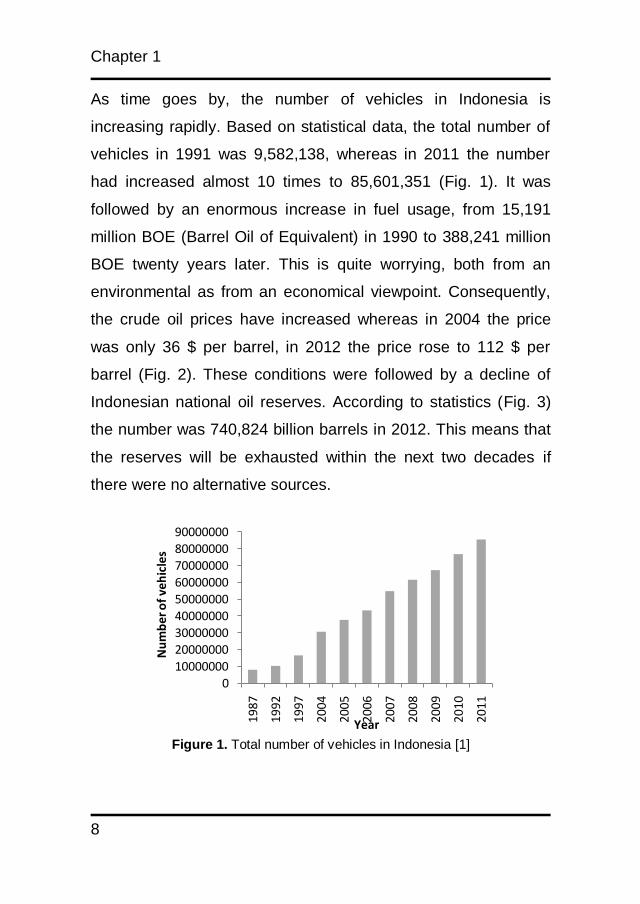
As time goes by, the number of vehicles in Indonesia is
increasing rapidly. Based on statistical data, the total number of
vehicles in 1991 was 9,582,138, whereas in 2011 the number
had increased almost 10 times to 85,601,351 (Fig. 1). It was
followed by an enormous increase in fuel usage, from 15,191
million BOE (Barrel Oil of Equivalent) in 1990 to 388,241 million
BOE twenty years later. This is quite worrying, both from an
environmental as from an economical viewpoint. Consequently,
the crude oil prices have increased whereas in 2004 the price
was only 36 $ per barrel, in 2012 the price rose to 112 $ per
barrel (Fig. 2). These conditions were followed by a decline of
Indonesian national oil reserves. According to statistics (Fig. 3)
the number was 740,824 billion barrels in 2012. This means that
the reserves will be exhausted within the next two decades if
there were no alternative sources.
Figure 1. Total number of vehicles in Indonesia [1]
0
10000000
20000000
30000000
40000000
50000000
60000000
70000000
80000000
90000000
1987
1992
1997
2004
2005
2006
2007
2008
2009
2010
2011
Nu
mb
er o
f ve
hic
les
Year

Introduction and scope of thesis
9
One hot issue that is connected with the increase of fuel usage is
global warming. It affects weather and climate. The mainland
and water temperature increase and they lead to the occurrence
of storms with great power and cause forest fires. This produces
thick smoke and has negative effects to the human respiratory
system. Other effects of global warming are the thawing of
glaciers at both poles, the rise of the sea level posing a potential
flooding in some areas, the disruption of the ecological balance
in the ocean and polar areas that might lead to the extinction of
animals and plants species. The most dangerous effect is
sociocultural when there is a war between human beings to fight
for certain region.
Figure 2. The price of crude oil in the world [2]
0
20
40
60
80
100
120
2004 2005 2006 2007 2008 2009 2010 2011 2012
Price ($)/barrel
Year
Chapter 1
10
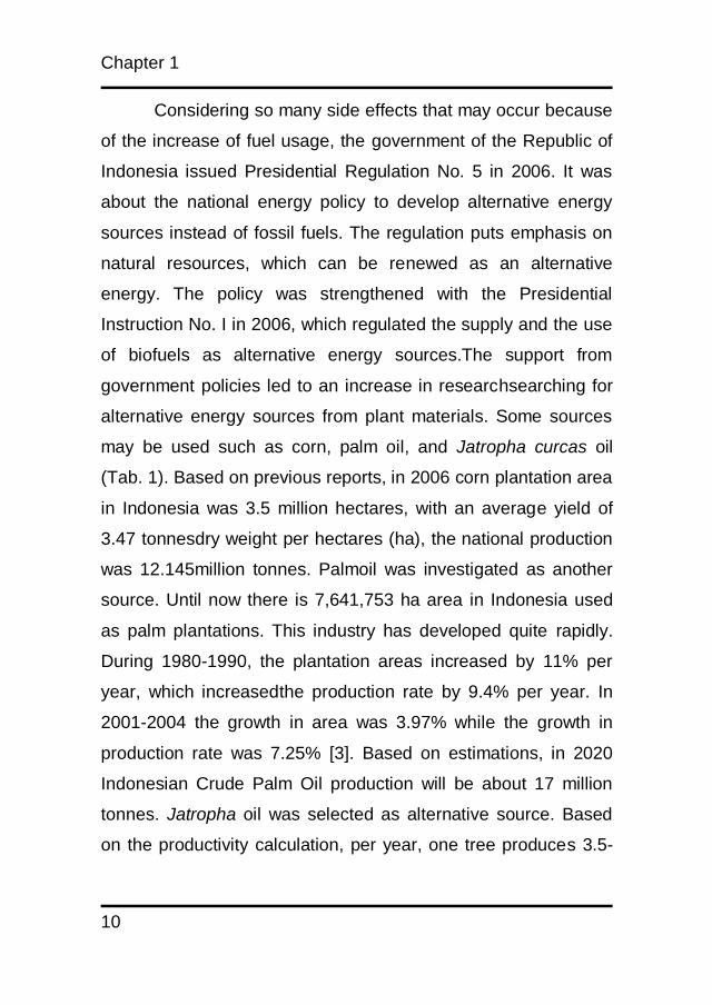
Considering so many side effects that may occur because
of the increase of fuel usage, the government of the Republic of
Indonesia issued Presidential Regulation No. 5 in 2006. It was
about the national energy policy to develop alternative energy
sources instead of fossil fuels. The regulation puts emphasis on
natural resources, which can be renewed as an alternative
energy. The policy was strengthened with the Presidential
Instruction No. I in 2006, which regulated the supply and the use
of biofuels as alternative energy sources.The support from
government policies led to an increase in researchsearching for
alternative energy sources from plant materials. Some sources
may be used such as corn, palm oil, and Jatropha curcas oil
(Tab. 1). Based on previous reports, in 2006 corn plantation area
in Indonesia was 3.5 million hectares, with an average yield of
3.47 tonnesdry weight per hectares (ha), the national production
was 12.145million tonnes. Palmoil was investigated as another
source. Until now there is 7,641,753 ha area in Indonesia used
as palm plantations. This industry has developed quite rapidly.
During 1980-1990, the plantation areas increased by 11% per
year, which increasedthe production rate by 9.4% per year. In
2001-2004 the growth in area was 3.97% while the growth in
production rate was 7.25% [3]. Based on estimations, in 2020
Indonesian Crude Palm Oil production will be about 17 million
tonnes. Jatropha oil was selected as alternative source. Based
on the productivity calculation, per year, one tree produces 3.5-
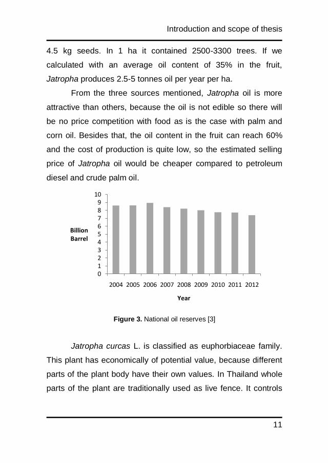
Introduction and scope of thesis
11
4.5 kg seeds. In 1 ha it contained 2500-3300 trees. If we
calculated with an average oil content of 35% in the fruit,
Jatropha produces 2.5-5 tonnes oil per year per ha.
From the three sources mentioned, Jatropha oil is more
attractive than others, because the oil is not edible so there will
be no price competition with food as is the case with palm and
corn oil. Besides that, the oil content in the fruit can reach 60%
and the cost of production is quite low, so the estimated selling
price of Jatropha oil would be cheaper compared to petroleum
diesel and crude palm oil.
Figure 3. National oil reserves [3]
Jatropha curcas L. is classified as euphorbiaceae family.
This plant has economically of potential value, because different
parts of the plant body have their own values. In Thailand whole
parts of the plant are traditionally used as live fence. It controls
0123456789
10
2004 2005 2006 2007 2008 2009 2010 2011 2012
Billion Barrel
Year

Chapter 1
12
soil erosion. J. curcas L.is also known as the source of biodiesel,
because from the seed, oil can be isolated by direct
compression. This oil is used as a biofuel, candle and soap
production, lighting and lubricant. In Europe the deoiled
seedcake is believed to be suitable as animal feedstock and
biofertilizer. In some rural areas in Indonesia the latex was
traditionally used for treating toothache.
Since J. curcas L. is considered as a future source of
biodiesel, many people in Indonesia plant it in a huge plantation.
they think if they can produce high amounts of oil from J. curcas
L., it will replace the petroleum usage. Although, J. curcas L.is
known to have many other usages, but the farmer did not realize
they thought this plant only produced oil without any beneficial
usage. So, they will be loss. This situation leads to a new
concept that J. curcas L. should not only be used as biodiesel
source, but it should give additional values to a farmer who
plants this crop.
Introduction and scope of thesis
13
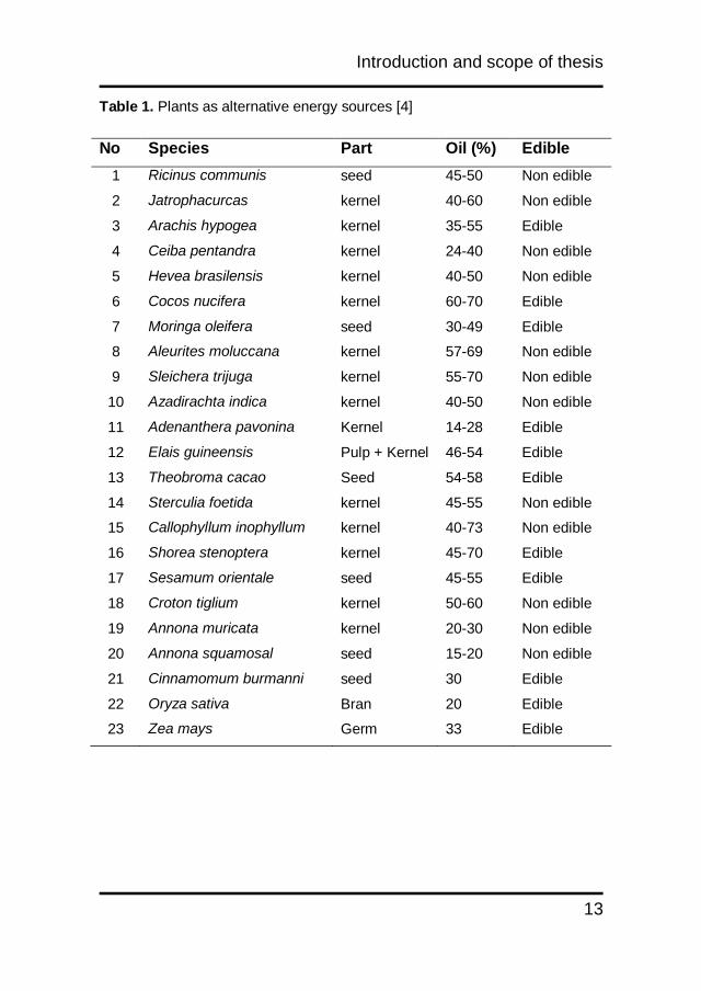
Table 1. Plants as alternative energy sources [4]
No Species Part Oil (%) Edible
1 Ricinus communis seed 45-50 Non edible
2 Jatrophacurcas kernel 40-60 Non edible
3 Arachis hypogea kernel 35-55 Edible
4 Ceiba pentandra kernel 24-40 Non edible
5 Hevea brasilensis kernel 40-50 Non edible
6 Cocos nucifera kernel 60-70 Edible
7 Moringa oleifera seed 30-49 Edible
8 Aleurites moluccana kernel 57-69 Non edible
9 Sleichera trijuga kernel 55-70 Non edible
10 Azadirachta indica kernel 40-50 Non edible
11 Adenanthera pavonina Kernel 14-28 Edible
12 Elais guineensis Pulp + Kernel 46-54 Edible
13 Theobroma cacao Seed 54-58 Edible
14 Sterculia foetida kernel 45-55 Non edible
15 Callophyllum inophyllum kernel 40-73 Non edible
16 Shorea stenoptera kernel 45-70 Edible
17 Sesamum orientale seed 45-55 Edible
18 Croton tiglium kernel 50-60 Non edible
19 Annona muricata kernel 20-30 Non edible
20 Annona squamosal seed 15-20 Non edible
21 Cinnamomum burmanni seed 30 Edible
22 Oryza sativa Bran 20 Edible
23 Zea mays Germ 33 Edible
Chapter 1
14
The aim of this thesis is to give an overview of the
additional values of Jatropha curcas L. plant by characterization
of its natural products that can be used as a safe pharmaceutical
product. In addition the detoxicification of the plantcake allowing
it to be used for animal stock has been researched. This thesis is
a part of larger project for valorization Jatropha curcas L.
plantation, especially in Indonesia.
Recent developments in the technology of detoxification
process and application of this ethnomedicinal plant to new fields
of experimental medicine are reviewed in chapter 2. In this
chapter recent data on biological activities, concepts and
strategies for turning a toxic plant into a valuable crop with high
pharmaceutical value are also discussed.
A group of toxic compounds, which are relevant to study
in J. curcas, are phorbol esters (PEs) since they are known as
tumour promoter. In analysing those phorbol esters,
phorbolmyristic acetate is used as a standard. This compound
has two isomers which are α and β. In chapter 3 the differences
between both isomers are discussed using LC-UV and LC-MS.
Selecting wrong standard can lead to quantification error of PEs.
From different parts of J. curcas some interesting
compounds were isolated. Some of them were investigated for
their biological activity, but others were only chemically analysed.
Curcacycline A and B were isolated from J. curcas latex. In
chapter 4 full synthetic approach, structure elucidation and
Introduction and scope of thesis
15
biological activities of both curcacyclines were described. Some
assays like antibiotic, cytotoxic, ecotoxic and mutagenic activities
were investigated to shed light on the pharmacological activity of
those compounds.
J. curcas seeds contain up to 60% of oil. The residues
from oil processing are called Jatropha kernel meal. This meal
can be used for animal feed. But the use is limited since it
contained toxic substances. Some studies have been done to
detoxify the meal and in this thesis the detoxified meal has been
analysed. In chapter 5, the validation of detoxification processes
are performed using cytotoxic and mutagenic assays. Colo 205
and OVCAR3 were used as cell lines in a cytotoxicity test while
Salmonella typhimuriumTA 98 and TA mix (TA 7001, TA 7002,
TA 7003, TA 7004, TA 7005, TA 7006) were used in a
mutagenicity assay.
Chapter 1
16
References
1. Indonesian Statistic Centre, 2013, Perkembangan Jumlah
Kendaraan Bermotor 1987-2012.
2. Indonesian Directorate of Energy, 2013, Statistik Minyak Bumi
3. Directorate General of cultivated plantation production, 2005,
Pokok-Pokok Rencana Makro Pengembangan Agribisnis
Komoditi Perkebunan 2005-2009
4. Soerawidjaja, T. H., 2006, Fondasi-Fondasi Ilmiah dan
Keteknikan dari Teknologi Pembuatan Biodiesel, Yogyakarta

CHAPTER 2
Rational use of Jatropha curcas L. in food and
medicine: from toxicity problems to safe
applications
Muhamad Insanu, Chryssa Dimaki, Richard Wilkins, John
Brooker, Piet van der Linde, Oliver Kayser
Published in : Phytochemistry reviews, 2013, 12 (1) : 107-119
Chapter 2
18
Abstract
Jatropha curcas L. has become an important plant for biorefinery
and production of biodiesel. From its ethnobotanical use, the
plant is known for several activities which are associated with
high toxicity. The latest development in engineering technology
enables detoxification of native oil and other parts of the plant for
new pharmaceutical purposes. Hence a revised look to the rich
metabolic spectra of partly structurally rare secondary compound
becomes an interesting field of research to be explored. In this
review, we discuss recent developments in the technology of
detoxification process and give insight about how this
ethnomedicinal plant can be applied to new fields of
experimental medicine. The review highlights recent data on
biological activities and discussed concepts and strategies for
turning a poison plant into a valuable crop with high
pharmaceutical potential.

Rational use of Jatropha curcas L.
19
2.1 Introduction
Jatropha curcas L. widely known as physic nut or purging nut, is
one of the oldest members of Euphorbiaceae. From the fossils
founded in Belem, Peru, the age of this plant is approximately 70
million years. The name was given by Karl von Linné in 1743
which means doctor (iatros) and food (trophe) [1]. The plant is
found in tropical regions of Africa, South America, South East
Asia and India [2]. Jatropha curcas L. is classified as a large
shrub or a small perennial tree because it can attain 5 m in
height, while under several conditions the height can reach 8 or
10 m [3]. It has soft wood with subtle grey bark and when it is
cut, it produces white and milky latex [4].
Jatropha curcas L.is a plant with multiple uses and
considerable economic potential. In the tropical countries, it
serves as a live fence in the fields and settlements and in arid
areas it is cultivated to control soil erosion. The deoiled
seedcake can be used for organic fertilizer without any
detectable phorbol ester both in the crops and soil [5].
Jatropha curcas L. has a potential for controlling
environmental pollution. Grounded seeds of J. curcas L. have
been demonstrated as an effective natural coagulant for
industrial effluent. Treatment of contaminated waters or soils is
an approach that gains popularity. Although the conventional
physical, chemical and thermal waste treatments are fast and
controllable, it requires high energy that renders them very costly.
Chapter 2
20
This plant is also known as source of biodiesel, the seed consist
of 60-68% of kernel which contain up to 60% oil depends on
geographical location (humidity, altitude, temperature, etc.). The
oil can be used directly or in methylester form as biodiesel [6-
9].The oil has been traditionally used for soap or candle
production, lighting and lubricant. It was observed that this fatty
acid composition of the oil was suitable for human nutrition. The
kernel also contains a high amount of crude protein (up to 32%),
which could be used as an animal feed [10].
Jatropha curcas L. is known as two genotypes, the toxic
and non-toxic. The difference between both types, is the
presence of phorbol esters in the seed. Non-toxic varieties from
Mexico contain very low to undetectable amounts of phorbol
ester while the other one contain up 3500 ppm. No differences
were found in the level of amino acids, trypsin inhibitor, lectin,
phytate,curcin and saponin between these two genotypes [7, 11-
13].
The objectives of this review are to make a validation of
the secondary natural products of Jatropha curcas L., to review
its toxic principles and the possibility of detoxification process for
safe use in animal feed and pharmaceutical purposes.

Rational use of Jatropha curcas L.
21
2.2 Chemical Composition
2.2.1 Compounds from the primary metabolism
Chemical analysis of Jatropha curcas L. revealed the presence
of primary metabolism in the seeds of the plant. Chemical
analysis of Jatropha curcas L. kernel and seed meal as well as
the fatty acid composition of the oil was reported [14] (Tab. 1 and
2). The presence of cis-11-eicosaenoic acid (20:1) and cis-
11,14-eicosadienoic acid (20:2) in Jatropha curcas L. seed oil
from four Mexican provenances was investigated. The same
study revealed that the content of starch and total soluble sugars
was below 6%, while the levels of essential amino acids except
for lysine were higher than those of FAO/WHO reference protein
for a five year old child on a dry matter basis [15].
Table 1. Average chemical composition of J. curcas L. kernel and meal
Constituent Kernel (%) Defatted Meal (%)
Dry matter 94.2-96.9 100
Crude protein 22.2-27.2 56.4-63.8
Lipid 56.8-58.4 1.0-1.5
Ash 3.6-4.3 9.6-10.4
Neutral detergent fibre 3.5-3.8 8.1-9.1
Acid detergent fibre 2.4-3.0 5.7-7.0
Acid detergent lignin 0.0-0.02 0.1-0.4
Gross Energy (MJ/Kg) 30.5-31.1 18.0-18.3
Chapter 2
22
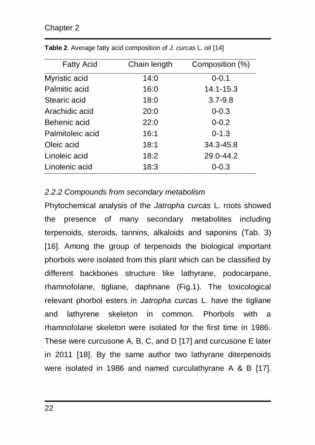
Table 2. Average fatty acid composition of J. curcas L. oil [14]
Fatty Acid Chain length Composition (%)
Myristic acid 14:0 0-0.1
Palmitic acid 16:0 14.1-15.3
Stearic acid 18:0 3.7-9.8
Arachidic acid 20:0 0-0.3
Behenic acid 22:0 0-0.2
Palmitoleic acid 16:1 0-1.3
Oleic acid 18:1 34.3-45.8
Linoleic acid 18:2 29.0-44.2
Linolenic acid 18:3 0-0.3
2.2.2 Compounds from secondary metabolism
Phytochemical analysis of the Jatropha curcas L. roots showed
the presence of many secondary metabolites including
terpenoids, steroids, tannins, alkaloids and saponins (Tab. 3)
[16]. Among the group of terpenoids the biological important
phorbols were isolated from this plant which can be classified by
different backbones structure like lathyrane, podocarpane,
rhamnofolane, tigliane, daphnane (Fig.1). The toxicological
relevant phorbol esters in Jatropha curcas L. have the tigliane
and lathyrene skeleton in common. Phorbols with a
rhamnofolane skeleton were isolated for the first time in 1986.
These were curcusone A, B, C, and D [17] and curcusone E later
in 2011 [18]. By the same author two lathyrane diterpenoids
were isolated in 1986 and named curculathyrane A & B [17].

Rational use of Jatropha curcas L.
23
Lathyrane Podocarpane Rhamnofolane Daphnane Tigliane
From aerial parts and stem of Jatropha curcas L., three lathyrane
types ((4E)-15-O-Acetyl-15-epijatrogrossidentadione, (14E)-14-
O-Acetyl-5,6-epoxygrossien-tadione, (4E)-15-epijatrogros-
sidenta-dione)), two podocarpane types (3β-acetoxy-12-
methoxy-13-methylpodo-carpane-8,11,13-trien-7-one, 3β,12-di-
hydroxy-13-methylpodocarpane 8,10,13-triene), one dinorditer-
penetype (heudelotinone) and three deoxy-preusomerins
(Palmarumycin CP1, JC1, and JC2) were isolated [19, 20]. The
presence of alkaloids in Jatropha curcas L. was marked by 5-
OH-pyrrolidin-2-one and pyrimidin-2,4-dione [21], while the
presence of coumarins was marked by marmesin, tomentin,
propacin and jatrophin [19, 22]. Flavonoids like apigenin, vitexin
and isovitexin were also found in leaves [21, 23]. Two
cyclopeptides were isolated from latex. Curcacycline A possess
eight amino acids in the order c[Gly-Leu-Leu-Gly-Thr-Val-Leu-
Leu] and curcacycline B possess nine amino acids which were
c[Gly-Ile-Leu-Gly-Ser-Pro-Ile-leu-Leu]. Curcacycline B can bind
to human cyclophilin B and increase by 60% its peptidyl-prolil cis
trans isomerase (PPI-ase) activity at 30µM [24, 25].
Figure 1. Basic structures of Jatropha curcas L. diterpenoid skeletons

Chapter 2
24
2.3 Human toxicity and case reports
Acute poisoning with seeds of Jatropha curcas L. was reported.
Abdu reported intoxication in two children aged three and five
[26]. In 2005, twenty children were admitted to hospital in India
because of Jatropha curcas L. seed ingesting. The age varied
between 8-13 years. All cases showed complain of vomiting,
diarrhea, abdominal pain, sensation in the throat. Vomiting was
the predominant symptom (95%), diarrhea (50%), headache
(40%), asymptomatic (5%). Intravenous fluid and antiemetic
were given to the children, the recovery rate was six hours and
after 24 hours they were discharged from medical services [27].
Table 3. Phytochemical compounds present in J. curcas L.
No Chemical compounds Type Sources Ref.
1. 5-OH-pyrrolidin-2-one Alkaloid Leaf [21]
2. Pyrimidine-2,4-dione Alkaloid Leaf [21]
3. 2-methylanthraquinone Antraquinone Aerial part [19]
4. Marmesin Coumarin Root [22]
5. Tomentin Coumarin Root [20]
6. Propacin Coumarino-Lignan Root [22]
7. Jatrophin Coumarino-lignane Root [22]
8. Curcacyline-A Cyclic peptide Latex [24]
9. Curcacycline-B Cyclic peptide Latex [25]
10. (4E)-15-O-Acetyl-15-epi-
jatrogrossidentadione
Diterpenes Aerial part [19]
11. (14E)-14-O-Acetyl-5,6- Diterpenes Aerial part [19]
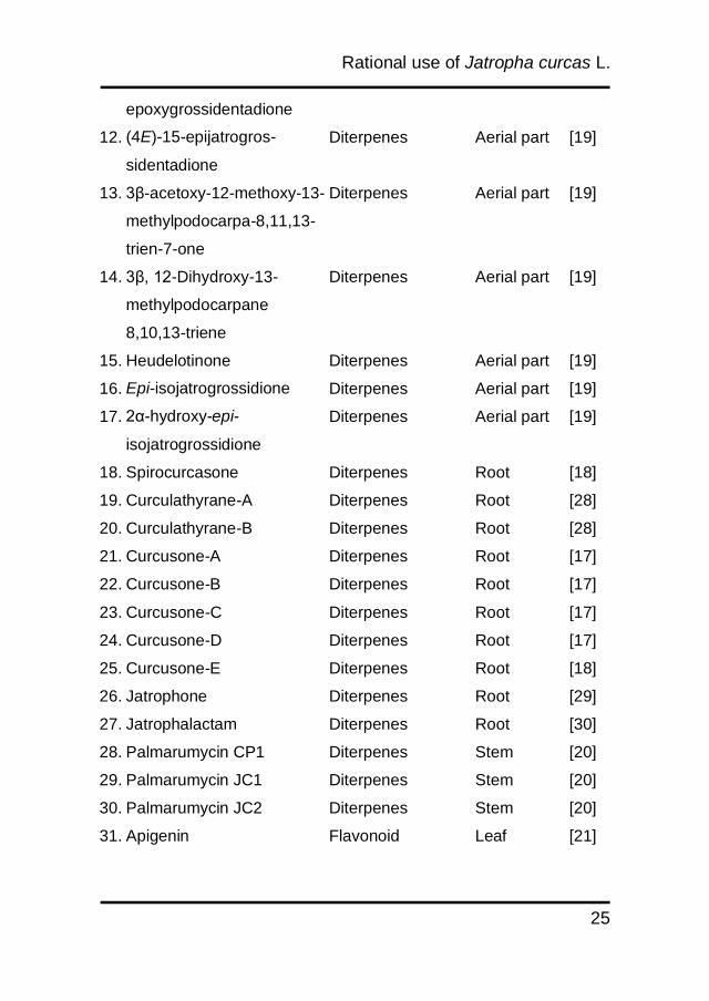
Rational use of Jatropha curcas L.
25
epoxygrossidentadione
12. (4E)-15-epijatrogros-
sidentadione
Diterpenes Aerial part [19]
13. 3β-acetoxy-12-methoxy-13-
methylpodocarpa-8,11,13-
trien-7-one
Diterpenes Aerial part [19]
14. 3β, 12-Dihydroxy-13-
methylpodocarpane
8,10,13-triene
Diterpenes Aerial part [19]
15. Heudelotinone Diterpenes Aerial part [19]
16. Epi-isojatrogrossidione Diterpenes Aerial part [19]
17. 2α-hydroxy-epi-
isojatrogrossidione
Diterpenes Aerial part [19]
18. Spirocurcasone Diterpenes Root [18]
19. Curculathyrane-A Diterpenes Root [28]
20. Curculathyrane-B Diterpenes Root [28]
21. Curcusone-A Diterpenes Root [17]
22. Curcusone-B Diterpenes Root [17]
23. Curcusone-C Diterpenes Root [17]
24. Curcusone-D Diterpenes Root [17]
25. Curcusone-E Diterpenes Root [18]
26. Jatrophone Diterpenes Root [29]
27. Jatrophalactam Diterpenes Root [30]
28. Palmarumycin CP1 Diterpenes Stem [20]
29. Palmarumycin JC1 Diterpenes Stem [20]
30. Palmarumycin JC2 Diterpenes Stem [20]
31. Apigenin Flavonoid Leaf [21]
Chapter 2
26
2.4 Toxic principles of Jatropha curcas L.
Jatropha curcas L. toxicity is mainly characterized by the
presence of phorbol ester and ribosome inactivating proteins
(RIP). From the last group of compound one of the representing
is curcin. It can be classified as type I ribosome inactivating
protein. The mechanism of action is due to depurination of the α-
32. Vitexin Flavonoid Leaf [23]
33. Isovitexin Flavonoid Leaf [23]
34. Curcin Lectin Seed [31]
35. Tetradecyl-E-ferulate Lignane Aerial part [19]
36. 12-deoxy-16-hydroxy
phorbol-C13-C16 diesters
Phorbol ester Seed [9,
32]
37. Factor 1 Phorbol ester Seed [9]
38. Factor 2 Phorbol ester Seed [9]
39. Factor 3 Phorbol ester Seed [9]
40. Factor 4 Phorbol ester Seed [9]
41. Factor 5 Phorbol ester Seed [9]
42. Factor 6 Phorbol ester Seed [9]
43. -sitosterol Phytosterol Phytosterol [33]
44. Curcain Protease Protease [34]
45. Stigmasterol Triterpenes Leaf [33]
46. 3-O-(Z)-coumaroyl oleanolic
acid
Triterpenes Aerial part [19]
47. Acetoxyjatropholone Diterpenes Root [18]
48. Multidione Diterpenes Root [35]
Rational use of Jatropha curcas L.
27
sarcin loop of large rRNA. Curcin can demolish N-glycosidic
linkage between polyphosphate backbone of the 28S rRNA and
adenine at A4324 (alpha sarcin loop) of the rat liver ribosome. This
will block protein translation. Curcin (28.2 kD) consist of 251
amino acids with the composition:Asx(31), Val(26), Leu(24),
Glx(22), Ala(22), Lys(18), Ser(16), Thr(15), Gly(15), Ile(14),
Tyr(14), Phe(12), Pro(9), Met(2), Arg(7), His(2), Cys(1) and
Trp(1)[36]. Its biosynthesis is induced in leaves under stress
conditions like drought, temperature and fungal infection, serving
thus plant defense purposes [37]. It was stored in endosperm
and tegmen of Madagascar and Mexican varieties [13]. Previous
result reported that the curcin gene coding region has similar
amino acid sequences with gelonin, bryodin, trichosantin,
momorcharin, ricin A-chain and abrin A-chain [38]. The gene of
the protein was inserted into the PQE30 vector. After introduction
into E. coli this protein was successfully expressed in strain M15.
Although the yield in this observation was low, the result showed
that 0.5 mM IPTG was suggested as an optimum inducer [39].
It was reported that curcin strongly inhibit protein
synthesis in reticulosyte lysate, gastric cancer cell line (SGC-
7901), mouse myeloma cell line (sp 2/0), and human hepatoma
with IC50 (95% confidence limit) of 0.19 (0.11-0.27) nmol/L, 0.23
(0.15-0.32) mg/L, 0.66 (0.35-0.97) mg/L, 3.16 (2.74-3.58) mg/L,
respectively [40]. Curcin showed no activity on Hela and MRC
cells (human embryo lung diploid cell line).
Chapter 2
28
In vivo application of curcin was done in mice. After 12
hours of curcin administration, some symptoms appeared in the
animal, such as hypersensitivity, declining pineal and corneal
reflexes, locomotorious activity, losing of grip strength and
righting reflex, defecations and palpebral closure. Autopsy on
dead animal showed hyperemia of the intestine and wounds in
the spleen, pancreas and liver. Death occurred after 48 hours of
curcin administration. LD50 of curcin was 9.11 IU intraperitoneal
[31].
In nature we find four types of diterpenoid esters. The
basic structure of the compounds is a tigliane, daphnane,
ingenane, and lathyrane skeleton. The classification was based
on their basic pattern containing tri and tetracyclic ring systems
[41]. Tigliane is the basic skeleton of phorbol ester (PE).
Hydroxylation is found in the position C12 and C13 of tigliane
backbone. Esterification with various fatty acids gives a broad
spectrum of phorbol ester in this plant [42].The target of PE are
phospolipid membrane receptors. They activate protein kinase C
(PKC), that is important for signal transduction leading to cell
differentiation and cell growth regulation [43, 44]. Under normal
physiological conditions, diacylglycerol (DAG) activates PKC,
and enhances PKC‟s affinity to bind phosphatidyl serine(PS)-
containing membranes. Whereas DAG is easily metabolized, PE
is not and, therefore, PE acts as an agonist of DAG and
uncontrollably activates the PKC with the consequence of
Rational use of Jatropha curcas L.
29
increasing cell proliferation. The toxic principle is that PKC
activity can hardly be turned down and the system is out of
regulatory control [45]. Carcinogenesis experiments on mouse
skin revealed that phorbol esters stimulate tumour growth but do
not induce tumours. Phorbol esters thus acts as cocarcinogens.
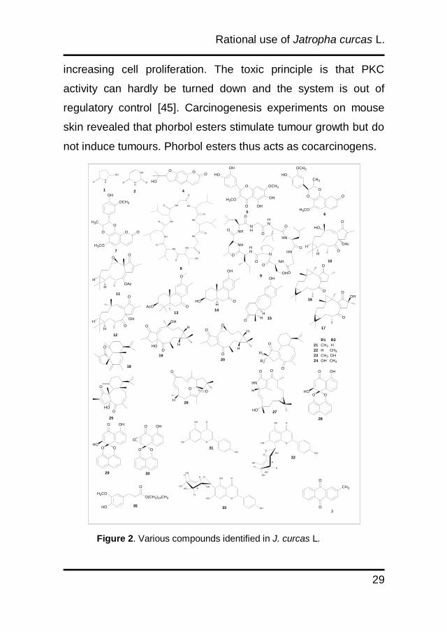
Figure 2. Various compounds identified in J. curcas L.
NHO
OH
NH
NH
OO
NH
O
HN
O
HN
O
HN
HN
O
OHO
NH
O
NH
O NH
O
O
O
OH
HO
OH
O
O
OH
HO
OH
HO
OH
H
HO
H
OHH
HO
O
O
OH
HO
OH
OH
O
HH
HO
HOH
HHO
HO
O OO
O
OH
H3CO
O OH
OH
OCH3
HO
H3CO
O
O
O
CH3
HO
OCH3
O
O
O
OH
OCH3
O
H3CO
H3C O
OH
H
HO
OAc
NHO
NH
O
OO
NH
OHO
HNO
HN
O
O
NH
HN
O
HN
NO
OAcH
H
O
O
OH
HOH
O
AcO O
O
OH
HOH
O
OH
HH
O
O O
O
OH
O
O
OHO H
H
OH
O
O
H
H
O
R1
R2
O
O
O
O
HO
OO
O
H
OO
H
HO
HN
OH
O O
O
O
O O
O OH
HO
O O
O OH
HO
H3CO
HO
O(CH2)13CH3
O
1 2 4
56
7
8
10
9
11
12
1314
15
16
17
18
1920
R1 R2
21 CH3 H
22 H CH3
23 CH3 OH
24 OH CH3
25
26
27
28
3029
31
33
32
35
O
O
CH3
3

Chapter 2
30
OH
H
H
H
O
H
OH
HOOH
H
OH
HOH
O
H
OH
HOOH
H
O
H
O
O
O
H
O
H
O
O
H
OH
HOOH
O
O
O
H
OH
HOOH
H
O
H
O
O
O
O
O
O
O
H
OH
HOOH
H
O
H
O
H
OH
HOOH
H
O
H
O
O
O
HO
O
O
OH
O
H
HO
O
OH
H COOH
1
2
34
5 6
7
8
9
10
11
12
13
14
15
16
17
18
36
3738
40,41
39
42
45
48
47
46
H
HO
H H
H
43
Figure 2. Continued
2.5 Detoxification Process
2.5.1 Detoxification of biomass
Many attempts have been performed to eliminate antinutritional
components and toxic principles (trypsin inhibitor, lectins, phytate,
phorbol esters) from the meal. Mexican people roasted the meal
before it was eaten. Roasting the meal could only effect trypsin
inhibitor and lectin activity [11], but the other components were
Rational use of Jatropha curcas L.
31
not affected by heat treatment [10]. The moist heat was more
effective in decreasing lectin activity than dry heat[46]. It was
observed that trypsin inhibitors and lectin were fully inactivated
by double solvent extraction using hexane and ethanol coupled
with moist heat treatment (20% moisture, 126oC, 2 bar, 10 min)
[47]. Double solvent extraction (petroleum ether and ethanol)
mixed with chemical treatment using 0.07% NaHCO3 eliminate
95.8% phorbol ester content. This treatment is accepted as the
best method to reduce lectin activity [15]. The article described a
complex detoxification strategy with protein extraction at basic
pH followed by isoelectric precipitation and finally steam injection
with different steps. By this procedure the level of trypsin
inhibitors, phytate, tannins and saponin has reduced by more
than 90% while toxic phorbol esters were not detected anymore
in the meal [48].
Beside elimination of phorbol ester using physical and
chemical methods, the influence of manganese chloride (MnCl2)
and N-ethylmaleimid in reducing phorbol ester biosynthesis in
callus cultures were observed. Two concentrations of MnCl2
were used (2 mM and 3 mM) and the content of phorbol ester in
callus cultures was reduced to 30.5% and 30.6% respectively
after 7 days. When N-ethylmaleimid was given to the callus in
three different concentrations (0.6, 0.9, and 1.2 mM) the content
of phorbol ester reduced to 26.6%, 6.2% and 32.2% respectively
after 21 days [49].
Chapter 2
32
2.5.2 Detoxification process for animal feed
Different animals show different physiological reactions to
detoxified Jatropha curcas L. meal. For example, increasing time
of heat treatment of Jatropha meal impacted the growth rate of
fishes. Heat treatment provokes the loss of amino acid and
structural changes in Jatropha curcas L. proteins. These
changes made them difficult to be digested by fish trypsin,
leading to low protein efficiency ratio and protein productive
value [50]. It was reported that pigs which consumed treated
Jatropha curcas L. meal, showed adverse effects with low level
of percent packed cell volume, serum glucose, cholesterol
concentration, serum alpha amylase activity (p<0.001) and
serum triglyceride.
A method that comprises three major steps was patented.
The first step was adding methanol and sodium hydroxide to
form a mixture. Second step was heating the mixture, and final
stap was separating it to obtain detoxified constituents. Analytical
profiling of these materials using HPLC showed that PE was
below the detection limit. Total crude protein content, available
lysine value and protein digestibility were the same for both
treated and untreated meal. Biological evaluation showed that
these products were not harmful to mollusk and carps (Cyprinus
caprio) [51].
Single step extraction with anazeotrop mixture of ethyl
acetate and methanol at 62oC and 1.2 bar with mixing rate 10
Rational use of Jatropha curcas L.
33
rpm, 6 cycles of 1 hour each was developed and patented.
Desolventising was at 100oC for 80 min in a vertical steam
desolventiser. For removing antinutrient factors, the meal was
autoclaved with 120oC moist heat for 60 min. The recovery of oil
was more than 50% of kernel weight while the content of the oil
in the meal was less than 0.5% by weight. No differences in
protein content and no residual PE was detected in the meal.
Bioassays were carried out to determine remaining toxicity
processed meal using Brine shrimps and Drosophila larvae
assays. Based on these experiments, the processed meal
showed no significant toxic effect on the larvae while 100%
mortality was shown in control group with unprocessed meal [52].
The newest patent for removing phorbol ester from
organic material was applied using microbial approach. Bacillus
subtilis var natto (0.004-0.2 part by mass) with stirring and
fermentation processes (37-50oC for two to four weeks) were
selected for decomposing phorbol ester. Fermented Jatropha
curcas seedcake showed better weight gain, feed intake and
health condition than unfermented seedcake in mice. All of
values were almost the same in fermented and soya bean
groups [53].
Detoxification of Jatropha curcas L. meal was done using
hydrolysis by enzymes (cellulase and pectinase) continued with
ethanol washing was investigated. These treatments decreased
the level of phorbol ester, trypsin inhibitor, lectin activity, tannin
Chapter 2
34
and saponin to tolerable values. There were enhancement in
crude protein and in vitro protein digestibility values from 60% to
75% and 82%to 92% respectively [54].
2.6 Biological Activities of Jatropha curcas L.
2.6.1 Antinutrient compounds in Jatropha curcas L.
The Jatropha curcas L. plant synthesizes some compounds that
act as anti-nutrients or toxins in living organisms. Antinutrients
have been characterized as substances that interfere with food
utilization of animals. These phytochemicals can be generally
divided into four groups: i. factors affecting protein digestion such
as trypsin inhibitors, tannins, lectins; ii. factors affecting mineral
utilization, which include phytates, gossypol pigments, oxalates,
glucosinolates; iii. Antivitamins; iv. miscellaneous substances like
mycotoxins, cyanogens, nitrate, alkaloids, phyto-estrogens and
saponins [55]. High level of trypsin inhibitors content caused low
in vitro rumen degradable proteins [10].The effect of trypsin
inhibitor was decreasing protein digestibility, due to the
interference with pancreatic proteolytic enzymes [56]. Phytate
decreased bioavailability of protein and minerals (Ca2+,Mg2+,
Cu3+, Fe3+)by forming complexes with them [57, 58].
Rational use of Jatropha curcas L.
35
2.6.2 Antimicrobial and antiparasitic
The latex of Jatropha curcas L. has strong antimicrobial activity
when it was applied to both gram positive and negative bacteria
[59].
Not only toxic to bacteria, Jatropha curcas L. latex was
also toxic to parasites. No embryonation was found in ova of
Ascaris lumbricoides and Necator americanus incubated in pure
and 1:1 diluted latex. Mosquito eggs could not hatch in the latex
of Jatropha curcas L. [59]. The methanol extract of the oil was
also tested at two larvae stages (cercariae and miracidia) of
Schistosoma mansoni. The extract showed toxicity to both of the
larvae, but cercariae was more sensitive to the extract than
miracidia. Decreasing the growth of Schistosoma sp. became
important, since these species cause schistosomiasis which is
one of the most serious parasitic diseases after malaria [60].
2.6.3 Antiviral activity
Water extract of stem bark of J. curcas L. showed good
suppressive effects on HIV growth. The effect was determined
by HIV-1 induced cytopathic effect on MT-4 cells, HIV-1 reversed
transcriptase (RT) assay and HIV protease assay (PR) to identify
the mode of action on inhibiting HIV growth while no inhibitory
effects were found in both HIV-RT and HIV-PR assays. It was
concluded that the results were significant and the activity had a

Chapter 2
36
high selective index, although the mode of action is still unknown
[61].
2.6.4 Insecticidal activity
Insecticidal activities of oil containing phorbol ester or
concentrated phorbol ester fractions have been recorded for a
large number of different insects like Mandura sexta, Helicoverpa
armigera, Aphis gossypii, Pectinophora gossypiella, Empoasca
biguttula, Callosobruchus chinensis, Sitophillus zeamays,
Phtorimaea opercullewla, Sesamia calamitis, Busseola fusca,
Periplaneta americana, Blatella germanica, Oncopeltus fasciatus,
Phaedon cochliariae, Platella xylostella, spodoptera frugiperda,
Mycus persicae, Callosobruchus maculatus and Dinarmus
basalis [62, 63]. Jatropherol I, a phorbol ester diterpene,
influenced some midgut enzymes of Bombyx mori.This
influenced general esterase, carboxylesterase and
acetylcholinesterase. Jatropherol It also caused pathological
changes in endoplasmatic reticulum, chromatin, lysosome,
mitochondria, and microvilli. LD50 values of these ester were
0.579 g/mL, 0.217 g/mL, 0.158 g/mL after 48, 72 and 120 h
administration, respectively [64].
2.6.5 Antisnail activity
Controlling snails is important because they act as the vector
host of parasites that caused schistosomiasis (see above).
Rational use of Jatropha curcas L.
37
Seeds of Jatropha curcas L. were tested against Biomphalaria
glabrata, Bulinus truncatus, Bulinus natalensis, Oncomelania
quadrasi and Oncomelania hupensis. Methanolic extract of
crude oil was reported to have high toxicity against all snails [60,
65].
2.7 Biosafety Studies
2.7.1 Acute toxicity in mice
Toxicological studies of Jatropha curcas L. seed have been
conducted in mice. Adam reported that 1% seed material in diet
did not cause mortality, but 5-50% of seed material was lethal in
mice. During the experiment, mice suffered from diarrhea, they
could not keep normal posture, lost their appetite (related to the
taste of the food), decreased motor activity, showed depression
and had increased respiration rate. The highest dose caused
100% mortality four days after feeding. Organs of death animals
were examined and pathological changes were found such as
catarrhal enteritis, erosion and widespread infarct of intestinal
mucosa. Hemorrhage and congestion were detected in small
intestine, heart and lungs. Other side effects were the increased
hepatic and renal fat accumulation. There were blood clots in
fecal material, but they were not found in peritoneal cavity.
Hemorrhage and dilation of ascending colon were also detected
in almost all groups [26, 66]. Crude oil and its fraction when
Chapter 2
38
applied as topical application were toxic to the dorsal skin of the
mice. Oil fraction caused eye hemorrhage, face swelling, and
skin irritation while only small effect of damage caused by crude
oil [6]. Acute toxicity of methanol extracts of Jatropha curcas L.
seed was determined in mice intraperitoneally. The LD50 was
25.19 mg/kg body mass [67]. Another experiment of acute
toxicity to male mice was carried out [68]. Phorbol esters were
isolated from the oil and administered intragastricly to the
animals. LD50 was determined at 27.34 mg/kg body weight and
the highest dose (36 mg/kg body weight) caused abnormality of
cortical neurons and cardiac muscle fibers. Glomerular sclerosis,
atrophy in kidney and hemorrhage in the lung was shown in
mice given a dose higher than 32.4 mg/kg body mass, while no
changes were found in the animal organ at the lowest dose
(21.26 mg/kg body mass) [68].
2.7.2 Acute Toxicity in rat
Acute toxicity of Jatropha oil was also observed in rats. Based on
the result, the LD50 was 6 mL/kg body weight.At higher doses (9
and 15 mL/kg body mass) all of the rats died during the
experiment. In topical application, oil fraction caused skin
irritation and edema [6].Various extracts from the fruit of the
Jatropha curcas L. also had an abortive effect to rats. They
influenced the early stage of pregnancy [69]. Methanol extract of
the seeds caused low hemoglobin concentration, low packed cell
Rational use of Jatropha curcas L.
39
volumes and also low red blood cell counts, but mean
corpuscular volume & mean corpuscular hemoglobin values
were high. These results indicated macrocytic anemia [67].
2.7.3 Acute toxicity in goat
Jatropha curcas L. seeds were also administered to goats. The
doses ranged between 0.25 to 10 g/kg/day. During dosing period,
some symptoms occurred in the goat like diarrhea and loosing
ability to keep normal posture. Autopsy showed hemorrhage in
the kidneys, lungs, reticulum, spleen and rumen. Necrosis and
degeneration of the liver were marked by the elevation of
glutamate oxalacetate transaminase and arginase activity, and
depletion of glucose level in the goat serum [70]. The same
symptoms were also found when the extract was given to a
goatling, Abdel Gadir reported that the mortality of them occurred
between day 7 and 21 after extract administration [71].
2.8 Conclusion and Perspective
Jatropha curcas L. has a great potential and value for cultivation
as economic crop for biodiesel production. Not only because of
the source of non-edible renewable biodiesel, but also containing
secondary metabolites with interesting biological activities. As
discussed, these secondary natural compounds were isolated
and elucidated from different parts of Jatropha curcas L. Some
experiments have been conducted in the past for validating
Chapter 2
40
pharmacological uses. Curcin, a ribosome inactivating protein is
a potential lead compound as anticancer and immune-
suppressive drug. Curcusone B isolated from the root also has a
good prospect for antiproliferative effect since it inhibits the
growth of cancer cell lines. Jatropherol I has insecticidal activity
because it changes pathological condition in endoplasmic
reticulum, chromatin, lysosome, mitchondria and microvilli of
insects. Curcacycline A shows moderate dose dependent
inhibition on human T-cell proliferation while curcacycline B can
bind to human cyclophilin B and increase its Peptidyl-prolil cis
trans isomerase.
Phorbol ester has become a major issue regarding
Jatropha curcas L. oil and toxicity. Since it is dissolved well in
the oil, intoxication risk for workers in the oil producing industry is
high, here more research is necessary to avoid the chronic harm
of the risk. It was reported that multiple steps in oil refining
process like degumming, deodorization, neutralization processes
(with alkali hydroxides), bleaching and stripping process reduce
the level of phorbol ester significantly. Detoxified oil can be used
as a candidate of new basis for ointment and dermal application
preparations. Further investigation is still needed for oil safety,
because direct contact of remaining phorbol ester with skin have
to be avoided in all cases. Studies about indoor pollution by
Jatropha curcas L. oil should be conducted, in order to give clear

Rational use of Jatropha curcas L.
41
explanation between combustion products from the oil and
related toxicity.
Jatropha curcas L. pressed cake contains a high protein
yield. It can be used as animal feed, but the utilization is limited
due to its toxic principles. Development strategy in detoxification
process of the pressed cake has become high of interest.
Physical and chemical treatments have been investigated in
toxic removal. For safety reason, biosafety assays have been
observed for this detoxified product to various in vitro and in vivo
studies. These results show that detoxification of pressed cake is
possible and safe for further use.
Recent progress on Jatropha curcas L. studies indicates
that all parts of this plant are valuable. Utilization of the plant
could improve valorization for huge plantation.
Chapter 2
42
References
1. Becker K, Makkar HPS. Jatropha curcas : A potential source
for tomorrow's oil and biodiesel. Lipid Tech. 2008;20(5):104-7.
2. Schmook B, Seralta-Peraza L. J. curcas : distribution and
uses in the Yucatan Peninsula of Mexico. Gübitz G, Trabi M,
Mittelbach M, editors1997.
3. Divakara BN, Upadhyaya HD, Wani SP, Gowda CLL. Biology
and genetic improvement of Jatropha curcas L.: A review. Appl
Energ. 2010;87(3):732-42.
4. Horiuchi T, Fujiki H, Hirota M, Suttajit M, Suganuma M,
Yoshioka A, et al. Presence of tumor promoters in the seed oil of
Jatropha curcas L. from Thailand. Jpn J Cancer Res.
1987;78(3):223-6. Epub 1987/03/01.
5. Srinophakun P, Titapiwatanakun B, Sooksathan I, Punsuvon
V. Prospect of deoiled Jatropha curcas seedcake as fertilizer for
vegetables crops- a case study. J Agr Sci. 2012;4(3):211-26.
6. Gandhi VM, Cherian KM, Mulky MJ. Toxicological studies on
ratanjyot oil. Food Chem Toxicol. 1995;33(1):39-42.
Rational use of Jatropha curcas L.
43
7. Makkar HPS, Becker K, Sporer F, Wink M. Studies on
Nutritive Potential and Toxic Constituents of Different
Provenances of Jatropha curcas. J Agr Food Chem.
1997;45(8):3152-7.
8. Asseleih LMC, Plumbley RA, Hylands PJ. Purification and
partial characterization of a hemagglutinin from seeds of
Jatropha Curcas. J Food Biochem. 1989;13(1):1-20.
9. Haas W, Sterk H, Mittelbach M. Novel 12-deoxy-16-
hydroxyphorbol diesters isolated from the seed oil of Jatropha
curcas. J Nat Prod. 2002;65(10):1434-40. Epub 2002/10/26.
10. Aderibigbe AO, Johnson COLE, Makkar HPS, Becker K,
Foidl N. Chemical composition and effect of heat on organic
matter- and nitrogen-degradability and some antinutritional
components of Jatropha meal. Anim Feed Sci Tech. 1997;67(2-
3):223-43.
11. Makkar HPS, Becker K, Schmook B. Edible provenances of
Jatropha curcas from Quintana Roo state of Mexico and effect of
roasting on antinutrient and toxic factors in seeds. Plant Food
Hum Nutr. 1998;52(1):31-6.
Chapter 2
44
12. Makkar HPS, Aderibigbe AO, Becker K. Comparative
evaluation of non-toxic and toxic varieties of Jatropha curcas for
chemical composition, digestibility, protein degradability and
toxic factors. Food Chem. 1998;62(2):207-15.
13. He W, King AJ, Khan MA, Cuevas JsA, Ramiaramanana Dl,
Graham IA. Analysis of seed phorbol-ester and curcin content
together with genetic diversity in multiple provenances of
Jatropha curcas L. from Madagascar and Mexico. Plant Physiol
Bioch. 2011;49(10):1183-90.
14. Gubitz GM, Mittelbach M, Trabi M. Exploitation of the tropical
oil seed plant Jatropha curcas L. Bioresource Technol.
1999;67(1):73-82.
15. Martínez-Herrera J, Siddhuraju P, Francis G, Dávila-Ortíz G,
Becker K. Chemical composition, toxic/antimetabolic
constituents, and effects of different treatments on their levels, in
four provenances of Jatropha curcas L. from Mexico. Food
Chem. 2006;96(1):80-9.
16. Aiyelaagbe OO, Adeniyi BA, Fatunsin OF, Arimah BD. In
vitro antimicrobial activity and phytochemical analysis of
Jatropha curcas roots. Int J Pharm. 2007;3(1):106-10.
Rational use of Jatropha curcas L.
45
17. Naengchomnong W, Thebtaranonth Y, Wiriyachitra P,
Okamoto KT, Clardy J. Isolation and structure determination of
four novel diterpenes from Jatropha curcas. Tetrahedron Lett.
1986;27(22):2439-42.
18. Chianese G, Fattorusso E, Aiyelaagbe OO, Luciano P,
SchroÌder HC, MuÌller WEG, et al. Spirocurcasone, a diterpenoid
with a novel carbon skeleton from Jatropha curcas. Org Lett.
2011;13(2):316-9.
19. Ravindranath N, Ravinder Reddy M, Ramesh C, Ramu R,
Prabhakar A, Jagadeesh B, et al. New lathyrane and
podocarpane diterpenoids from Jatropha curcas. Chem Pharm
Bull 2004;52(5):608-11. Epub 2004/05/11.
20. Ravindranath N, Reddy MR, Mahender G, Ramu R, Kumar
KR, Das B. Deoxypreussomerins from Jatropha curcas: are they
also plant metabolites? Phytochemistry. 2004;65(16):2387-90.
Epub 2004/09/24.
21. Staubmann R, Schubert-Zsilavecz M, Hiermann A, Kartnig T.
A complex of 5-hydroxypyrrolidin-2-one and pyrimidine-2,4-dione
isolated from Jatropha curcas. Phytochemistry. 1998;50(2):337-
8.
Chapter 2
46
22. Naengchomnong W, Tarnchompoo B, Thebtaranonth Y. (+)-
jatrophol, (+)-marmesin, propacin and jatrophin from the roots of
Jatropha curcas (Euphorbiaceae). J Sci Soc. 1994;20(2):73-83.
23. Subramanian SS, Nagarajan S, Sulochana N. Flavonoids of
some Euphorbiaceous plants. Phytochemistry 1971;10(10):2548-
9.
24. Van den Berg AJ, Horsten SF, Kettenes-van den Bosch JJ,
Kroes BH, Beukelman CJ, Leeflang BR, et al. Curcacycline A-a
novel cyclic octapeptide isolated from the latex of Jatropha
curcas L. FEBS Lett. 1995;358(3):215-8. Epub 1995/01/30.
25. Auvin C, Baraguey C, Blond A, Lezenven F, Pousset J-L,
Bodo B. Curcacycline B, a cyclic nonapeptide from Jatropha
curcas enhancing rotamase activity of cyclophilin. Tetrahedron
Lett. 1997;38(16):2845-8.
26. Abdu-Aguye I, Sannusi A, Alafiya-Tayo RA, Bhusnurmath
SR. Acute toxicity studies with Jatropha curcas L. Hum Exp
Toxicol. 1986;5(4):269-74.
27. Kulkarni ML, H. Sreekar, K.S. Keshavamurthy, Shenoy N.
Jatropha curcas - Poisoning. Indian J Pediatr. 2005;72(1):75-6.
Rational use of Jatropha curcas L.
47
28. Naengchomnong W, Thebtaranonth Y, Wiriyachitra P,
Okamoto KT, Clardy J. Isolation and structure determination of
two novel lathyrenes from Jatropha curcas. Tetrahedron Lett.
1986;27(47):5675-8.
29. Pletsch M, Charlwood BV. Accumulation of diterpenoids in
cell and root-organ cultures of Jatropha species. J Plant Physiol
1997;150(1-2):37-45.
30. Wang X-C, Zheng Z-P, Gan X-W, Hu L-H. Jatrophalactam, A
Novel Diterpenoid Lactam Isolated from Jatropha curcas.
Organic Lett. 2009;11(23):5522-4.
31. Stirpe F, Pession-Brizzi A, Lorenzoni E, Strocchi P,
Montanaro L, Sperti S. Studies on the proteins from the seeds of
Croton tiglium and of Jatropha curcas. Toxic properties and
inhibition of protein synthesis in vitro. Biochem J. 1976;156(1):1-
6. Epub 1976/04/15.
32. Adolf W, Opferkuch H, Hecker E. Irritant phorbol derivatives
from four Jatropha species. Phytochemistry. 1984;23(1):129-32.
33. Morton JF. Atlas of medicinal plants of middle America.
Thomas CC, editor. IL: Springfield; 1981.

Chapter 2
48
34. Nath LK, Dutta SK. Extraction and purification of curcain, a
protease from the latex of Jatropha curcas Linn. J Pharm
Pharmacol. 1991;43(2):111-4. Epub 1991/02/01.
35. Das B, Laxminarayana K, Krishnaiah M, Srinivas Y, Raju TV.
Multidione, a novel diterpenoid from Jatropha multifida.
Tetrahedron Lett. 2009;50(34):4885-7.
36. Lin J, Zhou X, Wang J, Jiang P, Tang K. Purification and
characterization of curcin, a toxic lectin from the seed of
Jatropha curcas. Prep Biochem Biotech. 2010;40(2):107 - 18.
37. Wei Q, Ming H, Ying XX, XinShen Z, Fang C. Expression of
a ribosome inactivating protein (curcin 2) in Jatropha curcas is
induced by stress. J Biosciences 2005;30(3):351-7.
38. Lin J, Li Y, Zhou X, Tang K, Chen F. Cloning and
characterization of a curcin gene encoding a ribosome
inactivating protein from Jatropha curcas. DNA Sequence.
2003;14(4):311-7.
39. Luo M, Liu W, Yang X, Xu Y, Yan F, Huang P, et al. Cloning,
expression, and antitumor activity of recombinant protein of
curcin. Russ J Plant Physl+. 2007;54(2):202-6.
Rational use of Jatropha curcas L.
49
40. Lin J, Yan F, Tang L, Chen F. Antitumor effects of curcin
from seeds of Jatropha curcas. Acta Pharmacol Sin.
2003;24(3):241-6. Epub 2003/03/06.
41. Dimitrijevic SM, Humer U, Shehadeh M, Ryves WJ, Hassan
NM, Evans FJ. Analysis and purification of phorbol esters using
normal phase HPLC and photodiode-array detection. J
Pharmaceut Biomed. 1996;15(3):393-401.
42. Goel G, Makkar HPS, Francis G, Becker K. Phorbol esters:
structure, biological activity, and toxicity in animals. Int J Toxicol.
2007;26(4):279-88.
43. Clemens M, Trayner I, Menaya J. The role of protein kinase
C isoenzymes in the regulation of cell proliferation and
differentiation. J Cell Sci. 1992;103(4):881-7.
44. Nishizuka Y. Intracellular signaling by hydrolysis of
phospholipids and activation of protein kinase C. Science.
1992;258(5082):607-14.
45. Segal A, Van Duuren BL, Mate U. The identification of
phorbol myristate acetate as a new metabolite in mouse skin.
Cancer Res. 1975;35(8):2154-9.

Chapter 2
50
46. Aregheore EM, Makkar HPS, Becker K. Assessment of lectin
activity in a toxic and a non-toxic variety of Jatropha curcas using
latex agglutination and haemagglutination methods and
inactivation of lectin by heat treatments. J Sci Food Agr.
1998;77(3):349-52.
47. Chivandi E, Mtimuni JP, Read JS, Makuza SM. Effect of
processing method on phorbol esters concentration, total
phenolics, trypsin inhibitor activity and the proximate composition
of the Zimbabwean Jatropha curcas provenance: A potential
livestock feed. Pak J Biol Sci. 2004;7(6):1001-5.
48. Devappa RK, Swamylingappa B. Biochemical and nutritional
evaluation of Jatropha protein isolate prepared by steam
injection heating for reduction of toxic and antinutritional factors.
J Sci Food Agr. 2008;88(5):911-9.
49. Wirasutisna K, Artri, Elfahmi. Reducing of phorbol ester
content in callus cultures of physic nut (Jatropha curcas L.) using
manganese chloride and n-ethylmaeimid. Pharmacog J.
2011;3(20):42-6.
50. Makkar HPS, Becker K. Nutritional studies on rats and fish
(carp Cyprinus carpio) fed diets containing unheated and heated

Rational use of Jatropha curcas L.
51
Jatropha curcas meal of a non-toxic provenance. Plant Food
Hum Nutr 1999;53(3):183-92.
51. Makkar HPS, Becker K, inventors; Method for detoxifying
plant constituents. European patent EP 2 397 240 A1. 2010 22
September 2010.
52. Brooker J, inventor; Methods for detoxifying oil seed crops.
Great Britain patent 2466353. 2010 24 June 2010.
53. He Y, inventor; Method for removing phorbol ester form
organic material, method for producing organic material having
high protein content, organic material having high protein
content, method for producing feed, and feed. European patent
EP 2397240A13. 2011 21 December 2011.
54. Xiao J, Zhang H, Niu L, Wang X, Lu X. Evaluation of
detoxification methods on toxic and antinutritional composition
and nutritional quality of proteins in Jatropha curcas Meal. J Agr
Food Chem. 2011;59(8):4040-4.
55. Francis G, Makkar HP, Becker K. Antinutritional factors
present in plant-derived alternate fish feed ingredients and their
effects in fish. Aquaculture. 2001;199(3/4):197-227.
Chapter 2
52
56. White C, Campbell D, Combs G. Effect of moisture and
processing temperature on activities of trypsin inhibitor and
urease in soybeans fed swine. Huisman J, Van der Poel T,
Liener I, editors. Wageningen: Pudoc; 1989.
57. Reddy NR, Pierson MD. Reduction in antinutritional and toxic
components in plant foods by fermentation. Food Res Int.
1994;27(3):281-90.
58. Richardson NL, Higgs DA, Beames RM, McBride JR.
Influence of dietary calcium, phosphorus, zinc and sodium
phytate level on cataract incidence, growth and histopathology in
juvenile chinook salmon (Oncorhynchus tshawytscha). J Nutr.
1985;115(5):553-67.
59. Fagbenro-Beyioku AF, Oyibo WA, Anuforom BC.
Disinfectant/antiparasitic activities of Jatropha curcas. E Afr Med
J. 1998;75(9):508-11. Epub 1999/09/24.
60. Rug M, Ruppel A. Toxic activities of the plant Jatropha
curcas against intermediate snail hosts and larvae of
schistosomes. Trop Med Int Health. 2000;5(6):423-30.
61. Matsuse IT, Lim YA, Hattori M, Correa M, Gupta MP. A
search for anti-viral properties in Panamanian medicinal plants.:
Rational use of Jatropha curcas L.
53
The effects on HIV and its essential enzymes. J
Ethnopharmacol. 1998;64(1):15-22.
62. Sauerwin M, Sporer F, Wink M. Insect toxicity of phorbol
esters from Jatropha curcas seed oil. Planta Med.
1993;59(7):686.
63. Wink M, C. Koschmieder, M. Sauerwein, Sporer F. Phorbol
esters of Jatropha curcas - biological and potential applications.
GM Gübitz, M Trabi, Mittelbach M, editors. Graz: DBV; 1997.
64. Jing L, Fang Y, Ying X, Wenxing H, Meng X, Syed M N, et al.
Toxic impact of ingested Jatropherol-I on selected enzymatic
activities and the ultrastructure of midgut cells in silkworm,
Bombyx mori L. J Appl Entomol. 2005;129(2):98-104.
65. Yasuraoka K, Hashiguchi J, Blas BL, editors. Laboratory
assessment of the molluscicidal activity of the plant Jatropha
curcas against Oncomelania snail. Philippine-Japan joint
conference on schistosomiasis research and control; 1980;
Manila.
66. Adam SEI. Toxic effects of Jatropha curcas in mice.
Toxicology. 1974;2(1):67-76.
Chapter 2
54
67. Oluwole FS, Bolarinwa AF. Jatropha curcas extract causes
anaemia in rat. Phytother Res. 1997;11(7):538-9.
68. Li C-Y, Devappa RK, Liu J-X, Lv J-M, Makkar HPS, Becker
K. Toxicity of Jatropha curcas phorbol esters in mice. Food
Chem Toxicol. 2010;48(2):620-5.
69. Goonasekera MM, Gunawardana VK, Jayasena K,
Mohammed SG, Balasubramaniam S. Pregnancy terminating
effect of Jatropha curcas in rats. J Ethnopharmacol.
1995;47:117-23.
70. Adam SEI, Magzoub M. Toxicity of Jatropha curcas for
goats. Toxicology. 1975;4(3):388-9.
71. Abdel Gadir WS, Onsa TO, Ali WEM, El Badwi SMA, Adam
SEI. Comparative toxicity of Croton macrostachys, Jatropha
curcas and Piper abyssinica seeds in Nubian goats. Small
Ruminant Res. 2003;48(1):61-7.
CHAPTER 3
Development of tandem mass spectrometry for
dehydroxy phorbol esters and phorbol myristate
acetate analysis
Muhamad Insanu, Pieter Tepper, Margot Jeronimus, Hjalmar
Permentier, Wim Quax, Oliver Kayser
Chapter 3
56
Abstract
Two stereoisomers of Phorbol Myristate Acetate (α and ) (PMA)
were used as standards in Dehydroxy Phorbol Esters (DHPEs)
analysis. Both PMA have different pharmacological effect and
analytical profile. The aim of this research was to develop a new
LC-MS/MS method for discrimination of both PMA and DHPEs,
for improved qualitative and quantitative determination and
pharmacological assessment. Precursor ion scan, product ion
scan and multiple reaction monitoring (MRM) MS experiments
have been applied to analyze DHPE, α and PMA. The two
stereoisomers have different retention times in reversed phase
HPLC (Rt α= 23.0, =23.2 min) while DHPEs were eluted
between 22 and 23 min, MS/MS fragmentation yields major
product ions at m/z 311 and 293 for α, PMA. The relative
intensity of both ions is different (m/z 311: >α, m/z 293: <α). In
addition, the UVλmax differs between both stereoisomers (: 239
nm, α: 234 nm). An LC-MS/MS method was developed for
explicit discrimination between phorbol myristic acetate
stereoisomers and DHPEs which may aid in identification in
complex plant extracts of Jatropha curcas L.

Analysis of phorbol esters
57
3.1 Introduction
Jatropha curcas L. is known as the rich source of diterpenes.
There are about 20 compounds were reported from different part
of J. curcas[1-6]. Among these diterpenes there is one group
which became an interesting subject for researcher called
phorbol esters (PEs). They act as cocarcinogen since they can
activate protein kinase C (PKC) that is important for signal
transduction leading to cell differentiation and cell growth
regulation. The reaction will not be stopped because these PEs
cannot be metabolized by the enzyme [7-9]. Six phorbol esters
were isolated from the seed of J. curcas L.[10]. Chemically they
have tigliane skeletons which C12 and C13 were esterified with
fatty acid [11]. Phorbol esters from J.curcas L. which have a 12-
deoxy-16-hydroxyphorbol skeleton have been investigated as
mouse skin irritants with weak tumour promoting activity [12, 13].
Many attempts were made to analyze and quantify PEs in
different samples like seed, oil, kernel, stem and bark. The most
widely used method for quantifying PEs is HPLC with diode array
detector using phorbol myristic acetate (PMA) as standard since
its first detection in 1967 [14-16].
Due to their complex structure, close structural
relationship and high diversity this has been an ongoing
challenge. One stereoisomer of PMA, namely -PMA (Fig.3.1),
has been used as analytical standard for UV and HPLC
Chapter 3
58
detection ignoring the possibility of different analytical profiles of
different stereochemical structures. This matters for biological
assays where both isomers have significantly different
pharmacological activities [17]. Herein we suggest a more
sophisticated LC-MS/MS method for direct differentiation of both
isomers based on multiple reactions monitoring to allow
simultaneous quantification.
3.2 Experimental
3.2.1 Chemicals
α- and -phorbol myristate acetate were obtained from Sigma
Aldrich. Identity and purity have been checked before use by 1H
NMR. All stock solutions (1 mg/mL) have been stored at 4o C for
a maximum of 4 weeks and have been replaced. All other
chemicals were obtained from Biosolve B.V., Valkenswaard,
The Netherlands.
3.2.2 High Performance Liquid Chromatography (UV detector)
Low pressure gradient chromatography was performed using a
Phenomenex Kinetex C18 column 100A, (100 x 4.6 mm, 2.6 µm).
The injection volume was 20 µL and the flow rate was set at 0.8
mL/min at 22oC. The mobile phase comprised 100% acetonitrile
(0.1% formic acid) (solvent A) and acetonitrile 5% in water
(0.1 % formic acid) (solvent B).

Analysis of phorbol esters
59
3.2.3 High Performance Liquid Chromatography (MS detector)
The mass spectrometer was interfaced to a Phenomenex
Kinetex C18 column (100A, 100 x 4.6 mm, 2.6 µm). The mobile
phase comprised 5% acetonitril in H2O (solvent A) and 100%
acetonitrile (solvent B) with a flow rate 0.5 mL/min. Post column
addition was used for ionization with 75% AcN + 2 mM
ammonium acetate and the flow rate was 0.2 mL/min. The
separation was done using 70% solvent B for 25 min, followed
by gradient eluent 75 to 95 % B in 5 min, 95% B in 5 min, 95%-
70% B in 2 min, and 70% B for the last 4 min. The positive ion
mode was employed and spectra were obtained with a spray
voltage of 5.2 kV. The source temperature was 500oC (SCIEX
API 3000 triple quadrupole mass spectrometer). Data processing
was performed using Analyst version 1.5.1 software (MDS Sciex,
Concord, Canada).
3.3 Result & Discussion
Although α-PMA has the same molecular weight and chemical
formula as β-PMA (Fig. 1), both compounds show different
retention times at 23.0 min and 23.2 min for α-PMA and β-PMA
(Fig. 2), respectively.α-PMA and β-PMA both give MS peaks at
m/z 617, 634, and 677 corresponding to the protoned,
ammoniated ion and protonated acetate adduct forms,
respectively.Analysing the MS/MS spectra of the m/z 617 and

Chapter 3
60
634 precursorions it is obvious that both stereoisomers show
different paths of fragmentation (Fig. 3).
a b
Figure 1. Structure of α-PMA (a) and -PMA (b)
a b
Figure 2. Representative LC-MS chromatogram for α-PMA and -PMA (a),
DHPEs (b).
The difference in intensity of the m/z 311 and 293 fragment
peaks is of high importance for selective discrimination of - and
O
H3C
OHOH
OH
HH
O
O
H3C(H2C)11H2C
H3C
O
O
CH3
H
CH3
CH3
O
H3C
OHOH
OH
H
O
O
H3C(H2C)11H2C
H3C
O
O
CH3
H
CH3
CH3
H
21 22 23 24 25
0
10000000
20000000
30000000
40000000
50000000
60000000
70000000
Inte
nsity (
cp
s)
Retention times (min)
21 22 23 24 25
0.00E+000
2.00E+008
4.00E+008
6.00E+008
inte
nsity (
cp
s)
Retention time (min)

Analysis of phorbol esters
61
α-PMA. For -PMA a precursor ion scan of m/z 311 showed
peaks at m/z 599.8, 618.0, 634.8, and 677 and for m/z 293 at
599.8, 617.9, 634.8, and 677 (Fig. 3a). In contrast, α-PMA
showed only two precursor peaks for m/z 311 at 618.0, 634.8,
and 677 and for m/z 293 at 617.9, 634.8, and 677 (Fig. 3b). Both
stereoisomers are fragmented by eliminating their ester groups
as free acids (X-OH) resulting in a diterpene nucleus of m/z 311,
which can subsequently lose water to form the m/z 293 ion. The
presence of the precursor ion at m/z 600 can be explained by the
more facile loss of H2O during ionization and entrance of the
mass analyser (up-front fragmentation) of -PMA.
a b
Figure 3. Precursor ion scan of the m/z 311 product ion of α-PMA (a) and -
PMA (b)
By calculation the 3d structural the close neighbourhood
of the proton H at C1 and the hydroxyl group at C8 in cis position
350 400 450 500 550 600 650 700 750 800
-200000
0
200000
400000
600000
800000
1000000
1200000
1400000
Inte
nsi
ty (
Cp
s)
m/z (Da)
400 450 500 550 600 650 700 750 800
0
100000
200000
300000
400000
500000
600000
Inte
nsity (
Cp
s)
m/z (Da)
Chapter 3
62
21 22 23 24 25
0
500000
1000000
1500000
2000000
2500000
Inte
nsity (
cp
s)
Retention time (min)
21 22 23 24 25
0
100000
200000
300000
400000
500000
Inte
nsity (
cp
s)
Retention Times (min)
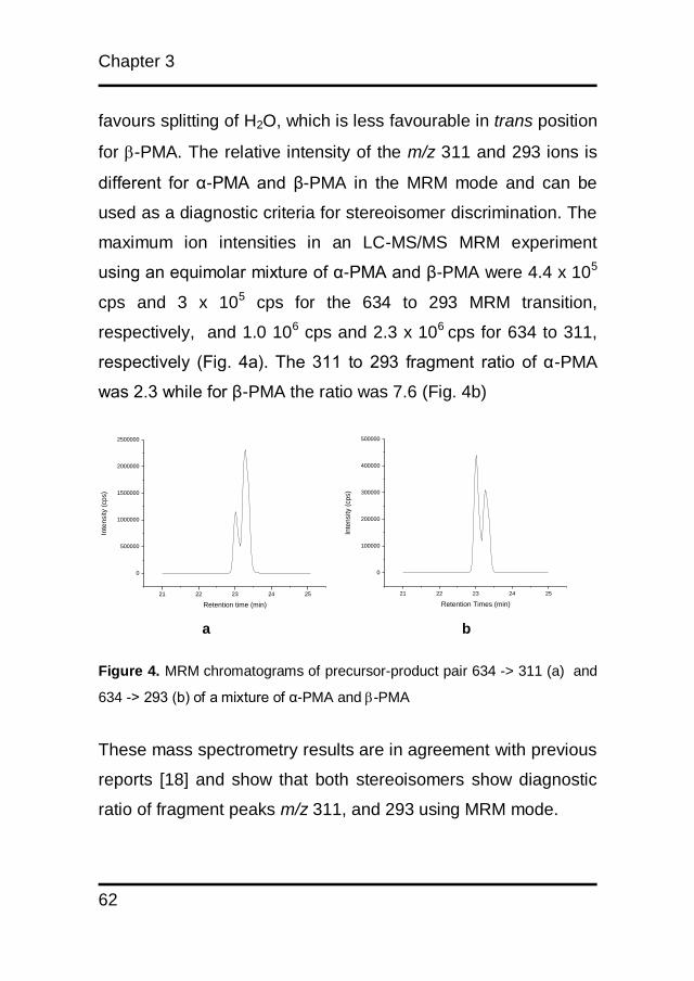
favours splitting of H2O, which is less favourable in trans position
for -PMA. The relative intensity of the m/z 311 and 293 ions is
different for α-PMA and β-PMA in the MRM mode and can be
used as a diagnostic criteria for stereoisomer discrimination. The
maximum ion intensities in an LC-MS/MS MRM experiment
using an equimolar mixture of α-PMA and β-PMA were 4.4 x 10⁵
cps and 3 x 10⁵ cps for the 634 to 293 MRM transition,
respectively, and 1.0 106 cps and 2.3 x 10⁶ cps for 634 to 311,
respectively (Fig. 4a). The 311 to 293 fragment ratio of α-PMA
was 2.3 while for β-PMA the ratio was 7.6 (Fig. 4b)
a b
Figure 4. MRM chromatograms of precursor-product pair 634 -> 311 (a) and
634 -> 293 (b) of a mixture of α-PMA and -PMA
These mass spectrometry results are in agreement with previous
reports [18] and show that both stereoisomers show diagnostic
ratio of fragment peaks m/z 311, and 293 using MRM mode.
Analysis of phorbol esters
63
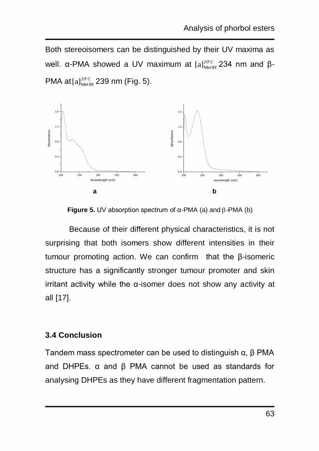
Both stereoisomers can be distinguished by their UV maxima as
well. α-PMA showed a UV maximum at C20
MeOH[a] 234 nm and β-
PMA at C20
MeOH[a] 239 nm (Fig. 5).
a b
Figure 5. UV absorption spectrum of α-PMA (a) and -PMA (b)
Because of their different physical characteristics, it is not
surprising that both isomers show different intensities in their
tumour promoting action. We can confirm that the β-isomeric
structure has a significantly stronger tumour promoter and skin
irritant activity while the α-isomer does not show any activity at
all [17].
3.4 Conclusion
Tandem mass spectrometer can be used to distinguish α, β PMA
and DHPEs. α and β PMA cannot be used as standards for
analysing DHPEs as they have different fragmentation pattern.
200 250 300 350 400
0.0
0.4
0.8
1.2
1.6
ab
so
rba
nce
wavelength (nm)
200 250 300 350 400
0.0
0.4
0.8
1.2
1.6
Ab
so
rba
nce
Wavelength (nm)
Chapter 3
64
References
1. Ravindranath N, Reddy MR, Mahender G, Ramu R, Kumar
KR, Das B. Deoxypreussomerins from Jatropha curcas: are they
also plant metabolites? Phytochemistry. 2004;65(16):2387-90.
Epub 2004/09/24.
2. Chianese G, Fattorusso E, Aiyelaagbe OO, Luciano P,
SchroÌder HC, MuÌller WEG, et al. Spirocurcasone, a diterpenoid
with a novel carbon skeleton from Jatropha curcas. Org Lett.
2011;13(2):316-9.
3. Naengchomnong W, Thebtaranonth Y, Wiriyachitra P,
Okamoto KT, Clardy J. Isolation and structure determination of
four novel diterpenes from Jatropha curcas. Tetrahedron Lett.
1986;27(22):2439-42.
4. Naengchomnong W, Thebtaranonth Y, Wiriyachitra P,
Okamoto KT, Clardy J. Isolation and structure determination of
two novel lathyrenes from Jatropha curcas. Tetrahedron Lett.
1986;27(47):5675-8.
5. Pletsch M, Charlwood BV. Accumulation of diterpenoids in cell
and root-organ cultures of Jatropha species. J Plant Physiol
1997;150(1-2):37-45.
6. Wang X-C, Zheng Z-P, Gan X-W, Hu L-H. Jatrophalactam, A
Novel Diterpenoid Lactam Isolated from Jatropha curcas.
Organic Lett. 2009;11(23):5522-4.
Analysis of phorbol esters
65
7. Clemens M, Trayner I, Menaya J. The role of protein kinase C
isoenzymes in the regulation of cell proliferation and
differentiation. J Cell Sci. 1992;103(4):881-7.
8. Nishizuka Y. Intracellular signaling by hydrolysis of
phospholipids and activation of protein kinase C. Science.
1992;258(5082):607-14.
9. Segal A, Van Duuren BL, Mate U. The identification of phorbol
myristate acetate as a new metabolite in mouse skin. Cancer
Res. 1975;35(8):2154-9.
10. Haas W, Sterk H, Mittelbach M. Novel 12-Deoxy-16-
hydroxyphorbol Diesters Isolated from the Seed Oil of Jatropha
curcas. Journal of Natural Products. 2002;65(10):1434-40.
11. Goel G, Makkar HPS, Francis G, Becker K. Phorbol esters:
structure, biological activity, and toxicity in animals. International
Journal of Toxicology. 2007;26(4):279-88.
12. Adolf W, Opferkuch HJ, Hecker E. Irritant phorbol derivatives
from four Jatropha species. Phytochemistry. 1984;23(1):129-32.
13. Hirota M, Suttajit M, Suguri H, Endo Y, Shudo K, Wongchai
V, et al. A new tumor promoter from the seed oil of Jatropha
curcas L., an intramolecular diester of 12-deoxy-16-
hydroxyphorbol. Cancer Res. 1988;48(20):5800-4. Epub
1988/10/15.

Chapter 3
66
14. Makkar HPS, Becker K. Jatropha curcas, a promising crop
for the generation of biodiesel and value-added coproducts.
European Journal of Lipid Science and Technology.
2009;111(8):773-87.
15. Martínez-Herrera J, Siddhuraju P, Francis G, Dávila-Ortíz G,
Becker K. Chemical composition, toxic/antimetabolic
constituents, and effects of different treatments on their levels, in
four provenances of Jatropha curcas L. from Mexico. Food
Chem. 2006;96(1):80-9.
16. Hecker E, Bartsch H, Bresch H, Gschwendt M, Härle B,
Kreibich G, et al. Structure and stereochemistry of the tetracyclic
diterpene phorbol from croton tiglium L. Tetrahedron Letters.
1967;8(33):3165-70.
17. Van Duuren BL, Tseng S-S, Segal A, Smith AC, Melchionne
S, Seidman I. Effects of Structural Changes on the Tumor-
promoting Activity of Phorbol Myristate Acetate on Mouse Skin.
Cancer Research. 1979;39(7 Part 1):2644-6.
18. Solomon JJ, Van Duuren BL, Tseng SS. Chemical ionization
mass spectrometry of the tumor promoter related 4aα-phorbol
esters. Biomedical Mass Spectrometry. 1978;5(2):164-9.

CHAPTER 4
Curcacycline a and b – new pharmacological
insights to an old drug
Muhamad Insanu, Jana Anggadiredja, Oliver Kayser
Published in : International Journal of Applied Research in
Natural Products, 2012, 5 (2), 26-34
Chapter 4
68
Abstract
Two cyclic peptides were isolated from the latex of Jatropha
curcas L., namely curcacycline A and B. In the past both
compounds have not been fully structure elucidated, today
modern structure elucidation has been applied by more
sophisticated bioanalytical techniques. Additionally, for the first
time the structure was confirmed by a full synthetic approach
and biological assays have been carried out to shed light on
the pharmacological activity of both curcacyclines. Screening of
antibiotic activity of curcacycline A was done against Bacillus
subtilis ATCC 6633, Staphylococcus aureus ATCC 6538,
Eschericia coli ATCC 8939, Pseudomonas aeruginosa ATCC
9027 and Candida albicans ATCC 10231 using paper disk
diffussion method. Curcacycline A inhibited the growth of B.
subtilis and P. aeruginosa with inhibition zone was ranged
between 6.5 – 10.3 mm. Cytotoxic test show that it decreased
the level of OVCAR 3 (ovarium cancer cell) at a concentration
of 1 mg/mL, while no effect was found on Colo205 (human
colon cancer cell). Curcacycline B did not have an effect on
both cell lines. Brine shrimp cytotoxicity assay showed LD50 of
curcacycline A and B at 49.85 µg/mL and 15.84 µg/mL,
respectively. Both curcacyclines did not have a mutagenic
effect on Salmonella typhimurium TA-98 and TA-mix (TA 7001,
TA 7002, TA 7003, TA 7004, TA 7005, TA 7006). This study
provides data on the safety as well the efficacy for curcacycline

Curcacycline a and b
69
A and B. Curcacyline A possesses antimicrobial and cytotoxic
activities. Based on AMES II assay, curcacycline A and B were
proven not to be mutagenic. Both curcacyclines also can be
synthesized in the lab. These data will be essential to develop
curcacyclines as a new drug candidate for managing infectious
and cancer diseases.

Chapter 4
70
4.1 Introduction
Cyclic peptides are formed into a ring via amide ester or
disulfide bonds [1]. Because of no exposure of C,N-terminal
groups to exopeptides, they are quite stable to enzymatic
degradation [2]. Since they have less zwitterionic character,
they can cross membranes more readily than linear peptides.
Finally, their rigid structure leads them to exhibit higher affinity
and selectivity for binding with protein [3]. Cyclic peptides show
a wide range of activities such as as antibiotic (gramicidin),
anthelmintic (yunnanin F), antineoplastic (dolastatin 3),
phytotoxic (tentoxin, HC-toxin), insecticidal (destruxin family)
and serve as cytostatic agent (chlamydocin) or antiviral agent
(sansalvamide A) [4-11]. Naturally cyclic peptides are non
ribosomely biosynthesized by complex multi enzyme in the
cytosol. These peptides have been found in organisms, like
fungi, bacteria, tunicates, mollusks, coelenterates, sponges and
crustaceans as well as in animals. In higher plants cyclic
peptides have been found only in 26 families, 65 genera and
120 species [1, 12-14].
A rich source of cyclic peptides is the family of
Euphorbiaceae, especially the genus of Jatropha. A number of
cyclic peptides have been characterized from this genus.
Podacycline A, B and C have been isolated from Jatropha
podagrica while chevalierin A, B and C from J. chevalieri.
Curcacycline a and b
71
Labaditin and biobollein isolated from Jatropha multifida show
selective inhibition of the classical pathway of the human
complement system. Integerrimide A and B, isolated from
Jatropha integerrimide inhibit basic cytoskeleton-dependent
cellular processes. Pohlianin A, B and C from Jatropha
pohliana show antimalarial effect. Mahafacyclin A, B and C
have been extracted from Jatropha mahafalensis while
cyclogossine A and B have been extracted from Jatropha
gossypifolia. Originally curcacycline A and B have been
isolated from Jatropha curcas latex (Fig. 1a and 1b).
Curcacycline A is an octapeptide with moderate dose
depending inhibitory activity on human T-cell proliferation and
the classical pathway complement system. Curcacycline B, a
cyclic nonapeptide, enhances rotamase activity of cyclophillin
[15-23].
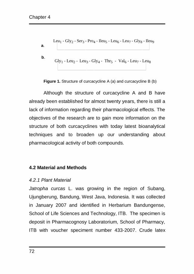
Chapter 4
72
Gly1 - Leu2 - Leu3 - Gly4 - Thr5 - Val6 - Leu7 - Leu8
Leu1 - Gly2 - Ser3 - Pro4 - Ileu5 - Leu6 - Leu7 - Gly8 - Ileu9
a.
b.
Figure 1. Structure of curcacycline A (a) and curcacycline B (b)
Although the structure of curcacycline A and B have
already been established for almost twenty years, there is still a
lack of information regarding their pharmacological effects. The
objectives of the research are to gain more information on the
structure of both curcacyclines with today latest bioanalytical
techniques and to broaden up our understanding about
pharmacological activity of both compounds.
4.2 Material and Methods
4.2.1 Plant Material
Jatropha curcas L. was growing in the region of Subang,
Ujungberung, Bandung, West Java, Indonesia. It was collected
in January 2007 and identified in Herbarium Bandungense,
School of Life Sciences and Technology, ITB. The specimen is
deposit in Pharmacognosy Laboratorium, School of Pharmacy,
ITB with voucher speciment number 433-2007. Crude latex
Curcacycline a and b
73
was collected early in the morning (7.00-10.00 am) by cutting
off leaf stalks and adding few drops of methanol (MeOH) to
prevent the latex from extra foaming. The latex was kept at -
20°C before use.
4.2.2 Extraction and isolation by MPLC
To 250 mL of latex 250 mL of deionized water was added, the
suspension was extracted with 3 x 500 mL of ethyl acetate.
Ethyl acetate was removed using rotary evaporator. The
viscous extract was dissolved in 5 mL MeOH and fractionated
on Sephadex LH-20 (Pharmacia, 4 x 25 cm) using MeOH as an
eluent at a flow rate 10 mL/min. An amount of 50 mL for each
fractions was colleted and monitored for cyclic peptides by thin
layer chromatography, 20 µL were spotted onto silica gel 60
GF254 (Merck), the plate was developed with CHCl3 : MeOH
(85:15 v/v), and cyclic peptides were visualized by Cl2/o-
tolidine. Fractions containing cyclic peptides were further
separated by column chromatography using a Büchi C-690
series borosilicate glass column (40x 450 mm) pack with sillica
gel 60, (Merck). Methanol and chloroform were used as mobile
phase. The solvent flow was set at 30 mL/min, and every 15
mL of fractions were collected. Thin layer chromatography
analysis was conducted on silica gel plates Merck F254 and use
o-tolidine/Cl2 as visualizing agent.
Chapter 4
74
4.2.3 HPLC
Low pressure gradient chromatography was performed using a
LiChrospher-100 C18 column (250 x 4.6 mm with 5µm packing).
The injection volume was 20 µL and the flow rate was set at 1
mL/min at room temperature. The mobile phase comprised
100% H2O + 0.1% TFA (solvent A) and 100% Acetonitrile +
0.1% TFA (solvent B) The separation was commenced at 30%
solvent B for 5 min, followed by gradient eluent 30 to 60% B in
10 min, in 5 min 60% B, in further 4 min to 95%B, and for 6 min
95% B.
4.2.4 HPLC/MS
The mass spectrometer was interfaced to a Vydac C18 column
(150 x 2.1 mm, 5 µm particle size). The mobile phase
comprised 100% H2O + 0.1% formic acid (solvent A) and 100%
acetonitrile + 0.1% Formic acid. The separation was done using
30% solvent B for 5 min, followed by gradient eluent 30 to 60%
B in 10 min, 60% B in 5 min, 95% B in 4 min, and 95% B for the
last 6 min. The injection volume was 20 µL and the flow rate
1mL/min at room temperature. The positive ion mode was
employed and spectra were obtained with a spray voltage of
5.2 kV. The source temperature was 500oC. Data processing
was performed using Analyst version 1.42 software (MDS
Sciex, Concord, Canada).
Curcacycline a and b
75
4.2.5 NMR
Curcacycline A and B were dissolved in (deuterated DMSO,
d6). 1H NMR and 13C NMR were recorded by 400 MHz Varian
NMR, while 2D NMR was recorded by 500 MHz Varian. Tetra
Methyl Silan was used as internal standard. Data processing
was performed by MestReNova 6.02.
Curcacycline A white crystalline, Rf = 0.7 (See extraction and
isolation); UV λmax (methanol) : 220 nm; IR max : 3290, 2960,
2873, 1640, 1520, 1260, 1157, 1126 ; ESI MS m/z (rel. int.) :
767 (2.2 x 10³), 655 (1.3 x 10⁵), 542 (1.8 x 10⁵), 485 (1.0 x 10⁵),
372 (1.2 x 10⁵), 259 (3.2 x 10⁴), 160 (5.0 x 10³) ; 1H NMR (400
MHz, dmso) δ 8.67 (t, J = 5.7 Hz, 1H), 8.63 – 8.54 (m, 1H),
8.51 (s, 1H), 8.17 (d, J = 8.5 Hz, 1H), 8.03 (s, 1H), 7.73 (d, J =
7.5 Hz, 1H), 7.53 (d, J = 8.9 Hz, 1H), 7.39 (d, J = 9.9 Hz, 1H),
5.34 (d, J = 10.8 Hz, 1H), 4.76 (dd, J = 9.9, 3.7 Hz, 1H), 4.60
(dd, J = 14.2, 8.3 Hz, 1H), 4.37 – 4.22 (m, 2H), 4.12 – 4.05 (m,
1H), 3.96 – 3.79 (m, 4H), 3.45 (d, J = 5.2 Hz, 1H), 3.40 (d, J =
4.9 Hz, 1H), 3.15 (d, J = 5.2 Hz, 1H), 2.50 – 2.46 (m, 1H), 2.32
(td, J = 13.8, 6.9 Hz, 1H), 1.69 – 1.18 (m, 13H), 1.08 – 0.69 (m,
32H) ; 13C NMR (101 MHz, dmso) δ 172.94, 172.90, 172.86,
170.99, 170.48, 169.42, 169.20, 79.59, 69.13, 58.70, 56.90,
53.36, 52.93, 51.00, 42.90, 40.55, 40.34, 40.14, 39.93, 39.72,
39.51, 39.30, 31.11, 28.73, 24.69, 24.55, 24.29, 23.50, 23.26,
22.99, 22.78, 22.62, 22.10, 21.83, 19.61, 18.91, 17.08.
Chapter 4
76
Curcacycline B white crystal, Rf : 0.66 (see extraction and
isolation) ; UV λmax (methanol) : 220 nm; IR max : 3290, 2960,
2873, 1644, 1520, 1259, 1027, 800. ESI MS m/z (rel. int.) :
864.4 (4.9 x 10⁴), 777.4 (1.3 x 10⁴), 720.4 (1.37 x 10⁴), 607 (3.6
x 104), 494 (3,1 x 10⁴), 437 (4.1 x 10⁴), 324 (3.2 x 10⁴), 211.4
(2.9 x 10⁴) ; 1H NMR (500 MHz, dmso) δ 8.72 (s, 1H), 8.63 (s,
1H), 8.54 (s, 1H), 8.32 (s, 4H), 8.25 (s, 1H), 8.10 (s, 1H), 8.02
(s, 1H), 7.69 (d, J = 7.1 Hz, 1H), 7.46 (d, J = 9.5 Hz, 1H), 7.36
(d, J = 8.3 Hz, 1H), 6.88 (d, J = 9.0 Hz, 1H), 6.54 (s, 1H), 6.11
– 6.07 (m, 1H), 5.76 (s, 2H), 4.63 (s, 1H), 4.45 (s, 1H), 4.39 –
4.24 (m, 3H), 4.20 – 4.08 (m, 4H), 4.04 (dd, J = 14.2, 7.1 Hz,
3H), 3.98 – 3.76 (m, 6H), 3.60 (s, 1H), 3.52 (d, J = 5.7 Hz, 1H),
3.48 – 3.41 (m, 2H), 3.36 (s, 49H), 3.18 (d, J = 5.1 Hz, 4H),
2.57 – 2.45 (m, 4H), 2.14 – 2.06 (m, 2H), 2.00 (s, 3H), 1.87 (d,
J = 9.3 Hz, 2H), 1.71 (s, 1H), 1.66 – 1.57 (m, 3H), 1.54 (d, J =
7.4 Hz, 2H), 1.49 – 1.35 (m, 4H), 1.25 (s, 1H), 1.23 – 1.11 (m,
4H), 1.23 – 0.98 (m, 7H), 1.23 – 0.73 (m, 26H).
4.2.6 Cyclic Peptides synthesis
Peptides were synthesized by ALMAC Sciences.Each peptide
was assembled on 0.3 mmol scale using pre-loaded chlorotityl
chloride resin and an automated peptide synthesizer. Each
amino acid was incorporated as an active ester in a single

Curcacycline a and b
77
couple cycle. The resin bound peptide was treated with 10%
TFE in DCM, 10 mL for two hours. The solvent was evaporated
under vacuum and the residue was lyophilized from
water/acetonitrile (1:1) to yield the crude protected linear
peptide. The crude linear peptide (50 mg) was dissolved in
DMF (50ml). HOBt (3eq), PyBOP (3eq) and DIEA (3eq) were
added to solution. The mixture was stirred at room temperature
for 72 hours. The solvent was evaporated under vacuum and
the residue lyphilised from water/acetonitrile 1:1 to yield the
crude protected peptide. The crude protected peptide was
dissolves in DMF (2 mL) and diluted with water and lyophilized
to yield crude product. The crude product was dissolved in
DMF (2mL) and diluted with water. The resulting precipitate
was triturated and isolated by filtration to provide final product.
4.2.7 Cytotoxicity test with Human ovarian cancer cell-line
OVCAR3
The cancer cells were routinely cultured in DMEM (Gibco®),
supplemented with 10% of heat-inactivated fetal bovine serum
(Gibco®) and 1% penicillin/streptomycin (Gibco®), at 37oC in the
presence of 5% CO2. The addition of paclitaxel reference
compound (plant and fungal extracts as well as pure
components thereof) dissolved in 1 % DMSO. The solution was
sterillized by filtration (0.2 µm) and diluted with cell culture
Chapter 4
78
medium, in consecutive 2-fold dilutions. It was preceded by
seeding the test cells into 96-well plates (5000 cells/well) for 24
h incubation. Cell viability was assessed 72 h later by means of
the MTS assay (CellTiter 96® AQueousNon-Radioactive Cell
Proliferation Assay, Promega Corporation, USA), following the
instructions of the manufacturer [24].
4.2.8 Cytotoxicity test with Human colon cancer cell-line
Colo205
The cancer cells were routinely cultured in RPMI (Invitrogen®),
supplemented with 10% of heat inactivated fetal bovine serum
(Invitrogen®) and 1% penicillin/streptomycin (Invitrogen®), at
37oC in the presence of 5% CO2. The addition of TRAIL and
paclitaxel reference compounds dissolved in 1% DMSO. The
solution was sterillized by filtration (0.2 µm) and diluted with cell
culture medium, in consecutive 2-fold dilutions. It was preceded
by seeding the test cells into 96-well plates (5000 cells/well) for
24 h incubation. Cell viability was assesed 24 h later by means
of the MTS assay (CellTiter 96® AQueousNon-Radioactive Cell
Proliferation Assay, Promega Corporation, USA), following the
instructions of the manufacturer. All assays were performed in
triplicate [24].
Curcacycline a and b
79
4.2.9 Brine Shrimp assay
Brine shrimps (Artemia salina) were hatched using brine shrimp
eggs in a conical shaped vessel (1L), filled with sterile artificial
seawater (prepared using sea salt 38 g/L and adjusted to pH
8.5 using 1N NaOH) under constant aeration for 48 h. After
hatching, active nauplii free from egg shells were collected from
brighter portion of the hatching chamber and used for the
assay. Hundred microliters of suspension of nauplii containing
about (9-12) larvae was added into 96-well microtiter plate.
Curcacyclines were dissolved in sea salt water with
concentration ranging between 1-1000 μL/mL. Into each well
curcacyclines were added 50 μL and incubated for 24 h (22-
28oC). The plates were examined under a microscope (12.5x)
and the number of dead nauplii in each well counted. Then 100
μL of methanol was added to each well. After 10 min, total
number of shrimp in each well were counted and recorded.
Calculation of LC50 values for each assay was done by taking
average of three experiments using a Finney Probit analysis
[25-28].
4.2.10 AMES II Test
Carcinogenicity test was determined by AMES kit from
Xenomatric Article number E10-213 and E10-213-S1. Two
bacteria were used, Salmonella typhimurium TA-98 and
Chapter 4
80
S.typhimurium TA-Mix (TA 7001, TA 7002, TA 7003, TA 7004,
TA 7005, TA 7006). These bacteria were exposed to six
concentrations (1000; 500; 250; 125; 62.5; 31.25; 15.63; 7.82;
3.9 g/mL) of samples, as well as positive and negative control,
for 90 min in medium containing histidine to support
approximately two cell divisions. After 90 min, the exposure
cultures were diluted in pH indicator medium lacking histidine,
and aliquoted into 48 wells of 384 well plates. Within two days,
cells which have undergone the reversion to His will grow into
colonies. Metabolism by the bacterial colonies reduces the pH
of the medium, changing colour of reagent in the well
(Xenometric®) [29].
4.2.11 Antibacterial Activity
The antibacterial activity of the curcacycline A was evaluated
by the paper-disk agar diffusion method against the two Gram-
positive bacteria Staphylococcus aureus (ATCC 6538) and
Bacilus subtilis (ATCC 6633), two gram-negative bacteria
Escherichia coli (ATCC8939) and Pseudomonas aeruginosa
(ATCC9027), one fungi Candida albicans (ATCC10231).
Organisms were maintained on Müller-Hinton agar (MH)
(BIORAD). Inoculum were prepared by diluting overnight (24 h
at 37oC) cultures in Muller Hinton Broth medium to
approximately 106 CFU/mL. Absorbent disks (Whatman disc No.
Curcacycline a and b
81
3, 6 mm diameter) were impregnated with 10 µL of curcacycline
A and then placed on the surface of inoculated Petri dishes (90
mm). Positive control discs of tetracycline (10 µg/disc) were
included in each essay. Diameters of growth inhibition zones
were measured after incubation at 37oC for 24 h.
4.3 Result and Discussion
4.3.1 Structure elucidation
After extraction and synthesis, curcacycline A and B were
obtained as white crystals. They were UV active with a
maximum wavelength (λmax) 220 nm, but no absorption was
found at 260 and 280 nm. HPLC chromatogram showed their
retention time at 15.02 and 16.93 for curcacycline A and B,
respectively. They showed positive reaction with Cl2/o-
tolidineon tlc spots. No reactions were detected with nynhidrine
reagent. These results proved that both compounds were
expected cyclopeptides without C and N terminal. The Infra-
Red spectra of these compounds exhibited typical bands at
functional group area which were 3290, 2960, 2873, 1640,
1520 cm-1 indicating the present of (-NH), (-OH) and (C=O),
respectively. Because of different amino acid sequences both
curcacyclines showed different finger print area (Fig. 2a and
2b).

Chapter 4
82
4500 4000 3500 3000 2500 2000 1500 1000 500
0.4
0.3
0.2
0.1
0.0
ab
so
rba
nce
(a
rb u
nit)
wavenumber (cm-1)
4500 4000 3500 3000 2500 2000 1500 1000 500
0.5
0.4
0.3
0.2
0.1
0.0
ab
so
rba
nce
(a
rb u
nit)
wave number (cm-1)
a.
b.
Figure 2. Infra-red spectrum of curcacycline A(a) and curcacycline B(b)
ESI-MS of curcacycline A indicated a molecular ion peak at m/z
767 corresponding to C37H67N8O9 and with fragments m/z 655
(M+, -Leu), 542 (M+, -Leu), 485 (M+, -Gly), 372 (M+, -Leu), 259
(M+, Leu), 160 (M+, -Val) (Fig. 3a).1HNMR revealed eight
protons at 8.67, 8.63, 8.51, 8.17, 8.03, 7.53, and 7.39
indicating amide protons.

Curcacycline a and b
83
Conformation with 13C NMR and COSY spectra (Table 1)
revealed that this compound was identical to previous
compound reported by van den Berg et al., 1995.
a.
b.
Figure 3. Fragmentation pattern of curcacycline A (a) and curcacyline B (b)
by ESI-MS
0 200 400 600 800 1000
0
100000
200000
300000
400000
500000
600000
-Val
-Leu
-Leu
160
259
372
485
542655
Inte
nciti
es (c
ps)
m/z
767
-Leu-Leu
-Gly
0 200 400 600 800 1000 1200
0
20000
40000
60000
80000
100000
-Leu
-Gly
-Ileu
-Leu
-Gly
-Ser
211
324437
494
607
720
777
Inte
nsiti
es (c
ps)
m/z amu
864-Leu
Chapter 4
84
Table 1. COSY spectrum data of curcacycline A (500 MHz varian NMR, d6, DMSO, TMS as internal standard)
Fragmentation pattern of curcacycline B showed m/z 864 and a
molecular ion peak at m/z 777 (M+, -Ser), 720 (M+, -Gly), 607
(M+, -Leu), 494 (M+, -Ileu), 437 (M+, -Gly), 324 (M+,-Leu),
211(M+, -Leu) corresponding to C42H73N9O10 (Fig. 3b). 1H NMR
revealed only eight amide protons at 8.72, 8.59 7.69, 7.46,
7.35, 8.00, 8.25, 6.88 while proline had no amide proton.
Comparing to COSY spectra of this compound (Tab. 2)
Amino acid
NH αH βH Other
Gly1 8.63 3.92 - - - 3.40 - - -
Leu2 7.56 4.64 1.4 CH3 0.85 1.34
Leu3 8.54 3.87 1.55 CH3 0.87 1.38 0.84
Gly4 8.71 3.91 - - - 3.43 - - -
Thr5 7.42 4.79 4.32 CH3 0.95 OH 5.38
Val6 7.76 4.1 2.34 CH3 0.85 0.85
Leu7 8.22 4.36 1.66 CH3 1.54 1.25
Leu8 8.07 3.94 1.43 CH3 0.86

Curcacycline a and b
85
indicated that this compound is similar to curcacycline B
reported by Auvin et al.,1997.
Table 2. COSY data of curcacycline B (500 MHz varian NMR, d6, DMSO,
TMS as internal standard)
Amino acid
NH αH βH Other
Leu1 8.72 3.78 1.54 - - - - 1.44 - -
Gly2 8.59 3.98 - - - - 3.37 - - -
Ser3 7.69 4.63 4.13 OH 6.03 - - 3.51 - -
Pro4 - - - - - - - - - -
Ile5 7.46 4.15 1.71 - - - - - - -
Leu7 8.00 3.96 1.62 - - - - 1.45 - -
Gly8 8.25 3.83 - - - - 3.44 - - -
Ileu9 6.88 4.25 1.90 - -
The yield of the synthesized compounds was low. Based on
ESI-MS chromatogram and 1H-NMR spectra both synthethic
curcacyclines have similar characteristic with isolated
curcacyclines.
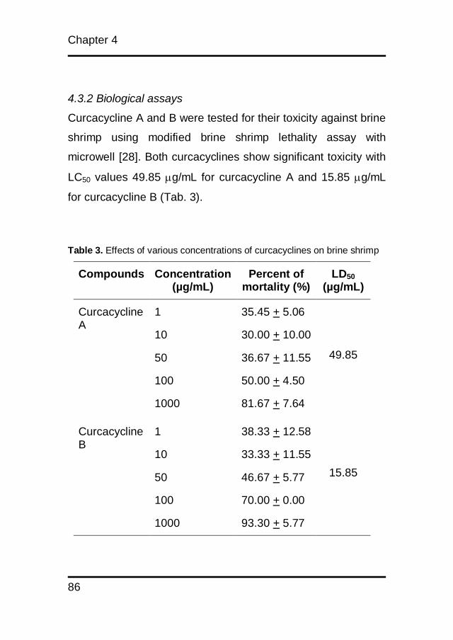
Chapter 4
86
4.3.2 Biological assays
Curcacycline A and B were tested for their toxicity against brine
shrimp using modified brine shrimp lethality assay with
microwell [28]. Both curcacyclines show significant toxicity with
LC50 values 49.85 g/mL for curcacycline A and 15.85 g/mL
for curcacycline B (Tab. 3).
Table 3. Effects of various concentrations of curcacyclines on brine shrimp
Compounds Concentration (µg/mL)
Percent of mortality (%)
LD50
(µg/mL)
Curcacycline A
1 35.45 + 5.06
49.85
10 30.00 + 10.00
50 36.67 + 11.55
100 50.00 + 4.50
1000 81.67 + 7.64
Curcacycline B
1 38.33 + 12.58
15.85
10 33.33 + 11.55
50 46.67 + 5.77
100 70.00 + 0.00
1000 93.30 + 5.77
Curcacycline a and b
87
Mutagenic activity was determined using Salmonella
typhimurium TA-98 and TA-Mix (AMES II assay). The results
are depicted in Fig. 5a and 5b. All concentrations of
curcacyclines A and B did not cause mutation on TA-98 and
TA-mix because only 10 wells (out of 48) gave positives
reaction while the positive control 2-nitrofluorene and 4-
nitroquinoline N-oxide (2-NF/4-NQO) had 46 positive wells (out
of 48), which were statistically not significant. Cytotoxic
properties of these peptides were further investigated using
MTS assay. Human ovarium cancer cell (OVCAR3) and human
colo cancer cell (Colo 205) were used. The results are
summarized in Fig. 6a and 6b. To ensure proliferation arrest,
TRAIL (TNF-related apoptosis-inducing ligand) and paclitaxel
was used as positive control. TRAIL is a protein that induced
apoptosis while paclitaxel stabilize microtubule polymer which
block mitosis progression [24, 30, 31].
Ovarian cancer cells were treated with paclitaxel (IC50 =
4.89 ng/ml) and human colon cancer cells were treated with
TRAIL (IC50 = 4.89 ng/mL). We found that IC50 of curcacycline A
on OVCAR3 was 724.44 µg/mL while IC50 of curcaycline B was
above 1000 µg/ml. Both curcacyclines did not have any effect
on Colo 205 (IC50> 1000 µg/mL).
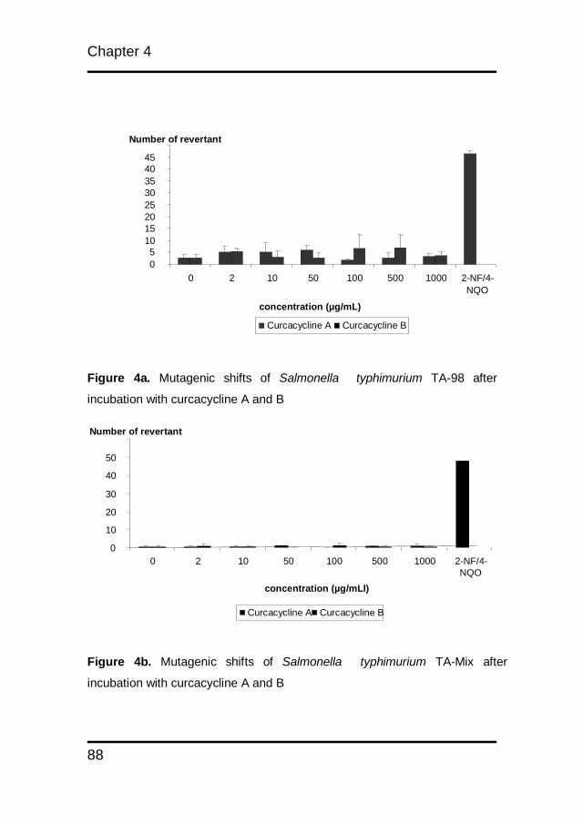
Chapter 4
88
Figure 4a. Mutagenic shifts of Salmonella typhimurium TA-98 after
incubation with curcacycline A and B
Figure 4b. Mutagenic shifts of Salmonella typhimurium TA-Mix after
incubation with curcacycline A and B
0
10
20
30
40
50
0 2 10 50 100 500 1000 2-NF/4- NQO
concentration (µg/mLl)
Number of revertant
Curcacycline A Curcacycline B
0 5
10 15 20 25 30 35 40 45
0 2 10 50 100 500 1000 2-NF/4- NQO
concentration (µg/mL)
Number of revertant
Curcacycline A Curcacycline B
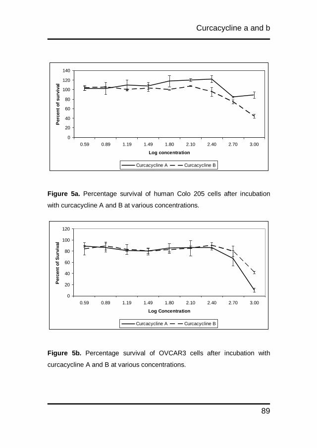
Curcacycline a and b
89
0
20
40
60
80
100
120
140
0.59 0.89 1.19 1.49 1.80 2.10 2.40 2.70 3.00
Log concentration
Perc
en
t o
f su
rviv
al
Curcacycline A Curcacycline B
Figure 5a. Percentage survival of human Colo 205 cells after incubation
with curcacycline A and B at various concentrations.
Figure 5b. Percentage survival of OVCAR3 cells after incubation with
curcacycline A and B at various concentrations.
0
20
40
60
80
100
120
0.59 0.89 1.19 1.49 1.80 2.10 2.40 2.70 3.00
Log Concentration
Perc
en
t o
f S
urv
ival
Curcacycline A Curcacycline B
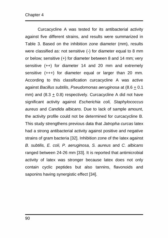
Chapter 4
90
Curcacycline A was tested for its antibacterial activity
against five different strains, and results were summarized in
Table 3. Based on the inhibition zone diameter (mm), results
were classified as: not sensitive (-) for diameter equal to 8 mm
or below; sensitive (+) for diameter between 8 and 14 mm; very
sensitive (++) for diameter 14 and 20 mm and extremely
sensitive (+++) for diameter equal or larger than 20 mm.
According to this classification curcacycline A was active
against Bacillus subtilis, Pseudomonas aeruginosa at (8.6 + 0.1
mm) and (8.3 + 0.8) respectively. Curcacycline A did not have
significant activity against Escherichia coli, Staphylococcus
aureus and Candida albicans. Due to lack of sample amount,
the activity profile could not be determined for curcacycline B.
This study strengthens previous data that Jatropha curcas latex
had a strong antibacterial activity against positive and negative
strains of gram bacteria [32]. Inhibition zone of the latex against
B. subtilis, E. coli, P. aeruginosa, S. aureus and C. albicans
ranged between 24-26 mm [33]. It is reported that antimicrobial
activity of latex was stronger because latex does not only
contain cyclic peptides but also tannins, flavonoids and
saponins having synergistic effect [34].
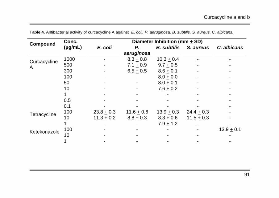
Curcacycline a and b
91
Table 4. Antibacterial activity of curcacycline A against E. coli, P. aeruginosa, B. subtilis, S. aureus, C. albicans.
Compound Conc. (µg/mL)
Diameter Inhibition (mm + SD)
E. coli P. aeruginosa
B. subtilis S. aureus C. albicans
Curcacycline A
1000 - 8.3 + 0.8 10.3 + 0.4 - -
500 - 7.1 + 0.9 9.7 + 0.5 - -
300 - 6.5 + 0.5 8.6 + 0.1 - -
100 - - 8.0 + 0.0 - -
50 - - 8.0 + 0.1 - -
10 - - 7.6 + 0.2 - -
1 - - - - -
0.5 - - - - -
0.1 - - - - -
Tetracycline 100 23.8 + 0.3 11.6 + 0.6 13.9 + 0.3 24.4 + 0.3 -
10 11.3 + 0.2 8.8 + 0.3 8.3 + 0.6 11.5 + 0.3 -
1 - - 7.9 + 1.2 - -
Ketekonazole 100 - - - - 13.9 + 0.1
10 - - - - -
1 - - - - -
Curcacycline a and b
They have different mechanism for inhibiting bacterial
activity. Tannins and saponins influence the bacterial cell wall
while flavonoids inhibit bacterial enzymes [35, 36]. Although the
mechanistic action of curcacyline A is still unclear, based on
previous research [3] cyclic peptides exhibit antibacterial activity
because of their ability to cross membranes easily and their
higher selective affinity to bind with certain protein.
The Ames II test revealed no mutagenic activity for both
compounds without S9 mixture. At all concentration the
mutagenic conversion was as low as the negative control. This is
the first report about mutagenic activity studies of cyclic peptides
obtain from Jatropha curcas L. latex.
4.4 Conclusion
From ethnopharmacological point, Jatropha curcas L. latex has
been traditionally used for treating dental problems in Indonesia
because of antimicrobial activity. This claim is proven by
previous research which showed antimicrobial activity of latex
against gram positive and negative bacteria. Curcacycline A and
B were isolated over twenty years ago. Isolated curcacycline A
and B were subjected to latest analytical techniques like NMR-
spectroscopy and HPLC/MS to confirm amino acid sequences.
Furthermore, both structures were confirmed by an organic
synthesis approach. Here we report for the first time about
Curcacycline a and b
93
antimicrobial, mutagenic, cytotoxic and ecotoxic activity. The
results showed that curcacycline A has antimicrobial activity
against Bacillus subtilis and Pseudomonas aeruginosa and
cytotoxic activity against OVCAR 3. No mutagenic effects were
found on curcacycline A and B against Salmonella typhimurium
TA-98 and TA-100. These data show that curcacycline A would
be potential for new and safe antimicrobial candidate.

Chapter 4
94
References
1. Pomilio AB, Battista ME, Vitale AA. Naturally-Occurring
Cyclopeptides: Structures and Bioactivity. Current Organic
Chemistry: Bentham Science Publishers Ltd.; 2006. p. 2075-121.
2. Wu L, Lu Y, Zheng Q-T, Tan N-H, Li C-M, Zhou J. Study on
the spatial structure of annomuricatin A, a cyclohexapeptide from
the seeds of Annona muricata. Journal of Molecular Structure.
2007;827(1-3):145-8.
3. Sakai R, Rinehart KL, Kishore V, Kundu B, Faircloth G, Gloer
JB, et al. Structure and Activity Relationships of the
Didemnins1,2. Journal of Medicinal Chemistry.
1996;39(14):2819-34.
4. Izumiya N, Kato T, Waki M. Synthesis of biologically active
cyclic peptides. Biopolymers. 1981;20(9):1785-91.
5. Païs M, Das BC, Ferron P. Depsipeptides from Metarhizium
anisopliae. Phytochemistry. 1981;20(4):715-23.
6. Schmidt U, Utz R, Lieberknecht A, Griesser H, Potzolli B, Bahr
J, et al. Amino Acids and Peptides; 60.1 Synthesis of Biologically
Active Cyclopeptides; 10. Synthesis of 16 Structural Isomers of
Dolastatin 3; II:2 Synthesis of the Linear Educts and the
Cyclopeptides. Synthesis. 1987;1987(03):236-41.

Curcacycline a and b
95
7. Kawai M, Gardner JH, Rich DH. Stereoselective synthesis and
absolute configuration of epoxyketones in chlamydocin and
related cyclic tetrapeptides. Tetrahedron Letters.
1986;27(17):1877-80.
8. Jacquier R, Verducci J. Nouvelle synthese de la tentoxine.
Tetrahedron Letters. 1984;25(26):2775-8.
9. Poojary B, Belagali SL. Synthetic studies on cyclic
octapeptides: Yunnanin F and Hymenistatin. European Journal
of Medicinal Chemistry. 2005;40(4):407-12.
10. Hwang Y, Rowley D, Rhodes D, Gertsch J, Fenical W,
Bushman F. Mechanism of Inhibition of a Poxvirus
Topoisomerase by the Marine Natural Product Sansalvamide A.
Molecular Pharmacology. 1999;55:1049.
11. Rich DH, Gardner JH. Synthesis of the Cytostatic Cyclic
Tetrapeptide, Chlamydocin. Tetrahedron Letters.
1983;24(48):5305-8.
12. Cavelier-Frontin F, Achmad S, Verducci J, Jacquier R, Pèpe
G. How to perform small peptide cyclizations. Journal of
Molecular Structure: THEOCHEM. 1993;286:125-30.
13. Tang Y-Q, Yuan J. A Cyclic Antimicrobial Peptide Produced
in Primate Leukocytes by the Litigation of Two Truncated.
Science. 1999;286(5439):498.

Chapter 4
96
14. Kelecom A. Secondary metabolites from marine
microorganisms. Anais da Academia Brasileira de Ciências.
2002;74:151-70.
15. Baraguey C, Auvin-Guette C, Blond A, Cavelier F, Lezenven
F, Pousset J-L, et al. Isolation, structure and synthesis of
chevalierins A, B and C, cyclic peptides from the latex of
Jatropha chevalieri. Journal of the Chemical Society, Perkin
Transactions 1. 1998(18):3033-40.
16. Kosasi S, Hart LA, van Dijk H, Labadie RP. Inhibitory activity
of Jatropha multifida latex on classical complement pathway
activity in human serum mediated by a calcium-binding
proanthocyanidin. Journal of ethnopharmacology. 1989;27(1-
2):81-9.
17. Auvin-Guette C, Baraguey C, Blond A, Pousset J-L, Bodo B.
Cyclogossine B, a Cyclic Octapeptide from Jatropha
gossypifolia. Journal of Natural Products. 1997;60(11):1155-7.
18. Auvin C, Baraguey C, Blond A, Lezenven F, Pousset J-L,
Bodo B. Curcacycline B, a cyclic nonapeptide from Jatropha
curcas enhancing rotamase activity of cyclophilin. Tetrahedron
Letters. 1997;38(16):2845-8.
19. Baraguey C, Blond A, Correia I, Pousset J-L, Bodo B, Auvin-
Guette C. Mahafacyclin A, a cyclic heptapeptide from Jatropha
Curcacycline a and b
97
mahafalensis exhibiting [beta]-bulge conformation. Tetrahedron
Letters. 2000;41(3):325-9.
20. Horsten SFAJ, van den Berg AJJ, Kettenes-van den Bosch
JJ, Leeflang BR, Labadie RP. Cyclogossine A: A Novel Cyclic
Heptapeptide Isolated from the Latex of Jatropha gossypifolia.
Planta Med. 1996;62(01):46,50.
21. Mongkolvisut W, Sutthivaiyakit S, Leutbecher H, Mika S,
Klaiber I, Moller W, et al. Integerrimides A and B, Cyclic
Heptapeptides from the Latex of Jatropha integerrima. Journal of
Natural Products. 2006;69(10):1435-41.
22. van den Berg AJ, Horsten SF, Kettenes-van den Bosch JJ,
Kroes BH, Beukelman CJ, Leeflang BR, et al. Curcacycline A--a
novel cyclic octapeptide isolated from the latex of Jatropha
curcas L. FEBS Lett. 1995;358(3):215-8. Epub 1995/01/30.
23. Schmidt VU, Beuttler T, Lieberknecht A, Griesser H.
Aminosäuren und peptide - XXXXII. Synthese von Chlamydocin
+ epi-Chlamydocin. Tetrahedron Letters. 1983;24(34):3573-6.
24. Staniek A, Woerdenbag HJ, Kayser O. Screening the
endophytic flora of Wollemia nobilis for alternative paclitaxel
sources. Journal of Plant Interactions. 2010;5(3):189-95.
25. Meyer BN, Ferrigni NR, Putnam JE, Jacobsen LB, Nichols
DE, McLaughlin JL. Brine Shrimp: A Convenient General
Chapter 4
98
Bioassay for Active Plant Constituents. Planta Med.
1982;45(05):31,4.
26. Krishnaraju AV, Rao, T.V.N., Sundararaju, D., Vanisree, M.,
Tsay, H.S., and Subbaraju, G.V.,. Assessment of Bioactivity of
Indian Medicinal Plants Using Brine Shrimp (Artemia salina)
Lethality Assay. International Journal of Applied Science and
Engineering. 2005;3(2):125-34.
27. Wanyoike GN, Chhabra SC, Lang'at-Thoruwa CC, Omar SA.
Brine shrimp toxicity and antiplasmodial activity of five Kenyan
medicinal plants. Journal of ethnopharmacology.
2004;90(1):129-33.
28. Solis PN, Wright CW, Anderson MM, Gupta MP, Phillipson
JD. A microwell cytotoxicity assay using Artemia salina (brine
shrimp). Planta Medica. 1993;59(3):250-2.
29. Fluckiger-Isler S, Baumeister M, Braun K, Gervais V, Hasler-
Nguyen N, Reimann R, et al. Assessment of the performance of
the Ames II assay: a collaborative study with 19 coded
compounds. Mutation Research/Genetic Toxicology and
Environmental Mutagenesis. 2004;558(1-2):181-97.
30. Wiley S, Schooley K, Smolak P, Din W, Huang C, Nicholl J,
et al. Identification and characterization of a new member of the
TNF family that induces apoptosis. Immunity. 1995;3(6):673-82.
Curcacycline a and b
99
31. Bharadwaj R, Yu H. The spindle checkpoint, aneuploidy, and
cancer. Oncogene. 2004;23(11):2016-27.
32. Fagbenro-Beyioku AF, Oyibo WA, Anuforom BC.
Disinfectant/antiparasitic activities of Jatropha curcas. E Afr Med
J. 1998;75(9):508-11. Epub 1999/09/24.
33. Oyi AR, Onaolapo J, Haruna AK, Morah CO. Antimicrobial
screening and stabaility studies of the crude extract of Jatropha
curcas L. latex (Euphorbiaceae). Nigerian Journal of
Pharmaceutical Sciences. 2007;6(2):14-20.
34. Levens M, Vandan-Berghe DA, Marten J, Vihen T, Lomiveas
EC. Screening of higher plants for biological activity. Planta
Medica. 1979;36:311-2.
35. Iwu M, Igboko OA, Okeniyi CD, Tempesta MS. Inhibition of
the enzyme activity of aldosoreductase of some flavonoids by
some flavonoids. J Pharm Pharmacology. 1990;42:290-2.
36. Pathak D, Pathak L, Singla AK. Inhibition of Xanthine-
oxidase activity by some flavonoids. Fitoterapia. 2002;62:371-85.
CHAPTER 5
Validation of detoxification process for Jatropha
curcas L. kernel meal for use as animal feed
Muhamad Insanu, Rita Setoikromo, Pieter Tepper, C.M.
Jeronimus-Stratingh, Hjalmar Permentier, Piet van der Linde,
John Brooker, Oliver Kayser, Wim J. Quax
Detoxification process
101
Abstract
Processed and detoxified Jatropha curcas L. kernel meal (JKM)
has been tested for its potential as an animal feedstock. The
detoxification process has been validated by determining
cytotoxicity, mutagenicity and ecotoxicity of fractions along the
down streaming process. Cytotoxic activity was tested against
Colo205 and OVCAR3 cells indicating high toxicity for
unprocessed Jatropha kernel material but no detectable
cytotoxicity for final processed JKM. Mutagenic activity was
investigated by AMES II assay using Salmonella typhimurium
TA-98 and TA-Mix. No mutagenic activity was detected in both
Salmonella strains after incubation with all processed material.
Ecotoxicity conducted by the brine shrimp lethality assay did not
show significant toxicity for all processed fractions either. This
report concludes that the detoxification process opens a way for
the valorization of JKM as a feed constituent.
Chapter 5
102
5.1 Introduction
Jatropha curcas L. is a tropical plant, widely known as physic nut
or purging nut. It is drought resistant and can be cultivated in a
wide variety of soils. The plant is mainly cultivated for the high oil
content of its seed (up to 40%). This oil is traditionally used for
soap or candle manufacture, lighting or as lubricant, but in recent
time it is introduced as alternative source as second generation
of biodiesel [1-3]. J. curcas L. oil received considerable
economic potential for fulfilling local energy requirements in rural
areas, but today it is well introduced for running trucks or even
aircrafts. The seed also contains a high level of protein (up to
28%), which composition is excellent to use it as an animal feed
[3-5]. However, the presence of anti-nutritional factors and toxic
components make the seed kernel unsuitable for direct use in an
animal‟s diet. Some seed constitutents affect protein digestion
like trypsin inhibitors (TIs) and lectins) or mineral utilization as
known for phytates being presence [6]. Others, including
saponins, curcin and phorbol esters (PEs) have direct or indirect
toxic effects on cellular functions and membrane transport.
Phorbol esters (especially phorbol myristate acetate (PMA)),
have been studied extensively in medical research because of
their known tumor promoting activity [7]. Previous research
reported six diterpenoid esters from the seed of this plant, all of
them containing a 12-deoxy-16-hydroxyphorbol (DHPE) skeleton
[8]. Phorbol esters act as skin irritants [9] with weak tumour
Detoxification process
103
promoting activity [10]. The targets of phorbol esters are
membrane receptors, which activate protein kinase c (PKC), an
important step in signal transduction, cell differentiation and
regulation of cell growth. PKC is activated by diacylglycerol
(DAG) and phorbol esters are known to be agonists of DAG,
which bind stronger to the complex than DAG, thereby pushing
the cell to proliferation.
Many attempts have been made to eliminate the anti-
nutritional and toxic components (trypsin inhibitors, phorbol
esters, curcin, lectins, phytate) from Jatropha kernel meal. In
Mexico Jatropha genotypes that exist do not contain phorbol
esters in the seed, but the other anti-nutritional factors are
present. These genotypes are being used as traditional food
after roasting, indicating that all anti-nutritional and toxic
components can be inactivated by heat.
Various approaches were used to eliminate the phorbol
esters from the kernels. Double solvent extraction (petroleum
ether and ethanol) combined with a chemical treatment using
0.07% NaHCO3 eliminated 95.8% of the phorbol ester content
[11]. This treatment was also suggested to be the best to reduce
lectin activity. Protein extraction of Jatropha curcas L. meal
under alkaline conditions and isoelectric precipitation before
steam injection and washing treatment decreased the level of
trypsin inhibitor, phytate, and saponins by more than 90%, and
toxic phorbol esters were not detected anymore in the meal [12].
Chapter 5
104
The company Quinvita, formerly D1 Oil, has recently developed
and patented (WO2010070264) a one-step solvent extraction
using an azeotrope mixture of ethyl acetate and methanol
combined with a standard heat treatment to produce industrial
quantities of detoxified Jatropha curcas L. kernel meal for use in
animal feeds [13]. However, before commercial quantities of feed
can be produced and incorporated into animal feed, it is
essential that the meal is assayed exhaustively for the absence
of any traces of potentially toxic components. This paper
describes the analyses of various processed and raw meal
samples for the presence of phorbol esters and potential
cytotoxic and mutagenic agents.
5.2 Materials and Methods
5.2.1 Materials
All of the sample meals were obtained from D1 oil company,
Innovation centre, innovation way, York, YO105DG, [13].
Jatropha curcas L. seeds were obtained from plants grown under
standardized conditions in Cape Verde, in 2008.
5.2.2 Seed processing and detoxification
About 400 kg Jatropha curcas L. seeds were shipped to CREOL,
Pessac, France. The seeds were cracked and dehulled using
standard mechanical seed processing equipment. The kernels
Detoxification process
105
were flaked and stored until use. 80 kg of flakes were solvent
extracted essentially according to in a Guedu 500 L batch
extractor with a mixture of ethylacetate and methanol at 60oC six
times [13]. The remaining 35 kg of meal was desolventised and
heat treated at 130oC for 20 min. Phorbol ester (PE) fractions
were prepared from the seed of toxic and non-toxic varieties of J.
curcas (stored at room temperature). The kernel was milled,
batch extracted with hexane then soxhlet extracted with hexane
for 3 hours. The defatted meal contained 0.46% oil. This meal
was than separated into 5 samples of 3.5 g each that were
soxhlet extracted for 1 hour with methanol and filtered through a
0.2 μm syringe filter. Crude J. curcas oil was batch extracted into
an excess of methanol at 60°C. The methanol fraction was
separated from the oil by gravity. Methanol was removed under
vacuum using a rotary evaporator to the point of dryness. The
remainder containing PE was dissolved in methanol and the
residual oil removed by back-extracting the methanol phase into
hexane. The methanolic solution was reduced in volume and
analyzed for absence of PE. Jatropha curcas kernel from two
varieties (toxic and non-toxic), and processed JKM were
methanol soxhlet extracted for 3 hours. The methanolic solution
was filtered and dried by rotary evaporation at 30oC. The residue
was dissolved in methanol to 1000 μg/mL for further analysis.

Chapter 5
106
5.2.3 Soxhlet Extraction
Meal was transferred into a thimble, covered with cotton and
placed in a soxhlet. The soxhlet was equipped with a condenser
and placed on a round bottom flask (500 mL), which was filled
with Methanol (300 mL), boiling chips and glass pearls. The
MeOH was refluxed for 4 h on a steam bath with which an
average of 9 cycles was obtained. After cooling in an ice bath the
flask was kept at 4ºC in the fridge in anticipation of further
procedures. The methanol extracts of Jatropha kernel, defatted,
oil,
5.2.4 Cytoxicity test with OVCAR3 cell-line
The cancer cells were routinely cultured in DMEM (Gibco®),
suplemented with 10% of heat-inactivated fetal bovine serum
(Gibco®) and 1% penicillin/streptomycin (Gibco®), at 37oC in the
presence of 5% CO2. The addition of paclitaxel reference
compound (plant and fungal extracts as well as pure
components thereof) dissolved in 1% DMSO. The solution was
sterillized by filtration (0.2 µm) and diluted with cell culture
medium, in consecutive 2-fold dilutions. It was preceded by
seeding the test cells into 96-well plates (5000 cells/well) for 24 h
incubation. Cell viability was assessed 72 h later by means of the
MTS assay (CellTiter 96® AQueousNon-Radioactive Cell
Proliferation Assay, Promega Corporation, USA), following the
instructions of the manufacturer [14].
Detoxification process
107
5.2.5 Cytotoxicity test with Colo205 cell-line
The cancer cells were routinely cultured in RPMI (Invitrogen®),
supplemented with 10% of heat inactivated fetal bovine serum
(Invitrogen®) and 1% penicillin/streptomycin (Invitrogen®), at
37oC in the presence of 5% CO2. The addition of TRAIL and
paclitaxel reference compounds dissolved in 1% DMSO. The
solution was sterillized by filtration (0.2 µm) and diluted with cell
culture medium, in consecutive 2-fold dilutions. It was preceded
by seeding the test cells into 96-well plates (5000 cells/well) for
24 h incubation. Cell viability was assesed 24 h later by means
of the MTS assay (CellTiter 96® AQueousNon-Radioactive Cell
Proliferation Assay, Promega Corporation, USA), following the
instructions of the manufacturer. All assays were performed in
triplicate [14].
5.2.6 Toxicity assay
Cell viability was evaluated by the MTS cell proliferation assay.
The conversion of MTS into aqueous, soluble formazan by
dehydrogenase enzymes in mitochondria of metabolically active
cells is measured by colorimetric method. Cells were washed
using PBS. An amount of 500 µL trypsin was added to the flask.
The cells was incubated for 2 min. at 37oC and deactivated by
trypsine with 5 mL medium which contain 10% FBS. Cell number
and viability was determined by tryphan blue and it was
suspended to final concentration of 1x10⁵ cells/mL in cultivating
Chapter 5
108
medium. To all wells of the plate 100μL of the cell suspension
(10,000 cells) were dispensed. They were incubated for 24 hours
at 37°C in a humidified, 5% CO2 atmosphere. In the next day, 20
μL of MTS solution was added. The plate was incubated for 1–4
hours at 37°C in a humidified, 5% CO2 atmosphere. The
absorbance was recorded at 490 nm using ELISA plate reader.
5.2.7 Mutagenicity assay
Mutagenicity test was determined by AMES kit from Xenomatric.
Two bacteria were used, Salmonella typhimurium TA-98 and S.
typhimurium TA-Mix (TA 7001, TA 7002, TA 7003, TA 7004, TA
7005, TA 7006). These bacteria were exposed to six
concentrations (1000; 500; 250; 125; 62.5; 31.25; 15.63; 7.82;
3.9 g/mL) of samples, as well as positive and negative control,
for 90 min in medium containing histidine to support
approximately two cell divisions. After 90 min, the exposure
cultures were diluted in pH indicator medium lacking histidine,
and aliquoted into 48 wells of 384 well plates. Within two days,
cells which have undergone the reversion to His will grow into
colonies. Metabolism by the bacterial colonies reduces the pH of
the medium, changing colour of reagent in the well
(Xenometric®) [15].
Detoxification process
109
5.2.8 Brine shrimp assay
Brine shrimp (Artemia salina) eggs were hatched in a conical
shaped vessel of 1 liter, filled with sterile artificial seawater
(prepared using sea salt 38 g/L and adjusted to pH 8.5 using 1 N
NaOH) under constant aeration for 48 hours. After hatching,
active nauplii free from egg shells were collected from a bright
portion of the hatching chamber and used for the assay.
5.2.9 HPLC-MS analysis
The mass spectrometer was interfaced to a Phenomenex
Kinetex C18 column (100A, 100 x 4.6 mm, 2.6 µm). The mobile
phase comprised 5% acetonitril in H2O (solvent A) and 100%
acetonitrile (solvent B) with a flow rate 0.5 mL/min. Post column
addition was used for ionization with 75% acetonitrile + 2 mM
ammonium acetate and the flow rate was 0.2 mL/min. The
separation was done using 70% solvent B for 25 min, followed
by gradient eluent 75 to 95% B in 5 min, 95% B in 5 min, 95%-
70% B in 2 min, and 70% B for the last 4 min. The positive ion
mode was employed and spectra were obtained with a spray
voltage of 5.2 kV. The source temperature was 500oC (SCIEX
API 3000 triple quadrupole mass spectrometer). Data processing
was performed using Analyst version 1.5.1 software (MDS Sciex,
Concord, Canada).

Chapter 5
110
5.2.10 Statistics
The results of experiment were analized by t-test.
5.3 Result and discussion
All processed fractions of the Jatropha curcas L. meal
detoxification process were tested for cytotoxicity, ecotoxicity
and mutagenicity. Depending on the step of detoxification
reduced toxicity was determined up to no cytotoxicity or
ecotoxicity in the final fractions that are considered to be feed
stock. Phorbolesters (PE) as main responsible constitutents for
toxicity but also mutagenicity were not detected in the final
processed fraction by LC-MS (Fig. 1). This result may correlate
with absence of any toxicity and mutagenicity, but still authors
are aware that other constituents may cause toxic effects (e.g.
lectines).
The presence of phorbol esters were observed by HPLC
MS using precursor ion scan 311 and multiple reactions
monitoring (Fig. 1). Based on testing of fractions from the
detoxification process, there was a signal of PE in kernel,
defatted meal the oil from toxic genotypes. While no phorbol
esters were detected in nontoxic genotypes and processed
Jatropha mealAs first committed step towards process validation
acute cytoxicity was determined with COLO205 and OVCAR-3
cell.
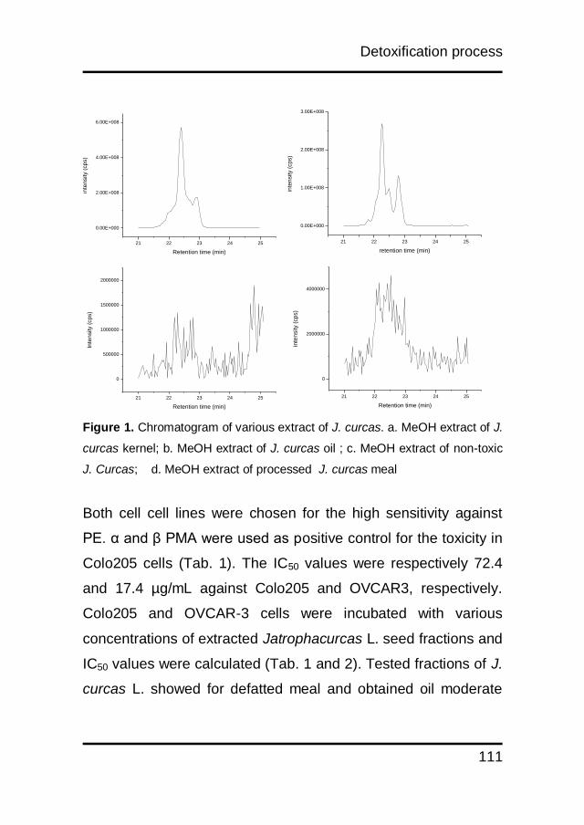
Detoxification process
111
Figure 1. Chromatogram of various extract of J. curcas. a. MeOH extract of J.
curcas kernel; b. MeOH extract of J. curcas oil ; c. MeOH extract of non-toxic
J. Curcas; d. MeOH extract of processed J. curcas meal
Both cell cell lines were chosen for the high sensitivity against
PE. α and β PMA were used as positive control for the toxicity in
Colo205 cells (Tab. 1). The IC50 values were respectively 72.4
and 17.4 µg/mL against Colo205 and OVCAR3, respectively.
Colo205 and OVCAR-3 cells were incubated with various
concentrations of extracted Jatrophacurcas L. seed fractions and
IC50 values were calculated (Tab. 1 and 2). Tested fractions of J.
curcas L. showed for defatted meal and obtained oil moderate
21 22 23 24 25
0.00E+000
1.00E+008
2.00E+008
3.00E+008
inte
nsity (
cp
s)
retention time (min)
21 22 23 24 25
0.00E+000
2.00E+008
4.00E+008
6.00E+008
inte
nsity (
cp
s)
Retention time (min)
21 22 23 24 25
0
500000
1000000
1500000
2000000
Inte
nsity (
cp
s)
Retention time (min)
21 22 23 24 25
0
2000000
4000000
inte
nsity (
cp
s)
Retention time (min)
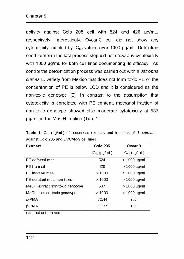
Chapter 5
112
activity against Colo 205 cell with 524 and 426 μg/mL,
respectively. Interestingly, Ovcar-3 cell did not show any
cytotoxicity indicted by IC50 values over 1000 μg/mL. Detoxified
seed kernel in the last process step did not show any cytotoxicity
with 1000 μg/mL for both cell lines documenting its efficacy. As
control the detoxification process was carried out with a Jatropha
curcas L. variety from Mexico that does not form toxic PE or the
concentration of PE is below LOD and it is considered as the
non-toxic genotype [5]. In contrast to the assumption that
cytotoxicity is correlated with PE content, methanol fraction of
non-toxic genotype showed also moderate cytotoxicity at 537
μg/mL in the MeOH fraction (Tab. 1).
Table 1 IC50 (µg/mL) of processed extracts and fractions of J. curcas L.
against Colo 205 and OVCAR-3 cell lines
Extracts Colo 205 Ovcar 3
IC50 (µg/mL) IC50 (µg/mL)
PE defatted meal
PE from oil
PE inactive meal
PE defatted meal non-toxic
MeOH extract non-toxic genotype
MeOH extract toxic genotype
α-PMA
β-PMA
524
426
> 1000
> 1000
537
> 1000
72.44
17.37
> 1000 µg/ml
> 1000 µg/ml
> 1000 µg/ml
> 1000 µg/ml
> 1000 µg/ml
> 1000 µg/ml
n.d
n.d
n.d : not determined
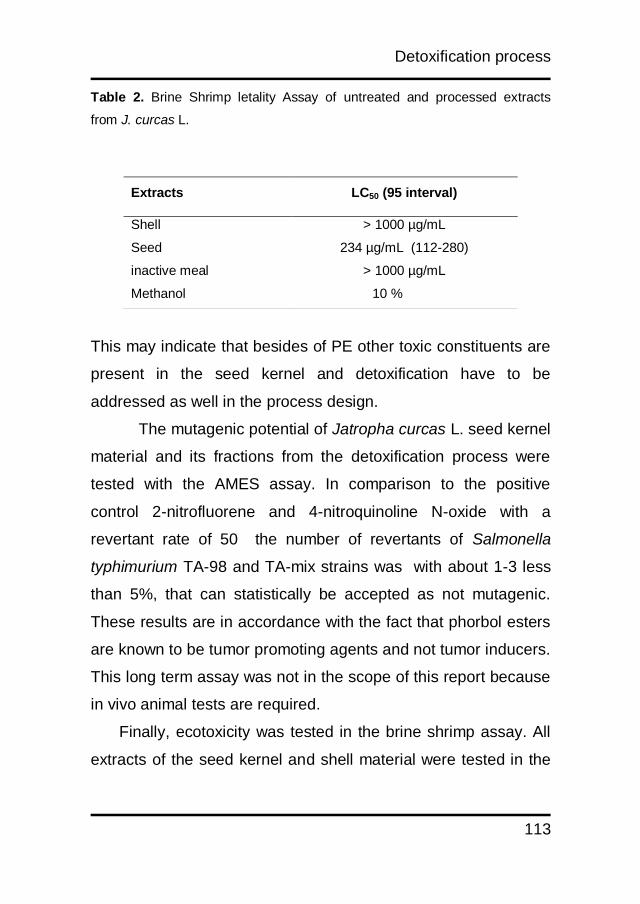
Detoxification process
113
Table 2. Brine Shrimp letality Assay of untreated and processed extracts
from J. curcas L.
This may indicate that besides of PE other toxic constituents are
present in the seed kernel and detoxification have to be
addressed as well in the process design.
The mutagenic potential of Jatropha curcas L. seed kernel
material and its fractions from the detoxification process were
tested with the AMES assay. In comparison to the positive
control 2-nitrofluorene and 4-nitroquinoline N-oxide with a
revertant rate of 50 the number of revertants of Salmonella
typhimurium TA-98 and TA-mix strains was with about 1-3 less
than 5%, that can statistically be accepted as not mutagenic.
These results are in accordance with the fact that phorbol esters
are known to be tumor promoting agents and not tumor inducers.
This long term assay was not in the scope of this report because
in vivo animal tests are required.
Finally, ecotoxicity was tested in the brine shrimp assay. All
extracts of the seed kernel and shell material were tested in the
Extracts LC50 (95 interval)
Shell
Seed
inactive meal
Methanol
> 1000 µg/mL
234 µg/mL (112-280)
> 1000 µg/mL
10 %
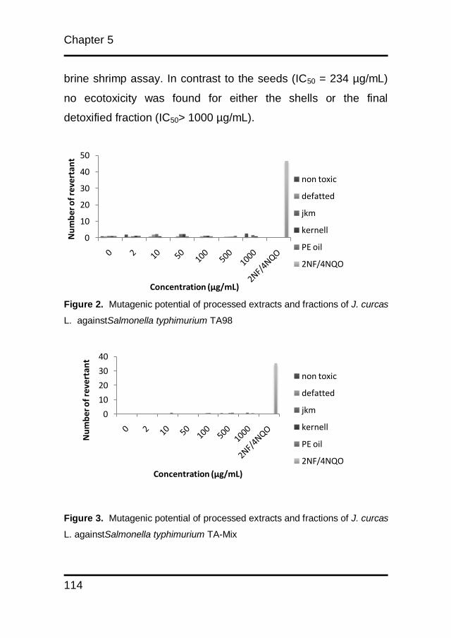
Chapter 5
114
0
10
20
30
40
Nu
mb
er o
f re
vert
ant
Concentration (µg/mL)
non toxic
defatted
jkm
kernell
PE oil
2NF/4NQO
brine shrimp assay. In contrast to the seeds (IC50 = 234 µg/mL)
no ecotoxicity was found for either the shells or the final
detoxified fraction (IC50> 1000 µg/mL).
Figure 2. Mutagenic potential of processed extracts and fractions of J. curcas
L. againstSalmonella typhimurium TA98
Figure 3. Mutagenic potential of processed extracts and fractions of J. curcas
L. againstSalmonella typhimurium TA-Mix
0
10
20
30
40
50
Nu
mb
er o
f re
vert
ant
Concentration (µg/mL)
non toxic
defatted
jkm
kernell
PE oil
2NF/4NQO
Detoxification process
115
5.4 Conclusion
The proposed detoxification process is efficient to separate toxic
principles from the Jatropha curcas seed kernel to obtain a
protein extract being safe to be used as feed stock. No final
fraction showed any mutagenic potential and none showed any
ecotoxicity or cytotoxicity in cell culture testing. By functional
biological assay, only intermediate fractions of the raw kernel
were toxic. These combined results confirm the effectiveness of
the extraction procedure and demonstrate that the processed
meal product can be valorized as potential feed stock. The
applied technical detoxification process may lead to increase
technical capabilities for biorefining of Jatropha curcas L. waste
material and to give new resources for cattle and chicken
production in areas where land resources are limited and fed
supply is critical to raise productive livestock.
Chapter 5
116
References
1. Gandhi VM, Cherian KM, Mulky MJ. Toxicological studies on
ratanjyot oil. Food Chem Toxicol. 1995;33(1):39-42. Epub
1995/01/01.
2. Makkar HPS, Becker K, Sporer F, Wink M. Studies on
Nutritive Potential and Toxic Constituents of Different
Provenances of Jatropha curcas. Journal of Agricultural and
Food Chemistry. 1997;45(8):3152-7.
3. Asseleih LMC, Plumbley RA, Hylands PJ. Purification and
Partial Characterization Of a Hemagglutinin From Seeds of
Jatropha Curcas. Journal of Food Biochemistry. 1989;13(1):1-20.
4. Aderibigbe AO, Johnson COLE, Makkar HPS, Becker K, Foidl
N. Chemical composition and effect of heat on organic matter
and nitrogen degradability and some antinutritional components
of Jatropha meal. Animal Feed Science and Technology.
1997;67(2-3):223-43.
5. Makkar HPS, Aderibigbe AO, Becker K. Comparative
evaluation of non-toxic and toxic varieties of Jatropha curcas for
chemical composition, digestibility, protein degradability and
toxic factors. Food Chemistry. 1998;62(2):207-15.

Detoxification process
117
6. Francis G, Makkar HP, Becker K. Antinutritional factors
present in plant-derived alternate fish feed ingredients and their
effects in fish. Aquaculture. 2001;199(3/4):197-227.
7. Baird WM, Boutwell RK. Tumor-promoting Activity of Phorbol
and Four Diesters of Phorbol in Mouse Skin. Cancer Research.
1971;31(8):1074-9.
8. Haas W, Sterk H, Mittelbach M. Novel 12-deoxy-16-
hydroxyphorbol diesters isolated from the seed oil of Jatropha
curcas. J Nat Prod. 2002;65(10):1434-40. Epub 2002/10/26.
9. Adolf W, Opferkuch HJ, Hecker E. Irritant phorbol derivatives
from four Jatropha species. Phytochemistry. 1984;23(1):129-32.
10. Hirota M, Suttajit M, Suguri H, Endo Y, Shudo K, Wongchai
V, et al. A new tumor promoter from the seed oil of Jatropha
curcas L., an intramolecular diester of 12-deoxy-16-
hydroxyphorbol. Cancer Res. 1988;48(20):5800-4. Epub
1988/10/15.
11. Martínez-Herrera J, Siddhuraju P, Francis G, Dávila-Ortíz G,
Becker K. Chemical composition, toxic/antimetabolic
constituents, and effects of different treatments on their levels, in
Chapter 5
118
four provenances of Jatropha curcas L. from Mexico. Food
Chemistry. 2006;96(1):80-9.
12. Devappa RK, Swamylingappa B. Biochemical and nutritional
evaluation of Jatropha protein isolate prepared by steam
injection heating for reduction of toxic and antinutritional factors.
J Sci Food Agr. 2008;88(5):911-9.
13. Brooker J, inventor; Methods for detoxifying oil seed crops.
Great Britain patent 2466353. 2010 24 June 2010.
14. Staniek A, Woerdenbag HJ, Kayser O. Screening the
endophytic flora of Wollemia nobilis for alternative paclitaxel
sources. Journal of Plant Interactions. 2010;5(3):189-95.
15. Fluckiger-Isler S, Baumeister M, Braun K, Gervais V, Hasler-
Nguyen N, Reimann R, et al. Assessment of the performance of
the Ames II assay: a collaborative study with 19 coded
compounds. Mutation Research/Genetic Toxicology and
Environmental Mutagenesis. 2004;558(1-2):181-97.

CHAPTER 6
Discussion, Summary and Perspective
Chapter 6
120
6.1 General discussion
The process of finding renewable energy sources is still very
interesting issue. The increase of crude oil price every year
make the Indonesian government budget increased, because
they should provide subsidy to reduce the price of the fuel. At the
first time J. curcas provided a new of hope as an alternative
energy sources, because of the low production costs.
Unfortunately this influence on the low selling price, thus making
the profit margins of the farmers were low, it made them look for
other plant to crop. This situation was not expected considering J.
curcas L. has other potential and it can provide a lot of benefits.
Naturally, Jatropha curcas L. produced different kinds of
secondary metabolites, i.e alkaloid, diterpenes, flavonoid,
cyclopeptides and phorbol esters. The two last groups were
studied in this thesis. Cyclopeptides were usually found in
marine organism. In higher plant, almost all member of Jatropha
genus produce these compounds. They can be found mostly in
latex. Cyclopeptides were grouped as oligopeptide because they
were constructed from 3-10 amino acids. They were unique,
because they cannot be visualized by nynhydrin and H2SO4
reagents since they do not have C and N terminal. They also
have different pharmacological activities, like anticancer,
antifungi, and antimicrobial effect. Two cyclopeptides were
isolated from J. curcas latex, they were curcacycline A and B. In
this thesis we found that curcacycline A had antimicrobial effect
Discussion, summary and perspective
121
against Bacillus subtillis and Pseudomonas aeruginosa which
represent positive and negative gram bacteria. So it is potential
to be developed as new antimicrobial. Phorbol esters (PEs)
became an interesting topics although they were toxic and
potential for promoting tumour. Curcin, a ribosome inactivating
protein is a potential lead compound as anticancer and
immunosuppressive drug. Curcusone B isolated from the root
also has good prospects to be used for its antiproliferative effect,
since it inhibits the growth of cancer cell lines. Jatropherol I has
insecticidal activity, because it induces apathological condition in
endoplasmic reticulum, chromatin, lysosome, mitchondria and
microvilli of insects. Phorbol ester has become a major issue
regarding Jatropha curcas L. oil and toxicity. Since it is dissolved
well in the oil, intoxication risk for workers in the oil producing
industry is high. It was reported that multiple steps in the oil
refining process like degumming, deodorization, neutralization
(with alkali hydroxides), bleaching and stripping processes
reduce the level of phorbol ester significantly. Detoxified oil can
be used as a candidate ingredient for ointment and dermal
application preparations.
Jatropha curcas L. pressed cake contains a high protein
yield. It can be used as animal feed, but the utilization is limited
due to its toxic ingredients. A strategy to develop a detoxification
process of the pressed cake has become of high interest.
Physical and chemical treatments have been investigated in
Chapter 6
122
toxic removal. For safety reason, biosafety assays have been
observed for this detoxified product to various in vitro and in vivo
studies. The results will show more evidence whether
detoxification of pressed cake is possible and safe for further use.
In analyzing DHPEs content, HPLC with Dioda Array Detector
was used with β-phorbol myristic acetate as standard. Based on
our findings, this was not correct and can lead to quantification
error. We proved using MRM in tandem mass spectrometer that
both compounds had different fragmentation patterns. This
method gave more advantage because we were able to see the
signal of the sample in smaller concentration.
Developing method for seed detoxification play an
important role for giving J. curcas more valuable, seed of J.
curcas without its toxic contents can be used for animal feed and
fertilizer. In order to test the safety of J. curcas meal, we can use
in vitro and in vivo methods. Several animals have been used for
this assay, they were mice, rat, goat and fish. The meal were
given repeatedly, and some biochemical change were evaluated
to see the influence of it to the animal. In this study, we used in
vitro assays to validate the effectiveness of detoxification
processes. J. curcas meals were applied to the cell culture (for
the carcinogenicity test), Salmonellatyphimurium TA 98 and TA
mix, the results showed that all processed meal were not
cytotoxic and mutagenic.
Discussion, summary and perspective
123
From the data above investigation on latex and J. curcas
meal can be applied for new pharmaceutical product and food
application. These conditions will give more additional economic
values to the farmer who want to crop this plant.
6.2 Summary
Jatropha curcas L. has a great potential and value for
being cultivated as economic crop for biodiesel production. It is
not only a source of non-edible renewable oil, but it also contains
secondary metabolites with interesting biological activities. As
discussed (chapter 2), these secondary natural compounds
were isolated from different parts of Jatropha curcas L. and
characterized. Some experiments have been conducted in the
past for validating pharmacological uses to know their efficacies.
In phorbol ester studies, α dan β Phorbol myristic acetate
(PMA) were used as standards. Although α and β are
stereoisomers, they showed different physicochemical
characteristics (chapter 3). Their maximum wavelengths and
retention times are different. Based on our result using tandem
mass spectrometry, we can confirm that the α dan β PMA also
have different fragmentation pattern with dehydroxy phorbol
ester (DHPE) in J. curcas. Because of that, the use of α dan β
PMA in DHPE analysis is not suitable.
From ethnopharmacological point of view, Jatropha
curcas L. latex has been traditionally used for treating dental
Chapter 6
124
problems in Indonesia, because of its antimicrobial activity. This
claim is proven by previous research which showed antimicrobial
activity of latex against gram positive and gram negative
bacteria. Curcacycline A and B were isolated over twenty years
ago. Isolated curcacycline A and B have now been subjected to
the latest analytical techniques like NMR-spectroscopy and
HPLC/MS to confirm the amino acid sequences. Furthermore,
both structures were confirmed by an organic synthesis
approach. Here we report for the first time about antimicrobial,
mutagenic, cytotoxic and ecotoxic activity of curcacycline A and
B (chapter 4). The results showed that curcacycline A has
antimicrobial activity against Bacillus subtilis and Pseudomonas
aeruginosa and cytotoxic activity against OVCAR 3. No
mutagenic effects were found on curcacycline A and B against
Salmonella typhimurium TA-98 and TA-100. These data showed
that curcacycline A would be potentially a new and safe
antimicrobial candidate.
Different techniques of seedcake detoxification were
developed. To validate the detoxification results, two assays
were done, mutagenicity and cytotoxicity (chapter 5). None of
the extract showed any mutagenic potential and none showed
any cytotoxicity in cell culture testing. By biological assay, only
extracts of raw kernel were toxic. All other extracts had no
significant effect on the survival of brine shrimps. These
combined results confirm the effectiveness of the extraction
Discussion, summary and perspective
125
procedure and demonstrate that the processed meal product has
no mutagenic potential and no detrimental effects on cells in
culture, or in biological assay.
6.3 Perspective
Because J. curcas L. produced many natural products with
interesting pharmacological activities. More efforts should be
done to prove their efficacies (in vitro and in vivo). Not only their
efficacies but their safeties should be observed. Beside doing
toxicity tests by cell culture and brine shrimp test, in vivo studies
in higher animal can be an option. The tests include acute
toxicity test to know LD50 of each compounds and subchronic
test to know the safety of those compounds if they were given in
repeated doses. Beside general toxicity test, we have done
special test for mutagenicity, additional work with s-9 fraction can
be done for additional information if the compounds were
metabolized by liver. Further investigation is still needed for oil
safety, because direct contact of remaining phorbol ester with
skin has to be avoided in all cases. Studies about indoor
pollution by Jatropha curcas L. oil should be conducted, in order
to give a clear distinction from combustion products from the oil
and related toxicity.

Chapter 6
126
In analytical work, validation method for analyzing phorbol
esters should be done using HPLC/MS. This validation work
should cover accuracy, precision, linearity, limit of detection
(LOD), limit of quantification (LOQ), specificity, and robustness.
So the development of this analytical method can be accepted
by others.
Recent progress on Jatropha curcas L. studies indicates
that all parts of this plant are valuable. Utilization of all plant parts
and ingredients can help to increase the economic potential of
Jatropha curcas L. and it might increase the economic value of
huge plantations.

APPENDIX
Nederlandse en Indonesische samenvatting
List of Publication
Acknowledgements
Appendix
128
Nederlandse samenvatting
Jatropha curcas L. heeft als gewas een groot potentieel en
economische waardevoorde productie van biodiesel. Het
isnietalleen een hernieuwbare bron voor olie, maar bevat
ooksecundairemetabolieten met interessante biologische
activiteiten. Hoofdstuk 2 van dit proefschrift beschrijft de
eigenschappen van deze natuurlijke stoffen afkomstig uit de
verschillende delen van het Jatropha curcas L. plant. In het
verleden zijn deze stoffen gebruikt als referentiestof bij de
analyse van farmacologische actieve verbindingen.
In studies naar werking en toxiciteit van phorbol ester worden α
en βphorbol myristic acetaat (PMA) gebruikt als standaards.
Hoewel α en β stereo-isomeren zijn, vertonen ze verschillende
fysisch-chemische karakteristieken (hoofdstuk 3). Zo zijn de
maximale golflengte in de spectrofotometer en de retentie tijden
bij chromatografie verschillend. Door onze resultaten met
tandem massa spectrometrie kunnen we bevestigen dat de α en
β PMA ook verschillende fragmentatie patronen hebben in
vergelijking tot dehydroxyphorbol ester (DPHE) in J. curcas L.
Daarhalve is het gebruik van α of β PMA als referentiestof in
DHPE analyse niet geschikt.
Appendix
129
Vanuit de ethnopharmacologie is bekend dat Jatropha curcas L.
latex gebruikt kan worden voor de behandeling van
tandheelkundige problemen in Indonesië, vanwege de
antimicrobiële activiteit. Deze bevinding is in eerder onderzoek
gedaan, waarin antimicrobiële activiteit van de latex tegen gram
positieve en gram negatieve bacteriën aangetoond werd.
Curcacycline A en B werden ruim twintig jaar geleden geïsoleerd
uit J. curcas L.. Geïsoleerd curcacycline A en B zijn nu
onderworpen aan de meest recente analytische technieken zoals
NMR-spectroscopie en HPLC/MS om de aminozuurvolgorde
bevestigen. Bovendien werden beide structuren bevestigd door
een organische synthese benadering. Hier rapporteren we voor
het eerst over de antimicrobiële, mutagene, cytotoxische en
ecotoxische activiteit van curcacycline A en B (hoofdstuk 4).
De resultaten laten zien dat curcacycline A antimicrobiële
werking heeft tegen Bacillus subtilis en Pseudomonas
aeruginosa en cytotoxische activiteit tegen OVCAR3 cellen.
Geen mutagene effecten werden gevonden van curcacycline A
en B op Salmonella typhimurium TA-98 en TA-100. Deze
gegevens laten zien dat curcacycline A een potentieel nieuwe en
veilige antimicrobiële verbinding is.
Verschillende technieken van detoxificatie van oliezaadkoek zijn
ontwikkeld. Om de detoxificatie resultaten te valideren werden
twee testen gedaan, mutageniteit en cytotoxiciteit (hoofdstuk 5).
Appendix
130
Geen van de extracten toonde mutagene activiteit en ook werd
geen cytotoxiciteit in celcultuur gemeten. Uit biologische testen
bleek dat alleen het extract uit ruwe kernel giftig is. Alle andere
extracten hadden geen significant effect op de overleving van
pekelkreeftjes. Deze gecombineerde resultaten bevestigen de
doeltreffendheid van de extractie procedure en laten zien dat de
verwerkte koek product geen mutagene werking heeft. Ook zijn
er geen aanwijzingen voor toxische effecten op cellijnen of in
een eenvoudig diermodel.
Kesimpulan
Jatropha curcas L. memiliki potensi dan nilai ekonomi
yang besar sebagai produsen biodiesel. Bukan hanya sebagai
sumber bahan bakar terbarukan, tetapi juga memiliki kandungan
metabolit kimia dengan aktivitas biologis yang menarik. Seperti
yang telah dibahas sebelumnya (chapter 2), metabolit sekunder
tersebut telah diisolasi dan dikarakterisasi dari berbagai bagian
tubuh Jatropha curcas L. Beberapa penelitian telah dilakukan
dalam upaya pemastian efek farmakologinya. Curcin, suatu
senyawa penginaktivasi ribosom protein adalah senyawa
potensial sebagai antikanker dan penekan sistem imun tubuh.
Curcusone B yang diisolasi dari akar juga memiliki prospek yang
baik untuk digunakan sebagai antiproliperatif, karena
kemampuannya dalam menghambat pertumbuhan sell kanker.
Appendix
131
Jathoperol I memiliki aktivitas sebagai insektisida, karena dapat
menginduksi patologi pada reticulum endoplasmik, kromatin,
lisosom, mitokondria dan mikrovilli serangga. Curcacycline A
menunjukkan inhibisi pada proliferasi sel T sedangkan
curcacycline B dapat mengikat cyclophilin manusia sehingga
dapat meningkatkan peptidyl-prolil cis trans isomerase.
Phorbol ester menjadi permasalahan utama pada
pengolahan minyak jarak pagar. Karena kelarutannya baik
dalam minyak, resiko intoksikasi pada pekerja pengolahan
minyak jarak sangatlah besar. Untuk itu diperlukan beberapa
penelitian untuk menelaah bahaya tersebut. Telah dilaporkan
bahwa proses pengolahan bertingkat pada pengolahan minyak
seperti degumming, deodorization, neutralization (dengan alkali
hidroksida), bleaching, dan stripping dapat mereduksi kadar
phorbol ester secara signifikan. Hasil pengolahan minyak
tersebut dapat digunakan sebagai kandidat bahan dasar salep
atau sediaan kulit lainnya. Penelitian lanjut diperlukan untuk
melihat keamanan minyak, karena kontak langsung phorbol
ester dengan kulit harus dihindari. Studi tentang polusi akibat
pembakaran pun harus dilakukan, sehingga dapat memberikan
penjelasan apakah pengaruh tersebut akibat minyak atau proses
pembakarannya.
Limbah pengolahan minyak Jatropha curcas L.
(seedcake) diketahui memiliki kandungan protein yang tinggi.
Appendix
132
Limbah tersebut dapat digunakan sebagai bahan makanan
hewan, akan tetapi penggunaannya terbatas oleh kandungan
senyawa toksiknya. Strategi untuk upaya detoksifikasi menjadi
fokus utama. Upaya yang telah dilakukan adalah proses fisika
dan kimia. Untuk alasan keamanan, uji toksisitas telah dilakukan
baik secara in vitro dan in vivo. Hasilnya dapat digunakan
sebagai bukti apakah hasil proses detoksifikasi tersebut aman
digunakan untuk proses selanjutnya.
Dalam penelitian phorbol ester, senyawa yang sering
digunakan sebagai standard adalah α dan β phorbol miristik
asetat (PMA), walaupun kedua senyawa tersebut adalah
stereoisomer akan tetapi memperlihatkan karakteristik
fisikokimia yang berbeda (chapter 3), tidak heran kedua
senyawa tersebut memiliki aktivitas promoter tumor yang
berbeda. Kedua senyawa tersebut juga memiliki karakteristik
fragmentasi ionisas yang berbeda dengan senyawa dehidroksi
phorbol ester (DHPE) phorbol ester yang terdapat pada Jatropha
curcas L. Sehingga pada penelitian ini disimpulkan bahwa
penggunaan α dan β PMA sebagai standard tidaklah tepat untuk
analisis DHPE.
Dari sudut pandang etnofarmakologi, getah J. curcas
telah digunakan secara tradisional oleh masyarakat untuk
mengatasi permasalahan gigi. Terutama sebagai antimikroba.
Klaim khasiat tersebut telah dibuktikan dari beberapa penelitian
Appendix
133
sebelumnya, yang menunjukkan aktivitas getah pohon jarak
pagar terhadap beberapa bakteri gram positif dan gram negatif.
Curcacyline A dan B telah diisolasi lebih dari 20 tahun yang lalu.
Isolasi dan karakterisasi curcacycline A dan B pada penelitian ini
menggunakan spektroskopi NMR dan HPLC/MS untuk
mengkonfirmasi urutan asam amino yang ada. Selain itu, upaya
sintesis organiknya pun telah berhasil dilakukan. Pada penelitian
ini dilaporkan untuk pertama kali mengenai aktivitas antimikroba,
mutagenisitas, sitotoksisitas, dan ekotoksisitas dari curcacycline
A dan B (chapter 4). Hasil penelitian menunjukkan bahwa
curcacycline A memiliki aktivitas antimikroba terhadap Bacillus
subtilis dan Pseudomonas aeruginosa dan aktivitas sitotoksik
terhadap sel OVCAR 3. Tidak ditemukan efek mutagenik
terhadap Salmonella typhimurium TA-98 dan TA-mix. Hasil
tersebut menunjukkan bahwa curcacycline A potensial
dikembangkan sebagai antimikroba.
Beberapa teknik telah dikembangkan untuk detoksifikasi
ampas J. curcas. Untuk memvalidasi proses detoksifikasi, tiga
metode digunakan. Uji mutagenik menggunakan metode AMES
test II, uji sitotoksik menggunakan sel kanker, dan uji
ekotoksisitas menggunakan brine shrimp. Hasil penelitian
menunjukkan tidak ada ekstrak yang memiliki efek sitotoksik
ataupun mutagenik. Uji ekotoksisitas menunjukkan bahwa
ekstrak dari biji J. curcas yang belum diolah toksik terhadap
larva Artemia salina. Hal ini membuktikan bahwa upaya
Appendix
134
detoksifikasi yang telah dilakukan tidak menunjukkan efek yang
berarti sehingga dapat dikatakan proses tersebut telah efektif.
Berdasarkan penelitian terhadap tanaman J. curcas
mengindikasikan bahwa seluruh bagian tanaman sangat
berharga. Penggunaan seluruh bagian tanaman akan dapat
meningkatkan nilai ekonomi J. curcas dan memberikan
keuntungan bagi petani yang menanamnya.
Appendix
135
List of Publications
1. Muhamad Insanu, Jana Anggadireja, Oliver Kayser, 2010,
Isolation of curcacycline A from latex of Jatropha curcas
and its antibiotic and cytotoxic effect, Planta Medica, 76
(12), P-435
2. Muhamad Insanu, Jana Anggadireja, Oliver Kayser, 2012,
Curcacycline a and b – new pharmacological insights to
an old drug,International Journal of Applied Research in
Natural Products, 5 (2), 26-34
3. Muhamad Insanu, Chryssa Dimaki, Richard Wilkins, John
Brooker, Piet van der Linde, Oliver Kayser, 2013, Rational
use of Jatropha curcas L. In food and medicine : from
toxicity problems to safe applications, Phytochemistry
reviews, 12 (1), 107-119
Appendix
136
Acknowledgements
Finally this thesis is DONE !! it became possible through the help
and support from everyone, please allow me to dedicate my
acknowledgment of gratitude toward the following significant
advisors and contributors:
First and formost,I would like to express my deep gratitude to
Professor Oliver Kayser and Professor Wim J Quax my research
supervisors, without their assistance and dedicated involvement
in every step throughout the process, this thesis would have
never been accomplished. I would like to thank you very much
for your support and understanding over these past „seven‟
years.
I would also like to thank Dr. Robert Manurung and Professor
Erik Heeres who opened the door for me to go to the
Netherlands and also involving me on KNAW SPIN programme.
I am particularly grateful for the assistance given by Rita, for
helping me working with cell culture during my experiment also
reading and correcting my thesis. U are so “Indonesian” Rita…
Also to Ronald and Pieter that encourage me to have an interest
in HPLC machine, thank you.
Appendix
137
Janita and Yvonne, My special thanks are extended for you, for
willingness to give your time and helping me with administration
in Groningen.
Thank you for the support from MS department, Hjalmar, Margot
and Annie.
I wish to acknowledge the help provided by Decanate School of
Pharmacy ITB, Prof. Dr. Tutus Gusdinar, Prof. Dr. Daryono Hadi,
Prof. Sukrasno, Dr. Marlia Singgih, and Dr. Rahmana Emran, for
supporting me during the time I spent at the ITB.
I would also like to thank the Pharmaceutical biology group :
Gerrit, Robert, Remco, Louis, Gani, Octavia, Carlos, Agata,
Gudrun, Pol, Mariette, Hans, Vinod, Magda, Elena, Bertjan, Art,
Mariana, and Eveline for the discussion and also some “parties”
on the weekend .
I would like to express my very great appreciation to
Pharmaceutical Biology research group, School of pharmacy
ITB, Prof Dr. Komar Ruslan (never forget to give me wise advice
for life….thanks pak ), Prof. Dr. Asep Gana, Dra. Siti
Kusmardiyani, M.Sc., Dr. Irda Fidrianny, Dr. Asyari Nawawi, Dr.
Elfahmi, Rika Hartati, M.Si., Hegar Pramastya, M.Si.
I would like to give a special thanks to my colleagues and friends
in Jatropha group : Dianika, Laura, Louis, Erna, Pak Gani, pak

Appendix
138
Ahmad, Pak Herman. It is always nice to have discussion with
people who are in the same topics.
I would like to express my sincere thanks to my Bapak dan Ibu
kos : Kang Nandang and teh Nisa who allowed me spent the day
and night in their house when i first arrived, also Guntur and
Intan when I prepared going back for good. To Saturday nights
gank, Arramel, and Iging you are my partners in crime, Puti &
meisya, desti dan daanish best wishes for you all.
To PPIG, with no particular order i would like to thank mbak Puri,
teh Uyung, Robby & Lia, Iqbal & Eryth, Novian, kang Hendi,
Rachmawati, mas Adhi, Fajar, Febi, pak Iswandi, kang Intan &
teh Enci, mas Teguh & mbak Tessa, mbak Tina, Faizah & Panji,
Fajar, Rizkiya Kudus, Mira, Aditya, Neng, mbak ari, Dini, mbak
Ike, mas Indra & Senaz, Helmi, Mckenzie, mbak Tina, Yusuf, pak
Adji Kusworo (i found another family while staying in Groningen).
Most importantly, none of this could have happened without my
family. My parents (ibu Yiyis dan Bapak Yuswir), this PhD thesis
is dedicated for both of you as a testament to your unconditional
love and encouragement. Your prayer for me was what
sustained me thus far. Words cannot express how grateful I am
to my mother-in law (Ambu), father-in-law (Abah), for all of the
supports that you‟ve made on my behalf. I would also like to
thank all of my brothers and sisters Gema, Beta & Raja, Gina,

Appendix
139
and Gani who supported me and motivated me to strive towards
my goal. At the end, I would like express appreciation to my
beloved wife Gemi, and my beautiful girls Mahira and Miura
whom Ispent sleepless nights with and who always gave me
motivation to finish this PhD.