Embed Size (px)
Citation preview
University of Groningen
Molecular dynamics study of the folding of hydrophobin SC3 at a hydrophilic/hydrophobicinterfaceZangi, R.; de Vocht, M.L.; Robillard, G.T.; Mark, A.E.
Published in:Biophysical Journal
DOI:10.1016/S0006-3495(02)75153-9
IMPORTANT NOTE: You are advised to consult the publisher's version (publisher's PDF) if you wish to cite fromit. Please check the document version below.
Document VersionPublisher's PDF, also known as Version of record
Publication date:2002
Link to publication in University of Groningen/UMCG research database
Citation for published version (APA):Zangi, R., de Vocht, M. L., Robillard, G. T., & Mark, A. E. (2002). Molecular dynamics study of the folding ofhydrophobin SC3 at a hydrophilic/hydrophobic interface. Biophysical Journal, 83(1), 112 - 124.https://doi.org/10.1016/S0006-3495(02)75153-9
CopyrightOther than for strictly personal use, it is not permitted to download or to forward/distribute the text or part of it without the consent of theauthor(s) and/or copyright holder(s), unless the work is under an open content license (like Creative Commons).
Take-down policyIf you believe that this document breaches copyright please contact us providing details, and we will remove access to the work immediatelyand investigate your claim.
Downloaded from the University of Groningen/UMCG research database (Pure): http://www.rug.nl/research/portal. For technical reasons thenumber of authors shown on this cover page is limited to 10 maximum.
Download date: 26-02-2019
Molecular Dynamics Study of the Folding of Hydrophobin SC3 at aHydrophilic/Hydrophobic Interface
Ronen Zangi,* Marcel L. de Vocht,† George T. Robillard,†‡ and Alan E. Mark**Department of Biophysical Chemistry and †Department of Biochemistry, University of Groningen, and ‡Biomade Technology Foundation,Nijenborgh 4, 9747 AG Groningen, The Netherlands
ABSTRACT Hydrophobins are fungal proteins that self-assemble at hydrophilic/hydrophobic interfaces into amphipathicmembranes. These assemblages are extremely stable and posses the remarkable ability to invert the polarity of the surfaceon which they are adsorbed. Neither the three-dimensional structure of a hydrophobin nor the mechanism by which theyfunction is known. Nevertheless, there are experimental indications that the self-assembled form of the hydrophobins SC3and EAS at a water/air interface is rich with �-sheet secondary structure. In this paper we report results from moleculardynamics simulations, showing that fully extended SC3 undergoes fast (�100 ns) folding at a water/hexane interface to anelongated planar structure with extensive �-sheet secondary elements. Simulations in each of the bulk solvents result in amainly unstructured globular protein. The dramatic enhancement in secondary structure, whether kinetic or thermodynamicin origin, highlights the role interfaces between phases with large differences in polarity can have on folding. The partitioningof the residue side-chains to one of the two phases can serve as a strong driving force to initiate secondary structureformation. The interactions of the side-chains with the environment at an interface can also stabilize configurations thatotherwise would not occur in a homogenous solution.
INTRODUCTION
Hydrophobins are small fungal proteins (�100 amino acidsresidues) that self-assemble at hydrophilic/hydrophobic in-terfaces (e.g., water/air, water/oil, and water/solid-hydro-phobic-surface) into amphiphatic membranes. They are re-sponsible for many functions in fungal growth anddevelopment, such as the formation of hydrophobic surfacesfound in aerial hyphae, spores, and fruiting bodies (Wostenet al., 1994; Talbot et al., 1996; Wessels, 1997; Wosten andWessels, 1997).
Hydrophobins exhibit interfacial activity and are amongthe most surface-active biomolecules known. Based on thehydrophaty patterns and on the solubility of the assembledmembranes, hydrophobins are classified into two classes, Iand II (Wessels, 1994). In the class I hydrophobins, theamphipathic membrane is highly insoluble and on the hy-drophobic side it is characterized by a rodlet pattern thatresembles that of amyloid proteins. Assemblages formed byclass II hydrophobins are more soluble and do not formrodlet structures.
SC3, a glycosylated hydrophobin that is secreted bySchizophyllum commune (Wessels et al., 1991; Wosten etal., 1993), is currently the most surface-active proteinknown (with a maximal lowering of the water surfacetension from 72 mJ/m�2 to 24 mJ/m�2). It is also the mostextensively studied hydrophobin to date. SC3 hydrophobinis released into the growth medium in a water-soluble form
that subsequently self-assembles into insoluble films at thewater/air interface. SC3 is a class I hydrophobin and isposttranslationally modified with 16 to 22 O-linked man-nose residues being attached to the N-terminal part of thepeptide chain.
Class I hydrophobins are characterized by a specifichydropathy pattern in their primary sequence and the strictconservation of eight cysteine residues that form four disul-fide bridges.
The secondary structure content of soluble and assembledSC3 hydrophobin as inferred from Fourier transform infra-red, and circular dichroism (CD) studies (de Vocht et al.,1998) is summarized in Table 1.
The soluble form of SC3 contains on average �40%�-sheet structure, which increases to 65% on self-assem-bly. This enhancement of �-sheet structure at the water/air interface plays an important role in the properties ofhydrophobins, specifically in the ability to reduce thesurface tension and to organize into the characteristicrodlet structure.
The secondary structure content of SC3 at the interfacebetween water and an apolar liquid is not known. However,rodlet formation is observed at a water/oil interface, sug-gesting that SC3 adopts a primarily �-sheet structure at thisinterface (Wosten et al., 1994).
No tertiary structure of hydrophobins is available. It hasnot been possible to obtain high-quality nuclear magneticresonance spectra of the soluble form of SC3 due to aggre-gation. It has also not been possible to stabilize the solubleform by the addition of sodium dodecyl sulfate, ethanol ordimethyl sulfoxide. Secondary structure prediction by aprofile-fed neural network system predicts that the N-ter-minal segment before the first cysteine residue (which con-tains the mannose residues) consists primarily of loop struc-
Received for publication September 27, 2001, and in final form March 13,2002.
Address reprint requests to Alan E. Mark, Department of BiophysicalChemistry, University of Groningen, Nijenborg 4, 9747 AG Groningen,The Netherlands. Tel.: 31-50-363-4457; E-mail: [email protected].
© 2002 by the Biophysical Society
0006-3495/02/07/112/13 $2.00
112 Biophysical Journal Volume 83 July 2002 112–124

ture. The remaining part of SC3, the cysteine-rich region,consists of alternating �-sheet and loop structures.
The cysteine-rich region is the region where the enhance-ment of the �-sheet structure takes place upon assembly atan interface. This is evident from studies on a truncated SC3in which 26 of the 31 N-terminal amino acids were removed(Fig. 1). Neither the functionality of the protein nor theconformational changes that occur upon assembly wereaffected. Truncated SC3 is able to self-assemble and to formrodlets. It shows the same degree of surface tension reduc-tion but weaker binding to hydrophilic surfaces (de Vocht etal., 1998).
The origin of the strong interaction between hydropho-bins and hydrophobic surfaces is unknown. Protein adsorp-tion is often characterized by a loss of secondary andtertiary structure and may be associated with an increase inentropy of the peptide and enhanced rotational mobilityaround the polypeptide backbone. In contrast, hydrophobinsshow an increase in secondary structure upon adsorptionindicating that specific conformational changes give rise tothe strong hydrophobic adhesive properties.
The water/air interface apparently acts as a catalyst forthe formation of rodlets but is not required for stability.
After drying down a solution of SC3 a rodlet layer with athickness of �10 nm is obtained. The diameter of a typical100 amino acid globular protein is �3 nm. Thus, the rodletsmust be comprised of more than one protein layer or theprotein must be highly elongated in shape.
Interfacial folding is observed not just in hydrophobinsbut also in other protein systems such as transmembraneproteins, toxins, antibiotics, and some hormones. Experi-mental studies have shown that certain naturally occurring(Cornell et al., 1993; Wu et al., 1995; Perez-Paya et al.,1997; Berneche et al., 1998), synthetic peptides (Tamm etal., 1989; Takahashi, 1990; Bechinger, 1996; Russell et al.,1998), and fragments of larger proteins (Segrest et al., 1994;Chernomordik et al., 1994; Voglino et al., 1998; Johnson etal., 1998), can readily form amphipathic structures at hy-drophilic/hydrophobic interfaces. The initial orientation ofthe peptides is in general parallel to the interface (Ishiguroet al., 1993; Bechinger et al., 1993, 1998; Wu et al., 1995;Cajal et al., 1996). However, at sufficiently high concentra-tion and/or in the presence of an electric field, some adopta perpendicular orientation (Wu et al., 1995; Biggin andSansom, 1996; Lear et al., 1997).
As it is difficult to study the process of adsorption atinterfaces in atomic detail experimentally, several groupshave turned to molecular dynamics (MD) computer simu-lation techniques to investigate interfacial folding. Chipotand Pohorille (1998b) showed in a simulation study of anundecamer of poly-L-leucine at a water/hexane interfacethat when placed initially on the water side in a random coilconformation, the peptide translocated toward the hexanephase and underwent interfacial folding into an �-helix. The
FIGURE 1 Primary structure of truncated SC3 showing the four loops formed by the four disulfide bridges. Cysteine residues are yellow. Hydrophilicand hydrophobic residues are blue and red, respectively.
TABLE 1 Percentage of specific secondary structureelements in SC3 as inferred from ATR-FTIR spectroscopy
�-Helix �-Sheet �-Turn Coil
Soluble 23 41 16 20Assembly at water/air 16 65 9 10
Data taken from reference (de Vocht et al., 1998).
Interfacial Folding of Hydrophobin SC3 113
Biophysical Journal 83(1) 112–124

helical peptide was largely buried in hexane yet remainedadsorbed at the interface with a parallel orientation. Therapid coil to helix transition, which occurred within 36 ns,suggested that at interfaces elements of secondary structuremay form before slower, long range tertiary contacts aremade.
The placement of an amphipathic peptide or protein at aninterface between a hydrophilic and a hydrophobic phase(which in effect represents an external electric field) intro-duces an additional parameter that can determine the natureof the free energy surface and its minimum. Amino acidresidues of the peptide or protein will partition into therespective phases according to their hydrophobicity. Thiswill preorganize the structure in solution leading to rapidsecondary structure formation. There are indications that insome cases the optimum conformation is insensitive to thenature of the hydrophobic phase. Some amphipathic pep-tides adopt the same secondary structure at water/mem-brane, water/alkane, and water/air interfaces (DeGrado andLear, 1985; Chung et al., 1992; Blondelle et al., 1995).
Although the partitioning of residues into one of thephases can initiate secondary structure formation, transi-tions between different amphipathic structures at an inter-face could require the surmounting of high free energybarriers. Nonoptimized folded structures with a satisfactorypartitioning of the residues may represent local minimum onthe free energy surface and impede folding (Chipot et al.,1999).
In this paper, MD simulation techniques are used to studythe initial stages of folding of hydrophobin SC3 at a water/hexane interface. The behavior of SC3 in bulk water andbulk hexane is also investigated for comparison. We findthat fully extended SC3 undergoes rapid folding at a water/hexane interface to a structure with extensive �-sheet con-tent. Simulations in each of the bulk phases result in amainly unstructured globular protein. The enhancement ofsecondary structure, whether kinetically or thermodynami-cally determined, highlights the role interfaces with a largedifference in polarity can have in catalyzing folding. Thepartitioning of the residue side-chains between the twophases provides a strong driving force to initiate secondarystructure formation. Furthermore, the interaction of givenside-chains within a hydrophobic or hydrophilic environ-ment can initially stabilize the creation of structural ele-ments that might otherwise not occur in a homogenoussolution.
MATERIALS AND METHODS
The MD simulations were preformed using the GROMACS packageversion 2.0 (Berendsen et al., 1995; van der Spoel et al., 1999) with theGROMOS96 (43A2) force field (van Gunsteren et al., 1996). SC3 wassimulated in three different environments: a water/hexane interface, bulkwater, and bulk hexane. Water was described by the simple point charge(SPC) model (Berend-sen et al., 1981). The hexane model was taken fromthe GROMOS96 force field version 43A2 in which the equilibrium distri-
bution of dihedral angles in alkanes is reproduced better then in previouslypublished force field versions (Schuler and van Gunsteren, 2000).
To maintain the system at a constant temperature of 300 K, a Berendsenthermostat (Berendsen et al., 1984) was applied using a coupling time of0.1 ps. The pressure was maintained by coupling to a reference pressure of1 bar. A coupling time of 1.0 ps was used for the simulations at theinterface and in bulk water, whereas a coupling constant of 2.0 ps was usedfor the simulation in bulk hexane (Berendsen et al., 1984). The values ofthe isothermal compressibility were set to 10.6 � 10�5, 4.5 � 10�5,16.7 � 10�5 bar�1 for water/hexane, water and hexane simulations,respectively. For the evaluation of the nonbonded interactions a twin rangecutoff of 0.9 and 1.4 nm was used. Interactions within the shorter cutoffwere updated every step, whereas interactions within the longer cutoff wereupdated every five steps. For the systems that contained water, the timestep used was 0.002 ps. However, because the GROMOS96 force field usesa united atom model for CH2 and for CH3 groups a larger time step wasused for the simulations in bulk hexane. After 14 ns from the point thesulfur-sulfur (S-S) bonds were formed, the time step was increased from0.002 to 0.004 ps. At the same time the mass of the polar hydrogen atomswithin the protein, which were treated explicitly, was increased to 4 amu.The increased mass of the hydrogen was subtracted from the mass of theheavy atom to which the hydrogen was bonded leaving the total massunchanged. The overall result is the removal of high frequency vibrationalmotion involving the hydrogen atoms allowing an increase of the integra-tion time step (Feenstra et al., 1999).
Water bond distances and angles were constrained using the SETTLEalgorithm (Miyamoto and Kollman, 1992), whereas the hexane and theprotein bond distances were constrained using the SHAKE algorithm(Ryckaert et al., 1977) with a geometric tolerance of 1 � 10�4.
The system simulated was that of an 86 amino acid truncated form ofSC3, the same as that used in the corresponding experimental studies. Inthis form of SC3, 29 N-terminal residues of the native SC3 (up to twoamino acids before the first cysteine) were removed and substituted by thesequence Gly-His-Pro (see Fig. 1). This three amino acid sequence ischaracteristic of many hydrophobins at this position. The N and the Ctermini were protonated and deprotonated, respectively. The negativecharges carried by the residues Asp-38 and Glu-68 resulted in a totalcharge of �2e.
The starting structure of the protein in the three simulations was a fullyextended conformation of truncated SC3 constructed using the programWHATIF (Vriend, 1990). In the simulation of the water/hexane interfacethe extended protein was aligned on the interface.
The simulation cell was a rectangular periodic box with the minimumdistance between the protein and the box walls set to 0.75 nm, so that theprotein did not directly interact with its own periodic image given thecutoff. The box dimensions were changed several times during the simu-lations. This was necessary because the protein underwent very largechanges in shape (especially at the beginning) and for reasons of efficiencyas initially large amounts of solvent were required to solvate the extendedstructures (�79,000 atoms in the case of bulk water). The extendedconformation enforced a single long axis on the simulation box. As theprotein collapsed the other two axes had to be increased to prevent theprotein being restricted in any direction. To change the box dimensions, theprotein configuration and the maximum possible volume of solvent aroundit (as determined by the new box dimensions) were kept. A new region ofpreequilibrated solvent molecules was then added to the extended direc-tion. In the water/hexane simulations, the length of the axis perpendicularto the interface was held constant when changing the box size. The lengthof this axis fluctuated (due to the pressure coupling) around 4.7 nmthroughout the simulation.
To form the four disulfide bridges starting from the extended conformation of the protein, distance constraints between the pairs of sulfur atomsthat form disulfide bridges were imposed. A coupling parameter � wasused to gradually reduced the constraint distance from that in the fullyextended conformation (� is 0) to an S-S distance of 0.21 nm (� is 1).During this process, each of the S-S distances were decreased at each step
114 Zangi et al.
Biophysical Journal 83(1) 112–124

by a distance corresponding to �� � 1 � 10�5. Once the four S-Sdistances reached 0.21 nm the distance restraint was replaced by a bondconstraint between the sulfur atoms of 0.204 nm as given in the force fieldused. The process of S-S bond formation was performed separately in eachof the three different environments. In the simulations at the interface andin water the process was split into two stages to allow for a reduction in thesize of the system.
The degree of amphipathy of the protein was estimated from its meanstructural hydrophobic moment (Eisenberg et al., 1982), ��� H�,
��� H� �1
N �i�1
N
Hi � S� i (1)
in which N denotes the number of amino acid residues in the protein, Hi isthe hydrophobicity of residue i and S�i is the unit vector pointing from the�-carbon atom of the ith residue to the center of mass of its side-chain. Thevalues for the hydrophobicities, Hi, were taken from Wolfenden et al.(1981). Because in glycine the side chain hydrogens are incorporated intothe �-carbon, which is treated as a united atom, the contribution of glycineresidues to the hydrophobic moment was not considered.
The analysis of the secondary structure elements was based on theDSSP (define secondary structure of proteins) definitions (Kabsch andSander, 1983). The figures of the protein, and thereby the secondarystructure assignment shown in the figures, were generated using the visu-alization package MOLMOL (Koradi et al., 1996).
Details regarding the simulations in the three environments are given inTable 2. Note that in the simulation at the water/hexane interface thesystem was relaxed for 2.4 ns before the process of disulfide bridgeformation was initiated. This was to allow the orientation of the residueside-chains in the extended conformation (initially placed arbitrarily) torelax and insert into their preferred phase. The time required for theside-chains to reorient was in the order of a few hundred picoseconds.
A second simulation of truncated SC3 at the water/hexane interface wasperformed for 51 ns. The starting conformation was taken from the firstsimulation at the interface after the formation of the sulfur bonds (i.e., at t �0) but with new random velocities. This simulation is labeled water/hexane-2.
All simulations were performed on a PentiumIII based linux cluster.The 135-ns trajectory at the interface required approximately the equiva-lence of five months processing time on a 866 MHz dual processor node.
RESULTS
In the initial stage of the relaxation process, after the S-Sbonds were formed, the peptide chain contracted rapidly(within picoseconds) as the linear conformation of the seg-ment of peptide between the second and third loops, whichwas unaffected by the formation of the disulfide bridges, ishighly unfavorable. Following this initial phase, the system
continued to collapse but at a slower rate (nanoseconds).The radius of gyration of SC3 as a function of time in eachof the three environments is shown in Fig. 2.
The radius of gyration in bulk water and bulk hexane was1.2 and 1.4 nm, respectively. At the interface, the value ofthe radius of gyration was 1.9 nm. This reflects the largevalue of the interface-plane component as is also apparentfrom inspection of the final structure.
The structures of SC3 at selected points along each tra-jectory are plotted in Figs. 3, 4, and 5.
The structure formed at the interface is essentially planarwith the peptide lying along the interfacial plane throughoutthe trajectory. In contrast the structures in the bulk solventsare globular. There is a clear enhancement of secondarystructure formation at the interface compared with that inthe bulk solvents. This is mainly in a form of �-sheet,although there is a small region (5–7 residues long) with�-helical structure. A plot of the secondary structure, de-fined using the DSSP algorithm, as a function of time isshown in Fig. 6.
The formation of �-sheets at the interface was ob-served to be a dynamic process especially at the initialstage of the trajectory. In few cases small segmentsfolded then unfolded only to later refold with the sameresidues or with others. However, the number of suchevents decreases as the folding of SC3 progresses. Theaddition of the third and the fourth strands to the two-stranded �-sheet segment initially formed is partialysequential.
FIGURE 2 Radius of gyration (nanometers) of SC3 as a function of time atthe water/hexane interface, in water and in hexane. The zero time indicates thepoint at which the disulfide bridges were formed. The values were calculatedevery 400 ps and averaged over a window of five adjacent points.
TABLE 2 Details of the procedures used to conduct thesimulations in the three environments
Simulation of SC3 in:
Water/hexane Water Hexane
Initial # of water molecules 7972 25,990 –Initial # of hexane molecules 1436 – 6780Number of steps of energy
minimization30 30 530
Relaxation time before S-S (ns) 2.4 – –Time of S-S (ps) 400 400 200Relaxation time after S-S (ns) 135 100 100
S-S denotes the disulfide-bridge forming process.
Interfacial Folding of Hydrophobin SC3 115
Biophysical Journal 83(1) 112–124

Selected structures from the second simulation at thewater/hexane interface as well as the DSSP plot are shownin Fig. 7.
The extended starting conformation used in all the sim-ulations is highly unstable. The simulations give rise tononequilibrium trajectories and large variations in the struc-ture of the protein as a function of time (especially in the
initial part of the trajectory) are observed. This makes anystatistical analysis of the trajectories problematic and re-quires a somewhat arbitrary choice of when the system has“equilibrated.” In the analysis that follows, the first 50 nswere excluded in the case of the longer trajectories in eachof the three environments, whereas for the shorter simula-tion at the interface, water/hexane-2, only the first 25 ns
FIGURE 3 Structures of SC3 from the simulation at the water/hexane after 0, 10, 50, and 135 ns. The sulfur atoms in the cysteine residues are shownin yellow.
FIGURE 4 Structures of SC3 from the simulation in water after 0, 10, 50, and 100 ns. The sulfur atoms in the cysteine residues are shown in yellow.
116 Zangi et al.
Biophysical Journal 83(1) 112–124

were discarded. The average secondary structure content foreach simulation is summarized in Table 3.
The major difference between the two simulations at theinterface is the lower percentage of �-sheet structure and theabsence of �-helix in the second simulation. However, thesame general mechanism of folding at the interface, that ofan essentially quasi two-dimensional system yielding a pla-nar folded structure with the very rapid formation of �-sheetwas found in both simulations.
The hydrophobic moment (Fig. 8) and the solvent acces-sible surface area of the protein were calculated from thetrajectories and are summarized in Table 4.
As expected the alignment of the side-chains leads to amore amphipathic structure in the simulation at the interfacethen in either of the two bulk phases. The value of ��� H� atthe interface is at least twice as high as the value obtainedin water or in hexane.
Truncated SC3 contains 41 hydrophilic and 45 hydropho-bic residues. Therefore, it is reasonable to expect that,qualitatively, the behavior of the collapsed structure inwater and in hexane would be similar (although differentresidues would be exposed to solvent) and that the value ofthe hydrophobic moment would be comparable. This is thecase even though the radius of gyration and the solventaccessible surface area are higher in hexane.
To determine the time scale on which side-chains orien-tate toward their preferred solvent, and to obtain values of��� H� when no constraints on the sulfur atoms were imposed,a series of short (2 ns) simulations were performed from afully extended conformation. The simulations were con-ducted in the three environments and during the 2 ns sim-ulated neither the collapse of the structure nor any second-
ary structure formation was observed. The orientation ofside-chains is relatively fast (�200 ps) and the value of��� H� averaged over the last 1 ns is 0.95, 0.40, and 0.27 forthe simulations at the interface, in water, and in hexane,respectively. Thus, there is a marked decrease in the valueof ��� H� at the interface as the protein folds and attainssecondary structure. The average value of the hydrophobicmoment in the second simulation at the interface is higherthan in the longer simulation. This is to be expected as thepercentage of �-sheet is smaller and more residues are freeto orientate toward their preferred phase.
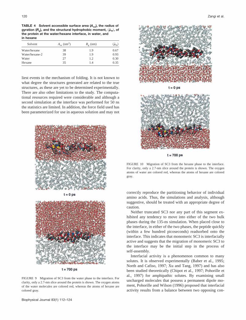
To test the tendency of SC3 to reside at the interface, twosimulations in which SC3 was placed in the bulk phases butclose to the interface were performed. In the first simula-tion, the peptide was placed in the water phase, and in thesecond, the peptide was placed in the corresponding mannerbut in the hexane phase. In both cases, the starting confor-mation was taken from the extended simulation at the waterhexane interface (after 135 ns). Each system was simulatedfor 4 ns. The behavior of the protein with respect to theinterface in the two simulations was very similar. Within afew hundred picoseconds the protein had fully readsorbedon the interface. Figs. 9 and 10 show the initial position ofSC3 with respect to the interface as well as the position after700 ps from the simulations with SC3 starting in water andin hexane, respectively. The time-scale for the migration tothe interface and the final interfacial position were compa-rable in both cases.
Averages of the energy components (Lennard-Jones andCoulomb) were calculated for the interactions inside theprotein and for the interactions between the protein and thesolvent. The results are shown in Table 5.
FIGURE 5 Structures of SC3 from the simulation in hexane after 0, 10, 50, and 100 ns. The sulfur atoms in the cysteine residues are shown in yellow.
Interfacial Folding of Hydrophobin SC3 117
Biophysical Journal 83(1) 112–124

The strongest intraprotein interactions occur when theprotein is solvated in hexane. As hexane is uncharged,electrostatic interactions act only between atoms within theprotein and it is most likely that electrostatic interactionsdrive the collapse of the protein in this solvent. Surprisingly,the value of the protein-protein interaction energy is slightlyhigher in the simulation in water than at the water/hexaneinterface. The primary contribution to the protein-proteinelectrostatic interaction at the interface is due to the forma-tion of backbone-backbone hydrogen bonds within elementsof �-sheet. In water, little regular secondary structure hasformed. Nevertheless, the overall intraprotein electrostatic
interaction is comparable with that on the interface. Themain difference is the stronger Lennard-Jones interactions,a result of adopting a compact globular structure enforcedby the hydrophilic surroundings. The weaker Lennard-Jonesinteractions within the protein at the water/hexane interfaceare compensated by interactions between the protein and thesolvent. Summing all contributions, the total energy insidethe protein and of the protein with the solvent is slightlymore favorable in water simulation than at the interface.However, as the energetic (or enthalpic) contribution to theoverall free energy of the system for the protein at theinterface includes both interactions that involve the protein
FIGURE 6 Secondary structure assignment of the protein (DSSP) as a function of time for the simulations in the three environments.
118 Zangi et al.
Biophysical Journal 83(1) 112–124

(intraprotein interactions and interactions with solvent) andinteractions between solvent molecules, it is not possible tojudge which state should be more stable based on theseconsiderations alone. The entropic contributions to the bind-ing of SC3 at the interface are difficult to assess. The translational entropy in one dimension and the rotational entropyaround two axes is lost when the protein is adsorbed ontothe interface. The configurational entropy of the backboneon the interface is likely to differ significantly from that inthe bulk solvents reflecting the different folded structures inthose environments. The dominant effect is, however, mostlikely to be the gain in water enthalpy when the proteinadsorb to the interface as a large area of hydrophobicsurface (protein � hexane) is buried.
DISCUSSION
The simulations carried out in this study were designed tocapture prominent trends in the folding of SC3 at a water/
hexane interface as compared with the folding in the twobulk phases. As folding, in general, is a process that ischaracterized by time scales much longer than that whichcan be currently reached in simulations, the processes thesimulations describe can plausibly only constitute the ear-
FIGURE 7 Structures of SC3 from the second simulation at the water/hexane interface after 25 and 51 ns. In the lower panel the DSSP plot is drawn.
FIGURE 8 Hydrophobic moment of SC3, as defined in Eq. 1, as afunction of time from the simulations at the interface and in the bulksolvents. The values were calculated every 400 ps and averaged over awindow of five adjacent points.
TABLE 3 Percentage of secondary structure in SC3 from thesimulations at the interface and in the bulk solvents
Solvents �-Helix �-Sheet �-Bridge �-Turn Bend Coil
Water/hexane 6 36 2 5 19 32Water/hexane-2 0 26 5 8 20 41Water 0 7 6 4 33 50Hexane 1 1 3 10 43 42
Interfacial Folding of Hydrophobin SC3 119
Biophysical Journal 83(1) 112–124
liest events in the mechanism of folding. It is not known towhat degree the structures generated are related to the truestructures, as these are yet to be determined experimentally.There are also other limitations to the study. The computa-tional resources required were considerable and although asecond simulation at the interface was performed for 50 nsthe statistics are limited. In addition, the force field used hasbeen parameterized for use in aqueous solution and may not
correctly reproduce the partitioning behavior of individualamino acids. Thus, the simulations and analysis, althoughsuggestive, should be treated with an appropriate degree ofcaution.
Neither truncated SC3 nor any part of this segment ex-hibited any tendency to move into either of the two bulkphases during the 135-ns simulation. When placed close tothe interface, in either of the two phases, the peptide quickly(within a few hundred picoseconds) readsorbed onto theinterface. This indicates that monomeric SC3 is interfaciallyactive and suggests that the migration of monomeric SC3 tothe interface may be the initial step in the process ofself-assembly.
Interfacial activity is a phenomenon common to manysolutes. It is observed experimentally (Baber et al., 1995;North and Cafiso, 1997; Xu and Tang, 1997) and has alsobeen studied theoretically (Chipot et al., 1997; Pohorille etal., 1997) for amphipathic solutes. By examining smalluncharged molecules that possess a permanent dipole mo-ment, Pohorille and Wilson (1996) proposed that interfacialactivity results from a balance between two opposing con-
FIGURE 9 Migration of SC3 from the water phase to the interface. Forclarity, only a 2.7-nm slice around the protein is shown. The oxygen atomsof the water molecules are colored red, whereas the atoms of hexane arecolored gray.
FIGURE 10 Migration of SC3 from the hexane phase to the interface.For clarity, only a 2.7-nm slice around the protein is shown. The oxygenatoms of water are colored red, whereas the atoms of hexane are coloredgray.
TABLE 4 Solvent accessible surface area (Asa), the radius ofgyration (Rg), and the structural hydrophobic moment, ��� H�, ofthe protein at the water/hexane interface, in water, andin hexane
Solvent Asa (nm2) Rg (nm) ��� H�
Water/hexane 38 1.9 0.67Water/hexane-2 39 1.9 0.93Water 27 1.2 0.30Hexane 35 1.4 0.35
120 Zangi et al.
Biophysical Journal 83(1) 112–124

tributions, 1) that the loss of free energy associated withcavity formation is lower in the nonpolar phase, and 2) thatthe gain from electrostatic solute-solvent interactions islarger in water. In the case of SC3 we infer that the strongelectrostatic interaction between the water molecules, lead-ing to a high free energy cost for cavity formation, is amajor factor contributing to the free energy minimum at theinterface. The accumulation at interfaces has also beenreported for terminally blocked amino acids and for shortamphiphilic or amphipathic peptides at water/hexane (Po-horille and Wilson, 1993; Chipot and Pohorille, 1997,1998a), water/membrane (Kaiser and Kezdy, 1987; Jacobsand White, 1989; Brown and Huestis, 1993; Pohorille andWilson, 1994; Damodaran et al., 1995; Blondelle et al.,1995), and water/air (Cornut et al., 1996) interfaces.
In the present study, secondary structure formation(mostly �-sheet) occurred much more rapidly when thepeptide was allowed to fold at the interface than in bulksolvent. Clearly, the coexistence of two phases with differ-ent polarity provides a driving force that enhances second-ary structure formation. Hydrophobins are amphiphatic withalternating segments (1–10 amino acids) of hydrophilic/hydrophobic residues. The forces acting on the side-chainsdue the solvent molecules in each phase creates a tendencyfor the hydrophilic residues to migrate toward the water andthe hydrophobic residues to migrate toward the hexane.Because the alternating hydrophilic/hydrophobic segmentsare small, the effect is to restrict the sampling of conforma-tional space. In some cases, the local structure formed bythe motion of the side-chains toward one of the two phasesserves as an initial nucleus for secondary structure forma-tion. The folding process becomes quasi two-dimensional,yielding a planar conformation of the folded protein.
In the simulations, folded SC3 at the water/hexane inter-face is planar as opposed to globular in water. The numberof short-range inter-protein interactions is, therefore, higherin the globular fold. The similarity in the protein-proteininteraction energy in those two cases is possible only due tothe extensive hydrogen bonding network formed in the�-sheet arrangement at the interface. This secondary struc-ture enhancement defines a disorder3order transition thatis also found in proteins other than hydrophobins.
In a previous computational study of interfacial foldinginvolving an undecamer peptide consisting of a sequence ofLeu and Gln residues capable of forming an amphipathic�-helix, it was shown that if the peptide was placed on thewater side in a nonamphipathic �-sheet conformation, thepeptide would still migrate to the water/hexane interfaceand adopt a nonhelical amphipathic conformation withnearly optimal amphipathy (Chipot et al., 1999). Almost nononamphipathic conformations were observed at the inter-face. Interfacial folding most likely goes through a series ofamphipathic intermediates. In the present study, in additionto the secondary structure enhancement, SC3 at the interfaceis also more amphipathic than the folded globular structuresT
AB
LE5
Lenn
ard
-Jo
nes
and
Co
ulo
mb
inte
ract
ion
ener
gie
s(K
J/m
ol)
for
the
sim
ulat
ions
of
SC
3in
the
thre
een
viro
nmen
ts
Inte
ract
ion
type
Wat
er/h
exan
eW
ater
/hex
ane-
2W
ater
Hex
ane
p-p
p-w
p-h
p-p
p-w
p-h
p-p
p-w
p-p
p-h
Cou
lom
b�
2711
(169
)�
4250
(435
)–
�28
26(2
57)
�41
35(3
61)
–�
2667
(220
)�
4981
(327
)�
4278
(83)
–L
enna
rd-J
ones
�21
11(2
8)�
483
(128
)�
1352
(92)
�20
40(4
9)�
519
(63)
�13
77(6
7)�
2529
(106
)�
1050
(59)
�22
24(3
5)�
2113
(32)
Tot
al�
4822
�47
33�
1352
�48
66�
4654
�13
77�
5196
�60
31�
6502
�21
13
Abb
revi
atio
ns:
p,pr
otei
n;w
,w
ater
;h,
hexa
ne.
The
num
bers
inth
epa
rent
hese
sar
eth
eva
lues
ofth
ero
otm
ean
squa
rede
viat
ions
.
Interfacial Folding of Hydrophobin SC3 121
Biophysical Journal 83(1) 112–124

in water or in hexane. This again suggests a kinetic and/orthermodynamic relationship between amphipathy and fold-ing.
The disorder3order transition, upon moving from a bulkphase to the interface, observed in the simulations of SC3 isalso observed experimentally (see Table 1). The effect hasbeen reported to be even more pronounced in another classI hydrophobin, EAS, from the ascomycete Neurosporacrassa (Mackay et al., 2001). Mackay et al. showed throughanalysis of NOESY spectra that in aqueous solution EAS ismonomeric and essentially unstructured except for a smallregion of three-stranded antiparallel �-sheet that is probablystabilized by the four disulfide bridges. CD spectra, how-ever, revealed a dramatic increase of �-sheet structure uponself-assembly at an interface.
The simulations in water show only qualitative agreementwith the experimental estimates of secondary structure con-tent. The amount of �-helix structure shows the largestdiscrepancy. A comparison between the secondary structurecontent inferred experimentally and that found in the sim-ulation at the interface is not straightforward. The experi-mental results correspond to a fully aggregated assembly ofproteins that strongly interact with each other. This intro-duces considerable uncertainty. Nevertheless, the enhance-ment of �-sheet structure at the interface, in comparisonwith the structure in aqueous solution in the simulations isclear.
In an idealized minimum energy configuration of a pro-tein at a water/hexane interface it is expected that all hy-drophilic side-chains would point toward the water and allhydrophobic side-chains would point toward the hexane. Atthe same time, to optimize the intramolecular energy thepeptide must adopt a �-sheet structure with the formation ofinterbackbone hydrogen bonds. If, however, the primarysequence does not consist of alternating hydrophilic andhydrophobic residues both criteria cannot be satisfied si-multaneously. The �-sheet conformation forces mismatchesbetween the type of the side-chain and the phase into whichit projects. In the case of SC3 there are 19 of the possible 86such mismatches in the extended simulation. This explainsthe smaller value of ��� H� at the interface in the fully foldedprotein as compared with that of the extended structure andalso to that of the second shorter simulation in which less�-sheet has formed. These mismatches may, nevertheless,play a role in driving the aggregation of SC3 hydrophobininto rodlets.
The simulation of the monomeric form of SC3 at ahydrophobic/hydrophilic interface has allowed us to iden-tify the effects of this unique environment on the folding ofa protein evolved naturally to be interfacially active. Theactual mechanism of hydrophobin folding and self-assem-bly is not known. The investigation of interfacial assemblyin atomic detail by experimental means is still intractable. Itwell may be that the existence of an interface is needed toinitiate or facilitate conformational rearrangements associ-
ated with other aggregating or fibril forming proteins(Schladitz et al., 1999). In SC3 the conformational rear-rangements needed to initiate self-assembly most likelyinvolve �-sheet formation driven by the two-dimensionalinterface. Nevertheless, it is possible that the parallel to theinterface configuration of the monomeric SC3, found in thissimulation is only transitory and that the final orientation offully assembled SC3 polymer is perpendicular to the inter-face as occurs in the self-assembly of Langmuir monolayersat an air/water interface. The elongated rodlike molecules ofwhich such monolayers can be formed posses a hydrophilichead and a hydrocarbon tail. At low concentration wherethere is a high surface area per molecule, the system is bestdescribed by a gas-like phase where the position of eachmolecule is at the air/water interface with a parallel orien-tation of their long axis. As the concentration is increasedthe system undergoes a phase transition. The condensedphase is characterized by strong interactions between themolecules the orientation of which is perpendicular to theinterface with the polar head groups pointing to the waterand the hydrophobic tail facing the air (Langmuir, 1933;Stenhagen, 1955).
The folded states of peptides and proteins are determinedby a delicate balance between many factors that characterizethe thermodynamics of the system. The introduction of aninterface can greatly effect the kinetics and mechanism ofthe folding process. This clearly is one role of chaperones.Amphipathic proteins can be driven rapidly toward theirfolded state. Hydrophobins have evolved to function only athydrophilic/hydrophobic interfaces. They have been se-lected to remain unstructured in other environments. Assuch they not only have interesting technological propertiesbut also have the potential to teach us more in regard to howand why proteins fold.
This research has been supported by a Marie Curie Fellowship of theEuropean Community, the Fifth Framework Programme, under contractnumber MCFI-1999-00161.
REFERENCES
Baber, J., J. F. Ellena, and D. S. Cafiso. 1995. Distribution of generalanesthetics in phospholipid bilayers determined using 2H NMR and1H-1H NOE spectroscopy. Biochemistry. 34:6533–6539.
Bechinger, B. 1996. Towards membrane protein design: pH-sensitive to-pology of histidine-containing polypeptides. J. Mol. Biol. 263:768–775.
Bechinger, B., M. Zasloff, and S. J. Opella. 1993. Structure and orientationof the antibiotic peptide magainin in membranes by solid-state nuclearmagnetic resonance spectroscopy. Prot. Sci. 2:2077–2084.
Bechinger, B., M. Zasloff, and S. J. Opella. 1998. Structure and dynamicsof the antibiotic peptide PGLa in membranes by solution and solid-statenuclear magnetic resonance spectroscopy. Biophys. J. 74:981–987.
Berendsen, H. J. C., J. P. M. Postma, W. F. van Gunsteren, A. DiNola, andJ. R. Haak. 1984. Molecular dynamics with coupling to an external bath.J. Chem. Phys. 81:3684–3690.
Berendsen, H. J. C., J. P. M. Postma, W. F. van Gunsteren, and J. Hermans.1981. Interaction models for water in relation to protein hydration. In
122 Zangi et al.
Biophysical Journal 83(1) 112–124

Intermolecular Forces. B. Pullman, editor. D. Reidel Publishing Com-pany, Dordrecht, The Netherlands 331–342.
Berendsen, H. J. C., D. van der Spoel, and R. van Drunen. 1995. GRO-MACS: a message-passing parallel molecular dynamics implementation.Comp. Phys. Commun. 91:43–56.
Berneche, S., M. Nina, and B. Roux. 1998. Molecular dynamics simulationof melittin in a dimyristoylphosphatidylcholine bilayer membrane. Bio-phys. J. 75:1603–1618.
Biggin, P. C., and M. S. Sansom. 1996. Simulation of voltage-dependentinteractions of �-helical peptides with lipid bilayers. Biophys. Chem.60:99–110.
Blondelle, S. E., J. M. Ostreh, R. A. Houghten, and E. Perez-Paya. 1995.Induced conformational states of amphipathic peptides in aqueous/lipidenvironments. Biophys. J. 68:351–359.
Brown, J. W., and W. H. Huestis. 1993. Structure and orientation of abilayer-bound model tripeptide: a 1H NMR study. J. Phys. Chem.97:2967–2973.
Cajal, Y., F. Rabanal, M. A. Alsina, and F. Reig. 1996. A fluorescence andCD study on the interaction of synthetic lipophilic hepatitis B viruspreS(120–145) peptide analogues with phospholipid vesicles. Biopoly-mers. 38:607–618.
Chernomordik, L., A. N. Chanturiya, E. Suss-Toby, E. Nora, and J.Zimmer berg. 1994. An amphipathic peptide from the C-terminal regionof the human immunodeficiency virus envelope glycoprotein causespore formation in membranes. J. Viriol. 68:7115–7123.
Chipot, C., B. Maigret, and A. Pohorille. 1999. Early events in the foldingof an amphipathic peptide: a multinanosecond molcular dynamics study.Proteins Struct. Funct. Gen. 36:383–399.
Chipot, C., and A. Pohorille. 1997. Structure and dynamics of smallpeptides at aqueous interfaces a multi-nanosecond molecular dynamicsstudy. J. Mol. Struct. (THEOCHEM). 398/399:529–535.
Chipot, C., and A. Pohorille. 1998a. Conformational equilibria of termi-nally blocked single amino acids at the water-hexane interface: a mo-lecular dynamics study. J. Phys. Chem. B. 102:281–290.
Chipot, C., and A. Pohorille. 1998b. Folding and translocation of theundecamer of poly-L-leucine across the water-hexane interface. a mo-lecular dynamics study. J. Am. Chem. Soc. 120:11912–11924.
Chipot, C., M. A. Wilson, and A. Pohorille. 1997. Interactions of anes-thetics with the water-hexane interface: a molecular dynamics study.J. Phys. Chem. B. 101:782–791.
Chung, L. A. D., J. Lead, and W. F. DeGrado. 1992. Fluorescence studiesof the secondary structure and orientation of a model ion channel peptidein phospholipid vesicles. Biochemistry. 31:6608–6616.
Cornell, W. D., P. Cieplak, C. I. Bayly, and P. A. Kollman. 1993. Appli-cation of RESP charges to calculate conformational energies, hydrogenbond energies and free energy of solvation. J. Am. Chem. Soc. 115:9620–9631.
Cornut, I., B. Desbat, M. J. Turlet, and J. Dufourcq. 1996. In situ study bypolarization modulated Fourier transform infrared spectroscopy of thestructure and orientation of lipids and amphipathic peptides at theair-water interface. Biophys. J. 70:305–312.
Damodaran, K. V., K. M. Merz, and B. P. Gaber. 1995. Interaction of smallpeptides with lipid bilayers. Biophys. J. 69:1299–1308.
de Vocht, M. L., K. Scholtmeijer, E. W. van der Vegte, O. M. H. de Vries,N. Sonveaux, H. A. B. Wosten, J.-M. Ruysschaert, G. Hadziioannou,J. G. H. Wessels, and G. T. Robillard. 1998. Structural characterizationof the hydrophobin SC3, as a monomer and after self-assembly athydrophobic/hydrophilic interfaces. Biophys. J. 74:2059–2068.
DeGrado, W. F., and J. D. Lear. 1985. Induction of peptide conformationat apolar/water interfaces: a study with model peptides of definedhydrophobic periodicity. J. Am. Chem. Soc. 107:7684–7689.
Eisenberg, D., R. M. Weiss, and T. C. Terwilliger. 1982. The helicalhydrophobic moment: a measure of the amphiphilicity of a helix. Na-ture. 299:371–374.
Feenstra, A. K., B. Hess, and H. J. C. Berendsen. 1999. Improvingefficiency of large time-scale molecular dynamics simulations of hydro-gen-rich systems. J. Comp. Chem. 20:786–798.
Ishiguro, R., N. Kimura, and S. Takahashi. 1993. Orientation of fusion-active synthetic peptides in phospholipid bilayers: determination byFourier transform infrared spectroscopy. Biochemistry. 32:9792–9797.
Jacobs, R. E., and S. H. White. 1989. The nature of the hydrophobicbinding of small peptides at the bilayer interface: implications for theinsertion of transbilayer helices. Biochemistry. 28:3421–3437.
Johnson, J. E., N. M. Rao, S. W. Hui, and R. B. Cornell. 1998. Confor-mation and lipid binding properties of four peptides derived from themembrane-binding domain of CFP: phosphocholine cytidylifransferase.Biochemistry. 37:9509–9519.
Kabsch, W., and C. Sander. 1983. Dictionary of protein secondarystructure: pattern recognition of hydrogen-bonded and geometrical fea-tures. Biopolymers. 22:2577–2637.
Kaiser, E. T. and F. J. Kezdy. 1987. Peptides with affinity for membranes.Annu. Rev. Biophys. Biophys. Chem. 16:561–581.
Koradi, R., M. Billeter, and K. Wuthrich. 1996. MOLMOL: a program fordisplay and analysis of macromolecular structures. J. Mol. Graphics.14:51–55.
Langmuir, I. 1933. Oil lenses on water and the nature of monomolecularexpanded films. J. Chem. Phys. 1:756–776.
Lear, J. D., J. P. Schneider, P. K. Kienker, and W. F. DeGrado. 1997.Electrostatic effect on ion selectivity and rectification in designed ionchannel peptides. J. Am. Chem. Soc. 119:3212–3217.
Mackay, J. P., J. M. Matthews, R. D. Winefield, L. G. Mackay, R. G.Haverkamp, and M. D. Templeton. 2001. The hydrophobin EAS islargely unstructured in solution and functions by forming amyloid-likestructures. Structure. 9:83–91.
Miyamoto, S. and P. A. Kollman. 1992. SETTLE: an analytical version ofthe SHAKE and RATTLE algorithms for rigid water models. J. Comp.Chem. 13:952–962.
North, C. and D. S. Cafiso. 1997. Contrasting membrane localization andbehavior of halogenated cyclobutanes that follow or violate the Meyer-Overton hypothesis of general anesthetic potency. Biophys. J. 72:1754–1761.
Perez-Paya, E., J. Dufourcq, L. Braco, and C. Abad. 1997. Structuralcharacterisation of the natural membrane-bound state of melittin: afluorescence study of dansylated analogue. Biochim. Biophys. Acta.1329:223–236.
Pohorille, A. and M. A. Wilson. 1993. Isomerization reactions at aqueousinterfaces. In Proceedings on the 26 Jerusalem Symposium on QuantumChemistry and Biochemistry, Reaction Dynamics in Clusters and Con-densed Phases. B. Pullman and R. D. Levine, editors, Vol. 26. Kluwer,Dordrecht, The Netherlands 207–226.
Pohorille, A. and M. A. Wilson. 1994. Interaction of a model peptide witha water-bilayer system. In Structure and Reactivity in Aqueous Solution:Characterization of Chemical and Biological Systems. C. J. Cramer andD. G. Truhlar, editors. ACS Symposium Series No. 568. ACS, Wash-ington, DC. 395–408.
Pohorille, A. and M. A. Wilson. 1996. Excess chemical potential of smallsolutes across water-membrane and water-hexane interfaces. J. Chem.Phys. 104:3760–3773.
Pohorille, A., M. A. Wilson, and C. Chipot. 1997. Interaction of alcoholsand anesthetics with the water-hexane interface: a molecular dynamicsstudy. Prog. Colloid Polym. Sci. 103:29–40.
Russell, C. J., D. S. King, T. E. Thorgeirsson, and Y. K. Shin. 1998. Denovo design of a peptide which partitions between water and phospho-lipid bilayers as a monomeric �-helix. Prot. Eng. 11:539–547.
Ryckaert, J. P., G. Ciccotti, and H. J. C. Berendsen. 1977. Numericalintegration of the cartesian equations of motion of a system withconstraints; molecular dynamics of n-alkanes. J. Comp. Phys. 23:327–341.
Schladitz, C., E. P. Vieira, H. Hermel, and H. Mohwald. 1999. Amyloid-�-sheet formation at the air-water interface. Biophys. J. 77:3305–3310.
Schuler, L. and W. F. van Gunsteren. 2000. On the choice of dihedral anglepotential energy functions for n-alkanes. Mol. Sim. 25:301–319.
Segrest, J. P., D. W. Garber, C. G. Brouillette, S. C. Harvey, and G. M.Anantharamaiah. 1994. The amphipathic �-helix: a multifunctional
Interfacial Folding of Hydrophobin SC3 123
Biophysical Journal 83(1) 112–124

structural motif in plasma apolipoproteins. Adv. Prot. Chem. 45:303–369.
Stenhagen, E. 1955. Surface films. In Determination of Organic Structuresby Physical Methods. E. A. Braude and F. C. Nachod, editors. Vol. 1.Academic Press Inc, New York, NY. 325–371.
Takahashi, S. 1990. Conformation of membrane fusion-active 20-residuepeptides with or without lipid bilayers: implication of �-helix formationfor membrane fusion. Biochemistry, 29:6257–6264.
Talbot, N. J., M. J. Kershaw, G. E. Wakley, O. M. H. de Vries, J. G. H.Wessels, and J. E. Hamer. 1996. MPG1 encodes a fungal hydrophobininvolved in surface interactions during infection-related developementof Magnaporthe grisea. Plant Cell. 8:985–999.
Tamm, L. K., J. M. Tomich, and M. H. Saier. 1989. Membrane incorpo-ration and induction of secondary structure of synthetic peptides corre-sponding to the N-terminal signal sequences of the glucitol and mannitolpermeases of Escherichia coli. J. Biol. Chem. 264:2587–2592.
van der Spoel, D., B. Hess, K. A. Feenstra, E. Lindahl, and H. J. C.Berendsen. 1999. GROMACS User Manual version 2.0. Nijenborgh 4,9747 AG Groningen, The Netherlands. Internet: http://md.chem.rug.nl/�gmx.
van Gunsteren, W. F., S. R. Billeter, A. A. Eising, P. H. Hunenberger, P.Kruger, A. E. Mark, W. R. P. Scott, and I. G. Tironi. 1996. BiomolecularSimulation: GROMOS96 Manual and User Guide. BIOMOS b.v., Zu-rich, Groningen.
Voglino, L., T. J. McIntosh, and S. A. Simon. 1998. Modulation of thebinding of signal peptides to lipid bilayers by dipoles near the hydro-carbon-water interface. Biochemistry. 37:12241–12252.
Vriend, G. 1990. WHAT IF: a molecular modeling and drug designprogram. J. Mol. Graph. 8:52–56.
Wessels, J. G. H. 1994. Developmental regulation of fungal cell wallinformation. Annu. Rev. Phytopathol. 32:413–437.
Wessels, J. G. H. 1997. Hydrophobins, proteins that change the nature offungal surface. Adv. Microb. Physiol. 38:1–45.
Wessels, J. G. H., O. M. H. de Vries, S. A. Asgeirsdottir, and F. H. J.Schuren. 1991. Hydrophobins genes involved in formation of aerialhyphae and fruit bodies in Schizophyllum commune. Plant Cell.3:793–799.
Wolfenden, R., L. Andersson, P. M. Cullis, and C. C. B. Southgate. 1981.Affinities of amino acid side chains for solvent water. Biochemistry.20:849–855.
Wosten, H. A. B., O. M. H. de Vries, and J. G. H. Wessels. 1993.Interfacial self-assembly of a fungal hydrophobin into a hydrophobicrodlet layer. Plant Cell. 5:1567–1574.
Wosten, H. A. B., F. H. J. Schuren, and J. G. H. Wessels. 1994. Interfacialself-assembly of a hydrophobin into an amphipathic protein membranemediates fungal attachment to hydrophobic surfaces. EMBO J. 13:5848–5854.
Wosten, H. A. B., and J. G. H. Wessels. 1997. Hydrophobins frommolecular structure to multiple functions in fungal development. Myco-science. 38:363–374.
Wu, Y., K. He, S. J. Ludtke, and H. W. Huang. 1995. X-ray diffractionstudy of lipid bilayer membranes interacting with amphiphilic helicalpeptides: diphytanoyl phosphatidylcholine with alamethicin at low con-centrations. Biophys. J. 68:2361–2369.
Xu, Y., and P. Tang. 1997. Amphiphilic sites for general anesthetic action?evidence from 129Xe-{1H} intermolecular nuclear overhauser effects.Biochim, Biophys. Acta, 1323:154–162.
124 Zangi et al.
Biophysical Journal 83(1) 112–124