Embed Size (px)
Citation preview
University of Groningen
Interaction between ERAP Alleles and HLA Class I Types Support a Role of AntigenPresentation in Hodgkin Lymphoma DevelopmentJiang, Peijia; Veenstra, Rianne N; Seitz, Annika; Nolte, Ilja M; Hepkema, Bouke G; Visser,Lydia; van den Berg, Anke; Diepstra, ArjanPublished in:Cancers
DOI:10.3390/cancers13030414
IMPORTANT NOTE: You are advised to consult the publisher's version (publisher's PDF) if you wish to cite fromit. Please check the document version below.
Document VersionPublisher's PDF, also known as Version of record
Publication date:2021
Link to publication in University of Groningen/UMCG research database
Citation for published version (APA):Jiang, P., Veenstra, R. N., Seitz, A., Nolte, I. M., Hepkema, B. G., Visser, L., van den Berg, A., & Diepstra,A. (2021). Interaction between ERAP Alleles and HLA Class I Types Support a Role of AntigenPresentation in Hodgkin Lymphoma Development. Cancers, 13(3), 1-13. [414].https://doi.org/10.3390/cancers13030414
CopyrightOther than for strictly personal use, it is not permitted to download or to forward/distribute the text or part of it without the consent of theauthor(s) and/or copyright holder(s), unless the work is under an open content license (like Creative Commons).
The publication may also be distributed here under the terms of Article 25fa of the Dutch Copyright Act, indicated by the “Taverne” license.More information can be found on the University of Groningen website: https://www.rug.nl/library/open-access/self-archiving-pure/taverne-amendment.
Take-down policyIf you believe that this document breaches copyright please contact us providing details, and we will remove access to the work immediatelyand investigate your claim.
Downloaded from the University of Groningen/UMCG research database (Pure): http://www.rug.nl/research/portal. For technical reasons thenumber of authors shown on this cover page is limited to 10 maximum.
cancers
Article
Interaction between ERAP Alleles and HLA Class I TypesSupport a Role of Antigen Presentation in Hodgkin LymphomaDevelopment
Peijia Jiang 1,2 , Rianne N. Veenstra 1, Annika Seitz 1, Ilja M. Nolte 3 , Bouke G. Hepkema 4, Lydia Visser 1,Anke van den Berg 1 and Arjan Diepstra 1,*
�����������������
Citation: Jiang, P.; Veenstra, R.N.;
Seitz, A.; Nolte, I.M.; Hepkema, B.G.;
Visser, L.; van den Berg, A.; Diepstra,
A. Interaction between ERAP Alleles
and HLA Class I Types Support a
Role of Antigen Presentation in
Hodgkin Lymphoma Development.
Cancers 2021, 13, 414. https://
doi.org/10.3390/cancers13030414
Academic Editor: Sylvia Hartmann
Received: 8 January 2021
Accepted: 19 January 2021
Published: 22 January 2021
Publisher’s Note: MDPI stays neutral
with regard to jurisdictional claims in
published maps and institutional affil-
iations.
Copyright: © 2021 by the authors.
Licensee MDPI, Basel, Switzerland.
This article is an open access article
distributed under the terms and
conditions of the Creative Commons
Attribution (CC BY) license (https://
creativecommons.org/licenses/by/
4.0/).
1 Department of Pathology and Medical Biology, University of Groningen, University Medical CentreGroningen, 9700 RB Groningen, The Netherlands; [email protected] (P.J.); [email protected] (R.N.V.);[email protected] (A.S.); [email protected] (L.V.); [email protected] (A.v.d.B.)
2 Department of Laboratory Medicine, Shenyang Huanggu National Defense Hospital, Shenyang 110032, China3 Department of Epidemiology, University of Groningen, University Medical Centre Groningen,
9700 RB Groningen, The Netherlands; [email protected] Department of Laboratory Medicine, University of Groningen, University Medical Centre Groningen,
9700 RB Groningen, The Netherlands; [email protected]* Correspondence: [email protected]
Simple Summary: Hodgkin lymphoma (HL) is a common lymphoma in young adults derived fromB cells. Emerging evidence suggests that antigen presentation by the malignant B cells is criticallyinvolved in HL pathogenesis. In fact, genetic variants of the antigen presenting Human LeukocyteAntigens (HLA) are strongly associated with HL susceptibility. Interestingly, the endoplasmicreticulum aminopeptidase (ERAP)1 and ERAP2 genes, that code for enzymes that process antigens,also appear to be associated. In this study, we show that genetic variants of ERAP genes stronglyaffect expression levels of ERAP1 and ERAP2. In addition, we find that certain ERAP variants interactwith specific HLA class I types in HL patients. This suggests that mechanisms that determine therepertoire of antigens that are presented to the immune system, affect the chance of developing HL.Our findings therefore support a prominent role of antigen presentation in HL susceptibility.
Abstract: Genetic variants in the HLA region are the strongest risk factors for developing Hodgkinlymphoma (HL), suggesting an important role for antigen presentation. This is supported by anotherHL-associated genomic region which contains the loci of two enzymes that process endogenousproteins to peptides to be presented by HLA class I, i.e., endoplasmic reticulum aminopeptidase 1(ERAP1) and ERAP2. We hypothesized that ERAP and HLA class I type interact in HL susceptibility,as shown previously for several autoimmune diseases. We detected ERAP1 and ERAP2 expression intumor cells and cells in the microenvironment in primary HL tissue samples. Seven ERAP SNPs andERAP1 haplotypes showed strong associations with RNA and protein levels of ERAP1 and ERAP2in LCLs and HL cell lines. Analysis of HLA class I types, ERAP SNPs and ERAP haplotypes bydirect genotyping or imputation from genome-wide association data in 390 HL patients revealedsignificant interactions between HLA-A11, rs27038 and the rs27038 associated ERAP haplotype, aswell as between HLA-Cw2 and rs26618. In conclusion, our results show that ERAP and HLA class Iinteract in genetic susceptibility to HL, providing further evidence that antigen presentation is animportant process in HL susceptibility and pathogenesis.
Keywords: Hodgkin lymphoma; ERAP1; ERAP2; SNP; HLA class I; susceptibility
1. Introduction
Hodgkin lymphoma (HL) is a heterogeneous malignancy originating from germinalcenter (GC) B cells. It is divided into classical HL (cHL) and nodular lymphocyte predomi-nant HL (NLPHL), accounting for about 95% and 5% of all HL cases, respectively. In the
Cancers 2021, 13, 414. https://doi.org/10.3390/cancers13030414 https://www.mdpi.com/journal/cancers
Cancers 2021, 13, 414 2 of 13
Western world, the tumor cells are positive for Epstein-Barr virus (EBV) in around 30% ofthe cases [1]. Clustering of HL within families and increased prevalence in monozygotic ascompared to dizygotic twins indicates a clear genetic component in the susceptibility toHL [2]. The association between specific human leukocyte antigen (HLA) alleles and therisk to develop HL further confirms presence of a genetic susceptibility component [3–7].In fact, the HLA region is the strongest genetic determinant of HL susceptibility in thegeneral population as consistently shown in several genome-wide association studies(GWAS) [8–13].
Epidemiological studies demonstrate an increased incidence of HL in patients withautoimmune diseases [14–16], possibly indicating a shared pathogenic mechanism. Thesepotential associations are further strengthened by the finding of shared susceptibility locibetween HL and psoriasis, ulcerative colitis and multiple sclerosis [9,12,17]. The rs27524SNP associated with psoriasis [18], is also associated with susceptibility to HL in combinedGWAS screening and validation cohorts [9]. This SNP maps within the endoplasmicreticulum amino peptidase (ERAP)1 and ERAP2 gene loci on 5q15. ERAP1 and ERAP2are zinc metallopeptidases of the M1 aminopeptidase family [19]. Both enzymes trimendogenous degraded proteins to peptides to make them available for antigen presentationin the context of HLA class I [20–22]. These peptides need to have a specific length toeffectively bind to the antigen binding groove of the HLA class I molecule [20,23,24].
The associations found for SNPs in the ERAP region with psoriasis [18], Behçet’sdisease [25], ankylosing spondylitis [26], and Birdshot chorioretinopathy [27] are restrictedto patients carrying HLA class I type Cw6, B51, B27, and A29, respectively. This indicatesthat ERAP variants influence the composition of the pool of endogenous peptides that canbe presented by specific HLA class I types and as such affect disease susceptibility. ERAP1is highly polymorphic and multiple common isoforms exist in the general population.Functional analysis of ERAP1 missense SNPs revealed an effect on the trimming efficiencyof specific substrates [28]. In addition, multiple ERAP1 SNPs are strong expression quan-titative trait loci (eQTLs) [29,30]. ERAP1 haplotype combinations were shown to haveeven more pronounced susceptibility effects [31]. ERAP2 has only one missense SNP,rs2549782, with clear differences in trimming efficiency between the two SNP alleles [32].This SNP is in perfect linkage disequilibrium (LD) with rs2248374, a splice-site SNP thataffects ERAP2 splicing and results in a transcript isoform with an extended exon 10 regioncontaining a premature stop-codon. The very strong eQTL effect observed for this SNPcan be explained by degradation of the ERAP2 transcript via the nonsense mediated decay(NMD) pathway [33]. Thus, by altering ERAP1 and ERAP2 activity and/or expression,ERAP SNPs can have pronounced effects on the availability and repertoire of antigenicpeptides available for presentation by HLA class I molecules [34].
We hypothesized that the association of rs27524 with HL susceptibility is relatedto differences in expression levels, trimming efficiencies and/or specificities of ERAP1and ERAP2 protein variants and may be restricted to a subgroup of HL patients carryingspecific HLA types, similar to the findings in autoimmune diseases. In this study, we firstdetermined eQTL effects of ERAP SNPs and haplotypes in (HL) cell lines, EBV immortal-ized lymphoblastoid cells (LCLs) and HL tissue samples. We then investigated interactionsbetween single ERAP SNPs and haplotypes with HLA-class I types in HL patients.
2. Results2.1. ERAP SNP Genotyping, ERAP1 Haplotype Reconstruction and HLA Type Imputation
ERAP SNP genotyping was successful for all control and HL derived LCLs, lymphomaand leukemia derived cell lines and non-GWAS HL patients. The minor allele frequencies(MAFs) were similar to those of the Utah residents with Northern and Western Europeanancestry (CEU) population from 1000 Genomes project (Table S1). MAFs of the typed andimputed SNPs from the GWAS HL cases were similar to each other and to the CEU frequen-cies (Table S1). HLA imputation of the GWAS HL cases showed an overall concordancerate of 98.2% with the PCR sequence-specific oligonucleotide probe-based HLA typing
Cancers 2021, 13, 414 3 of 13
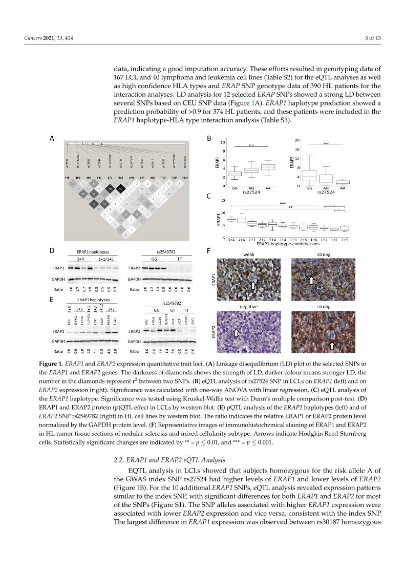
data, indicating a good imputation accuracy. These efforts resulted in genotyping data of167 LCL and 40 lymphoma and leukemia cell lines (Table S2) for the eQTL analyses as wellas high confidence HLA types and ERAP SNP genotype data of 390 HL patients for theinteraction analyses. LD analysis for 12 selected ERAP SNPs showed a strong LD betweenseveral SNPs based on CEU SNP data (Figure 1A). ERAP1 haplotype prediction showed aprediction probability of >0.9 for 374 HL patients, and these patients were included in theERAP1 haplotype-HLA type interaction analysis (Table S3).
Cancers 2021, 13, x FOR PEER REVIEW 3 of 14
2. Results
2.1. ERAP SNP Genotyping, ERAP1 Haplotype Reconstruction and HLA Type Imputation
ERAP SNP genotyping was successful for all control and HL derived LCLs, lym-
phoma and leukemia derived cell lines and non-GWAS HL patients. The minor allele
frequencies (MAFs) were similar to those of the Utah residents with Northern and
Western European ancestry (CEU) population from 1000 Genomes project (Table S1).
MAFs of the typed and imputed SNPs from the GWAS HL cases were similar to each
other and to the CEU frequencies (Table S1). HLA imputation of the GWAS HL cases
showed an overall concordance rate of 98.2% with the PCR sequence-specific oligonu-
cleotide probe-based HLA typing data, indicating a good imputation accuracy. These
efforts resulted in genotyping data of 167 LCL and 40 lymphoma and leukemia cell lines
(Table S2) for the eQTL analyses as well as high confidence HLA types and ERAP SNP
genotype data of 390 HL patients for the interaction analyses. LD analysis for 12 selected
ERAP SNPs showed a strong LD between several SNPs based on CEU SNP data (Figure
1A). ERAP1 haplotype prediction showed a prediction probability of >0.9 for 374 HL pa-
tients, and these patients were included in the ERAP1 haplotype-HLA type interaction
analysis (Table S3).
Figure 1. ERAP1 and ERAP2 expression quantitative trait loci. (A) Linkage disequilibrium (LD) plot of the selected SNPs
in the ERAP1 and ERAP2 genes. The darkness of diamonds shows the strength of LD, darker colour means stronger LD,
the number in the diamonds represent r2 between two SNPs. (B) eQTL analysis of rs27524 SNP in LCLs on ERAP1 (left)
and on ERAP2 expression (right). Significance was calculated with one-way ANOVA with linear regression. (C) eQTL
analysis of the ERAP1 haplotype. Significance was tested using Kruskal-Wallis test with Dunn’s multiple comparison
post-test. (D) ERAP1 and ERAP2 protein (p)QTL effect in LCLs by western blot. (E) pQTL analysis of the ERAP1 haplo-
types (left) and of ERAP2 SNP rs2549782 (right) in HL cell lines by western blot. The ratio indicates the relative ERAP1
ERAP2 protein level normalized by the GAPDH protein level. (F) Representative images of immunohistochemical
Figure 1. ERAP1 and ERAP2 expression quantitative trait loci. (A) Linkage disequilibrium (LD) plot of the selected SNPs inthe ERAP1 and ERAP2 genes. The darkness of diamonds shows the strength of LD, darker colour means stronger LD, thenumber in the diamonds represent r2 between two SNPs. (B) eQTL analysis of rs27524 SNP in LCLs on ERAP1 (left) and onERAP2 expression (right). Significance was calculated with one-way ANOVA with linear regression. (C) eQTL analysis ofthe ERAP1 haplotype. Significance was tested using Kruskal-Wallis test with Dunn’s multiple comparison post-test. (D)ERAP1 and ERAP2 protein (p)QTL effect in LCLs by western blot. (E) pQTL analysis of the ERAP1 haplotypes (left) and ofERAP2 SNP rs2549782 (right) in HL cell lines by western blot. The ratio indicates the relative ERAP1 or ERAP2 protein levelnormalized by the GAPDH protein level. (F) Representative images of immunohistochemical staining of ERAP1 and ERAP2in HL tumor tissue sections of nodular sclerosis and mixed cellularity subtype. Arrows indicate Hodgkin Reed-Sternbergcells. Statistically significant changes are indicated by ** = p ≤ 0.01, and *** = p ≤ 0.001.
2.2. ERAP1 and ERAP2 eQTL Analysis
EQTL analysis in LCLs showed that subjects homozygous for the risk allele A ofthe GWAS index SNP rs27524 had higher levels of ERAP1 and lower levels of ERAP2(Figure 1B). For the 10 additional ERAP1 SNPs, eQTL analysis revealed expression patternssimilar to the index SNP, with significant differences for both ERAP1 and ERAP2 for mostof the SNPs (Figure S1). The SNP alleles associated with higher ERAP1 expression wereassociated with lower ERAP2 expression and vice versa, consistent with the index SNP.The largest difference in ERAP1 expression was observed between rs30187 homozygous
Cancers 2021, 13, 414 4 of 13
minor as compared to major allele carriers with an effect size of 74%. The ERAP2 SNPrs2549782 showed a very strong eQTL effect for ERAP2, with a lower expression level forsubjects carrying the T allele (Figure S1). This ERAP2 SNP was also significantly associatedto expression levels of ERAP1.
ERAP1 haplotype eQTL analyses were done for haplotype combinations that wereobserved in ≥5 individuals. This revealed significant differences between ERAP1 andERAP2 expression levels in individuals with different haplotype combinations. Haplotype3 and 4 heterozygous and haplotype 4 homozygous individuals were associated with highERAP1 expression levels. These two haplotypes both contain the risk allele of the GWASindex SNP. Haplotypes associated with a low ERAP1 expression included haplotypes 1 and5 that do not include the risk allele of the index SNP (Figure 1C). Haplotypes associatedwith high ERAP1 expression levels showed low ERAP2 expression levels and vice versa.(Figure 1C and Figure S2). Individuals with the ERAP1 haplotype 3+4 combination had thehighest ERAP1 expression with a 128% higher expression level compared to individualswith a haplotype 1+5 combination.
The ERAP2 SNP rs2549782 showed a strong eQTL effect on ERAP2 expression in thecell line panel including 40 lymphoma and leukemia cell lines. No significant eQTL effectson ERAP1 and ERAP2 expression were observed for ERAP1 SNPs and haplotypes in thecell line panel (data not shown).
2.3. Protein QTL Effects
Effects of ERAP1 haplotypes and the ERAP2 SNP genotype on protein levels weretested in eight LCLs that were selected based on a homozygous genotype for the ERAP2NMD associated SNP rs2549782. The ERAP1 haplotype distribution of these eight casesincluded four LCLs with at least one haplotype 3 allele, and four LCLs with at leastone haplotype 1 allele. In line with the results on the transcript level, haplotype 3 wasassociated with high ERAP1 protein and haplotype 1 with low ERAP1 protein levels. TheGG genotype of rs2549782 was associated with high expression of ERAP2 and the TTgenotype of rs2549782 was associated with loss of ERAP2 expression (Figure 1D). Thesefindings are consistent with the eQTL results. Western blot analysis of HL cell lines revealeda similar pQTL pattern as observed for the LCLs (Figure 1E and Figure S3).
2.4. Immunohistochemistry
Immunohistochemistry was done on 10 cHL (5 nodular sclerosis and 5 mixed cellular-ity) cases with membranous HLA class I expression in Hodgkin Reed-Sternberg cells [35]and with homozygosity of the minor (n = 5) or major (n = 5) allele of the ERAP2 NMD SNP.Positive ERAP1 staining was observed in Hodgkin Reed-Sternberg cells in all cases. ERAP2was observed in both Hodgkin Reed-Sternberg cells and in cells in the microenvironmentin the 5 cases that were homozygous for the minor G-allele of the ERAP2 SNP. The 5 caseshomozygous for the T-allele were consistently negative for ERAP2, both in Hodgkin Reed-Sternberg cells and cells in the microenvironment (Figure 1F). No obvious differences inERAP1 or ERAP2 staining intensities were observed between Hodgkin Reed-Sternbergcells and reactive cells.
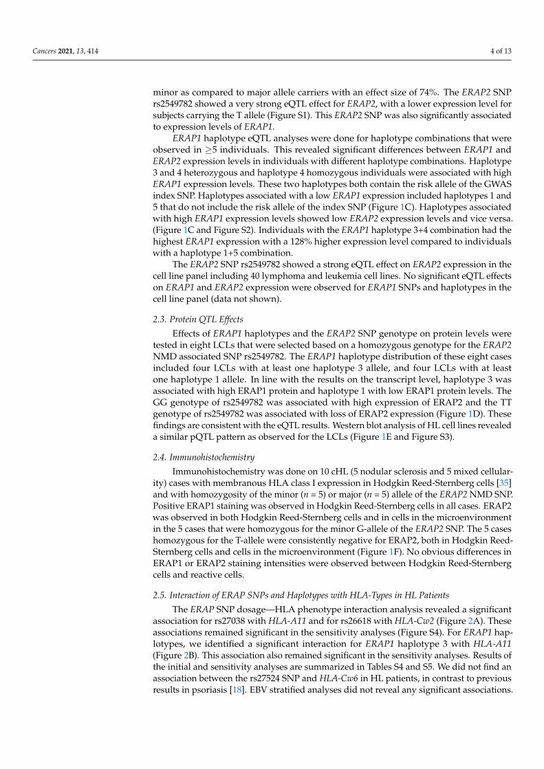
2.5. Interaction of ERAP SNPs and Haplotypes with HLA-Types in HL Patients
The ERAP SNP dosage—HLA phenotype interaction analysis revealed a significantassociation for rs27038 with HLA-A11 and for rs26618 with HLA-Cw2 (Figure 2A). Theseassociations remained significant in the sensitivity analyses (Figure S4). For ERAP1 hap-lotypes, we identified a significant interaction for ERAP1 haplotype 3 with HLA-A11(Figure 2B). This association also remained significant in the sensitivity analyses. Results ofthe initial and sensitivity analyses are summarized in Tables S4 and S5. We did not find anassociation between the rs27524 SNP and HLA-Cw6 in HL patients, in contrast to previousresults in psoriasis [18]. EBV stratified analyses did not reveal any significant associations.
Cancers 2021, 13, 414 5 of 13
Cancers 2021, 13, x FOR PEER REVIEW 5 of 14
differences in ERAP1 or ERAP2 staining intensities were observed between Hodgkin
Reed-Sternberg cells and reactive cells.
2.5. Interaction of ERAP SNPs and Haplotypes with HLA-Types in HL Patients
The ERAP SNP dosage—HLA phenotype interaction analysis revealed a significant
association for rs27038 with HLA-A11 and for rs26618 with HLA-Cw2 (Figure 2A). These
associations remained significant in the sensitivity analyses (Figure S4). For ERAP1 hap-
lotypes, we identified a significant interaction for ERAP1 haplotype 3 with HLA-A11
(Figure 2B). This association also remained significant in the sensitivity analyses. Results
of the initial and sensitivity analyses are summarized in Tables S4 and S5. We did not
find an association between the rs27524 SNP and HLA-Cw6 in HL patients, in contrast to
previous results in psoriasis [18]. EBV stratified analyses did not reveal any significant
associations.
Figure 2. Association analysis of ERAP1 and ERAP2 SNPs and haplotypes with HLA phenotype in
HL patients. (A) SNPs are ordered by chromosomal position. Genotyped or best guess ERAP gen-
otypes and typed or best guess HLA phenotypes were used. (B) ERAP1 haplotypes are ordered
from high to low frequency in the CEU population. The best guess ERAP1 haplotypes and typed or
best guess HLA phenotypes were used. Samples were excluded if the maximum probability of a
haplotype combination was <0.9. Logistic regression analysis was used to determine significance of
associations. A p-value of <0.005 (dashed line) was considered significant.
The ERAP SNP genotype—HLA genotype analysis revealed interactions for rs26618
with HLA-Cw2, and for rs27038 with HLA-A11 and HLA-A68. These interactions remained
significant in the sensitivity analyses (Figure S5). For ERAP1 haplotypes, four significant
interactions with HLA genotypes were identified, i.e., ERAP1 haplotype 3 with HLA-A11
and HLA-B35, haplotype 4 with HLA-Cw7 and haplotype 7 with HLA-Cw2 (Figure S5).
Results of the initial and sensitivity analysis are summarized in Tables S4 and S5. Again, no
significant associations were found in EBV+ and EBV− subgroups.
Figure 2. Association analysis of ERAP1 and ERAP2 SNPs and haplotypes with HLA phenotypein HL patients. (A) SNPs are ordered by chromosomal position. Genotyped or best guess ERAPgenotypes and typed or best guess HLA phenotypes were used. (B) ERAP1 haplotypes are orderedfrom high to low frequency in the CEU population. The best guess ERAP1 haplotypes and typedor best guess HLA phenotypes were used. Samples were excluded if the maximum probability of ahaplotype combination was <0.9. Logistic regression analysis was used to determine significance ofassociations. A p-value of <0.005 (dashed line) was considered significant.
The ERAP SNP genotype—HLA genotype analysis revealed interactions for rs26618with HLA-Cw2, and for rs27038 with HLA-A11 and HLA-A68. These interactions remainedsignificant in the sensitivity analyses (Figure S5). For ERAP1 haplotypes, four significantinteractions with HLA genotypes were identified, i.e., ERAP1 haplotype 3 with HLA-A11and HLA-B35, haplotype 4 with HLA-Cw7 and haplotype 7 with HLA-Cw2 (Figure S5).Results of the initial and sensitivity analysis are summarized in Tables S4 and S5. Again,no significant associations were found in EBV+ and EBV− subgroups.
3. Discussion
In this study we explored the relevance of the previously reported association of anERAP SNP with HL susceptibility in the context of specific HLA types. We showed strongeQTL effects of ERAP SNPs and haplotypes consistent with previous studies [30,36]. Therisk allele (A allele) of the GWAS index SNP rs27524 and haplotypes 3 and 4 carrying thisrisk allele were associated with high ERAP1 and low ERAP2 expression. The effect ofERAP1 SNPs and haplotypes on ERAP2 and vice versa can be attributed to the strong LD ofvariants in this genomic region. With regards to HL susceptibility, significant interactionswere observed between ERAP1 SNP rs27038 and HLA-A11, ERAP1 SNP rs26618 and HLA-Cw2, and ERAP1 haplotype 3 and HLA-A11. This suggests that mechanisms that determinethe repertoire of antigens that are presented by HL precursor cells, affect the chance ofdeveloping full-blown HL.
Proper tumor-specific antigen presentation is an essential process in the induction of afunctional anti-tumor immune response and requires expression of HLA class I and ERAPproteins. In many cancer types, tumor cells attempt to escape from immune responsesby abolishing cell surface HLA class I expression [37]. ERAP1 and ERAP2 expression isalso frequently altered in melanoma, acute myeloid leukemia, gastric adenocarcinoma,HPV-induced malignancies and renal clear cell carcinoma, as another immune escape
Cancers 2021, 13, 414 6 of 13
mechanism [38–41]. In HL, expression of membranous HLA class I by tumor cells isretained in a significant proportion of cases [35,42,43]. The differences we observed inERAP expression patterns in HLA class I positive HL cases were associated with thegenotype of the subjects and there was no down regulation of ERAP expression in thetumor cells relative to normal cells in the tumor microenvironment. Thus, the tumor cellsof HLA class I positive HL cases show normal ERAP1 and ERAP2 expression patterns.This means that, in combination with the previously reported expression of other antigenprocessing proteins such as TAP1 and TAP2, the essential components of the antigenpresentation pathway are present in the tumor cells of HL [44].
The function of ERAP molecules has not been studied in HL, but in other diseases andmodel systems ERAP is strongly linked to antigen presentation and immune responses.Deficiency in ERAP1 expression was shown to result in unstable and highly immunogenicpeptides presented in the context of HLA class I [45]. Indeed, altered ERAP1 expressioninfluences the effectiveness of cytotoxic CD8+ T-cell anti-tumor immune responses in thecontext of solid cancer, melanoma and a T-cell lymphoma mouse model [40,46,47]. Anotherstudy showed that lack of ERAP2 expression decreased NK cell activation in choriocarci-noma cell lines [48]. In addition, differential substrate specificity based on combinations ofERAP missense SNPs affects the peptidome, which may reduce recognition of tumor cellsby CD8+ T cells and NK cells [47,49]. For example, ERAP1 missense SNP alleles rs30187-Tand rs27044-C affect peptide specificity, alter the peptide pool available for being presentedin the context of HLA class I, and potentially facilitate immune evasion [28,50]. Besidesthe direct effects of missense SNPs on expression levels and substrate specificity, the ratiobetween ERAP1 and ERAP2 may also influence peptide trimming efficiency due to theirfunctional coordination by forming a heterodimer [51]. We showed that the A allele of thers27524 HL associated risk SNP results in high ERAP1 and low ERAP2 expression, whichaffects peptide trimming efficiency and is expected to associate with a restricted range ofpeptides in the peptidome. This may result in a skewed anti-tumor CD8+ T cell responseand contribute to escape of HL tumor cells from effective anti-tumor immune surveillance.This concept fits well with ERAP-dependent mechanisms as proposed for multiple othercancer types [40,46,47].
We demonstrated a significant interaction between the C allele of rs26618 and HLA-Cw2 in HL patients. Rs26618 is a missense SNP that is expected to define peptide specificityand the C allele is associated with high ERAP2 expression. We also identified interactions ofthe rs27038 A allele and the associated haplotype 3 with HLA-A11. HLA-A11 was previouslyreported to be associated with HL [52], although this association was not confirmed inlater studies [53,54]. This HLA type can present peptides from EBV and induce cytotoxicT cell responses [55,56]. Both the intronic rs27038 A allele and the ERAP1 haplotype 3are associated with high ERAP1 and low ERAP2 expression. Thus, this combination ofalleles might have an impact on the presentation of tumor-associated antigenic peptidesby HLA-A11. Unfortunately, due to a relatively small group size of EBV+ HL patients,we could not determine whether there was an EBV specific effect and a potential relationshould be further explored in a larger EBV+ HL cohort. Regarding HL subtype, we considerthe interactions to reflect cHL, as these interactions remained significant in the sensitivityanalyses that excluded NLPHL cases.
In previous studies on autoimmune diseases, interactions of ERAP were shown withknown HLA susceptibility types, i.e., rs27524 with HLA-Cw6 in psoriasis [18], rs17482078with HLA-B51 in Behçet’s disease [25], rs30187 with HLA-B27/HLA-B40 in ankylosingspondylitis [26], and rs7705093 with HLA-A29 in birdshot chorioretinopathy [27]. Incontrast, the HLA types identified in this HLA-ERAP interaction study, i.e., HLA-A11 andHLA-Cw2, have not been associated with HL susceptibility [6,7]. We also did not findERAP-HLA interactions with HLA types that have previously been associated with HLsusceptibility. This might be caused by less strong associations between ERAP and HLAtypes in HL and limited power due to low frequencies of certain HLA risk types in ourHL cohort. Nonetheless, the interactions observed in the current study suggest that ERAP
Cancers 2021, 13, 414 7 of 13
missense SNPs have an effect on the peptide pool available for being presented by specificHLA types and as such influence susceptibility to HL.
4. Materials and Methods4.1. Patient and Control Samples
Patients included in this study were retrieved from previously published HLA typ-ing (n = 332) and GWAS (n = 304) studies, with an overlap of 278 HL patients [6,11].Most patients were diagnosed with HL in multiple centers in the Northern region of theNetherlands between 1987–2010. In addition, 56 new HL patients were included who werediagnosed between 2010–2012 at the University Medical Centre Groningen, the Nether-lands (Figure S6). There were 397 cHL and 17 NLPHL cases, according to the World HealthOrganization classification system [57]. EBV status was determined previously by EBERin situ hybridization (ISH) [6,9]. Additionally, 97 healthy controls were included in thisstudy. This research has been approved by the Medical Ethical Committee of the UniversityMedical Centre Groningen on 10 November 2011 (ethic code: METc 2004/219) and wasconducted in accordance with the Declaration of Helsinki. Informed consent was given byall patients.
4.2. LCL Generation and Cell Line Culture
LCLs were generated by infection of peripheral blood mononuclear cells (PBMCs)from 97 anonymized healthy donors and 70 successfully treated HL patients with B95.8EBV virus using standard procedures. Forty-four of these HL patients were also includedin the HLA typing study and 26 were from the new HL cases. LCLs were cultured inRPMI 1640 (Lonza BioWhittaker, Walkersville, MD, USA) supplemented with 10% Fe-tal Bovine Serum (FBS, HyClone Thermo Scientific, Waltham, MA, USA) and 100U/mLof Penicillin/Streptomycin and 1% Ultraglutamine (BioWhittaker, Basel, Switzerland).All HL cell lines except DEV were obtained from DSMZ (German Collection of Microor-ganisms and Cell Cultures GmbH, Braunschweig, Germany). The DEV cell line wasmade in our group [58]. These cell lines were cultured in RPMI 1640 with 20% FBS(DEV, L540 and HDLM2), 10% (KMH2, L591 and L1236) or 5% (L428) FBS and Peni-cillin/Streptomycin and Ultraglutamine. SUPHD1 was cultured in McCoys5A with 20%FBS and Penicillin/Streptomycin and Ultraglutamine. Thirty-two additional lymphomaand leukemia cell lines were used to extend the cell line panel (Table S2). These cell lineswere cultured as described previously [59,60]. Mycoplasma tests were done at a regularbasis and cell line identities were confirmed through short tandem repeat (STR) analysis.
4.3. ERAP SNP Selection, Genotyping, Imputation and Haplotype Reconstruction
We selected all ERAP1 and ERAP2 missense SNPs with a minor allele frequency (MAF)above 5% based on public data from CEU cohort of 1000 Genomes phase 3(ftp://ftp.1000genomes.ebi.ac.uk/vol1/ftp/release/20130502). This resulted in 10 SNPs,9 mapping to the ERAP1 locus and one mapping to the ERAP2 locus. In addition, weincluded (1) the index rs27524 SNP identified in our previous GWAS study [9], and (2)rs27038 and rs13160562 SNPs showing a strong eQTL effect according to the GTEx portal(https://www.gtexportal.org/home/) and a recent study by Harson et al. [30]. LD analysisfor all of the 13 above mentioned SNPs in the CEU cohort of the 1000 Genomes phase3 using Haploview v4.2 [61] showed perfect LD between rs2287987 and rs17482078. Soonly rs2287987 was selected for genotyping. This resulted in a final selection of 11 SNPsmapping at the ERAP1 locus and one ERAP2 SNP.
For 304 HL cases, SNP data were extracted from our previous GWAS data by PLINKv1.07 (http://pngu.mgh.harvard.edu/purcell/plink/) [62] (Figure S6). Six (rs27524,rs13160562, rs30187, rs10050860, rs26618, rs2549782) of the 12 SNPs selected for anal-ysis were genotyped within the GWAS. The remaining six SNPs were imputed usingthe HRC reference panel (Version r1.1 2016) on the Michigan Imputation Server (https://imputationserver.sph.umich.edu/) [63]. The average probability was >99% for each of
Cancers 2021, 13, 414 8 of 13
the SNPs. The genotype with the highest probability was used for the analyses (i.e., bestguess genotype). Eight of the 304 × 6 imputed genotypes with an imputation quality scorer2 < 0.95 were excluded in the sensitivity analyses.
For the 97 healthy controls, 110 HL patients not included in the GWAS and for the40 cell lines, genomic DNA was isolated following routine protocols. SNP genotyping wasdone in triplicate on the Taqman 7900HT fast real-time PCR system using Taqman assays(Thermo Fisher Scientific, Waltham, MA, USA) (Table S1) in a final volume of 5 µL using 5 to10 ng DNA in a 384-well plate (Applied Biosystems, Waltham, MA, USA). Genotypes werecalled by QuantStudio™ Real-Time PCR Software. As a quality check, frequencies of theSNPs were compared to those of the CEU population from the 1000 Genomes Project [64].
ERAP1 haplotype reconstructions were carried out using genotype data of the 11 se-lected ERAP1 SNPs using PHASE v2.1.1 [65,66]. The haplotype combination with thehighest probability (i.e., best guess haplotype) was assigned. Samples with haplotypecombinations with predicted probabilities of <0.9 were excluded (n = 16) from the interac-tion analyses.
4.4. HLA Typing and Imputation
For 332 HL cases, HLA types were determined using a sequence-specific oligonu-cleotide PCR method as described in our previous study [6]. For 32 of the new HL cases,HLA typing was performed following the same approach. For 26 HL patients (including9 cHL and 17 NLPHL) from the GWAS and for 12 HL patients for whom HLA typing failedfor some of the HLA class I alleles, HLA types were defined using the SNP-based HLAimputation R-package HIBAG [67]. Data not meeting the imputation quality thresholdswere set to missing. Eight poorly imputed HLA types with a prediction probability <0.8were excluded in the indicated sensitivity analyses. HLA imputation was done for all HLcases included in the GWAS. The imputation quality was assessed for the 278 patientsthat were included both in the GWAS and the HLA typing cohort by determining theconcordance between real and imputed HLA types.
4.5. qRT-PCR
Total RNA was isolated from LCL and lymphoma cell lines using TRIzol® reagent(Thermo Fisher Scientific, Waltham, MA, USA) following the protocol of the manufacturer.Concentration of RNA samples was measured on the Nanodrop-1000 spectrophotometerand quality was checked on a 1% agarose gel. cDNA was synthesized from 500 ng totalRNA in 20 µL reaction volume using Superscript II Reverse Transcriptase Kit and Randomprimers in accordance with the protocol of the manufacturer (Thermo Fisher Scientific,Waltham, MA, USA). The qPCR was performed in a total volume of 10 µL with 300 nMprimers, 1 ng cDNA and 5 µL SYBR® Green Real-Time PCR Master Mixes (Thermo FisherScientific Inc., Waltham, MA, USA) on the Lightcycler 480 (Roche, Penzberg, Germany),and in triplicate for each sample. Relative expression levels were determined using 2-∆Cpusing TATA-Box Binding Protein (TBP) as a housekeeping gene. Primer sequences used foramplification of ERAP1, ERAP2 and TBP are listed in Table S6.
4.6. Western Blot
Cells (about 1–20 × 106) of eight LCLs that were selected based on homozygosity ofthe ERAP2 SNP and cells of the eight Hodgkin lymphoma cell lines were lysed in cell lysisbuffer (#9803, Cell Signaling Technology, Danvers, MA, USA) with 1 mM phenylmethane-sulphonyl fluoride (PMSF) on ice for 30–45 min. The protein concentration was measuredusing the Pierce™ BCA Protein Assay Kit (#23227, Thermo Fisher Scientific Inc., Waltham,MA, USA). Twenty µg of protein was electrophoresed on a 6% SDS-PAGE gel and trans-ferred to a nitrocellulose membrane using standard protocols. Blots were blocked in TBST +5% ELK milk powder for 60 min and incubated with primary antibodies (Goat anti-humanERAP1 antibody 1:500, AF2334, R&D Systems, Minneapolis, USA; and Goat anti-humanERAP2 antibody, 1:1000, AF3830, R&D Systems) overnight at 4 ◦C. After washing, blots
Cancers 2021, 13, 414 9 of 13
were incubated with HRP-conjugated rabbit anti-goat antibody (#P0449, Dako, Glostrup,Denmark, 1:1000 dilution), which was followed by an incubation with HRP-conjugatedgoat anti-rabbit antibody (#P0448, Dako, Glostrup, Denmark, 1:1000 dilution). SuperSignalWestPico Chemiluminescent Substrate (#34078, Thermo Fisher Scientific Inc., Waltham,MA, USA) was used to visualize ERAP1 and ERAP2 protein. Glyceraldehyde-3-PhosphateDehydrogenase (GAPDH) was used as a housekeeping gene.
4.7. Immunohistochemistry
Primary formalin-fixed paraffin-embedded (FFPE) tissue sections of 10 cHL cases (5nodular sclerosis, 5 mixed cellularity) selected based on membranous expression of HLAclass I and ERAP2 SNP genotype (5 homozygous minor and 5 homozygous major allele ofthe index SNP) were deparaffinized using xylene and ethanol and subjected to heat-inducedantigen retrieval in 1mM EDTA pH = 8.0 for 15 min. Endogenous peroxidase was blockedusing 0.3% H2O2 for 30 min and avidin and biotin were blocked using the avidin/biotinblocking kit (# SP-2001, Vector Laboratories, Burlingame, CA, USA). Goat anti-humanERAP1 antibody (AF2334, R&D Systems, Minneapolis, MN, USA) at 1:50 dilution andGoat anti-human ERAP2 antibody (AF3830, R&D Systems, Minneapolis, MN, USA) at1:100 dilution in PBS + 1% BSA were incubated at room temperature for 60 min. Afterwashing, slides were incubated with biotin-conjugated rabbit anti-goat antibody (#6165-08,Southern Biotech, Birmingham, AL, USA) at 1:100 dilution for 30 min, followed by anincubation with streptavidin-HRP (#P0397, Dako, Glostrup, Denmark) at 1:300 dilution atroom temperature for 30 min. 3,3′-Diaminobenzidine (DAB) staining was used to visualizethe protein and slides were counterstained with hematoxylin. Scoring was performed in anunbiased way, without knowing the ERAP1 and ERAP2 genotypes. We scored the stainingintensity for ERAP1 and ERAP2 based on the pattern in the majority of the Hodgkin Reed-Sternberg cells as negative, weak, intermediate or strong. For both ERAP1 and ERAP2,staining patterns were evaluated separately for Hodgkin Reed-Sternberg cells and cells inthe microenvironment.
4.8. Statistical Analysis
One-way analysis of variance (ANOVA) with a post-hoc test for linear trend fornumber of alleles and comparison between the mean of each genotype was employed totest for eQTL effects of ERAP1 and ERAP2 SNP genotypes. A Kruskal-Wallis test with aDunns post-hoc test was applied to analyze the eQTL effect of ERAP1 haplotypes. Analyseswere performed using GraphPad Prism 5.0 (GraphPad Software, Inc., San Diego, CA, USA)and python3.7 with packages of SciPy (https://www.scipy.org/).
Association analysis testing for an interaction effect of ERAP (additive model) andHLA phenotypes were performed in PLINK v1.07 using logistic regression by comparingmean number of risk alleles of ERAP SNP allele or ERAP1 haplotype between HLA typecarriers and non-carriers. In addition, ERAP—HLA genotype interactions were determinedby comparing observed with expected frequencies using Chi-square tests. ERAP1 haplo-type analyses were performed for individuals for whom the probability of the haplotypereconstruction was >0.9 using PHASE. We tested all of the 18 HLA types with an allelefrequency >5% in the study population. Power calculations using the online Clincalc tool(https://clincalc.com/stats/Power.aspx) revealed a power of 80% to detect a relative riskratio of interaction of 1.28 and more. Considering multiple testing correction and LD inboth the HLA and the ERAP gene regions we regarded a p-value < 0.005 as significant.
ERAP allele dosage/HLA phenotype association analyses were done on the entiregroup of 390 HL cases using best guess genotypes for all ERAP SNP and HLA imputationresults. Since imputation results can be imprecise, we performed a sensitivity analysis bysetting imputed SNP genotypes and imputed HLA types with an imputation accuracy(r2) of <0.95 and <0.8, respectively, to missing (n = 390). Another sensitivity analysiswas done only including cases with direct HLA typing data (n = 364). This resulted inexclusion of 9 cHL and all 17 NLPHL cases as they only had imputed HLA types. In
Cancers 2021, 13, 414 10 of 13
addition, we performed ERAP genotype/HLA genotype association analyses using thesame sensitivity groups as described above. Besides analyzing the total HL group, we alsoexplored associations in the EBV positive and negative subgroups (only using best guessERAP genotypes and HLA-types of all 390 cases without filtering for imputation quality).
5. Conclusions
In conclusion, our data indicate a role of ERAP1 and ERAP2 variants in the devel-opment of HL in the context of specific HLA types. Given the importance of ERAP1 andERAP2 in trimming antigenic peptides and the known and very strong associations ofthe HLA region with HL susceptibility, our data support an important role of antigenpresentation in HL susceptibility and pathogenesis.
Supplementary Materials: The following are available online at https://www.mdpi.com/2072-6694/13/3/414/s1, Figure S1. Overview of eQTL effects of all 12 missense SNPs in ERAP1 andERAP2. Figure S2. eQTL analysis of ERAP1 haplotype on ERAP2 expression in lymphoblastoidcell lines. Figure S3. Western blot of ERAP1 and ERAP2 protein. Figure S4. Sensitivity analysesfor testing the association between genotypes of 12 missense SNPs of the ERAP1 and ERAP2 geneloci and HLA-phenotype in HL patients. Figure S5. Sensitivity analyses for testing the associationbetween 12 missense SNPs genotypes of the ERAP1 and ERAP2 gene loci and HLA-genotype HL.Figure S6. Schematic representation of the HL cases included in this study. Table S1. Minor allelefrequencies of selected SNPs in the ERAP1 and ERAP2 genes in LCL controls and HL patients. TableS2. Cell lines used in this study. Table S3. ERAP1 haplotypes in CEU controls and HL patients. TableS4. ERAP SNP—HLA allele interactions with (nearly) significant associations. Table S5. ERAP1haplotype—HLA type interactions with (nearly) significant associations. Table S6. Sequence of theprimers used for qRT-PCR.
Author Contributions: P.J., R.N.V. and A.S. did SNP genotyping, qPCR and eQTL analyses. P.J., L.V.,A.v.d.B. and A.D. performed SNP imputation and immunohistochemical data analysis. I.M.N. didstatistical analyses. B.G.H. contributed by HLA typing and HLA imputation analysis. P.J., A.v.d.B.and A.D. designed the study and wrote the manuscript. All authors have read and agreed to thepublished version of the manuscript.
Funding: This work was supported by a grant from the Dutch Cancer Society (KWF RUG 2014-6698)and by a grant from de Stichting De Cock—Hadders (2018-18).
Institutional Review Board Statement: The study was conducted according to the guidelines of theDeclaration of Helsinki, and approved by the Ethics Committee of the University Medical CentreGroningen on 10 November 2011 (ethic code METc 2004/219).
Informed Consent Statement: Informed consent was obtained from all subjects involved in thestudy.
Data Availability Statement: SNP genotype data from CEU cohort of 1000 Genomes phase 3 arepublicly available (ftp://ftp.1000genomes.ebi.ac.uk/vol1/ftp/release/20130502).
Acknowledgments: We would like to thank James D. McKay, International Agency for Research onCancer, WHO, Lyon, France, for helpful discussions.
Conflicts of Interest: The authors declare no conflict of interest.
References1. Lee, J.H.; Kim, Y.; Choi, J.W.; Kim, Y.S. Prevalence and prognostic significance of Epstein-Barr virus infection in classical Hodgkin’s
lymphoma: A meta-analysis. Arch. Med. Res. 2014, 45, 417–431. [CrossRef] [PubMed]2. Mack, T.M.; Cozen, W.; Shibata, D.K.; Weiss, L.M.; Nathwani, B.N.; Hernandez, A.M.; Taylor, C.R.; Hamilton, A.S.; Deapen, D.M.;
Rappaport, E.B. Concordance for Hodgkin’s disease in identical twins suggesting genetic susceptibility to the young-adult formof the disease. N. Engl. J. Med. 1995, 332, 413–418. [CrossRef] [PubMed]
3. Diepstra, A.; Niens, M.; te Meerman, G.J.; Poppema, S.; van den Berg, A. Genetic susceptibility to Hodgkin’s lymphoma associatedwith the human leukocyte antigen region. Eur. J. Haematol. Suppl. 2005, 66, 34–41. [CrossRef] [PubMed]
4. Diepstra, A.; Niens, M.; Vellenga, E.; van Imhoff, G.W.; Nolte, I.M.; Schaapveld, M.; van der Steege, G.; van den Berg, A.;Kibbelaar, R.E.; te Meerman, G.J.; et al. Association with HLA class I in Epstein-Barr-virus-positive and with HLA class III inEpstein-Barr-virus-negative Hodgkin’s lymphoma. Lancet 2005, 365, 2216–2224. [CrossRef]
Cancers 2021, 13, 414 11 of 13
5. Niens, M.; van den Berg, A.; Diepstra, A.; Nolte, I.M.; van der Steege, G.; Gallagher, A.; Taylor, G.M.; Jarrett, R.F.; Poppema, S.; teMeerman, G.J. The human leukocyte antigen class I region is associated with EBV-positive Hodgkin’s lymphoma: HLA-A andHLA complex group 9 are putative candidate genes. Cancer Epidemiol. Biomark. Prev. 2006, 15, 2280–2284. [CrossRef]
6. Huang, X.; Kushekhar, K.; Nolte, I.; Kooistra, W.; Visser, L.; Bouwman, I.; Kouprie, N.; Veenstra, R.; van Imhoff, G.; Olver, B.; et al.HLA Associations in Classical Hodgkin Lymphoma: EBV Status Matters. PLoS ONE 2012, 7, e39986. [CrossRef]
7. Niens, M.; Jarrett, R.F.; Hepkema, B.; Nolte, I.M.; Diepstra, A.; Platteel, M.; Kouprie, N.; Delury, C.P.; Gallagher, A.; Visser, L.; et al.HLA-A*02 is associated with a reduced risk and HLA-A*01 with an increased risk of developing EBV + Hodgkin lymphoma.Blood 2007, 110, 3310–3315. [CrossRef]
8. Enciso-Mora, V.; Broderick, P.; Ma, Y.; Jarrett, R.F.; Hjalgrim, H.; Hemminki, K.; van den Berg, A.; Olver, B.; Lloyd, A.; Dobbins,S.E.; et al. A genome-wide association study of Hodgkin’s lymphoma identifies new susceptibility loci at 2p16.1 (REL), 8q24.21and 10p14 (GATA3). Nat. Genet. 2010, 42, 1126–1130. [CrossRef]
9. Urayama, K.Y.; Jarrett, R.F.; Hjalgrim, H.; Diepstra, A.; Kamatani, Y.; Chabrier, A.; Gaborieau, V.; Boland, A.; Nieters, A.; Becker,N.; et al. Genome-wide association study of classical Hodgkin lymphoma and Epstein-Barr virus status-defined subgroups. J.Natl. Cancer Inst. 2012, 104, 240–253. [CrossRef]
10. Frampton, M.; da Silva Filho, M.I.; Broderick, P.; Thomsen, H.; Forsti, A.; Vijayakrishnan, J.; Cooke, R.; Enciso-Mora, V.; Hoffmann,P.; Nothen, M.M.; et al. Variation at 3p24.1 and 6q23.3 influences the risk of Hodgkin’s lymphoma. Nat. Commun. 2013, 4, 2549.[CrossRef]
11. Cozen, W.; Timofeeva, M.N.; Li, D.; Diepstra, A.; Hazelett, D.; Delahaye-Sourdeix, M.; Edlund, C.K.; Franke, L.; Rostgaard, K.;Van Den Berg, D.J.; et al. A meta-analysis of Hodgkin lymphoma reveals 19p13.3 TCF3 as a novel susceptibility locus. Nat.Commun. 2014, 5, 3856. [CrossRef] [PubMed]
12. Sud, A.; Thomsen, H.; Law, P.J.; Forsti, A.; Filho, M.I.D.S.; Holroyd, A.; Broderick, P.; Orlando, G.; Lenive, O.; Wright, L.; et al.Genome-wide association study of classical Hodgkin lymphoma identifies key regulators of disease susceptibility. Nat. Commun.2017, 8, 1892-1. [CrossRef]
13. Sud, A.; Thomsen, H.; Orlando, G.; Forsti, A.; Law, P.J.; Broderick, P.; Cooke, R.; Hariri, F.; Pastinen, T.; Easton, D.F.; et al.Genome-wide association study implicates immune dysfunction in the development of Hodgkin lymphoma. Blood 2018, 132,2040–2052. [CrossRef] [PubMed]
14. Gelfand, J.M.; Shin, D.B.; Neimann, A.L.; Wang, X.; Margolis, D.J.; Troxel, A.B. The risk of lymphoma in patients with psoriasis. J.Investig. Dermatol. 2006, 126, 2194–2201. [CrossRef]
15. Fallah, M.; Liu, X.; Ji, J.; Forsti, A.; Sundquist, K.; Hemminki, K. Hodgkin lymphoma after autoimmune diseases by age atdiagnosis and histological subtype. Ann. Oncol. 2014, 25, 1397–1404. [CrossRef]
16. Hemminki, K.; Liu, X.; Ji, J.; Forsti, A. Origin of B-Cell Neoplasms in Autoimmune Disease. PLoS ONE 2016, 11, e0158360.[CrossRef] [PubMed]
17. Khankhanian, P.; Cozen, W.; Himmelstein, D.S.; Madireddy, L.; Din, L.; van den Berg, A.; Matsushita, T.; Glaser, S.L.; More, J.M.;Smedby, K.E.; et al. Meta-analysis of genome-wide association studies reveals genetic overlap between Hodgkin lymphoma andmultiple sclerosis. Int. J. Epidemiol. 2016, 45, 728–740. [CrossRef] [PubMed]
18. Strange, A.; Capon, F.; Spencer, C.C.; Knight, J.; Weale, M.E.; Allen, M.H.; Barton, A.; Band, G.; Bellenguez, C. A genome-wideassociation study identifies new psoriasis susceptibility loci and an interaction between HLA-C and ERAP; Genetic Analysis ofPsoriasis Consortium & the Wellcome Trust Case Control Consortium 2. Nat. Genet. 2010, 42, 985–990.
19. Tsujimoto, M.; Hattori, A. The oxytocinase subfamily of M1 aminopeptidases. Biochim. Biophys. Acta 2005, 1751, 9–18. [CrossRef]20. Serwold, T.; Gonzalez, F.; Kim, J.; Jacob, R.; Shastri, N. ERAAP customizes peptides for MHC class I molecules in the endoplasmic
reticulum. Nature 2002, 419, 480–483. [CrossRef]21. York, I.A.; Chang, S.C.; Saric, T.; Keys, J.A.; Favreau, J.M.; Goldberg, A.L.; Rock, K.L. The ER aminopeptidase ERAP1 enhances or
limits antigen presentation by trimming epitopes to 8–9 residues. Nat. Immunol. 2002, 3, 1177–1184. [CrossRef]22. Saveanu, L.; Carroll, O.; Lindo, V.; Del Val, M.; Lopez, D.; Lepelletier, Y.; Greer, F.; Schomburg, L.; Fruci, D.; Niedermann, G.;
et al. Concerted peptide trimming by human ERAP1 and ERAP2 aminopeptidase complexes in the endoplasmic reticulum. Nat.Immunol. 2005, 6, 689–697. [CrossRef] [PubMed]
23. Serwold, T.; Gaw, S.; Shastri, N. ER aminopeptidases generate a unique pool of peptides for MHC class I molecules. Nat. Immunol.2001, 2, 644–651. [CrossRef] [PubMed]
24. Saric, T.; Chang, S.C.; Hattori, A.; York, I.A.; Markant, S.; Rock, K.L.; Tsujimoto, M.; Goldberg, A.L. An IFN-gamma-inducedaminopeptidase in the ER, ERAP1, trims precursors to MHC class I-presented peptides. Nat. Immunol. 2002, 3, 1169–1176.
25. Kirino, Y.; Bertsias, G.; Ishigatsubo, Y.; Mizuki, N.; Tugal-Tutkun, I.; Seyahi, E.; Ozyazgan, Y.; Sacli, F.S.; Erer, B.; Inoko, H.; et al.Genome-wide association analysis identifies new susceptibility loci for Behcet’s disease and epistasis between HLA-B*51 andERAP. Nat. Genet. 2013, 45, 202–207.
26. Evans, D.M.; Spencer, C.C.; Pointon, J.J.; Su, Z.; Harvey, D.; Kochan, G.; Oppermann, U.; Dilthey, A.; Pirinen, M.; Stone, M.A.;et al. Interaction between ERAP1 and HLA-B27 in ankylosing spondylitis implicates peptide handling in the mechanism forHLA-B27 in disease susceptibility. Nat. Genet. 2011, 43, 761–767. [CrossRef]
27. Kuiper, J.J.; Van Setten, J.; Ripke, S.; Van’T Slot, R.; Mulder, F.; Missotten, T.; Baarsma, G.S.; Francioli, L.C.; Pulit, S.L.; De Kovel,C.G.; et al. A genome-wide association study identifies a functional ERAP2 haplotype associated with birdshot chorioretinopathy.Hum. Mol. Genet. 2014, 23, 6081–6087.
Cancers 2021, 13, 414 12 of 13
28. Reeves, E.; Edwards, C.J.; Elliott, T.; James, E. Naturally occurring ERAP1 haplotypes encode functionally distinct alleles withfine substrate specificity. J. Immunol. 2013, 191, 35–43. [CrossRef]
29. Gabrielsen, I.S.; Viken, M.K.; Amundsen, S.S.; Helgeland, H.; Holm, K.; Flam, S.T.; Lie, B.A. Autoimmune risk variants in ERAP2are associated with gene-expression levels in thymus. Genes Immun. 2016, 17, 406–411. [CrossRef]
30. Hanson, A.L.; Cuddihy, T.; Haynes, K.; Loo, D.; Morton, C.J.; Oppermann, U.; Leo, P.; Thomas, G.P.; Le Cao, K.A.; Kenna, T.J.;et al. Genetic Variants in ERAP1 and ERAP2 Associated With Immune-Mediated Diseases Influence Protein Expression and theIsoform Profile. Arthritis Rheumatol. 2018, 70, 255–265. [CrossRef]
31. Kuiper, J.J.W.; Setten, J.V.; Devall, M.; Cretu-Stancu, M.; Hiddingh, S.; Ophoff, R.A.; Missotten, T.O.A.R.; Velthoven, M.V.; DenHollander, A.I.; Hoyng, C.B.; et al. Functionally distinct ERAP1 and ERAP2 are a hallmark of HLA-A29-(Birdshot) Uveitis. Hum.Mol. Genet. 2018, 27, 4333–4343. [CrossRef] [PubMed]
32. Evnouchidou, I.; Birtley, J.; Seregin, S.; Papakyriakou, A.; Zervoudi, E.; Samiotaki, M.; Panayotou, G.; Giastas, P.; Petrakis,O.; Georgiadis, D.; et al. A common single nucleotide polymorphism in endoplasmic reticulum aminopeptidase 2 induces aspecificity switch that leads to altered antigen processing. J. Immunol. 2012, 189, 2383–2392. [CrossRef] [PubMed]
33. Andrés, A.M.; Dennis, M.Y.; Kretzschmar, W.W.; Cannons, J.L.; Lee-Lin, S.-Q.; Hurle, B.; Schwartzberg, P.L.; Williamson, S.H.;Bustamante, C.D.; Nielsen, R.; et al. Balancing Selection Maintains a Form of ERAP2 that Undergoes Nonsense-Mediated Decayand Affects Antigen Presentation. PLoS Genet. 2010, 6, e1001157. [CrossRef]
34. López de Castro, J.A.; Alvarez-Navarro, C.; Brito, A.; Guasp, P.; Martín-Esteban, A.; Sanz-Bravo, A. Molecular and pathogeniceffects of endoplasmic reticulum aminopeptidases ERAP1 and ERAP2 in MHC-I-associated inflammatory disorders: Towards aunifying view. Mol. Immunol. 2016, 77, 193–204. [CrossRef]
35. Nijland, M.; Veenstra, R.N.; Visser, L.; Xu, C.; Kushekhar, K.; van Imhoff, G.W.; Kluin, P.M.; van den Berg, A.; Diepstra, A.HLA dependent immune escape mechanisms in B-cell lymphomas: Implications for immune checkpoint inhibitor therapy?Oncoimmunology 2017, 6, e1295202. [CrossRef] [PubMed]
36. Ardlie, K.G.; DeLuca, D.S.; Segre, A.V.; Sullivan, T.J.; Young, T.R.; Gelfand, E.T.; Trowbridge, C.A.; Maller, J.B.; Tukiainen, T. TheGenotype-Tissue Expression (GTEx) pilot analysis: Multitissue gene regulation in humans. Science 2015, 348, 648–660. [CrossRef]
37. Garrido, F. MHC/HLA Class I Loss in Cancer Cells. Adv. Exp. Med. Biol. 2019, 1151, 15–78. [CrossRef] [PubMed]38. Compagnone, M.; Cifaldi, L.; Fruci, D. Regulation of ERAP1 and ERAP2 genes and their disfunction in human cancer. Hum.
Immunol. 2019, 80, 318–324. [CrossRef]39. Fruci, D.; Ferracuti, S.; Limongi, M.Z.; Cunsolo, V.; Giorda, E.; Fraioli, R.; Sibilio, L.; Carroll, O.; Hattori, A.; van Endert, P.M.; et al.
Expression of endoplasmic reticulum aminopeptidases in EBV-B cell lines from healthy donors and in leukemia/lymphoma,carcinoma, and melanoma cell lines. J. Immunol. 2006, 176, 4869–4879. [CrossRef]
40. Steinbach, A.; Winter, J.; Reuschenbach, M.; Blatnik, R.; Klevenz, A.; Bertrand, M.; Hoppe, S.; von Knebel Doeberitz, M.;Grabowska, A.K.; Riemer, A.B. ERAP1 overexpression in HPV-induced malignancies: A possible novel immune evasionmechanism. Oncoimmunology 2017, 6, e1336594. [CrossRef]
41. Stoehr, C.G.; Buettner-Herold, M.; Kamphausen, E.; Bertz, S.; Hartmann, A.; Seliger, B. Comparative expression profiling forhuman endoplasmic reticulum-resident aminopeptidases 1 and 2 in normal kidney versus distinct renal cell carcinoma subtypes.Int. J. Clin. Exp. Pathol. 2013, 6, 998–1008. [PubMed]
42. Jones, K.; Wockner, L.; Brennan, R.M.; Keane, C.; Chattopadhyay, P.K.; Roederer, M.; Price, D.A.; Cole, D.K.; Hassan, B.; Beck,K.; et al. The impact of HLA class I and EBV latency-II antigen-specific CD8(+) T cells on the pathogenesis of EBV(+) Hodgkinlymphoma. Clin. Exp. Immunol. 2016, 183, 206–220. [CrossRef]
43. Fletcher, L.B.; Veenstra, R.N.; Loo, E.Y.; Hwang, A.E.; Siddiqi, I.N.; Visser, L.; Hepkema, B.G.; Nolte, I.M.; van den Berg, A.; Cozen,W.; et al. HLA expression and HLA type associations in relation to EBV status in Hispanic Hodgkin lymphoma patients. PLoSONE 2017, 12, e0174457. [CrossRef]
44. Murray, P.G.; Constandinou, C.M.; Crocker, J.; Young, L.S.; Ambinder, R.F. Analysis of major histocompatibility complex classI, TAP expression, and LMP2 epitope sequence in Epstein-Barr virus-positive Hodgkin’s disease. Blood 1998, 92, 2477–2483.[CrossRef] [PubMed]
45. Hammer, G.E.; Gonzalez, F.; James, E.; Nolla, H.; Shastri, N. In the absence of aminopeptidase ERAAP, MHC class I moleculespresent many unstable and highly immunogenic peptides. Nat. Immunol. 2007, 8, 101–108. [CrossRef] [PubMed]
46. Keller, M.; Ebstein, F.; Burger, E.; Textoris-Taube, K.; Gorny, X.; Urban, S.; Zhao, F.; Dannenberg, T.; Sucker, A.; Keller, C.; et al. Theproteasome immunosubunits, PA28 and ER-aminopeptidase 1 protect melanoma cells from efficient MART-126-35 -specific T-cellrecognition. Eur. J. Immunol. 2015, 45, 3257–3268. [CrossRef]
47. Cifaldi, L.; Lo Monaco, E.; Forloni, M.; Giorda, E.; Lorenzi, S.; Petrini, S.; Tremante, E.; Pende, D.; Locatelli, F.; Giacomini, P.; et al.Natural killer cells efficiently reject lymphoma silenced for the endoplasmic reticulum aminopeptidase associated with antigenprocessing. Cancer Res. 2011, 71, 1597–1606. [CrossRef]
48. Warthan, M.D.; Washington, S.L.; Franzese, S.E.; Ramus, R.M.; Kim, K.R.; York, T.P.; Stratikos, E.; Strauss, J.F.; Lee, E.D. The roleof endoplasmic reticulum aminopeptidase 2 in modulating immune detection of choriocarcinoma. Biol. Reprod. 2018, 98, 309–322.[CrossRef]
49. Evnouchidou, I.; Kamal, R.P.; Seregin, S.S.; Goto, Y.; Tsujimoto, M.; Hattori, A.; Voulgari, P.V.; Drosos, A.A.; Amalfitano, A.;York, I.A.; et al. Cutting Edge: Coding single nucleotide polymorphisms of endoplasmic reticulum aminopeptidase 1 can affect
Cancers 2021, 13, 414 13 of 13
antigenic peptide generation in vitro by influencing basic enzymatic properties of the enzyme. J. Immunol. 2011, 186, 1909–1913.[CrossRef]
50. Goto, Y.; Hattori, A.; Ishii, Y.; Tsujimoto, M. Reduced activity of the hypertension-associated Lys528Arg mutant of humanadipocyte-derived leucine aminopeptidase (A-LAP)/ER-aminopeptidase. FEBS Lett. 2006, 580, 1833–1838. [CrossRef]
51. Evnouchidou, I.; Weimershaus, M.; Saveanu, L.; van Endert, P. ERAP1-ERAP2 dimerization increases peptide-trimming efficiency.J. Immunol. 2014, 193, 901–908. [CrossRef] [PubMed]
52. Forbes, J.F.; Morris, P.J. Analysis of HL-A antigens in patients with Hodgkin’s disease and their families. J. Clin. Investig. 1972, 51,1156–1163. [CrossRef] [PubMed]
53. Dolcetti, R.; Frisan, T.; Sjoberg, J.; De Campos-Lima, P.O.; Pisa, P.; De Re, V.; Gloghini, A.; Rizzo, S.; Masucci, M.G.; Boiocchi,M. Identification and characterization of an Epstein-Barr virus-specific T-cell response in the pathologic tissue of a patient withHodgkin’s disease. Cancer Res. 1995, 55, 3675–3681.
54. Chu, P.G.; Chang, K.L.; Chen, W.G.; Chen, Y.Y.; Shibata, D.; Hayashi, K.; Bacchi, C.; Bacchi, M.; Weiss, L.M. Epstein-Barr virus(EBV) nuclear antigen (EBNA)-4 mutation in EBV-associated malignancies in three different populations. Am. J. Pathol. 1999, 155,941–947. [CrossRef]
55. Gavioli, R.; Kurilla, M.G.; de Campos-Lima, P.O.; Wallace, L.E.; Dolcetti, R.; Murray, R.J.; Rickinson, A.B.; Masucci, M.G. MultipleHLA A11-restricted cytotoxic T-lymphocyte epitopes of different immunogenicities in the Epstein-Barr virus-encoded nuclearantigen 4. J. Virol. 1993, 67, 1572–1578. [CrossRef]
56. Lee, S.P.; Tierney, R.J.; Thomas, W.A.; Brooks, J.M.; Rickinson, A.B. Conserved CTL epitopes within EBV latent membrane protein2: A potential target for CTL-based tumor therapy. J. Immunol. 1997, 158, 3325–3334.
57. Swerdlow, S.H.; Campo, E.; Harris, N.L.; Jaffe, E.S.; Pileri, S.A.; Stein, H.; Thiele, J. WHO Classification of Tumours of Haematopoieticand Lymphoid Tissues, 4th ed.; IARC Press: Lyon, France, 2017.
58. Poppema, S.; De Jong, B.; Atmosoerodjo, J.; Idenburg, V.; Visser, L.; De Ley, L. Morphologic, immunologic, enzymehistochemicaland chromosomal analysis of a cell line derived from Hodgkin’s disease. Evidence for a B-cell origin of Sternberg-Reed cells.Cancer 1985, 55, 683–690. [CrossRef]
59. Gibcus, J.H.; Tan, L.P.; Harms, G.; Schakel, R.N.; de Jong, D.; Blokzijl, T.; Moller, P.; Poppema, S.; Kroesen, B.J.; van den Berg, A.Hodgkin lymphoma cell lines are characterized by a specific miRNA expression profile. Neoplasia 2009, 11, 167–176. [CrossRef]
60. de Jong, M.R.W.; Langendonk, M.; Reitsma, B.; Herbers, P.; Lodewijk, M.; Nijland, M.; van den Berg, A.; Ammatuna, E.; Visser, L.;van Meerten, T. WEE1 inhibition synergizes with CHOP chemotherapy and radiation therapy through induction of prematuremitotic entry and DNA damage in diffuse large B-cell lymphoma. Ther. Adv. Hematol. 2020, 11, 2040620719898373. [CrossRef]
61. Barrett, J.C.; Fry, B.; Maller, J.; Daly, M.J. Haploview: Analysis and visualization of LD and haplotype maps. Bioinformatics 2005,21, 263–265. [CrossRef]
62. Purcell, S.; Neale, B.; Todd-Brown, K.; Thomas, L.; Ferreira, M.A.; Bender, D.; Maller, J.; Sklar, P.; de Bakker, P.I.; Daly, M.J.; et al.PLINK: A tool set for whole-genome association and population-based linkage analyses. Am. J. Hum. Genet. 2007, 81, 559–575.[CrossRef] [PubMed]
63. Das, S.; Forer, L.; Schonherr, S.; Sidore, C.; Locke, A.E.; Kwong, A.; Vrieze, S.I.; Chew, E.Y.; Levy, S.; McGue, M.; et al. Next-generation genotype imputation service and methods. Nat. Genet. 2016, 48, 1284–1287. [CrossRef] [PubMed]
64. Sudmant, P.H.; Rausch, T.; Gardner, E.J.; Handsaker, R.E.; Abyzov, A.; Huddleston, J.; Zhang, Y.; Ye, K.; Jun, G.; Fritz, M.H.; et al.An integrated map of structural variation in 2,504 human genomes. Nature 2015, 526, 75–81. [CrossRef]
65. Stephens, M.; Smith, N.J.; Donnelly, P. A new statistical method for haplotype reconstruction from population data. Am. J. Hum.Genet. 2001, 68, 978–989. [CrossRef] [PubMed]
66. Stephens, M.; Scheet, P. Accounting for decay of linkage disequilibrium in haplotype inference and missing-data imputation. Am.J. Hum. Genet. 2005, 76, 449–462. [CrossRef] [PubMed]
67. Zheng, X.; Shen, J.; Cox, C.; Wakefield, J.C.; Ehm, M.G.; Nelson, M.R.; Weir, B.S. HIBAG–HLA genotype imputation with attributebagging. Pharm. J. 2014, 14, 192–200. [CrossRef]


![Joseph "Erap" Estrada complete biography (ultimate the best powerepoint! ) [tagalog]](https://img.pdfslide.us/doc/110x75/5473aa72b4af9fc9688b45a0/joseph-erap-estrada-complete-biography-ultimate-the-best-powerepoint-tagalog.jpg)