Embed Size (px)
Citation preview

University of Groningen
Electron microscopy of cyanobacterial membrane proteinsFolea, Ioana Mihaela
IMPORTANT NOTE: You are advised to consult the publisher's version (publisher's PDF) if you wish to cite fromit. Please check the document version below.
Document VersionPublisher's PDF, also known as Version of record
Publication date:2008
Link to publication in University of Groningen/UMCG research database
Citation for published version (APA):Folea, I. M. (2008). Electron microscopy of cyanobacterial membrane proteins s.n.
CopyrightOther than for strictly personal use, it is not permitted to download or to forward/distribute the text or part of it without the consent of theauthor(s) and/or copyright holder(s), unless the work is under an open content license (like Creative Commons).
Take-down policyIf you believe that this document breaches copyright please contact us providing details, and we will remove access to the work immediatelyand investigate your claim.
Downloaded from the University of Groningen/UMCG research database (Pure): http://www.rug.nl/research/portal. For technical reasons thenumber of authors shown on this cover page is limited to 10 maximum.
Download date: 31-05-2018

Chapter 4
59
�������
Unraveling of new photosynthetic strategies for adaptation to
low iron environments:
The optimization of both antenna and electron transfer
in a photosystem I-IsiA supercomplex
Devendra Chauhan, I. Mihaela Folea, R. Kou�il, Cara Lubner, Felisa Wolfe-Simon, John
Golbeck, Egbert J. Boekema and Petra Fromme
Abstract
Cyanobacteria are aquatic photoautotrophs and an important primary producer in
many areas of the ocean. Their ability to generate molecular oxygen by oxygenic
photosynthesis caused one of the biggest changes in the Earth’s atmosphere. Iron, which is
essential for the aquatic ecosystems, is scarce in the ocean and often limits cyanobacterial
growth. Cyanobacteria prevail over iron deficiency by expressing a number of genes
including the “Iron stress induced”gene, IsiA, which encodes a 36 kDa membrane protein,
IsiA, and causes various structural and functional alterations in the cell. Here, we report that
nano-molar concentrations of iron leads to the formation of a giant supercomplex in the
thermophilic cyanobacterium Thermosynechococcus elongatus, where a Photosystem I trimer
is surrounded by double rings of 43 IsiA molecules. Electron microscopic projection maps at
15 Å resolution shows that the supercomplex consists of a PSI trimer encircled by two
complete rings of IsiA with 18 copies in the inner ring and 25 IsiA proteins in the outer ring.
This supercomplex has a molecular weight of 3.2 MDa and is thereby the largest PSI
membrane protein complex that has been isolated to date. In this supercomplex the
chlorophyll-a/P700 ratio was 285±5, which corresponds to 855±15 chlorophyll-a/PSI-trimer.
Most strikingly, we identified for the first time that the rate of electron transfer from
Photosystem I to flavodoxin was increased by 900% in this supercomplex, compared to the
rate of flavodoxin reduction of PSI trimer. These results imply that the formation of the PSI-
IsiA supercomplex does not only raises the PSI antenna size by a factor of 3, but also
immensely increases the efficiency of electron transfer from PSI to flavodoxin and hence
compensates for oxidative stress in various environmental conditions.

Chapter 4
60
Introduction
Cyanobacteria inhabit various aquatic environments ranging from hot springs and
fresh water lakes to the oceans, whereas some species are even able to live up to 200 meters
deep. Due to inorganic precipitation and biological uptake, the concentrations of iron in many
of these aquatic environments and particularly in the open oceans are very low, in the range
of 0.05-2.5 nM (Martin and Fitzwater 1988; Richard and Butler 1991). Some remote “High-
nutrient, low-chlorophyll” (HNLC) regions with surface water iron concentrations of 20-50
pM have also been identified (Martin et al. 1994). These HNLC regions represent about 20%
of world’s oceans and are generally characterized by the presence of high macronutrients but
low phytoplankton growth and corresponding low levels of chlorophyll. It has been
experimentally proven that iron deficiency is the limiting factor for this slow phytoplankton
growth in the oceans (Martin and Fitzwater 1988; Coale et al. 1996; Boyd et al. 2000).
Cyanobacteria, primarily present in the oceans are severely affected by iron deficiency
whereas they account for approximately half of the carbon assimilation and oxygen
production through oxygenic photosynthesis and contribute substantially in the global
primary productivity (Martin and Fitzwater 1988; Richard and Butler 1991: Martin et al.
1994; Behrenfeld et al. 1996).
Oxygenic photosynthesis starts with a complex multi-step light driven electron
transfer process in the thylakoid membrane, where the conversion of light energy from the
sun into a transmembrane charge separation is catalyzed by two multi-subunit membrane
protein complexes: photosystem I (PSI) and photosystem II (PSII). PSII uses the light energy
to extract electrons from water, which is split into O2 and protons (Loll et al. 2005).
Plastoquinone (PQ) is the final electron acceptor in PSII. Upon double reduction, it binds 2H+
at the citoplasmic side and transfers the electrons to another membrane bound protein
complex, the cytochrome b6f complex. At the luminal side of the thylakoid membrane, the
electrons are passed from Cytochrome b6f complex to PSI by a small soluble electron carrier
plastocyanin. Cyanobacteria can also use Cytochrome c6 (Cyt. c6) as the electron carrier. PSI
catalyzes the second step of the electron transfer chain by capturing the light energy by a large
internal antenna system of 96 chla + 22 carotene molecules and funnels it to the core of the
complex with more than 99.99% efficiency (Jordan et al. 2001). This excitation energy is
used to pump electrons against the potential gradient from the inner (luminal) side of the
thylakoid membrane to ferredoxin or flavodoxin at the cytoplasmic (stromal) side through a
series of redox carriers. The electron transfer chain of PSI contains of 6 chl a molecules, a

Chapter 4
61
pair of phyloquinones and 3 Fe4-S4 iron sulfur clusters. Ferredoxin transfers electrons to FNR
(Ferredoxin-NADP oxidoreductase) that reduces NADP+ to NADPH (nicotinamide adenine
dinucleotide phosphate-oxidase), which is one of the primary energy products of the
photosynthesis. The electron transfer reactions are coupled to the transfer of protons from the
stromal to the luminal side of the membrane and �pH + �� drive the synthesis of ATP by the
ATP synthase complex. The ATP and NADPH produced by the light reactions are used in the
light-independent (“dark”) reactions for the synthesis of organic compounds from CO2. In the
lack of NADP+ or additional requirement of ATP, cells switch to a different mechanism called
cyclic-photophosphorylation. It does not involve PSII and produces neither NADPH nor O2.
Electron transfer reactions in PSI drive a cyclic electron flow where the electrons are shuttled
between PSI and Cyt. b6f complex. This cyclic electron flow around PSI produces an
electrochemical H+ gradient across the membrane, which is used to power ATP synthesis
(Fork and Herbert 1993; Bendall and Manasse 1995).
Under nutrient replete growth conditions, with a sufficient amount of iron in the
medium (~2�M) (Rippka 1988), cyanobacterial PSI occurs predominantly as a trimer in vivo
and the PSI to PSII ratio is generally very high (at least five). In oxygenated water and at
natural pH the biosynthesis of the photosynthetic apparatus is heavily affected by limitations
of free iron because PSI, PSII and Cytochrome b6f complex, all need iron for the essential
electron transfer processes. This problem is most prominent for PSI because it contains 12
iron atoms per monomer in three Fe4-S4 clusters (Jordan et al. 2001). Cyanobacteria respond
to Fe deficiency by a very effective mechanism that involves the lowering of the PSI to PSII
ratio (Guikema and Sherman 1983), and induction of the IsiAB operon which encodes a
chlorophyll-a binding protein IsiA (CP43’) (Burnap et al. 1993; Park et al. 1999). IsiA is a
light harvesting antenna protein that binds to PSI. IsiA shows homologies to the PSII core
antenna protein CP43 and is also called CP43’. In 2001, two groups found simultaneously
that 18 copies of IsiA protein can assemble into a ring like structure which surrounds PSI
trimer in the thylakoids of short term iron deficient Synechococcus PCC 7942 (Boekema et
al. 2001) and Synechocystis PCC 6803 cells (Bibby et al. 2001). Besides iron depletion,
excessive light stress (Havaux et al. 2005), oxidative stress (Yousef et al. 2003), salt stress
(Vinnemeyer et al. 1998) and heat-stress (Kojima et al. 2006) can also induce the expression
of the isiA gene. Spectroscopic measurements have indicated that the chlorophylls and
carotenoids of IsiA harvest light for the PSI in a very efficient way and established the view
that IsiA works as an auxiliary antenna system for the PSI (Andrizhiyevskaya et al. 2002;
Melkozernov et al. 2003; Andrizhiyevskaya et al. 2004). Furthermore, it is discussed that

Chapter 4
62
IsiA monomers may function as dissipaters of excitation energy, thereby playing a
photoprotective role (Sandström et al. 2001).
Iron is involved in all general metabolic processes as cofactor of metalloenzymes and
proteins. Prolonged severe iron depletion in the cyanobacterial growth medium leads to
chlorosis due to down regulation of the PsaL/I subunits (Singh et al. 2003). Because subunit
PsaL functions in the trimerization, its absence leads to monomerization of the PSI trimer
(Kou�il et al. 2005; Ivanov et al. 2006). Under these conditions isolation of PSI-IsiA complex
from thylakoids in intact natural form, for in vitro studies is not possible, as the complexes
are instable and dissociate upon detergent solubilization. However, most natural habitats of
cyanobacteria are iron deficient, but they are not entirely iron depleted. Apparently, neither
the high micromolar iron content of most of the growth media used for cultivation of
photosynthetic organisms in the laboratory nor complete iron deficiency mimic the most
abundant environmental state for fast growth and biomass production in the ocean, where
iron concentrations are generally in the subnanomolar-nanomolar range (Fig. 1).
The minimal Fe needs of open-ocean cyanobacteria usually are met via the fallout of
Fe-rich atmospheric dust (Duce 1986). Recently, the surface water iron enhancement from
0.6 nM to 4 nM has been measured after the Saharan dust deposition in the Atlantic Ocean
(De Jong et al. 2007). In situ iron fertilization of the ocean also shows that 1-4 nM final
concentrations of iron in the seawater of the HNLC areas facilitate the phytoplanktons bloom
(Martin et al.1994; Coale et al. (1996); Boyd et al. 2000). Moreover, the influence of iron
availability on the physiology of the costal cyanobacterium Synechococcus sp. PCC 7002
shows a non-linear response between iron availability and cell proliferation (Charles and
Steven 1995). Interestingly, in this study the growth rate in nanomalar iron conditions (1.38
doubling day-1) was similar to the growth rate in sufficient iron conditions (1.4 doubling day-
1). All these observations indicate that cyanobacteria can efficiently adapt to grow in
nanomolar iron environments. The discrepancy between iron availability and requirements
raises questions about the mechanisms that cyanobacteria use to control iron homeostasis. To
study this hypothesis, we grew the thermophilic cyanobacterium Thermosynechococcus
elongatus photoautotrophically in a medium with 2.5 nM iron for a prolonged time period, in
terms of total iron content these conditions represent most of the cyanobacterial habitats in
the world’s ocean (Martin et al. 1988; Richard et al. 1991; Takata et al. 2004). Under these
conditions, we were able to characterize a new PSI-IsiA supercomplex with complete double
rings, consisting of 43 copies of IsiA. These particles are up till now the largest membrane-
bound photosynthetic particles found. In vitro, kinetic analysis of these particles showed a 9-

Chapter 4
63
fold increase of the electron transfer rates from PSI to flavodoxin, which is described for the
first time and elucidates a remarkable feature of the IsiA protein. It further explains for the
first time how cyanobacteria can adapt in a variety of stress conditions.
Figure 1. Correlation of oceanic intake of Fe by dust and photosynthetic productivity. This map pinpoints possible iron-limited regions of the world's oceans by seeing which phytoplankton-rich areas are also areas that received iron from wind-blown dust. Areas with high levels of chlorophyll from phytoplankton and high levels of dust deposition (high correlation coefficients) are indicated in red. Dust deposition was calculated by a 3-year modeled climatology for the years 1996-1998. The chlorophyll measurements are from 1998 observations from the SeaWiFS (Sea-viewing Wide Field-of-view Sensor) instrument on the OrbView-2 satellite. See also: http://www.lcamediabase.com/exweb/blooms/index.html
Materials and methods
Cell growth and purification of the PSI-IsiA supercomplex
For standard growth conditions, Thermosynechococcus elongatus were grown
photoautotrophically at 56 ºC in a 130 liter photobioreactor in iron containing medium D
(Rippka 1988), under fluorescent cold white light at an irradiance of 100-�mol photon m−2 s−1
with a mixture of saturated air and 2% (v/v) CO2. For growth under iron deficiency, 12 g of
fresh-weight cells were washed three times with 18.2 m� nanopure water and suspended in a
160 liters acid-washed glass container, filled with iron omitted D medium at 56 ºC. The
medium was vigorously agitated with air containing 5% (v/v) CO2 to ensure sufficient CO2

Chapter 4
64
supply as well as homogeneity and equal temperature distribution. The culture was
illuminated with tungsten halogen lamps, (25-�mol photon m−2 s−1), and an additional
illumination of 25-�mol photon m−2 s−1 was given to a dense culture on the 10th day. A daily
absorption spectrum was taken to monitor the health of the cells and the formation of PSI-
IsiA supercomplex. In the beginning, cells were grown without any additional iron
supplement for 11 days, which leads to consumption of iron within the cells (ferretin) as well
as in the surroundings (iron as impurity). On the 12th day 5 ml of 2.0 mM FeCl3 aqueous
solution was added in the culture. At day 20, illumination was raised to total 70 �mol photon
m−2 s−1. Cells were harvested after 30 days. A previously described method was modified for
the preparation of thylakoid membranes (Fromme and Witt 1998). Cells were washed and
resuspended in 10 mM CaCl2, 10 mM MgCl2, 500 mM Mannitol and 20 mM MES buffer at
pH 6.5, and passed twice through a chilled cell-microfluidizer at 12,000 psi. Unbroken cells
were removed by centrifugation at 4,000Xg for 10 minutes at 4 ºC and thylakoid membranes
were harvested from the supernatant. After adjusting the chlorophyll-a concentration to 1.0
mM (Porra et al. 1989), membranes were solubilized with 1.5% (w/v) ultra-pure n-dodecyl-�-
D-maltoside (�-DDM) (less than 0.01% alpha isomer, from Glycon) at 4 ºC for 12 hours and
then centrifuged at 20,000Xg for 20 minutes to remove insoluble materials. Subsequently, the
detergent extract was ultracentrifuged (Beckman Coulter) at 200,000Xg at 4 ºC for 1 hour.
The upper dark green portion of the sediment was collected and the protein complexes were
further purified by FPLC/HPLC anion exchange chromatography using Mono-Q column (GE
Healthcare). A 25 mM – 300 mM MgSO4 buffer gradient was used for elution. The
supercomplex elute in a peak at ~200 mM MgSO4 . They were concentrated by ultrafiltration
using 100K cut-off ultrafiltration spin filters (Millipore). The samples were further purified by
two size exclusion chromatography runs using a Superose-6 (Amersham biosciences) column.
Purified PSI-IsiA supercomplex was again concentrated, and stored in a buffer solution
containing 20 mM MES, pH 6.5, 10mM CaCl2, 100mM MgSO4, 0.4 M Mannitol and 0.03%
(w/v) � -DDM at –80 °C.
Electron microscopy and single-particle analysis
Aliquots of purified protein were applied on carbon-coated glow-discharged grids and
negatively stained with a 2% solution of uranyl acetate. EM was performed on a Philips
CM120 electron microscope. Semiautomated data acquisition was used to record images at a
magnification of 80,000 with a Gatan 4000 SP 4K slow-scan CCD camera (Gatan,

Chapter 4
65
Pleasanton, CA). The pixel size used was 3.75 Å at the specimen level (after binning the
images). Single-particle analysis was performed with Groningen Image Processing software
(GRIP). Projections were aligned and subjected to multivariate statistical analysis (MSA).
After MSA, particles were classified, summed and class sums were used in a next cycle of
multireference alignment, MSA and classification (Penczek et al. 1992; van Heel et al. 2000).
Resolution was measured using Fourier-ring correlation and the 3� criterion (van Heel 1987).
Dual-beam spectrophotometery
Dissolved PSI crystals and PSI-IsiA supercomplex were assayed for Chl/P700 ratio by
measuring P700 oxidation. The proteins were suspended in a buffer containing 20 mM MES,
pH 6.4, 100 mM MgSO4 and 0.03 % �-dodecylmaltoside. The sample (5 ml) was equally
divided between two glass cuvettes (2.5 ml each). 10 �l ascorbate (0.5 M) was added to one
cuvette and 10 �l potassium ferricyanide (0.1 M) was added to the other. The absorption
difference spectrum (reduction-oxidation) was measured between 650-850 nm using a Cary
spectrophotometer. The Chl: P700 ratios were calculated using an extinction coefficient of 64,0
M-1 cm-1 for P700 (Porra et al. 1989).
Laser-flash induced absorption spectroscopy
Charge recombination kinetics from (FA/FB)- to P700+ were measured by monitoring
the flash-induced absorbance change at 700 nm. The sample contained PSI-IsiA
supercomplexes at a concentration of 45 μM chlorophyll in 50 mM MOPS pH 7.0, 0.04% n-
dodecyl- �-maltoside, 10 mM MgCl2, 10 mM NaCl, 10 mM sodium ascorbate and 10 μM
DCPIP. The sample was placed in a 1 cm x 1 cm (2.5 ml volume) quartz cuvette. As a
control, the reoxidation kinetics of the PSI trimer (obtained from dissolved PSI crystals) was
measured under identical conditions. Actinic illumination was provided by a Nd:YAG laser
(Quanta-Ray DCR-11, Spectra-Physics, Mountain View, CA) operated in the second harmonic
(532 nm) with a 7 ns duration and an energy of 2 mJ/cm2. The measuring beam was derived
from a 400 W tungsten-halogen lamp (model 66057, Oriel Corp., Stratford, CT), which was
passed sequentially through a 1/4 m monochromator (Model 82-410, Jarrel-Ash. Co.,
Waltham, MA) and a shutter (Uniblitz model T132, Vincent Associates, Rochester, NY). The
beam was monitored using a reverse-biased Si photodiode (PIN10D, UDT Sensors, Inc.,
Hawthorne, CA), which was shielded from stray light with a narrow-band interference filter
centered at 700 nm (Corion). The shutter was opened 3 ms before the onset of the laser flash.
The signal from the photodiode was amplified with a TM502A differential amplifier

Chapter 4
66
(Tektronix, Beaverton, OR), digitized with a DSA601 digital oscilloscope (Tektronix), and
processed on a Power Macintosh computer (Model 9500, Apple, Cupertino, CA) that was
interfaced with the digitizer via an IEEE-488 bus (PCI-GPIB, National Instruments, Austin,
TX). The electronic bandwidth of the detection system was 1 MHz. Typically, 6-12 transients
were averaged. Kinetic traces were analyzed by fitting the multiexponential decay using the
Marquardt least-squares algorithm program in Igor-Pro (Wavemetrics, Lake Oswego, OR).
Steady state kinetics mesurements
The ability of PSI-IsiA supercomplex to reduce flavodoxin was investigated using steady state
kinetics. The rate of flavodoxin photoreduction was measured in a 2.5 ml volume (1 cm
quartz cuvette) using the same sample composition as for the laser-flash induced absorption
studies except for the addition of 5 μM cytochrome c6 from Synechocystis sp. PCC 6803, 50
mM MgCl2 and 30 μM flavodoxin. The reduction of flavodoxin was measured by the change
in the absorption at 580 nm. Steady state rates were determined using a Cary 50 (Varian
Associates) spectrophotometer fitted with appropriate narrow band and interference filters
attached to the surface of the photodiode. The quartz cuvette was illuminated on one side
using white light from a 100-watt lamp.
Immunoblot analysis
Aliquots of total protein extract (as mentioned) were separated in SDS-PAGE (Bio-Rad).
Proteins were electrophoretically transferred onto PVDF membranes (0.2 �m pore size) and
immunodecorated with primary antibodies raised against PsaC (Agrisera, product number
AS04 042). Binding of the antibodies was monitered with a secondary antibody (goat anti-
rabbit horse radish peroxidase conjugated, from Agrisera) and immunolabeled bands were
visualized by ECL reagent (GE healthcare). Images of the blots were obtained using a CCD
imager.
Results
Isolation and biochemical characterization of the supercomplex
Thermosynechococcus elongatus was grown photo-autotrophically under iron deficient
conditions for 11 days to deplete the intracellular Fe storage of the cells in form of ferritin. On
the 12th day 2.5 nM iron was added to the growth medium. We observed by electron
microscopy that in the beginning of the iron-deficiency a large number of PSI with single
rings are formed in the membrane and after adding nM iron, within 3-4 weeks they further
Chapter 4
67
become surrounded by a second ring of IsiA. The number of double ring complexes was
increased with the duration of the culture. It should be noticed that we did not see any reverse
reaction of IsiA disappearance after adding nM iron to the growth medium; the culture
remained dark green without any sign of chlorosis during the complete growth cycle. This
proves that these borderline iron conditions were tolerated very well and fast cell growth
under these conditions indicates that the cells do not suffer from decrease of any physiological
activity. After 30 days cells were harvested and PSI-IsiA supercomplex was purified from the
thylakoids by ion exchange chromatography and two subsequent size exclusion
chromatography steps (not shown).
Previously, we had studied the response of cyanobacteria to prolonged complete iron
depletion and found that cells undergo chlorosis and most of supercomplexes lack the stromal
PSI subunits (PsaC, PsaD and PsaE), (Devendra and Fromme unpublished). To test the
hypothesis weather this decrease in stromal subunit content might be an obligate feature of
PSI-IsiA supercomplex formation or indicates the decrease of Photosystem I, induced by
severe iron deficiency, we analyzed the protein composition of the complex grown in nano-
molar iron conditions by SDS gel electrophoresis. The results show that the complex grown
under nM iron concentrations does contain PsaC, PsaD and PsaE, however quantification
from silver stained SDS-gel is difficult. We have therefore analyzed the PsaC content of the
PSI-IsiA supercomplex, PSI and cells grown under normal conditions and nM iron
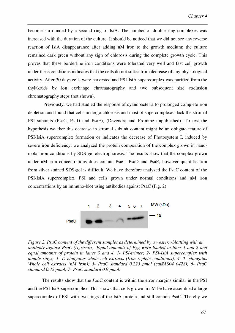
concentrations by an immuno-blot using antibodies against PsaC (Fig. 2).
Figure 2. PsaC content of the different samples as determined by a western-blottting with an antibody against PsaC (Agrisera). Equal amounts of P700 were loaded in lines 1 and 2 and equal amounts of protein in lanes 3 and 4. 1- PSI-trimer; 2- PSI-IsiA supercomplex with double rings; 3- T. elongatus whole cell extracts (Iron replete conditions); 4- T. elongatus Whole cell extracts (nM iron); 5- PsaC standard 0.225 pmol (cat#AS04 042S); 6- PsaC standard 0.45 pmol; 7- PsaC standard 0.9 pmol.
The results show that the PsaC content is within the error margins similar in the PSI
and the PSI-IsiA supercomplex. This shows that cells grown in nM Fe have assembled a large
supercomplex of PSI with two rings of the IsiA protein and still contain PsaC. Thereby we

Chapter 4
68
were able to show that the reduction of the PsaC content is not directly correlated to the
formation of the PSI-IsiA supercomplex, and is only induced under severe iron depletion. To
ensure that the PsaC subunit still contains the terminal Fe-S clusters FA and FB, we have
analyzed the iron content of the PSI-IsiA sample and PSI in form of dissolved crystals by
Inductively Coupled Plasma (ICP) measurement (a spectrometry technique for elemental
analysis). For the intact complex, we would expect 12 Fe/P700 as PSI contains 3 Fe4-S4
clusters. The Fe content of PSI was determined as 13.8 ± 2 Fe/P700 and the iron content of the
PSI-IsiA was determined to be 12.1 ± 2 Fe/P700 (data not shown), which corresponds to the
intact supercomplex.
The reduction-oxidation spectra of P700 in this supercomplex was analyzed for the
determination of the ratio of Chl to P700 in a dual beam spectrophotometer, for comparison,
the spectra of dissolved crystals of trimeric PS I were also analyzed in parallel using the same
setup. The Chl/P700 content of the PSI trimer was determined to be 95 ± 1 Chl/P700, which is in
agreement with the crystal structure of the complex that unraveled 96 Chl/P700 (Jordan et al.
2001). In PSI-IsiA supercomplex the Chl/P700 ratio was increased by a factor of 3 to 285 ± 5.
If we assume that one IsiA protein contains 13 chlorophyll molecules as its counterpart CP43
in PSII (Loll et al. 2005), this would lead to a Chl content of 559 Chl for the IsiA double ring
with 43 IsiA proteins/PSI-trimer, plus 3 x 96 Chl for the PSI core which adds up to 847
Chl/PSI trimer. This corresponds to 282 Chl/P700, which is in agreement with the
determination of Chl/P700 in the PSI-IsiA43 supercomplex. The molecular weight of this
supercomplex is ~3.2 kDa. Interestingly, in this supercomplex, the size of this auxiliary
antenna system is significantly larger than the PSI trimer itself. We also found that these
particles are stable at room temperature and sustain at high medium salt concentration (100
mM MgSO4) in the buffers, which are essential for stability and solubility of the detergent-
solubilized complexes.
Structure of PSI-IsiA supercomplex. Single-particle analysis
Purified PSI-IsiA supercomplexes were analyzed with transmission electron
microscopy. Under conditions where the supercomplexes were isolated from cells grown at
nM iron concentration, large circular particles with a diameter of 40.5 nm were abundant.
A large data set of single particle projections was selected from the images and
subjected to alignment procedures and statistical analysis. A map with intact circular particles
at 15 Å resolution shows trimeric PSI surrounded by a double ring of IsiA. In these particles,
the inner ring contains 18 copies of the IsiA protein and the outer ring 25 proteins (Fig. 3).

Chapter 4
69
Figure 3. Structural comparison between PSI-IsiA supercomplex with complete double rings (PSI-IsiA43) and the PSI-trimer. The resolution of the structures is 15 Å resolution. Both complexes are shown in a top view from the stromal side. (A) Single particle electron microscopy image of complete PSI-IsiA double ring particles from TS. elongatus, grown under nano-molar iron. The PSI-trimer in the middle is surrounded by 43 IsiA molecules, 18 IsiA form the inner ring, while the outer ring is formed by 25 IsiA molecules. (B) Projection map of the PSI structure at 15 Å resolution, derived from the X-ray structure of PSI at 2.5 Å resolution; the resolution was lowered to 15 Å to allow the direct comparison with the EM map of the PSI-IsiA43 supercomplex. The white arrow points to the largest difference between the maps. Scale bar is 100 Å.
We were also able to identify assembly intermediates of the supercomplex, which contained
either 16 or 19 copies of IsiA in the outer rings. (Fig. 4B, C). Empty IsiA rings without any
central mass were absent. The configuration of the outer ring in the complete and incomplete
supercomplex is different. This becomes clear after imposing a 3-fold rotational symmetry. If
applied to the complete outer ring, densities become faded out because 25 is not a multiple of
3 (Fig. 4D), whereas they become stronger in the fragments (Fig. 4E, F). This indicates that
the fragments have the IsiA copies at positions which, if extrapolated, would give a ring of 24
copies. Instead the full ring has a very astonishing symmetry mismatch with the perfect 3-fold
symmetry of the inner core. We also overlayed the positions of IsiA in the complete and
incomplete outer ring (red and blue bars, Fig 4.). From this overlay it is immediately obvious
that the spacing between the IsiA molecules in the outer ring is slightly smaller in the intact
ring compared to the assembly intermediates. This result may indicate that the IsiA protein
must have some intrinsic flexibility which allows the molecules to slightly reduce the space
between the individual subunits in the final assembly stage of the complex.

Chapter 4
70
Figure 4. Positions of IsiA copies in the outer ring of the PSI-IsiA supercomplex. (A) complete supercomplex; (B,C) incomplete supercomplex. (D-F) A 3-fold symmetry was imposed on the images of A, B and C. Positions of the IsiA copies in the outer ring in the complete particle (red bars) and in the largest fragment (blue bars) have been indicated. These spacing markers from the fully assembled ring are superimposed onto the assembly intermediates in the center picture. The overlay shows that the spacing between the IsiA complexes is slightly larger in the partial assembled complexes.
In EM images, the size and the "resolution" of IsiA complexes differ significantly
between the outer and inner ring. The IsiA monomers in the inner ring show much more
structural details than any of the IsiA proteins in the single ring PSI-IsiA complexes that have
been described before, while the proteins in the outer ring show less structural details and also
appear to be larger in size than the IsiA monomers in the inner ring, being comparable in the
size and resolution to the reported electron micrographs of the single-ring IsiA complexes
(Boekema et al. 2001; Bibby et al. 2001). The calculated area which is occupied by each IsiA
monomer in outer ring is ~8 nm2 larger than the inner ring. The major reason for the size
difference may be the presence of the detergent shell around the outer rings. However, the
strongly increased resolution of the IsiA molecules in the inner ring may indicate that the
assembly of the second ring leads to a slightly tighter packing and higher order of the IsiA
complexes in the first ring, which interact on both the peripheries and are compactly packed,
whereas the IsiA molecules in the second ring show a higher degree of flexibility due to their
interaction with the membrane/detergent micelle.

Chapter 4
71
Characterization of the electron transport chain by laser-flash induced absorption
spectroscopy
The electron transport chain in the complex was characterized by laser flash induced
absorption spectroscopy at 700 nm. The results (Fig. 5A, B) show homogenous re-reduction
kinetics with more than 80% of the back reaction occurring with kinetics of 82 ms indicative
of a recombination of the electron from the terminal Fe4-S4 clusters to P700. No significant �s
kinetics, which would be indicative of a loss of the terminal Fe4-S4 clusters are observed in
the sample, which suggests that the PSI-IsiA complex with double rings contains an intact
acceptor site with FA and FB clusters present.
Steady-state flavodoxin reduction assay
The second protein that is strongly induced with IsiA under iron deficiency is flavodoxin
(Burnap et al. 1993). It replaces the iron-containing protein ferredoxin under iron deficiency.
Despite their similar functional role, size, structure and cofactor content, the structures of both
proteins are completely different. Ferredoxin is a small Fe2-S2 protein (~100 amino acids, ~11
kDa); the two iron atoms are tetrahedrally coordinated both by inorganic sulfur atoms and by
conserved cysteine residues from peptide backbone (Hatanaka et al. 1997). In contrast,
flavodoxin is a comparatively larger flavin mononucleotide (FMN) binding protein (~170
amino acids, ~18-20 kDa) characterized by an open twisted structure consisting of five
standard parallel �-sheets, surrounded by �-helices (Rao et al. 1993). On the stromal side,
both flavodoxin and ferredoxin can serve as a soluble, redox electron carrier and transport
electrons between PSI and FNR. Flavodoxin replaces Fferredoxin under iron deficiency. We
measured the kinetics of flavodoxin reduction in the PSI and PSI-IsiA supercomplex thereby
analyzing the combined effect of both proteins that are induced under iron deficiency (IsiA,
B) on the PSI efficiency. To our surprise the rate of flavodoxin reduction in PSI-IsiA
supercomplex was ~9 times higher than the flavodoxin reduction rate of PSI (Fig. 6A,B).

Chapter 4
72
A B
Figure 5. P700+ recombination kinetics of PSI-trimers and the PSI-IsiA supercomplex at 700nm. The kinetics was determined as described in materials and methods. (A) P700+ reoxidation kinetics of PSI-trimers (T. elongatus wild type). Dissolved PSI crystals were used for this experiment. (B) P700+ reoxidation kinetics of PSI-IsiA supercomplexes isolated from cells that have been grown at 2.5 nano molar iron. The kinetics is very similar in both samples. The reoxidation of P700 is dominated by ms kinetics that are indicative of (FA/FB )-
back-reactions, with kinetic constants of 46ms for PSI (74.2%) and 82ms for the PSI-IsiA supercomplexes (84.4 %).
Discussion
Surface waters of most of the oceans in the world are iron deficient. The iron levels in
coastal areas, where most of the photosynthetic biomass is produced, are in the nanomolar
range (Takata et al. 2004) and thereby higher than in the open ocean, but still limiting for
photosynthetic growth. Cyanobacteria have adapted to grow efficiently in these low iron
environments by minimizing their iron requirements. Besides for the photosynthetic
machinery, iron is also essential for several other enzymatic and respiratory activities in the
cell. Therefore, a minimal amount of iron in the medium is necessary to balance out supply all
the basic physiological functions. Although, cyanobacteria have a very sophisticated
mechanism for short-term iron deficiency, this mechanism fails in dealing with against
prolonged complete iron depletion.
Omitting iron from the cyanobacterial growth medium triggers several new proteins in
the photosynthetic machinery (Burnap et al. 1993). In the beginning of iron depletion, the
cells consume cellular iron stored in ferritins (Keren et al. 2004) (iron storage proteins) and
from the surroundings (iron in chemical matrix of the medium, as impurity) and grow for a
couple of weeks until they have used up all internal storage and external iron. Once all the
iron in the environment and and inside the cells is consumed, the cells undergo growth
retardation, with doubling times decreasing to the time intervals of weeks or even months.

Chapter 4
73
Under these circumstances, studies of the effect of prolonged iron deficient conditions on the
photosynthetic apparatus of the cell are difficult because the iron-below-the threshold limit
causes severe chlorosis, and further cellular changes, which makes difficult to discriminate
between protective mechanisms and pre-stages of senescence and cellular death.
(A) (B)
(A) (B)
(C) Figure 6. Steady state flavodoxin reduction kinetics of (A) PSI-trimers and (B)PSI-IsiA43 supercomplexes (C) Comparison of the efficiency of the PSI and the supercomplex in terms of flavodoxin reduction.
In this study we planned to focus on cellular growth in a subnanomolar to nanomolar
iron environments. We found that ~0.5-2.5 nM concentrations of iron in the growth medium
of T. elongatus, which mimics most of the iron-limited conditions in aquatic environments,
does not only stabilize the PSI-trimer but also facilitates the formation of large amount of
PSI-IsiA supercomplexes with complete double IsiA rings.
Structure of the supercomplex
In electron microscopy (EM), we identified for the first time PSI-IsiA particles with complete
double rings by electron microscopy and single particle analysis. By single particle analysis a
15 Å resolution was obtained. The structure is one of the highest resolution structures for a
PSI-IsiA antenna supercomplex and allows us to draw conclusion about the internal
organization at the subunit level. Even after overnight solubilization of the thylakoid
membranes and several purification steps at room temperature, the complex is stable and

Chapter 4
74
contains 43 IsiA monomers/PSI-trimer with 18 copies in inner and 25 in outer ring. This
stability is a unique feature of this novel giant supercomplex and mainly depends upon two
types of protein-protein interactions. i.e. 1. interactions between IsiA monomers 2. PSI-IsiA
interactions. The electron density clearly shows the presence of all stromal subunits including
PsaC, which is a further confirmation of our western-blot results.
The new results, which show the presence of the intact acceptor side of PSI, including
PsaC and the terminal Fe-S clusters under nM iron conditions demonstrates that this decrease
in PsaC content is not related to IsiA induction, but it may be a part of PSI degradation in
severe iron deficiency and related to recycling of iron. However, functional analysis of such
particles might be interesting to see if there is any alternation in the electron transfer
mechanism.
In previous long-term iron deficient studies, hollow IsiA double rings structures of up
to 35 subunits (14+21) were observed, lacking monomeric or trimeric PSI (Yeremenko et al.
2004). Larger regular IsiA structures were not observed. As our novel PSI-IsiA supercomplex
has 43 IsiA molecules it shows that interactions with PSI are important for getting even larger
ordered structures. To our knowledge, this supercomplex is the maximal auxiliary antenna
complex for the PSI trimer, since we could not find any PSI trimer particle in the thylakoid
extract larger than this complex, e.g., no incomplete or complete triple rings have been
identified.
From this EM analysis and previous studies, we can also make some general
conclusions about the interactions of the PSI and IsiA. In a previous study monomeric PSI
was investigated with a mutant of the PsaL gene. (In the absence of the PsaL subunit, PSI
trimers cannot be formed anymore.) However, if mutant cells without this subunit are grown
in iron deficient conditions, most of the IsiA accumulates in incomplete rings at the PsaF/J
side of the complex (Kou�il et al. 2005b). The role of PsaF and PsaJ in the binding of IsiA to
the PSI core trimer was investigated in a PsaF/PsaJ double mutant of Synechocystis PCC 6803
(Kou�il et al. 2003). The data show that PSI-IsiA supercomplexes can still be formed, but the
PSI-IsiAcomplex is very unstable. The IsiA ring consists of 17 units in this mutant, instead of
18 in PSI-IsiA complexes from wild-type cells. This indicates that PsaF and PsaJ are not
absolutely required for the assembly of IsiA to PSI, but may be important for the stabilization
and structural integrity of the complex. The results also showed that the size of the ring
around the photosystem is strongly determined by the circumference of the PSI trimer, in
addition to specific, but still unknown PSI-IsiA interactions sites.

Chapter 4
75
The identification of assembly intermediates of the complex, that contain 16 and 19
subunits in an incomplete outer ring, indicate that the symmetry mismatch between the inner
ring, that consists of 18 subunits and the outer ring that consists of 25 subunits is already not
yet established in the assembly phase of the complex formation. The detailed analysis of the
position of the outer ring IsiA subunits in the three different assembly intermediates indicate,
that the IsiA complexes are more densely packed in the complete double ring than in the
assembly intermediates. This may suggest that the IsiA protein must contain an intrinsic
flexibility of the structural elements that allows it to show a more loosely or tightly packing.
In this respect it is also very intriguing to see the increase in the structural order and
resolution of the IsiA proteins of the inner ring upon binding of the outer IsiA ring. It shows
that despite of its conserved interactions with PSI, IsiA has tremendous ability to adjust its
interactions according to requirements. This flexible nature is another remarkable feature of
the PSI-IsiA supercomplex.
A comparison of EM images of the PSI-IsiA43 supercomplex with the structure of the
PSI trimer at 15 Å resolution, derived from the 2.5 Å resolution X-ray structure (Fig. 3)
showed the same major structural features for the PSI trimer. One difference is, however, at
the periphery. It can be seen that the interface between the monomers is more open in the X-
ray map (white arrow, Fig. 3) whereas the electron microscopy map of the new PSI-IsiA43
supercomplex shows more density, in particular at the position of the PsaK subunit. The PsaK
subunit is only loosely bound to the PSI-trimer (see Fig.2, chapter 1 for the structure), which
led to the fact that it was partially disordered in the X-ray structure at 2.5 Å resolution (Jordan
et al. 2001). No structural details of side chains were visible and the protein was only modeled
by a C-alpha backbone trace (Jordan et al. 2001). The extra density in the electron
microscopic images of the new PSI-IsiA43 supercomplex may indicate that the flexibility of
PsaK may be an important feature of PSI. PsaK may play a role in the interaction with the
IsiA protein and gets tightly packed in a slightly altered position upon assembly of the PSI-
IsiA43 supercomplex.
Previous EM studies showed that terminal electron acceptor flavodoxin docks to PSI
(Mühlenhoff et al. 1996) close to the stromal subunits PsaC and PsaD at a stromal hump that
is located on top of PsaA. A previous electron microscopy analysis showed that flavodoxin
extends the outer boundary of the PSI trimer (Mühlenhoff et al. 1996). A comparison of these
images with our EM pictures of the PSI-IsiA43 supercomplex, indicates that flavodoxin docks
at a position where parts of the electron transfer protein could interact with the IsiA ring. This
could provide some indication why PSI-IsiA43 shows an increase in flavodoxin reduction by a

Chapter 4
76
factor of 9. It is also possible that the core subunits of PSI that are involved in docking of
flavodoxin might undergo slight conformational changes that allow a more efficient docking
and electron transfer from the terminal FeS cluster in PSI to flavodoxin.
The induction of the IsiA and flavodoxin is not only limited to iron reduction. The co-
induction of flavodoxin in oxidative stresses with IsiA, even in iron replete conditions, shows
that both proteins are synergistic in their functionality and may represent a universal cellular
mechanism that allows the cells to adapt to oxidative stress (see below). Only the induction of
both proteins allows the simultaneous optimization of the antenna size and electron transfer
chain in PSI.
Functional analysis of the supercomplex
The Chl/P700 ratio in this complex was found to be 285 ± 5, which is approximately 3 times
higher than the PSI. The binding of IsiA increased the light absorption cross-section area of
PSI trimer by 300%. A relationship between iron deficiency and formation of a 3 times bigger
light harvesting antenna system is very interesting. How are these facts related to each other?
Synthesis of peripheral antenna phycobilisomes is iron consuming, therefore iron deficiency
leads to their degradation (Singh and Sherman 2000) and PSI acquires light from a new
auxiliary chlorophyll antenna system. Moreover, IsiA is expressed under different stress
conditions (Havaux et al. 2005; Yousef et al. 2003; Vinnemeier et al. 1998; Kojima et al.
2006). So IsiA may have more functions than just being a substitute for the water-soluble
phycobilisome antenna, which rapidly degrades under iron limitation. If we analyze all the
conditions in which IsiA is expressed, we find that they share a common trait: oxidative
stress. In the thylakoid membrane, excess light energy or any limitation in the electron
transfer chain, can over-reduce the PQ pool, which leads to a back log of the excitation energy
in the light-harvesting antenna. Consequently, reactive oxygen species are generated which
can cause oxidative damage and photoinhibition. It has been recently shown, that iron
deficiency also causes oxidative stress (Latifi et al. 2005); we postulate here that induction of
IsiA is related to the alterations in the electron transfer chain. Therefore, all stress conditions
that affect the smooth transfer of electrons from water to NADP+ through photosystem II,
cytochrome b6f, plastocyanin + cytochrome c6, PSI and FNR, also originate oxidative stress,
thereby inducing the expression of IsiA. Alternation in the electron transfer chain, induction
of IsiA in a cytochrome c6-deficient mutant (Ardelean et al. 2002), induction of IsiA in a PSI-
PsaFJ-null mutant (Jeanjean et al. 2003), respectively, are further supporting this idea. These
studies show that outside the natural environmental stresses there are much more possibilities

Chapter 4
77
of de-repression (induction) of the isiAB operon. We want to introduce the hypothesis that
redox state of the PQ pool, over-reduction of the electron receptor sites in PSI/PSII or
elevated lumenal acidity may control the induction of IsiA. In the case of iron deficiency,
reduction of PQ pool due to reduction of the number of PSI complexes in the cell might be the
primary reason to trigger the induction of IsiA.
The kinetic study presented here show that the efficiency of the electron transport
between PSI and flavodoxin is increased by nearly one order of magnitude in the PSI-IsiA43
supercomplex. Thereby we were able to show that the primary function of IsiA is to optimize
the efficiency of PSI, by a 3 fold- increase of the antenna size and the remarkable 9!-fold
increase of the rate of electron transfer from PSI to flavodoxin. The higher flavodoxin
reduction rate in this supercomplex is a new and remarkable feature of the PSI-IsiA
supercomplex. We suggest that this increase in PSI efficiency allows the cells to balance the
excitation energy and electron transfer reactions between the two photosystems. As iron
deficiency heavily induces the IsiA but also limits the number of PSI trimers, it seems that
under a well-balanced mechanism, cyanobacteria restrict the iron consumption by
synthesizing more efficient PSI-IsiA supercomplexes. PSI can promote the generation of the
high energy molecules (NADPH, ATP) by two ways i.e., it is involved in non-cyclic (linear)
and cyclic electron transfer. As during IsiA formation, a decrease in linear electron flow and
increase in cyclic flow around PSI has been reported (Michel and Pistorius 2004), we can
interpret that raise in PSI efficiency having the following benefits: 1. The system needs fewer
PSI complexes and can minimize its iron requirements; 2. Re-direction of iron and maybe
also chlorophyll; 3. Improvement of redox balance, due to cyclic electron flow. Consequently,
the cells experience less oxidative stress. Here, PSI is tuning the overall physiology of the cell
in addition to its significant contribution in energy production.
Acknowledgements
We thank Dr. W. Keegstra and Dr. G.T. Oostergetel for discussions and help with EM data
processing. This work was supported by NSF grant MCB-0417142 (to P.F.) and by a NWO-
TOP grant of the Netherlands Organisation for Scientific Research NWO (to E.J.B.).
References Andrizhiyevskaya, E.G., Schwabe, T.M.E., Germano, M., D’Haene, S., Kruip, J., van Grondelle, R. and Dekker J.P. (2002) Spectroscopic properties of PSI-IsiA supercomplexes from the cyanobacterium Synechococcus PCC 7942. Biochim. Biophys. Acta 1556, 265– 272.

Chapter 4
78
Andrizhiyevskaya, E.G., Frolov, D., van Grondelle, R. and Dekker, J.P. (2004) Energy transfer and trapping in the Photosystem I complex of Synechococcus PCC 7942 and in its supercomplex with IsiA. Biochim. Biophys. Acta 1656, 104-113. Ardelean, I., Matthijs, H.C.P., Havaux, M., Joset, F. and Jeanjean, R. (2002) Unexpected changes in photosystem I function in a cytochrome c6-deficient mutant of the cyanobacterium Synechocystis PCC 6803. FEMS Micro. Lett. 213, 113-119. Behrenfeld, M., Bale, A., Kolber, Z., Aiken, J. and Falkowski, P. (1996) Conformation of iron limitation of phytoplankton photosynthesis in the equatorial pacific ocean. Nature 383, 508-511. Bendall, D. S. and Manasse, R. S. (1995) Cyclic photophosphorylation and electron transport. Biochim. Biophys. Acta 1229, 23-38. Bibby, T. S., Nield, J. and Barber, J. (2001) Iron deficiency induces the formation of an antena ring around trimeric photosystem I in cyanobacteria. Nature 412, 743-745. Boekema, E.J., Hifney, A., Yakushevska, A.E., Piotrowski, M., Keegstra, W., Berry, S., Michel, K.P., Pistorius, E.K., and Kruip, J. (2001) A giant chlorophyll-protein complex induced by iron-deficiency in cyanobacteria. Nature 412, 745-748. Boyd, P.W., Watson, A.J., et al. (2000) A mesoscale phytoplankton bloom in the polar southern Ocean stimulated by iron fertilization. Nature 407, 695-702. Burnap, R.L., Troyan, T. and Sherman, L.A. (1993) The highly abundant chlorophyll-protein of iron-deficient Synechococcus sp. PCC 7942 (CP43’) is encoded by the isiA gene. Plant Physiol. 103, 893-902. Charles, G. T. and Steven, W.W. (1995) Physiological changes in the coastal marine cyanobacterium Synechococcus sp. PCC 7002 exposed to low ferric ion levels. Marine chemistry 50, 207-217. Coale, K.H. et al. (1996) A massive phytoplankton bloom induced by an ecosystem-scale iron fertilization experiment in the equatorial Pacific Ocean. Nature 383, 495-501.
De Jong, J.T.M., et al. (2007) Inputs of iron, manganese and aluminium to surface waters of the Northeast Atlantic Ocean and the European continental shelf. Marine Chemistry, 107, 120-142. Duce, R.A. (1986) The impact of atmospheric nitrogen, phosphorus and iron species on marine biological productivity. pp 497-529 In: P. Buat-Menard (ed.), The role of air-sea exchange in geochemical cycling. Reidel, Dordrecht, The Netherlands. Fork, D. C. and Herbert, S. K. (1993) Electron transport and photophosphorylation by photosystem I in vivo in plants and cyanobacteria. Photosynth. Res. 36, 149-168. Fromme, P. and Witt, H.T. (1998) Improved isolation and crtstallization of photosystem I for structural analysis. Biochim. Biophys. Acta 1365, 175-184.

Chapter 4
79
Guikema, J. and Sherman, L.A. (1983) Organization and function of chlorophyll in membranes of cyanobacteria during iron starvation. Plant Physiol. 73, 250-256. Hatanaka, H., Tanimura, R., Kotoh, S. and Inagaki, F. (1997) Solution structure of ferredoxin from the thermophilic cyanobacterium Synechococcus elongatus and its thermostability. J. Mol. Biol. 268, 922-933. Havaux, M., Guedeney, G., Hagemann, M., Yeremenko, N., Matthijs, H.C.P. and Jeanjean. R. (2005)The chlorophyll-binding protein IsiA is inducible by high light and protects the cyanobacterium synechocystis PCC6803 from photooxidative stress. FEBS Letters 579, 2289-2293. Ivanov, A. G., Krol, M., et al. (2006) Iron deficiency in cyanobacteria causes monomerization of photosystem I trimers and reduces the capacity for state transitions and the effective absorption cross section of photosystem I in vivo. Plant Physiol. 141, 1436-1445. Jeanjean, R., Zuther, E., Yeremenko, N., Havaux, M., Matthijs, H.C.P. and Hagemann, M.A. (2003) A photosystem 1 psaFJ-null mutant of the cyanobacterium Synechocystis PCC 6803 expresses the isiAB operon under iron replete conditions. FEBS Letters 549, 52-56. Jordan, P., Fromme, P., Witt, H.T., Klukas, O., Saenger, W. and Krauss, N. (2001) Three-dimensional structure of cyanobacterial photosystem I at 2.5 Å resolution. Nature 411, 909-917. Keren, N., Aurora, R. and Pakrasi, B. (2004) Critical Roles of Bacterioferritins in Iron Storage and Proliferation of Cyanobacteria. Plant Physiol. 135, 1666-1673. Kojima, K., Toyoko, S-M., Kikuchi, T. and Nakamoto, H. (2006) Roles of the cyanobacterial isiABC operon in protection from oxidative and heat stresses. Physio. Planta. 128, 507-519. Kou�il, R. Yeremenko, S. D’Haene, A. E. Yakushevska, W. Keegstra, H.C.P. Matthijs, J.P. Dekker and E.J. Boekema (2003) Photosystem I trimers from Synechocystis PCC 6803 lacking the PsaF and PsaJ subunits bind an IsiA ring of seventeen units. Biochim. Biophys. Acta 1607, 1-4. Kou�il, R., Arteni, A.A., Lax, J., Yeremenko, N., D’Haene, S., Rögner, M., Matthijs, H.C.P., Dekker, J.P. and Boekema, E.J. (2005a) Structure and functional role of supercomplexes of IsiA and Photosystem I in cyanobacterial photosynthesis. FEBS Letters 579, 3253-3257. Kou�il, R., Yeremenko, N., D’Haene, S., Oostergetel, G.T., Matthijs, H.C.P., Dekker, J.P. and Boekema, E.J. (2005b) Supercomplexes of IsiA and Photosystem I in a mutant lacking subunit PsaL. Biochim. Biophys. Acta 1706, 262-266. Latifi, A., Jeanjean, R., Lemeille, S., Havaux, M., Zhang, C.C. (2005) Iron Starvation Leads to Oxidative Stress in Anabaena sp. Strain PCC 7120. J. Bacteriol. 187(18), 6596-6598. Loll, B., Kern, J., Saenger, W., Zouni, A. and Biesiadka, J. (2005) Towards complete cofactor arrangement in the 3.0 Å resolution structure of photosystem II. Nature 438, 1040-1044.

Chapter 4
80
Martin, J.H. and Fitzwater, S.E. (1988) Iron deficiency limits phytoplankton growth in the north-east Pacific subarctic. Nature 331, 341-343. Martin, J.H., Coale, et al. (1994). Testing the iron hypothesis in ecosystems of the equatorial Pacific Ocean. Nature 371, 123-129. Melkozernov, A.N., Bibby, T.S., Lin, S., Barber, J. and Blankenship, R.E. (2003) Time-resolved absorption and emission show that the CP43 ' antenna ring of iron-stressed Synechocystis sp PCC6803 is efficiently coupled to the photosystem I reaction center core.
Biochemistry, 42, 3893–3903.
Michel, K.P. and Pistorius, E.K. (2004) Adaptation of the photosynthetic electron transport
chain in cyanobacteria to iron deficiency: The function of IdiA and IsiA. Physiol. Plant. 120,
36-50. Mühlenhoff, U., Kruip, J., Bryant, D.A., Rögner, M., Sétif, P. and Boekema, E. (1996) Characterization of a redox-active cross-linked complex between cyanobacterial photosystem I and its physiological acceptor flavodoxin. EMBO J., 15, 488-497. Park, Y.P., Sandstrom, S., Gustafsson, P. and Öquist, G. (1999) Expression of the isiA gene is essential for the survival of the cyanobacterium Synechococcus sp. PCC 7942 by protecting photosystem II from excess light under iron limitation. Mol. Microbiol. 32, 123-129.
Penczek, P., Radermacher, M. and Frank, J. (1992) Three-dimensional reconstruction of
single particles embedded in ice. Ultramicroscopy 40, 33-53. Porra, R.J., Thompson, W.A. and Kriedemann, W.E. (1989) Determination of accurate extinction coefficients and simultaneous equations for assaying chlorophylls a and b extracted with four different solvents: verification of the concentration of chlorophyll standards by atomic absorption spectroscopy. Biochim. Biophys. Acta 975, 384-394. Rao, S. T., Wu, S,, Satyshur, A., Ling, K.Y., Kung, C. and Sundaralingam, M. (1993) Structure of the oxidized long chain flavodoxin from Anabaena 7120 at 2 Å resolution. Prot. Sci. 1, 1413-1427. Richard, T. and Butler A. (1991) Investigation of the mechanism of iron acquisition by marine bacteria Alteromonas luteoviolaceus: Characterization of siderophore production. Limnol. Oceanogr. 36, 1783-1792. Rippka, R. (1988) Isolation and purification of cyanobacteria. Methods in Enzymo. 167, 3-27. Sandström, S., Park Y.I., Öquist G. and Gustafsson, P. (2001) CP43’, the isiA Gene Product, Functions as an Excitation Energy Dissipator in the Cyanobacterium Synechococcus sp. PCC 7942. Photochem. Photobiol. 74, 431-437. Singh, A.K. and Sherman, L.A. (2000) Identification of Iron-Responsive, Differential Gene Expression in the Cyanobacterium Synechocystis sp. Strain PCC 6803 with a Customized Amplification Library. J. Bacteriol. 182, 3536-3543.
Chapter 4
81
Singh, A.K., McIntyre, M. and Sherman, L.A. (2003) Microarray Analysis of the Genome-Wide Response to Iron Deficiency and Iron Reconstitution in the Cyanobacterium Synechocystis sp. PCC 6803. Plant Physiol. 132, 1825-1839. Takata, H., Kuma, K., Iwade, S., Yamajyoh, Y., Yamaguchi, A., Takag,i S., Sakaoka, K., Yamashita, Y., Tanoue, E., Midorikawa, T., Kimura, K. and Nishioka, J. (2004) Spatial variability of iron in the surface water of the northwestern North Pacific Ocean. Marine Chemistry 86, 139-157. Van Heel, M. (1987) Similarity measures between images. Ultramicroscopy 21, 95-99. Van Heel, M., Gowen, B., Matadeen, R., Orlova, E.V., Finn, R., Pape, T., Cohen, D., Stark, H., Schmidt, R., Schatz, M. and Patwardhan, A (2000) Single-particle electron cryo-microscopy: towards atomic resolution Quart. Rev. Bioph. 33, 307-369. Vinnemeier, J., Kunert, A. and Hagemann, M. (1998) Transcriptional analysis of the IsiAB operon in salt-stressed cells of the cyanobacterium Synechocystis sp. PCC 6803. FEMS Microbio. Lett. 169, 323-330. Yeremenko, N., Kouril, R., Ihalainen, J.A., D’Haene, S., van Oosterwijk, N., Andrizhiyevskaya, E.G., Keegstra, W., Dekker, H.L., Hagemann, M., Boekema, E.J., Matthijs H.C.P. and Dekker J.P. (2004) (2004) Supramolecular Organization and Dual Function of the IsiA Chlorophyll-Binding Protein in Cyanobacteria. Biochemistry 43, 10308-10313. Yousef, N., Pistorius, E.K. and Michel, K.P. (2003) Comparative analysis of idiA and isiA transcription under iron starvation and oxidative stress in Synechococcus elongatus PCC 7942 wild-type and selected mutants. Arch. Microbiol. 180, 471-483.