Embed Size (px)
Citation preview

University of Groningen
DNA replication at the single-molecule levelStratmann, S.A.; Oijen, A.M. van
Published in:Chemical Society Reviews
DOI:10.1039/c3cs60391a
IMPORTANT NOTE: You are advised to consult the publisher's version (publisher's PDF) if you wish to cite fromit. Please check the document version below.
Document VersionPublisher's PDF, also known as Version of record
Publication date:2014
Link to publication in University of Groningen/UMCG research database
Citation for published version (APA):Stratmann, S. A., & Oijen, A. M. V. (2014). DNA replication at the single-molecule level. Chemical SocietyReviews, 43(4), 1201-1220. https://doi.org/10.1039/c3cs60391a
CopyrightOther than for strictly personal use, it is not permitted to download or to forward/distribute the text or part of it without the consent of theauthor(s) and/or copyright holder(s), unless the work is under an open content license (like Creative Commons).
The publication may also be distributed here under the terms of Article 25fa of the Dutch Copyright Act, indicated by the “Taverne” license.More information can be found on the University of Groningen website: https://www.rug.nl/library/open-access/self-archiving-pure/taverne-amendment.
Take-down policyIf you believe that this document breaches copyright please contact us providing details, and we will remove access to the work immediatelyand investigate your claim.
Downloaded from the University of Groningen/UMCG research database (Pure): http://www.rug.nl/research/portal. For technical reasons thenumber of authors shown on this cover page is limited to 10 maximum.
Download date: 07-12-2021

This journal is©The Royal Society of Chemistry 2014 Chem. Soc. Rev., 2014, 43, 1201--1220 | 1201
Cite this: Chem. Soc. Rev., 2014,
43, 1201
DNA replication at the single-molecule level
S. A. Stratmann and A. M. van Oijen*
A cell can be thought of as a highly sophisticated micro factory: in a pool of billions of molecules – metabolites,
structural proteins, enzymes, oligonucleotides – multi-subunit complexes assemble to perform a large
number of basic cellular tasks, such as DNA replication, RNA/protein synthesis or intracellular transport. By
purifying single components and using them to reconstitute molecular processes in a test tube, researchers
have gathered crucial knowledge about mechanistic, dynamic and structural properties of biochemical
pathways. However, to sort this information into an accurate cellular road map, we need to understand
reactions in their relevant context within the cellular hierarchy, which is at the individual molecule level
within a crowded, cellular environment. Reactions occur in a stochastic fashion, have short-lived and not
necessarily well-defined intermediates, and dynamically form functional entities. With the use of single-
molecule techniques these steps can be followed and detailed kinetic information that otherwise would be
hidden in ensemble averaging can be obtained. One of the first complex cellular tasks that have been
studied at the single-molecule level is the replication of DNA. The replisome, the multi-protein machinery
responsible for copying DNA, is built from a large number of proteins that function together in an intricate
and efficient fashion allowing the complex to tolerate DNA damage, roadblocks or fluctuations in subunit
concentration. In this review, we summarize advances in single-molecule studies, both in vitro and in vivo,
that have contributed to our current knowledge of the mechanistic principles underlying DNA replication.
1. Introduction
Life is as dynamic as its environment. Many key cellularprocesses cannot be described as outcomes from static associa-tions of molecular components, but instead rely on an intricatespatial and temporal orchestration of many molecular players.For example, the conversion of chemical energy into mechanicalwork allows the transport of vesicles and molecules within thecytosol, along a membrane or between cells. On the single-molecule level, kinesins and other motor proteins move alongthe cytoskeletal filaments, transporter proteins shuffle metabolitesbetween compartments, and multi-subunit complexes like repli-somes, ribosomes, or the respiratory chain support an efficientmaintenance and balancing of anabolism and catabolism.
Both fluorescence- and force-based single-molecule studieshave provided fascinating new insights into some of theseelaborate biological processes, such as cytoskeletal dynamics,1–3
ATP synthesis,4,5 RNA and DNA polymerization,6–8 and viralpackaging.9,10 The more recent developments in live-cell single-molecule imaging allow us to record the cellular micro-management in real time, as has been demonstrated for examplefor transcription-factor dynamics,11 protein-expression rates,12
and signalling pathways13 (reviewed to a greater detail in ref. 14).
What type of knowledge do we obtain from experimentsmonitoring individual molecules? Ensemble-averaging bulkassays provide information about the reaction rates of a poolof catalysts and, by synchronizing reactions, kinetic studies canreveal the first few transitions of a multi-step process. However,loss of synchronization due to the stochastic nature of chemicalreactions will render it challenging to obtain kinetic parametersof short-lived intermediate states. Single-molecule studiescapture the probabilities of reaction steps or conformationalchanges of an individual enzyme during any arbitrary pointalong a multi-step process and provide information onunderlying heterogeneities in the dynamic behaviour of thepopulation.15,16 Watching individual reactions at work tells usnot only about the stochasticity of consecutive pathways, but alsoabout any temporal correlation: does an enzymatic reaction forexample display non-markovian behaviour, i.e. are reaction stepsaffected by preceding paths?17 One of the earliest single-molecule fluorescence studies demonstrated such a memoryeffect in a flavoenzyme: autocorrelations of on and off dwelltimes of the redox-cofactor FAD(H2) resolved heterogeneouskinetic rates, caused by conformational changes within theprotein, that had been previously masked in bulk experi-ments.16 Similarly, single-molecule analyses of the RecBCDhelicase of Escherichia coli could decipher subpopulations ormicrostates of the enzymatic complex that differ in the velocityof DNA unwinding.18 Here, conformational changes that
Zernike Institute for Advanced Materials, Centre for Synthetic Biology,
University of Groningen, The Netherlands. E-mail: [email protected]
Received 1st November 2013
DOI: 10.1039/c3cs60391a
www.rsc.org/csr
Chem Soc Rev
REVIEW ARTICLE
Publ
ishe
d on
06
Janu
ary
2014
. Dow
nloa
ded
by U
nive
rsity
of
Gro
ning
en o
n 20
/02/
2014
13:
54:0
0.
View Article OnlineView Journal | View Issue

1202 | Chem. Soc. Rev., 2014, 43, 1201--1220 This journal is©The Royal Society of Chemistry 2014
are adopted in the absence of the ligand/substrate ATP are‘‘memorized’’ by the active RecBCD upon ATP addition andresult in distinct rates of progression along the DNA template.
The actual chemical conversions in enzymatic reactionstypically proceed on a sub-picosecond time scale. However, thelimiting steps in catalysis are often the crossing of thermalactivation barriers and the diffusive process necessary to mediateassociation of two reactants, which last orders of magnitude longerand are consequently the parameters to follow in single-moleculestudies.19,20 Typical fluorescence assays rely for example on visua-lizing a chromophore coupled to a molecule of interest andmonitoring the appearance and disappearance of its signal asthe labelled component is binding to and dissociating from areaction partner molecule (Fig. 1). Binding lifetimes can beextracted and a probability distribution generated that containsthe kinetic rates of the observed reaction. Several reviews on single-molecule enzymology provide excellent descriptions of enzymatickinetics based on single-molecule reaction probabilities.16,21
In addition to successes in resolving single-protein kinetics,recent developments have focused on the visualization of proteindynamics and complex assemblies in real time, usually using
fluorescence co-localization or fluorescence (Forster) resonanceenergy transfer (FRET) methods. These approaches have allowed,for example, the observation of the dimerization of EGF receptorsin living cells, the complex formation of a reconstituted functionalvesicle fusion construct of t- and v-SNARE proteins, or the Arp2/3-mediated branch formation on growing actin filaments.13,22,23
Studies of the replisome, the machinery responsible for DNAreplication, face the challenge of revealing the various andfrequently transient interactions of the numerous enzymes thatare involved.24–26 The multi-component replisome is loadedon the DNA template in tight coordination with the cell cycle;it proceeds with a speed of up to thousand nucleotides per second(for certain bacterial systems), corrects wrongly incorporatednucleotides to an accuracy of about one mistake per 109 nt andtriggers repair processes upon detection of depurination, deami-nation, or pyrimidine-dimer formation.27,28 Coordination of sucha wide array of tasks, each on their own representing formidablemolecular challenges, requires a finely tuned and balanced set ofenzymatic activities. Building on the large base of knowledge wehave on the individual components of the replication reaction,derived from many decades of genetic, biochemical and structuralstudies, single-molecule approaches represent a powerfulapproach to unravel the intricacies of how the various enzy-matic activities at the replication fork are coordinated.
The process of replication needs to deal with a variety ofmolecular hurdles. For example, the antiparallel nature of thedouble-stranded DNA template imposes an asymmetry on thereplication machinery, whose DNA polymerases can onlysynthesize in one particular direction. Besides the need for thisasymmetric coordination, other obstacles have to be tackled, suchas crowding effects and roadblocks caused by transcription-relatedprocesses and repair activities that take place simultaneously onthe same DNA template. How exactly cells meet those challenges isa subject particularly well-suited for single-molecule studies –requiring methods to observe the spatiotemporal behaviour ofindividual molecules in a biologically relevant environment.
In this review, we describe recent developments in single-molecule research on the replisome in vitro and in vivo. First wehighlight a section to the main technological developments interms of microscope setups, design of fluorophores and labellingmethods. Referring to the replication systems of the bacteriophagesT7 and T4, of Escherichia coli (E. coli), and of eukaryotic cells, weguide the reader through the different aspects of important single-molecule studies that have contributed to a better understanding ofthe basic mechanics of DNA replication and organization.
2. Experimental strategies to imagesingle molecules
Single-molecule techniques are typically categorized into twoclasses that we want to outline briefly: fluorescence microscopyallows the recording of the emitted photons of a fluorophore-labelled molecule of interest and is particularly applicable fordetecting conformational changes within the protein of interest orits localization. Force-based measurement techniques, like atomic
Fig. 1 Extraction of single-molecule kinetics from the observation of onand off times. These on- and off times can represent a variety of functional orstructural transitions such as binding/unbinding, conformational transitionsor chemical reactions. (A) On- and off times of an observed fluorescentemitter are recorded and the photon count per molecule is tracked overtime and fitted to an appropriate function. (B) The time scales for on andoff times are sorted in a distribution that provides the kinetic parameters ofthe individual reaction.
Review Article Chem Soc Rev
Publ
ishe
d on
06
Janu
ary
2014
. Dow
nloa
ded
by U
nive
rsity
of
Gro
ning
en o
n 20
/02/
2014
13:
54:0
0.
View Article Online

This journal is©The Royal Society of Chemistry 2014 Chem. Soc. Rev., 2014, 43, 1201--1220 | 1203
force microscopy (AFM), magnetic tweezers, optical traps or flow-stretching setups, are useful in characterizing mechanical proper-ties such as DNA topology or force exertion by motor proteins.
2.1 Getting proteins to shine
As early as the 1970s, it was demonstrated that single proteinmolecules labelled with a large number of dyes could be detectedusing an optical microscope:29 Tomas Hirschfeld coupled roughlya hundred fluorescein dyes to a single antibody, swept a dilutesolution of these constructs along a tightly focused laser beam,and observed bursts of fluorescence each corresponding to asingle antibody. Not until two decades later, absorption andfluorescence measurements of single chromophores were success-fully performed at cryogenic temperatures where absorptioncross-sections are highest and photo-induced damage lowest.30–32
Initially these studies were performed on doped molecular crystals,whereas later cryogenic single-molecule approaches were appliedto study pigment–protein complexes.33 Near-field scanning micro-scopy approaches demonstrated the feasibility to repeatedlyimage chromophores within biological samples at ambient tem-perature,34 but were later joined by even more powerful andtechnically less-demanding far-field methods, mainly confocaland total-internal-reflection (TIRF) microscopy. Since then, greatadvances in high-sensitivity detection devices, in the engineeringof photostable dyes and fluorescent proteins, and labellingstrategies have pushed the sensitivity and resolution limits to apoint where single molecules can be observed over timescalesfrom milliseconds to minutes and down to spatial resolutions of afew nanometres. Furthermore, advances in live-cell imaging haveenabled such experiments in a cellular context. Here, additionalfactors have to be considered in terms of cell viability (nutrients,CO2, photodamage due to decomposition of fluorophores andradical release) and fluorophore choice (uptake, label selectivityand specificity). Even though these developments are relativelyrecent and many novel methods are still coming to fruition,single-molecule approaches are already revolutionizing the waymechanistic questions of biological systems are answered.
2.1.1 Hardware technology. The optical instrumentationrequired for single-molecule imaging and tracking can be roughlydivided into two modes of operation: wide-field imaging andscanning confocal microscopy (Fig. 2). Both approaches have
their advantages and need to be adapted to the actual question inconsideration of both spatial and temporal resolution.
Wide-field imaging is a frequently used method to followreactions at the single-molecule level in real time, i.e. to trackparticles and observe fast dynamics. In epifluorescence micro-scopy, a large sample volume is excited, limiting the signal-to-noise ratio in the region-of-interest. However, thin samples,either reconstituted isolated compounds or flat cells, can beanalysed with single-molecule sensitivity, as shown for micro-tubule gliding on kinesins or live-cell protein expression.35,36
Being proposed already in the 1950s but not fully developeduntil several decades later,37,38 TIRF microscopy has proven tobe exceptionally useful in improving signal detection. Here, atthe coverglass/solution interface an evanescent field is inducedthat decays exponentially in the z plane and limits the excitedvolume to about 100 nm.
In confocal imaging, a diffraction-limited focus is positionedwithin the sample volume and scanned orthogonally to theoptical axis.39 The use of pinholes results in the selective detec-tion of only in-focus fluorescence, while suppressing mostout-of-focus background. In contrast to a TIRF setup, confocalmicroscopy allows the scanning of samples in three dimensionswith a large penetration depth. However, the limiting factor isthe scanning speed of the focal spot through the sample.Spinning-disk confocal setups employ a broad laser illuminationthat is focused by a large array of microlenses on a Nipkow disk,achieving high frame rates of up to 1000 frames per second.40
Non-linear two-photon techniques use optical sectioning aswell, but here focussing relies on the probability of two-photonabsorption, which is proportional to the square of the excita-tion intensity. A main advantage is that the required lower-energy wavelengths reduce photodamage of the fluorophores aswell as scattering in tissue samples. Depths of several hundredmicrometres are achievable with this method, as for exampledemonstrated in fascinating work on intact organs in livingorganisms.41
Technological developments in optical microscopy havecontributed to a gradual improvement in spatial resolution,but with the size of the smallest resolvable structures stillsimilar to the diffraction limit. The recent breakthroughs insuper-resolution imaging, however, have allowed the imaging
Fig. 2 Fluorescence microscopy designs frequently used in single-molecule studies. In epifluorescence microscopy, the light source illuminates theentire sample. In confocal microscopy, a pinhole is used to illuminate specifically the focal plane, thus reducing background fluorescence. By installing aNipkow spinning disk, the sample is illuminated at multiple points within the focal plane simultaneously. In total-internal-reflection fluorescence (TIRF)microscopy, the incident laser is reflected from the coverglass surface, creating an exponentially decaying evanescent field on top of the surface, whichreduces the thickness of the illuminated volume to about 100 nm.
Chem Soc Rev Review Article
Publ
ishe
d on
06
Janu
ary
2014
. Dow
nloa
ded
by U
nive
rsity
of
Gro
ning
en o
n 20
/02/
2014
13:
54:0
0.
View Article Online

1204 | Chem. Soc. Rev., 2014, 43, 1201--1220 This journal is©The Royal Society of Chemistry 2014
of fluorescently labelled structures down to length scales thatare an order of magnitude smaller than the diffraction limit.Super-resolution methods find their basis in the reduction of thepoint-spread function (PSF) in excitation, as in stimulated emis-sion depletion (STED), ground-state depletion (GSD) or structuredillumination microscopy (SIM), or in the modulation of thefluorophore’s emission, as in photoactivated localization micro-scopy (PALM) and stochastic optical reconstruction microscopy(STORM) (Fig. 3). STED microscopy is based on the illuminationof the sample with a doughnut-shaped beam profile. The excita-tion beam is narrowed by an overlaying ring-shaped longer-wavelength depletion beam, that forces the dyes into the groundstate.42 The higher the intensity of the depletion laser, thenarrower the PSF becomes. In a similar design, but typically usingonly one wavelength, GSD brings the fluorophores to their lowesttriplet dark state in the outer ring. An alternative approach tosuper-resolution imaging is enabled by the wide-field methodsPALM and STORM, utilizing the stochastic activation of fluoro-phores that are photoactivatable or photoswitchable. The activa-tion of a few fluorophores in the field of view allows each of themto be individually imaged and to be fit by a two-dimensionalpoint-spread function and thus each of their centroid positions tobe obtained with sub-diffraction-limited precision. It is the sum ofseveral cycles of activation–centroid detection–bleaching/inactivationthat leads to the reconstruction of the complete object of interest.The super-resolution techniques PALM and STORM have also playedimportant roles recently in resolving intracellular dynamic processesat the single-molecule level (e.g. ref. 43 and 44).
2.1.2 Fluorophore technology. One of the major challengesin modern fluorescence microscopy is the engineering of appro-priate dyes and the specific attachment to the biomolecule of
choice. The properties required of chromophores for single-molecule imaging are demanding: the photostability in termsof lifetime and (absence of) blinking must be high, the con-jugated molecular structure must be soluble and stable, and thefluorophore’s dimensions and physicochemical propertiesshould not interfere with protein conformations and function.For sub-nanometre tracking, high quantum yields and largeStokes shifts are especially important.
In general, three categories of probes can be differentiated:fluorescent proteins, organic dyes and quantum dots. Beinggenetically encoded as fusion constructs to the protein ofinterest, fluorescent proteins are labels with absolute specificityand represent a standard approach for in vivo imaging. Limita-tions are their photostability and brightness, as well as thebulkiness of the 25 kDa structure that potentially interferes withenzyme functionality. Organic dyes are significantly smaller andoften display better photophysical properties. The commercialavailability of dyes is enormous; brightness, stability, and solu-bility can be chosen with great flexibility. The major bottlenecksare the specificity end efficiency of the labelling chemistry and,for in vivo studies, the need for electroporation or alternativemethods to introduce the dyes into the cell. Finally, quantumdots, fluorescent nanocrystals of 5–20 nm diameter, can beengineered in highly sophisticated ways, with extinction coeffi-cients several times higher than those of organic dyes. Extremelyhigh brightness and the resultant high signal-to-noise ratiosallow nanometre-tracking of individual molecules.45 The largesize, however, can influence the mobility and conformationalflexibility of the labelled protein.
Fluorescent proteins. Fluorescent proteins (FPs) consist of arigid b-barrel composed of 11 b-sheets that surround a centrala-helix containing the chromophore.46 The naturally occurringvariants have been extensively tuned in terms of brightness,emission range, photostability, monomeric character, andmaturation rate.47,48 Due to the strong autofluorescence ofendogenous cellular fluorophores (flavins, NADH, amino acids)at wavelengths below 500 nm, the development of red-shiftedFPs is one central consideration for in vivo imaging.
Newly developed classes of FPs with photoconvertible or photo-switchable chromophores allow super-resolution imaging even in ahigh-concentration environment, as only a limited fraction withinthe excitation field is switched on. Photoconvertible FPs such asKaede, KikGR, Dendra and Eos are subject to a peptide-backbonecleavage step when illuminated with a 405 nm laser, leading to anenlargement of the conjugated system by an additional imidazolering, which corresponds to a green-to-red shift in fluorescence.48,49
The photoswitchable FP Dronpa has an excitation maximum at503 nm, and can be switched off and on several times by strong488 nm and weak 405 nm illumination, respectively. Alternatively,the green fluorescent Padron is switched on by blue excitation andoff by UV light. The combination of those opposite switchingbehaviours allows two-color tracking in live cell imaging.50 In termsof photochemistry, crystal structures of Dronpa suggest that thecis–trans isomerization and protonation of the chromophore areresponsible for the different fluorescent states.51
Fig. 3 Super-resolution techniques. In STED, fluorophores are excited tothe S1 state (green) and return to the ground state S0 spontaneously whileemitting photons (yellow). An intense red-shifted doughnut-shaped depletionlaser beam (red) forces molecules into the ground state without them emittingfluorescence. As a result, only a sub-diffraction-limited area in the center ofthe depletion laser remains in the excited state and will be observable throughthe emission of a yellow fluorescent photon. A similar excitation geometry isused in ground state depletion (GSD) microscopy. However, instead ofrendering the fluorophores around a point of interest nonfluorescent bydepleting the fluorescent excited state, they are brought into a long-liveddark state. In PALM and STORM, molecules are switched on at low spatialdensities, their positions determined with sub-diffraction-limited precision,and irreversibly photobleached. Repeating this procedure for a large numberof molecules results in sub-diffraction-limited images (adapted from ref. 191).
Review Article Chem Soc Rev
Publ
ishe
d on
06
Janu
ary
2014
. Dow
nloa
ded
by U
nive
rsity
of
Gro
ning
en o
n 20
/02/
2014
13:
54:0
0.
View Article Online
This journal is©The Royal Society of Chemistry 2014 Chem. Soc. Rev., 2014, 43, 1201--1220 | 1205
Further progress in the design of fluorescent protein tags,especially far-red fluorescent as well as switchable probes, incombination with novel microscopy techniques will continue toprovide powerful tools for in vivo imaging.
Organic dyes and their coupling to proteins. The main chal-lenge in the use of organic dyes is a highly efficient and specificlabelling reaction to the target protein. Several strategies existfor selective chemical tagging that can be basically subdividedinto the introduction of a protein domain, a short peptide or aunique amino acid.52
A successful method to specifically couple an organic dye to aprotein is the fusion to a target protein of an additional proteindomain that itself binds the organic dye tightly and selectively.Prominent protein-domain fusion constructs are the commer-cially available dehalogenase and alkylguanosine transferase tags(HaloTag and SNAP tag, respectively). The HaloTag technologytakes advantage of a self-labelling step of a 33 kDa-sized dehalo-genase enzyme. The reaction catalysed by this enzyme consists of(1) a nucleophilic displacement of a halide ion from an alkanechain that is transferred to an aspartate residue, (2) histidinecatalysed hydrolysis, finally regenerating the aspartate. Mutagenesisof the active-site histidine residue locks the dehalogenase in step 1,allowing specific labelling with a customized fluorescent alkanemoiety.53 The 20 kDa sized O6-alkylguanine-DNA alkyltransferase(hAGT) enzyme transfers an alkyl group from guanosine derivativesto its active site cysteine residue, allowing for the subsequentcovalent coupling of alkyl-modified fluorophores.54
Smaller peptide tags are particularly advantageous wheninternal labelling positions are required. The Tsien labdeveloped a biarsenic tagging technology that depends on thehigh affinity of thiols to arsenic.55,56 The probes 40,50-bis(1,3,2-dithioarsolan-2-yl)fluorescein (FlAsH) and the chemicallysimilar resorufin-based ReAsH are non-fluorescent when boundto ethane dithiol (EDT), but fluoresce green and red, respectively,when a tetracysteine sequence CCXXCC replaces EDT. Anotherstrategy relies on the incorporation of an aldehyde tagging peptidesequence LCTPSR into the target protein.57 A co-expressed formyl-glycine-generating enzyme converts the cysteine’s thiol group intoan aldehyde that specifically reacts with hydrazide-functionalizedmolecules to produce a hydrazone. Other self-labelling tags arethe hexa-histidine peptide or the Texas-red-binding aptamer,chelating with Ni–NTA-derivatized fluorophores or bindingthe Texas-red fluorophore with nano- to picomolar bindingaffinity.58,59
Cysteines are usually less abundant in proteins and due totheir high reactivity towards maleimide thioesters they are apopular target for in vitro labelling. If cysteine mutagenesis is notfavourable because of limitations related to protein function-ality, the introduction of unnatural amino acids, as pioneered bythe Schultz lab, represents an alternative approach. Co-expressedorthogonal tRNAs and aminoacyl tRNA synthetases incorporate arange of unnatural amino acids in response to amber stopcodons or quadruplet codons.60–63
Despite the intrinsic bottleneck of selectivity in labelling, theadvantage of organic dyes lies in the nearly unlimited options
for fluorescence characteristics. Not only are dyes available thatcover the entire spectral range, but also many fluorescentcompounds have been developed with properties that can beexternally modified by optical inputs. For example, cagedchromophores can be activated by UV light, and several cyaninedyes can be coupled to construct activator–reporter FRET pairs.64,65
2.2 Trapping and pulling at individual DNA molecules
Force spectroscopy methods are frequently applied for charac-terizing mechanical properties of biomolecules at the single-molecule level, such as topological changes in DNA moleculesor force exertion by individual motor proteins. In the context ofstudying DNA replication at the single-molecule level, suchtechniques are often used to stretch the DNA substrate and toprobe the mechanical consequences of replication on the DNA(conversion between single- and double-stranded DNA,8,66
change in supercoiling67), or to observe the motion of proteinsalong DNA.68,69 Detailed reviews about the instrumentaldesigns can be found elsewhere.70–73
In trapping techniques, one end of the biomolecule ofchoice is stably attached to a surface and the other one trappedwith a magnetically or optically controlled bead or an AFM tip.Optical tweezers trap dielectric beads within a focused laserbeam. The electromagnetic field polarizes the particle that isforced into the steep gradient at the focal spot. Spatial resolu-tions of down to 0.1 nm with sub-millisecond time resolutionsare feasible by applying forces of about 0.1 to 100 pN.70 Magnetictraps have a slightly lower spatial resolution of about 2 to 10 nm,can apply forces over a large range from pico- to nano-Newtons,and therefore are particularly useful in the measurement andmanipulation of DNA topology. By attaching DNA on one end toa surface and on the other to a paramagnetic bead, the polymeris constrained and can be accurately controlled and placed in aparticular topological conformation with defined twist andwrithe.74 Prominent topoisomerase experiments are performedon magnetically manipulated plectonemic DNA, as the ATP-dependent double-strand breaks remove two turns, thus chan-ging the linking number by two.75 Flow-stretching techniquesrely on the hydrodynamic dragging of one-end anchored poly-mers in a microfluidic device. DNA-bead tethers are for exampleuseful in tracking length changes of the molecule during thetime course of replication76 (Fig. 4).
Combining the strengths of fluorescence imaging andmechanical approaches, recent developments have allowed theobservation of DNA-based single-molecule fluorescence whileexerting well-defined stretching forces on the DNA template.77,78
For example, Holliday-junction recombination events and con-formational changes could be followed by creating FRET pairswithin the four-stranded complex, tethered to an optical trap.79
The angstrom resolution of FRET signals combined with sub-pNforces in the optical trap established a highly controlled systemfor controlling and following conformations of DNA structures.Such hybrid techniques, allowing both the tracking of fluores-cent molecules and the detection of the chemomechanicalreactions, hold tremendous power in understanding the manyfacets of multi-protein machineries acting on DNA.
Chem Soc Rev Review Article
Publ
ishe
d on
06
Janu
ary
2014
. Dow
nloa
ded
by U
nive
rsity
of
Gro
ning
en o
n 20
/02/
2014
13:
54:0
0.
View Article Online

1206 | Chem. Soc. Rev., 2014, 43, 1201--1220 This journal is©The Royal Society of Chemistry 2014
3. Replication machineries
Genomic DNA replication consists of three distinct phases:initiation, elongation and termination. The complexity of cell-cycle timing, its coupling to DNA synthesis, and in general themolecular details of DNA synthesis vary tremendously amongstthe taxonomic domains. However, the main principles of thereplication machinery are conserved: ring-structured replicativehelicases encircle single-stranded DNA and couple the energyreleased from nucleotide hydrolysis to directional movement.The subsequent unwinding of the DNA provides a template forpolymerases to synthesize the daughter strands by catalysingthe coupling of an incoming nucleotide to the ribose 30 hydroxylgroup of the previously incorporated nucleotide. All known
polymerases display this requirement of directionality: onlyDNA synthesis from the 50 to the 30 end allows for a continuationof synthesis (accompanied by the backwards removal of incor-rectly incorporated nucleotides). With the antiparallel nature ofdouble-stranded DNA, such a directional requirement for DNAsynthesis results in a picture in which DNA is synthesizedcontinuously on the so-called leading strand, with the leading-strand DNA polymerase acting in the same direction as thehelicase is moving, and with the lagging-strand DNA polymerasepolymerizing in a discontinuous fashion, giving rise to shortstretches of DNA named Okazaki fragments (Fig. 5A and B).
A special class of polymerases known as primases synthesizeshort oligo-ribonucleotide primers that are used as startingtemplate for the lagging-strand DNA polymerase. The timing ofthe enzymatic steps at the lagging strand, i.e. priming, utiliza-tion of the primer by the polymerase and its extension into anOkazaki fragment, is of importance for the orchestration of acoupled replication reaction: a process in which continuoussynthesis on the leading strand is tightly coordinated with thediscontinuous synthesis on the lagging strand.
Research on the replisome of the bacteriophage T4 initiated theidea of the trombone model that reconciles a symmetric replica-tion fork, containing two DNA polymerases moving in the samedirection, with the underlying asymmetry of the DNA template.80
The formation of a looped structure in the lagging strand reorientsthe polymerase while synthesizing an Okazaki fragment, until arelease event triggers the recycling of the polymerase to the nextOkazaki fragment. The formation of a DNA loop and the closeproximity of the lagging-strand DNA polymerase to the replisome
Fig. 4 Force manipulation setups. DNA molecules are attached on oneside to the coverglass surface and coupled to a bead that is stretched in ahydrodynamic flow, or trapped magnetically or optically.
Fig. 5 Replisome proteins and fork architecture in viruses, bacteria and eukaryotes. (A) The replication fork of bacteriophage T7. Two polymerases gp5,each associated with an E. coli thioredoxin molecule, bind to the hexameric helicase–primase gp4. The primase domain of gp4 synthesizes shortribonucleotide primers that are handed over to the lagging-strand polymerase for elongation into Okazaki fragments. The unwound single-strandedregions of the template DNA are covered by gp2.5 proteins (adapted from ref. 66). (B) The replication fork of E. coli. Two copies of the DNA polymeraseholoenzyme are associated with b clamps on the leading and lagging strand. Three DnaG molecules associate with the DnaB helicase to synthesizeprimers on the lagging strand that is partly covered by SSB tetramers (adapted from ref. 192). (C) Comparison of the replisome components in phage,E. coli and eukaryotes.
Review Article Chem Soc Rev
Publ
ishe
d on
06
Janu
ary
2014
. Dow
nloa
ded
by U
nive
rsity
of
Gro
ning
en o
n 20
/02/
2014
13:
54:0
0.
View Article Online

This journal is©The Royal Society of Chemistry 2014 Chem. Soc. Rev., 2014, 43, 1201--1220 | 1207
result in a short travel distance to the next primer after comple-tion of Okazaki fragment synthesis by the polymerase. Thepresence of replication loops is supported by several lines ofevidence obtained from bacteriophage replication machineriesgenerating Okazaki fragments of about 1000 to 2000 bp.In eukaryotes however, the much shorter Okazaki fragments(100 to 200 base pairs) make such a looping scenario less likelyand certainly more difficult to observe.
In addition to the mechanistic demands placed on replicationdue to the antiparallel nature of duplex DNA, copying genomicstretches of DNA inside the cell comes with several other mole-cular challenges. Roadblocks such as nucleosomes need to bedealt with, the topology of the DNA needs to be controlled, andreplication needs to be regulated and coordinated with othercellular activities such as DNA repair and recombination. As willbe laid out in the remainder of this review, single-moleculebiophysical techniques have begun to significantly contribute toour understanding of the molecular aspects of each of theseprocesses. We will illustrate these efforts by starting with simplereplication model systems, focusing on only the activities at thefork, followed by zooming out and considering the interplay ofreplication with topology, nucleosomes and the overall cell cycle.
3.1 Model systems for single-molecule studies
The main operating principles of the replisome are highlyconserved across phages, bacteria and eukaryotes (Fig. 5),although the involved enzyme classes are structurally notnecessarily homologous. Replication complexes that are wellunderstood in terms of their composition, assembly and func-tioning are the ones of the bacteriophages T7 and T4, as well asthat of E. coli. The much higher complexity of the eukaryoticreplisome and of the cell-cycle checkpoints that regulate thestart and progression of replication still requires further bio-chemical research in order to completely model the process ofDNA duplication.81,82 In the following sections, we will discussbriefly the biochemical properties of these systems, beforefocussing on single-molecule studies.
3.1.1 Bacteriophage T7. As one of the simplest replicationmachineries in terms of the number of proteins involved, thebacteriophage T7 replisome has proven to be a powerful plat-form to study the coordination of leading and lagging-strandsynthesis, both at the ensemble and single-molecule level. Onlyfour proteins (Fig. 5A) are needed to assemble a replication forkthat proceeds with high processivity and stability, while alsoexhibiting remarkable dynamics in its interactions and compo-sition. The DNA helicase–primase gene product 4, gp4, isresponsible for both DNA unwinding and RNA primer deposi-tion on the lagging strand. The N-terminal half of this bifunc-tional protein supports the primase activity. Faced away fromthe ds–ssDNA junction, the N-terminal zinc-binding domain(ZBD) scans the single-stranded lagging strand as it is extrudedby the C-terminal helicase domain. After recognition of a signalsequence, a tetraribonucleotide primer is synthesized by theRNA-polymerase domain.83 The ZBD remains associated withthe primer and hands it off to the lagging-strand polymerase.84
The C-terminal helicase domain of gp4 hydrolyses dTTP to
translocate along ssDNA in 50 to 30 direction and displaces thecomplementary strand to unwind dsDNA. Gp4 exists as ahexamer as well as a heptamer in solution, but functions onssDNA in its hexameric conformation.85,86 As the T7 replisomelacks a helicase-loading protein in comparison to other systems(see below), it is hypothesized that the loss of one subunitfacilitates the loading mechanism.86 Alternatively or concomi-tantly, a loading site within the primase domain that interactswith the DNA may participate in the ring-opening mechanismrequired for loading on DNA.86,87 The T7 DNA polymerase, acomplex of gp5 with the E. coli thioredoxin protein as aprocessivity factor, synthesizes new DNA with one copy of thecomplex on the leading strand and one on the lagging strand.Gp5 on its own displays a processivity of only about 80 nt, butwhen bound to thioredoxin with a very low Kd of 5 nM,88 itsbinding lifetime to the primer–template, and thus its proces-sivity, is increased ten-fold.89,90 The activities of gp4 and gp5,unwinding and synthesis, are highly synergistic, so that a fullyreconstituted T7 replisome achieves a processivity of >17 kbp inleading-strand synthesis, while Okazaki fragments are generatedin the lagging-strand loops approximately every 1–2 kbp.91 Finally,the ssDNA-binding protein gp2.5 binds and protects the transi-ently exposed single-stranded DNA on the lagging strand.91,92
Beyond this classical ssDNA-binding role, gp2.5 is also importantin mediating protein–protein interactions and regulating hand-offevents at the replication fork.93,94
3.1.2 Bacteriophage T4. After its initial reconstitutionin vitro by Alberts and coworkers in 1975,95 the bacteriophageT4 replisome has been one of the most intensively studiedreplication systems. Detailed knowledge exists of the variousprotein structures, protein-interaction sites and enzyme kinetics,together forming an ideal basis for biophysical studies. A keyproperty of the T4 system is its conceptual similarity to thereplication systems of higher-order replisomes: like these, it con-tains ring-shaped clamp proteins that anchor the polymerases atthe fork, clamp-loader proteins and helicase-loader proteins.The lower complexity, however, in terms of the total number ofinvolved proteins or the regulation of replication initiation, hasallowed the manipulation and study of its molecular mecha-nisms by single-molecule approaches.
The T4 replisome is composed of eight proteins (Fig. 5C),subdivided into the primosome (gp41 helicase, gp59 helicaseloader, gp32 ssDNA-binding protein, gp41 primase) and thereplicase/holoenzyme (gp43 polymerase, gp45 clamp, gp44/62clamp loader).96,97 The hexameric helicase loader has a highaffinity for gp32-coated DNA segments at replication forks andcoordinates the loading of the hexameric helicase.98 Equimolaramounts of helicase loader and helicase were shown to befavourable for the helicase unwinding activity, pointing to a1 : 1 binding stoichiometry,99 analogous to the DnaB–DnaCcomplex in E. coli, as described below. The primase gp61associates with the helicase on the lagging strand and synthe-sizes pentaribonucleotide primers to initiate Okazaki-fragmentsynthesis.100,101 Reminiscent of the fused helicase–primaseT7 gp4, gp61 shows maximal priming activity when present ina 6 : 1 molar ratio with the hexameric helicase.102 As for T7,
Chem Soc Rev Review Article
Publ
ishe
d on
06
Janu
ary
2014
. Dow
nloa
ded
by U
nive
rsity
of
Gro
ning
en o
n 20
/02/
2014
13:
54:0
0.
View Article Online

1208 | Chem. Soc. Rev., 2014, 43, 1201--1220 This journal is©The Royal Society of Chemistry 2014
most likely a primer hand-off mechanism to either the ssDNA-binding protein or the polymerase exists that prevents theprimer from melting.103 On both DNA strands, the polymerasegp43 associates with a trimeric sliding processivity clamp gp45that prevents it from falling off the template and that is loadedby the gp44/62 clamp-loader complex.104 This pentamericcomplex is required to break up the ring-shaped clamp in orderto thread the double-stranded DNA through the clamp openingat the primer–template hybrid segment. The clamp-loadercomplex belongs to the class of AAA+ (ATPases Associated withdiverse cellular Activities) proteins. However, in comparison tomost other AAA+ enzymes that are hexameric, clamp loadersdisplay one open interface instead, and form a spiral-likestructure allowing access to the DNA-binding substrate. Theloader works as a molecular switch: in its ATP-bound form ithas a high affinity for the open homotrimeric clamp, but in itsADP bound or empty conformation this affinity is loosened.104
Once fully assembled, the T4 replisome proceeds up to 20 kbpalong the template with a velocity of about 250 nt s�1.105
3.1.3 Escherichia coli. A better understanding of not onlyreplication elongation but also initiation and termination ismade possible by the study of replication in single-cell modelorganisms such as E. coli. While still much simpler than theeukaryotic replication system, E. coli has to employ similarstrategies in its ability to control the starting and ending ofreplication. Further, it also relies on efficient methods to dealwith DNA lesions and resolve topological structures.
To initiate the formation of a replication fork, the initiatorprotein DnaA assembles at a unique origin containing a 245-bplong specific sequence, known as the oriC locus. The oriCconsists of five 9 bp-DnaA boxes and three AT-rich 13-bp
segments, the DNA-unwinding elements, that melt upon DnaAbinding106–109 (Fig. 6A). DnaA is a DNA-dependent AAA+ familyATPase that oligomerizes upon DNA binding and inducesorigin unwinding driven by ATP hydrolysis,110–112 possibly viainducing locally negative supercoiling in the AT-rich segments.Histone-like proteins (HU/IHF) support the separation of the twostrands at the replication fork by stabilizing DNA bending.113
DnaA recruits the prepriming complex, composed of the hexa-meric helicase DnaB and its loader protein DnaC, along theunwound DNA region. DnaC interacts with both DnaB and DnaAand allows the helicase loading on both sides of the asymmetricreplication bubble.114 Like many other regulatory proteins, DnaCis a dual switch AAA+ protein – the ATP-bound form preferentiallybinds to ssDNA and inhibits DnaB unwinding activity. DnaBassociation triggers hydrolysis, and the formation of DnaC–ADPprovides the starting signal for fork progression.115
During the initiation process, the polymerases are loaded atthe replication fork to finally start elongation (Fig. 5B). E. coliexpresses at least five different polymerases, specialized tosupport either replication (Pol III), Okazaki-fragment maturation(Pol I), repair (Pol I, II), or translesion synthesis (II, IV and V).The replicative Pol III is a multi-subunit complex, assembledfrom ten different proteins (Fig. 5C).116 The core enzyme consistsof the polymerase a, the 30–50 proofreading exonuclease e, and y,which stimulates the exonuclease activity. The holoenzymeincludes the dimeric b clamp and the clamp-loader complex,either gt2dd0wc or t3dd0wc, with the t subunits binding to a,dimerizing the core and thus being critical for dissociation of thepolymerases.117 The w subunit binds to the single-strandedbinding protein SSB on the lagging strand, and c bridges wand g. DnaG primase coordinates Okazaki fragment initiation
Fig. 6 Replication initiation. (A) E coli. DnaA oligomerizes at the oriC locus and recruits DnaB–C complexes to the unwound region. DnaG moleculesassociate with DnaB and prime synthesis of the daughter DNA strands. (B) Eukaryotes. The DNA-bound origin-recognition-complex (ORC) recruits Cdc6,Ctd1 and MCM2-7 to assemble the pre-replication complex (pre-RC). Upon MCM2-7 phosphorylation by CDKs and association with GINS and Cdc45,the active Cdc45–MCM2-7–GINS (CMG) complex unwinds the template DNA. RPA molecules protect the single-stranded region and Pol a primes thepolymerization elongation reaction.
Review Article Chem Soc Rev
Publ
ishe
d on
06
Janu
ary
2014
. Dow
nloa
ded
by U
nive
rsity
of
Gro
ning
en o
n 20
/02/
2014
13:
54:0
0.
View Article Online
This journal is©The Royal Society of Chemistry 2014 Chem. Soc. Rev., 2014, 43, 1201--1220 | 1209
and interacts directly with DnaB at the replication fork.118–120
The replication forks proceed around the circular chromosomeuntil encountering each other again at the termination (ter) sites.Once there, they are sterically blocked by Tus proteins, that aretightly bound to the Ter sites and inhibit DnaB unwinding activity,finally resulting in the disassembly of the replisome.121,122
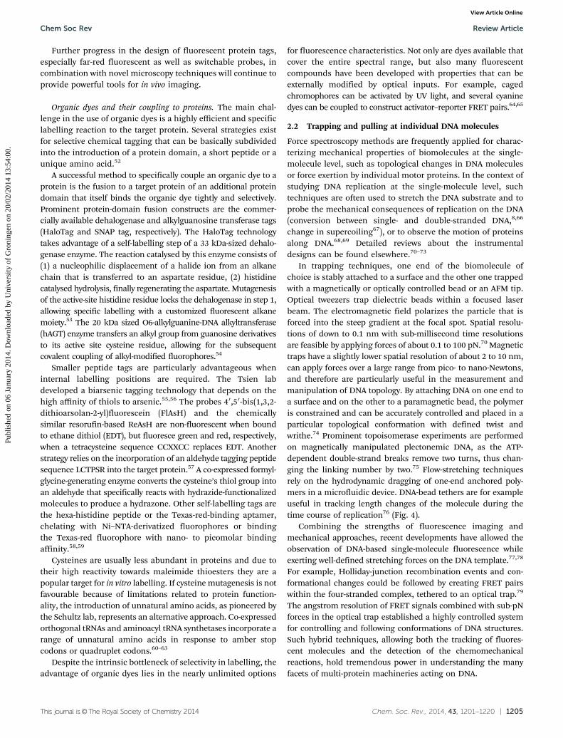
3.1.4 Eukaryotic systems. After having established the salientproperties of phage and bacterial replisomes, an enormousamount of progress has recently been made in deciphering themolecular mechanisms underlying eukaryotic replication. How-ever, in comparison to the previously described model systems,our understanding of eukaryotic replication is still far less com-plete. Not only the exact composition of the eukaryotic replicationmachinery remains unclear, but also the regulation of the replica-tion reaction in terms of posttranslational modifications like PCNAubiquitination or cell-cycle checkpoints is complicated and chal-lenging to address with classical biochemical approaches.123–125
Additionally, the details of the structural arrangement of chromo-somes need to be considered as parameters that influence replica-tion initiation and regulation. For example, histones have to bedisplaced during unwinding, but replaced onto the nascent DNAstrands to preserve epigenetic information.126
Due to their size, each eukaryotic chromosome contains a largenumber of replication origins, onto which the replication initia-tion complexes assemble. To assure that each origin can act as asite of replication initiation maximally once per cell cycle, alicensing process starts in the G1 phase.127,128 A pre-replicationcomplex (pre-RC) is assembled on each of the origins in a processthat is started by the binding of the hexameric origin-recognitioncomplex ORC129 (Fig. 6B). The ORC recruits first the cell divisioncycle proteins cdc6/cdc18 and cdt1, followed by the heterohexa-meric helicase MCM2-7. During the following S phase, these pre-RCs can be used as a platform to recruit polymerases, a primase,and numerous other replication factors to assemble a functionalreplisome. Once phosphorylated by cyclin-dependent proteinkinases (Cdks),130,131 the MCM2-7 hexamer associates with thecofactors cdc45 and GINS to form the actively unwinding CMGcomplex.132–134 The heterotrimeric replication protein A (RPA)functions as ssDNA-binding protein, coating the lagging strandduring fork progression.135,136 During replication, the poly-merases have to be switched according to their catalytic proper-ties: the Pol a–primase complex synthesizes 7–10 nt long RNAprimers and extends these by about 15 deoxynucleotides, beforethe pentameric replication factor C (RPC), analogous to the E. coliclamp-loader complex, displaces Pol a and hands the templateover to the lagging-strand DNA polymerase Pol d, whereas Pol emost likely acts on the leading strand.137 The trimeric PCNA(proliferating cell nuclear antigen) fulfils similar tasks to thebacterial b clamp, increasing the polymerase’s processivity.138,139
Replication is regulated in accordance with the cell-cyclesignalling. Cdks phosphorylate their target proteins, eitheractivating them, like the MCM subunits, directly inactivatingthem, or labelling them for proteolytic degradation, like cdc6/cdc18.140,141 In this way, secondary loading events at the originsites are prevented and it is ensured that DNA is only copiedonce during every cell cycle.
As described above, the processes of replisome assemblyand fork progression are highly dynamic, but tightly coordi-nated. Biochemical studies characterized the basic replicationarchitecture, as well as enzymatic activities of the isolatedcomponents. This body of knowledge on function and structurehas been critical to allow the single-molecule studies that weoutline in the following sections.
3.2 Replication-fork assembly pathways
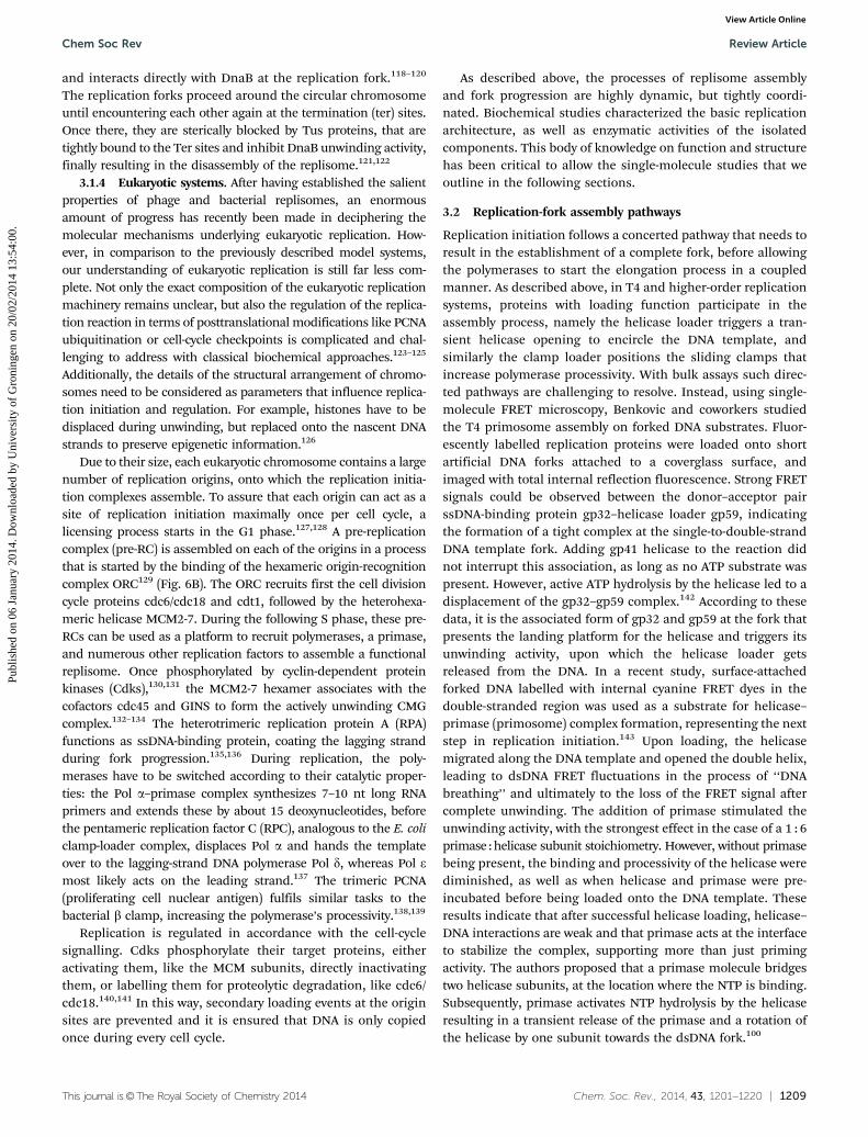
Replication initiation follows a concerted pathway that needs toresult in the establishment of a complete fork, before allowingthe polymerases to start the elongation process in a coupledmanner. As described above, in T4 and higher-order replicationsystems, proteins with loading function participate in theassembly process, namely the helicase loader triggers a tran-sient helicase opening to encircle the DNA template, andsimilarly the clamp loader positions the sliding clamps thatincrease polymerase processivity. With bulk assays such direc-ted pathways are challenging to resolve. Instead, using single-molecule FRET microscopy, Benkovic and coworkers studiedthe T4 primosome assembly on forked DNA substrates. Fluor-escently labelled replication proteins were loaded onto shortartificial DNA forks attached to a coverglass surface, andimaged with total internal reflection fluorescence. Strong FRETsignals could be observed between the donor–acceptor pairssDNA-binding protein gp32–helicase loader gp59, indicatingthe formation of a tight complex at the single-to-double-strandDNA template fork. Adding gp41 helicase to the reaction didnot interrupt this association, as long as no ATP substrate waspresent. However, active ATP hydrolysis by the helicase led to adisplacement of the gp32–gp59 complex.142 According to thesedata, it is the associated form of gp32 and gp59 at the fork thatpresents the landing platform for the helicase and triggers itsunwinding activity, upon which the helicase loader getsreleased from the DNA. In a recent study, surface-attachedforked DNA labelled with internal cyanine FRET dyes in thedouble-stranded region was used as a substrate for helicase–primase (primosome) complex formation, representing the nextstep in replication initiation.143 Upon loading, the helicasemigrated along the DNA template and opened the double helix,leading to dsDNA FRET fluctuations in the process of ‘‘DNAbreathing’’ and ultimately to the loss of the FRET signal aftercomplete unwinding. The addition of primase stimulated theunwinding activity, with the strongest effect in the case of a 1 : 6primase : helicase subunit stoichiometry. However, without primasebeing present, the binding and processivity of the helicase werediminished, as well as when helicase and primase were pre-incubated before being loaded onto the DNA template. Theseresults indicate that after successful helicase loading, helicase–DNA interactions are weak and that primase acts at the interfaceto stabilize the complex, supporting more than just primingactivity. The authors proposed that a primase molecule bridgestwo helicase subunits, at the location where the NTP is binding.Subsequently, primase activates NTP hydrolysis by the helicaseresulting in a transient release of the primase and a rotation ofthe helicase by one subunit towards the dsDNA fork.100
Chem Soc Rev Review Article
Publ
ishe
d on
06
Janu
ary
2014
. Dow
nloa
ded
by U
nive
rsity
of
Gro
ning
en o
n 20
/02/
2014
13:
54:0
0.
View Article Online
1210 | Chem. Soc. Rev., 2014, 43, 1201--1220 This journal is©The Royal Society of Chemistry 2014
The primase–helicase interaction is crucial in the course ofreplication, as it determines the rate and coupling of leadingand lagging-strand synthesis. The T4 primosome studies dis-cussed above suggested that a single primase molecule perhelicase hexamer is sufficient for the formation and stabili-zation of a primosome complex. However, the reconstitution ofthe complete replication fork is necessary to obtain informationabout the number of primase molecules within the replisomeduring a coupled replication reaction. Bulk assays gave lines ofevidence for a multimeric primase organization within thereplication fork of T4.144 Stoichiometry measurements of theisolated E. coli DnaB hexameric helicase–DnaG primase alsoindicated the presence of several primases per helicase hexamer,namely three molecules,120 suggesting a mechanistic need for acooperatively functioning multimeric primase complex.
The visualization at the single-molecule level of the sub-sequent steps of loading of the gp43 DNA polymerase at thefork further increased our understanding of T4 fork assembly.By using single-molecule FRET imaging and reconstituting theprimosome–holoenzyme assembly pathway on a forked DNAtemplate in vitro, the mechanism of ordered association offluorescently labelled enzymes was demonstrated that preventspremature replication initiation by the leading-strand poly-merase before the helicase is loaded.145 In the initiallyassembled complex of the helicase loader and the polymeraseon a forked DNA substrate, the helicase loader locks thepolymerase in an inactive state. The loading of the helicaselikely disrupts this complex, displaces the loader and forms thefunctional leading-strand replisomal complex with the poly-merase (and the sliding clamp) (Fig. 7). Taken together, theseFRET-based studies provide a model of T4 assembly, whichconsists of (1) binding of the gp59 helicase loader on a DNAfork that is coated with the gp32 ssDNA-binding protein;(2) gp43/gp45 (polymerase/clamp) loading and interaction ofthe gp43 polymerase with the gp59 helicase loader, that blockspolymerization activity; (3) gp41 helicase loading and ATP-hydrolysis dependent disassembly of the gp59 helicase loader,
(4) association of the gp61 primase with the gp41 helicase.These single-molecule experiments on T4 replication initiationare a beautiful example of how on/off switching of enzymaticactivity can be accomplished by protein–protein interactions.
3.3 Leading and lagging-strand coordination
Achieving coordinated replication in an asymmetric polymeri-zation configuration is a general requirement for all replisomes,but the question remains how the discontinuously actinglagging-strand polymerase can keep up with the leading-strandpolymerase. Priming on the lagging strand inherently slowsdown lagging-strand synthesis due to the relatively slow rNTPpolymerization kinetics146,147 and the time needed to recruit apolymerase to this new primer. Different single-molecule assaysfor T7 and T4 replication provided distinct views on coordina-tion mechanisms that we want to outline here.
The T7 replisome has served as an attractive model systemto understand the functioning of the individual proteins as wellas the behaviour of the whole replisome. Pioneering optical-trapping studies helped for example to understand themechanochemical properties of the isolated gp5/thioredoxinDNA polymerase.8,148 Here, it was demonstrated how DNAtemplate tension induces switching between active polymeriza-tion and backtracking accompanied by the exonucleolyticremoval of nucleotides. These experiments showed that therate-limiting step in the catalytical cycle of T7 polymerase isforce dependent, an observation that resulted in mechanisticinsights into the orientation of the DNA in the polymeraseactive site.149
Tethered-bead experiments with 48.5-kb long lambda-phageDNA, anchored to a coverglass surface and stretched hydro-dynamically in a flow cell, provided a direct read-out forreplication of the fully reconstituted T7 replisome andaddressed the mechanism with which leading-strand andlagging-strand syntheses are coupled (Fig. 8A).66 Here, a beadwas attached to the unreplicated, parental end of a forked DNAconstruct, the T7 replisome components loaded onto the fork,and leading-strand synthesis initiated. At the applied stretchingforce of B2 pN, ssDNA is more compact than dsDNA andconversion from double-stranded parental DNA into single-stranded lagging-strand DNA can be monitored by visualizingthe gradual motion of the tethered bead towards the anchoringpoint as the total length of the DNA construct decreases. Bycomparing leading-strand synthesis traces in the presence orabsence of primer synthesis, either via removal of the zinc-binding-domain of gp4 or via the omittance of ribonucleotides,the authors could demonstrate bead-stalling events of severalseconds that were related to priming activity and that precededlagging-strand loop formation and subsequent (fast) release.Consistent with these experiments is a model in which primaseactivity transiently halts the progression of the entire fork,preventing leading-strand synthesis from outpacing lagging-strand synthesis.
Another possible mechanism of coupling between the lead-ing and lagging strand is the display of differential rates for thetwo polymerases at the fork.150 High-resolution sequencing gels
Fig. 7 T4 replication initiation. Benkovic and coworkers used forked DNAsubstrates coupled to a microscope coverglass surface to reconstitute theinitiation pathway of T4.142,145,193 Gp32 and gp59 bind to the fork structureand recruit the polymerase gp43 together with the clamp gp45 thatis installed on the DNA strand by the loader complex gp44/62. Gp59catalyses loading of the helicase gp61 and is displaced by the latter uponDNA unwinding. Primase molecules (gp61) associate with the helicase toform a stable primosome complex.
Review Article Chem Soc Rev
Publ
ishe
d on
06
Janu
ary
2014
. Dow
nloa
ded
by U
nive
rsity
of
Gro
ning
en o
n 20
/02/
2014
13:
54:0
0.
View Article Online

This journal is©The Royal Society of Chemistry 2014 Chem. Soc. Rev., 2014, 43, 1201--1220 | 1211
provided evidence for leading-strand synthesis not beingdelayed during priming, but rather, the leading-strand poly-merase synthesizing slower than the lagging-strand poly-merase. By positioning internal DNA FRET pairs next to theT7 priming sequence, the authors investigated the conforma-tion of the lagging-strand template. They observed increasingFRET acceptor signals in the course of a loop formation eventbringing the labelled DNA segments in close proximity to eachother, and suggested these signals to be priming loops formedby the lagging-strand between the helicase and the primasedomain of gp4 (Fig. 8B). In such a configuration, DNA synthesiscan continue without interruption and primers can be synthe-sized concomitantly with DNA polymerization.
Where the T7 system is unique in that the primase andhelicase functions are present in the same protein, the T4replisome, as most other replication systems, utilizes twodifferent proteins for these enzymatic activities. As describedin the previous section, the T4-based DNA synthesis reactioncan be reconstituted in vitro and magnetic tweezers have beenused as a single-molecule approach to unravel the coordination
between unwinding and priming.151 In these studies, a DNAconstruct containing a hairpin structure was stretched and itsextension measured while the helicase and primase wereloaded onto the artificial fork (Fig. 8C). The setup allowed adistinction between helicase pausing, priming loop formationand primosome disassembly during primer synthesis, and bothloop growth and primase displacement were detected. Whenadditional replication proteins, ssDNA-binding protein, clampand clamp loader, were applied, looping appeared more oftenthan in the primosome-only complex, although disassemblyremained predominant. As a comparative control, a fusionconstruct of primase and helicase exclusively primed in alooping configuration.
These different scenarios of pausing/looping/disassemblyshown for T7 and T4 indicate a need for replication systemsto adapt to their particular composition and structure, theavailable number of proteins at the fork and the relativelydifferent needs for processivity and stability in replicating aphage genome of a few tens of kbp or a Mbp-long bacterialgenome. The plasticity of the fork may permit all described
Fig. 8 Coordinated replication. (A) T7 replisome reconstitution in a hydrodynamic DNA flow-stretching assay. A bead attached to one end of the DNAtemplate can be followed by bright-field microscopy and from its trajectory the replication kinetics are extracted. Pausing events in the single-moleculetrajectories indicate primer synthesis reactions and lagging strand loop releases are visible as instantaneous DNA lengthening (adapted from ref. 66).(B) T7 replisome reconstitution in a single-molecule FRET assay. Cy3 (green)–Cy5 (red) FRET pairs are installed next to a priming sequence on a DNAtemplate attached to a coverglass surface. Upon helicase/polymerase loading the Cy5 FRET signal increases, suggesting the formation of a priming loop(adapted from ref. 150). (C) T4 primosome–replisome reconstitution in a magnetic trap. A DNA hairpin structure is attached to a coverglass surface and amagnetic bead. The active unwinding by the helicase can be followed by measuring the extension of the DNA template. Priming loops and loop releaseevents are extracted from the bead trajectories (adapted from ref. 151).
Chem Soc Rev Review Article
Publ
ishe
d on
06
Janu
ary
2014
. Dow
nloa
ded
by U
nive
rsity
of
Gro
ning
en o
n 20
/02/
2014
13:
54:0
0.
View Article Online

1212 | Chem. Soc. Rev., 2014, 43, 1201--1220 This journal is©The Royal Society of Chemistry 2014
mechanisms interchangeably, thus being more robust towardsany obstacles.
3.4 Polymerase dynamics
Besides an efficient coupling between leading and lagging-strand synthesis, a processive replication reaction requires astable association of the polymerases within the replicationfork. Since every new Okazaki fragment requires the recruit-ment of a lagging-strand polymerase, either DNA polymerasesfrom solution need to associate with a newly synthesizedprimer or the lagging-strand DNA polymerase needs to berecycled efficiently to support the synthesis of multiple Okazakifragments. Both polymerase exchange and recycling on thelagging strand are feasible, with the first scenario relying onsufficient protein concentrations around the fork so as to notimpede the overall reaction kinetics.
In T7, the gp5/trx DNA polymerase was shown to employ twobinding modes of different tightness to the gp4 helicase,152,153
indicating the possibility of multiple distinct steps in therecruitment and utilization of polymerases at the replicationfork. Considering gp4 to be hexameric within the replicationfork, potentially the weak interaction site between the acidicC-terminal tail of gp4 and a basic patch within the thioredoxin-binding domain (TBD) on gp5154 could result in a reservoir ofpolymerases being bound to the replisome. Interestingly,a similar electrostatic interaction could be found between theC terminus of the gp2.5 ssDNA-binding protein and the gp5polymerase,152 potentially further increasing the local concen-tration of polymerases around the replication fork. Such a localexcess of polymerases would enable a rapid replacement of apolymerase after dissociation and thus would support a highlyprocessive replication reaction. Ensemble-averaging dilutionand competition experiments highlighted the dual behaviourresulting from polymerase switching and recycling: the proces-sivity of the T7 replisome is not diminished by dilution,enforcing the hypothesis of efficient recycling.155 On the otherhand, by using a mutant gp5 that is resistant to inhibition bydideoxynucleotides in competition experiments, rapid exchangeof polymerases was observed, as well.153 Recently, a directobservation of exchange kinetics was feasible in a single-molecule study by tracking fluorescently labelled polymerases69
(Fig. 9A and B). Here, a DNA substrate tagged with a fluorescentquantum dot at one site was anchored to a coverglass surface andthe T7 leading-strand polymerase and helicase preassembledat the fork. The replication reaction could be followed by trackingthe quantum dot moving towards the DNA attachment point.Upon addition of fluorescently labeled polymerases to thereplication reaction, signals from those polymerases newlyarriving at the replication fork were detected, suggesting thatexcess polymerases stayed associated with the replication forkfor several tens of seconds, occupying the available dockingsites on the gp4 helicase and ready to replace the synthesizingDNA polymerase.
The mechanism of DNA polymerase exchange and recyclingis important in the context of lagging-strand synthesis andreplication-loop release. Replication loops are formed in the
lagging strand due to the fact that the lagging-strand DNApolymerase remains associated with the rest of the replisomewhile synthesizing new DNA in a direction that is opposite tothe direction of movement of the rest of the replisome. A newloop is formed for every new Okazaki fragment that is synthe-sized and released before the initiation of the next one. Twopathways of loop release and Okazaki fragment initiation havebeen proposed.156–158 In the collision model the polymerase isreleased upon encountering the 50 end of the previous Okazakifragments, thus resulting in the release of the replication loop.In the signalling model, the synthesis of a new primer triggersloop release, before the Okazaki fragment is finished (Fig. 9C).Using single-molecule approaches that rely on the length
Fig. 9 Polymerase exchange dynamics of the T7 replisome. (A) Unlabeledleading-strand polymerase and helicase are preassembled on DNA. Uponinitiation of the reaction, DNA synthesis occurs and is observed as short-ening of the DNA by tracking the DNA-template anchored quantum dot(B, middle panel). Upon introduction of fluorescently labeled polymerasesto the reaction, fluorescent spots appear at the position of the replicationfork and remain there for several seconds (B, bottom panel). Lagging-strand synthesis is not taking place as ribonucleotides are excluded fromthe reaction (adapted from ref. 69). (C) Collision vs. signalling model of theT7 replisome. The hexameric gp4 (blue) translocates along the laggingstrand while unwinding the DNA template and priming the Okazakifragments (O.F.). The polymerases gp5 (green), complexed with thiore-doxin, are bound to gp4 and synthesize the leading and laggingstrands. Gp2.5 molecules (red) coat and protect the single-strandedDNA that has been extruded behind the helicase. In the collision model,the replication loop is released when the lagging-strand polymerasecollides with the 50 terminus of the previous Okazaki fragment. In thesignalling model, the synthesis of a new primer triggers the release of thereplication loop before the nascent Okazaki fragment is completed(adapted from ref. 194).
Review Article Chem Soc Rev
Publ
ishe
d on
06
Janu
ary
2014
. Dow
nloa
ded
by U
nive
rsity
of
Gro
ning
en o
n 20
/02/
2014
13:
54:0
0.
View Article Online
This journal is©The Royal Society of Chemistry 2014 Chem. Soc. Rev., 2014, 43, 1201--1220 | 1213
measurement of a single DNA molecule as it is being replicated,as depicted in Fig. 8A, the formation and release of such replica-tion loops have been directly observed and their dynamic proper-ties analysed.76 These studies revealed that both models areoperative during the T7 replication reaction and may serve jointlyas a redundancy mechanism to ensure timely loop release. Forboth mechanisms, however, it is unclear whether the polymeraseis recycled to initiate the synthesis of the next Okazaki fragmentor whether it stays behind to fill the remaining gap in theprevious Okazaki fragment (in the signalling mechanism) orsimply dissociates in solution (in the collision mechanism).Combining the observation of DNA length changes during coor-dinated leading and lagging-strand replication while monitoringthe arrival and departure of fluorescently labelled polymerases atthe fork, as has been reported for leading-strand synthesis,69 isan approach that likely will shed more light on these dynamicaspects of the replisome.
3.5 In vivo studies on the E. coli replisome
Extensive work has been done to decipher the interactionsbetween partners within the bacterial replisome (furtherreviewed in ref. 28, 106 and 159–166). Here we focus on recentstudies that particularly concentrate on the dynamics of repli-cation in the context of the living cell. Recent in vivo single-molecule studies have provided considerable insight into thespatial and temporal properties of the bacterial replisome andthe underlying protein dynamics.
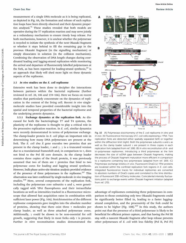
3.5.1 Exchange dynamics at the replication fork. As dis-cussed for both the bacteriophage T7 and T4 systems, theplasticity of the replisome is thought to play a critical role inthe processive replication reaction. In E. coli, similar dynamicswere recently demonstrated in terms of polymerase exchange.The clamp-loader protein in E. coli plays an important role indetermining how many DNA polymerases are present at thefork. The E. coli dna X gene encodes two proteins that arepresent in the clamp loader, t and g. g is a truncated versiondue to a translational frameshift and, in comparison to t, doesnot bind to the Pol III core domain. As the clamp loadercontains three copies of the DnaX protein, it was previouslyassumed that two of them are t proteins that bind to twopolymerase cores for leading and lagging strand synthesis.However, bulk-phase active-site titration analysis provided evidenceof the presence of three polymerases in the replisome.167 Thisobservation was later confirmed by single-molecule in vivo imagingstudies.168 Here, several components of the replication fork,including the polymerase core subunits a and e, were geneti-cally tagged with YPet fluorophores and their intracellularlocations as well as intensities tracked in a fluorescence micro-scope that allowed fast acquisition rates in combination withsufficient laser power (Fig. 10A). Stoichiometries of the differentreplisome components gave insights into the absolute numberof proteins, showing that there exist three t molecules perclamp loader, as well as three attached polymerase cores.Additionally, g could be shown to be non-essential for cellgrowth, suggesting that likely in most forks only t is present.Further in vitro reconstitution studies demonstrated the
advantage of replisomes containing three polymerases in com-parison to those containing only two: Okazaki fragments couldbe significantly better filled in, leading to a faster lagging-strand completion, and the processivity of the fork could beshown to be higher (Fig. 10B).169 In this study, the authorspoint out that the presence of a third polymerase is likely to bebeneficial for efficient primer capture, and that having the Pol IIIstay with a nascent Okazaki fragment after loop release preventsother polymerases of E. coli with less fidelity (Pol II, Pol IV)
Fig. 10 (A) Polymerase stoichiometry of the E. coli replisome in vitro andin vivo. (A) Fluorescence microscopy of E. coli cells expressing e-YPet. Tworeplication forks are detected either spatially separated (left) or togetherwithin the diffraction limit (right). Both the polymerase subunits a and e aswell as the clamp loader subunit t are present in three copies in eachreplication fork (adapted from ref. 168). (B) In vitro reconstitution of di- andtri-polymerase replisomes. Introducing a third polymerase at the forkdecreases the size of ssDNA gaps between Okazaki fragments, makingthe process of Okazaki fragment maturation more efficient in comparisonto a replisome containing two polymerases (adapted from ref. 169). (C)Polymerase exchange kinetics in vivo. Fluorescent DnaQ (e)-YPet proteinsare localized within the confined replication fork regions in E. coli cells.Integrated fluorescence traces for individual replisomes are convertedto absolute numbers of DnaQ copies and correlated to the time distribu-tion of fluorescent SSB-mCherry molecules. Coincidental intensity fluctua-tions point to exchange events within Okazaki fragment initiation (adaptedfrom ref. 170).
Chem Soc Rev Review Article
Publ
ishe
d on
06
Janu
ary
2014
. Dow
nloa
ded
by U
nive
rsity
of
Gro
ning
en o
n 20
/02/
2014
13:
54:0
0.
View Article Online

1214 | Chem. Soc. Rev., 2014, 43, 1201--1220 This journal is©The Royal Society of Chemistry 2014
from filling the remaining gap on the template. If there arethree potential binding sites for the polymerase core complex,are the dynamics of polymerase exchange similar to the T7system, as described above? In another in vivo study, both SSBand polymerase core e subunits were fluorescently tagged andtheir signals correlated (Fig. 10C).170 Correlating protein fluc-tuations at the fork suggested that for every Okazaki fragment anew polymerase associates on the lagging strand and, inaddition, that it also proceeds faster than the leading-strandpolymerase. An additional third polymerase in close vicinitywould allow a rapid restart of synthesis after polymerasedissociation from the replisome upon loop release.
3.5.2 Obstacles along the template. Replication forks arelikely to face various obstacles during their progressionthrough large genomic stretches of DNA: lesions in the DNAand proteins bound to the DNA, such as transcribing RNApolymerases, may block the template and histone-like proteinsintroduce twist and bending of the DNA that may presenthurdles for the replisome.
Those potential blockades might stall the replication forkand provoke a (partial) disassembly. Experimental approachesto characterize such stalling events involve for example UVirradiation or the use of thermosensitive replication mutants,as for DnaB.171 With single-molecule detection sensitivity in livecells, the disassembly kinetics could be followed in real timeafter DnaB arrest by tracking the different replisome compo-nents encoded as fusion constructs to fluorescent proteins.172
In this study, the authors observed not only remarkably distincttime scales of polymerase core dissociation, which may point toa stronger interaction of the leading-strand polymerase withthe replisome than that of the lagging-strand polymerase, butalso revealed the initiation of a fork rescue process with RecAfilaments replacing SSB.
The double-helical structure of DNA itself also poses a barrierfor replication. The torsional energy that builds up during replica-tion or transcription gives rise to a number of topological chal-lenges the replisome needs to deal with. The exact nature oftopological deformation depends on how the replication forkproceeds. If the replisome is free to rotate, precatenanes behindthe fork can be generated. Without rotation, positive supercoilingin front of the fork is the cost for unwinding activity.173 In bothcases, topoisomerases of type I and II are indispensable in dealingwith the intertwined regions. A recent study addressed thetorsional stress on the replisome in the context of coupledreplication.174 Here the authors argued that the dissociationmechanism of the lagging-strand polymerase and thus itsexchange kinetics are important not only in the context ofrapid primer capture, but also in terms of DNA stress release.The concept of the signalling model in which primer synthesistriggers loop release (Fig. 9C) could thus be extended to atorsion-dependent displacement of the lagging-strand polymerase.
3.5.3 Chromosome structure. Globally, replication is coordi-nated with the process of chromosome segregation. Structuralmaintenance of chromosome (SMC) proteins, in E. coli theMukBEF complex, act to organize the chromosome during thecell cycle. Recently, individual copies of the MukBEF complex
could be imaged using fluorescence microscopy175 (Fig. 11).Not only could the stoichiometry of the functional minimalunit of the complex be resolved by counting the fluorescentlytagged components, but also the salient details of the ATP-dependent DNA-modelling pathway could be studied. StableMukBEF complexes were found to accumulate at several dis-crete spots in the cell to form higher-order structures that areengaged in organizing the chromosome. Making use of ATPase-deficient mutant strains, the authors could show that in thesespots the minimal MukBEF units exchange via ATP binding andhydrolysis cycles. Due to MukBEF multimerization, segments ofDNA can be caught without losing contact with other ones, sothat in sum, MukBEF works as a step-by-step remodeller,grabbing and bringing together distal segments of the chromo-some (Fig. 11B).
A recent study, based on pull-down and topology/superhelicityrelaxation assays in bulk, looked into the interaction of MukBwith ParC, a subunit of topoisomerase IV.176 Here, MukBenhanced ParC-catalysed relaxation of negatively supercoiledand knotted DNA, exemplifying how multiple protein inter-actions and complexes are involved in the topological configu-ration of the chromosome that interacts with the replicationprogression.
3.6 Replication in the context of eukaryotic cell division
The eukaryotic genome is highly structured into chromatin,which is a critical requirement not only for proper cell division,but also for the storage of epigenetic information such ashistone modifications. The regulation of replication, includingaspects like the establishment of pre-replication complexes,
Fig. 11 MukBEF organization in E. coli. (A) Using live-cell PALM, diffusingand immobile MukB-PAmCherry molecules can be tracked and intensitiesintegrated to estimate the number of proteins per spot. (B) ‘‘Rock-climber’’model for the action of SMC complexes in cells. MukBEF complexesconsist of dimers of dimeric 4 : 4 : 2 MukB : E : F molecules that associateon DNA. ATP hydrolysis triggers release from the DNA segment; howeveras the multimeric complexes exhibit several DNA binding sites, completerelease from the chromosome is inhibited (adapted from ref. 175).
Review Article Chem Soc Rev
Publ
ishe
d on
06
Janu
ary
2014
. Dow
nloa
ded
by U
nive
rsity
of
Gro
ning
en o
n 20
/02/
2014
13:
54:0
0.
View Article Online
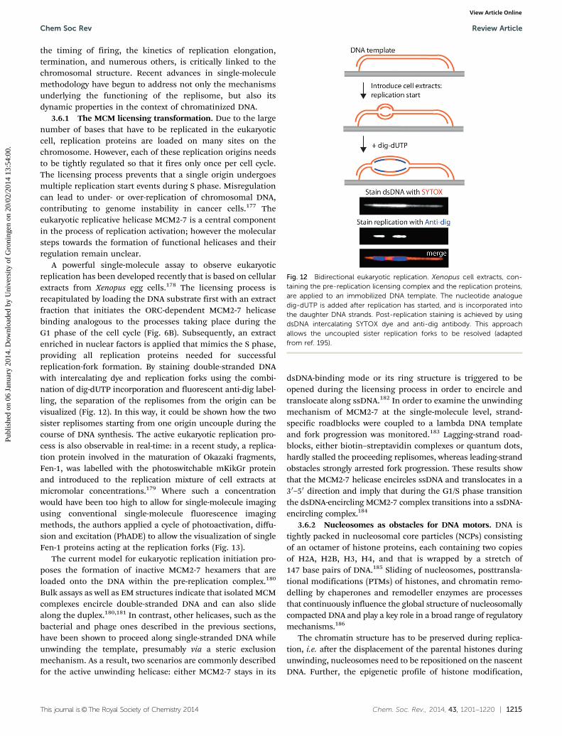
This journal is©The Royal Society of Chemistry 2014 Chem. Soc. Rev., 2014, 43, 1201--1220 | 1215
the timing of firing, the kinetics of replication elongation,termination, and numerous others, is critically linked to thechromosomal structure. Recent advances in single-moleculemethodology have begun to address not only the mechanismsunderlying the functioning of the replisome, but also itsdynamic properties in the context of chromatinized DNA.
3.6.1 The MCM licensing transformation. Due to the largenumber of bases that have to be replicated in the eukaryoticcell, replication proteins are loaded on many sites on thechromosome. However, each of these replication origins needsto be tightly regulated so that it fires only once per cell cycle.The licensing process prevents that a single origin undergoesmultiple replication start events during S phase. Misregulationcan lead to under- or over-replication of chromosomal DNA,contributing to genome instability in cancer cells.177 Theeukaryotic replicative helicase MCM2-7 is a central componentin the process of replication activation; however the molecularsteps towards the formation of functional helicases and theirregulation remain unclear.
A powerful single-molecule assay to observe eukaryoticreplication has been developed recently that is based on cellularextracts from Xenopus egg cells.178 The licensing process isrecapitulated by loading the DNA substrate first with an extractfraction that initiates the ORC-dependent MCM2-7 helicasebinding analogous to the processes taking place during theG1 phase of the cell cycle (Fig. 6B). Subsequently, an extractenriched in nuclear factors is applied that mimics the S phase,providing all replication proteins needed for successfulreplication-fork formation. By staining double-stranded DNAwith intercalating dye and replication forks using the combi-nation of dig-dUTP incorporation and fluorescent anti-dig label-ling, the separation of the replisomes from the origin can bevisualized (Fig. 12). In this way, it could be shown how the twosister replisomes starting from one origin uncouple during thecourse of DNA synthesis. The active eukaryotic replication pro-cess is also observable in real-time: in a recent study, a replica-tion protein involved in the maturation of Okazaki fragments,Fen-1, was labelled with the photoswitchable mKikGr proteinand introduced to the replication mixture of cell extracts atmicromolar concentrations.179 Where such a concentrationwould have been too high to allow for single-molecule imagingusing conventional single-molecule fluorescence imagingmethods, the authors applied a cycle of photoactivation, diffu-sion and excitation (PhADE) to allow the visualization of singleFen-1 proteins acting at the replication forks (Fig. 13).
The current model for eukaryotic replication initiation pro-poses the formation of inactive MCM2-7 hexamers that areloaded onto the DNA within the pre-replication complex.180
Bulk assays as well as EM structures indicate that isolated MCMcomplexes encircle double-stranded DNA and can also slidealong the duplex.180,181 In contrast, other helicases, such as thebacterial and phage ones described in the previous sections,have been shown to proceed along single-stranded DNA whileunwinding the template, presumably via a steric exclusionmechanism. As a result, two scenarios are commonly describedfor the active unwinding helicase: either MCM2-7 stays in its
dsDNA-binding mode or its ring structure is triggered to beopened during the licensing process in order to encircle andtranslocate along ssDNA.182 In order to examine the unwindingmechanism of MCM2-7 at the single-molecule level, strand-specific roadblocks were coupled to a lambda DNA templateand fork progression was monitored.183 Lagging-strand road-blocks, either biotin–streptavidin complexes or quantum dots,hardly stalled the proceeding replisomes, whereas leading-strandobstacles strongly arrested fork progression. These results showthat the MCM2-7 helicase encircles ssDNA and translocates in a30–50 direction and imply that during the G1/S phase transitionthe dsDNA-encircling MCM2-7 complex transitions into a ssDNA-encircling complex.184
3.6.2 Nucleosomes as obstacles for DNA motors. DNA istightly packed in nucleosomal core particles (NCPs) consistingof an octamer of histone proteins, each containing two copiesof H2A, H2B, H3, H4, and that is wrapped by a stretch of147 base pairs of DNA.185 Sliding of nucleosomes, posttransla-tional modifications (PTMs) of histones, and chromatin remo-delling by chaperones and remodeller enzymes are processesthat continuously influence the global structure of nucleosomallycompacted DNA and play a key role in a broad range of regulatorymechanisms.186
The chromatin structure has to be preserved during replica-tion, i.e. after the displacement of the parental histones duringunwinding, nucleosomes need to be repositioned on the nascentDNA. Further, the epigenetic profile of histone modification,
Fig. 12 Bidirectional eukaryotic replication. Xenopus cell extracts, con-taining the pre-replication licensing complex and the replication proteins,are applied to an immobilized DNA template. The nucleotide analoguedig-dUTP is added after replication has started, and is incorporated intothe daughter DNA strands. Post-replication staining is achieved by usingdsDNA intercalating SYTOX dye and anti-dig antibody. This approachallows the uncoupled sister replication forks to be resolved (adaptedfrom ref. 195).
Chem Soc Rev Review Article
Publ
ishe
d on
06
Janu
ary
2014
. Dow
nloa
ded
by U
nive
rsity
of
Gro
ning
en o
n 20
/02/
2014
13:
54:0
0.
View Article Online

1216 | Chem. Soc. Rev., 2014, 43, 1201--1220 This journal is©The Royal Society of Chemistry 2014
mainly acetylation and phosphorylation, has to be transmittedfrom the parental to the daughter DNA. Some hints about theunderlying mechanisms were provided by a study that demon-strated that the MCM2 helicase subunit of yeast binds histonescooperatively with the FACT (facilitates chromatin transcrip-tion) complex, which acts as a histone chaperone.126 As shownby immunoprecipitation, FACT is part of the replisome by
interaction with MCM2, independent of concomitant histonebinding. Although those multi-interaction processes are difficult todecipher, several single-molecule studies have begun to givesignificant insight into DNA-nucleosome dynamics (reviewed ingreater detail elsewhere187). Using optical tweezers, the unzippingmechanism of single NCPs could be tracked and an energy land-scape described for the distinct interaction domains, revealing thatthe dyad domain association is particularly strong.187 In anotherrecent study, magnetic tweezers as well as FRET microscopy wereapplied to distinguish the effects of post-transcriptional modifica-tions depending on their exact positions. It appeared that espe-cially near the dyad, post-transcriptional modifications speed uphistone disassembly processes, whereas those localized near theDNA entry/exit site regulate the unwrapping pathway.188 Similarly,by installing DNA FRET pairs on the template, the compaction asrelated to CpG island methylation could be quantified, pointing toa positive correlation between the amount of methylated sites andcompaction.189
Further single-molecule studies of eukaryotic replication ina cellular context will likely give exciting insights into thecomplex processes involved in DNA metabolism. In combi-nation with cell biological studies, theoretical modelling andmechanistic biochemical work, such single-molecule experi-ments will be necessary to not only provide fine-grained kineticinformation on the various molecular processes but also enablea much more quantitative view of the molecular processestaking place inside living cells and as such play a role inbridging experimental and theoretical studies.
4. Conclusions and outlook
The statement ‘‘the whole is greater than the sum of its parts’’applies to many biological systems, be it the complicatedpathways of signal transduction, cytoskeletal reconstructionin cell division, or as described in this review, DNA replication.Initial single-molecule studies concentrated on the isolatedDNA motors, quantifying the force generation and processivityof polymerases or unwinding activity of helicases, and laidthe basis for studies of the entire replisome. Single-moleculeexperiments merely represent a category of approaches fromthe toolbox of biophysics and biochemistry. In combinationwith structural, biochemical, genetic and other approaches,they provide us with a tool that has proven to be powerful inrevealing new features about the inner workings of individualmotors and larger units that self-organize into multifunctionalcomplexes. The understanding of the replisome, isolated orinside the living cell, gives us powerful information aboutcomplex cellular structures. The central theme, the plasticityof the replication fork in its ability to exchange componentsduring replication, is remarkable considering the processivityand robustness of coupled replication.
Ultimately, a key goal is to construct a holistic picture of thecell and how it is built up from many molecular pathways. Duringthe last few years, reductionist and bottom-up approaches toconstruct a synthetic cell have been explored and have already led
Fig. 13 The concept of PhADE (photoactivation, diffusion and excitation).(A) The photoswitchable mKikGR protein is activated by 405 nm light totransition from its green to red fluorescence state. In a TIRF setup, onlythose molecules within the evanescent field near the coverglass surfaceare activated. Waiting before imaging in the red channel until the activatedproteins in solution have diffused to outside the evanescent field allowsthe selective visualization of only those proteins bound to the DNA. (B) Inorder to follow the eukaryotic replication fork progression in real time, thereplication protein Fen1 is tagged with mKikGr protein and added to theXenopus cell extract (see text for details). The kymograph recapitulatesthe growth and separation of the replication forks (adapted from ref. 179).
Review Article Chem Soc Rev
Publ
ishe
d on
06
Janu
ary
2014
. Dow
nloa
ded
by U
nive
rsity
of
Gro
ning
en o
n 20
/02/
2014
13:
54:0
0.
View Article Online
This journal is©The Royal Society of Chemistry 2014 Chem. Soc. Rev., 2014, 43, 1201--1220 | 1217
to intriguing results. The reconstitution of compartments orunits of increasing intricacy is one key step towards recapitulat-ing and understanding cellular complexity, and several single-molecule studies, many of them focussing on cytoskeletalsystems, rely on this method. Highly related to cellular com-plexity is the crowded nature of the intracellular environment.Going forward, it is important to characterize and understandhow biochemical pathways work not only in the context ofhighly purified and homogeneous systems, but also in thecontext of molecular crowding.190 Single-molecule approacheshave begun to significantly impact how complex biochemicalpathways are studied and further integration with other toolsand mechanistic insights will undoubtedly result in significantcontributions to our understanding of the molecular principlesunderlying life.
References
1 M. K. Gardner, M. Zanic, C. Gell, V. Bormuth andJ. Howard, Cell, 2011, 147, 1092–1103.
2 M. Tomishige, N. Stuurman and R. D. Vale, Nat. Struct.Mol. Biol., 2006, 13, 887–894.
3 R. D. Vale, T. Funatsu, D. W. Pierce, L. Romberg, Y. Haradaand T. Yanagida, Nature, 1996, 380, 451–453.
4 K. Adachi, K. Oiwa, M. Yoshida, T. Nishizaka andK. Kinosita Jr., Nat. Commun., 2012, 3, 1022.
5 H. Noji, R. Yasuda, M. Yoshida and K. Kinosita Jr., Nature,1997, 386, 299–302.
6 E. A. Abbondanzieri, W. J. Greenleaf, J. W. Shaevitz,R. Landick and S. M. Block, Nature, 2005, 438, 460–465.
7 D. A. Schafer, J. Gelles, M. P. Sheetz and R. Landick,Nature, 1991, 352, 444–448.
8 G. J. Wuite, S. B. Smith, M. Young, D. Keller andC. Bustamante, Nature, 2000, 404, 103–106.
9 K. Aathavan, A. T. Politzer, A. Kaplan, J. R. Moffitt,Y. R. Chemla, S. Grimes, P. J. Jardine, D. L. Andersonand C. Bustamante, Nature, 2009, 461, 669–673.
10 Y. R. Chemla, K. Aathavan, J. Michaelis, S. Grimes, P. J. Jardine,D. L. Anderson and C. Bustamante, Cell, 2005, 122, 683–692.
11 J. Elf, G. W. Li and X. S. Xie, Science, 2007, 316, 1191–1194.12 Y. Taniguchi, P. J. Choi, G. W. Li, H. Chen, M. Babu,
J. Hearn, A. Emili and X. S. Xie, Science, 2010, 329, 533–538.13 Y. Sako, S. Minoghchi and T. Yanagida, Nat. Cell Biol.,
2000, 2, 168–172.14 T. Xia, N. Li and X. Fang, Annu. Rev. Phys. Chem., 2013, 64,
459–480.15 S. Chaudhury and O. A. Igoshin, PLoS One, 2010, 5, e12364.16 H. P. Lu, L. Xun and X. S. Xie, Science, 1998, 282,
1877–1882.17 L. Edman and R. Rigler, Proc. Natl. Acad. Sci. U. S. A., 2000,
97, 8266–8271.18 B. Liu, R. J. Baskin and S. C. Kowalczykowski, Nature, 2013,
500, 482–485.19 S. C. Kou, B. J. Cherayil, W. Min, B. P. English and X. S. Xie,
J. Phys. Chem. B, 2005, 109, 19068–19081.
20 W. Min, B. P. English, G. Luo, B. J. Cherayil, S. C. Kou andX. S. Xie, Acc. Chem. Res., 2005, 38, 923–931.
21 Q. Chen, R. Groote, H. Schonherr and G. J. Vancso,Chem. Soc. Rev., 2009, 38, 2671–2683.
22 W. Bae, M. G. Choi, C. Hyeon, Y. K. Shin and T. Y. Yoon,J. Am. Chem. Soc., 2013, 135, 10254–10257.
23 B. A. Smith, K. Daugherty-Clarke, B. L. Goode and J. Gelles,Proc. Natl. Acad. Sci. U. S. A., 2013, 110, 1285–1290.
24 B. M. Alberts, Philos. Trans. R. Soc., B, 1987, 317, 395–420.25 S. J. Benkovic, A. M. Valentine and F. Salinas, Annu. Rev.
Biochem., 2001, 70, 181–208.26 N. Y. Yao and M. O’Donnell, Mol. BioSyst., 2008, 4,
1075–1084.27 T. A. Kunkel and K. Bebenek, Annu. Rev. Biochem., 2000, 69,
497–529.28 A. M. van Oijen, Mol. BioSyst., 2007, 3, 117–125.29 T. Hirschfeld, Appl. Opt., 1976, 15, 2965–2966.30 W. E. Moerner and L. Kador, Phys. Rev. Lett., 1989, 62,
2535–2538.31 W. E. Moerner and W. P. Ambrose, Phys. Rev. Lett., 1991,
66, 1376.32 M. Orrit and J. Bernard, Phys. Rev. Lett., 1990, 65,
2716–2719.33 A. M. van Oijen, M. Ketelaars, J. Kohler, T. J. Aartsma and
J. Schmidt, Science, 1999, 285, 400–402.34 E. Betzig and R. J. Chichester, Science, 1993, 262,
1422–1425.35 A. Yamada, T. Yamaga, H. Sakakibara, H. Nakayama and
K. Oiwa, J. Cell Sci., 1998, 111(Pt 1), 93–98.36 J. Yu, J. Xiao, X. Ren, K. Lao and X. S. Xie, Science, 2006,
311, 1600–1603.37 E. J. Ambrose, Nature, 1956, 178, 1194.38 D. Axelrod, J. Cell Biol., 1981, 89, 141–145.39 E. H. Stelzer, I. Wacker and J. R. De Mey, Semin. Cell Biol.,
1991, 2, 145–152.40 A. Nakano, Cell Struct. Funct., 2002, 27, 349–355.41 F. Helmchen and W. Denk, Nat. Methods, 2005, 2, 932–940.42 S. W. Hell and J. Wichmann, Opt. Lett., 1994, 19, 780–782.43 F. V. Subach, G. H. Patterson, M. Renz, J. Lippincott-
Schwartz and V. V. Verkhusha, J. Am. Chem. Soc., 2010,132, 6481–6491.
44 S. Uphoff, R. Reyes-Lamothe, F. Garza de Leon, D. J.Sherratt and A. N. Kapanidis, Proc. Natl. Acad. Sci. U. S. A.,2013, 110, 8063–8068.
45 S. R. Nelson, M. Y. Ali and D. M. Warshaw, Methods Mol.Biol., 2011, 778, 111–121.
46 M. Ormo, A. B. Cubitt, K. Kallio, L. A. Gross, R. Y. Tsien andS. J. Remington, Science, 1996, 273, 1392–1395.
47 G. J. Kremers, S. G. Gilbert, P. J. Cranfill, M. W. Davidsonand D. W. Piston, J. Cell Sci., 2011, 124, 157–160.
48 N. C. Shaner, G. H. Patterson and M. W. Davidson, J. CellSci., 2007, 120, 4247–4260.
49 A. M. van Oijen, Curr. Opin. Biotechnol., 2011, 22, 75–80.50 M. Andresen, A. C. Stiel, J. Folling, D. Wenzel, A. Schonle,
A. Egner, C. Eggeling, S. W. Hell and S. Jakobs, Nat.Biotechnol., 2008, 26, 1035–1040.
Chem Soc Rev Review Article
Publ
ishe
d on
06
Janu
ary
2014
. Dow
nloa
ded
by U
nive
rsity
of
Gro
ning
en o
n 20
/02/
2014
13:
54:0
0.
View Article Online
1218 | Chem. Soc. Rev., 2014, 43, 1201--1220 This journal is©The Royal Society of Chemistry 2014
51 M. Andresen, A. C. Stiel, S. Trowitzsch, G. Weber,C. Eggeling, M. C. Wahl, S. W. Hell and S. Jakobs,Proc. Natl. Acad. Sci. U. S. A., 2007, 104, 13005–13009.
52 M. Z. Lin and L. Wang, Physiology, 2008, 23, 131–141.53 G. V. Los, L. P. Encell, M. G. McDougall, D. D. Hartzell,
N. Karassina, C. Zimprich, M. G. Wood, R. Learish, R. F.Ohana, M. Urh, D. Simpson, J. Mendez, K. Zimmerman,P. Otto, G. Vidugiris, J. Zhu, A. Darzins, D. H. Klaubert,R. F. Bulleit and K. V. Wood, ACS Chem. Biol., 2008, 3,373–382.
54 A. Juillerat, T. Gronemeyer, A. Keppler, S. Gendreizig,H. Pick, H. Vogel and K. Johnsson, Chem. Biol., 2003, 10,313–317.
55 B. A. Griffin, S. R. Adams and R. Y. Tsien, Science, 1998,281, 269–272.
56 S. R. Adams, R. E. Campbell, L. A. Gross, B. R. Martin,G. K. Walkup, Y. Yao, J. Llopis and R. Y. Tsien, J. Am. Chem.Soc., 2002, 124, 6063–6076.
57 X. Shi, Y. Jung, L. J. Lin, C. Liu, C. Wu, I. K. Cann andT. Ha, Nat. Methods, 2012, 9, 499–503.
58 E. G. Guignet, R. Hovius and H. Vogel, Nat. Biotechnol.,2004, 22, 440–444.
59 K. M. Marks, M. Rosinov and G. P. Nolan, Chem. Biol.,2004, 11, 347–356.
60 C. J. Noren, S. J. Anthony-Cahill, M. C. Griffith andP. G. Schultz, Science, 1989, 244, 182–188.
61 L. Wang, A. Brock, B. Herberich and P. G. Schultz, Science,2001, 292, 498–500.
62 J. C. Anderson, N. Wu, S. W. Santoro, V. Lakshman, D. S.King and P. G. Schultz, Proc. Natl. Acad. Sci. U. S. A., 2004,101, 7566–7571.
63 L. Bossi and J. R. Roth, Cell, 1981, 25, 489–496.64 M. Bates, B. Huang and X. Zhuang, Curr. Opin. Chem. Biol.,
2008, 12, 505–514.65 M. Bates, B. Huang, G. T. Dempsey and X. Zhuang, Science,
2007, 317, 1749–1753.66 J. B. Lee, R. K. Hite, S. M. Hamdan, X. S. Xie, C. C.
Richardson and A. M. van Oijen, Nature, 2006, 439,621–624.
67 T. Strick, J. Allemand, V. Croquette and D. Bensimon, Prog.Biophys. Mol. Biol., 2000, 74, 115–140.
68 C. M. Etson, S. M. Hamdan, C. C. Richardson and A. M.van Oijen, Proc. Natl. Acad. Sci. U. S. A., 2010, 107, 1900–1905.
69 J. J. Loparo, A. W. Kulczyk, C. C. Richardson and A. M. vanOijen, Proc. Natl. Acad. Sci. U. S. A., 2011, 108, 3584–3589.
70 K. C. Neuman and A. Nagy, Nat. Methods, 2008, 5, 491–505.71 I. De Vlaminck and C. Dekker, Annu. Rev. Biophys., 2012,
41, 453–472.72 W. J. Greenleaf, M. T. Woodside and S. M. Block, Annu. Rev.
Biophys. Biomol. Struct., 2007, 36, 171–190.73 D. Dulin, J. Lipfert, M. C. Moolman and N. H. Dekker,
Nat. Rev. Genet., 2013, 14, 9–22.74 T. R. Strick, J. F. Allemand, D. Bensimon, A. Bensimon and
V. Croquette, Science, 1996, 271, 1835–1837.75 T. R. Strick, V. Croquette and D. Bensimon, Nature, 2000,
404, 901–904.
76 S. M. Hamdan, J. J. Loparo, M. Takahashi, C. C. Richardsonand A. M. van Oijen, Nature, 2009, 457, 336–339.
77 R. Zhou, M. Schlierf and T. Ha, Methods Enzymol., 2010,475, 405–426.
78 P. Gross, G. Farge, E. J. Peterman and G. J. Wuite, MethodsEnzymol., 2010, 475, 427–453.
79 S. Hohng, R. Zhou, M. K. Nahas, J. Yu, K. Schulten,D. M. Lilley and T. Ha, Science, 2007, 318, 279–283.
80 B. M. Alberts, J. Barry, P. Bedinger, T. Formosa, C. V.Jongeneel and K. N. Kreuzer, Cold Spring Harbor Symp.Quant. Biol., 1983, 47(Pt 2), 655–668.
81 T. J. Kelly and G. W. Brown, Annu. Rev. Biochem., 2000, 69,829–880.
82 H. Masai, S. Matsumoto, Z. You, N. Yoshizawa-Sugata andM. Oda, Annu. Rev. Biochem., 2010, 79, 89–130.
83 S. J. Lee and C. C. Richardson, J. Biol. Chem., 2001, 276,49419–49426.
84 M. Kato, T. Ito, G. Wagner, C. C. Richardson andT. Ellenberger, Mol. Cell, 2003, 11, 1349–1360.
85 E. A. Toth, Y. Li, M. R. Sawaya, Y. Cheng and T. Ellenberger,Mol. Cell, 2003, 12, 1113–1123.
86 D. J. Crampton, M. Ohi, U. Qimron, T. Walz andC. C. Richardson, J. Mol. Biol., 2006, 360, 667–677.
87 P. Ahnert, K. M. Picha and S. S. Patel, EMBO J., 2000, 19,3418–3427.
88 D. E. Johnson and C. C. Richardson, J. Biol. Chem., 2003,278, 23762–23772.
89 S. Tabor, H. E. Huber and C. C. Richardson, J. Biol. Chem.,1987, 262, 16212–16223.
90 B. Akabayov, S. R. Akabayov, S. J. Lee, S. Tabor,A. W. Kulczyk and C. C. Richardson, Proc. Natl. Acad. Sci.U. S. A., 2010, 107, 15033–15038.
91 S. J. Lee and C. C. Richardson, Curr. Opin. Chem. Biol.,2011, 15, 580–586.
92 S. M. Hamdan and C. C. Richardson, Annu. Rev. Biochem.,2009, 78, 205–243.
93 S. Ghosh, S. M. Hamdan and C. C. Richardson, J. Biol.Chem., 2010, 285, 18103–18112.
94 S. Ghosh, B. Marintcheva, M. Takahashi and C. C. Richardson,J. Biol. Chem., 2009, 284, 30339–30349.
95 C. F. Morris, N. K. Sinha and B. M. Alberts, Proc. Natl. Acad.Sci. U. S. A., 1975, 72, 4800–4804.
96 N. K. Sinha, C. F. Morris and B. M. Alberts, J. Biol. Chem.,1980, 255, 4290–4293.
97 M. M. Spiering, S. W. Nelson and S. J. Benkovic, Mol.BioSyst., 2008, 4, 1070–1074.
98 S. R. Arumugam, T. H. Lee and S. J. Benkovic, J. Biol.Chem., 2009, 284, 29283–29289.
99 K. D. Raney, T. E. Carver and S. J. Benkovic, J. Biol. Chem.,1996, 271, 14074–14081.
100 D. Jose, S. E. Weitzel, D. Jing and P. H. von Hippel,Proc. Natl. Acad. Sci. U. S. A., 2012, 109, 13596–13601.
101 D. M. Hinton and N. G. Nossal, J. Biol. Chem., 1987, 262,10873–10878.
102 A. M. Valentine, F. T. Ishmael, V. K. Shier and S. J.Benkovic, Biochemistry, 2001, 40, 15074–15085.
Review Article Chem Soc Rev
Publ
ishe
d on
06
Janu
ary
2014
. Dow
nloa
ded
by U
nive
rsity
of
Gro
ning
en o
n 20
/02/
2014
13:
54:0
0.
View Article Online

This journal is©The Royal Society of Chemistry 2014 Chem. Soc. Rev., 2014, 43, 1201--1220 | 1219
103 S. W. Nelson, R. Kumar and S. J. Benkovic, J. Biol. Chem.,2008, 283, 22838–22846.
104 B. A. Kelch, D. L. Makino, M. O’Donnell and J. Kuriyan,Science, 2011, 334, 1675–1680.
105 D. C. Mace and B. M. Alberts, J. Mol. Biol., 1984, 177,295–311.
106 M. J. Davey, D. Jeruzalmi, J. Kuriyan and M. O’Donnell,Nat. Rev. Mol. Cell Biol., 2002, 3, 826–835.
107 J. P. Erzberger, M. M. Pirruccello and J. M. Berger, EMBO J.,2002, 21, 4763–4773.
108 K. E. Duderstadt and J. M. Berger, Crit. Rev. Biochem. Mol.Biol., 2008, 43, 163–187.
109 K. E. Duderstadt, K. Chuang and J. M. Berger, Nature, 2011,478, 209–213.
110 K. Sekimizu, D. Bramhill and A. Kornberg, Cell, 1987, 50,259–265.
111 B. Y. Yung, E. Crooke and A. Kornberg, J. Biol. Chem., 1990,265, 1282–1285.
112 C. Margulies and J. M. Kaguni, J. Biol. Chem., 1996, 271,17035–17040.
113 W. Messer, F. Blaesing, D. Jakimowicz, M. Krause,J. Majka, J. Nardmann, S. Schaper, H. Seitz, C. Speck,C. Weigel, G. Wegrzyn, M. Welzeck and J. Zakrzewska-Czerwinska, Biochimie, 2001, 83, 5–12.
114 M. L. Mott, J. P. Erzberger, M. M. Coons and J. M. Berger,Cell, 2008, 135, 623–634.
115 M. J. Davey, L. Fang, P. McInerney, R. E. Georgescu andM. O’Donnell, EMBO J., 2002, 21, 3148–3159.
116 Z. Kelman and M. O’Donnell, Annu. Rev. Biochem., 1995,64, 171–200.
117 R. E. Georgescu, I. Kurth, N. Y. Yao, J. Stewart, O. Yurievaand M. O’Donnell, EMBO J., 2009, 28, 2981–2991.
118 L. Rowen and A. Kornberg, J. Biol. Chem., 1978, 253,758–764.
119 Y. B. Lu, P. V. Ratnakar, B. K. Mohanty and D. Bastia,Proc. Natl. Acad. Sci. U. S. A., 1996, 93, 12902–12907.
120 A. V. Mitkova, S. M. Khopde and S. B. Biswas, J. Biol. Chem.,2003, 278, 52253–52261.
121 G. S. Khatri, T. MacAllister, P. R. Sista and D. Bastia, Cell,1989, 59, 667–674.
122 T. M. Hill and K. J. Marians, Proc. Natl. Acad. Sci. U. S. A.,1990, 87, 2481–2485.
123 J. T. Fox, K. Y. Lee and K. Myung, FEBS Lett., 2011, 585,2780–2785.
124 K. Y. Lee and K. Myung, Mol. Cells, 2008, 26, 5–11.125 R. A. Sclafani and T. M. Holzen, Annu. Rev. Genet., 2007, 41,
237–280.126 M. Foltman, C. Evrin, G. De Piccoli, R. C. Jones, R. D.
Edmondson, Y. Katou, R. Nakato, K. Shirahige andK. Labib, Cell Rep., 2013, 3, 892–904.
127 C. Li and J. Jin, Protein Cell, 2010, 1, 227–236.128 L. N. Truong and X. Wu, J. Mol. Cell Biol., 2011, 3, 13–22.129 M. Mechali, Nat. Rev. Mol. Cell Biol., 2010, 11, 728–738.130 M. Lei and B. K. Tye, J. Cell Sci., 2001, 114, 1447–1454.131 D. I. Lin, P. Aggarwal and J. A. Diehl, Proc. Natl. Acad. Sci.
U. S. A., 2008, 105, 8079–8084.
132 S. E. Moyer, P. W. Lewis and M. R. Botchan, Proc. Natl.Acad. Sci. U. S. A., 2006, 103, 10236–10241.
133 N. Marinsek, E. R. Barry, K. S. Makarova, I. Dionne,E. V. Koonin and S. D. Bell, EMBO Rep., 2006, 7, 539–545.
134 Y. P. Chang, G. Wang, V. Bermudez, J. Hurwitz andX. S. Chen, Proc. Natl. Acad. Sci. U. S. A., 2007, 104,12685–12690.
135 M. P. Longhese, P. Plevani and G. Lucchini, Mol. Cell. Biol.,1994, 14, 7884–7890.
136 A. Georgaki, B. Strack, V. Podust and U. Hubscher, FEBSLett., 1992, 308, 240–244.
137 P. M. Burgers, J. Biol. Chem., 2009, 284, 4041–4045.138 G. Prelich, C. K. Tan, M. Kostura, M. B. Mathews, A. G. So,
K. M. Downey and B. Stillman, Nature, 1987, 326, 517–520.139 G. Prelich, M. Kostura, D. R. Marshak, M. B. Mathews and
B. Stillman, Nature, 1987, 326, 471–475.140 B. O. Petersen, J. Lukas, C. S. Sorensen, J. Bartek and
K. Helin, EMBO J., 1999, 18, 396–410.141 X. H. Hua, H. Yan and J. Newport, J. Cell Biol., 1997, 137,
183–192.142 Z. Zhang, M. M. Spiering, M. A. Trakselis, F. T. Ishmael,
J. Xi, S. J. Benkovic and G. G. Hammes, Proc. Natl. Acad. Sci.U. S. A., 2005, 102, 3254–3259.
143 W. Lee, D. Jose, C. Phelps, A. H. Marcus and P. H. vonHippel, Biochemistry, 2013, 52, 3157–3170.
144 J. Yang, J. Xi, Z. Zhuang and S. J. Benkovic, J. Biol. Chem.,2005, 280, 25416–25423.
145 J. Xi, Z. Zhang, Z. Zhuang, J. Yang, M. M. Spiering,G. G. Hammes and S. J. Benkovic, Biochemistry, 2005, 44,7747–7756.
146 R. J. Sheaff and R. D. Kuchta, Biochemistry, 1993, 32,3027–3037.
147 J. R. Swart and M. A. Griep, Biochemistry, 1995, 34,16097–16106.
148 B. Maier, D. Bensimon and V. Croquette, Proc. Natl. Acad.Sci. U. S. A., 2000, 97, 12002–12007.
149 A. Goel, M. D. Frank-Kamenetskii, T. Ellenberger andD. Herschbach, Proc. Natl. Acad. Sci. U. S. A., 2001, 98,8485–8489.
150 M. Pandey, S. Syed, I. Donmez, G. Patel, T. Ha andS. S. Patel, Nature, 2009, 462, 940–943.
151 M. Manosas, M. M. Spiering, Z. Zhuang, S. J. Benkovic andV. Croquette, Nat. Chem. Biol., 2009, 5, 904–912.
152 S. M. Hamdan, B. Marintcheva, T. Cook, S. J. Lee, S. Taborand C. C. Richardson, Proc. Natl. Acad. Sci. U. S. A., 2005,102, 5096–5101.
153 D. E. Johnson, M. Takahashi, S. M. Hamdan, S. J. Lee andC. C. Richardson, Proc. Natl. Acad. Sci. U. S. A., 2007, 104,5312–5317.
154 S. J. Lee, B. Marintcheva, S. M. Hamdan and C. C. Richardson,J. Biol. Chem., 2006, 281, 25841–25849.
155 Z. Debyser, S. Tabor and C. C. Richardson, Cell, 1994, 77,157–166.
156 X. Li and K. J. Marians, J. Biol. Chem., 2000, 275, 34757–34765.157 C. A. Wu, E. L. Zechner, J. A. Reems, C. S. McHenry and
K. J. Marians, J. Biol. Chem., 1992, 267, 4074–4083.
Chem Soc Rev Review Article
Publ
ishe
d on
06
Janu
ary
2014
. Dow
nloa
ded
by U
nive
rsity
of
Gro
ning
en o
n 20
/02/
2014
13:
54:0
0.
View Article Online

1220 | Chem. Soc. Rev., 2014, 43, 1201--1220 This journal is©The Royal Society of Chemistry 2014
158 J. Yang, S. W. Nelson and S. J. Benkovic, Mol. Cell, 2006, 21,153–164.
159 H. J. Geertsema and A. M. van Oijen, Curr. Opin. Struct.Biol., 2013, 5, 788–793.
160 A. Robinson and A. M. van Oijen, Nat. Rev. Microbiol., 2013,11, 303–315.
161 R. E. Georgescu, N. Y. Yao and M. O’Donnell, FEBS Lett.,2010, 584, 2596–2605.
162 A. Johnson and M. O’Donnell, Annu. Rev. Biochem., 2005,74, 283–315.
163 K. E. Duderstadt, R. Reyes-Lamothe, A. M. van Oijen andD. J. Sherratt, Cold Spring Harbor Perspect. Biol., 2013, DOI:10.1101/cshperspect.a010157.
164 G. Wijffels, B. Dalrymple, K. Kongsuwan and N. E. Dixon,IUBMB Life, 2005, 57, 413–419.
165 P. M. Schaeffer, M. J. Headlam and N. E. Dixon, IUBMBLife, 2005, 57, 5–12.
166 A. Robinson, R. J. Causer and N. E. Dixon, Curr. DrugTargets, 2012, 13, 352–372.
167 P. McInerney, A. Johnson, F. Katz and M. O’Donnell,Mol. Cell, 2007, 27, 527–538.
168 R. Reyes-Lamothe, D. J. Sherratt and M. C. Leake, Science,2010, 328, 498–501.
169 R. E. Georgescu, I. Kurth and M. E. O’Donnell, Nat. Struct.Mol. Biol., 2012, 19, 113–116.
170 G. Lia, B. Michel and J. F. Allemand, Science, 2012, 335,328–331.
171 J. J. Belle, A. Casey, C. T. Courcelle and J. Courcelle,J. Bacteriol., 2007, 189, 5452–5462.
172 G. Lia, A. Rigato, E. Long, C. Chagneau, M. Le Masson,J. F. Allemand and B. Michel, Mol. Cell, 2013, 49,547–557.
173 R. Bermejo, M. S. Lai and M. Foiani, Mol. Cell, 2012, 45,710–718.
174 I. Kurth, R. E. Georgescu and M. E. O’Donnell, Nature,2013, 496, 119–122.
175 A. Badrinarayanan, R. Reyes-Lamothe, S. Uphoff, M. C.Leake and D. J. Sherratt, Science, 2012, 338, 528–531.
176 R. Hayama, S. Bahng, M. E. Karasu and K. J. Marians,J. Biol. Chem., 2013, 288, 7653–7661.
177 J. J. Blow and P. J. Gillespie, Nat. Rev. Cancer, 2008, 8,799–806.
178 H. Yardimci, A. B. Loveland, A. M. van Oijen andJ. C. Walter, Methods, 2012, 57, 179–186.
179 A. B. Loveland, S. Habuchi, J. C. Walter and A. M. vanOijen, Nat. Methods, 2012, 9, 987–992.
180 C. Evrin, P. Clarke, J. Zech, R. Lurz, J. Sun, S. Uhle, H. Li,B. Stillman and C. Speck, Proc. Natl. Acad. Sci. U. S. A.,2009, 106, 20240–20245.
181 D. Remus, F. Beuron, G. Tolun, J. D. Griffith, E. P. Morrisand J. F. Diffley, Cell, 2009, 139, 719–730.
182 M. L. Bochman and A. Schwacha, Microbiol. Mol. Biol. Rev.,2009, 73, 652–683.
183 Y. V. Fu, H. Yardimci, D. T. Long, T. V. Ho, A. Guainazzi,V. P. Bermudez, J. Hurwitz, A. van Oijen, O. D. Scharer andJ. C. Walter, Cell, 2011, 146, 931–941.
184 A. Costa, I. Ilves, N. Tamberg, T. Petojevic, E. Nogales,M. R. Botchan and J. M. Berger, Nat. Struct. Mol. Biol.,2011, 18, 471–477.
185 K. Luger, A. W. Mader, R. K. Richmond, D. F. Sargent andT. J. Richmond, Nature, 1997, 389, 251–260.
186 J. L. Killian, M. Li, M. Y. Sheinin and M. D. Wang,Curr. Opin. Struct. Biol., 2012, 22, 80–87.
187 M. A. Hall, A. Shundrovsky, L. Bai, R. M. Fulbright, J. T. Lisand M. D. Wang, Nat. Struct. Mol. Biol., 2009, 16, 124–129.
188 M. Simon, J. A. North, J. C. Shimko, R. A. Forties, M. B.Ferdinand, M. Manohar, M. Zhang, R. Fishel, J. J. Ottesenand M. G. Poirier, Proc. Natl. Acad. Sci. U. S. A., 2011, 108,12711–12716.
189 J. S. Choy, S. Wei, J. Y. Lee, S. Tan, S. Chu and T. H. Lee,J. Am. Chem. Soc., 2010, 132, 1782–1783.
190 B. Akabayov, S. R. Akabayov, S. J. Lee, G. Wagner andC. C. Richardson, Nat. Commun., 2013, 4, 1615.
191 Y. Xu, T. J. Melia and D. K. Toomre, Curr. Opin. Chem. Biol.,2011, 15, 822–830.
192 N. A. Tanner, S. M. Hamdan, S. Jergic, K. V. Loscha,P. M. Schaeffer, N. E. Dixon and A. M. van Oijen,Nat. Struct. Mol. Biol., 2008, 15, 998.
193 R. D. Smiley, Z. Zhuang, S. J. Benkovic and G. G. Hammes,Biochemistry, 2006, 45, 7990–7997.
194 S. M. Hamdan and A. M. van Oijen, J. Biol. Chem., 2010,285, 18979–18983.
195 H. Yardimci, A. B. Loveland, S. Habuchi, A. M. van Oijenand J. C. Walter, Mol. Cell, 2010, 40, 834–840.
Review Article Chem Soc Rev
Publ
ishe
d on
06
Janu
ary
2014
. Dow
nloa
ded
by U
nive
rsity
of
Gro
ning
en o
n 20
/02/
2014
13:
54:0
0.
View Article Online