Embed Size (px)
Citation preview

University of Groningen
Detoxification of LPS by alkaline phosphataseTuin, Annemarie
IMPORTANT NOTE: You are advised to consult the publisher's version (publisher's PDF) if you wish to cite fromit. Please check the document version below.
Document VersionPublisher's PDF, also known as Version of record
Publication date:2007
Link to publication in University of Groningen/UMCG research database
Citation for published version (APA):Tuin, A. (2007). Detoxification of LPS by alkaline phosphatase: application of a new concept in sepsis andinflammatory bowel disease. s.n.
CopyrightOther than for strictly personal use, it is not permitted to download or to forward/distribute the text or part of it without the consent of theauthor(s) and/or copyright holder(s), unless the work is under an open content license (like Creative Commons).
Take-down policyIf you believe that this document breaches copyright please contact us providing details, and we will remove access to the work immediatelyand investigate your claim.
Downloaded from the University of Groningen/UMCG research database (Pure): http://www.rug.nl/research/portal. For technical reasons thenumber of authors shown on this cover page is limited to 10 maximum.
Download date: 26-02-2019

Chapter 6
Oral administration of alkaline
phosphatase ameliorates colitis
Annemarie Tuin, Alie de Jager-Krikken, Lisette Bok1, Willem Raaben2,
Markwin P. Velders2, Dirk K.F. Meijer, Klaas Poelstra and Gerard Dijkstra1
Department of Pharmacokinetics and Drug Delivery, University Centre for Pharmacy,
University of Groningen, The Netherlands 1 Department of Gastroenterology and Hepatology, University Medical Centre Groningen,
The Netherlands 2 AM-Pharma, Bunnik, The Netherlands
Submitted
Chapter 6
114
Abstract Background & Aims: Crohn’s disease (CD) and ulcerative colitis (UC) are chronic
multifactorial inflammatory bowel diseases with unknown etiology, but a
dysregulated mucosal immune response to gut-derived bacterial antigens is
thought to be involved. Toll-like receptor ligands, especially lipopolysaccharide
(LPS), seem to contribute in the maintenance of the disease. Previously, we showed
that the enzyme alkaline phosphatase (AP) is able to detoxify LPS and the aim of
this study was therefore to examine its role in inflammatory bowel diseases.
Methods: We examined intestinal AP (iAP) mRNA expression and LPS-
dephosphorylation in intestinal biopsies of control persons and IBD patients, and
we studied the effect of orally administered acid-protected enteric coated iAP-
tablets on the progression of dextran sodium sulphate-induced colitis in rats.
Results: In healthy persons, iAP mRNA and protein expression was high in the
ileum relative to the colon. iAP mRNA expression was not altered in CD patients,
but it was markedly reduced in UC patients when inflamed tissue was compared
to non-inflamed tissue. Oral administration of iAP-tablets to colitic rats resulted in
a significant attenuation of colonic inflammation as reflected by reduced mRNA
levels for TNFα, IL-1β, IL-6 and iNOS, a reduced iNOS-staining and inflammatory
cell influx, and a significantly improved morphology of the intestinal wall.
Conclusions: The present study shows that epithelial iAP mRNA expression is
clearly reduced in UC patients. The rat colitis model showed that oral
administration of iAP can not only replenish the intestinal tract with active AP-
enzymes, but also results in a significant reduction of gut inflammation. This may
provide new opportunities for the treatment of IBD.
Oral administration of alkaline phophatase ameliorates colitis
115
Introduction Crohn's disease (CD) and ulcerative colitis (UC) are inflammatory bowel diseases
(IBD) of the digestive tract that are thought to result from inappropriate and
ongoing activation of the mucosal immune system driven by the presence of the
normal luminal flora (1). The exact causes of IBD are still unclear but
environmental factors, genetic predisposition and immunologic disorders are
suggested to be involved. Mutations in several genes, like NOD2/CARD15 during
CD (2) and TLR4 during CD and UC (3; 4), seem to predispose for IBD. The
intracellular protein encoded by the NOD2 gene is thought to interact with
bacterial products like peptidoglycans (5; 6) and TLR4 is the signaling receptor for
lipopolysaccharide (LPS). Deficiencies in response mechanisms against bacterial
products, including LPS, thus seem to be an important factor in IBD.
Alkaline phosphatase (AP) has been found to dephosphorylate LPS (7-10), which
results in the formation of a non-toxic lipid A group within the LPS molecule. In
general, the lipid A group of LPS harbours 2 phosphate groups that are responsible
for the toxicity of LPS and AP was shown to remove at least one of these
phosphate groups. This enzyme is abundantly present along the microvilli in the
small intestine of all species (11), indicating a possible role in the protection of the
host against endotoxins.
As AP is able to detoxify LPS and response mechanisms against LPS are changed
during IBD, we wondered whether the levels of AP are changed in the intestines of
IBD patients. Therefore, iAP mRNA expression and LPS-dephosphorylation in
intestinal biopsies of control persons and IBD patients were determined.
Furthermore, we studied the efficacy of orally administrated acid-resistant iAP-
tablets on dextran sodium sulphate-induced colitis in rats. In this study, we show
that epithelial iAP expression is decreased in UC patients and that oral iAP
administration ameliorates LPS-mediated symptoms in colitic rats. In colon
biopsies of IBD patients, a response to LPS was only observed when the epithelial
layer was affected by ulcerations. These observations provide novel insights and a
rationale for new therapeutic strategies against IBD through augmentation of LPS
detoxification in the intestinal lumen.
Chapter 6
116
Materials & Methods
Patient characteristics / specimen collection Intestinal mucosal biopsy specimens were obtained during endoscopy following
informed consent (approved by the Ethics Committee of the University Medical
Centre Groningen) from patients with Crohn’s disease (CD), ulcerative colitis (UC)
and control subjects. Patient characteristics are described in table 1. Diagnosis of
IBD was established by endoscopic and histopathological examination. The group
control subjects were referred to our endoscopy centre because of polyp
surveillance or changed stool frequency. In control subjects, biopsies were obtained
from 4 different intestinal areas (ileum, ascending colon, transverse colon, and
rectum). Biopsies from IBD patients were obtained from the rim of ulceration’s or
aphtoid lesions if present and from macroscopic non-inflamed areas using a
standard biopsy forceps. Intestinal specimens were immediately snap-frozen in
liquid nitrogen for mRNA and protein analysis or liquid nitrogen-cooled
isopentane for immunohistochemical staining, and stored at –80°C until further
processing. For LPS incubation experiments, biopsies from the transverse colon
were immediately incubated after endoscopy.
Table 1: Patient’s characteristics
Total number
(males/females)
Mean age
(years)
Medication
(without/with)
CD 10(1/9) 37 (20-80) 7/3 Azathioprine (2), corticosteroids (2)
5-aminosalicylic acid (1)
UC 10(7/3) 37 (27-59) 4/6 Azathioprine (1), corticosteroids (3)
5-aminosalicylic acid (5)
Healthy controls 8(4/4) 56 (36-77) 8/0 Polyps (2)
Enzymehistochemical detection of AP activity LPS-dephosphorylation by human iAP was examined in cryostat sections (5 µm) of
biopsies of human ileum and colon (ascendens, descendens and rectum) with LPS
as a substrate as described previously (12). The specificity of this staining has been
demonstrated before using the iAP-inhibitor L-phenylalanine (13). LPS was
omitted in control incubations.

Oral administration of alkaline phophatase ameliorates colitis
117
Incubation of human intestinal biopsies Per patient, 8 biopsies were collected and immediately put in 6-well plates
containing 2 ml William’s medium E supplemented with glucose (final conc. 25
mM), gentamicin (final conc. 50 µg/ml), amphotericin B (final conc. 2.5 µg/ml) and
1% human serum. Of the 8 biopsies, 2 were incubated in medium only (controls), 2
in medium plus 500U AP, 2 in medium plus 10 µg/ml LPS and 2 in medium plus
500U AP and 10 µg/ml LPS. After 4 hr incubation in a CO2-incubator, biopsies were
snap-frozen in liquid nitrogen and stored at –80°C until RNA isolation.
RNA isolation and real-time PCR RNA was isolated from incubated human intestinal biopsies using the QIAGEN
RNeasy Mini Kit and subsequently converted to cDNA with the Promega Reverse
Transcription System. The cDNA was amplified with appropriate primers (Table 2)
by quantitative real-time PCR using SYBR Green (Applied Biosystems) and
products were detected using the ABI PRISM 7900HT Detection System. Relative
quantification of the genes was calculated using the comparative threshold cycle
(CT) method as described by Van de Bovenkamp, using GAPDH as a
housekeeping gene (14).
Table 2: Primers used for amplification of the listed genes in human cDNA.
Gene Forward primer Reversed primer
GAPDH CCATCACCATCTTCCAGGAG CCTGCTTCACCACCTTCTTG
iAP ACGCGGCAATGAGGTCATCT CCGCCAAGGATCACGTCAAT
IL-1β GGGCCTCAAGGAAAAGAATC TTCTGCTTGAGAGGTGCTGA
TNFα CGTCTCCTACCAGACCAAGG CCAAAGTAGACCTGCCCAGA
Villin TGACCCTGAGACCCCATC TCAGCAGTGATCTGGCTCCA
CD14 CGCAACACAGGAATGGAGAC CCAGCGAACGACAGATTGAG
TLR4 GGCTTGTCCAGTCTCGAAGT GAGGTCCAGGAAGGTCAAGT

Chapter 6
118
In vivo experiments To examine whether exogenous iAP affects experimental colitis, a colonic
inflammation was induced in male Sprague-Dawley rats by dextran sodium
sulphate (DSS). The rats were divided in four groups; 1: normal drinking water
and placebo-tablets (n=5), 2: normal drinking water and iAP-tablets (n=5), 3: 5%
DSS in drinking water and placebo-tablets (n=10) and 4: 5% DSS in drinking water
and iAP-tablets (n=10). Both the iAP- and placebo-tablets had a diameter of 5.3 mm
and an enteric coating, consisting of eudragit L, triethylcitraat and talc, to prevent
dissolution in the stomach, which would destroy the activity of acid-sensitive AP
enzymes. The pH at which the tablets dissoluted was determined at 5.5. The AP-
tablets contained 1250 glycine units iAP (specific activity: 1035 units/mg protein),
as determined by a standard enzyme activity assay.
Table 3: Primers used for the amplification of the listed genes in rat cDNA.
Gene Forward primer Reversed primer
GAPDH CCATCACCATCTTCCAGGAG CCTGCTTCACCACCTTCTTG
TNFα ATGTGGAACTGGCAGAGGAG GGCCATGGAACTGATGAGAG
IL-1β AGGCAGTGTCACTCATTGTG GGAGAGCTTTCAGCTCACAT
IL-6 CCGGAGAGGAGACTTCACAG ACAGTGCATCATCGCTGTTC
iNOS CGTTCGATGTTCAAAGCAAA CCCTGGACTTCTCACTCTGC
IL-10R GCCCAGAGACTCTCGATGAC AAGACCCTTCCTTTCCCAGA
villin TGTGGAACTGGCAGGGAG GGGGTGGGTCTTGAGGTATT
Oral treatment consisted of daily administration of a tablet, under
isofluran/O2/N2O anaesthesia, from day 1 to 7 after start of the DSS administration.
From day 1 to 8, the rats were daily weighed, their consumption of drinking water
was measured and their condition was scored using a standard scoring-procedure.
At day 8, the rats were anaesthetized with isofluran/O2/N2O and sacrificed by heart
puncture. Faeces were collected for measurement of AP activity. The colon was
harvested and scored macroscopically by examining whether there was distension

Oral administration of alkaline phophatase ameliorates colitis
119
(score 1), partial distension (score 0.5) or no distension (score 0) and whether the
serosa was thickened (score 1), partially thickened (score 0.5) or not thickened at all
(score 0). The proximal, middle and distal part of the colon were scored separately
and all scores were summed. Thereafter, the colon was weighed, the length was
measured and tissues samples of the distal part were stored for RNA isolation and
real-time PCR analysis, as described above, for several genes (Table 3).
The rest of the colon was filled with Tissue-Tek, rolled up and frozen in
isopentane for histochemical analysis.
AP activity in faeces Homogenates of approximately 1 gram/ml rat faeces in water were centrifuged at
2000 rpm to spin down insoluble materials. The supernatant was removed and
centrifuged at 13000 rpm to completely remove insoluble materials. Samples were
diluted 0, 2, 4, 8, 16, 32, 64 and 128 times in a 96-wells plate in 0.05 M ammediol
buffer containing 2 mM MgCl2. After addition of 10 µl of 10 mg/ml 4-nitrophenyl
phosphate disodium salt, the plate was incubated for 30 min at 37°C. The reaction
was stopped by adding 105 µl 1N NaOH. The ODs were measured at 405 nm on a
Thermomax microplate reader.
Immunohistochemistry The H&E staining was performed according to standard procedures.
Myeloperoxidase (MPO) activity in activated neutrophils was visualized according
to Poelstra (15). This staining was inhibitable by catalase. The staining for iNOS
was done according to standard indirect immunoperoxidase techniques with a
rabbit polyclonal antibody directed against iNOS and GARPO (DAKO) as the
secondary antibody. The iNOS antibody was developed in the laboratory of dr. H.
Moshage (University of Groningen, The Netherlands) and has been described
previously (16). Peroxidase activity was visualized with 3-amino-9-ethylcarbazole.
The staining for villin was performed like the iNOS-staining using a goat
polyclonal antibody against villin (sc-7672, Santa Cruz) and RAGPO and GARPO
as a secondary respectively third antibody (both from DAKO).

Chapter 6
120
Statistical analysis Statistical analysis of patient data was done by an unpaired two-tailed Student’s t-
test, assuming similar variances, and expressed as the mean ± the S.D. The data of
the animal experiment were subjected to a non-parametric one-sided Mann-
Whitney U test, because these data were not normally distributed. Differences
were considered significant at p< 0.05.
Results
LPS-dephosphorylation and iAP mRNA levels in healthy human intestinal
biopsies To investigate whether the human small intestine and colon have LPS-
dephosphorylating activity, cryostat sections of biopsies of human terminal ileum,
colon ascendens, colon transversum and rectum of healthy persons were
examined. Results from the enzymehistochemical analysis showed that the cells in
sections of human ileum have a high LPS-dephosphorylating activity as
demonstrated by a brown lead sulphate precipitate along the apical side of the
microvilli of the enterocyte (Fig. 1A). In contrast, LPS-dephosphorylation was
absent in human colon sections; colon ascendens, colon transversum and rectum
(Fig. 1B, C and D). These reactions were also negative when a conventional
substrate for AP (β-glycerophosphate) was used. Occasional cells stained positive,
which probably reflects AP activity in macrophages and endothelial cells of small
blood vessels. When LPS was omitted from the incubation medium, no staining
was detected (Fig. 1E).
In addition to LPS-dephosphorylation, the biopsies were inspected for their
intestinal AP mRNA levels. As previous studies have shown that AP can
dephosphorylate LPS (7; 8), AP expression levels may be indicative for the LPS-
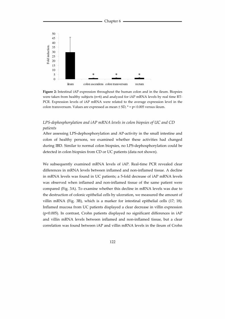
detoxifying capacity of the intestine. iAP mRNA levels in the human ileum were
found to be about 30 times higher than those in the human colon (Fig. 2).

Oral administration of alkaline phophatase ameliorates colitis
121
Figure 1: LPS dephosphorylation by sections of the intestine of a normal healthy person (a
full color version of this figure is depicted on page 172). A brown staining was clearly visible
along the apical side of the microvilli of the enterocyte in the terminal ileum (A). In contrast,
biopsies from the colon ascendens (B), colon transversum (C) and rectum (D), showed
hardly any LPS-dephosphorylating activity along the enterocytes. Occasional cells stained
positive (arrows). Control sections (without LPS) were completely negative (E).
Magnification 200*.
A B
C D
E
Chapter 6
122
0
5
10
15
20
25
30
35
40
45
50
ileum colon ascendens colon transversum rectum
Fold
in
du
ctio
n. .
Figure 2: Intestinal iAP expression throughout the human colon and in the ileum. Biopsies
were taken from healthy subjects (n=6) and analyzed for iAP mRNA levels by real time RT-
PCR. Expression levels of iAP mRNA were related to the average expression level in the
colon transversum. Values are expressed as mean ± SD, * = p< 0.005 versus ileum.
LPS-dephosphorylation and iAP mRNA levels in colon biopsies of UC and CD
patients After assessing LPS-dephosphorylation and AP-activity in the small intestine and
colon of healthy persons, we examined whether these activities had changed
during IBD. Similar to normal colon biopsies, no LPS-dephosphorylation could be
detected in colon biopsies from CD or UC patients (data not shown).
We subsequently examined mRNA levels of iAP. Real-time PCR revealed clear
differences in mRNA levels between inflamed and non-inflamed tissue. A decline
in mRNA levels was found in UC patients; a 5-fold decrease of iAP mRNA levels
was observed when inflamed and non-inflamed tissue of the same patient were
compared (Fig. 3A). To examine whether this decline in mRNA levels was due to
the destruction of colonic epithelial cells by ulceration, we measured the amount of
villin mRNA (Fig. 3B), which is a marker for intestinal epithelial cells (17; 18).
Inflamed mucosa from UC patients displayed a clear decrease in villin expression
(p<0.005). In contrast, Crohn patients displayed no significant differences in iAP
and villin mRNA levels between inflamed and non-inflamed tissue, but a clear
correlation was found between iAP and villin mRNA levels in the ileum of Crohn
* * *

Oral administration of alkaline phophatase ameliorates colitis
123
patients (R2=0.98, p=0.0001), which links the iAP expression to the intact intestinal
epithelium.
A
B
iAP
0,0
0,5
1,0
1,5
2,0
not inflamed inflamed
Fold
ind
uct
ion
Villin
0,0
0,5
1,0
1,5
2,0
not inflamed inflamed
Fo
ld i
nd
ucti
on
*
*
Figure 3: iAP (A) and villin (B) mRNA expression in non-inflamed and inflamed intestinal
tissue of UC patients (n=7). Total RNA was isolated from biopsies of non-inflamed and
inflamed intestinal tissue. A decreased iAP expression strongly correlated with a decreased
expression of the epithelial marker villin. * = p<0.005.
Responsiveness of human colon biopsies to LPS Human colonic biopsies from healthy persons and IBD patients were tested for
their responsiveness upon exposure to LPS. Biopsies from healthy volunteers
showed at best only a very low induction of inflammation-related genes after
incubation with LPS.

Chapter 6
124
It is known that during CD and UC, the LPS receptor TLR4 is upregulated on colon
epithelium (3; 19) and therefore it could be envisioned that biopsies of CD and UC
patients would react more vigorously upon incubation with LPS. However, also in
the biopsies of patients, hardly any inflammatory response was observed. mRNA
levels of the inflammatory genes TNFα and IL-1β upon LPS incubation in three UC
patients were not significantly induced compared to biopsies incubated without
LPS. In two of the three tested CD patients an induction of inflammatory genes
after incubation with LPS was observed. However, no effects of iAP could be
determined. The two patients that reacted upon LPS had a reduced villin
expression and higher expression levels of the LPS receptors TLR4 and CD14.
These data indicate that only when the intestinal barrier is damaged due to
ulceration, LPS is able to induce an inflammatory response.
Effect of oral administration of enteric-coated iAP-tablets on DSS-induced colitis
in rats To examine whether oral administration of iAP affects experimental colitis,
inflammation was induced in the colon in male Sprague-Dawley rats by oral intake
of dextran sodium sulphate (DSS).
Oral administration of AP in tablets resulted in a higher AP activity within the
intestine. AP activity in faeces at 24 hr after treatment had risen from 0.13
units/gram faeces in rats that received normal drinking water plus placebo-tablets
to 7.08 units/gram faeces in rats that received normal drinking water plus AP-
tablets. The total recovery of AP activity from acid-resistant AP-tablets in the
intestine is approximately 30% as determined previously (20).
The daily consumption of drinking water by the rats showed no significant
differences, confirming that both treated and untreated rats received the same
amount of DSS.
In this study also no difference in weight loss between the different groups that
received DSS in their drinking water was found. The rats in the AP-treated and
placebo-treated group lost 11.0% and 11.9% of their initial body weight,
Oral administration of alkaline phophatase ameliorates colitis
125
respectively. The control groups, i.e. the groups that received normal drinking
water plus either placebo- or AP-tablets, showed an average increase of body
weight of 4.5% respectively 4.6%.
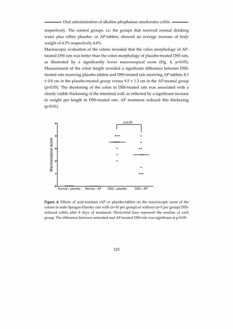
Macroscopic evaluation of the colons revealed that the colon morphology of AP-
treated DSS rats was better than the colon morphology of placebo-treated DSS rats,
as illustrated by a significantly lower macroscopical score (Fig. 4, p<0.05).
Measurement of the colon length revealed a significant difference between DSS-
treated rats receiving placebo-tablets and DSS-treated rats receiving AP-tablets; 8.1
± 0.8 cm in the placebo-treated group versus 9.5 ± 1.3 cm in the AP-treated group
(p<0.05). The shortening of the colon in DSS-treated rats was associated with a
clearly visible thickening of the intestinal wall, as reflected by a significant increase
in weight per length in DSS-treated rats. AP treatment reduced this thickening
(p<0.01).
Normal + placebo Normal + AP DSS + placebo DSS + AP0
1
2
3
4
5 p<0.05
Ma
cro
sco
pic
al sco
re
Figure 4: Effects of acid-resistant iAP or placebo-tablets on the macroscopic score of the
colons in male Sprague-Dawley rats with (n=10 per group) or without (n=5 per group) DSS-
induced colitis after 8 days of treatment. Horizontal bars represent the median of each
group. The difference between untreated and AP-treated DSS-rats was significant at p<0.05.

Chapter 6
126
In the distal part of the colon, relative mRNA expression levels of the following
genes were determined: TNFα, IL-1β, IL-6, iNOS, IL-10R and villin. The mRNA
levels of the inflammatory genes, TNFα, IL-1β, IL-6, iNOS and IL-10R were all
strongly upregulated in DSS-treated rats. Expression levels of all these genes were
significantly lower in DSS-rats that received AP-tablets compared to DSS-rats that
received placebo-tablets (p<0.05). Only two out of ten rats treated with AP still had
elevated levels of TNFα, IL-1β, IL-6, iNOS and IL-10R. In contrast, mRNA levels
for the epithelial marker villin, which were profoundly reduced in placebo-treated
DSS-rats, were significantly increased in colitic rats receiving AP-tablets (p<0.05).
Interestingly, the two rats that displayed aberrant cytokine, iNOS and IL-10R
levels, also had reduced villin levels. In other words, the outliners were the same
rats for all parameters. These data indicate that the epithelial layer in the colon of
rats receiving DSS plus placebo-tablets is severely damaged whereas the epithelial
layer in rats receiving DSS plus AP-tablets is significantly less damaged.
Figure 5: Effects of oral treatment with acid-resistant iAP or placebo-tablets on mRNA
expression of several genes in male Sprague-Dawley rats with (n=10 per group) or without
(n=5 per group) DSS-induced colitis after 7 days of treatment (Figure on page 127). Dot plots
show the expression levels of TNFα, IL-1β, IL-6, iNOS, IL-10R and villin mRNA in the distal
part of the rat colons. Horizontal bars represent the median of each group. Expression levels
were normalized to GAPDH. For each parameter, the difference between untreated and AP-
treated DSS-rats was significant at p<0.05.

Oral administration of alkaline phophatase ameliorates colitis
127
TNFαααα
Normal + plac. Normal + AP DSS + plac. DSS + AP0
25
50
75
100
125
150
Fo
ld in
du
cti
on
IL-1 ββββ
Normal + plac. Normal + AP DSS + plac. DSS + AP0
200
400
600
800
1000
1200
1400
1600
1800
Fo
ld in
du
cti
on
IL-6
Normal + plac. Normal + AP DSS + plac. DSS + AP0
500
1000
1500
2000
2500
3000
Fo
ld in
du
cti
on
iNOS
Normal + plac. Normal + AP DSS + plac. DSS + AP0
5000
10000
15000
20000
25000
Fo
ld in
du
cti
on
IL-10R
Normal + plac. Normal + AP DSS + plac. DSS + AP0
100
200
300
400
500
600
700
800
900
Fo
ld in
du
cti
on
villin
Normal + plac. Normal + AP DSS + plac. DSS + AP0.00
0.25
0.50
0.75
1.00
1.25
Fo
ld in
du
cti
on
Chapter 6
128
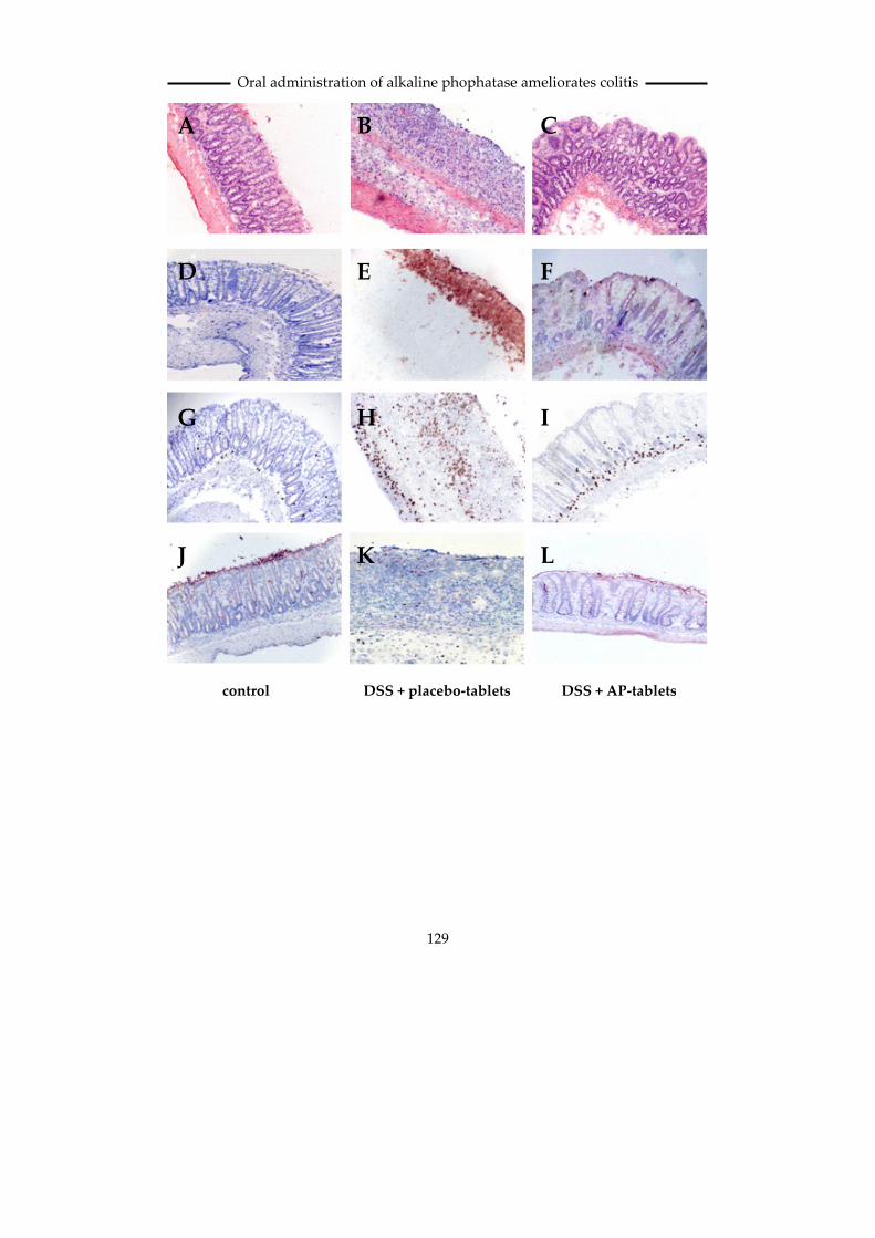
The colon was also histochemically evaluated by H&E, iNOS, MPO and villin
staining. The colons of DSS rats receiving AP-tablets were clearly less damaged
than those of DSS rats that received placebo-tablets (Fig. 6C versus fig. 6B). The
epithelial lining of the colon was more intact, confirming the PCR results for villin
mRNA levels, and in line with the macroscopical data (Fig. 4), the serosa was less
thickened in AP-treated rats. In both groups of rats that received normal drinking
water, no staining for the inflammation marker iNOS was observed (Fig. 6D).
However, in placebo-treated DSS-rats, all colon segments displayed a profound
iNOS staining (Fig. 6E). In contrast, colons from AP-treated DSS-rats showed no or
only minor iNOS staining (Fig. 6F). The amount of MPO-positive cells in the colon
was highly increased in DSS-treated rats receiving placebo-tablets (Fig. 6H)
compared to normal rats (Fig. 6G). Administration of AP-tablets to DSS-treated
rats markedly reduced this influx of MPO-positive cells (Fig. 6I). The epithelial
marker villin was abundantly present along the epithelial lining of the mucosa in
control rats (Fig. 6J), but completely absent in colons of DSS-rats that received
placebo-tablets (Fig. 6K). In contrast, DSS-rats that received AP-tablets displayed a
clear villin-staining along the epithelial cells which was comparable to control rats
(Fig. 6L), which is in line with the PCR data for villin mRNA.
Taken together, all data indicate that the inflammation of the colon and the
structural damage after DSS exposure is significantly reduced upon AP treatment.
Figure 6: H&E staining (A, B, C), histochemical staining for iNOS (D, E, F) and activated
neutrophils (G, H, I) and villin (J, K, L) in male Sprague-Dawley rats with DSS-induced
colitis after 7 days of treatment with placebo (mid-panel) or acid-resistant iAP-tablets (right-
hand column). Normal rats are depicted in the left-hand column. Note the mucosal
ulceration and thickening of the serosa in placebo-treated colitic rats (B) compared to oral
iAP-treated colitic rats (C). The inflammation marker iNOS was absent in non-colitic rats
(D), enhanced in placebo-treated colitic rats (E), and strongly decreased in oral iAP-treated
colitic rats (F). The amount of activated neutrophils was low in non-colitic rats (G), high in
placebo-treated colitic rats (H) and clearly reduced in iAP-treated colitic rats (I). The red
staining for the epithelial marker villin was visible along the epithelial lining in non-colitic
rats (J), absent in placebo-treated colitic rats (K) and retained in iAP-treated DSS rats (L).
Magnification of all figures is 100*. A full color version of this article can be found on page
173)
Oral administration of alkaline phophatase ameliorates colitis
129
A B C
E D F
G H I
J K L
DSS + AP-tablets DSS + placebo-tablets control
Chapter 6
130
Discussion Inflammatory bowel disease (IBD) is one of the major chronic inflammatory
diseases, affecting several millions of people world-wide. It is believed that the
inflammation of the damaged gut is the result of an inappropriate ongoing
activation of the mucosal immune system triggered by components of the normal
luminal flora (1). When the sub-epithelial layers of the intestinal wall are exposed
to LPS due to damage, macrophages residing in these layers start to produce
cytokines and chemoattractants (21), leading to the migration of high amounts of
neutrophils to the site of damage and local activation. Production of high amounts
of ROS by accumulated neutrophils will cause local damage and further enhance
inflammation. A damaged intestinal barrier sometimes also leads to elevated LPS
levels in the blood caused by translocation of luminal LPS and/or bacterial
translocation, which in turn may induce elevated levels of circulating cytokines
like TNFα, IL-1β, IL-6 and IL-17 (22; 23). This may increase the intestinal
permeability even further. Several reports have shown that the enzyme alkaline
phosphatase (AP) is able to dephosphorylate and detoxify LPS (7-10). The resulting
product, LPS with a monophosphoryl lipid A moiety, is non-toxic (24) and may
even antagonize LPS (25). Because inflammatory bowel diseases are associated
with a perpetuating inflammatory response towards intestinal bacterial products,
in particular LPS, administration of LPS-detoxifying molecules might attenuate the
inflammatory response during periods of severe inflammation.
We first studied intestinal AP expression in normal healthy persons with
enzymehistochemistry and real-time PCR techniques. The results obtained with
both techniques corresponded with each other and showed a strong LPS-
dephosphorylation in the human ileum along the lining of the villi whereas the
human colon showed almost no LPS-dephosphorylation. This difference was
confirmed by real-time PCR; the human ileum showed much higher iAP mRNA
levels than the human colon. Previous studies have demonstrated that this LPS-
dephosphorylation was caused by AP activity (7; 8) and the localization of this
activity here is also in agreement with the reported localization of iAP in the small
intestine (11; 26). The reason for the lack of LPS-dephosphorylation in the colon is
unknown. Because bacterial titers in the colon are high, the barrier function may be

Oral administration of alkaline phophatase ameliorates colitis
131
sufficiently strong under physiological circumstances and LPS receptors are not
expressed in the colon epithelium in the normal situation (19; 27; 28), leading to a
hyporesponsiveness to LPS within the colon. So, LPS detoxification may not be
required in the colon. AP activity within the colon might also be blocked by
dephosphorylated LPS, which is a known inhibitor of AP activity (9). In the ileum,
where intestinal nutrient uptake is high, LPS-dephosphorylation mechanisms may
be more relevant.
Although LPS-dephosphorylation in the colon in the healthy control group was
low, we observed decreased mRNA iAP levels in the colon of UC patients. These
decreases were paralleled by decreases in the expression levels of the epithelial
marker villin, suggesting that the decrease in iAP expression is most likely due to
loss of the epithelial cell layer.
LPS-dephosphorylation was shown to be associated with reduced toxicity of this
bacterial product (7-10; 29) and this may be relevant in view of the role of LPS in
the pathology of IBD. We therefore explored whether administration of calf
intestinal AP (ciAP) might affect DSS-induced colitis, a model for human UC in
rats (30). The DSS-model is characterized by colonic epithelial damage, diarrhea,
bloody faeces, decrease of body weight, colon shortening and neutrophilic
infiltration in the intestinal wall (31). Treatment with AP will not affect DSS-
induced damage but only the secondary damage caused after destruction of the
epithelial layer (32). Intestinal AP appeared to display a profound effect on the
colon. All parameters reflecting the colon condition at the macroscopical level
showed that the colon was significantly improved in diseased rats receiving AP-
tablets.
Measurement of drinking water consumption showed that both treated and
untreated groups received equal amounts of DSS, leading to a decreased water-
resorption capacity of the colon and diarrhea, and subsequently weight loss in both
groups. Nevertheless, significant effects were found on mRNA levels of all genes
examined; mRNA levels of inflammatory genes were strongly decreased,
indicating a reduced inflammation within the colon wall, and villin mRNA levels
were strongly enhanced in the colons of AP-treated rats, reflecting a more intact
epithelial cell layer. Also intestinal wall thickness and integrity were nearly normal
in all AP-treated colitic rats, and the patchy staining for iNOS found in all DSS-rats

Chapter 6
132
treated with placebo-tablets was completely absent in all rats treated with AP. So,
these data demonstrate a significant therapeutic effect of oral administration of
iAP-tablets. Although AP activity in the colon is normally low or absent,
administration of exogenous iAP seems therefore beneficial. Apparently, during
disease, when damage to the colon wall occurs and infiltration of CD14 / TLR4
receptor-positive cells has taken place, shielding mechanisms are affected and LPS-
hyperresponsive cells infiltrate in the tissue. So only when the normal barrier is
affected, a high concentration of AP may exert a clinical relevant function.
The role of the barrier and the effect of LPS-responsive cells that subsequently
infiltrate in the tissue became also clear in human biopsies. Biopsies of normal
healthy persons appeared to be quite insensitive to stimulation with LPS. This
apparent hyporesponsiveness might be due to the absence of TLR4 in human
intestinal epithelial cells (27; 33). After examining the response upon LPS in
biopsies from 3 CD and 3 UC patients, we only found induction of mRNA for
inflammatory genes in biopsies of 2 CD patients. Biopsies of these two patients also
displayed low villin mRNA levels indicating that in these patients the epithelial
layer was affected. It has been described that IBD patients have higher expression
levels of the LPS receptor TLR4 (3; 19), but in contrast to these reports, we could
not detect such differences. Since there was no response upon LPS in most cases,
no significant effect of AP on the LPS-response was noted either.
In summary, this study shows for the first time that epithelial iAP expression is
decreased in UC patients and that the iAP-mediated endogenous LPS-detoxifying
activity is therefore challenged in these patients. Importantly, oral iAP
administration was found to reduce LPS-mediated inflammatory effects in colitic
rats. These data provide new insights into the role of iAP during IBD and support
the notion that oral administration of iAP to UC patients may be therapeutically
effective. This prompted us to a phase II proof of concept study in patients with
severe UC which is currently ongoing.

Oral administration of alkaline phophatase ameliorates colitis
133
Reference List
1. Podolsky DK.
Inflammatory bowel disease. N Engl J
Med 2002;347:417-429.
2. Hugot JP, Chamaillard
M, Zouali H, Lesage S, Cezard JP,
Belaiche J, Almer S, Tysk C, O'Morain
CA, Gassull M, Binder V, Finkel Y, Cortot
A, Modigliani R, Laurent-Puig P, Gower-
Rousseau C, Macry J, Colombel JF,
Sahbatou M, Thomas G. Association of
NOD2 leucine-rich repeat variants with
susceptibility to Crohn's disease. Nature
2001;411:599-603.
3. Cario E, Podolsky DK.
Differential alteration in intestinal
epithelial cell expression of toll-like
receptor 3 (TLR3) and TLR4 in
inflammatory bowel disease. Infect
Immun 2000;68:7010-7017.
4. Franchimont D,
Vermeire S, El HH, Pierik M, Van SK,
Gustot T, Quertinmont E, Abramowicz
M, Van GA, Deviere J, Rutgeerts P.
Deficient host-bacteria interactions in
inflammatory bowel disease? The toll-
like receptor (TLR)-4 Asp299gly
polymorphism is associated with Crohn's
disease and ulcerative colitis. Gut
2004;53:987-992.
5. Inohara N, Ogura Y,
Fontalba A, Gutierrez O, Pons F, Crespo
J, Fukase K, Inamura S, Kusumoto S,
Hashimoto M, Foster SJ, Moran AP,
Fernandez-Luna JL, Nunez G. Host
recognition of bacterial muramyl
dipeptide mediated through NOD2.
Implications for Crohn's disease. J Biol
Chem 2003;278:5509-5512.
6. Girardin SE, Boneca IG,
Viala J, Chamaillard M, Labigne A,
Thomas G, Philpott DJ, Sansonetti PJ.
Nod2 is a general sensor of
peptidoglycan through muramyl
dipeptide (MDP) detection. J Biol Chem
2003;278:8869-8872.
7. Poelstra K, Bakker
WW, Klok PA, Kamps JA, Hardonk MJ,
Meijer DK. Dephosphorylation of
endotoxin by alkaline phosphatase in
vivo. Am J Pathol 1997;151:1163-1169.
8. Poelstra K, Bakker
WW, Klok PA, Hardonk MJ, Meijer DK.
A physiologic function for alkaline
phosphatase: endotoxin detoxification.
Lab Invest 1997;76:319-327.
9. Bentala H, Verweij WR,
Huizinga-Van der Vlag A, Loenen-
Weemaes AM, Meijer DK, Poelstra K.
Removal of phosphate from lipid a as a
strategy to detoxify lipopolysaccharide.
Shock 2002;18:561-566.
10. Beumer C, Wulferink
M, Raaben W, Fiechter D, Brands R,
Seinen W. Calf intestinal alkaline
phosphatase, a novel therapeutic drug for
lipopolysaccharide (LPS)-mediated
diseases, attenuates LPS toxicity in mice
and piglets. J Pharmacol Exp Ther
2003;307:737-744.
11. Domar U, Nilsson B,
Baranov V, Gerdes U, Stigbrand T.
Expression of intestinal alkaline
Chapter 6
134
phosphatase in human organs.
Histochemistry 1992;98:359-364.
12. Tuin A, Huizinga-Van
der Vlag A, Loenen-Weemaes AM, Meijer
DK, Poelstra K. On the role and fate of
LPS-dephosphorylating activity in the rat
liver. Am J Physiol Gastrointest Liver
Physiol 2006;290:G377-G385.
13. Tillyer CR, Rakhorst S,
Colley CM. Multicomponent analysis for
alkaline phosphatase isoenzyme
determination by multiple linear
regression. Clin Chem 1994;40:803-810.
14. van de Bovenkamp M,
Groothuis GM, Draaisma AL, Merema
MT, Bezuijen JI, van Gils MJ, Meijer DK,
Friedman SL, Olinga P. Precision-cut
liver slices as a new model to study
toxicity-induced hepatic stellate cell
activation in a physiologic milieu. Toxicol
Sci 2005;85:632-638.
15. Poelstra K, Hardonk
MJ, Koudstaal J, Bakker WW.
Intraglomerular platelet aggregation and
experimental glomerulonephritis. Kidney
Int 1990;37:1500-1508.
16. Vos TA, Gouw AS,
Klok PA, Havinga R, van Goor H,
Huitema S, Roelofsen H, Kuipers F,
Jansen PL, Moshage H. Differential
effects of nitric oxide synthase inhibitors
on endotoxin-induced liver damage in
rats. Gastroenterology 1997;113:1323-
1333.
17. Aono K, Isobe K,
Kiuchi K, Fan ZH, Ito M, Takeuchi A,
Miyachi M, Nakashima I, Nimura Y. In
vitro and in vivo expression of inducible
nitric oxide synthase during experimental
endotoxemia: involvement of other
cytokines. J Cell Biochem 1997;65:349-358.
18. Hofer D, Drenckhahn
D. Cytoskeletal markers allowing
discrimination between brush cells and
other epithelial cells of the gut including
enteroendocrine cells. Histochem Cell
Biol 1996;105:405-412.
19. Hausmann M,
Kiessling S, Mestermann S, Webb G,
Spottl T, Andus T, Scholmerich J,
Herfarth H, Ray K, Falk W, Rogler G.
Toll-like receptors 2 and 4 are up-
regulated during intestinal inflammation.
Gastroenterology 2002;122:1987-2000.
20. Eriksson HJ, Verweij
WR, Poelstra K, Hinrichs WL, de Jong GJ,
Somsen GW, Frijlink HW. Investigations
into the stabilisation of drugs by sugar
glasses: II. Delivery of an inulin-stabilised
alkaline phosphatase in the intestinal
lumen via the oral route. Int J Pharm
2003;257:273-281.
21. Ramos CD, Heluy-Neto
NE, Ribeiro RA, Ferreira SH, Cunha FQ.
Neutrophil migration induced by IL-8-
activated mast cells is mediated by
CINC-1. Cytokine 2003;21:214-223.
22. Fujino S, Andoh A,
Bamba S, Ogawa A, Hata K, Araki Y,
Bamba T, Fujiyama Y. Increased
expression of interleukin 17 in
inflammatory bowel disease. Gut
2003;52:65-70.

Oral administration of alkaline phophatase ameliorates colitis
135
23. Street ME, de'Angelis
G, Camacho-Hubner C, Giovannelli G,
Ziveri MA, Bacchini PL, Bernasconi S,
Sansebastiano G, Savage MO.
Relationships between serum IGF-1,
IGFBP-2, interleukin-1beta and
interleukin-6 in inflammatory bowel
disease. Horm Res 2004;61:159-164.
24. Schromm AB,
Brandenburg K, Loppnow H, Zahringer
U, Rietschel ET, Carroll SF, Koch MH,
Kusumoto S, Seydel U. The charge of
endotoxin molecules influences their
conformation and IL-6-inducing capacity.
J Immunol 1998;161:5464-5471.
25. Astiz ME, Rackow EC,
Still JG, Howell ST, Cato A, Von Eschen
KB, Ulrich JT, Rudbach JA, McMahon G,
Vargas R, . Pretreatment of normal
humans with monophosphoryl lipid A
induces tolerance to endotoxin: a
prospective, double-blind, randomized,
controlled trial. Crit Care Med 1995;23:9-
17.
26. Benjawatanapon C, Bell
LE, Williams L. Ultrastructural
localisation of alkaline phosphatase in
adult human large intestine. Gut
1982;23:134-136.
27. Naik S, Kelly EJ, Meijer
L, Pettersson S, Sanderson IR. Absence of
Toll-like receptor 4 explains endotoxin
hyporesponsiveness in human intestinal
epithelium. J Pediatr Gastroenterol Nutr
2001;32:449-453.
28. Nakata K, Inagawa H,
Nishizawa T, Kohchi C, Soma GI. Specific
messenger RNA expression for signal
transduction molecules by
lipopolysaccharide in intestinal
macrophages. Clin Exp Immunol
2006;143:484-493.
29. Xu Q, Lu Z, Zhang X. A
novel role of alkaline phosphatase in
protection from immunological liver
injury in mice. Liver 2002;22:8-14.
30. Elson CO, Sartor RB,
Tennyson GS, Riddell RH. Experimental
models of inflammatory bowel disease.
Gastroenterology 1995;109:1344-1367.
31. Rumi G, Tsubouchi R,
Nishio H, Kato S, Mozsik G, Takeuchi K.
Dual role of endogenous nitric oxide in
development of dextran sodium sulfate-
induced colitis in rats. J Physiol
Pharmacol 2004;55:823-836.
32. Cooper HS, Murthy
SN, Shah RS, Sedergran DJ.
Clinicopathologic study of dextran
sulfate sodium experimental murine
colitis. Lab Invest 1993;69:238-249.
33. Abreu MT, Vora P,
Faure E, Thomas LS, Arnold ET, Arditi
M. Decreased expression of Toll-like
receptor-4 and MD-2 correlates with
intestinal epithelial cell protection against
dysregulated proinflammatory gene
expression in response to bacterial
lipopolysaccharide. J Immunol
2001;167:1609-1616.

136