Embed Size (px)
Citation preview

University of Groningen
Bridging the gap between in vitro and in vivo evaluation of biomaterials-associated infectionsSubbiahdoss, Guruprakash
IMPORTANT NOTE: You are advised to consult the publisher's version (publisher's PDF) if you wish to cite fromit. Please check the document version below.
Document VersionPublisher's PDF, also known as Version of record
Publication date:2010
Link to publication in University of Groningen/UMCG research database
Citation for published version (APA):Subbiahdoss, G. (2010). Bridging the gap between in vitro and in vivo evaluation of biomaterials-associatedinfections Groningen: s.n.
CopyrightOther than for strictly personal use, it is not permitted to download or to forward/distribute the text or part of it without the consent of theauthor(s) and/or copyright holder(s), unless the work is under an open content license (like Creative Commons).
Take-down policyIf you believe that this document breaches copyright please contact us providing details, and we will remove access to the work immediatelyand investigate your claim.
Downloaded from the University of Groningen/UMCG research database (Pure): http://www.rug.nl/research/portal. For technical reasons thenumber of authors shown on this cover page is limited to 10 maximum.
Download date: 14-05-2018


Bridging the gap between in vitro and in vivo
evaluation of biomaterials-associated infections
Guruprakash Subbiahdoss
“Bridging the gap between in vitro and in vivo evaluation of biomaterials-associated infections” by Guruprakash Subbiahdoss Department of BioMedical Engineering University Medical Center Groningen Cover: Represents the race between bacteria and tissue cells for the biomaterial surface. Bacteria shown in green and tissue cells with cell membrane in red and nucleus in blue. The cover has been designed by Sumitha. Printing of this thesis was financially supported by: RuG, W.J. Kolff Institute, Heraeus Medical. Copyright © 2010 by G Subbiahdoss ISBN: 978-90-367-4479-9 (printed version) ISBN: 978-90-367-4480-5 (electronic version)

Bridging the gap between in vitro and in vivo
evaluation of biomaterials-associated infections
Proefschrift
ter verkrijging van het doctoraat in de
Medische Wetenschappen
aan de Rijksuniversiteit Groningen
op gezag van de
Rector Magnificus, dr. F. Zwarts,
in het openbaar te verdedigen op
woensdag 22 september 2010
om 13.15 uur
door
Guruprakash Subbiahdoss
geboren op 20 juli 1980
te Tiruchendur, India

Promotores: Prof. dr. H.C. van der Mei
Prof. dr. ir. H.J. Busscher
Copromoter: Dr. R. Kuijer
Beoordelingscommissie: Prof. dr. R.A. Bank
Prof. dr. S.K. Bulstra
Prof. dr. J.M. van Dijl

Paranimfen: Sumitha Rani Baluchamy
Ekaterina Ovchinnikova


Contents Chapter 1 General introduction and aim of this thesis 1 Chapter 2 Microbial biofilm growth versus tissue integration: 13
“The race for the surface” experimentally studied Chapter 3 Microbial biofilm growth versus tissue integration 31
on biomaterials with different wettabilities and a polymer-brush coating
Chapter 4 Bacterial strain-specific effects on mammalian cell growth 49
on poly(methylmethacrylate) Chapter 5 Bacterial biofilm formation versus mammalian cell growth 65
on titanium-based mono- and bi-functional coatings Chapter 6 Mammalian cell growth versus biofilm formation 87
on biomaterials surfaces in a post-operative contamination model in vitro
Chapter 7 A new method to study the simultaneous interaction 103
between bacteria, macrophages and osteoblasts on a biomaterial implant surface
Chapter 8 General discussion 119 Summary 129 Samenvatting 135 Acknowledgements 143


CHAPTER 1
General Introduction
Chapter 1 Biomaterial-Associated Infections
Biomaterials play an important role in modern medicine in the restoration of tissue,
organ or body function. For example, a current estimate of the number of total hip
replacements in the world amounts approximately one million per year and knee
replacements more than 250,000. More than 10% of all hospitalized patients are using
urinary catheters, biomaterial implants or other medical devices. The use of
biomaterial implants and medical devices is mainly restricted by complications due to
biomaterial-associated infections (BAI). On average, BAI occurs in approximately 0.5
– 6% [1,2] of all cases, strongly depending on the implant site, and more often in
cases of trauma or revision surgery [3-5]. The presence of a biomaterial in the body
significantly compromises the host to cope with invading microorganisms. There are
various routes along which microorganisms can enter the body and develop a BAI in
the case of permanent implants [6]. The best-documented route is direct
contamination of an implant during surgery (peri-operative contamination). BAI can
also be initiated immediately post-surgery during hospitalization (post-operative
contamination) or microbial spreading through blood from infections elsewhere in the
human body [6-8]. These different routes of infection will be briefly discussed.
Routes of Infection
Peri-operative contamination means that an implant becomes contaminated with
bacteria before implantation into the human body or during the implantation process.
During the implantation of a biomaterial implant, microorganisms from the skin can
reach the implant surface. It is known that during a surgical procedure of 1 h, the total
number of bacteria carrying particles falling on a wound is about 270 cm-2. The
2
General Introduction bacterial counts are generally higher during periods of activity and when more people
are present in the operating room [9]. More recent, through the use of modern, better
ventilated operating theatres (20 changes of air per hour) and impermeable patient and
personnel clothing, peri-operative bacterial contamination levels may well be less
[10].
The second route of infection is post-operative contamination which may
occur during the period of hospitalization immediately post-surgery, caused by direct
contamination of open wounds or by the use of invasive devices like infusion tubes,
drains and catheters. Both peri-operative and post-operative infections can cause BAI
many years after implantation, because bacteria on a biomaterial implant surface are
known to be able to stay on the implant in a low metabolic state for several years [6].
As a third route, BAI can result from hemotogenous spreading of bacteria
from infections elsewhere in the body, which upon adhering to a biomaterial implant
surface form a biofilm. This includes skin infections, surgical or dental interventions,
pneumonia, abscesses or bacteriuria, which can cause temporal or chronic bacteremia
resulting in BAI [7]. It is also shown that macrophages play a major role in
transporting bacteria to the implant surface, as some strains are capable to survive
within macrophages [11,12].
Microbial Adhesion to Biomaterials
The introduction of bacteria by any of the above routes to the biomaterial surface is
the initial step in the development of BAI. Microorganisms can reach the biomaterial
surface as early as during implantation and interact with bare substratum surfaces, not
even covered with a conditioning film, i.e. a film of adsorbed serum or plasma
3
Chapter 1 proteins [8]. In general, microbial adhesion is mediated by the specific interactions
between cell surface structures and specific molecular groups on the substratum
surface, or by non-specific interaction forces, such as Lifshitz-Van der Waals,
electrostatic charge, acid-base interactions and Brownian motion forces. Irrespective
of the presence or absence of a conditioning film, the physico-chemical surface
properties of the biomaterial and microorganisms play an important role in the
adhesion process. Conditioning films on biomaterial surfaces and on bacterial cell
surfaces play important roles too by changing the physico-chemical surface
properties. Most proteins in conditioning films are capable of reducing microbial
adhesion, but fibronectin and fibrinogen have been shown to promote the adhesion of
certain Staphylococcus epidermidis and Staphylococcus aureus strains [13]. Microbial
adhesion mechanisms in post-operative infections or hematogeneous infections are
unclear. Biomaterial implants are usually not completely integrated with host tissue,
especially when they consist of metal parts which are not easily colonized by host
tissue cells. An uncovered metal surface can be a site for microbial colonization.
Moreover, it is possible that by repeated hinging of orthopedic implants, cell/tissue
damage can occur, providing adhesive sites for microorganisms [6].
Once microorganisms adhere to the biomaterial surface, the second step is the
subsequent growth of the adhering bacteria, accompanied by production of
Extracellular Polymeric Substances (EPS), leading to biofilm formation, which
protects the infecting organisms against host immune system and antibiotic treatment
[14,15]. Eradication of a biofilm is difficult because the populating bacteria are
protected from the immune system and antibiotics. Bacteria within biofilms generally
require 500-5000 times higher doses of antibiotics than planktonic ones suspended in
4
General Introduction body fluids [16]. In the majority of cases, the final outcome of a BAI is removal of the
implant.
Pathogens Causing BAI
In general, S. epidermidis and S. aureus are the most frequently isolated pathogens
from infected biomaterial surfaces. Additionally isolated organisms include
Escherichia coli and Pseudomonas aeruginosa [6,15,17]. Almost 50% of infections
associated with catheters, artificial joints and heart valves are caused by S.
epidermidis [18]. S. aureus is the cause of around 23% of infections associated with
prosthetic joints [18]. P. aeruginosa is the causative organism of around 12% of all
hospital acquired urinary track infections, 10% of bloodstream infections and 7% of
hip-joint infections [19]. S. epidermidis are the common cause of late infections [18].
Another factor that plays an important role in the pathogenesis of BAI is the
bacterial virulence [18]. S. aureus and P. aeruginosa infections usually progress much
more aggressively than BAI caused by S. epidermidis [18,19]. S. aureus appears more
frequently in acute infections within 4 weeks after surgery, compared to S.
epidermidis. S. epidermidis is most commonly implicated in delayed septic loosening
of total joint prostheses [20] or even in presumed a-septic loosening [21], indicating
its low virulence with only minor clinical symptoms of infection. Pseudomonas is also
much more virulent than S. epidermidis, which is ascribed to the more aggressive
endotoxins in the slime. The low virulence of S. epidermidis strains compared to S.
aureus or P. aeruginosa is due to the lack of additional genes responsible for
producing tissue damaging toxins [18,19,22]. In S. epidermidis infections, biofilm
5
Chapter 1 formation is considered the only virulence factor and therefore infections are usually
sub-acute or chronic [23-25].
Mammalian Cell Adhesion to Biomaterials
Mammalian cell adhesion to a biomaterial implant surface is crucial for the successful
integration of the implant within the host tissue. In vivo, when a biomaterial is
implanted, it becomes immediately coated with proteins (conditioning film) that are
adsorbed from the local body fluids. Depending on the body site, the surrounding
fluid can be saliva, urine, tissue fluid, serum or blood and the conditioning film
mostly consists of adsorbed proteins. The physico-chemical properties of the
biomaterial surface (chemical composition, hydrophobicity and surface charge)
control the nature of the adsorbed protein layer [26,27]. Cell adhesion to adsorbed
proteins is mediated through integrins and other receptors present within the cell
membrane. Upon adhesion to the protein film, a cascade of intracellular signaling
events is triggered. Therefore, controlling protein adsorption on the biomaterial
surface may be critical to control and direct cell responses to biomaterials.
Unfortunately, proteins like fibronectin and fibrinogen in conditioning films have
been shown to promote the adhesion of certain S. epidermidis and S. aureus strains
[13].
The Race for the Surface
In 1987, the orthopedic surgeon Anthony Gristina coined the term “race for the
surface” to describe the fate of a biomaterial implant in relation with the development
of BAI [6], as illustrated in Fig. 1. The fate of a biomaterial implant was depicted as a
6

General Introduction race between microbial adhesion and biofilm growth on an implant surface versus
tissue integration. If the race is won by tissue cells, then the surface is covered by
tissue and less vulnerable to bacterial colonization. On the other hand, if the race is
won by bacteria, the implant surface will become colonized by bacteria and tissue cell
functions are hampered by bacterial virulence factors [6]. Irrespective of the route of
infection, the fate of biomaterial implants depends mainly on the outcome of the so-
called ‘race for the surface’.
Fig. 1. Schematic diagram representing the race between bacteria and tissue cells for the biomaterial surface [6].
Bridging the Gap Between In Vitro and In Vivo Studies
In vivo, processes occurring at the biomaterial site are complex, involving multiple
cell types depending on the site, cytokines, chemokines, excretion of bacterial and cell
substances. Normalization of macrophage response on biomaterials allows appropriate
antibacterial activities, healing and integration. During infection, macrophages play a
key role in the elimination of bacterial colonization [17].
In the path towards reducing the risk of BAI, biomaterials research has been
focused on the development of biomaterials or coatings that can prevent bacterial
adhesion and stimulate mammalian cell growth. Till today, biomaterials or functional
7

Chapter 1 coatings were evaluated in vitro either for their ability to resist bacterial adhesion or
for their ability to support mammalian cell adhesion and proliferation based on the
concept of the race for the surface [28-33]. Shi et al. [28] showed that a surface
composed of chitosans and RGD-containing peptides discouraged bacterial adhesion
but enhanced cell attachment and alkaline phosphatase activity. Dexter et al. [30]
suggested that an optimal concentration of seeded 3T3 fibroblasts and conditions to
stimulate cell adhesion without stimulating bacterial adhesion, could probably reduce
infection. Ploux et al. [33] showed an opposite adhesion behavior of bacteria
compared to human osteoprogenitor cells on the nano-patterned surfaces prepared by
pulsed plasma polymerization and UV-irradiation.
However, none of the above studies have made an attempt to address the
simultaneous effects of the presence of bacteria and mammalian cells on a biomaterial
surface, which according to the concept of the ‘race for the biomaterial surface’, is
crucial for the ultimate fate of a biomaterial implant. The reason for this omission is,
that hitherto no methodology exists to this end. A proper method to study the race for
the surface on an experimental basis, would constitute a valuable bridge between in
vivo and in vitro studies.
Aim of this Thesis
The aim of this thesis is to develop a method that could bridge the gap between in
vitro and in vivo studies on BAI (Fig. 2).
8

General Introduction
Fig. 2. Schematic diagram representing the bridge connecting the gap between traditional in vitro and in vivo studies on BAI.
9

Chapter 1 References
1. Campoccia D, Montanaro L, Arciola CR. The significance of infection related to orthopedic devices and issues of antibiotic resistance. Biomaterials 2006;27:2331-2339.
2. Trampuz A, Zimmerli W. New strategies for the treatment of infections associated with prosthetic joints. Curr Opin Investig Drugs 2005;6:185-190.
3. Calhoun JH, Klemm K, Anger DM, Mader JT. Use of antibiotic-PMMA beads in the ischemic foot. Orthopedics 1994;17:453-457.
4. Darouiche RO. Treatment of infections associated with surgical implants. N Engl J Med 2004;350:1422-1429.
5. Mohr VD, Eickhoff U, Haaker R, Klammer HL. External fixation of open femoral shaft fractures. J Trauma 1995;38:648-652.
6. Gristina AG. Biomaterial-centered infection: microbial adhesion versus tissue integration. Science 1987;237:1588-1595.
7. Ahlberg A, Carlsson AS, Lindberg L. Hematogenous infection in total joint replacement. Clin Orthop Relat Res 1978;137:69-75.
8. Lidwell OM, Lowbury EJ, Whyte W, Blowers R, Stanley SJ, Lowe D. Airborne contamination of wounds in joint replacement operations: the relationship to sepsis rates. J Hosp Infect 1983;4:111-131.
9. Fitzgerald RH. Microbiologic environment of the conventional operating-room. Arch Surg 1979;114:772-775.
10. Verkkala K, Eklund A, Ojajarvi J, Tiittanen L, Hoborn J, Makela P. The conventionally ventilated operating theatre and air contamination control during cardiac surgery - bacteriological and particulate matter control garment options for low level contamination. Eur J Cardiothorac Surg 1998;14:206-210.
11. Wells CL, Maddaus MA, Simmons RL. Role of the macrophage in the translocation of intestinal bacteria. Arch Surg 1987;122:48-53.
12. Guo W, Andersson R, Ljungh A, Wang XD, Bengmark S. Enteric bacterial translocation after intraperitoneal implantation of rubber drain pieces. Scand J Gastroenterol 1993;28:393-400.
13. An YH, Friedman RJ. Concise review of mechanisms of bacterial adhesion to biomaterial surfaces. J Biomed Mater Res 1998;43:338-348.
14. Gristina AG, Rovere GD, Shoji H, Nicastro JF. An in vitro study of bacterial response to inert and reactive metals and to methyl methacrylate. J Biomed Mater Res 1976;10:273-281.
10
General Introduction 15. Gristina AG, Naylor PT, Myrvik QN. Musculoskeletal infection, microbial
adhesion, and antibiotic resistance. Infect Dis Clin North Am 1990;4:391-408.
16. Donlan RM, Costerton JW. Biofilms: Survival mechanisms of clinically relevant micro-organisms. Clin Microbiol Rev 2002;15:167-193.
17. Gristina AG. Implant failure and the immune-incompetent fibro-inflammatory zone. Clin Orthop Rel Res 1994;298:106-118.
18. Khalil H, Williams RJ, Stenbeck G, Henderson B, Meghji S, Nair SP. Invasion of bone cells by Staphylococcus epidermidis. Microb Infect 2007;9:460-465.
19. Van Delden C, Iglewski B.H. Cell-to-cell signalling and Pseudomonas aeruginosa infections. Emerging Infect Dis 1998;4:551-560.
20. Robinson DA, Enright MC. Multilocus sequence typing and the evolution of methicillin-resistant Staphylococcus aureus. Clin Microbiol Infect 2004;10:92-97.
21. Zimmerli W, Trampuz A, Ochsner PE. Current concepts: Prosthetic-joint infections. N Engl J Med 2004;351:1645-1654.
22. Massey RC, Horsburgh MJ, Lina G, Hook M, Recker M. Opinion - The evolution and maintenance of virulence in Staphylococcus aureus: a role for host-to-host transmission? Nat Rev Microbiol 2006;4:953-958.
23. Mckevitt AI, Bjornson GL, Mauracher CA, Scheifele DW. Amino-acid-sequence of a deltalike toxin from Staphylococcus epidermidis. Infect Immun 1990;58:1473-1475.
24. Raad I, Alrahwan A, Rolston K. Staphylococcus epidermidis: Emerging resistance and need for alternative agents. Clin Infect Dis 1998;26:1182-1187.
25. Vuong C, Otto M. Staphylococcus epidermidis infections. Microb Infect 2002;4:481-489.
26. Anderson JM. Biological responses to materials. Ann Rev Mater Res 2001;31:81-110.
27. Salthouse TN. Some aspects of macrophage behavior at the implant interface. J Biomed Mater Res 1984;18:395-401.
28. Shi ZL, Neoh KG, Kang ET, Poh C, Wang W. Bacterial adhesion and osteoblast function on titanium with surface-grafted chitosan and immobilized RGD peptide. J Biomed Mater Res Part A 2008;86A:865-872.
29. Shi Z, Neoh KG, Kang ET, Poh C, Wang W. Titanium with surface-grafted dextran and immobilized bone morphogenetic protein-2 for inhibition of bacterial adhesion and enhancement of osteoblast functions. Tissue Eng Part A 2009;15:417-426.
11

Chapter 1
12
30. Dexter SJ, Pearson RG, Davies MC, Camara M, Shakesheff KM. A comparison of the adhesion of mammalian cells and Staphylococcus epidermidis on fibronectin-modified polymer surfaces. J Biomed Mater Res 2001;56:222-227.
31. Harris LG, Tosatti S, Wieland M, Textor M, Richards RG. Staphylococcus aureus adhesion to titanium oxide surfaces coated with non-functionalized and peptide-functionalized poly(L-lysine)-grafted-poly(ethylene glycol) copolymers. Biomaterials 2004;25:4135-4148.
32. Maddikeri RR, Tosatti S, Schuler M, Chessari S, Textor M, Richards RG, Harris LG. Reduced medical infection related bacterial strains adhesion on bioactive RGD modified titanium surfaces: A first step toward cell selective surfaces. J Biomed Mater Res Part A 2008;84A:425-435.
33. Ploux L, Anselme K, Dirani A, Ponche A, Soppera O, Roucoules V. Opposite responses of cells and bacteria to micro/nanopatterned surfaces prepared by pulsed plasma polymerization and UV-Irradiation. Langmuir 2009;25:8161-8169.

CHAPTER 2
Microbial Biofilm Growth versus Tissue Integration: “The Race for the Surface” Experimentally Studied
Guruprakash Subbiahdoss, Roel Kuijer, Dirk W. Grijpma, Henny C. van der Mei, Henk J. Busscher
Acta Biomaterialia 2009; 5: 1399-1404
Republished with the permission of Elsevier, B.V

Chapter 2 Introduction
Biomaterials play a major role in modern medicine for the restoration of function,
frequently used examples being prosthetic joints or heart valves. Biomaterials-
associated infections (BAI) pose a serious complication, which is of growing concern
due to the increasing use of biomaterial implants and devices. On average, BAI occurs
in approximately 0.5 – 6% [1,2] of all cases, strongly depending on the implant site,
and more often in cases of trauma or revision surgery [3-5]. BAI is difficult to treat, as
the biofilm mode of growth protects the infecting organisms against the host immune
system and antibiotic treatment [6,7]. In most cases, the final outcome of a BAI is
removal of the implant. There are various routes along which a BAI can develop. The
best-documented route is direct contamination of the implant during surgery (peri-
operative contamination) or contamination during hospitalization [8-10]. Since
microorganisms can remain dormant for several years on a biomaterial surface [9,11]
inside the human body or in adjacent tissue [9], BAI can become clinically manifest
years after insertion of an implant. Moreover, late BAI can develop by microbial
spreading through blood from infections elsewhere in the human body, but evidence
for haematogenous spreading is mainly anecdotal.
In 1987, the orthopedic surgeon Anthony G. Gristina coined the term “race for
the surface” to describe the fate of biomaterial implants in relation with the
development of BAI [9]. The fate of a biomaterial implant was pictured as a race
between microbial adhesion and biofilm growth on an implant surface versus tissue
integration. If the race is won by tissue cells, then the surface is covered by tissue and
less vulnerable to bacterial colonization. On the other hand, if the race is won by
bacteria the implant surface will become rapidly covered by a biofilm and tissue cell
functions are hampered by bacterial virulence factors and toxins. To the aid of the
14
The race for the surface implant, its surface may generate an inflammatory reaction at the tissue interface,
resulting in the activation of the immune system which may hamper bacterial
colonization [9,12]. Unfortunately, microorganisms are frequently introduced on an
implant surface during surgery and in vivo, microorganisms start the race for the
surface before tissue integration can occur.
The concept of the race for the surface has been embraced by many
researchers in the field, but hitherto there has been no in vitro experimental
methodology forwarded to study the actual race. New biomaterials or functional
coatings are either evaluated for their ability to resist bacterial adhesion and biofilm
formation [13-16] or for their ability to support tissue cell adhesion and proliferation
[13,16-18]. The aim of this study is to describe an in vitro experimental methodology
to investigate the race for the surface between bacteria and tissue cells in a single
experiment.
Materials and Methods
U2OS cell culturing and harvesting. U2OS osteosarcoma cells were routinely
cultured in Dulbecco’s modified Eagle’s Medium (DMEM) -low glucose
supplemented with 10% fetal calf serum (FBS), 0.2 mM of ascorbic acid-2-phosphate
(AA2P) denoted in the paper as DMEM+FBS. U2OS cells were maintained at 37°C in
a humidified 5% CO2 atmosphere, and cells were passaged at 70 – 90% confluency
using trypsin/EDTA.
Bacterial growth conditions and harvesting. Staphylococcus epidermidis ATCC
35983, originally isolated from human blood of a patient with an infected
intravascular catheter, was used throughout this study. First, the strain was streaked on
15
Chapter 2 a blood agar plate from a frozen stock and grown overnight at 37°C. The plate was
then kept at 4°C. For each experiment, a colony was inoculated in 10 ml of tryptone
soy broth (TSB; OXOID, Basingstoke, United Kingdom) and cultured for 24 h. This
culture was used to inoculate a second culture in TSB, which was grown for 17 h prior
to harvesting. Bacteria were harvested by centrifugation at 5000 x g for 5 min at 10°C
and washed twice with sterile ultrapure water. Subsequently, the harvested bacteria
were sonicated on ice (3 x 10 s) in sterile PBS (10 mM potassium phosphate, 0.15 M
NaCl, pH 7.0) in order to break bacterial aggregates. This suspension was further
diluted in sterile PBS to a concentration of 3 x 10⁵ bacteria per ml.
Development of modified culture medium. In order to grow both S. epidermidis and
U2OS cells simultaneously, a suitable medium had to be developed. To this end,
bacterial medium (TSB) and tissue growth medium (DMEM+FBS) were combined in
different ratios and growth rates of both S. epidermidis and U2OS cells were
determined.
To determine U2OS cell growth, 1 ml U2OS cell suspension, containing
600,000 cells, was mixed in combined media with different amounts of TSB and
seeded into T25 cell culture flasks. After incubation at 37°C in a humidified 5% CO2
atmosphere for 48 h, cells were detached using trypsin-EDTA solution (Invitrogen,
Breda, The Netherlands) and counted in a Bürker-Türk counting chamber. During
incubation, cell adhesion, spreading and morphology were assessed every 24 h using
phase-contrast light microscopy.
S. epidermidis ATCC 35983 was inoculated from agar plates in 10 ml of the
combined media consisting of different amounts of TSB and (DMEM+FBS) for 24 h.
16

The race for the surface This culture was used to inoculate a second culture in combined media, which was
grown overnight. Bacteria were counted using a Bürker-Türk counting chamber.
The medium composition showing optimal S. epidermidis and U2OS cell
growth was chosen for further studies and will be denoted as “modified culture
medium” in the remainder of this study.
Substratum. For ease of use (optimal transparency), glass was used as a substratum
surface. Microscope glass slides were cleaned in a 2% RBS 35 detergent solution
(Omniclean, Breda, The Netherlands) under sonication and thoroughly rinsed in
demineralized water, methanol, water again and finally washed with sterile ultrapure
water. This cleansing yielded full spreading of water, immediately after cleaning.
The race for the surface under static conditions. Glass slides were exposed in Petri
dishes to different concentrations of S. epidermidis ATCC 35983 and incubated at
37°C for 30 min. Subsequently, the bacterial suspensions were removed by rinsing
with PBS. Images of adhering bacteria were obtained using a CCD camera (Basler
AG, Germany) mounted on a phase-contrast microscope (Leica Microsystems Ltd,
Germany) with a x30 objective and bacterial adhesion was expressed as the number of
bacteria adhering cm-2. Subsequently, U2OS cells suspended in modified culture
medium were seeded on bacterial-coated glass plates to a density of 20,000 cells cm-2.
S. epidermidis and U2OS cells were maintained at 37°C in a humidified 5% CO2 for
48 h. Images were obtained using Leica DMIL microscope (Leica Microsystems Ltd,
Germany) at x10 magnification after 48 h and analyzed using Scion image software.
The race for the surface under flow conditions. The parallel plate flow chamber
and image analysis system have been described in detail previously [19]. The flow
17

Chapter 2 chamber used was equipped with heating elements and kept at 37°C throughout the
experiment. Bacterial and cellular deposition were observed with a CCD camera
(Basler AG, Germany) mounted on a phase-contrast microscope Leica DM2000
(Leica Microsystems Ltd, Germany) with a x30 objective for bacteria and x10
objective for tissue cells.
Prior to each experiment, all tubes and the flow chamber were filled with
sterile PBS, taking care to remove all air bubbles from the system. Once the system
was filled, and before the addition of the bacterial suspension, PBS was allowed to
flow through the system at a shear rate of 11 s-1. Then, the bacterial suspension in
PBS was perfused through the chamber at the same shear rate and images were
obtained as a function of time. As soon as the desired density of adhering bacteria
(102 cm-2 or 105 cm-2), was reached, flow was switched to sterile PBS in order to
remove the bacterial suspension from the tubes and chamber. Subsequently, a U2OS
cells suspension in modified culture medium was allowed to enter the flow chamber.
Once the entire volume of buffer inside the chamber was replaced by cell suspension,
flow was stopped for 1.5 h in order to allow cells to adhere and spread on the
substratum. Finally, modified culture medium supplemented with 2% HEPES was
perfused through the system at a low shear rate of 0.14 s-1 for 48 h without
recirculation, and images were obtained continuously and analyzed real-time.
After 48 h of medium flow, the samples from the flow chamber were prepared
for immunocytochemical staining to assess U2OS cell morphology and spreading. For
fixation, glass slides with S. epidermidis and U2OS cells were placed in a Petri dish
with 30 ml of 3.7% formaldehyde in cytoskeleton stabilization buffer (CS; 0.1 M
Pipes, 1 mM EGTA, 4% (w/v) polyethylene glycol 8000, pH 6.9). After 5 min, the
fixation solution was replaced by 30 ml of fresh CS for another 5 min. Subsequently
18

The race for the surface cells were incubated in 0.5% Triton X-100 for 3 min, rinsed with PBS and stained for
30 min with 5 ml PBS containing 49 µl DAPI and 2 µg ml-1 of TRITC-Phalloidin.
The cells on the glass slide were washed 4 times in PBS and examined with confocal
laser scanning microscopy (Leica DMRXE with confocal TCS SP2 unit). The number
of adhering cells per unit area and the average area per spread cell were determined
using Scion image software. For each density of adhering bacteria, 6 images (900 x
700 µm) per sample were randomly chosen and analyzed.
In order to assess the viability of U2OS cells adhering to the glass slide after
48 h of flow, vitality staining solution (2 µM calcein-AM and 3.4 µM ethidium
homodimer-1 (Molecular Probes Inc.) in PBS) was directly added to the adhering
U2OS cells, after which slides were left for 15 min in the dark at room temperature
with a coverslip on top.
Statistics. Experiments for each density of adhering bacteria both under static and
flow conditions were carried out in triplicate. Data are represented as a mean with
standard deviation. Statistical ANOVA analysis was performed, followed by a Tukey
HSD post-hoc test and a P-value of <0.05 was considered significant.
Results
Development of a modified culture medium. U2OS cells were cultured in different
ratios of DMEM+FBS and TSB media, but did not show any growth in media
containing more than 30% TSB (see Fig. 1). No changes in cell morphology were
observed, when U2OS cells were cultured in media containing 2% and 4% TSB
compared to cells grown in 100% DMEM+FBS. For higher percentages of TSB,
change in U2OS cell morphology and subsequent cell death were observed.
19
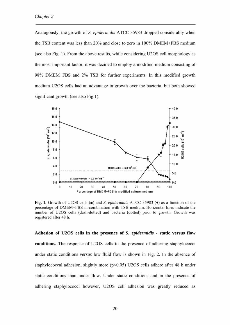
Chapter 2 Analogously, the growth of S. epidermidis ATCC 35983 dropped considerably when
the TSB content was less than 20% and close to zero in 100% DMEM+FBS medium
(see also Fig. 1). From the above results, while considering U2OS cell morphology as
the most important factor, it was decided to employ a modified medium consisting of
98% DMEM+FBS and 2% TSB for further experiments. In this modified growth
medium U2OS cells had an advantage in growth over the bacteria, but both showed
significant growth (see also Fig.1).
Fig. 1. Growth of U2OS cells (■) and S. epidermidis ATCC 35983 (♦) as a function of the percentage of DMEM+FBS in combination with TSB medium. Horizontal lines indicate the number of U2OS cells (dash-dotted) and bacteria (dotted) prior to growth. Growth was registered after 48 h.
Adhesion of U2OS cells in the presence of S. epidermidis - static versus flow
conditions. The response of U2OS cells to the presence of adhering staphylococci
under static conditions versus low fluid flow is shown in Fig. 2. In the absence of
staphylococcal adhesion, slightly more (p<0.05) U2OS cells adhere after 48 h under
static conditions than under flow. Under static conditions and in the presence of
adhering staphylococci however, U2OS cell adhesion was greatly reduced as
20

The race for the surface compared to the control, i.e. in the absence of adhering bacteria (p<0.05). Under
medium flow conditions, the number of adhering U2OS cells was significantly
reduced for both staphylococcal concentrations as compared to the control (p<0.05).
Fig. 2. Number of U2OS cells seeded on a glass substratum after 48 h under static conditions (■) and under medium flow (□) and in the absence and presence of adhering S. epidermidis ATCC 35983. Error bars represent the standard deviation over three replicates, with separate bacterial and cell cultures. *,° Significantly different (p<0.05) from the control (absence of staphylococci).
Under static conditions and irrespective of the number of S. epidermidis initially
present, floating granular particles were observed and interpreted as cell debris,
indicating U2OS cell death. In the presence of S. epidermidis, U2OS cells showed
better spreading and survival rate under flow as compared to static conditions. This is
due to the continuous flow, which likely removes the majority of bacterial endotoxins
produced. Bacterial endotoxin-induced cell death was confirmed by culturing U2OS
cells in growth medium consisting of 50% fresh modified growth medium and 50%
supernatant from a 48 h mixed U2OS cell and bacterial culture. No growth and
subsequent U2OS cell death was observed in this mix of fresh and spent medium,
21

Chapter 2 confirming that bacterial endotoxins are responsible for cell death during the race for
the surface under static conditions. Fluorescent dead-live stain, comprising of calcein-
AM and ethidium homodimer-1, also confirmed that adhering U2OS cells were alive
in the presence of S. epidermidis after 48 h under flow, as shown in Fig. 3.
Fig. 3. CSLM live-dead images of U2OS cells after 48 h on glass under medium flow in the absence and presence of adhering staphylococci. Bar denotes 75 µm.
Influence of the number of adhering S. epidermidis on adhesion and spreading of
U2OS cells under medium flow. Adhesion and spreading of U2OS cells after 1.5 and
48 h of growth in the presence of different number of adhering S. epidermidis are
shown in Fig. 4. After 48 h the number of adhering bacteria had also increased due to
growth, which made it difficult to analyze the cell number and spreading. For this
reason, the adhering U2OS cells were stained and analyzed by CLSM (see Fig. 5).
Fig. 4. U2OS cells adhesion and spreading after 1.5 h (top series) and 48 h (bottom series) under medium flow on a glass substratum in the absence and presence of adhering staphylococci. Bar denotes 75 µm. U2OS cells are differentiated by a contour line from S. epidermidis biofilm.
22

The race for the surface
U2OS cells showed highest spreading in the absence of staphylococci. The
extent of spreading was reduced with increasing density of adhering staphylococci
and in the presence of adhering staphylococci many non-spread U2OS cells were
observed. Quantitative analysis of cell spreading in the absence and presence of
staphylococci is shown in Fig. 6. After the initial adhesion of U2OS cells for 1.5 h,
the average area of U2OS cells on all surfaces was between 250 µm2 and 380 µm2 per
cell. After 48 h of flow, significant differences were observed in the average area per
cell as a result of varying densities of adhering S. epidermidis. In the absence of S.
epidermidis, the average area of a spread cell was 960 µm2 after 48 h, but in the
presence of 105 staphylococci cm-2 spreading decreased to approximately 390 µm2 per
cell.
Fig. 5. CLSM images of U2OS cells after 48 h on glass under medium flow in the absence and presence of adhering staphylococci. U2OS cells were stained with 5 ml PBS containing 49 µl DAPI and 2µg ml-1 of TRITC-Phalloidin. Bar denotes 75 µm.
23

Chapter 2
Fig. 6. Average area per spread U2OS cell after 1.5 h (□) and 48 h (■) of growth on glass under medium flow in the absence and presence of adhering S. epidermidis ATCC 35983. Error bars represent the standard deviation over three replicates with separate bacterial and cell cultures. * Significantly different (p<0.05) from the control (absence of staphylococci).
Discussion
This paper presents the first experimental set-up to study the race between bacteria
and tissue cells for a biomaterial surface in vitro. The need for such a system is
enormous at current, as many coatings that are propagated to attract low numbers of
adhering bacteria, such as polymer brush coatings [15,20], may also be expected to
support poor adhesion and spreading of tissue cells, which is currently stimulating the
development of bi-functional coatings that support cell spreading and repel
microorganisms at the same time. This development necessitates the use of a
methodology, as described in this manuscript. Under static culturing conditions,
U2OS cells did not have a chance to win the race for the surface and detached from
the surface in the presence of adhering staphylococci. Under medium flow, however,
U2OS cells remained adhering in the presence of adhering staphylococci and spread
24

The race for the surface more when the density of adhering staphylococci was lower. Therewith we have
presented a method for the evaluation of biomaterial coatings that encompasses tissue
cell adhesion and spreading as well as bacterial growth.
The effects of two different densities of adhering bacteria were evaluated in
this study. Bacteria were allowed to adhere prior to cell adhesion and spreading,
which is a fine-tuning of the model toward peri-operative bacterial contamination of
implant surfaces. It has been documented that during an operation procedure of 1 h
[21], the total number of bacteria-carrying particles falling on a wound is about 270
cm-2, while the risk of infection depends on the number of viable bacteria present in
the wound area at the time of wound closure. Bacterial counts are generally higher
during periods of activity or during increased numbers of personnel in the operation
theatre [21]. The presence of a biomaterial implant in the body usually stimulates
infection by a smaller inoculum of bacteria than in non-biomaterial surgery [22].
Hence, the densities of adhering bacteria prior to cell adhesion employed in this study
between 102 and 105 bacteria cm-2 may be considered relevant for minimum and
maximum contaminations occurring clinically.
U2OS cell response was studied under static conditions and flow. In vivo, fluid
is continuously flowing through the network of fine channels of osteocytes to
facilitate the diffusion of nutrients and waste products from the bone surface to deeply
buried osteocytes and vice versa [23]. Using computer simulation, Klein-Nulend and
co-workers [24] showed that flow rates are low in the region of bone immediately
ahead of the basic multicellular units. They also showed that cells die in this stagnant
area, possibly because of lack of nutrients. Similarly in our study under static
conditions, cell death was observed irrespective of different densities of adhering
bacteria. Under flow, detachment of cells was not observed and cells were alive due to
25
Chapter 2 the continuous incoming of fresh medium and removal of endotoxins. Hence,
conducting the race for a biomaterial surface between bacteria and tissue cells in vitro
under low flow may be considered clinically relevant, although exact flow rates as
occurring in vivo are unknown.
Several researchers predict the outcome of the race for the surface by studying
the bacteria-surface interactions and tissue cell-surface interactions separately, but this
is not how we interpret Gristina’s meaning of “the race for the surface”, neither do we
think this is the right way to study the possible fate of a biomaterial implant in vitro.
Many biomaterial coatings have been identified as non-adhesiveness to bacteria or
support tissue integration separately [6,13-15,17,18], but the combination of both
bacteria and tissue cells on the same biomaterials surface has never been studied. Shi
et al. [16] for instance, promoted a surface composed of chitosans and Arginine-
Glycine-Aspartic acid (RGD) peptide sequences. In separate experiments, it was
shown that these combined surfaces discouraged bacterial adhesion, and enhanced cell
attachment and Alkaline Phosphatase (ALP) activity. However, effects of bacterial
presence, including the influence of bacterial activities and toxins [12] on the tissue
cell attachment were not studied, which could completely change the fate of a
biomaterial implant according to the concept of “race for the biomaterial surface”.
The methodology forwarded here allows simultaneous growth of bacteria and tissue
cells on the same biomaterial surface and will be useful for the evaluation of new
functional and biomimetic surfaces.
Conclusion
A novel in vitro methodology to study the race between bacteria and tissue cells for a
biomaterial surface has been developed. Although due to the cell type chosen and use
26

The race for the surface of a staphylococcal strain, the methodology as described here may seem geared
toward orthopedic applications, we emphasize that its principles are equally
applicable to other implant systems, such as surgical meshes, or vascular grafts. Both
the absence and presence of flow, as well as the number of adhering bacteria appeared
to determine whether tissue cells were able to grow on a biomaterial surface. The
methodology forwarded is expected to become indispensable for in vitro evaluation of
bi-functionalized, biomimetic biomaterial coatings currently being developed in
different groups worldwide.
27

Chapter 2 References
1. Campoccia D, Montanaro L, Arciola CR. The significance of infection related to orthopedic devices and issues of antibiotic resistance. Biomaterials 2006;27:2331-2339.
2. Trampuz A, Zimmerli W. New strategies for the treatment of infections associated with prosthetic joints. Curr Opin Investig Drugs 2005;6:185-190.
3. Calhoun JH, Klemm K, Anger DM, Mader JT. Use of antibiotic-PMMA beads in the ischemic foot. Orthopedics 1994;17:453-457.
4. Darouiche RO. Treatment of infections associated with surgical implants. N Engl J Med 2004;350:1422-1429.
5. Mohr VD, Eickhoff U, Haaker R, Klammer HL. External fixation of open femoral shaft fractures. J Trauma 1995;38:648-652.
6. Gristina AG, Rovere GD, Shoji H, Nicastro JF. An in vitro study of bacterial response to inert and reactive metals and to methyl methacrylate. J Biomed Mater Res 1976;10:273-281.
7. Gristina AG, Naylor PT, Myrvik QN. Musculoskeletal infection, microbial adhesion, and antibiotic resistance. Infect Dis Clin North Am 1990;4:391-408.
8. Ahlberg A, Carlsson AS, Lindberg L. Hematogenous infection in total joint replacement. Clin Orthop Relat Res 1978;137:69-75.
9. Gristina AG. Biomaterial-centered infection: microbial adhesion versus tissue integration. Science 1987;237:1588-1595.
10. Lidwell OM, Lowbury EJ, Whyte W, Blowers R, Stanley SJ, Lowe D. Airborne contamination of wounds in joint replacement operations: the relationship to sepsis rates. J Hosp Infect 1983;4:111-131.
11. Gristina AG, Shibata Y, Giridhar G, Kreger A, Myrvik QN. The glycocalyx, biofilm, microbes, and resistant infection. Semin Arthroplasty 1994;5:160-170.
12. Gristina AG, Naylor P, Myrvik Q. Infections from biomaterials and implants: a race for the surface. Med Prog Technol 1988;14:205-224.
13. Dexter SJ, Pearson RG, Davies MC, Camara M, Shakesheff KM. A comparison of the adhesion of mammalian cells and Staphylococcus epidermidis on fibronectin-modified polymer surfaces. J Biomed Mater Res 2001;56:222-227.
14. Harris LG, Tosatti S, Wieland M, Textor M, Richards RG. Staphylococcus aureus adhesion to titanium oxide surfaces coated with non-functionalized and peptide-functionalized poly(L-lysine)-grafted-poly(ethylene glycol) copolymers. Biomaterials 2004;25:4135-4148.
28

The race for the surface
29
15. Maddikeri RR, Tosatti S, Schuler M, Chessari S, Textor M, Richards RG, Harris LG. Reduced medical infection related bacterial strains adhesion on bioactive RGD modified titanium surfaces: A first step toward cell selective surfaces. J Biomed Mater Res Part A 2008;84A:425-435.
16. Shi ZL, Neoh KG, Kang ET, Poh C, Wang W. Bacterial adhesion and osteoblast function on titanium with surface-grafted chitosan and immobilized RGD peptide. J Biomed Mater Res Part A 2008;86A:865-872.
17. Shi Z, Neoh KG, Kang ET, Poh C, Wang W. Titanium with surface-grafted dextran and immobilized bone morphogenetic protein-2 for inhibition of bacterial adhesion and enhancement of osteoblast functions. Tissue Eng Part A 2009;15:417-426.
18. Lussi JW, Falconnet D, Hubbell JA, Textor M, Csucs G. Pattern stability under cell culture conditions--a comparative study of patterning methods based on PLL-g-PEG background passivation. Biomaterials 2006;27:2534-2541.
19. Busscher HJ, Van der Mei HC. Microbial adhesion in flow displacement systems. Clin Microbiol Rev 2006;19:127-141.
20. Fundeanu I, Van der Mei HC, Schouten AJ, Busscher HJ. Polyacrylamide brush coatings preventing microbial adhesion to silicone rubber. Colloids Surf B-Biointerfaces 2008;64:297-301.
21. Fitzgerald RH. Microbiologic environment of the conventional operating-room. Arch Surg 1979;114:772-775.
22. Gristina AG. Implant failure and the immuno-incompetent fibro-inflammatory zone. Clin Orthop Relat Res 1994;298:106-118.
23. Jacobs CR, Yellowley CE, Davis BR, Zhou Z, Cimbala JM, Donahue HJ. Differential effect of steady versus oscillating flow on bone cells. J Biomechanics 1998;31:969-976.
24. Klein-Nulend J, Bacabac RG, Mullender MG. Mechanobiology of bone tissue. Pathol Biol (Paris) 2005;53:576-580.

CHAPTER 3
Microbial Biofilm Growth versus Tissue Integration on Biomaterials with Different Wettabilities and a
Polymer-Brush Coating
Guruprakash Subbiahdoss, Dirk W. Grijpma, Henny C. van der Mei,
Henk J. Busscher, Roel Kuijer
Journal of Biomedical Materials Research Part A 2010; 94: 533-538
Republished with the permission of Wiley Periodicals, Inc
Chapter 3
Introduction
Biomaterial implants are indispensable in human function restoration after damage to
the human body beyond natural repair. The number one cause of failure of biomaterial
implants is infection, partly as a cause of unsuccessful tissue integration.
Microorganisms involved in biomaterial-associated infection (BAI) are resistant to
antibiotics due to their biofilm mode of growth, and infected implants often have to be
removed before the infection can be fully eradicated from surrounding tissue and a
new implant can be inserted. On average, BAI occurs in approximately 0.5 – 6% of all
primary implant patients [1]. BAI has an at least two- to three-fold higher incidence in
revision surgery. Primary implants can become contaminated with microorganisms
during implant surgery (peri-operative contamination) or hospitalization [2], as the
onset of BAI. Whether or not microbial contamination eventually results in BAI,
depends on the outcome of the so-called ‘race for the surface’ between successful
tissue integration of the biomaterial implant and biofilm growth [2]. If this race is won
by tissue cells, then the biomaterial surface is covered by a cellular layer and less
vulnerable to biofilm formation. On the other hand, if the race is won by bacteria, the
implant surface will become colonized by bacteria and tissue cell functions are
hampered by bacterial virulence factors and toxins [2,3]. Since microorganisms are
frequently introduced on an implant surface during surgery, microorganisms have a
head start in this race for the surface.
A better understanding of the combined interaction of tissue cells and bacteria
on biomaterial surfaces is required in order to develop new biomaterials or functional
coatings that resist bacterial adhesion and biofilm formation [4-8] and simultaneously
support tissue cell adhesion and proliferation [5-9]. Since the development of
biomaterials and functional coatings, bacterial adhesion and biofilm formation are
32

The race for the surface on different biomaterials studied independently from tissue cell adhesion and proliferation, and the combined
outcome of these two interactions, i.e. the race for the surface, remains unknown.
Recently, an in vitro experimental methodology to investigate the race between
bacteria and tissue cells in a single experiment has been forwarded [10]. The outcome
of the race for the surface between staphylococci and tissue cells appeared dependent
on the number of bacteria present prior to cell seeding and the absence or presence of
fluid flow. Cells lost the race for the surface in the absence of flow due to
accumulation of bacterial endotoxins, but were able to grow in the presence of flow
due to continuous incoming of fresh medium and removal of endotoxins by the flow
[10].
Many biomaterials that are often used in the clinic have not yet been
investigated with respect to the influence of their surface properties on the outcome of
the race for the surface between bacteria and tissue cells. Surface wettability is one of
the important properties influencing bacterial or cellular interactions with
biomaterials. Gottenbos et al. [11]. showed relatively similar bacterial adhesion
between materials with different wettabilities. Schakenraad et al. [12]. reported that
tissue cells spread best on wettable and poorly on less wettable surfaces. Therefore the
aim of this paper is to determine the influence of wettability on the outcome of the
race for the surface on different biomaterial surfaces. In addition, a surface coated
with a hydrophilic polymer-brush is included, since these have been shown to
discourage microbial adhesion and biofilm formation [13].
Materials and Methods
Biomaterial surfaces. Polyethylene (PE) (Goodfellow, Cambridge, UK),
Poly(tetrafluoroethylene-co-hexafluoropropylene) (FEP) (Fluorplast, Raamsdonkveer,
33

Chapter 3
The Netherlands), poly (methyl methacrylate) (PMMA) (Vink Kunststoffen, Didam,
The Netherlands), Polystyrene (PS) (Colltec, Groningen, The Netherlands), High-
Throughput microArraying, multifunctional slide (HTA) (Greiner Bio One, Alphen
aan den Rijn, The Netherlands) and glass coated with a hydrophilic polyethylene
oxide (PEO) layer were used. All samples except hydrophilic PEO-coating were
rinsed thoroughly with ethanol (Merck, Darmstadt, Germany) and washed with sterile
ultrapure water before use.
Hydrophilic PEO-coated glass (polymer-brush coating) was prepared by first
cleaning microscope glass slides in a 2% RBS35 detergent solution (Omniclean,
Breda, The Netherlands) under sonication and thorough rinsing in demineralized
water, methanol, water again and finally washed with sterile ultrapure water. Glass
surfaces were made hydrophobic by application of a dimethyldichlorosilane coating
(DDS, Merck, Germany), yielding a water contact angle of 107 ± 2 degrees. Exposure
to a solution of 0.5 g l-1 Pluronic F-127 solution (PEO99PPO65PEO99, molecular
weight 12600; Sigma-Aldrich, USA) in phosphate buffered saline (PBS: 10 mM
potassium phosphate, 0.15 M NaCl, pH 7.0) for 20 min created a hydrophilic
polymer-brush coating, that has appeared stable and effective against bacterial
adhesion for at least 48 h [13].
Biomaterial surface characterization. The wettability of the surfaces was
determined by water contact angle measurements at room temperature with an image
analyzing system, using the sessile drop technique. Each value was obtained by
averaging five droplets on one sample.
The elemental surface composition of the biomaterial surfaces was measured
using X-ray Photoelectron Spectroscopy (XPS). The S-probe spectrometer (Surface
34

The race for the surface on different biomaterials Science Instruments. Mountain View, CA, USA) was equipped with an aluminium
anode (10 kV, 22 mA) and a quartz monochromator. The direction of photoelectron
collection angle was 55 degrees with the sample surface and the electron flood gun
was set at 10 eV. Broad spectrum survey scans (binding energy range of 1 to 1100
eV) were made at low resolution (pass energy, 150 eV). The area under each peak was
used to calculate peak intensities, yielding elemental surface concentrations for
carbon, oxygen, nitrogen, fluorine and silicon, after correction with sensitivity factors
provided by the manufacturer. Elemental surface compositions were expressed in
atom percentages of carbon, oxygen, silicon, fluorine and/or nitrogen. Two separate
measurements were taken on different spots of each biomaterial.
U2OS cell culturing and harvesting. U2OS osteosarcoma cells were routinely
cultured in Dulbecco’s modified Eagle’s Medium (DMEM) -low glucose
supplemented with 10% fetal calf serum (FBS), 0.2 mM of ascorbic acid-2-phosphate
(AA2P) and denoted in the paper as DMEM+FBS. U2OS cells were maintained at
37°C in a humidified 5% CO2 atmosphere, and cells were passaged at 70 – 90%
confluency using trypsin/EDTA.
Bacterial growth conditions and harvesting. Staphylococcus epidermidis ATCC
35983, originally isolated from human blood of a patient with an infected
intravascular catheter, was used throughout this study. First, the strain was streaked on
a blood agar plate from a frozen stock and grown overnight at 37°C. The plate was
then kept at 4°C. For each experiment, a colony was inoculated in 10 ml of tryptone
soy broth (TSB; OXOID, Basingstoke, United Kingdom) and cultured for 24 h. This
culture was used to inoculate a second culture in TSB, which was grown for 17 h prior
35

Chapter 3
to harvesting. Bacteria were harvested by centrifugation at 5000 x g for 5 min at 10°C
and washed twice with sterile ultrapure water. Subsequently, the harvested bacteria
were sonicated on ice (3 x 10 s) in sterile PBS (10 mM potassium phosphate, 0.15 M
NaCl, pH 7.0) in order to break bacterial aggregates. This suspension was further
diluted in sterile PBS to a concentration of 3 x 10⁶ bacteria per ml.
The race for the surface. The race for the surface was studied on the bottom plate of
a parallel plate flow chamber (175 x 17 x 0.75 mm3) prepared from the biomaterials
or coating under investigation, as described in detail before [10]. The flow chamber
was equipped with heating elements and kept at 37°C throughout the experiments.
Bacterial and U2OS deposition were observed with a CCD camera (Basler AG,
Germany) mounted on a phase-contrast microscope Leica DM2000 (Leica
Microsystems Ltd, Germany) with a 30x objective for bacteria and 10x objective for
tissue cells.
Prior to each experiment, all tubes and the flow chamber were filled with
sterile PBS, taking care to remove all air bubbles from the system. Once the system
was filled, and before the addition of the bacterial suspension, PBS was allowed to
flow through the system at a shear rate of 11 s-1. Then, the bacterial suspension in
PBS was perfused through the chamber at the same shear rate and phase-contrast
images were obtained and image-analyzed as a function of time. As soon as the
desired density of adhering bacteria (103 cm-2 or 105 cm-2), was reached, flow was
switched to sterile PBS to remove the bacterial suspension from the tubes and
chamber. Subsequently, a U2OS cell suspension in modified culture medium,
consisting of 98% DMEM+FBS and 2% TSB and suitable for the simultaneous
growth of U2OS cells and S. epidermidis [10], was allowed to enter the flow chamber.
36

The race for the surface on different biomaterials Once the entire volume of buffer inside the chamber was replaced by the cell
suspension, flow was stopped for 1.5 h in order to allow tissue cells to adhere and
spread on the substratum. Subsequently, phase contrast images (6 images, 900 x 700
µm each) were taken and the number of adhering cells per unit area and area per
spread cell were determined using Scion image software. Finally, modified culture
medium supplemented with 2% HEPES was perfused through the system at a low
shear rate of 0.14 s-1 for 48 h without recirculation, and phase-contrast images were
obtained continuously.
Immuno-cytochemical staining. After 48 h of flow, the biomaterial surfaces or
coating were prepared for immuno-cytochemical staining to assess the tissue cell
morphology and spreading. For fixation, surfaces with adhering bacteria and tissue
cells were placed in a Petri dish with 30 ml of 3.7% formaldehyde in cytoskeleton
stabilization buffer (CS; 0.1 M Pipes, 1 mM EGTA, 4% (w/v) polyethylene glycol
8000, pH 6.9). After 5 min, the fixation solution was replaced by 30 ml of fresh CS
for another 5 min. Subsequently tissue cells were incubated in 0.5% Triton X-100 for
3 min, rinsed with PBS and stained for 30 min with 5 ml PBS containing 49 µl DAPI
and 2 µg ml-1 of TRITC-Phalloidin. The cells on the surfaces were washed 4 times in
PBS and examined with confocal laser scanning microscopy (CLSM, Leica DMRXE
with confocal TCS SP2 unit). The number of adhering tissue cells per unit area and
the average area per spread cell were determined using Scion image software. For
each density of adhering bacteria, 6 images (900 x 700 µm) per sample were
randomly chosen and analyzed.
37

Chapter 3
Statistics. Experiments for each density of adhering bacteria on different surfaces
were carried out in triplicate. Data are represented as a mean with standard deviation.
Statistical ANOVA analysis was performed followed by a Tukey’s HSD post-hoc test
and a P-value of <0.05 was considered significant.
Results
Biomaterials wettability and surface composition. The water contact angles and
elemental surface compositions of the biomaterials and polymer-brush coating
evaluated are summarized in Table 1. The biomaterial surfaces extend over a
wettability range from 36 to 103 degrees, and are composed for the major part of
carbon and oxygen, with the exception of FEP, containing fluorine and HTA,
containing nitrogen. The polymer-brush coating has a wettability of 41 degrees, and
shows some silicon, originating from the glass underneath the thin polymer-brushes
consisting of carbon and oxygen.
Table 1. Water contact angle and elemental surface compositions of biomaterials and PEO-coating evaluated. ± indicates the standard deviation over three independently prepared end measured samples.
Biomaterial (degrees)
%C %O %F %N %Si
HTA 36 ± 3 80.1 ± 2.0 16.9 ± 0.6 - 3.1 ± 1.2
-
PMMA 73 ± 3 73.2 ± 0.2 25.9 ± 1.4 - - - PS 80 ± 2 94.8 ± 2.2 5.2 ± 2.2 - - - PE 95 ± 2 97.2 ± 1.2 2.8 ± 1.2 - - -
FEP 103 ± 1 27.6 ± 0.3 0.6 ± 0.3 72.1 ± 0.8
- -
Polymer-brush coating 1)
41 ± 5 59.5 ± 0.8 33.6 ± 0.3 - - 6.9 ± 0.9
1) data taken from Roosjen et al. [14].
38

The race for the surface on different biomaterials U2OS cell adhesion and spreading in the absence and presence of adhering S.
epidermidis. Immediately after seeding, U2OS cell adhesion and spreading was
observed in the absence and presence of adhering staphylococci on all biomaterial
surfaces evaluated, but not on the polymer-brush coating. At 1.5 h, there was no
significant difference between the number of adhering U2OS cells on the biomaterials
(25000 cells cm-2 on average), with the exception of FEP where far less adhering
tissue cells were found (approximately 12000 cells cm-2). On the polymer-brush
coating, however, the number of U2OS cells on the surface was 25000 cells cm-2 on
average at 1.5 h, but these tissue cells adhered loosely and were removed at the
applied shear rate of 0.14 s-1. Phase-contrast images of U2OS cell adhesion, spreading
and simultaneous biofilm formation of S. epidermidis after 24 h of growth on the
biomaterials evaluated and the polymer-brush coatings are shown in Fig. 1. On the
biomaterials, adhering staphylococci had grown into a biofilm, whereas at the same
time U2OS cell adhesion and spreading were observed. On the polymer-brush
coating, the adhering U2OS cells that could withstand the applied shear, did not
spread and remained rounded even after 24 h (see Fig. 1).
39

Chapter 3
Fig. 1. U2OS cell spreading and staphylococcal biofilm formation (initial number of staphylococci present is 103 cm-2) after 24 h of growth on biomaterials evaluated (HTA = High-Throughput microArraying, multifunctional slide, PMMA = Poly (methyl methacrylate), PS = Polystyrene, PE = Polyethylene, FEP = Poly (tetrafluoroethylene-co-hexafluoropropylene)) and a polymer-brush coating. Bar denotes 10 µm.
After 48 h of growth, the number of adhering bacteria had increased to the
extent that it impeded quantification of cellular adhesion and spreading and hence
adhering U2OS cells were immunocyto-stained for CLSM analysis (see Fig. 2) to
40

The race for the surface on different biomaterials derive the number and spread area of U2OS cells in the absence and presence of
staphylococci. In Fig. 3 it can be seen, that the % increase in number of adhering
U2OS cells was significantly reduced due to the presence of adhering staphylococci
on all biomaterials surfaces as compared to the control, i.e. in the absence of adhering
bacteria (p<0.05). The number of adhering U2OS cells on hydrophobic FEP showed a
significant reduction (p<0.01) in % increase with respect to the initial number of S.
epidermidis present.
Fig. 2. CLSM images of immunocyto-stained U2OS cells after 48 h of growth on the biomaterials evaluated (HTA = High-Throughput microArraying, multifunctional slide, PMMA = Poly (methyl methacrylate), PS = Polystyrene, PE = Polyethylene, FEP = Poly (tetrafluoroethylene-co-hexafluoropropylene)) and a polymer-brush coating in the absence (no staphylococci) and presence of adhering staphylococci (initial number of adhering staphylococci amounted either 103 cm-2 or 105 cm-2). Bar denotes 75 µm.
41

Chapter 3
Fig. 3. Percentage increase in the number of adhering U2OS cells after 48 h of growth with respect to their initial adhesion at 1.5 h as a function of the water contact angles of the biomaterials evaluated in the absence (●) and presence (■ – 103 cm-2, ▲ – 105 cm-2) of adhering S. epidermidis ATCC 35983. Error bars represent the standard deviation over three replicates, with separately cultured bacteria and tissue cells.
U2OS cells showed maximum spreading in the absence of staphylococci on all
biomaterials evaluated irrespective of their wettabilities, as compared to the presence
of staphylococci. U2OS cells spread best on hydrophilic surfaces (HTA and PMMA)
and showed the least spreading on the hydrophobic FEP surface. It is interesting to see
that the adhering tissue cells, spread equally well in the presence of staphylococci as
in their absence (see Fig. 4). Note that the total surface coverage of the U2OS cells is
different in the presence of staphylococci, which is caused by a lower number of the
adhering bacteria (see Fig. 2). Tissue cell spreading only appeared hampered due to
the presence of adhering staphylococci on the most hydrophobic surface, FEP. On the
polymer-brush coating, no increase in the number of adhering U2OS cells was
observed, neither did the adhering cells spread within 48 h, irrespective of the absence
or presence of staphylococci.
42

The race for the surface on different biomaterials
Fig. 4. Average area per spread U2OS cell after 48 h of growth as a function of the water contact angles of the biomaterial evaluated in the absence (●) and presence (■ – 103 cm-2, ▲ – 105 cm-2) of adhering S. epidermidis ATCC 35983. Error bars represent the standard deviation over three replicates, with separately cultured bacteria and tissue cells.
Discussion
This paper presents the first experimental study on the race between bacteria and
tissue cells on biomaterials with different wettabilities as well as on polymer – brush
coated glass. Two different densities of bacteria (103 and 105 bacteria cm-2) were
allowed to adhere prior to cell seeding, adhesion and spreading, which mimicks a
situation of peri-operative bacterial contamination of implant surfaces. In the past, it
has been documented that during a surgical procedure of 1 h, the total number of
bacteria carrying particles falling on the wound is approximately 270 per cm2. The
bacterial counts are generally higher during the periods of activity and correlate
positively with the number of personnel present in the operation theatre [15]. In more
recent, conventionally ventilated operation theatre (20 changes of air per hour) and the
use of impermeable patient and personnel clothing [16], bacterial contamination will
be far less, which makes, the bacterial adhesion densities chosen in our experiments a
worst case scenario.
43

Chapter 3
Amongst other material properties, surface wettability plays a major role in
bacterial or cellular interaction with biomaterials. Wettability of biomaterial surfaces
has been related to bacterial adhesion, cell adhesion and spreading. Gottenbos et al.
[11]. showed that adhesion of staphylococci is relatively similar on different
biomaterials, such as PMMA, PE and FEP, irrespective of differences in wettability.
Likewise, these biomaterials in our study showed similar biofilm formation
irrespective of their differences in wettability. The adhesion of staphylococci on the
polymer – brush coating was slow and not as strong as on the biomaterials evaluated.
A recent study by Nejadnik et al. [13]. showed that the polymer – brush coatings
reduced adhesion of staphylococci considerably but the few adhered bacteria still
formed a biofilm when allowed to grow. This biofilm formed on the polymer – brush
coating detached when exposed to high fluid shear [13].
In case of tissue cells, many papers [12,17-19] have suggested that optimal
tissue cell adhesion and spreading occurs at intermediate wettability (water contact
angle between 60 degrees and 80 degrees). Nevertheless, surfaces that are poorly
wettable still support appreciable cell adhesion [12]. Similarly, on the biomaterials, in
the absence of staphylococci, U2OS cells showed the best adhesion on hydrophilic
HTA and PMMA and appreciable cell adhesion on hydrophobic FEP (see Fig. 3). The
percentage adhesion of tissue cells with respect to surface wettabilities of biomaterials
followed the same trend as demonstrated earlier [20-23]. It is surprising that the
poorly wettable polymer PE was capable of supporting significant cell attachment and
spreading. This was also reported by Lydon et al. [23]. in the previous study. U2OS
cells showed no adhesion and spreading on a polymer – brush coating.
A combined bacterial and tissue cell adhesion and growth study on biomaterial
surfaces is novel. This study showed that in the presence of staphylococci, U2OS cell
44

The race for the surface on different biomaterials adhesion in terms of numbers of adhering cells, reduced significantly on all
biomaterial surfaces evaluated, as compared to a control, i.e. in the absence of
adhering bacteria (p<0.05). Spreading of adhering tissue cells was less affected by the
presence of bacteria except on hydrophobic FEP. This demonstrates that tissue cell
interactions with biomaterials are hampered by bacterial presence on all biomaterials,
establishing a tight race between bacteria and tissue cells for the biomaterial surface.
Clinically, this is the reason why antibiotics need to be used in order to substantially
reduce the risk of post-operative infection or BAI [24]. The polymer – brush coating
showed both reduced bacterial adhesion and tissue cell adhesion and spreading, which
makes it unsuitable for implant coatings, albeit for a different reason than valid for the
biomaterial surfaces evaluated. Therefore, this study emphasizes the need for the
development of bi-functional surfaces, discouraging bacterial adhesion and growth
and simultaneously supporting tissue cell adhesion and spreading.
Conclusion
This study demonstrates that tissue cell interactions with biomaterials were hampered
by biofilm formation on biomaterial surfaces and in fact most on a hydrophobic FEP
surface. This indicates that the race for the biomaterial surface a tight one, as can be
inferred from the relatively high rate of occurrence of BAI. As such, neither
hydrophobic nor hydrophilic surfaces aid in a decisive way to determine the outcome
of the race for the surface. Polymer-brush coatings appear less promising in
determining the race for the surface than expected on the reductions observed in
bacterial adhesion and growth on polymer-brush coatings, because they do not
support tissue cell adhesion and spreading. Surfaces, on which the race for the surface
can be won with certainty by the tissue cells, do not yet exist. Biomaterials
45

Chapter 3
engineering should aim for bi-functional surfaces, discouraging bacterial adhesion and
growth, but at the same time supporting tissue adhesion and spreading. Hitherto, these
two requirements have not been met by any of the biomaterials surfaces currently
used for biomedical implants and devices.
46

The race for the surface on different biomaterials References
1. Trampuz A, Zimmerli W. New strategies for the treatment of infections associated with prosthetic joints. Curr Opin Investig Drugs 2005;6:185-190.
2. Gristina AG. Biomaterial-centered infection: microbial adhesion versus tissue integration. Science 1987;237:1588-1595.
3. Gristina AG, Naylor P, Myrvik Q. Infections from biomaterials and implants: a race for the surface. Med Prog Technol 1988;14:205-224.
4. Gristina AG, Dobbins JJ, Giammara B, Lewis JC, DeVries WC. Biomaterial-centered sepsis and the total artificial heart. Microbial adhesion vs tissue integration. JAMA 1988;259:870-874.
5. Dexter SJ, Pearson RG, Davies MC, Camara M, Shakesheff KM. A comparison of the adhesion of mammalian cells and Staphylococcus epidermidis on fibronectin-modified polymer surfaces. J Biomed Mater Res 2001;56:222-227.
6. Harris LG, Tosatti S, Wieland M, Textor M, Richards RG. Staphylococcus aureus adhesion to titanium oxide surfaces coated with non-functionalized and peptide-functionalized poly(L-lysine)-grafted-poly(ethylene glycol) copolymers. Biomaterials 2004;25:4135-4148.
7. Maddikeri RR, Tosatti S, Schuler M, Chessari S, Textor M, Richards RG, Harris LG. Reduced medical infection related bacterial strains adhesion on bioactive RGD modified titanium surfaces: A first step toward cell selective surfaces. J Biomed Mater Res A 2008;84A:425-435.
8. Shi ZL, Neoh KG, Kang ET, Poh C, Wang W. Bacterial adhesion and osteoblast function on titanium with surface-grafted chitosan and immobilized RGD peptide. J Biomed Mater Res A 2008;86A:865-872.
9. Lussi JW, Falconnet D, Hubbell JA, Textor M, Csucs G. Pattern stability under cell culture conditions - A comparative study of patterning methods based on PLL-g-PEG background passivation. Biomaterials 2006;27:2534-2541.
10. Subbiahdoss G, Kuijer R, Grijpma DW, Van der Mei HC, Busscher HJ. Microbial biofilm growth vs. tissue integration: "The race for the surface" experimentally studied. Acta Biomater 2009;5:1399-1404.
11. Gottenbos B, Van der Mei HC, Busscher HJ. Initial adhesion and surface growth of Staphylococcus epidermidis and Pseudomonas aeruginosa on biomedical polymers. J Biomed Mater Res 2000;50:208-214.
12. Schakenraad JM, Busscher HJ, Wildevuur CR, Arends J. Thermodynamic aspects of cell spreading on solid substrata. Cell Biophys 1988;13:75-91.
13. Nejadnik MR, Van der Mei HC, Norde W, Busscher HJ. Bacterial adhesion and growth on a polymer brush-coating. Biomaterials 2008;29:4117-4121.
47

Chapter 3
48
14. Roosjen A, Kaper HJ, Van der Mei HC, Norde W, Busscher HJ. Inhibition of adhesion of yeasts and bacteria by poly(ethylene oxide)-brushes on glass in a parallel plate flow chamber. Microbiology 2003;149:3239-3246.
15. Fitzgerald RH. Microbiologic environment of the conventional operating-room. Arch Surg 1979;114:772-775.
16. Verkkala K, Eklund A, Ojajarvi J, Tiittanen L, Hoborn J, Makela P. The conventionally ventilated operating theatre and air contamination control during cardiac surgery - bacteriological and particulate matter control garment options for low level contamination. Eur J Cardiothorac Surg 1998;14:206-210.
17. Kishida A, Iwata H, Tamada Y, Ikada Y. Cell behavior on polymer surfaces grafted with nonionic and ionic monomers. Biomaterials 1991;12:786-792.
18. Tamada Y, Ikada Y. Fibroblast growth on polymer surfaces and biosynthesis of collagen. J Biomed Mater Res 1994;28:783-789.
19. Van Wachem PB, Beugeling T, Feijen J, Bantjes A, Detmers JP, Vanaken WG. Interaction of cultured human-endothelial cells with polymeric surfaces of different wettabilities. Biomaterials 1985;6:403-408.
20. Ikada Y. Surface modification of polymers for medical applications. Biomaterials 1994;15:725-736.
21. Saltzman WM, Parsonswingerter P, Leong KW, Lin S. Fibroblast and hepatocyte behavior on synthetic-polymer surfaces. J Biomed Mater Res 1991;25:741-759.
22. Van Wachem PB, Hogt AH, Beugeling T, Feijen J, Bantjes A, Detmers JP et al. Adhesion of cultured human-endothelial cells onto methacrylate polymers with varying surface wettability and charge. Biomaterials 1987;8:323-328.
23. Lydon MJ, Minett TW, Tighe BJ. Cellular interactions with synthetic-polymer surfaces in culture. Biomaterials 1985;6:396-402.
24. Hoffmann CEJ, Mcdonald PJ, Watts JM. Use of peroperative cefoxitin to prevent infection after colonic and rectal surgery. Ann Surg 1981;193:353-356.

CHAPTER 4
Bacterial Strain-Specific Effects on Mammalian Cell Growth on Poly(methyl methacrylate)

Chapter 4
Introduction
Biomaterial-associated infection (BAI) is a serious problem in modern medicine. BAI
is often difficult to treat, as the biofilm mode of growth protects the infecting
organisms against the host defense system and antibiotics [1]. In most cases, the final
outcome is removal of the infected implant. Biomaterial implants can become
contaminated in different ways. The best documented route is direct contamination of
the implant surface during surgery (peri-operative contamination) or contamination
during hospitalization. Whether or not microbial contamination eventually results in
BAI, depends on the outcome of the so-called ‘race for the surface’ between
successful tissue integration of the implant surface and biofilm formation [2]. If this
race is won by tissue cells, then the implant surface is covered by a cellular layer and
less vulnerable to biofilm formation. On the other hand, if the race is won by bacteria,
the implant surface will become colonized by bacteria and tissue cell functions are
hampered by bacterial virulence factors and toxins [2,3]. Since microorganisms are
frequently introduced on an implant surface during surgery, microorganisms have a
head start in this race for the surface. In the concept of the race for the surface, full
coverage of an implant surface in vivo by a viable tissue cell layer, intact cell
membrane and functional host defense mechanisms resist biofilm formation [4].
Previously, we proposed an in vitro model to experimentally determine the
influence of peri-operative bacterial contamination on the race for the surface, in
which adhesion, spreading and growth of mammalian cells on a biomaterial surface is
compared in the absence or presence of adhering bacteria [5]. The outcome of the
competition between contaminating Staphylococcus epidermidis ATCC 35983 and
U2OS cells on glass appeared to be dependent on the number of bacteria adhering
prior to mammalian cell seeding and the absence or presence of fluid flow. Cells lost
50

Bacterial strain-specific effects on mammalian cells the competition in the absence of flow due to accumulation of bacterial toxins, but
were able to grow under flow due to the continuous supply of fresh medium to and
removal of endotoxins from the interface on all commonly used biomaterial surfaces
[6]. Coverage of the surface by U2OS cells in the presence of adhering S. epidermidis
was reduced however, as compared to cell surface coverage in the absence of
adhering bacteria.
In general, S. epidermidis and Staphylococcus aureus are the most frequently
isolated pathogens from infected biomaterial implant surfaces. Additionally isolated
organisms include Escherichia coli and Pseudomonas aeruginosa [2,7]. Almost 50%
of infection associated with catheters, artificial joints and heart valves are caused by S.
epidermidis [8], whereas S. aureus is detected in approximately 23% of infections
associated with prosthetic joints [8]. P. aeruginosa is the causative organism of
around 12% of hospital acquired urinary tract infections, 10% of bloodstream
infections and 7% of hip joint infections [9].
It is known that bacterial virulence is a contributing factor in the pathogenesis
of BAI [10], but hitherto this aspect has not been included in our studies on the race
for the surface, and experiments have been confined to S. epidermidis ATCC 35983.
Therefore the aim of this study is to compare the influence of different strains of S.
epidermidis, S. aureus and P. aeruginosa on the outcome of the competition between
adhering bacteria and mammalian cells for the biomaterial surface in a peri-operative
contamination model.
Materials and Methods
Biomaterial. Poly(methyl methacrylate) (PMMA) (Vink Kunststoffen, Didam, The
Netherlands) was used as a substratum. Samples were rinsed thoroughly with ethanol
51

Chapter 4
(Merck, Darmstadt, Germany) and washed with sterile ultrapure water before use to
yield a water contact angle of 73 ± 3 degrees [6].
Mammalian cell culturing and harvesting. U2OS osteosarcoma cells were routinely
cultured in Dulbecco’s modified Eagles Medium (DMEM)-low glucose supplemented
with 10% fetal calf serum (FBS), 0.2mM ascorbic acid-2-phosphate (AA2P), denoted
in the paper as “DMEM+FBS”. Cells were maintained at 37°C in a humidified
atmosphere with 5% CO2, they were passaged at 70 – 90% confluency using
trypsin/EDTA.
Bacterial growth conditions and harvesting. The bacterial strains used in this study
were S. epidermidis ATCC 35983, S. epidermidis ATCC 35984, S. epidermidis 3399,
S. aureus ATCC 12600, S. aureus A20734, S. aureus 7388, P. aeruginosa DN7348,
P. aeruginosa 01, P. aeruginosa ATCC 27853. First, a strain was streaked on a blood
agar plate from a frozen stock and grown overnight at 37°C. The plate was then kept
at 4°C. For each experiment, a colony was inoculated in 10 ml of tryptone soya broth
(TSB; OXOID, Basingstoke, England) and cultured for 24 h. This culture was used to
inoculate a second culture, which was grown for 17 h prior to harvesting. Bacteria
were harvested by centrifugation at 5000 x g for 5 min at 10°C and washed twice with
sterile ultrapure water. Subsequently, the harvested bacteria were sonicated on ice (3 x
10 s) in sterile potassium phosphate buffer (PBS, 10 mM potassium phosphate, 0.15
M NaCl, pH 7.0) in order to break bacterial aggregates. This suspension was further
diluted in sterile PBS to a concentration of 3 x 10⁶ bacteria per ml. Prior to the
experiments, growth and biofilm formation of all bacterial strains in modified culture
52

Bacterial strain-specific effects on mammalian cells medium (98% DMEM+FBS and 2% TSB [5]) was confirmed by culturing bacteria in
this medium for 48 h.
Competitive assay for mammalian cell growth and biofilm formation. The
competitive assay was studied on the PMMA bottom plate of a parallel plate flow
chamber (175 x 17 x 0.75 mm3), as described in detail before [5]. The flow chamber
was equipped with heating elements and kept at 37°C throughout the experiments.
Bacterial and U2OS deposition were observed with a CCD camera (Basler AG,
Germany) mounted on a phase-contrast microscope Olympus BH-2 (Olympus,
Germany) with a 40x objective for bacteria and 10x objective for mammalian cells.
Prior to each experiment, all tubes and the flow chamber were filled with
sterile PBS, taking care to remove all air bubbles from the system. Once the system
was filled, and before the addition of the bacterial suspension, PBS was allowed to
flow through the system at a shear rate of 11 s-1. Then, a bacterial suspension in PBS
was perfused through the chamber at the same shear rate and phase-contrast images
were obtained. As soon as the desired density of adhering bacteria (103 cm-2), was
reached, flow was switched to sterile PBS to remove the bacterial suspension from the
tubes and chamber. Subsequently, a U2OS cell suspension (1.2 × 10⁵ cells ml-1) in
modified culture medium (98% DMEM+FBS and 2% TSB) was allowed to enter the
flow chamber. Once the entire volume of buffer inside the chamber was replaced by
the cell suspension, flow was stopped for 1.5 h in order to allow U2OS cells to adhere
and spread on the substratum surface. Subsequently, phase contrast images (nine
images, 900 x 700 µm each) were taken and the number of adhering cells per unit area
as well as the area per spread cell were determined using Scion image software.
Finally, modified culture medium supplemented with 2% HEPES was perfused
53

Chapter 4
through the system at a low shear rate of 0.14 s-1 for 48 h and phase-contrast images
were obtained continuously.
Immuno-cytochemical staining and determination of cell surface coverage. After
48 h of flow, the surfaces were prepared for immuno-cytochemical staining to assess
the mammalian cell morphology and spreading. For fixation, surfaces with adhering
bacteria and U2OS cells were placed in a Petri dish with 30 ml of 3.7% formaldehyde
in cytoskeleton stabilization buffer (CS; 0.1 M Pipes, 1 mM EGTA, 4% (w/v)
polyethylene glycol 8000, pH 6.9). After 5 min, the fixation solution was replaced by
30 ml of fresh CS for another 5 min. Subsequently U2OS cells were incubated in
0.5% Triton X-100 for 3 min, rinsed with PBS and stained for 30 min with 5 ml PBS
containing 49 µl DAPI and 2 µg ml-1 of TRITC-Phalloidin. The U2OS cells on the
surfaces were washed four times in PBS and examined with confocal laser scanning
microscopy (CLSM, Leica DMRXE with confocal TCS SP2 unit). Images (nine
images on different locations, 900 x 700 µm each) were taken. The number of
adhering U2OS cells per unit area and the average area per spread cell were
determined using Scion image software to yield the total coverage of the substratum
surface by mammalian cells.
Statistics. Data are presented as a mean with standard deviation. Statistical ANOVA
analysis was performed followed by a Tukey’s HSD post-hoc test and p < 0.05 was
considered significant.
54

Bacterial strain-specific effects on mammalian cells Results
Immediately after seeding, U2OS cell adhesion and spreading was observed in the
absence and presence of adhering bacteria on PMMA. After 1.5 h, the average
number of adhering U2OS cells on the PMMA surface was 2.5 × 10⁴ cells cm-2 with
an average area of 500 µm2 per cell. The spreading of U2OS cells on the PMMA
surface after 1.5 h was not significantly different in the absence or presence of
adhering bacteria, regardless of the strain involved. After 18 h of growth, U2OS cell
death was observed on PMMA in the presence of adhering S. aureus and P.
aeruginosa strains, whereas no cell death was observed in the presence of adhering S.
epidermidis strains (Fig. 1). The simultaneous growth of U2OS cells and S.
epidermidis was subsequently observed for a period of 48 h.
Fig. 1. Phase contrast images of U2OS cells after 18 h of growth in the presence of adhering bacteria (a – S. epidermidis ATCC 35983, b – S. aureus ATCC 12600 and c – P. aeruginosa ATCC 27853) on PMMA surfaces. Fig. 1a, U2OS cells are differentiated by a contour line from S. epidermidis biofilm. White arrows in Figs. 1b and 1c indicate U2OS cell death. The bar denotes 10 µm.
55
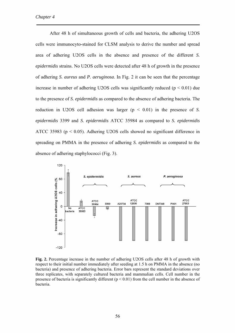
Chapter 4
After 48 h of simultaneous growth of cells and bacteria, the adhering U2OS
cells were immunocyto-stained for CLSM analysis to derive the number and spread
area of adhering U2OS cells in the absence and presence of the different S.
epidermidis strains. No U2OS cells were detected after 48 h of growth in the presence
of adhering S. aureus and P. aeruginosa. In Fig. 2 it can be seen that the percentage
increase in number of adhering U2OS cells was significantly reduced (p < 0.01) due
to the presence of S. epidermidis as compared to the absence of adhering bacteria. The
reduction in U2OS cell adhesion was larger (p < 0.01) in the presence of S.
epidermidis 3399 and S. epidermidis ATCC 35984 as compared to S. epidermidis
ATCC 35983 (p < 0.05). Adhering U2OS cells showed no significant difference in
spreading on PMMA in the presence of adhering S. epidermidis as compared to the
absence of adhering staphylococci (Fig. 3).
Fig. 2. Percentage increase in the number of adhering U2OS cells after 48 h of growth with respect to their initial number immediately after seeding at 1.5 h on PMMA in the absence (no bacteria) and presence of adhering bacteria. Error bars represent the standard deviations over three replicates, with separately cultured bacteria and mammalian cells. Cell number in the presence of bacteria is significantly different (p < 0.01) from the cell number in the absence of bacteria.
56

Bacterial strain-specific effects on mammalian cells
Fig. 3. Average area per adhering U2OS cell immediately after seeding at 1.5 h (□) and after 48 h (■) of growth on PMMA in the absence (no bacteria) and presence of adhering bacteria. Error bar represents the standard deviation over three replicates, with separately cultured bacteria and mammalian cells.
In the concept of the race for the surface, the total cell surface coverage of the
substratum surface by tissue cells is considered determinant for the fate of a
biomaterial implant. The surface coverages by U2OS cells at 1.5 h after seeding and
after 48 h of growth are shown in Fig. 4. The surface coverage by adhering U2OS
cells 1.5 h after seeding varied between 11% and 16% regardless of the absence or
presence of adhering bacteria. The cell surface coverage increased significantly after
48 h in the absence and in the presence of adhering S. epidermidis. In the presence of
adhering S. aureus and P. aeruginosa, reductions in cell surface coverages were
observed after 18 h of growth, indicating U2OS cell death and no U2OS cells were
detected on the PMMA surface after 48 h (Fig. 5).
57
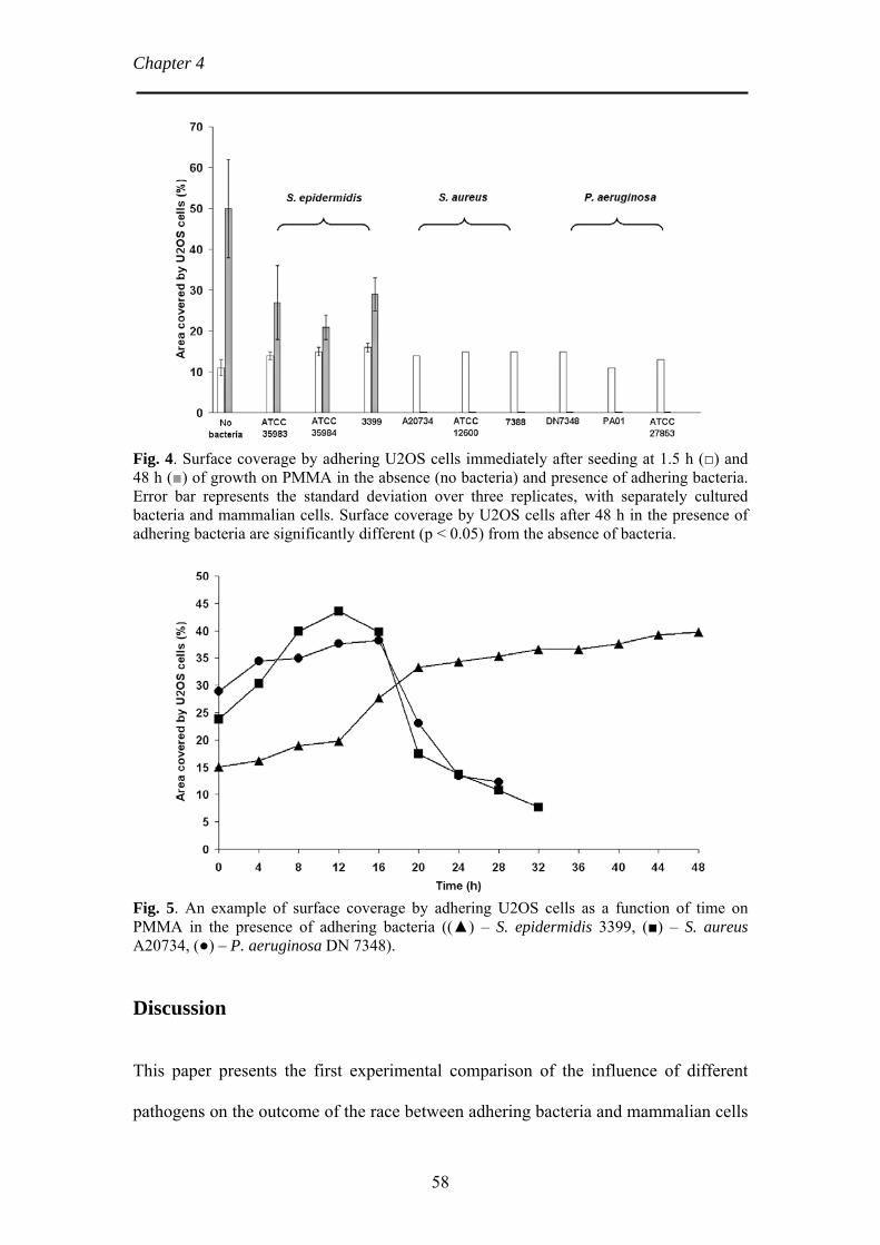
Chapter 4
Fig. 4. Surface coverage by adhering U2OS cells immediately after seeding at 1.5 h (□) and 48 h (■) of growth on PMMA in the absence (no bacteria) and presence of adhering bacteria. Error bar represents the standard deviation over three replicates, with separately cultured bacteria and mammalian cells. Surface coverage by U2OS cells after 48 h in the presence of adhering bacteria are significantly different (p < 0.05) from the absence of bacteria.
Fig. 5. An example of surface coverage by adhering U2OS cells as a function of time on PMMA in the presence of adhering bacteria ((▲) – S. epidermidis 3399, (■) – S. aureus A20734, (●) – P. aeruginosa DN 7348).
Discussion
This paper presents the first experimental comparison of the influence of different
pathogens on the outcome of the race between adhering bacteria and mammalian cells
58

Bacterial strain-specific effects on mammalian cells for a biomaterial surface. In our in vitro model, bacteria were allowed to adhere prior
to cell adhesion and spreading, which mimics the situation of peri-operative bacterial
contamination of implant surfaces. The number of bacteria adhering on the PMMA
surface prior to initiating mammalian cell adhesion and spreading was set to 103 cm-2.
In the past, it has been documented that during a surgical procedure of 1 h, the total
number of bacteria carrying particles falling on a wound is about 270 cm-2. The
bacterial counts are generally higher during periods of activity and when more people
are present in the operation theatre [11]. More recent, through the use of modern,
better ventilated operation theatres (20 changes of air per hour) and impermeable
patient and personnel clothing, peri-operative bacterial contamination may well be
less [12]. However, many surgical procedures in which implants are introduced in the
body last much longer than 1 h. Therefore, the level of bacterial contamination chosen
in our experiments is probably realistic of a worst case scenario. Despite these low
numbers, peri-operatively introduced organisms, particularly when of low virulence,
can survive on an implant surface for prolonged periods of time and later, during
periods of host immune depression, they proliferate and establish an infection with
clinical symptoms [13].
In the concept of the race for the surface, full coverage of a biomaterial surface
in vivo protects an implant against bacterial colonization [4]. Previously, in our model
for the ‘race for the surface’, all common biomaterial surfaces, including PMMA,
allowed S. epidermidis ATCC 35983 biofilm formation with a negative impact on the
coverage of the biomaterial surface by mammalian cells [6]. Yet, PMMA showed
better mammalian cell adhesion and spreading in the presence of adhering S.
epidermidis ATCC 35983 than other commonly used biomaterials [6]. The present
study confirms that U2OS cells are able to adhere, spread and grow in the presence of
59

Chapter 4
different S. epidermidis strains, but not in the presence of adhering S. aureus or P.
aeruginosa. The latter strains cause death of all adhering U2OS cells within 18 h,
despite removal of bacterial toxins by flow.
These observations are in line with clinical findings that BAI due to S. aureus
and P aeruginosa usually progresses much more aggressively than BAI caused by S.
epidermidis. S. aureus appears more frequently in acute infections, within 4 weeks of
surgery, of complicated total joint arthroplasty compared to S. epidermidis. S.
epidermidis is most commonly implicated in delayed septic loosening of total joint
prostheses [14] or even in presumed a-septic loosening [15], indicating its low
virulence with only minor clinical symptoms of infection. Pseudomonas is also much
more virulent than S. epidermidis, which is ascribed to the more aggressive
endotoxins in the slime. Pseudomonas slime injected into mice produces liver and
renal dysfunction and death within a short time, while no such effect was seen with S.
epidermidis slime [16,17]. In vascular grafts, pacemakers and orthopedic devices, S.
epidermidis is mostly found in low grade infections and a common cause of late
infections. In patients with low grade hip implant infections, only 5% of the patient
had a temperature of 37.8°C or higher and only 20% had wound drainage [18].
Buchholz and co-workers [19] showed that 84% of 64 patients with an infection by a
low virulent organism, were free from infection 2 years later. Alternatively, when S.
aureus was the causal organism, recurrent infection occurred in 28% of the patients
[19], and almost 50% of the patients with a Gram-negative bacterial infection
(Pseudomonas) experienced recurrent infection. This suggested a direct correlation
between the clinical outcome and the causative bacterial strain, in line with our in
vitro results.
60

Bacterial strain-specific effects on mammalian cells
The low virulence of S. epidermidis strains compared to S. aureus or P.
aeruginosa is due to the lack of additional genes responsible for producing severely
tissue damaging toxins [8,9,20]. In S. epidermidis infections, biofilm formation is
considered the only virulence factor and therefore infections are usually sub-acute or
chronic [21-23]. Amongst the S. epidermidis strains involved in this study, S.
epidermidis ATCC 35983 was considered not to produce slime, while S. epidermidis
3399 and S. epidermidis ATCC 35984 were slime producers [24]. Interestingly, a
significant reduction in number of adhering U2OS cells was observed in the presence
of S. epidermidis ATCC 35984 and S. epidermidis 3399 compared to S. epidermidis
ATCC 35983 in direct correlation to the slime producing capability of these strains.
This study demonstrates that U2OS cells are bound to lose the race for the
surface in the presence of adhering, highly virulent S. aureus or P. aeruginosa strains,
whereas spreading and growth of adhering U2OS cells, although impaired, still occurs
in the presence of adhering S. epidermidis strains. These results are in line with the
clinically documented progression of BAI by the different species included in this
study and highlight the potential use of our in vitro model for studies investigating the
effect of pathogen virulence on host tissue cells in BAI.
61

Chapter 4
References
1. Trampuz A, Zimmerli W. New strategies for the treatment of infections associated with prosthetic joints. Curr Opin Investig Drugs 2005;6:185-190.
2. Gristina AG. Biomaterial-centered infection: microbial adhesion versus tissue integration. Science 1987;237:1588-1595.
3. Gristina AG, Naylor P, Myrvik Q. Infections from biomaterials and implants: a race for the surface. Med Prog Technol 1998;14:205-224.
4. Gristina AG. Implant failure and the immune-incompetent fibro-inflammatory zone. Clin Orthop Rel Res 1994;298:106-118.
5. Subbiahdoss G, Kuijer R, Grijpma DW, Van der Mei HC, Busscher HJ. Microbial biofilm growth vs. tissue integration: "The race for the surface" experimentally studied. Acta Biomater 2009;5:1399-1404.
6. Subbiahdoss G, Grijpma DW, Van der Mei HC, Busscher HJ, Kuijer R. Microbial biofilm growth vs. tissue integration on biomaterials with different wettabilities and a polymer – brush coating. J Biomed Mater Res A 2010;DOI: 10.1002/jbm.a.32731.
7. Gristina AG, Costerton JW. Bacterial adherence to biomaterials and tissue. The significance of its role in clinical sepsis. J Bone Joint Surg Am 1985;67:264-273.
8. Khalil H, Williams RJ, Stenbeck G, Henderson B, Meghji S, Nair SP. Invasion of bone cells by Staphylococcus epidermidis. Microb Infect 2007;9:460-465.
9. Van Delden C, Iglewski BH. Cell-to-cell signalling and Pseudomonas aeruginosa infections. Emerg Infect Dis 1998;4:551-560.
10. Boelens JJ., Dankert J, Murk JL, Weening JJ, Van der Poll T, Dingemans KP, Koole L, Laman JD, Zaat SAJ. Biomaterial-associated persistence of Streptococcus epidermidis in pericatheter macrophages. J Infect Dis 2000;181:1337-1349.
11. Fitzgerald RH. Microbiologic environment of the conventional operating-room. Arch Surg 1979;114:772-775.
12. Verkkala K, Eklund A, Ojajarvi J, Tiittanen L, Hoborn J, Makela P. The conventionally ventilated operating theatre and air contamination control during cardiac surgery - bacteriological and particulate matter control garment options for low level contamination. Eur J Cardiothorac Surg 1998;14:206-210.
13. Dougherty SH. Pathobiology of infection in prosthetic devices. Rev Infect Dis 1988;10:1102-1117.
14. Robinson DA., Enright MC. Multilocus sequence typing and the evolution of methicillin-resistant Staphylococcus aureus. Clin Microbiol Infect 2004;10:92-97.
62

Bacterial strain-specific effects on mammalian cells
63
15. Zimmerli W, Trampuz A, Ochsner PE. Current concepts: Prosthetic-joint infections. N Engl J Med 2004;351:1645-1654.
16. Lynn M, Lotz L. Biochemical alterations in the mouse induced by Pseudomonas aeruginosa and its slime glycolipoprotein. Br J Exp Pathol 1984;65:275-281.
17. Peters G, Schumacher-Perdreau F, Jansen B, Bey M, Pulverer G. Biology of S. epidermidis extracellular slime. In: Pulverer G, Quie PG, Peters G, eds. Pathogenicity and clinical significance of coagulase-negative staphylococci. Stuttgart: Gustav Fischer Verlag, 1987;15-32.
18. Fitzgerald RH, Jones DR. Hip implant infection - Treatment with resection arthroplasty and late total hip-arthroplasty. Am J Med 1985;78:225-228.
19. Buchholz HW, Elson RA, Engelbrecht E, Lodenkamper H, Rottger J, Siegel A. Management of deep infection of total hip replacement. J Bone Joint Surg 1981;63B:342-353.
20. Massey RC, Horsburgh MJ, Lina G, Hook M, Recker M. Opinion - The evolution and maintenance of virulence in Staphylococcus aureus: a role for host-to-host transmission? Nat Rev Microbiol 2006;4:953-958.
21. Mckevitt AI, Bjornson GL, Mauracher CA, Scheifele DW. Amino-acid-sequence of a delta like toxin from Staphylococcus-epidermidis. Infect Immun 1990;58:1473-1475.
22. Raad I, Alrahwan A, Rolston K. Staphylococcus epidermidis: Emerging resistance and need for alternative agents. Clin Infect Dis 1998;26:1182-1187.
23. Vuong C, Otto M. Staphylococcus epidermidis infections. Microb Infect 2002;4:481-489.
24. Kiers PJM, Bos R, Van der Mei HC, Busscher HJ. The electrophoretic softness of the surface of Staphylococcus epidermidis cells grown in a liquid medium and on a solid agar. Microbiol-Uk 2001;147:757-762.

CHAPTER 5
Bacterial Biofilm Formation versus Mammalian Cell Growth on Titanium-Based Mono- and Bi-Functional
Coatings
Guruprakash Subbiahdoss, Bidhari Pidhatika, Geraldine Coullerez, Mirren Charnley, Roel Kuijer, Henny C. van der Mei, Marcus Textor,
Henk J. Busscher
European Cells and Materials, 2010; 19: 205-213
Republished with the permission of ECM Journal

Chapter 5 Introduction
Biomaterial-associated-infections (BAI) remain a major cause of failure of
biomaterial implants. Biomaterial implants may become contaminated with
microorganisms during implant surgery (peri-operative contamination) or during
hospitalization [1], causing the onset of BAI. Microorganisms involved in BAI are
protected from antibiotics due to their biofilm mode of growth. Consequently,
infected implants often have to be removed and a new implant can only be inserted
once complete eradication of the infected surrounding tissue has been performed.
Successful tissue integration depends on the outcome of the race for the surface
between microorganisms and tissue cells [1]. If this race is won by tissue cells, then
the biomaterial surface is covered by a cellular layer and is less vulnerable to biofilm
formation. On the other hand, if the race is won by bacteria, the implant surface will
become colonized by bacteria, and tissue cell functions are hampered by bacterial
virulence factors [2]. Since microorganisms are frequently introduced on an implant
surface during surgery, microorganisms often start the race for the surface before
tissue integration can even occur [2].
Biofilm formation occurs on all currently used biomaterials. Microbial
adhesion to biomaterials is determined by the physicochemical properties of the
implant surface [3]. Thus, modification of an implant surface may be able to prevent
bacterial adhesion and biofilm formation. Several surface modification methods [4-6],
such as hydrophilic polymer brush-type surfaces (in particular poly(ethylene glycol)
PEG), have been developed to prevent bacterial adhesion and biofilm formation [7].
However, such non-adhesive polymer coatings also resist adhesion, spreading and
growth of mammalian cells [6,8], which are required for successful tissue integration.
66

The race for the surface on functional coatings
In order to prepare bi-functional coatings which prevent microbial adhesion
while supporting tissue cell growth, the biologically inert poly(L-lysine)-graft-
poly(ethylene glycol) (PLL-g-PEG) copolymer was modified with the arginine-
glycine-aspartic acid (RGD) peptide sequence [8]. The RGD peptide is known as one
of the major recognition sites of integrin receptors through which mammalian cells
connect to their extracellular matrix molecules [9]. In earlier studies, reduced bacterial
adhesion on bi-functional PLL-g-PEG/PEG-RGD-modified surfaces has been
demonstrated separately from their ability to support mammalian cell growth [7,10].
However, a simultaneous study on bacterial biofilm formation and mammalian cell
growth, i.e. an actual “race for the surface”, has not been performed due to lack of
proper methodology. Recently, an in vitro experimental methodology to investigate
bacterial biofilm formation and mammalian cell growth in a single experiment was
developed [11]. The outcome of the competition between Staphylococcus epidermidis
and U2OS cells appeared to be dependent on the number of bacteria present prior to
cell seeding and the absence or presence of fluid flow. Mammalian cells lost the
competition in the absence of flow due to the accumulation of bacterial toxins, but
were able to grow under flow due to the continuous supply of fresh medium to and
removal of endotoxins from the interface. A further study of the race for the surface
on different biomaterials demonstrated that mammalian cell interactions with
biomaterials were hampered by bacterial biofilm formation on all commonly used
biomaterial surfaces [12].
This study aimed at evaluating titanium oxide surfaces modified with
biopassive PLL-g-PEG and bioactive PLL-g-PEG/PEG-RGD with respect to biofilm
formation and simultaneous mammalian cell adhesion, spreading and growth. In
67

Chapter 5 particular the hypothesis that the bi-functional, bioactive surface may favour
mammalian cell interaction over biofilm formation, is tested.
Materials and Methods
TiO2-coated glass slides. Microscope glass slides (76 × 26 mm) were purchased from
Menzel GmBH+Co KG, Braunschweig, Germany. The glass slides were sputter-
coated with a 21 nm thick TiO2 layer (reactive magnetron sputtering, Paul Scherrer
Institute, Villigen, Switzerland).
PLL-g-PEG and PLL-g-PEG/PEG-RGD. PLL-g-PEG and PLL-g-PEG/PEG-RGD
were purchased from Surface Solutions, AG, Switzerland. PLL-g-PEG is a copolymer
with a polycationic PLL backbone onto which non-functionalized (methoxy-
terminated) PEG chains are grafted. Grafting ratio, expressed as g, represents the ratio
between number of lysine units and number of grafted PEG chains (g = Lys/PEG).
PLL-g-PEG/PEG-RGD describes a copolymer with a PLL backbone onto which two
different types of PEG chains are grafted, i.e. non-functionalized PEG and RGD-
functionalized PEG [8].
The molecular weights of PLL, non-functionalized PEG and RGD-
functionalized PEG, are 20 kDa, 2 kDa, and 3.4 kDa, respectively. The grafting ratio
(g) of both copolymers PLL-g-PEG and PLL-g-PEG/PEG-RGD is 3.5. For the latter,
8% of all PEG chain carry the terminal, covalently linked peptide
GCRGYGRGDSPG.
Surface preparation and polymer adsorption. The copolymers were dissolved at a
0.25 mg ml-1 concentration in HEPES II buffer (10 mM HEPES and 150 mM NaCl,
68

The race for the surface on functional coatings adjusted to pH 7.4 by addition of NaOH). The polymer solutions were filter-sterilized
(0.22 µm filter), aliquoted and stored at -20°C until use. Prior to surface modification,
TiO2-coated glass slides were ultrasonicated for 10 min in a cleansing solution (300
mM hydrochloric acid and 1% detergent, Cobas Integra®, Roche Diagnostic,
Indianapolis, USA), rinsed with ultra pure water, followed by 10 min ultrasonication
in 2-propanol to remove the adventitious macroscopic contamination and blow-drying
under a stream of nitrogen. Subsequently, the TiO2-coated glass slides were cleaned
by means of oxygen plasma treatment for 2 min (Plasma cleaner/sterilizer PDC-32G,
Harrick scientific products Inc., USA). Copolymer solutions were subsequently
placed onto the pre-cleaned substrates completely covering their surfaces. Copolymer
adsorption was allowed to proceed for 1 h to provide a complete monolayer on the
surface, followed by extensive washing with HEPES II buffer, ultrapure water, and
finally blow-drying under a stream of nitrogen. Data on conformation of the used
copolymers adlayers and interaction with proteins have been described elsewhere
[13].
The copolymer-modified substrates were placed in a clean sample holder and
stored at 4°C until use. The surfaces are denoted as “TiO2” for bare titanium oxide
surface, “PEG” for PLL-g-PEG-modified and “PEG-RGD” for PLL-g-PEG/PEG-
RGD-modified surface. The surfaces were sterilized using 70% ethanol and washed
with sterile ultrapure water before use.
Bacterial growth conditions and harvesting. S. epidermidis ATCC 35983,
originally isolated from human blood of a patient with an infected intravascular
catheter, and known to produce polysaccharide integrin adhesin [14], was used
throughout this study. First, the strain was streaked on a blood agar plate from a
69

Chapter 5 frozen stock and grown overnight at 37°C. The plate was then kept at 4°C. For each
experiment, a colony was inoculated in 10 ml of tryptone soya broth (TSB; OXOID,
Basingstoke, England) and cultured for 24 h. This culture was used to inoculate a
second culture, which was grown for 17 h prior to harvesting. Bacteria were harvested
by centrifugation at 5000 x g for 5 min at 10°C and washed twice with sterile
ultrapure water. Subsequently, the harvested bacteria were ultrasonicated on ice (3 x
10 s) in sterile PBS (10 mM potassium phosphate, 0.15 M NaCl, pH 7.0) in order to
break bacterial aggregates. This suspension was further diluted in sterile PBS to a
concentration of 3 x 10⁶ bacteria per ml.
Mammalian cell culturing and harvesting. U2OS osteosarcoma cells were cultured
in Dulbecco’s modified Eagles Medium (DMEM)-low glucose supplemented with
10% fetal calf serum (FBS) and 0.2 mM of ascorbic acid-2-phosphate (AA2P) without
antibiotics, denoted “DMEM+FBS”. Osteoblasts were maintained at 37°C in a
humidified 5% CO2 atmosphere, and passaged at 70 – 90% confluency using
trypsin/EDTA.
Bacteria and tissue cell competitive adhesion assays. Competitive adhesion assays
were performed on the bottom plate of a parallel plate flow chamber (175 x 17 x 0.75
mm3) prepared from the biomaterial surfaces with or without polymer coatings under
investigation, as described in detail elsewhere [11]. Importantly, the flow chamber
was equipped with heating elements and kept at 37°C throughout the experiments.
Bacterial and U2OS cell deposition were observed with a CCD camera (Basler AG,
Germany) mounted on a phase-contrast Leica DM2000 microscope (Leica
70

The race for the surface on functional coatings Microsystems Ltd, Germany) with a 30x objective for bacteria and 10x objective for
mammalian cells.
Prior to each experiment, all tubes and the flow chamber were filled with
sterile PBS, to ensure all air bubbles were removed from the system. Prior to the
addition of bacterial suspension, sterile PBS was allowed to flow through the system
at a shear rate of 11 s-1. Subsequently, the bacterial suspension in PBS was perfused
through the chamber at the same shear rate for 2 h. Images were obtained
continuously and the number of bacteria per unit area were analyzed in real-time by
using proprietary software based on the Matlab Image processing Toolkit (The
MathWorks, MA, USA). The flow with shear rate of 11 s-1 was then re-initiated to
deliver sterile PBS to remove unattached bacteria from the tubes and flow chamber,
after which a U2OS cell suspension (6 × 10⁵ cells ml-1) in modified culture medium,
consisting of 98% DMEM+FBS and 2% TSB which is suitable for the simultaneous
growth of U2OS cells and S. epidermidis [11], was allowed to enter the flow chamber.
Once the entire volume of buffer inside the chamber was replaced by the cell
suspension, the flow was stopped for 1.5 h in order to allow tissue cells to adhere and
spread on the substratum. Subsequently, phase contrast images (nine images on
different locations, 900 x 700 µm2 each) were taken to determine the number of
adhering cells per unit area and the area per spread cell using Scion image analysis
software. Finally, modified culture medium supplemented with 2% HEPES was
perfused through the system at a low shear rate of 0.14 s-1 for 48 h, and phase-contrast
images were collected continuously. After 48 h of growth, the shear stress was
increased to 5.6 s-1 for 30 min, i.e. a value that is lower than the one used during
initial bacterial adhesion (11 s-1). Subsequently, U2OS cells and bacterial biofilm
71

Chapter 5 growth were monitored continuously. Control experiments with only U2OS cells (in
the absence of S. epidermidis) were performed using the above procedure.
Finally, the adhering U2OS cells were stained with TRITC-Phalloidin for
quantitative analysis. Briefly, the substrate surfaces with adhering bacteria and tissue
cells were fixed with 30 ml of 3.7% formaldehyde in cytoskeleton stabilization buffer
(CS; 0.1 M Pipes, 1 mM EGTA, 4% (w/v) polyethylene glycol 8000, pH 6.9). After 5
min, the fixation solution was replaced with 30 ml of fresh CS for another 5 min.
Subsequently, U2OS cells were incubated in 0.5% Triton X-100 for 3 min, rinsed
with PBS, stained for 30 min with 5 ml PBS containing 49 µl DAPI and 2 µg ml-1 of
TRITC-Phalloidin, washed four times in PBS and examined with confocal laser
scanning microscopy (CLSM, Leica DMRXE with confocal TCS SP2 unit equipped
with a water immersion lens). Images (nine images on different locations, 900 x 700
µm2 each) were taken and the number of adhering cells per unit area and the average
area per spread cell were determined.
Statistics. Experiments on different surfaces were performed in triplicate. Data are
represented as a mean with standard deviation. For statistical analysis ANOVA was
performed followed by a Tukey’s HSD post-hoc test and a p-value < 0.05 was
considered to be significant.
72

The race for the surface on functional coatings
73
Results
Bacterial adhesion. Initial adhesion of S. epidermidis after 2 h of flow at a shear rate
of 11 s-1 was significantly reduced to 3.6±1.8 × 103 cm-2 and 6.0±3.9 × 103 cm-2 on
both PEG and PEG-RGD coatings respectively, compared to 1.3±0.4 × 105 cm-2 on
the bare TiO2 surface (Fig. 1). This demonstrates that in the presence of RGD-peptide
sequences, the non-adhesive functionality of the PEG brush surface is still maintained.
Fig. 1. The number of adhering S. epidermidis ATCC 35983 as a function of time in a parallel plate flow chamber (shear rate 11 s-1) on mono-functional PEG coating (○) and a bi-functional PEG-RGD coating (□) as well as on a bare TiO2 surface (▲). Error bars represent the standard deviation over three replicates with separately cultured bacteria.
U2OS cell adhesion and spreading in the absence and presence of adhering S.
epidermidis. Immediately after seeding, U2OS cell adhesion and spreading was
observed in the absence and presence of adhering S. epidermidis on TiO2 surfaces and
PEG-RGD coatings, but not on the PEG coatings. At 1.5 h, there was no significant
difference in the number of adhering U2OS cells on the different surfaces (Table 1),
but cell spreading was greater on PEG-RGD coatings, as compared to PEG coatings
and TiO2 surfaces (p<0.01), irrespective of absence or presence of S. epidermidis
bacteria (Table 1).
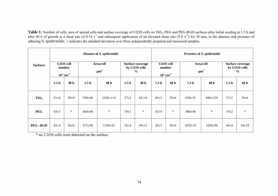
Chapter 5
Table 1: Number of cells, area of spread cells and surface coverage of U2OS cells on TiO2, PEG and PEG-RGD surfaces after initial seeding at 1.5 h and after 48 h of growth at a shear rate of 0.14 s-1 and subsequent application of an elevated shear rate (5.6 s-1) for 30 min, in the absence and presence of adhering S. epidermidis. ± indicates the standard deviation over three independently prepared and measured samples.
Absence of S. epidermidis
Presence of S. epidermidis
U2OS cell number
103 cm-2
Area/cell
µm2
Surface coverage by U2OS cells
%
U2OS cell number
103 cm-2
Area/cell
µm2
Surface coverage by U2OS cells
%
Surfaces
1.5 h
48 h
1.5 h
48 h
1.5 h
48 h
1.5 h
48 h
1.5 h
48 h
1.5 h
48 h
TiO2
51±4
59±9 550±48 1020±114 27±2 63±10 45±3 39±6 620±55 940±210 27±2 38±6
PEG
43±3
* 460±60 * 19±1 * 42±4 * 480±40 * 19±2 *
PEG –RGD
43±4
56±9 875±50 1180±82 36±4 69±11 43±3 50±9 1070±35 1024±96 44±4 54±10
* no U2OS cells were detected on the surface.
74

The race for the surface on functional coatings
After 48 h, the adhering S. epidermidis had grown into a biofilm on all
surfaces, but simultaneously U2OS cell adhesion and spreading was observed on TiO2
surfaces and PEG-RGD coatings (Fig. 2). On PEG coatings, U2OS cells that were
loosely adhered retained a rounded morphology up to 48 h (Fig. 2e). Upon application
of an elevated shear rate (5.6 s-1), biofilms detached from PEG and PEG-RGD
coatings and partially from TiO2 surfaces, whereas U2OS cell detachment only
occurred from the PEG coating (Fig. 2g,h and i).
Fig. 2. Phase-contrast images of U2OS cell adhesion and spreading after seeding at 1.5 h, after 48 h of growth at low shear (0.14 s-1) in the presence of adhering S. epidermidis as well as after an elevated shear (5.6 s-1) for 30 min at the end of the experiment, on PEG and PEG-RGD coatings, and on TiO2 control surfaces. At 1.5 h, well spread U2OS cells were observed on PEG-RGD (c) compared to PEG coating (b) and TiO2 control surfaces (a), for which cell spreading was less. At 48 h, biofilm formation and U2OS cell spreading were observed on TiO2 control surface (d) and PEG-RGD coating (f), whereas only biofilm formation and no cell spreading were seen on PEG coating (e). Upon application of an elevated shear, detachment of biofilm and U2OS cells was observed on PEG coatings (h), while only biofilm detachment occurred on PEG-RGD coating (i) and TiO2 surface (g). All images were taken at the same magnification. The bar denotes 10 µm.
After 48 h of growth and subsequent application of a higher shear, adhering
U2OS cells were immunocyto-stained for CLSM analysis (Fig. 3) to derive the
number and spread area of adhering U2OS cells in the absence and presence of
staphylococci. In Fig. 4 it can be seen that on a percentage basis, the number of
75

Chapter 5 adhering U2OS cells was significantly reduced on TiO2 surfaces in the presence of S.
epidermidis as compared to the control, i.e. in the absence of adhering bacteria (p <
0.05). In the presence of S. epidermidis, the percentage adhering U2OS cells
decreased on TiO2 surfaces in contrast to that on bi-functional PEG-RGD coatings
(Fig. 4) which showed an increase. U2OS cells showed no significant difference in
spreading on PEG-RGD coating as compared to a TiO2 surface in the presence or
absence of adhering S. epidermidis (Fig. 5 and Table 1).
Surface coverage of U2OS cells in the absence and presence of adhering S.
epidermidis. Immediately after seeding at 1.5 h, a significant increase in the surface
coverage of U2OS cells was observed on PEG-RGD coatings compared to TiO2
surfaces and PEG coatings, irrespective of the absence or presence of adhering S.
epidermidis (Table 1).
Fig. 3. CLSM images of U2OS cells after 48 h of growth at low shear (0.14 s-1) and subsequent application of an elevated shear (5.6 s-1) for 30 min on TiO2 surfaces, PEG and PEG-RGD coatings in the absence (a-c) and presence (d-f) of adhering S. epidermidis. U2OS cells were stained with 5 ml PBS containing 49 µl DAPI and 2 µg ml-1 of TRITC-phalloidin. All images were taken at the same magnification. The bar denotes 75 µm.
76

The race for the surface on functional coatings After 48 h of growth at low shear and subsequent application of an elevated shear for
30 min, TiO2 surface showed a significant decrease in surface coverage in the
presence of adhering staphylococci compared to the control, i.e. the absence of
adhering staphylococci. On PEG coatings, no adhering U2OS cells were detected on
the surface (Fig. 3b,e). Conversely PEG-RGD coatings in the presence of adhering
staphylococci showed no significant difference in surface coverage of U2OS cells
compared to cells cultured in the absence of S. epidermidis.
Fig. 4. The percentage increase in the number of adhering U2OS cells after 48 h of growth at low shear (0.14 s-1) and subsequent application of an elevated shear (5.6 s-1) for 30 min with respect to their initial number immediately after seeding at 1.5 h on TiO2 surfaces and PEG and PEG-RGD coatings in the absence (□) and presence (■) of adhering S. epidermidis ATCC 35983. Error bars represent the standard deviations over three replicates, with separately cultured bacteria and tissue cells. × denotes significance at p < 0.01, denotes significance at p < 0.05.
77

Chapter 5
Fig. 5. The average area per adhering U2OS cell immediately after seeding at 1.5 h (□) and after 48 h of growth at low shear (0.14 s-1) and subsequent application of an elevated shear (5.6 s-1) for 30 min (■) on TiO2 surfaces, PEG and PEG-RGD coatings in the absence and presence of adhering S. epidermidis ATCC 35983. Error bars represent the standard deviations over three replicates, with separately cultured bacteria and tissue cells. * denotes significance at differences at p < 0.01 compared with PEG-RGD.
Discussion
This paper is the first to experimentally demonstrate the advantage of bi-functional
versus mono-functional coatings in the prevention of infection on implant surfaces by
conducting the race for the surface between bacteria and mammalian cells, as
eventually determining the fate of a biomaterial implant. Bi-functional coatings
clearly remained non-adhesive to S. epidermidis, but at the same time supported
mammalian cell adhesion and spreading to a greater extent than mono-functional
coatings and a control surface, in this case a bare TiO2 surface. Titanium is widely
used in dental and orthopaedic implants, heart valves and vascular stents [15].
Titanium devices implanted in the body are covered with a layer of oxide, responsible
for the favourable biocompatibility of titanium implants. Tissue cells reactions on
TiO2 and pure titanium are very similar [16].
78

The race for the surface on functional coatings
In our in vitro model, bacteria were allowed to adhere for 2 h prior to cell
adhesion and spreading, which is considered to mimic the clinical situation where
implants become contaminated prior to implantation. The number of bacteria adhering
on PEG and PEG-RGD was 103 cm-2 and 105 cm-2 for TiO2, representing a reduction
in initial bacterial load on polymer brush coatings of two log-units. Note that in this
study we deliberately chose to fix the time for bacterial adhesion, and not to
contaminate all surfaces with the same number of organisms. This choice allows to
compare coatings and surfaces for their performance during a fixed surgical period. In
the past, it has been documented that during a surgical procedure of 1 h, the total
number of bacteria carrying particles falling on the wound is about 270 per cm2 [17].
The bacterial counts are generally higher during periods of activity and when more
people are present in the operation theatre. More recent, through the use of modern,
better ventilated operation theatres (20 changes of air per hour) and impermeable
patient and personnel clothing, peri-operative bacterial contamination may well be
less [18]. However, many surgical procedures in which implants are introduced in the
body last much longer than 1 h. Therefore, the bacterial adhesion densities chosen in
our experiments represent a moderate to worst case scenario.
Significant reduction of bacterial adhesion on PEG and PEG-RGD-coated
surfaces compared to that on bare TiO2 surface was previously reported [7]. In earlier
studies [19,20], it was reported that the presence of a peptide covalently attached to
PEG reduced bacterial adhesion similar to PEG coatings. In agreement with previous
studies our results show that S. epidermidis showed no significant difference in
adhesion on PEG coatings compared to PEG-RGD coatings indicating that the S.
epidermidis ATCC 35983 bacteria do not recognize the RGD, unlike mammalian cells
recognize RGD via integrin receptors.
79

Chapter 5
Cell adhesion to a biomaterial surface is a prerequisite for successful tissue
integration. Mammalian cells bind to the biomaterial surface through adsorbed
proteins and one of the main binding sites in these proteins is the RGD peptide
sequence [21]. On PEG coatings, hardly any protein adsorbed resulting in an
inhibition of cell adhesion and spreading [6,22]. Similarly, in our experiments U2OS
cells remained rounded on mono-functional PEG coatings up to 48 h. Such anti-
fouling behaviour is mainly due to steric repulsion between the hydrated uncharged
PEG chains and proteins [23]. However cell adhesion to mono-functional PEG-
coatings can be achieved by introducing specific bioligands, such as a cell-interactive
peptide (RGD) linked covalently to the non-fouling PEG, producing a bi-functional
PEG-RGD coating. Our results show an enhanced mammalian cell adhesion to the bi-
functional PEG-RGD coatings, in which already within 1.5 h after cell seeding, the
U2OS cells showed near maximum spreading, indicating recognition of the RGD-
sequence by adhesion receptors in the cell membrane [24]. The enhanced cell
attachment and spreading depends on the RGD-peptide surface density [25]. The
RGD-peptide surface density of around 5 pmol RGD peptide per cm2 applied here
was well above reported densities for cell adhesion of over 0.6 pmol RGD peptide per
cm2 [26]. The former was calculated [27] based on the copolymer composition (RGD
grafting density, NMR) and the adsorbed copolymer mass from in situ monitoring of
the copolymer adsorption using Optical Waveguide Lightmode Spectroscopy
(OWLS) (data not shown).
In the concept of the race for the surface, a full coverage of a biomaterial in
vivo by a viable tissue cell layer, intact cell membranes and functional host defense
mechanisms resist bacterial colonization [28]. Previously, in our model for the ‘race
for the surface’, all biomaterial surfaces allowed bacterial adhesion and biofilm
80

The race for the surface on functional coatings growth with a negative impact on the coverage of the biomaterial by mammalian cells
[12]. Similarly, the presence of adhering S. epidermidis decreased the surface
coverage of U2OS cells on TiO2 surface compared to their control, i.e. the absence of
S. epidermidis. In contrast to the TiO2 surface, the presence of RGD-peptides at the
surface significantly improved U2OS cell adhesion and spreading in the presence of S.
epidermidis, eventually resulting in similar surface coverage than the control, i.e. the
absence of S. epidermidis. Considering that cell adhesion and spreading are the signs
of cellular well-being, this is highly indicative for a protective effect of cell binding to
RGD in the presence of bacteria [29]. Even though complete coverage of the
biomaterial surface was not obtained after 48 h, significantly improved U2OS cell
adhesion and spreading on PEG-RGD coatings could eventually lead to complete
coverage of U2OS cells on biomaterial surfaces over longer periods of time. In the
concept of the race for the surface, one may speculate that PEG-RGD coated implants
will become more easily integrated by host tissue cells than other biomaterial surfaces
and thus better protect the implant against infection.
PEG coatings have been shown to be resistant to protein adsorption, biofilm
formation and cell attachment in vitro over periods of up to 4 weeks [8, 30-32].
Depending on the exact nature of the coating, degradation of this biocompatible
polymeric monolayer by hydrolytic/enzymatic activity has been reported. Another
concern is the mechanical robustness of the PEG coatings, and if handled with
surgical instruments, coatings may be easily damaged. Optichem® [32] and nano-
particle based polymer brush coatings [31] are mono-functional non-adhesive coatings
with greater robustness than can be obtained with monolayer coatings, like the PEG-
RGD coating evaluated here. The results of the present study, however, aid in
directing such further development of monolayer bi-functional coatings into more
81

Chapter 5 robust ones, suitable for clinical use. The early protection by bi-functional coatings is
believed to be clinically relevant, as it could prevent an implant from becoming
colonized during the important period of actual implantation, which is critical for the
long-term success of an implant [33,34].
Conclusion
This study demonstrates that biopassive, mono-functional PEG coatings and
bioactive, bi-functional PEG-RGD coatings have potential to reduce bacterial
adhesion and prevent firm adhesion of biofilms compared to common biomaterial
surfaces. Moreover, in contrast to mono-functional PEG coatings, bi-functional PEG-
RGD coatings allow cell adhesion and spreading. At the same time, adhesion and
spreading of mammalian cells is not detrimentally affected by the presence of
adhering S. epidermidis. Bi-functional coatings thus have a strong potential to reduce
the risk of infection in applications requiring tissue integration, such as in dental and
orthopaedic implants.
82

The race for the surface on functional coatings References
1. Gristina AG. Biomaterial-centered infection: microbial adhesion versus tissue integration. Science 1987;237:1588-1595.
2. Gristina AG, Naylor P, Myrvik Q. Infections from biomaterials and implants: a race for the surface. Med Prog Technol 1988;14:205-224.
3. Hermansson M. The DLVO theory in microbial adhesion. Coll Surf B-Biointerf 1999;14:105-119.
4. Morra M. On the molecular basis of fouling resistance. J Biomat Sci-Polymer Ed 2000;11:547-569.
5. Vasilev K, Michelmore A, Griesser HJ, Short RD. Substrate influence on the initial growth phase of plasma-deposited polymer films. Chem Comm 2009;24:3600-3602.
6. Schuler M, Trentin D, Textor M, Tosatti SGP. Biomedical interfaces: titanium surface technology for implants and cell carriers. Nanomedicine 2006;1:449-463.
7. Maddikeri RR, Tosatti S, Schuler M, Chessari S, Textor M, Richards RG, Harris LG. Reduced medical infection related bacterial strains adhesion on bioactive RGD modified titanium surfaces: A first step toward cell selective surfaces. J Biomed Mater Res 2008;84A:425-435.
8. VandeVondele S, Voros J, Hubbell JA. RGD-Grafted poly-l-lysine-graft-(polyethylene glycol) copolymers block non-specific protein adsorption while promoting cell adhesion. Biotechnol Bioeng 2003;82:784-790.
9. Lebaron RG, Athanasiou KA. Extracellular matrix cell adhesion peptides: Functional applications in orthopedic materials. Tissue Eng 2000;6:85-103.
10. Shi ZL, Neoh KG, Kang ET, Poh C, Wang W. Bacterial adhesion and osteoblast function on titanium with surface-grafted chitosan and immobilized RGD peptide. J Biomed Mater Res 2008;86A:865-872.
11. Subbiahdoss G, Kuijer R, Grijpma DW, Van der Mei HC, Busscher HJ. Microbial biofilm growth vs. tissue integration: "The race for the surface" experimentally studied. Acta Biomater 2009;5:1399-1404.
12. Subbiahdoss G, Grijpma DW, Van der Mei HC, Busscher HJ, Kuijer R. Microbial biofilm growth vs. tissue integration on biomaterials with different wettabilities and a polymer – brush coating. J Biomed Mater Res A 2010;DOI:10.1002/jbm.a.32731.
13. Pasche S, De Paul SM, Vörös J, Spencer ND, Textor M. Poly(L-Lysine)-graft-poly(ethylene glycol) assembled monolayers on niobium oxide surfaces: a quatitative study of the influence of polymer interfacial architecture on resistance
83
Chapter 5
to protein adsorption by ToF-SIMS and in-situ OWLS. Langmuir 2003;19:9216-9225.
14. Dhanawade NB, Kalorey DR, Srinivasan R, Barbuddhe SB, Kurkure NV. Detection of intercellular adhesion genes and biofilm production in Staphylococcus aureus isolated from bovine subclinical mastitis. Vet Res Commun 2010;34:81-89.
15. Brunette DM, Tengvall P, Textor M, Thomsen P. Titanium in medicine: Material Science, Surface Science, Engineering, Biological Responses and Medical Applications. 2001;Springer-Verlag; Heidelberg and Berlin.
16. Textor M, Sittig CE, Frauchiger V, Tosatti S, Brunette DM. Properties and Biological Significance of Natural Oxide Films on Titanium and Its Alloys. In Titanium in Medicine: Material Science, Surface Science, Engineering, Biological Responses and Medical Applications. Thomsen P. 2001;Springer-Verlag; Heidelberg and Berlin. pp 171-230.
17. Fitzgerald RH. Microbiologic environment of the conventional operating-room. Arch Surg 1979;114:772-775.
18. Verkkala K, Eklund A, Ojajarvi J, Tiittanen L, Hoborn J, Makela P. The conventionally ventilated operating theatre and air contamination control during cardiac surgery - bacteriological and particulate matter control garment options for low level contamination. Eur J Cardiothorac Surg 1998;14:206-210.
19. Harris LG, Tosatti S, Wieland M, Textor M, Richards RG. Staphylococcus aureus adhesion to titanium oxide surfaces coated with non-functionalized and peptide-functionalized poly(L-lysine)-grafted-poly(ethylene glycol) copolymers. Biomaterials 2004;25:4135-4148.
20. Wagner VE, Bryers JD. Poly(ethylene glycol)-polyacrylate copolymers modified to control adherent monocyte-macrophage physiology: Interactions with attaching Staphylococcus epidermidis or Pseudomonas aeruginosa bacteria. J Biomed Mater Res 2004;69A:79-90.
21. Hersel U, Dahmen C, Kessler H. RGD modified polymers: biomaterials for stimulated cell adhesion and beyond. Biomaterials 2003;24:4385-4415.
22. Tosatti S, De Paul SM, Askendal A, VandeVondele S, Hubbell JA, Tengvall P, Textor M. Peptide functionalized poly(L-lysine)-g-poly(ethylene glycol) on titanium: resistance to protein adsorption in full heparinized human blood plasma. Biomaterials 2003;24:4949-4958.
23. Jeon SI, Lee JH, Andrade JD, Degennes PG. Protein surface interactions in the presence of polyethylene oxide. 1. Simplified theory. J Coll Interf Sci 1991;142:149-158.
24. Rezania A, Healy KE. Biomimetic peptide surfaces that regulate adhesion, spreading, cytoskeletal organization, and mineralization of the matrix deposited by osteoblast-like cells. Biotechnol Progress 1999;15:19-32.
84

The race for the surface on functional coatings
85
25. Massia SP, Hubbell JA. An RGD spacing of 440nm is sufficient for integrin alpha-V-beta-3-mediated fibroblast spreading and 140nm for focal contact and stress fiber formation. J Cell Biol 1991;114:1089-1100.
26. Rezania A, Johnson R, Lefkow AR, Healy KE. Bioactivation of metal oxide surfaces. 1. Surface characterization and cell response. Langmuir 1999;15:6931-6939.
27. Barth KA, Coullerez G, Nilsson LM, Castelli R, Seeberger PH, Vogel V, Textor M. An engineered mannoside presenting platform: Escherichia coli adhesion under static and dynamic conditions. Adv Funct Mater 2008;18:1459-1469.
28. Gristina AG. Implant failure and the immune-incompetent fibro-Inflammatory zone. Clin Orthop Rel Res 1994;298:106-118.
29. Stupack DG, Cheresh DA. Get a ligand, get a life: integrins, signaling and cell survival. J Cell Sci 2002;115:3729-3738.
30. Fundeanu I, Van der Mei HC, Schouten AJ, Busscher HJ. Polyacrylamide brush coatings preventing microbial adhesion to silicone rubber. Coll Surf B-Biointerf 2008;64:297-301.
31. Holmes PF, Currie EPK, Thies JC, Van der Mei HC, Busscher HJ, Norde W. Surface-modified nanoparticles as a new, versatile, and mechanically robust nonadhesive coating: Suppression of protein adsorption and bacterial adhesion. J Biomed Mater Res 2009;91A:824-833.
32. Fernandez ICS, Van der Mei HC, Metzger S, Grainger DW, Engelsman AF, Nejadnik MR, Busscher HJ. In vitro and in vivo comparisons of staphylococcal biofilm formation on a cross-linked poly(ethylene glycol)-based polymer coating. Acta Biomater 2010;6:1119-1124.
33. Poelstra KA, Barekzi NA, Rediske AM, Felts AG, Slunt JB, Grainger DW. Prophylactic treatment of Gram-positive and Gram-negative abdominal implant infections using locally delivered polyclonal antibodies. J Biomed Mater Res 2002;60:206-215.
34. Colon G, Ward BC, Webster TJ. Increased osteoblast and decreased Staphylococcus epidermidis functions on nanophase ZnO and TiO2. J Biomed Mater Res 2006;78A:595-604.


CHAPTER 6
Mammalian Cell Growth versus Biofilm Formation on Biomaterials Surfaces in a Post-operative
Contamination Model In vitro
Guruprakash Subbiahdoss, Roel Kuijer, Henk J. Busscher, Henny C. van der Mei
Accepted in Microbiology

Chapter 6 Introduction
Biomaterial-associated infections (BAI) can develop from peri-operative microbial
contamination of implant surfaces during implantation, immediately post-surgery
during hospitalization or by late haematogenous spreading from infections elsewhere
in the body. Both peri-operative and post-operative contaminations can cause BAI
many years after implantation, as bacteria can stay dormant on an implant surface for
several years [1,2]. Microorganisms involved in BAI are resistant to antibiotics and
the host immune system due to their biofilm mode of growth, and biomaterial
implants with a biofilm have to be removed in most cases [3-5]. Irrespective of the
route of infection, the fate of a biomaterial implant depends mainly on the outcome of
the so-called ‘race for the surface’ between successful tissue integration of the
biomaterial implant and biofilm growth [6]. If this race is won by tissue cells, then the
biomaterial surface is fully integrated by tissue cells and less vulnerable to bacterial
biofilms. On the other hand, if the race is won by bacteria, the implant surface will
become colonized by bacteria and tissue cell functions are hampered by bacterial
virulence factors and toxins [6,7]. In the concept of the race for the surface, a full
surface coverage of a biomaterial in vivo by a viable tissue cell layer, intact cell
membrane and functional host defense mechanisms resist bacterial colonization [8].
Previously an in vitro experimental model for peri-operative bacterial
contamination of implant surfaces was forwarded and the effects of bacterial presence
on adhesion, spreading and growth of mammalian cells were determined in a single
experiment [9]. The outcome of the race for the surface between contaminating
Staphylococcus epidermidis and mammalian cells on glass appeared to be dependent
on the number of bacteria present prior to mammalian cell seeding and the absence or
presence of fluid flow. Cells lost the race for the surface in the absence of flow due to
88
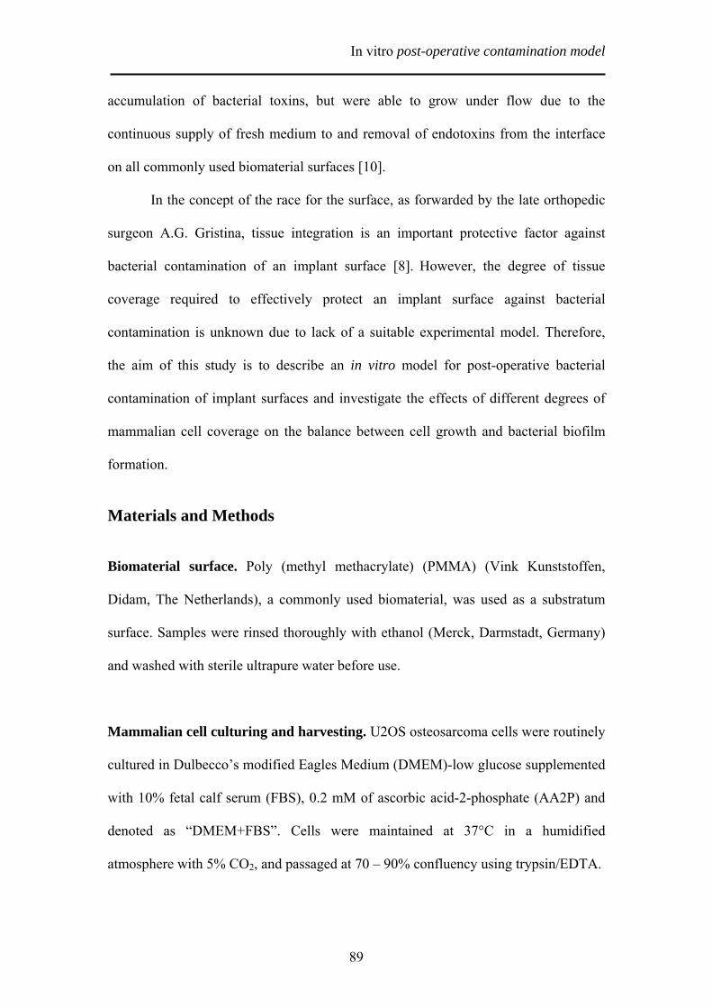
In vitro post-operative contamination model accumulation of bacterial toxins, but were able to grow under flow due to the
continuous supply of fresh medium to and removal of endotoxins from the interface
on all commonly used biomaterial surfaces [10].
In the concept of the race for the surface, as forwarded by the late orthopedic
surgeon A.G. Gristina, tissue integration is an important protective factor against
bacterial contamination of an implant surface [8]. However, the degree of tissue
coverage required to effectively protect an implant surface against bacterial
contamination is unknown due to lack of a suitable experimental model. Therefore,
the aim of this study is to describe an in vitro model for post-operative bacterial
contamination of implant surfaces and investigate the effects of different degrees of
mammalian cell coverage on the balance between cell growth and bacterial biofilm
formation.
Materials and Methods
Biomaterial surface. Poly (methyl methacrylate) (PMMA) (Vink Kunststoffen,
Didam, The Netherlands), a commonly used biomaterial, was used as a substratum
surface. Samples were rinsed thoroughly with ethanol (Merck, Darmstadt, Germany)
and washed with sterile ultrapure water before use.
Mammalian cell culturing and harvesting. U2OS osteosarcoma cells were routinely
cultured in Dulbecco’s modified Eagles Medium (DMEM)-low glucose supplemented
with 10% fetal calf serum (FBS), 0.2 mM of ascorbic acid-2-phosphate (AA2P) and
denoted as “DMEM+FBS”. Cells were maintained at 37°C in a humidified
atmosphere with 5% CO2, and passaged at 70 – 90% confluency using trypsin/EDTA.
89

Chapter 6 Bacterial growth conditions and harvesting. S. epidermidis ATCC 35983,
originally isolated from human blood of a patient with an infected intravascular
catheter, was used throughout this study. First, the strain was streaked on a blood agar
plate from a frozen stock and grown overnight at 37°C. The plate was then kept at
4°C. For each experiment, a colony was inoculated in 10 ml of tryptone soya broth
(TSB; OXOID, Basingstoke, England) and cultured for 24 h. This culture was used to
inoculate a second culture, which was grown for 17 h prior to harvesting. Bacteria
were harvested by centrifugation at 5000 x g for 5 min at 10°C and washed twice with
sterile ultrapure water. Subsequently, the harvested bacteria were sonicated on ice (3 x
10 s) in sterile PBS in order to break bacterial aggregates. This suspension was further
diluted in sterile PBS to a concentration of 3 x 10⁶ bacteria per ml.
Competitive assay for mammalian cell growth and biofilm formation. The
competitive assay was studied on the PMMA bottom plate of a parallel plate flow
chamber (175 x 17 x 0.75 mm3), as described in detail before [9]. The flow chamber
was equipped with heating elements and kept at 37°C throughout the experiments.
Bacterial and U2OS cell deposition were observed with a CCD camera (Basler AG,
Germany) mounted on a phase-contrast microscope Olympus BH-2 (Olympus,
Germany) with a 40x objective for bacteria and 10x objective for mammalian cells.
Prior to each experiment, all tubes and the flow chamber were filled with
sterile PBS, taking care to remove all air bubbles from the system. Once the system
was filled, PBS was allowed to flow through the system at a shear rate of 11 s-1. Then,
the U2OS cell suspension in modified culture medium, consisting of 98%
DMEM+FBS and 2% TSB suitable for the simultaneous growth of U2OS cells and S.
epidermidis [9], was allowed to enter the flow chamber. Once the entire volume of
90

In vitro post-operative contamination model buffer inside the chamber was replaced by the U2OS cell suspension, flow was
stopped for 1.5 h in order to allow cells to adhere and spread on the substratum.
Subsequently, phase contrast images (nine images, 900 x 700 µm each) were taken
and the number of adhering cells per unit area and the area per spread cell were
determined using Scion image software. Subsequently, modified culture medium
supplemented with 2% HEPES was perfused through the system at a low shear rate of
0.14 s-1 for 24 h. Experiments with three different U2OS cell densities (1.2 × 10⁵ cells
ml-1, 6 × 10⁵ cells ml-1 and 13 × 105 cells ml-1) were performed to attain different
degrees of initial cell coverage after 24 h prior to seeding of bacteria. After 24 h of
U2OS cell growth, bacterial suspension in PBS was perfused through the chamber at
shear rate of 11 s-1 and phase-contrast images were obtained as a function of time. As
soon as the desired density of adhering bacteria (103 cm-2, which is a relevant number
in implant contamination [11]), was reached (around 20 min), flow was switched to
sterile PBS to remove unattached bacteria from the tubes and flow chamber, after
which modified culture medium supplemented with 2% HEPES was perfused through
the system at a low shear rate of 0.14 s-1 for another 24 h. HEPES was added in order
to compensate for the absence of 5% CO2 during cell growth in the flow chamber.
Immuno-cytochemical staining and determination of U2OS cell surface coverage.
After simultaneous growth of bacteria and U2OS cells, surfaces were prepared for
immuno-cytochemical staining to assess the mammalian cell morphology and
spreading. For fixation, surfaces with adhering bacteria and U2OS cells were placed
in a Petri dish with 30 ml of 3.7% formaldehyde in cytoskeleton stabilization buffer
(CS; 0.1 M Pipes, 1 mM EGTA, 4% (w/v) polyethylene glycol 8000, pH 6.9). After 5
min, the fixation solution was replaced by 30 ml of fresh CS for another 5 min.
91

Chapter 6 Subsequently U2OS cells were incubated in 0.5% Triton X-100 for 3 min, rinsed with
PBS and stained for 30 min with 5 ml PBS containing 49 µl DAPI and 2 µg ml-1 of
TRITC-phalloidin. The cells on the surfaces were washed four times in PBS and
examined with confocal laser scanning microscopy (CLSM, Leica DMRXE with
confocal TCS SP2 unit). Images (nine images on different locations, 900 x 700 µm
each) were taken and the number of adhering cells per unit area and the average area
per spread cell were determined using Scion image software to yield the total
coverage of the substratum surface by mammalian cells.
Statistics. Data are presented as mean ± standard deviation of nine images. Statistical
ANOVA analysis was performed followed by a Tukey’s HSD post-hoc test and a p-
value of < 0.05 was considered significant.
Results
U2OS cells were allowed to adhere and spread for 24 h followed by S. epidermidis
adhesion to mimic post-operative infection. Subsequently, the simultaneous growth of
mammalian cells and S. epidermidis was observed for 24 h.
1.5 h after U2OS cell seeding, the average numbers of adhering U2OS cells on
the PMMA surface were 2.5 × 10⁴ cells cm-2, 8.2 × 10⁴ cells cm-2 and 17 × 104 cells
cm-2 for the different seeding densities, with an average area of the spread cells
between 380 µm2 and 540 µm2 per cell. After 24 h of U2OS cell growth, S.
epidermidis were allowed to adhere at a shear rate of 11 s-1 until 103 bacteria per cm2
were counted. The adhering U2OS cells were not affected during initial adhesion of S.
epidermidis.
92

In vitro post-operative contamination model
After 24 h of simultaneous growth of U2OS cells and S. epidermidis, the
number of adhering S. epidermidis was significantly higher (p < 0.01) on PMMA with
the lower cell seeding density (2.5 × 10⁴ cells cm-2 on average) as compared to the
number of adhering S. epidermidis on PMMA with higher U2OS cell seeding
densities. There was no significant difference in number of adhering S. epidermidis on
PMMA with U2OS cell seeding densities of 8.2 × 10⁴ cells cm-2 and 17 × 104 cells
cm-2 (Fig. 1).
Fig. 1. The number of adhering S. epidermidis ATCC 35983 after 24 h of simultaneous growth of bacterial and U2OS cells on PMMA with different initial numbers of U2OS cells present. Error bars represent the standard deviations (n = 9). * Significantly different (p < 0.01) from the lowest U2OS cell seeding density (2.5 × 10⁴ cells cm-2).
The adhering U2OS cells were immunocyto-stained for CLSM analysis in order to
derive their number and spread area after 48 h (Fig. 2). In Fig. 3 it can be seen, that
the %increase in number of adhering cells was significantly reduced due to the
presence of adhering staphylococci in all cases as compared to the control, i.e. in the
absence of adhering staphylococci (p < 0.01) . The average area per spread cell was
approximately 1000 µm2 at the two lower cell seeding densities, but at the highest cell
93

Chapter 6 seeding density of 17 × 104 cells cm-2, U2OS cells spread to only 460 µm2 per cell,
similar to control (Fig. 4).
Fig. 2. CLSM images of U2OS cells seeded to a density of (a) 2.5 × 10⁴ cells cm-2, (b) 8.2 × 10⁴ cells cm-2 and (c) 17 × 104 cells cm-2 after 48 h of growth in the presence of adhering S. epidermidis ATCC 35983 on PMMA. U2OS cells were stained with 5 ml PBS containing 49 µl DAPI and 2 µg ml-1 of TRITC-phalloidin. The bars denote 75 µm.
Fig. 3. Percentage increase in the number of adhering U2OS cells after 48 h of growth with respect to their initial number immediately after seeding at 1.5 h on PMMA in the absence (□) and presence (■) of adhering S. epidermidis ATCC 35983. Error bars represent the standard deviations (n = 9). *Significantly different (p < 0.01) from the lowest U2OS cell seeding density (2.5 × 10⁴ cells cm-2).
94

In vitro post-operative contamination model In the concept of the race for the surface, the total cell surface coverage of the
substratum by host tissue cells is determinant for the fate of an implant. The surface
coverage of U2OS cells at 1.5 h after seeding and after 48 h of growth are shown in
Fig. 5. After 1.5 h of U2OS cell seeding, the average surface coverages by U2OS cells
on PMMA surface were 12%, 33% and 65% for seeding densities 2.5 × 10⁴ cells cm-2,
8.2 × 10⁴ cells cm-2 and 17 × 104 cells cm-2, respectively. After 24 h of U2OS cell
growth, a slight increase of 5% - 10% in surface coverage was observed (data not
shown). After 48 h of growth in the absence of S. epidermidis, a significant increase
(p < 0.01) in surface coverage by adhering cells was observed compared to 1.5 h. In
the presence of S. epidermidis, a significant reduction (p < 0.01) in surface coverage
by adhering cells was observed at the lowest cell density (2.5 × 10⁴ cells cm-2) as
compared to the control, i.e. in the absence of adhering staphylococci. At the higher
cell densities, cell surface coverage was similar in the absence or presence of adhering
staphylococci.
Fig. 4. Average area per adhering U2OS cells immediately after seeding at 1.5 h (□) and after 48 h (■) of growth on PMMA in the absence and presence of adhering S. epidermidis ATCC 35983. Error bars represent the standard deviations (n = 9). *Significantly different (p < 0.05) from the lowest U2OS cell seeding density (2.5 × 10⁴ cells cm-2).
95
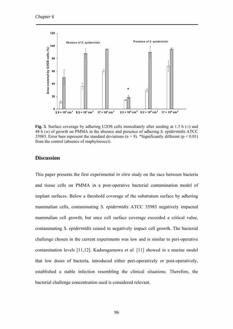
Chapter 6
Fig. 5. Surface coverage by adhering U2OS cells immediately after seeding at 1.5 h (□) and 48 h (■) of growth on PMMA in the absence and presence of adhering S. epidermidis ATCC 35983. Error bars represent the standard deviations (n = 9). *Significantly different (p < 0.01) from the control (absence of staphylococci).
Discussion
This paper presents the first experimental in vitro study on the race between bacteria
and tissue cells on PMMA in a post-operative bacterial contamination model of
implant surfaces. Below a threshold coverage of the substratum surface by adhering
mammalian cells, contaminating S. epidermidis ATCC 35983 negatively impacted
mammalian cell growth, but once cell surface coverage exceeded a critical value,
contaminating S. epidermidis ceased to negatively impact cell growth. The bacterial
challenge chosen in the current experiments was low and is similar to peri-operative
contamination levels [11,12]. Kaduragamuwa et al. [11] showed in a murine model
that low doses of bacteria, introduced either peri-operatively or post-operatively,
established a stable infection resembling the clinical situations. Therefore, the
bacterial challenge concentration used is considered relevant.
96

In vitro post-operative contamination model
The current study was conducted with S. epidermidis. Clinically, S.
epidermidis is one of the main causative organisms for BAI in particular chronic
prosthetic joint infections [13,14]. S. epidermidis can yield early post-operative
contamination as long as wound closure is incomplete. Alternatively, late
haematogenous spreading may occur from infections elsewhere in the body to an
implant surface. Notorious in this aspect are abscesses underneath the skin,
developing for instance after minor injuries. Also dental treatment is known to be a
cause of post-operative contamination of implant surfaces, as even routine inspection
of the dentition by a dentist or mouth hygienist may give rise to bacteraemia [15,16].
BAI due to S. epidermidis is usually low grade, since the organism lacks the genes to
produce toxins and tissue damaging exoenzymes, that are produced by, for instance
Staphylococcus aureus [17,18], another causative strain of BAI [13,14]. S. aureus
infections are therefore more aggressive than S. epidermidis ones, but at the same time
are more readily noticed and treated. In a sense this makes the low-grade BAI due to
S. epidermidis more troublesome, which motivates our current choice for S.
epidermidis to set up our post-operative contamination model.
PMMA is well-know for its use in ophthalmological, orthopedic and dental
applications. In orthopedic applications, PMMA-based bone cements are extensively
used for fixation of total joint replacements, as the material supports cell adhesion and
spreading [19-21]. In a peri-operative contamination model, PMMA showed better
tissue cell adhesion and spreading in the presence of adhering S. epidermidis than
other commonly used biomaterials [10].
In the concept of the race for the surface, complete surface coverage of a
biomaterial in vivo by viable tissue cells, combined with functional host defense
mechanisms resist the negative consequences of bacterial contamination [8]. Rapid
97

Chapter 6 and complete surface coverage of mammalian cells restricts the biomaterial surface
area available for bacterial adhesion and biofilm formation. Dexter et al. [22]
suggested that an optimal concentration of seeded 3T3 fibroblasts and conditions to
stimulate cell adhesion and surface coverage without stimulating bacterial adhesion
could probably reduce infection. However, the consequence of the combined presence
of mammalian cells and bacteria on a surface was not monitored. In this study, the
presence of S. epidermidis ATCC 35983 showed negative effects on cell surface
coverage by U2OS cells at the lowest initial cell density (2.5 × 10⁴ cells cm-2) as
compared to cell surface coverage in the absence of adhering staphylococci. In
contrast, surface coverage of U2OS cells after seeding higher initial cell densities was
not influenced by the presence of S. epidermidis. Therewith this study provides direct
experimental evidence for the Gristina postulate [8] that in vivo tissue integration
protects implant surfaces against bacterial colonization. Moreover, our study defines a
critical cell surface coverage needed in order to protect an implant surface against S.
epidermidis biofilm formation. We anticipate that this critical cell surface coverage
level will depend on the biomaterial surface characteristics as well as on the infecting
strain. With this model system we present a tool to assess the ‘race for the surface’
and compare different biomaterials, coatings and bacterial strains.
Conclusion
An in vitro method is presented to study the effects of post-operative bacterial
contamination on the interaction of mammalian cells with biomaterial implants. A
critical mammalian cell surface coverage was found, above which contaminating S.
epidermidis ATCC 35983 no longer had a negative impact on mammalian cell
growth. Thus following the concept of the race for successful implantation of a
98

In vitro post-operative contamination model biomaterial, survival of the implant will be more solidly assured if an implant is
rapidly integrated by tissue to above this critical cell surface coverage. This new
method and the concept of critical cell surface coverage will allow better evaluation of
biomaterial coatings prior to animal experiments or human trials, than based on
separate studies of microbial adhesion to or mammalian cell interactions with such
coatings.
99

Chapter 6 References
1. Singh R, Ray P, Das A, Sharma M. Role of persisters and small-colony variants in antibiotic resistance of planktonic and biofilm-associated Staphylococcus aureus: an in vitro study. J Med Microbiol 2009;58:1067-1073.
2. Proctor RA, Von Eiff C, Kahl BC, Becker K, McNamara P, Herrmann M, Peters G. Small colony variants: a pathogenic form of bacteria that facilitates persistent and recurrent infections. Nat Rev Microbiol 2006;4:295-305.
3. Gristina AG, Rovere GD, Shoji H, Nicastro JF. An in vitro study of bacterial response to inert and reactive metals and to methyl methacrylate. J Biomed Mater Res A 1976;10:273-281.
4. Gristina AG, Naylor P, Myrvik QN. Musculoskeletal infection, microbial adhesion, and antibiotic resistance. Infect Dis Clin North Am 1990;4:391-408.
5. Habash M, Reid G. Microbial biofilms: Their development and significance for medical device-related infections. J Clin Pharmacol 1999;39:887-898.
6. Gristina AG. Biomaterial-centered infection: microbial adhesion versus tissue integration. Science 1987;237:1588-1595.
7. Gristina AG, Naylor P, Myrvik Q. Infections from biomaterials and implants: a race for the surface. Med Prog Technol 1998;14:205-224.
8. Gristina AG. Implant failure and the immune-incompetent fibro-inflammatory zone. Clin Orthop Rel Res 1994;298:106-118.
9. Subbiahdoss G, Kuijer R, Grijpma DW, Van der Mei HC, Busscher HJ. Microbial biofilm growth vs. tissue integration: "The race for the surface" experimentally studied. Acta Biomater 2009;5:1399-1404.
10. Subbiahdoss G, Grijpma DW, Van der Mei HC, Busscher HJ, Kuijer R. Microbial biofilm growth vs. tissue integration on biomaterials with different wettabilities and a polymer – brush coating. J Biomed Mater Res A 2010;DOI: 10.1002/jbm.a.32731.
11. Kadurugamuwa JL, Sin L, Albert E, Yu, J, Francis K, DeBoer M, Rubin M, Kawahara CB, Parr TR, Contag PR. Direct continuous method for monitoring biofilm infection in a mouse model. Infect Immun 2003;71:882-890.
12. Fitzgerald RH, Jones DR. Hip implant infection - Treatment with resection arthroplasty and late total hip-arthroplasty. Am J Med 1985;78:225-228.
13. Young EJ, Sugarman B. Infections in prosthetic devices. Surg Clin North Am 1988;68:167-180.
14. Christensen GD, Baldassarri L, Simpson WA. Methods for studying microbial colonization of plastics. Methods Enzymol 1995;253:477-500.
100

In vitro post-operative contamination model
101
15. Okell CC, Elliott CD. Bacteriaemia and oral sepsis with special reference to the etiology of subacute endocarditis. Lancet 1935;2:869-875.
16. Ohara-Nemoto Y, Haraga H, Kimura S, Nemoto TK. Occurrence of staphylococci in the oral cavities of healthy adults and nasal-oral trafficking of the bacteria. J Med Microbiol 2008;57:95-99.
17. Vuong C, Otto M. Staphylococcus epidermidis infections. Microb Infect 2002;4:481-489.
18. Massey RC, Horsburgh MJ, Lina G, Hook M, Recker M. Opinion - The evolution and maintenance of virulence in Staphylococcus aureus: a role for host-to-host transmission? Nat Rev Microbiol 2006;4:953-958.
19. Lydon MJ, Minett TW, Tighe BJ. Cellular interactions with synthetic-polymer surfaces in culture. Biomaterials 1985;6:396-402.
20. Van Wachem PB, Beugeling T, Feijen J, Bantjes A, Detmers JP, Vanaken WG. Interaction of cultured human-endothelial cells with polymeric surfaces of different wettabilities. Biomaterials 1985;6:403-408.
21. Van Wachem PB, Hogt AH, Beugeling T, Feijen J, Bantjes A, Detmers JP, Vanaken WG. Adhesion of cultured human-endothelial cells onto methacrylate polymers with varying surface wettability and charge. Biomaterials 1987;8:323-328.
22. Dexter SJ, Pearson RG, Davies MC, Camara M, Shakesheff KM. A comparison of the adhesion of mammalian cells and Staphylococcus epidermidis on fibronectin-modified polymer surfaces. J Biomed Mater Res A 2001;56:222-227.

CHAPTER 7
A New Method to Study the Simultaneous Interaction Between Bacteria, Macrophages and Osteoblasts on a
Biomaterial Implant Surface

Chapter 7 Introduction
Biomaterial-associated infections (BAI) are a widespread complication that threatens
the longevity and functionality of indwelling biomaterial implants and devices and the
consequences in terms of medical care are severe. Despite of improved techniques and
highly sterile conditions in the operating theatre, peri-operative contamination by
microorganisms suspended in the air and from the skin flora continues to be the most
common pathway for the contamination of biomaterial implants and devices [1,2].
Staphylococcus aureus and Staphylococcus epidermidis are microorganisms
frequently isolated from BAI. S. epidermidis is found in almost 50% of the infections
associated with catheters, artificial joints and heart valves, while S. aureus is seen in
around 23% of the infections associated with prosthetic joints [3]. These commensals
from the skin adhere to the biomaterial surface and grow to form a biofilm. Bacteria
in their biofilm mode of growth are frequently more resistant to antibiotic treatment
and the host immune system than their planktonic counterparts. Hence, removal of an
infected implant or device is often the only remedy for a BAI. Surrounding tissue,
however, may remain compromised by bacterial presence for prolonged periods of
time after removal of the biomaterial [4,5] which severely lowers the prospects of a
secondary implant or device, since bacteria in tissue constitute a new source for BAI
to develop.
Whether or not BAI will occur, ultimately depends on the interaction between
the biomaterial, the bacterium involved and host cells. Host cells and bacteria battle to
proliferate and colonize a biomaterial surface. The general assumption is that on
surfaces with a high affinity for tissue cells, bacterial biofilm formation will be
limited, thus decreasing the risk of a BAI. Contrary, if bacteria grow and colonize
104

Bacteria-macrophages-osteoblasts interactions faster than tissue cells, bacterial toxins and virulence factors can impair cell functions
leading to BAI [6].
In a healthy host, the host immune system comes to the aid of tissue cells [7].
Macrophages are one of the most predominant immune cells that arrive within
minutes to hours at an implant site and can remain at a biomaterial surface for several
weeks to orchestrate the inflammation process and eventually foreign body reactions
[7]. During infection, macrophages detect bacteria via cell surface receptors that bind
to bacterial ligands and opsonines [8]. Subsequently, macrophages ingest pathogens
and activate cellular functions such as proliferation, secretion of proteins and
cytokines, and respiratory burst to destroy phagocyted microorganisms and recruit
other cells from the adaptive immune system [9]. However, it has been shown that the
presence of a foreign body may impair the host immune system and very low numbers
of adherent bacteria are already sufficient to create a BAI [6].
Currently, biomaterials research is strongly focused on the design of novel
functional coatings that reduce the risk of BAI by inhibiting bacterial adhesion and
stimulating tissue cell adhesion [10,11]. However, in the evaluation of these coatings,
bacterial and tissue cell adhesion are often considered as independent phenomena and
not as simultaneous events [10,11]. Furthermore, the response of the immune system
to bacterial colonization on these coatings is generally ignored. Recently, a novel in
vitro method has been forwarded to determine the influence of bacterial presence on
the outcome of the competition between bacteria and tissue cells in their attempt to
colonize or integrate a biomaterial surface [12].
In this study we present an extension of this model [12] to include also the
influence of macrophages on the outcome of the race for the surface between adhering
bacteria and tissue cells in a single experiment.
105
Chapter 7 Materials and Methods
Biomaterial. Poly (methyl methacrylate) (PMMA) (Vink Kunststoffen, Didam, The
Netherlands) was used as a substratum. Samples were rinsed thoroughly with 70%
ethanol (Merck, Darmstadt, Germany) and washed with sterile ultrapure water before
use. Water contact angles on thus cleaned PMMA were 73 ± 3 degrees, in line with
literature [13].
Tissue cell culturing and harvesting. U2OS osteosarcoma cells were routinely
cultured in Dulbecco’s modified Eagles Medium (DMEM)-low glucose supplemented
with 10% fetal calf serum (FBS, non-heat inactivated), 0.2 mM of ascorbic acid-2-
phosphate (AA2P). Cells were maintained at 37°C in a humidified atmosphere with
5% CO2, and passaged at 70 – 90% confluency using trypsin/EDTA.
Macrophages culturing and harvesting. J774 murine macrophages were routinely
cultured in Dulbecco’s modified Eagles Medium (DMEM)-high glucose
supplemented with 10% fetal calf serum (FBS, non-heat inactivated) and denoted in
the paper as “optimal medium”. Macrophages were maintained at 37°C in a
humidified atmosphere with 5% CO2, and passaged at 70 – 80% confluency by
scraping.
Bacterial growth conditions and harvesting. The bacterial strains used in this study
were S. epidermidis ATCC 35983 and S. aureus ATCC 12600. These strains were
cultured on blood agar plates and grown aerobically overnight at 37°C, and the plates
were kept at 4°C, never longer than two weeks. For each experiment, one colony was
used to inoculate 10 ml of tryptone soya broth (TSB, OXOID, Basingstoke, England)
106

Bacteria-macrophages-osteoblasts interactions and incubated for 24 h at 37°C in ambient air. This culture was used to inoculate
another culture in 200 ml TSB that was incubated overnight at 37°C prior to
harvesting. Bacteria were harvested by centrifugation (5 min at 5000 g at 10°C) and
washed twice with sterile ultrapure water. Bacteria were sonicated intermittently on
ice (30 s) in sterile phosphate buffered saline (PBS, 10 mM potassium phosphate, 150
mM NaCl, pH 6.8) to break bacterial aggregates, and resuspended in 200 ml sterile
PBS to a concentration of 3 x 10⁶ bacteria per ml. Prior to the experiments, growth
and biofilm formation of S. epidermidis ATCC 35983 and S. aureus ATCC 12600 in
optimal medium was confirmed by culturing bacteria in optimal medium for 24 h.
In vitro multiple cell type culture assays. The competition between bacteria and
U2OS cells for the colonization of PMMA in the absence of macrophages (control)
and in the presence of macrophages was assessed under laminar flow on the bottom
plate of a parallel plate flow chamber (175 x 17 x 0.75 mm3). Bacterial and cell
deposition were observed real-time using a CCD camera (Basler AG, Germany)
mounted on a phase-contrast microscope Olympus BH-2 (Olympus, Germany). The
flow chamber was equipped with heating elements and kept at 37°C throughout the
experiments.
Prior to each experiment, all tubes and the flow chamber were filled with
sterile PBS, taking care to remove all air bubbles from the system. Once the system
was filled, and before the addition of bacterial suspension, PBS was allowed to flow
through the system at a shear rate of 11 s-1. Then, a bacterial suspension in PBS was
perfused through the chamber at the same shear rate and phase-contrast images were
obtained. As soon as the desired density of adhering bacteria (103 cm-2), was reached,
flow was switched to sterile PBS to remove unbound bacteria and the bacterial
107

Chapter 7 suspension from the tubes and chamber. Subsequently, a cell suspension consisting of
U2OS cells (6 × 10⁵ cells ml-1) and J774 macrophages (12 × 105 cells ml-1) in
optimum medium was added to the flow chamber. Once the entire volume of buffer
inside the chamber was replaced by the cell suspension, flow was stopped for 1.5 h in
order to allow U2OS cells and macrophages to adhere and spread on the substratum
surface. Ultimately, “optimum medium” supplemented with 2% HEPES was perfused
through the system without recirculation at a shear rate of 0.14 s-1 for 24 h and phase-
contrast images were obtained continuously at 2 min intervals. Biofilm growth was
assessed in real-time by determining the numbers of adhering bacteria per unit area
using proprietary software based on the Matlab Image processing Toolkit (The
MathWorks, MA, USA).
At the end of the assay, surfaces were prepared for qualitative analysis to
assess U2OS cell and macrophages morphology and spreading. Cells adhering to
PMMA were fixed with citrated-acetone-formaldehyde fixative solution for 30 s and
stained with an alkaline-dye mixture (Sigma-Aldrich, Germany) (Naphtol AS-BI
phosphate, sodium nitrite, fast blue BB base) for 15 min. The samples were
subsequently rinsed with demineralized water and counterstained for 2 min with
neutral red solution. Then the samples were rinsed once again with demineralized
water, allowed to dry and phase-contrast images were taken on different places of the
sample. Differentiated U2OS osteosarcoma cells stained purple/blue (alkaline
phosphatase-positive) and macrophages were orange stained.
Results
Bacteria were allowed to adhere to the biomaterial surface prior to U2OS cell and
macrophage adhesion, mimicking a peri-operative contamination after which bacteria,
108

Bacteria-macrophages-osteoblasts interactions U2OS cells and macrophages were allowed to grow simultaneously for 24 h. Events
are illustrated as follows.
Migration of macrophages towards bacteria and phagocytosis. The number of
bacteria adhering to the PMMA surface prior to U2OS cells and macrophages
adhesion was set to 103 cm-2, using the image analysis system Subsequently, U2OS
cells and macrophages were allowed to adhere to the surface and the simultaneous
interactions of bacteria, macrophages and U2OS cells were observed by phase-
contrast microscopy. Figure 1 shows macrophage migration in the presence of U2OS
cells towards adhering bacteria and subsequent phagocytosis. Macrophage migration
towards bacteria and phagocytosis was similar on PMMA colonized by S. epidermidis
and S. aureus.
Fig. 1. Phase-contrast images of macrophage activity toward S. epidermidis ATCC 35983 on a PMMA surface in the presence of U2OS cells: macrophage migration towards S. epidermidis (images 1-5), bacterial clearance by phagocytosis (images 6-7) and further migration (images 8-12). The bar denotes 50 µm.
109

Chapter 7 Bacterial biofilm formation in the absence and presence of macrophages. Biofilm
growth was assessed over time by determining the numbers of bacteria adhering to
PMMA at different time points during the simultaneous growth of bacteria, U2OS
cells and macrophages (Fig. 2). In the presence of macrophages, reduction in the
numbers of adherent bacteria, both for S. epidermidis and S. aureus, was observed as
compared to controls (absence of macrophages). This effect was observed up to 20 h
of growth for S. epidermidis and up to 14 h for S. aureus. Thereafter macrophage
burst and release of ingested bacteria was observed.
Fig. 2. The numbers of adhering bacteria on PMMA as a function of time during the simultaneous growth of bacteria and U2OS cells in the absence and presence of macrophages in a parallel plate flow chamber (shear rate 0.14 s-1). S. epidermidis in the absence of macrophages (□), S. aureus in the absence of macrophages (○), S. epidermidis in the presence of macrophages (■) and, S. aureus in the presence of macrophages (●).
Bacterial-tissue cell interactions in the absence and presence of macrophages.
Immediately after seeding, U2OS cell adhesion and spreading on PMMA was
observed independently of whether macrophages were present or not. After 24 h of
simultaneous growth, U2OS cell death was observed in the presence of a S. aureus
110

Bacteria-macrophages-osteoblasts interactions biofilm irrespective of the absence or presence of macrophages. On the other hand,
colonizing S. epidermidis did not significantly affect U2OS cells and their adhesion
and spreading were similar both in the absence and presence of macrophages (see Fig.
3).
Fig. 3. Phase-contrast images of adhered cells to PMMA after 24 h of simultaneous growth of U2OS and S. epidermidis ATCC 35983 or S. aureus ATCC 12600 in the absence (upper images) and presence (lower images) of macrophages. Macrophages are orange-stained. The bar denotes 50 µm.
Discussion
This paper presents the first experimental model to study the simultaneous interaction
of macrophages-bacteria-osteoblasts on a biomaterial surface in a single experiment.
In our in vitro model, bacteria were allowed to adhere prior to adhesion of
macrophages and U2OS cells, which mimics a peri-operative bacterial contamination
of implant surfaces. The number of bacteria adhering on the PMMA surface prior to
macrophages and U2OS cell adhesion was set to 103 cm-2. In the past, it has been
111

Chapter 7 documented that during a surgical procedure of 1 h, the total number of bacteria
carrying particles falling on a wound is about 270 cm-2. The bacterial counts were
generally higher during periods of high activity and when more people were present in
the operation theatre [14]. Recently, through the use of modern, better ventilated
operation theatres (20 changes of air/h) and impermeable patient and personnel
clothing, peri-operative bacterial contamination is likely to be reduced [15]. However,
many surgical procedures in which implants are introduced in the body last longer
than 1 h. Therefore, the level of bacterial contamination chosen in our experiments is
probably realistic of a worst case scenario. Despite these low numbers, peri-
operatively introduced organisms, particularly when of low virulence, can survive on
an implant surface for prolonged periods of time and later, during periods of host
immune depression, they proliferate and establish an infection with clinical symptoms
[16].
The pathogenesis of BAI is complex and depends on factors such as bacterial
virulence, physicochemical properties of the biomaterial and alterations in the host
defense [17]. Previously, in our model for the competition between bacteria and tissue
cells, all common biomaterial surfaces, including PMMA, allowed S. epidermidis
ATCC 35983 biofilm formation with a negative impact on the coverage of the
biomaterial surface by tissue cells [13]. Yet, PMMA showed better cell adhesion and
spreading in the presence of adhering S. epidermidis ATCC 35983 than other
commonly used biomaterials [13]. Our present study supports our previous
observations that U2OS cells are able to adhere, spread and grow in the presence of S.
epidermidis ATCC 35983, and extend these observations to the absence and presence
of macrophages. On the other hand, in the presence of adhering S. aureus ATCC
12600, death of all adhering U2OS cells and macrophages within 18 h was observed
112

Bacteria-macrophages-osteoblasts interactions despite the suspected removal of the majority of the bacterial toxins by flow. These
observations are in line with clinical findings that BAI due to S. aureus usually
progresses much more aggressively than BAI caused by S. epidermidis. In S.
epidermidis infections, biofilm formation is considered the only virulence factor and
therefore infections are usually sub-acute or chronic. The low virulence of S.
epidermidis strains compared to S. aureus is due to the lack of additional genes
responsible for producing severely tissue damaging toxins [3,18].
In general, immune cells migrate, engulf and kill invading microorganisms
[19-21]. A previous study on the interaction between macrophages and colonizing S.
epidermidis, showed that macrophage behavior is surface dependent [22].
Macrophage migration towards bacteria and phagocytosis was enhanced on cross-
linked poly(ethylene)-glycol (PEG) based polymer coatings compared to the uncoated
substrata due to the weak adhesion of macrophages and bacteria to the PEG coating
[22]. In our study, macrophages migrate towards the bacteria on a PMMA surface and
engulfed the bacteria. The phagocytosis of bacteria by macrophages differs depending
on the virulence of the strain. In the presence of low virulent S. epidermidis, bacterial
biofilm growth was strictly reduced by the presence of macrophages up to 20 h
compared to only 14 h in the case of high virulent S. aureus biofilm growth. These
results are in line with previous studies showing that in both in vivo and in vitro the
uptake rate of bacteria by macrophages was inversely proportional to the virulence of
the bacteria [23,24]. Furthermore, macrophages disintegration and necrotic death has
been observed in vitro and in vivo due to overloading with ingested bacteria [25,26].
In this study it was observed that after a period of time macrophages become
exhausted and break open which leads to a burst release of bacteria. At least part of
these bacteria appeared to be active in the flow chamber. These findings suggest that
113

Chapter 7 J774 macrophages in this model are not able to kill all phagocytised bacteria.
Although the viability of the released bacteria was not assessed, several studies have
demonstrated that immune cells lose their ability to kill bacteria [25,21,27,28]. Leid et
al. [29] showed that leukocytes were able to migrate to S. aureus biofilms but failed to
phagocyte the bacteria. Neutrophils adjacent to Teflon cages, implanted in peritoneal
cavities, exhibited decreased bactericidal activity and reduced superoxide production
due to the increased production of S. epidermidis extracellular slime [30-33].
Watanabe et al. [28] demonstrated that engulfed S. aureus suppressed the production
of superoxide, resulting in the prolonged survival inside the macrophages. In a murine
model it was shown that high numbers of S. epidermidis could persist within
macrophages in peri-catheter tissue without showing any signs of inflammation [17].
Also S. epidermidis inside macrophages were not only viable but were able to
proliferate. In vivo, the local host defense was compromised because of the presence
of biomaterials, resulting in deficient intracellular killing of pathogens by
macrophages [17].
The influence of macrophages on the competition between bacteria and
mammalian cells is novel. This study demonstrates that despite the presence of
macrophages, mammalian cells lost the race for the surface in the presence of high
virulent S. aureus. In vivo, bacteria may well survive inside the macrophages for
prolonged periods of time. These bacterial will favor the development of BAI,
especially when certain physical conditions of the patients disturb the balance
between bacteria and the host response [17]. This model validated for bacteria-
macrophages-osteoblasts interactions in a flow chamber system resembles the in vivo
environment more closely than single-cell type cultures therewith providing an
important bridge between in vitro and in vivo studies. Even though, this study was
114
Bacteria-macrophages-osteoblasts interactions qualitatively analyzed, we believe that this methodology supported with quantitative
data, could be a suitable tool for evaluation of biomaterials based on infection models.
115

Chapter 7 References
1. Davis N, Curry A, Gambhir AK, Panigrahi H, Walker CR, Wilkins EG, Worsley MA, Kay PR. Intraoperative bacterial contamination in operations for joint replacement. J Bone Joint Surg Br 1999;81:886-889.
2. Hughes SP, Anderson FM. Infection in the operating room. J Bone Joint Surg Br 1999;81:754-755.
3. Khalil H, Williams RJ, Stenbeck G, Henderson B, Meghji S, Nair SP. Invasion of bone cells by Staphylococcus epidermidis. Microb Infect 2007;9:460-465.
4. Broekhuizen CA, De Boer L, Schipper K, Jones CD, Quadir S, Feldman RG, Dankert J, Vandenbroucke-Grauls CMJE, Weening JJ, Zaat SAJ. Peri-implant tissue is an important niche for Staphylococcus epidermidis in experimental biomaterial associated infection in mice. Infect Immun 2007;75:1129–1136.
5. Vuong C, Otto M. Staphylococcus epidermidis infections. Microb Infect 2002;4:481-489.
6. Gristina AG. Biomaterial-centered infection: microbial adhesion versus tissue integration. Science 1987;237:1588-1595.
7. Anderson JM. Inflammation, wound healing, and the foreign-body response. pp 296-304. In: B.D. Ratner, A.S. Hoffman, F.J. Schoen and J.E. Lemons (ed.), Biomaterials Science. An Introduction to Materials in Medicine. 2004;Elsevier, San Diego, CA.
8. Xia Z, Triffitt JT. A review on macrophage responses to biomaterials. Biomed Mater 2006;1:R1-9.
9. Berton G, Lowell CA. Integrin signalling in neuthrophils and macrophages. Cell Signal 1999;11:621-635.
10. Maddikeri RR, Tosatti S, Schuler M, Chessari S, Textor M, Richards RG, Harris LG. Reduced medical infection related bacterial strains adhesion on bioactive RGD modified titanium surfaces: A first step toward cell selective surfaces. J Biomed Mater Res 2008;84A:425-435.
11. Shi ZL, Neoh KG, Kang ET, Poh C, Wang W. Bacterial adhesion and osteoblast function on titanium with surface-grafted chitosan and immobilized RGD peptide. J Biomed Mater Res 2008;86A:865-872.
12. Subbiahdoss G, Kuijer R, Grijpma DW, Van der Mei HC, Busscher HJ. Microbial biofilm growth vs. tissue integration: "The race for the surface" experimentally studied. Acta Biomater 2009;5:1399-1404.
13. Subbiahdoss G, Grijpma DW, Van der Mei HC, Busscher HJ, Kuijer R. Microbial biofilm growth vs. tissue integration on biomaterials with different wettabilities
116

Bacteria-macrophages-osteoblasts interactions
and a polymer – brush coating. J Biomed Mater Res A 2010;DOI:10.1002/jbm.a.32731.
14. Fitzgerald RH. Microbiologic environment of the conventional operating-room. Arch Surg 1979;114:772-775.
15. Verkkala K, Eklund A, Ojajarvi J, Tiittanen L, Hoborn J, Makela P. The conventionally ventilated operating theatre and air contamination control during cardiac surgery - bacteriological and particulate matter control garment options for low level contamination. Eur J Cardiothorac Surg 1998;14:206-210.
16. Dougherty SH. Pathobiology of infection in prosthetic devices. Rev Infect Dis 1988;10:1102-1117.
17. Boelens JJ, Dankert J, Murk JL, Weening JJ, Van der Poll T, Dingemans KP, Koole L, Laman JD, Zaat SAJ. Biomaterial-associated persistence of Staphylococcus epidermidis in pericatheter macrophages. J Infect Dis 2000;181:1337-1349.
18. Massey RC, Horsburgh MJ, Lina G, Hook M, Recker M. Opinion - The evolution and maintenance of virulence in Staphylococcus aureus: a role for host-to-host transmission? Nat Rev Microbiol 2006;4:953-958.
19. Babior BM. Oxidants from phagocytes: agents of defense and destruction. Blood 1984;64:959-966.
20. Guenther F, Wabnitz GH, Stroh P, Prior B, Obst U, Samstag Y, Wagner C, Haensch GM. Host defence against Staphylococcus aureus biofilms infection: Phagocytosis of biofilms by polymorphonuclear neutrophils (PMN). Mol Immun 2009;46:1805-1813.
21. Kaplan SS, Heine RP, Simmons RL. Defensins impair phagocytic killing by neutrophils in biomaterial-related infection. Infect Immun 1999;67:1640-1645.
22. Saldarriaga Fernández IC, Da Silva Domingues JF, Van Kooten TG, Metzger S, Grainger DW, Busscher HJ, Van der Mei HC. Macrophage response to staphylococcal biofilm on cross-linked poly(ethylene) glycol polymer coatings in vitro. Eur Cells Mater.2010;Submitted.
23. Valtonen MV, Plosila M, Valtonen VV, Maekelae PH. Effect of the quality of lipopolysaccharide on mouse virulence of Salmonella enteritidis. Infect Immun 1975;12:828-832.
24. Valtonen M Role of phagocytosis in mouse virulence of Salmonella enteritidis recombinants with O-antigen 6,7 or 4,12. Infect Immun 1977;18:574-582.
25. Bonventre PF, Imhoff JG. Uptake of 3H-dihydrostreptomycin by macrophages in culture. Infect Immun 1970;2:89-95.
117

Chapter 7
118
26. Luhrmann A, Mauder N, Sydor T, Fernandez-Mora E, Schulze-Luehrmann J, Takai A, Haas S. Necrotic death of Rhodococcus equi-infected macrophages is regulated by virulence-associated plasmids. Infect Immun 2004;72:853-862.
27. Tofte RW, Petersoson PK, Kim Y, Quie PG. Influence of serum concentration on opsonization by the classical and alternative complement pathways. Infect Immun 1980;27:693-696.
28. Watanabe I, Ichiki M, Shiratsuchi A, Nakanishi Y. TLR2-Mediated survival of Staphylococcus aureus in macrophages: A novel bacterial strategy against host innate immunity. J Immunol 2007;178:4917-4925.
29. Leid JG, Shirtliff HG, Costerton JW, Stoodley P. Human leukocytes adhere to, penetrate, and to respond to Staphylococcus aureus biofilms. Infect Immun 2002;70:6339-6345.
30. Johnson GM, Lee DA, Regelmann WE. Interference with granulocyte function by Staphylococcus epidermidis slime. Infect Immun 1986;54:13-20.
31. Myrvit QN, Wagner W, Barth E, Wood P, Gristina AG. Effects of extracellular slime produced by Staphylococcus epidermidis on oxidative responses of rabbit alveolar macrophages. J Invest Surg 1989;2:381-389.
32. Shanbhag A, Yang J, Lilien J, Black J. Decreased neutrophil respiratory burst on exposure to cobaltchrome alloy and polystyrene in vitro. J. Biomed Mater Res 1992;26:185-195.
33. Zimmerli W, Waldvogel F. Pathogenesis of foreign body infection. J Clin Invest 1984;73:1191-1200.

CHAPTER 8
General Discussion

Chapter 8 Hitherto, biomaterials or functional coatings for application in dentistry or orthopedics
addressing infections are evaluated in vitro either for their ability to resist bacterial
adhesion and biofilm formation or for their ability to enhance mammalian cell
adhesion and proliferation [1-6]. However, according to Anthony Gristina, in vivo the
fate of a biomaterial implant can be depicted as a race between microbial adhesion
and biofilm formation on the implant surface versus tissue integration [7]. Even
though the concept of the race for the surface has been embraced by many researchers
in the field, unfortunately there has been no in vitro methodology forwarded to study
the actual race. In vivo, the processes occurring at a biomaterial surface are quite
complicated and involve multiple cell types, cytokines, chemokines, and secretion of
bacterial and cellular substances [8]. These processes are difficult to adequately
address with current in vitro models, but in vitro experiments are indispensable in
order to develop new functional materials and coatings. Moreover, well designed and
predictive in vitro experiments are often a requirement in order to obtain approval for
animal experiments and at the same time reduce the need for animal experiments.
Therefore, the goal of the studies described in this thesis was to bridge the gap
between the in vitro and in vivo evaluation of biomaterials-associated infections.
Bacterial Biofilm Growth versus Mammalian Cell Growth
As a first step, we developed a methodology to conduct the competition between
bacteria and mammalian cells on the biomaterial surface. The development of
methodologies were based on the two infection models: peri-operative and post-
operative contamination of biomaterial implants. In the methodology based on peri-
operative infection, Staphylococcus epidermidis ATCC 35983 was allowed to adhere
120
General discussion to a biomaterial surface prior to U2OS osteosarcoma cell adhesion and spreading to
mimic the clinical situation where an implant becomes contaminated prior to
implantation. We observed that the U2OS cells lost the competition in the absence of
flow due to the accumulation of bacterial toxins, but were able to grow under flow
due to continuous supply of fresh medium and remove the bacterial toxins from the
interface. In vivo fluid flows continuously through the net-work of fine channels of
osteocytes to facilitate the diffusion of nutrients and waste products [9]. In the concept
of the race for the surface, tissue integration is an important protective factor against
bacterial contamination of an implant surface [7]. Therefore, another methodology
was developed based on post-operative contamination to investigate the effects of
different degrees of mammalian cell coverage on the balance between bacterial
biofilm formation and mammalian cell growth. We observed that mammalian cell
growth was severely impaired when bacteria were introduced on surfaces with a low
initial mammalian cell density (2.5 × 10⁴ cells cm-2) but in the presence of higher
initial cell densities (8.2 × 10⁴ cells cm-2 and 17 × 104 cells cm-2), contaminating
staphylococci did not affect cell growth.
Results suggest that the methodologies based on these two infection models
could be well used in the evaluation of biomaterials infections, resembling the in vivo
situation more closely than traditional single cell type cultures.
Evaluation of Biomaterials
Biomaterials can be broadly classified into two main categories biopassive and
bioactive. Biopassive materials cannot induce any interfacial biological bond between
implants and host cells. Metallic materials such as cobalt-chromium-molybdenum
121

Chapter 8 alloys and titanium-aluminium-vanadium alloys are used in hip replacements [10] and
polymeric materials such as poly (methyl methacrylate) (PMMA),
(hydroxyethylmethacrylate), polyethylene oxide (PEO), phosphoryl choline are used
in the field of ophthalmology (i.e contact lenses, intraocular lenses, keratoprosthesis
optics and vitreous substitutes) [11] are some examples of biopassive materials. None
of the synthetic materials are truly inert but materials like phosphoryl choline and
polyethylene oxide are considerably promising in down-regulating biological
responses in ophthalmology [11].
Bioactive materials can attach directly with body tissues and form chemical
and biological bonds during the early stages of the post implantation period. Materials
such as tricalcium phosphates, hydroxyapatites and calcium sulphate are some
examples of bioactive materials [10]. Materials like tricalcium phosphates are
osteoconductive because osteoblasts adhere and deposit bony tissue on the surface.
Current efforts are being directed towards distilling the essence of cell adhesion
requirements in new biomaterials incorporating minimal peptide sequences from
adhesion molecules responsible for integrin binding. Biomaterials incorporated with
RGD (arginine-glycine-aspartic acid) enhances tissue integration [11].
Based on the Gristina’s ‘race for the surface’, the competition between S.
epidermidis ATCC 35983 and U2OS cells on different biomaterials showed that
mammalian cell interactions with biomaterials were hampered by bacterial biofilm
formation on all biomaterials used. Yet, PMMA showed better U2OS cell adhesion
and spreading in the presence of S. epidermidis compared to other biomaterials
(Chapter 3). This study suggest that the race for the surface depends on the physico-
chemical properties of the biomaterial surface and on the density of contaminating
bacteria. In a comparative study, bi-functional coatings (bioactive PLL-g-PEG/PEG-
122
General discussion RGD) showed better cell adhesion and spreading and cells were less affected by the
presence of S. epidermidis ATCC 35983 compared to mono-functional coatings
(biopassive PLL-g-PEG) and bare titanium oxide surfaces (Chapter 5).
Bacterial Strain-Specific Effects on the Competition Between
Bacterial Biofilm Growth versus Mammalian Cell Growth
Many different pathogens such as S. epidermidis, Staphylococcus aureus, Escherichia
coli and Pseudomonas aeruginosa can be isolated from infected biomaterial implant
surfaces, depending on their body site [7,8,12]. After an initial focus on S.
epidermidis, our study on the competition between bacteria and mammalian cells was
extended to different bacterial strains. We observed U2OS cell death within 18 h of
simultaneous growth of S. aureus or P. aeruginosa and U2OS cells despite removal of
bacterial toxins by flow, whereas U2OS cells were able to adhere, spread and grow in
the presence of S. epidermidis. These observations are inline with the clinical findings
that BAI due to highly virulent S. aureus and P. aeruginosa usually progresses much
more aggressively than BAI caused by low virulent S. epidermidis. This study
highlights the importance of bacterial virulence as a factor in the pathogenesis of BAI.
However, characterization and quantification of substances released by bacteria was
not performed. Hence, exact parameters responsible for mammalian cell death were
unknown. However, studies showed that in S. aureus, most extracellular and surface
attached virulence factors are regulated by the agr (accessory gene regulator) locus, a
quorum-sensing system and the expression of agr-regulated targets is bacterial cell
density-dependent. With increase in bacterial cell density agr becomes active, and
starts producing extracellular degradative exoenzymes and toxins [13,15]. A similar
cell-to-cell signalling (quorum-sensing) system plays a major role in the P.
123

Chapter 8 aeruginosa virulence factors [14]. In contrast to S. aureus and P. aeruginosa, S.
epidermidis produce a very limited number of tissue damaging exoenzymes and
toxins. The only toxin made by S. epidermidis is N-formylated alpha-helical peptide
-toxin, which plays a major role in the construction of a structured biofilm and in the
detachment from surfaces [15].
Limitations of the In Vitro Experimental Model
In our experiments, we have focused on the development of in vitro methodology for
the evaluation of new functional biomaterials and coatings to prevent biomaterial-
associated infection. Our in vitro results are inline with clinical findings and provide
additional information prior to animal experiments than in vitro studies addressing
solely bacterial or tissue cell responses to biomaterials. However, the bacterial
phenotypic changes occurring in vivo, resistance against antibiotics and host immune
system are not taken into account in our in vitro model, which constitutes a major
limitation. Secondly, the mechanism of interaction between host and biomaterial
implant cannot be studied. Nevertheless, the evaluation of biomaterials based on these
methodologies provides more information than can be obtained based on traditional in
vitro studies and therewith form a good basis for further animal experiments.
Future Research
The model provided here to study the race for the surface methodologies shows that
biomaterials contaminated even with very low number of bacteria can ultimately
harvest a biofilm capable of influencing mammalian cell adhesion and growth
depending on the virulence factor of the pathogens (Chapter 4). Results suggest the
124

General discussion need for development of biomaterials or coatings with selective toxicity to bacteria
and only stimulating mammalian cell adhesion and growth.
It would be interesting to answer research questions such as: i) What would be
the outcome of the race for the surface if both bacteria and mammalian cells are
introduced to the biomaterial surface at the same time? ii) Could a biomaterial surface
completely covered by a monolayer of mammalian cells prevent bacterial
colonization? ii) What would be the outcome of the race for the surface if the
biomaterial surface is pre-treated with physiologically relevant proteins?
Future experiments also includes characterization and quantification of the
substances released by bacteria prior to the race for the surface study in order to gain a
better insight on the influence of bacteria on mammalian cells adhesion and growth.
This in vitro methodology could be progressed towards resembling the in vivo
environment quite closely by incorporating antibiotics, growth factors and cytokines.
Furthermore, animal experiments should be performed in order to validate the in vitro
model, but pre-screening materials and coatings with the methodology developed here
may save a large number of animals.
The choice of cell type in this thesis was mainly focussed on the orthopedic
applications, but this methodology could be applicable for other cell types and implant
systems, such as oral implants, surgical meshes and vascular grafts.
125

Chapter 8 References
1. Shi ZL, Neoh KG, Kang ET, Poh C, Wang W. Bacterial adhesion and osteoblast function on titanium with surface-grafted chitosan and immobilized RGD peptide. J Biomed Mater Res Part A 2008;86A:865-872.
2. Shi Z, Neoh KG, Kang ET, Poh C, Wang W. Titanium with surface-grafted dextran and immobilized bone morphogenetic protein-2 for inhibition of bacterial adhesion and enhancement of osteoblast functions. Tissue Eng Part A 2009;15:417-426.
3. Dexter SJ, Pearson RG, Davies MC, Camara M, Shakesheff KM. A comparison of the adhesion of mammalian cells and Staphylococcus epidermidis on fibronectin-modified polymer surfaces. J Biomed Mater Res 2001;56:222-227.
4. Harris LG, Tosatti S, Wieland M, Textor M, Richards RG. Staphylococcus aureus adhesion to titanium oxide surfaces coated with non-functionalized and peptide-functionalized poly(L-lysine)-grafted-poly(ethylene glycol) copolymers. Biomaterials 2004;25:4135-4148.
5. Maddikeri RR, Tosatti S, Schuler M, Chessari S, Textor M, Richards RG, Harris LG. Reduced medical infection related bacterial strains adhesion on bioactive RGD modified titanium surfaces: A first step toward cell selective surfaces. J Biomed Mater Res Part A 2008;84A:425-435.
6. Ploux L, Anselme K, Dirani A, Ponche A, Soppera O, Roucoules V. Opposite responses of cells and bacteria to micro/nanopatterned surfaces prepared by pulsed plasma polymerization and UV-irradiation. Langmuir 2009;25:8161-8169.
7. Gristina AG. Biomaterial-centered infection: microbial adhesion versus tissue integration. Science 1987;237:1588-1595.
8. Gristina AG. Implant failure and the immune-incompetent fibro-inflammatory zone. Clin Orthop Related Res 1994;298:106-118.
9. Jacobs CR, Yellowley CE, Davis BR, Zhou Z, Cimbala JM, Donahue HJ. Differential effect of steady versus oscillating flow on bone cells. J Biomechanics 1998;31:969-976.
10. Willmann G. Coating of implants with hydroxyapatite material connections between bone and metal. Adv Eng Mater 1999;1:95-105.
11. Allan B. Closer to nature: new biomaterials and tissue engineering in ophthalmology. Br J Ophthalmol 1999;83:1235-1240.
12. Gristina AG, Naylor PT, Myrvik QN. Musculoskeletal infection, microbial adhesion, and antibiotic resistance. Infect Dis Clin North Am 1990;4:391-408.
13. Khalil H, Williams RJ, Stenbeck G, Henderson B, Meghji S, Nair SP. Invasion of bone cells by Staphylococcus epidermidis. Microb Infect 2007;9:460-465.
126

General discussion
127
14. Van Delden C, Iglewski B.H. Cell-to-cell signalling and Pseudomonas aeruginosa infections. Emerging Infect Dis 1998;4:551-560.
15. Vuong C, Otto M. Staphylococcus epidermidis infections. Microb Infect 2002;4:481-489.


Summary

Summary
130
Biomaterial-associated infections (BAI) are the major cause of implant failure and can
develop many years after implantation. BAI can develop from peri-operative
microbial contamination of implant surfaces during implantation, immediately post-
surgery during hospitalization or by late hematogenous spreading from infections
elsewhere in the body. The microorganisms involved in BAI are resistant to
antibiotics due to their biofilm mode of growth and the infected implant has to be
removed in most cases.
In 1987, the orthopedic surgeon Anthony G. Gristina coined the term “the race
for the surface” to describe the fate of biomaterial implants in relation with the
development of BAI. The fate of a biomaterial implant was depicted as a race between
microbial adhesion and biofilm growth on an implant surface versus tissue
integration. If the race is won by tissue cells, then the surface is covered by tissue and
less vulnerable to bacterial colonization. On the other hand, if the race is won by
bacteria the implant surface will become rapidly covered by a biofilm and tissue cell
functions will be hampered by bacterial virulence factors and toxins. The concept of
race for the surface has been embraced by many researchers in this field, but hitherto
there has been no in vitro methodology forwarded to study the race.
Chapter 1 reviews the main causes of implant failure and development of
BAI. Irrespective of the route of infection, the fate of a biomaterial implant depends
mainly on the outcome of the so-called “race for the surface”. In the past till now,
researchers predicted the outcome of the race for the surface by in vitro evaluation of
biomaterials or functional coatings either for their ability to resist bacterial adhesion
and biofilm formation or for their ability to support mammalian cell adhesion and
proliferation. However, the effects of the presence of bacteria including the influence
of bacterial activity and toxins on mammalian cell adhesion and proliferation remains

Summary
131
unknown, which could completely change the fate of a biomaterial implant according
to the concept of the “race for the biomaterial surface”. Therefore, the main aim of
this thesis is to develop a method that could bridge the gap between in vitro and in
vivo studies on BAI.
Chapter 2, as a first step towards bridging the gap between in vitro and in
vivo studies, an in vitro experimental methodology was developed based on Anthony
G. Gristina‘s concept of the “race for the surface”. This methodology was developed
based on the peri-operative contamination model in which the implant becomes
contaminated prior to implantation. After developing a suitable growth medium that
allowed both bacterial and mammalian cell growth in the parallel plate flow chamber,
the simultaneous growth of Staphylococcus epidermidis ATCC 35983 and U2OS
osteosarcoma cells was studied for a period of 48 h. The outcome of the competition
between S. epidermidis ATCC 35983 and U2OS cells appeared to be dependent on
the number of bacteria present prior to U2OS cell seeding and the absence or presence
of fluid flow. U2OS cells lost the competition in the absence of flow, probably due to
the accumulation of bacterial toxins, but were able to grow under flow due to the
continuous supply of fresh medium and removal of most endotoxins from the
interface.
This methodology based on the peri-operative contamination model was used
to determine the influence of biomaterial surfaces with different wettabilities and a
polymer brush coating on the outcome of the competition between S. epidermidis
ATCC 35983 and U2OS cells which is described in chapter 3. This study
demonstrated that the presence of S. epidermidis reduced U2OS cell growth on all
surfaces, but adhering U2OS cell spread equally well in the absence and presence of
staphylococci. A hydrophilic polymer-brush coating discouraged bacteria and cellular

Summary
132
adhesion and growth. This study highlights the non existence of a surface on which
the race for the surface can be won by mammalian cells and therefore emphasizes the
need for biofunctionalized surfaces that discourage biofilm formation and support
mammalian cell growth at the same time.
There are different pathogens involved in BAI, such as S. epidermidis,
Staphylococcus aureus, Escherichia coli and Pseudomonas aeruginosa. It is known
that bacterial virulence is a contributing factor in pathogenesis of BAI. In chapter 4,
the influence of different strains of S. epidermidis, S. aureus and P. aeruginosa on the
outcome of the competition between bacteria and mammalian cells for the biomaterial
surface was studied. U2OS cells are able to adhere, spread and grow in the presence
of adhering S. epidermidis strains but not in the presence of adhering S. aureus or P.
aeruginosa, which cause death of all adhering U2OS cells within 18 h, despite the
continuous medium flow. These observations are in line with clinical findings that
BAI due to S. aureus and P. aeruginosa usually progress much more aggressively
than BAI caused by S. epidermidis. This study highlights the potential of this in vitro
model to be used to investigate the effect of pathogen virulence on host tissue cells in
BAI.
In chapter 5, bacterial and cellular responses to poly(ethylene glycol) (PEG)
brush coatings on titanium oxide surfaces presenting the integrin-binding peptide
RGD (arginine-glycine-aspartic acid) (bioactive “PEG-RGD”) were compared to
mono-functional PEG brush coatings (biopassive “PEG”) and bare titanium oxide
(TiO2) under flow. In this chapter, bacteria were allowed to adhere for a fixed period
of 2 h on all surfaces prior to mammalian cell adhesion and spreading and
simultaneous growth of bacteria and mammalian cells was allowed for a period of 48
h. After 2 h, staphylococcal adhesion was reduced more than 20 fold on PEG and
Summary
133
PEG-RGD coatings, compared to the TiO2 surface. When allowed to grow for 48 h,
biofilms were formed on all surfaces. However, biofilms on PEG and PEG-RGD
coatings detached when exposed to higher shear. U2OS cells neither adhered nor
spread on the biopassive mono-functional coating PEG regardless of the presence of
biofilm. In contrast, in the presence of biofilm, U2OS cells adhered and spread on the
bioactive PEG-RGD coating with a significantly higher surface coverage than on bare
TiO2. This study highlights the potential of bifunctional coatings to reduce the risk of
infection in the applications requiring tissue integration.
In the concept of the race for the surface, as forwarded by the late orthopedic
surgeon Gristina, tissue integration is an important protective factor against bacterial
contamination of an implant surface. Therefore, chapter 6 was focused on the
development of an in vitro model based on post-operative bacterial contamination of
implant surfaces. The effect of differences in mammalian cell coverage on the balance
between mammalian cell growth and bacterial biofilm formation was investigated. In
this study, mammalian cells were allowed to adhere and spread for 24 h followed by
S. epidermidis adhesion to mimic post-operative infection. Subsequently, the
simultaneous growth of mammalian cells and S. epidermidis was observed for another
24 h under low shear conditions (0.14 s-1). Mammalian cell growth was severely
impaired when bacteria were introduced on surfaces with a low initial cell density (2.5
× 104 cells cm-2), but in the presence of higher initial cell densities (8.2 × 10⁴ cells cm-
2 and 17 × 104 cells cm-2), contaminating staphylococci did not affect cell growth. A
critical mammalian cell coverage (8.2 × 10⁴ cells cm-2) was determined, above which
the contaminating S. epidermidis ATCC 35983 no longer had a negative impact on
mammalian cell growth. This study demonstrates that a critical coverage by host cells

Summary
134
is needed to effectively protect a biomaterial implant against contaminating S.
epidermidis.
The success of an implant is a complex interplay between factors such as
physico-chemical properties of the biomaterial surface, bacterial virulence and
alteration in the immune system. As a second step in bridging the gap between in vitro
and in vivo studies, the influence of immune cells (macrophages) on the competition
between bacteria and mammalian cells was studied in chapter 7. This study based on
the peri-operative contamination model validates the interactions of multiple cell
types (bacteria-macrophages-osteoblast cells) which more closely resembles the in
vivo environment than single cell type cultures. Results showed that despite the
presence of macrophages, mammalian cells lost the race for the surface in the
presence of the highly virulent S. aureus.
In the general discussion chapter 8, the advantages and limitations of our
novel in vitro methodology are discussed. Even though the results from the in vitro
model are inline with the clinical findings, the importance of validating this in vitro
model with animal experiments are highlighted under future research. In vitro
methodologies based on two infection models as suggested in this thesis will allow
better evaluation of biomaterials and coatings prior to animal experiments or human
trials and possibly reduce the number of experimental animals.

Samenvatting

Samenvatting
136
Biomateriaal-geassocieerde infecties (BAI) zijn de belangrijkste oorzaak voor het
falen van implantaten en kunnen vele jaren na implantatie pas evident worden.
Biomateriaal-geassocieerde infecties kunnen zich ontwikkelen na contaminatie van
een implantaat met een micro-organisme tijdens de implantatie (peri-operatief), direct
na een operatie tijdens de ziekenhuisopname, of veel later doordat micro-organismen
vanuit wondjes elders in het lichaam zich verspreiden via de bloedbaan. Micro-
organismen in biomateriaal-geassocieerde infecties groeien in een biofilm waardoor
ze nagenoeg ongevoelig zijn voor antibiotica. In de meeste gevallen moet zo’n
implantaat worden verwijderd.
In 1987 introduceerde de orthopeed Anthony G. Gristina de term “Race for the
surface” om de ontwikkeling van een biomateriaal-geassocieerde infecties te
beschrijven op een geïmplanteerd biomateriaal. De uitkomst van de ‘race’ tussen
microbiële adhesie en groei èn integratie van het biomateriaal in het weefsel is een
belangrijke indicator voor een succesvolle implantatie. Als de ‘race’ wordt gewonnen
door weefselintegratie, dan wordt het oppervlak bedekt met cellen en weefsel en is
daardoor minder aantrekkelijk voor bacteriën. Als daarentegen de ‘race’ wordt
gewonnen door micro-organismen, dan wordt het oppervlak snel bedekt met een
biofilm, die toxines produceert, waardoor het functioneren van het weefsel sterk
negatief beïnvloed wordt. Het concept van de ‘Race for the surface” wordt algemeen
aanvaard in dit onderzoeksveld, maar tot voor kort was er geen methode beschikbaar
om deze ‘race’ in het laboratorium te bestuderen.
Hoofdstuk 1 geeft een overzicht van de belangrijkste oorzaken voor het
ontwikkelen van biomateriaal-geassocieerde infecties en daarmee het falen van
implantaten. Onafhankelijk van de manier waarop zo’n infectie is ontstaan hangt het
lot van het implantaat voornamelijk af van de zogenaamde ‘Race for the surface’. Tot

Samenvatting
137
op heden werd de uitkomst van de ‘race’ voorspeld door onderzoekers door in het
laboratorium te meten in hoeverre biomateriaal oppervlakken of coatings bestand
waren tegen microbiële adhesie en groei van een biofilm. In een tweede experiment
werd dan nagegaan of cellen uit weefsels konden hechten aan de oppervlakken en er
op konden groeien. De invloed van de aanwezigheid van micro-organismen op de
hechting van weefselcellen en hun groei bleef echter onbekend. Het belangrijkste doel
van de experimenten beschreven in dit proefschrift is het ontwikkelen van een
laboratorium techniek die een brug kan vormen tussen in vitro en in vivo studies van
biomateriaal-geassocieerde infecties.
In hoofdstuk 2 wordt als een eerste stap een laboratorium model beschreven
waarmee Anthony G. Gristina’s ‘Race for the surface’ kan worden bestudeerd. Als
basis voor de methode diende een peri-operatief model van besmetting, waarbij het
implantaat voor implantatie wordt besmet met een relatief lage dosis bacteriën. Na
ontwikkeling van een geschikt groeimedium voor zowel bacteriën als weefselcellen in
een parallelle plaat doorstroom kamer (‘flow chamber’) , werd de gelijktijdige groei
van Stafylococcus epidermidis ATCC 35983 en U2OS osteosarcoma cellen gedurende
48 uur bestudeerd. De uitkomst van de competitie tussen S. epiderimidis en U2OS
cellen bleek afhankelijk van het aantal bacteriën dat aanwezig was voordat de U2OS
cellen werden gezaaid en van de aanwezigheid van stroming. Zonder stroming van het
medium verloren de U2OS cellen de competitie, vermoedelijk door een opeenhoping
van bacteriële toxines. Bij stroming van het medium bleken de U2OS cellen in staat te
hechten en te groeien. De continue verversing van medium zorgde waarschijnlijk voor
verwijdering van de meeste endotoxines van het oppervlak.
Dezelfde methode, gebaseerd op het peri-operatieve model van besmetting,
werd gebruikt om de effecten van oppervlakte eigenschappen van biomaterialen op de

Samenvatting
138
uitkomst van de competitie tussen S. epidermidis ATCC 35983 en U2OS cellen te
bepalen. Materialen met oppervlakken verschillend in hydrofobiciteit of bekleed met
polymeer borsteltjes werden getest (Hoofdstuk 3). Deze studie toonde aan dat in de
aanwezigheid van S. epidermidis de groei van U2OS cellen werd beperkt op alle
oppervlakken, maar dat de spreiding van de cellen niet werd beïnvloed. De bekleding
van het oppervlak met hydrofiele polymeer borsteltjes voorkwam hechting en groei
van bacteriën en U2OS cellen bijna volledig. Deze studie geeft eens te meer aan dat er
geen oppervlak is waarop de ‘Race for the surface’ kan worden gewonnen door
weefselcellen en benadrukt de noodzaak van het ontwikkelen van bio-functionele
oppervlakken, die biofilmgroei ontmoedigen en weefselcelgroei bevorderen.
Bij biomateriaal-geassocieerde infecties kunnen verschillende pathogenen
betrokken zijn, zoals S. epidermidis, S. aureus, Escherichia coli, Pseudomonas
aeruginosa, etc. Het is bekend dat de bacteriële virulentie een rol speelt in de mate
waarin de infectie actief is. In hoofdstuk 4 wordt een studie beschreven waarin de
invloed van verschillende bacteriestammen van S. epidermidis, S. aureus en P.
aeruginosa werd gemeten op de groei en spreiding van U2OS cellen in het model,
zoals beschreven in hoofdstuk 2. U2OS cellen bleken in staat te hechten, spreiden en
groeien in de aanwezigheid van hechtende S. epidermidis, maar niet in de
aanwezigheid van hechtende S. aureus of P. aeruginosa, die alle resulteerden in dood
van de U2OS cellen binnen 18 uur ondanks stroming van het medium. Deze
uitkomsten komen overeen met klinische waarnemingen dat biomateriaal-
geassocieerde infecties met S. aureus of P. aeruginosa veel agressiever verlopen dan
infecties met S. epidermidis. Deze studie geeft het belang van het ontwikkelde model
aan voor het bestuderen van de effecten van pathogeniciteit van infecterende micro-
organismen op weefselcellen.

Samenvatting
139
In de studie beschreven in hoofdstuk 5 werden bekledingen van titanium oxide
oppervlakken met poly(ethylene glycol) (PEG) borsteltjes al dan niet voorzien van het
integrine-bindende peptide RGD (arginine-glycine-aspartine zuur) (PEG-RGD)
vergeleken in het in hoofdstuk 2 beschreven model. In deze studie kregen S.
epidermidis ATCC 35983 gedurende 2 uur de tijd te hechten aan een oppervlak,
waarna de weefselcellen werden gezaaid. De gelijktijdige groei van bacteriën en
weefselcellen werd gedurende 48 uur gevolgd. Na 2 uur bleek de bacteriële hechting
op de PEG- en PEG-RGD beklede oppervlakken 20-voudig gereduceerd te zijn ten
opzichte van kaal titanium oxide. Na 48 uur waren op alle drie de oppervlakken
biofilms te zien. De biofilms op PEG- en PEG-RGD beklede oppervlakken bleken los
te laten bij hogere stroming snelheden. Onafhankelijk van de aanwezigheid van een
biofilm bleken U2OS cellen niet op PEG beklede oppervlakken te hechten, laat staan
te groeien. Op de ‘bioactieve’ PEG-RGD bekleding daarentegen, bleken de U2OS
cellen goed te hechten en te spreiden ook in aanwezigheid van een biofilm. De
bedekking van het oppervlak met U2OS cellen was significant hoger vergeleken met
kaal titanium oxide. Met deze studie wordt aangetoond dat een ‘bioactieve’ coating
waardevol kan zijn om het risico van een infectie terug te dringen.
In het door Anthony G. Gristina beschreven concept van de ‘Race for de
surface’ is de integratie van het materiaal in het weefsel een belangrijke
beschermende factor tegen microbiële besmetting van het oppervlak. Daarom was de
studie beschreven in hoofdstuk 6 gericht op de ontwikkeling van een post-operatief
bacteriële contaminatie model van het oppervlak van een implantaat. De invloed van
de mate van bedekking van het oppervlak met weefselcellen op de balans tussen
weefselcelgroei en S. epidermidis biofilm vorming werd bestudeerd. In deze studie
werden verschillende aantallen weefselcellen gezaaid en konden ze hechten en

Samenvatting
140
spreiden gedurende 24 uur. Daarna werd een vast aantal S. epidermidis gezaaid en
werd de gelijktijdige groei van bacteriën en weefselcellen gevolgd in de tijd onder
vloeistofstroming (0.14 s-1). Weefselcelgroei werd ernstig geremd als bacteriën
werden gezaaid op oppervlakken met lage aantallen weefselcellen (2.5 x 10⁴ cellen
cm-2), maar bij hogere weefselceldichtheden (8.2 x 10⁴ en 17 x 104 cellen cm-2)
hadden de aanwezige S. epidermidis geen effect op de celgroei. Een kritische
weefselceldichtheid werd bepaald: 8.2 x 10⁴ cellen cm-2, waarboven aanwezige S.
epidermidis niet langer een negatief effect op de weefselcelgroei hadden. Deze studie
toont aan dat bij een zekere mate van bedekking van een biomateriaal oppervlak met
gastheercellen het implantaat effectief beschermd lijkt tegen besmetting door S.
epidermidis.
Het succes van een implantaat is een complex samenspel van factoren, zoals
de fysisch-chemische eigenschappen van het oppervlak, bacteriële virulentie en de
effectiviteit van het immuunsysteem. Als een tweede stap in het overbruggen van de
kloof tussen in vitro en in vivo studies, werd de invloed van immuuncellen
(macrofagen) op de competitie tussen bacteriën en weefselcellen bepaald. Deze
studie, beschreven in hoofdstuk 7, gebaseerd op het peri-operatieve contaminatie
model (hoofdstuk 2), valideert de interacties tussen meerdere celtypes (bacteriën,
macrofagen, en osteosarcomacellen) en benadert meer de in vivo situatie dan wanneer
slechts één celtype wordt gebruikt. De resultaten tonen aan dat ondanks de
aanwezigheid van macrofagen de weefselcellen de ‘Race for the surface’ verliezen in
de aanwezigheid van ‘hoog’ virulente S. aureus, gelijk de in vivo situatie
In de algemene discussie, hoofdstuk 8, worden de voordelen en beperkingen
van onze nieuwe in vitro methodiek besproken. Hoewel de resultaten van de studies
met de in vitro modellen overeenkomen met klinische bevindingen wordt het belang
Samenvatting
141
van validatie van de methode door middel van proefdierexperimenten benadrukt. De
beide in vitro modellen (peri-operatieve en post-operatieve contaminatie) beschreven
in dit proefschrift zullen leiden tot een betere evaluatie van biomaterialen en coatings,
voordat overgegaan wordt naar dierstudies of klinische experimenten en daarmee
mogelijk het aantal voor deze doeleinden gebruikte proefdieren reduceren.


Acknowledgements

Acknowledgements To begin with, I would like to sincerely thank my supervisors for offering me the PhD
position which has shown me a new direction in my career.
First of all, I would like to thank my promoters Henk and Henny for their constant
support in completing my PhD.
Henny, without your help it would not have been easy for me to finish my PhD within
four years.
Henk, I am really honored in working on the idea that you came up with. I enjoyed
working on this topic and my enthusiasm was stimulated by your positive approach. I
am heartly thankful for your encouragement, guidance and support from the initial to
the final level of this thesis.
Henk & Henny, the flexibility in meeting you both at your office, the hardworking
attitude and e-mail responses that I receive even during the weekends and in late
hours are very much impressive and motivational. I appreciate all your contributions
to make my PhD experience productive.
I would like to thank my co-promoter Roel, who taught me how cell culture is being
done. I appreciate your effort and valuable suggestions in this work. I am also
thankful for the help in writing the Dutch summary.
Dirk, I thank you for the guidance during the initial work with polymers. I am grateful
to you.
144

Acknowledgements I would like to acknowledge Prashant for his support in many ways both scientific and
personal.
I would like to thank Eefje for her patience in teaching me to culture bacteria and to
do flow chamber experiments.
I would like to thank the members of the reading committee for taking their time to
read this thesis.
I thank the following people Theo, Bastiaan, Willem and Gerhard for their helpful
discussions in this work.
Dear Wya, Ellen and Ina, thank you for your assistance with the administrative tasks.
I really appreciate your help.
I would like to extend my thanks to all my colleagues and friends for their help and
support at work and personal life.
I would like to acknowledge Marcus Textor Group, ETHZ for their collaboration. I
very much appreciate Bidhari for her enthusiasm and helping me in sending samples
when required.
Last but not least, I thank my family: my parents Subbiahdoss and Padma for giving
me life in the first place, educating me, for unconditioned support and encouragement
to pursue my interest even when the interest went beyond boundaries. My sister
145

Acknowledgements
146
Sathiya Bhama for her kindness and encouragement. And most of all, for my loving,
supporting, encouraging wife Sumitha whose faithful support during the final stage of
this PhD is so appreciated. Thank you.