Embed Size (px)
Citation preview
University of Groningen
Anti-Microbial Biopolymer Hydrogel Scaffolds for Stem Cell EncapsulationKuhn, Philipp T.; Rozenbaum, René; Perrels, Estelle; Sharma, Prashant; van Rijn, Patrick
Published in:Polymers
DOI:10.3390/polym9040149
IMPORTANT NOTE: You are advised to consult the publisher's version (publisher's PDF) if you wish to cite fromit. Please check the document version below.
Document VersionPublisher's PDF, also known as Version of record
Publication date:2017
Link to publication in University of Groningen/UMCG research database
Citation for published version (APA):Kuhn, P. T., Rozenbaum, R. T., Perrels, E., Sharma, P. K., & van Rijn, P. (2017). Anti-Microbial BiopolymerHydrogel Scaffolds for Stem Cell Encapsulation. Polymers, 9(4), [149]. DOI: 10.3390/polym9040149
CopyrightOther than for strictly personal use, it is not permitted to download or to forward/distribute the text or part of it without the consent of theauthor(s) and/or copyright holder(s), unless the work is under an open content license (like Creative Commons).
Take-down policyIf you believe that this document breaches copyright please contact us providing details, and we will remove access to the work immediatelyand investigate your claim.
Downloaded from the University of Groningen/UMCG research database (Pure): http://www.rug.nl/research/portal. For technical reasons thenumber of authors shown on this cover page is limited to 10 maximum.
Download date: 07-06-2018
polymers
Communication
Anti-Microbial Biopolymer Hydrogel Scaffolds forStem Cell Encapsulation
Philipp T. Kühn 1, René T. Rozenbaum 1, Estelle Perrels 1, Prashant K. Sharma 1
and Patrick van Rijn 1,2,*1 University of Groningen, University Medical Center Groningen, Department of Biomedical
Engineering-FB40, W.J. Kolff Institute for Biomedical Engineering and Materials Science-FB41, A.Deusinglaan 1, 9713 AV Groningen, The Netherlands; [email protected] (P.T.K.);[email protected] (R.T.R.); [email protected] (E.P.); [email protected] (P.K.S.)
2 University of Groningen, Zernike Institute for Advanced Materials, Nijenborgh 4, 9747 AG Groningen,The Netherlands
* Correspondence: [email protected]; Tel.: +31-50-361-6066
Academic Editor: Felix H. SchacherReceived: 31 March 2017; Accepted: 20 April 2017; Published: 22 April 2017
Abstract: Biopolymer hydrogels are an attractive class of materials for wound dressings andother biomedical applications because of their ease of use and availability from biomass. Here,we present a hydrogel formation approach based on alginate and chitosan. Alginate is conventionallycross-linked using multivalent ions such as Ca2+ but in principle any polycationic species canbe used such as polyelectrolytes. Exchanging the cross-linking Ca2+ ions partially with chitosan,which at pH 7 has available positive charges as well as good interactions with Ca2+, leads toan improved Young’s modulus. This gel is non-toxic to mammalian cells and hence allowsconveniently for stem cell encapsulation since it is based on two-component mixing and gel formation.Additionally, the chitosan is known to have a bactericidal effect which is retained when using it inthe alginate–chitosan gel formation and the formed hydrogels displayed bactericidal effects againstP. aeruginosa and S. aureus. The combination of anti-bacterial properties, inclusion of stem cells,and the hydrogel nature would provide an ideal environment for complex wound healing.
Keywords: hydrogel; alginate; chitosan; stem cell scaffold; anti-microbial
1. Introduction
Wound healing is a complex process, which is often treated using a wound dressing [1,2].Regular wound dressings only provide a protective barrier against contaminations and are notregarded as sufficient as it is known that wound dressings can modulate, support, and delegatethe healing process [3]. Hence the passive bandage is being replaced by novel active wounddressings [4–6]. Dressings have been developed to enhance wound healing and combating infectionsrather than only preventing them [4,7]. These are capable of creating an ideal environment, not justfor wound healing, but also for promoting tissue regeneration [4,5,7–10]. Structured scaffolds such asmembranes, electro-spun meshes, and hydrogels are considered as major developments in wounddressings [5,11,12]. A fibrous and “wet” system resembles the natural extracellular matrix to a greaterextent, thereby directing cellular processes that enable the wound to heal faster and with a lowerchance of scarring.
Materials most frequently used for wound dressing design are biopolymers such as alginate,chitin/chitosan, hyaluronic acid, and gelatin/collagen due to their biocompatibility, low toxicity,biodegradability, or resorption, and their ease of processing [6,7,11,13–15]. In particular, alginate and
Polymers 2017, 9, 149; doi:10.3390/polym9040149 www.mdpi.com/journal/polymers
Polymers 2017, 9, 149 2 of 9
chitosan are known and embraced for their ease of use and the possibility for chemical modifications.Chitosan has the additional feature of being anti-microbial [16,17].
Many systems have been described using either alginate or chitosan but the combination islimitedly used and even less as a hydrogel structure. Alginate–chitosan-based materials have beendeveloped in the form of anti-microbial nanoparticulates [18], microcapsules for drug delivery [19],polyelectrolyte dense membranes [20,21], and polyelectrolyte membrane coatings [22,23]. Recently,it was shown that rehydrated alginate–chitosan films enabled faster wound healing although thepreparation method described would not be tolerated by incorporated stem cells. [24] Including stemcells would be of particular interest, as these are considered excellent therapeutic components forpromoting wound healing and reducing fibrotic responses [8,9,24–26].
Here we developed a two-component mixing approach for creating anti-microbialalginate–chitosan hybrid hydrogels tolerated by human bone marrow-derived mesenchymal stemcells (hBM-MSC). The method can be used for both a preformed dressing system and a wound fillingsystem depending whether wounds are superficial (dermal) or irregularly shaped. The anti-microbialproperties of chitosan were tested against Pseudomonas aeruginosa (P. aeruginosa, Gram-negative,rod-shaped) and Staphylococcus aureus (S. aureus, Gram-positive, spherical), which are both importantmicrobes in wound infections [27,28].
2. Materials and Methods
All chemicals were obtained from Sigma-Aldrich, St. Louis, MO, USA, unless stated otherwise.Alginic acid sodium salt (viscosity of 15–20 cP, 1% in H2O) and chitosan (medium molecular weight,viscosity of 200–800 cP, 1 wt % in 1% acetic acid at 25 ◦C) were used for all experiments. Cell culturesflask and wells plates were purchased from Greiner Bio-one (Münster, Austria). Human bonemarrow-derived mesenchymal stem cells (Lonza™) were used for the cell experiments. hBM-MSCswere observed using a LEICA TCS SP2 CLSM (Leica Microsystems B.V., Amsterdam, The Netherlands)equipped with a 40× NA 0.80 water immersion objective. Cell viability was analyzed using an XTTassay (Applichem A8088, Panreac Applichem, Darmstadt, Germany). For anti-microbial studies,Staphylococcus aureus ATCC 29213 and Pseudomonas aeruginosa ATCC 43392, obtained from woundisolates, were used.
Gel formation: Gels were formed by mixing one volume of a 2 wt % alginate solution withone volume of electrolyte solution (chitosan/CaCl2). This results in a gel containing 1 wt % alginate.For the electrolyte solution, the overall amount of charge was kept constant. It was calculated that astock solution of 50 mM CaCl2 and a stock solution of 15.7 g/L chitosan, which equals 100 mM of themonomer, formally contain the same amount of positive charge (under the assumption that all aminegroups are protonated). The different gels were obtained by mixing the CaCl2 and chitosan solutionvarying the overall charge contribution of chitosan between 0 and 100%, and to reach the same finalvolume as the desired volume of alginate solution. These were mixed to obtain the final gel.
Cell encapsulation: For the cell encapsulation, hBM-MSCs (p7) were suspended in the alginatesolution in a concentration of 106 cells/mL, before gel preparation. Alginate solution was preparedusing DMEM (Dulbecco’s Modified Eagle Medium, Thermo Fischer Scientific, Waltham, MA, USA)rather than water, which ensured a higher viability of the cells. This solution was then used in amanner similar to that described above for the regular gel preparation. No significant difference ingel properties were found using DMEM instead of water. The medium was refreshed every 2 days.Cells were analyzed after 1 and 5 days of incubation.
Determining Young’s Modulus: The stiffness (Young’s modulus) of the gels was measured usingLLCT (Low Load Compression Tester, Wipotec Wiege–und positioniersysteme GmbH, Kaiserslautern,Germany). A uniaxial compression was performed measuring strain and stress in a non-destructiveway. Gel samples were applied on filter paper resting on a glass slide in order to avoid sampledisplacement during compression. A few microliters of water were added on top of the sampleensuring contact of the sample with the plunger before commencing compression. All measurements
Polymers 2017, 9, 149 3 of 9
were performed with a fixed strain rate of 5%/s and a maximum deformation of 20%. All measurementswere performed in triplicate. Obtained values for stress and strain were extrapolated using Hooke’sLaw. For determining stiffness, only the initial 10% of the stress vs. strain curve were taken into account.
Atomic Force Microscopy: The gels were imaged using atomic force microscopy (AFM) in termsof morphology. The gels were measured in their hydrated state using an atomic force microscope(AFM) model Dimension 3100 Nanoscope V (Veeco, Plainview, NY, USA) in contact mode and wetstate with 0.24 N/m tips. All data were processed using Nanoscope Analysis (Veeco, Version 1.70).Samples were prepared by consecutively adding a drop of alginate solution onto a glass substratefollowed by a drop of gelating solution. After gelation, excess liquid was carefully blotted from theside but leaving the gels still in their hydrated form.
Cell culture hBM-MSCs: Human bone marrow derived mesenchymal stem cells (hBM-MSC)were used. Cells were incubated at 37 ◦C, 5% CO2 at maximum humidity. Cell culture stock was keptin liquid nitrogen. Cells were counted with hemocytometer. Alpha-MEM complete (10% FBS and0.1% AA2P in Alpha-MEM) was used as the growth medium. Cells were harvested from culture flasksusing trypsin for 3–5 min at 37 ◦C. Cells were cultured at ~80% confluence and 30% was seeded in anew daughter flask.
Cytotoxicity assay (XTT): hBM-MSCs were used for determining cytotoxicity. A directcytotoxicity assay was performed by adding preformed gel directly to the cells. Cells were grown andharvested and seeded in a 96-well plate with a density of 2.5 × 10⁴ cells per well. Cells were incubatedfor 24 h to ensure sufficient adherence and were washed with PBS prior to the addition of the gel. Cellswere incubated for 24 and 120 h together with the gel. The medium was changed every 2 days. XTT andPMS were added to the cells and incubated for 3 h prior to analysis using absorbance measurements at485 and 690 nm. The absorbance at 480 nm was used for quantifying the metabolic activity observed asthe reduction of XTT, while measuring at 690 nm provided the nonspecific absorbance.
Live/dead staining: Live/dead staining was performed using PBS containing calcein AM (2 µM,Life Technology) and ethidium homodimer-1 (4 µM, Invitrogen molecular probes) to stain the cells,30 min prior to microscopy.
Bacterial culture conditions and harvesting: Staphylococcus aureus ATCC 29213 and Pseudomonasaeruginosa ATCC 43392, wound isolates, were taken from a frozen stock, and streaked over a blood agarplate, and incubated for 24 h at 37 ◦C. A single colony from the plate was taken to inoculate 10 mL oftryptic soya broth (TSB) and incubated for 24 h at 37 ◦C under static conditions. Two milliliters of thesecultures were added to 40 mL of TSB, and again incubated for 18 h at 37 ◦C at 150 rpm. Bacteria wereharvested by 5 min centrifugation at 5000 g, and washed twice with 10 mL of phosphate bufferedsaline (PBS, 10 mM potassium phosphate, 0.15 mM NaCl, pH 7.0). S. aureus ATCC 29213 was sonicated(Vibra cell model 275, Sonics and Material Inc., Danbury, CT, USA) three times for 10 seconds onice to break bacterial aggregates. Bacteria were counted using the Bürker Türk counting chamber,and diluted in PBS to 3 × 10⁸ colony forming units/mL.
Gel treatment conditions: Glass slides were cut in squares of 1 cm × 1 cm, and washed three timeswith 2% RBS35 (Omnilabo International BV, Breda, The Netherlands) in a sonication bath (TranssonicTP 690, Elma GmbH & Co, Singen, Germany). After the slides were rinsed with ultrapure water andplaced in methanol for 5 min, they were washed with ultrapure water, air-dried, and sterilized byautoclaving. Glass slides were placed in Petri dishes and 3 × 10⁸ CFU/mL of either S. aureus ATCC29213 or P. aeruginosa ATCC 43392 was added, and incubated at 30 RPM at 37 ◦C to let the bacteriaadhere to the glass slides. After one hour, glass slides were rinsed with PBS and either placed in a24-well plate (two component treatment) or in Petri dishes (preformed gel treatment). For the twocomponent treatment, alginate (0.5 mL) and Ca2+/chitosan mixtures (0.5 mL) were added at the sametime on top of the glass slides. The preformed gels were first washed with PBS before being placed onthe glass slides. The gels on the glass slides were incubated for three hours at 37 ◦C.
Bacterial viability analysis: Gels were taken off from the glass plates, and 30 µL of live/deadstaining solution (BacLightTM molecular Probes Europe BV, Leiden, The Netherlands) was pipetted
Polymers 2017, 9, 149 4 of 9
on top of the glass slides, and incubated in the dark for 30 min, after which the glass slides wereimmersed in PBS. Fluorescent microscopy (Leica DM 4000 B, Leica microsystems Heidelberg Gmbh,Heidelberg, Germany) was used to assess living and dead bacteria, and images were taken at least threerandom spots from each sample. Live/dead ratios were determined by using ImageJ (NIH, Bethesda,MD, USA). All experiments were performed in triplicate on three different days.
Statistical analysis: Data was tested for normality by using D’Agostino–Pearson andShapiro–Wilk tests (p < 0.05). When data was distributed normally, an ANOVA with Tukey’s post-hoctest was performed, while in cases of non-normal distribution, a Kruskall–Wallis test was performed,followed by a Dunns test. All statistical analysis was performed using Graphpad Prism (Version 5.00,GraphPad Software, San Diego, CA, USA).
3. Results
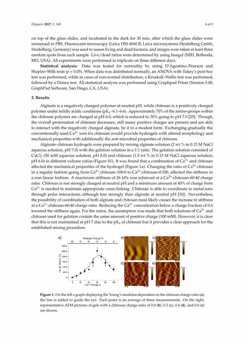
Alginate is a negatively charged polymer at neutral pH, while chitosan is a positively chargedpolymer under mildly acidic conditions (pKa~6.1–6.6). Approximately 70% of the amino-groups withinthe chitosan polymer are charged at pH 6.0, which is reduced to 30% going to pH 7.0 [29]. Though,the overall protonation of chitosan decreases, still many positive charges are present and are ableto interact with the negatively charged alginate, be it in a modest form. Exchanging gradually theconventionally used Ca2+ ions for chitosan would provide hydrogels with altered morphology andmechanical properties with additionally the anti-microbial properties of chitosan.
Alginate–chitosan hydrogels were prepared by mixing alginate solution (2 wt % in 0.15 M NaClaqueous solution, pH 7.0) with the gelation solution in a 1:1 ratio. The gelation solution consisted ofCaCl2 (50 mM aqueous solution, pH 8.0) and chitosan (1.5 wt % in 0.15 M NaCl aqueous solution,pH 6.0) in different volume ratios (Figure S1). It was found that a combination of Ca2+ and chitosanaffected the mechanical properties of the hydrogel (Figure 1a). Changing the ratio of Ca2+:chitosanin a regular fashion going from Ca2+:chitosan-100:0 to Ca2+:chitosan-0:100, affected the stiffness ina non-linear fashion. A maximum stiffness of 26 kPa was achieved at a Ca2+:chitosan-60:40 chargeratio. Chitosan is not strongly charged at neutral pH and a minimum amount of 40% of charge fromCa2+ is needed to maintain appropriate cross-linking. Chitosan is able to coordinate to metal-ionsthrough polar interactions although less strongly than alginate at neutral pH [30]. Nevertheless,the possibility of coordination of both alginate and chitosan most likely causes the increase in stiffnessat a Ca2+:chitosan-60:40 charge ratio. Reducing the Ca2+ concentration below a charge fraction of 0.6lowered the stiffness again. For the ratios, the assumption was made that both solutions of Ca2+ andchitosan used for gelation contain the same amount of positive charge (100 mM). However, it is clearthat this is not maintained at pH 7 due to the pKa of chitosan but it provides a clear approach for theestablished mixing procedure.
Polymers 2017, 9, 149 4 of 9
on top of the glass slides, and incubated in the dark for 30 min, after which the glass slides were immersed in PBS. Fluorescent microscopy (Leica DM 4000 B, Leica microsystems Heidelberg Gmbh, Heidelberg, Germany) was used to assess living and dead bacteria, and images were taken at least three random spots from each sample. Live/dead ratios were determined by using ImageJ (NIH, Bethesda, MD, USA). All experiments were performed in triplicate on three different days.
Statistical analysis: Data was tested for normality by using D’Agostino–Pearson and Shapiro–Wilk tests (p < 0.05). When data was distributed normally, an ANOVA with Tukey’s post-hoc test was performed, while in cases of non-normal distribution, a Kruskall–Wallis test was performed, followed by a Dunns test. All statistical analysis was performed using Graphpad Prism (Version 5.00, GraphPad Software, San Diego, CA, USA).
3. Results
Alginate is a negatively charged polymer at neutral pH, while chitosan is a positively charged polymer under mildly acidic conditions (pKa~6.1–6.6). Approximately 70% of the amino-groups within the chitosan polymer are charged at pH 6.0, which is reduced to 30% going to pH 7.0 [29]. Though, the overall protonation of chitosan decreases, still many positive charges are present and are able to interact with the negatively charged alginate, be it in a modest form. Exchanging gradually the conventionally used Ca2+ ions for chitosan would provide hydrogels with altered morphology and mechanical properties with additionally the anti-microbial properties of chitosan.
Alginate–chitosan hydrogels were prepared by mixing alginate solution (2 wt % in 0.15 M NaCl aqueous solution, pH 7.0) with the gelation solution in a 1:1 ratio. The gelation solution consisted of CaCl2 (50 mM aqueous solution, pH 8.0) and chitosan (1.5 wt % in 0.15 M NaCl aqueous solution, pH 6.0) in different volume ratios (Figure S1). It was found that a combination of Ca2+ and chitosan affected the mechanical properties of the hydrogel (Figure 1a). Changing the ratio of Ca2+:chitosan in a regular fashion going from Ca2+:chitosan-100:0 to Ca2+:chitosan-0:100, affected the stiffness in a non-linear fashion. A maximum stiffness of 26 kPa was achieved at a Ca2+:chitosan-60:40 charge ratio. Chitosan is not strongly charged at neutral pH and a minimum amount of 40% of charge from Ca2+ is needed to maintain appropriate cross-linking. Chitosan is able to coordinate to metal-ions through polar interactions although less strongly than alginate at neutral pH [30]. Nevertheless, the possibility of coordination of both alginate and chitosan most likely causes the increase in stiffness at a Ca2+:chitosan-60:40 charge ratio. Reducing the Ca2+ concentration below a charge fraction of 0.6 lowered the stiffness again. For the ratios, the assumption was made that both solutions of Ca2+ and chitosan used for gelation contain the same amount of positive charge (100 mM). However, it is clear that this is not maintained at pH 7 due to the pKa of chitosan but it provides a clear approach for the established mixing procedure.
Figure 1. On the left a graph displaying the Young’s modulus dependent on the chitosan charge ratio (a); the line is added to guide the eye. Each point is an average of three measurements. On the right, representative AFM pictures of gels with a chitosan charge ratio of 0.0 (b), 0.2 (c), 0.4 (d), and 0.6 (e) are shown.
Figure 1. On the left a graph displaying the Young’s modulus dependent on the chitosan charge ratio (a);the line is added to guide the eye. Each point is an average of three measurements. On the right,representative AFM pictures of gels with a chitosan charge ratio of 0.0 (b), 0.2 (c), 0.4 (d), and 0.6 (e)are shown.
Polymers 2017, 9, 149 5 of 9
It was expected that the addition of chitosan and altering the Ca2+ concentration would alter thegel morphology. Alg–Ca2+:Ch-100:0, Alg–Ca2+:Ch-80:20, Alg–Ca2+:Ch-60:40, and Alg–Ca2+:Ch-40:60were analyzed in the hydrated state using atomic force microscopy (AFM) to identify morphologicalchanges. It was found that, compared to the chitosan-free gel (Alg–Ca2+:Ch-100:0) and theAlg–Ca2+:Ch-80:20 and Alg–Ca2+:Ch-60:40 gels showed increased fiber formation. Especially forthe latter, long distinct fibers were observed, whereas gel fibers for the Ca2+:Ch-80:20 were shorterbut higher in numbers. The Alg–Ca2+:Ch-40:60 gel did not show as many fibers as the other chitosancontaining gels. The trend found for the stiffness of the gels could be correlated to the AFM analysisas chitosan induces fiber formation up until a point, after which that effect diminishes (Figure 1b–e,Figure S2).
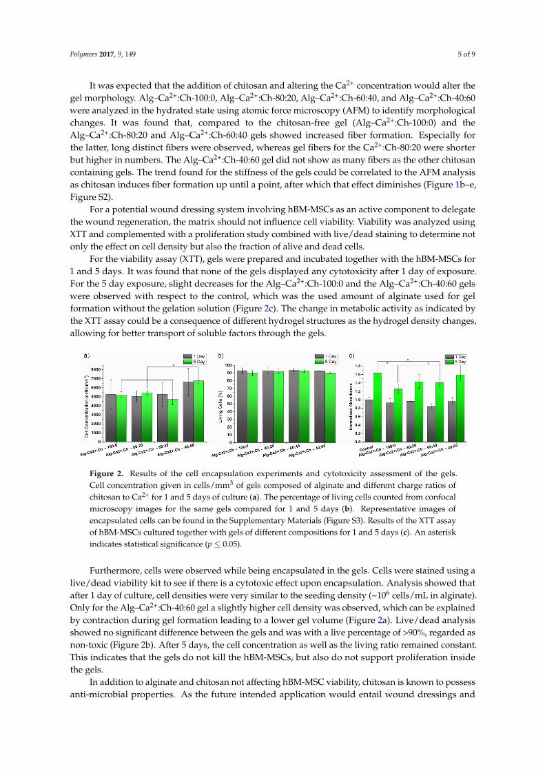
For a potential wound dressing system involving hBM-MSCs as an active component to delegatethe wound regeneration, the matrix should not influence cell viability. Viability was analyzed usingXTT and complemented with a proliferation study combined with live/dead staining to determine notonly the effect on cell density but also the fraction of alive and dead cells.
For the viability assay (XTT), gels were prepared and incubated together with the hBM-MSCs for1 and 5 days. It was found that none of the gels displayed any cytotoxicity after 1 day of exposure.For the 5 day exposure, slight decreases for the Alg–Ca2+:Ch-100:0 and the Alg–Ca2+:Ch-40:60 gelswere observed with respect to the control, which was the used amount of alginate used for gelformation without the gelation solution (Figure 2c). The change in metabolic activity as indicated bythe XTT assay could be a consequence of different hydrogel structures as the hydrogel density changes,allowing for better transport of soluble factors through the gels.
Polymers 2017, 9, 149 5 of 9
It was expected that the addition of chitosan and altering the Ca2+ concentration would alter the gel morphology. Alg–Ca2+:Ch-100:0, Alg–Ca2+:Ch-80:20, Alg–Ca2+:Ch-60:40, and Alg–Ca2+:Ch-40:60 were analyzed in the hydrated state using atomic force microscopy (AFM) to identify morphological changes. It was found that, compared to the chitosan-free gel (Alg–Ca2+:Ch-100:0) and the Alg–Ca2+:Ch-80:20 and Alg–Ca2+:Ch-60:40 gels showed increased fiber formation. Especially for the latter, long distinct fibers were observed, whereas gel fibers for the Ca2+:Ch-80:20 were shorter but higher in numbers. The Alg–Ca2+:Ch-40:60 gel did not show as many fibers as the other chitosan containing gels. The trend found for the stiffness of the gels could be correlated to the AFM analysis as chitosan induces fiber formation up until a point, after which that effect diminishes (Figures 1b–e, Figure S2).
For a potential wound dressing system involving hBM-MSCs as an active component to delegate the wound regeneration, the matrix should not influence cell viability. Viability was analyzed using XTT and complemented with a proliferation study combined with live/dead staining to determine not only the effect on cell density but also the fraction of alive and dead cells.
For the viability assay (XTT), gels were prepared and incubated together with the hBM-MSCs for 1 and 5 days. It was found that none of the gels displayed any cytotoxicity after 1 day of exposure. For the 5 day exposure, slight decreases for the Alg–Ca2+:Ch-100:0 and the Alg–Ca2+:Ch-40:60 gels were observed with respect to the control, which was the used amount of alginate used for gel formation without the gelation solution (Figure 2c). The change in metabolic activity as indicated by the XTT assay could be a consequence of different hydrogel structures as the hydrogel density changes, allowing for better transport of soluble factors through the gels.
Figure 2. Results of the cell encapsulation experiments and cytotoxicity assessment of the gels. Cell concentration given in cells/mm3 of gels composed of alginate and different charge ratios of chitosan to Ca2+ for 1 and 5 days of culture (a). The percentage of living cells counted from confocal microscopy images for the same gels compared for 1 and 5 days (b). Representative images of encapsulated cells can be found in the supplementary information (Figure S3). Results of the XTT assay of hBM-MSCs cultured together with gels of different compositions for 1 and 5 days (c). An asterisk indicates statistical significance (p ≤ 0.05).
Furthermore, cells were observed while being encapsulated in the gels. Cells were stained using a live/dead viability kit to see if there is a cytotoxic effect upon encapsulation. Analysis showed that after 1 day of culture, cell densities were very similar to the seeding density (~106 cells/mL in alginate). Only for the Alg–Ca2+:Ch-40:60 gel a slightly higher cell density was observed, which can be explained by contraction during gel formation leading to a lower gel volume (Figure 2a). Live/dead analysis showed no significant difference between the gels and was with a live percentage of >90%, regarded as non-toxic (Figure 2b). After 5 days, the cell concentration as well as the living ratio remained constant. This indicates that the gels do not kill the hBM-MSCs, but also do not support proliferation inside the gels.
In addition to alginate and chitosan not affecting hBM-MSC viability, chitosan is known to possess anti-microbial properties. As the future intended application would entail wound dressings and wound filling materials for regenerative medicine, the anti-microbial effect of the gels were tested towards two bacterial strains which have been isolated from wounds, namely P. aeruginosa and S. aureus. Both pose as a high medical risk as P. aeruginosa is an opportunistic nosocomial
Figure 2. Results of the cell encapsulation experiments and cytotoxicity assessment of the gels.Cell concentration given in cells/mm3 of gels composed of alginate and different charge ratios ofchitosan to Ca2+ for 1 and 5 days of culture (a). The percentage of living cells counted from confocalmicroscopy images for the same gels compared for 1 and 5 days (b). Representative images ofencapsulated cells can be found in the Supplementary Materials (Figure S3). Results of the XTT assayof hBM-MSCs cultured together with gels of different compositions for 1 and 5 days (c). An asteriskindicates statistical significance (p ≤ 0.05).
Furthermore, cells were observed while being encapsulated in the gels. Cells were stained using alive/dead viability kit to see if there is a cytotoxic effect upon encapsulation. Analysis showed thatafter 1 day of culture, cell densities were very similar to the seeding density (~106 cells/mL in alginate).Only for the Alg–Ca2+:Ch-40:60 gel a slightly higher cell density was observed, which can be explainedby contraction during gel formation leading to a lower gel volume (Figure 2a). Live/dead analysisshowed no significant difference between the gels and was with a live percentage of >90%, regarded asnon-toxic (Figure 2b). After 5 days, the cell concentration as well as the living ratio remained constant.This indicates that the gels do not kill the hBM-MSCs, but also do not support proliferation insidethe gels.
In addition to alginate and chitosan not affecting hBM-MSC viability, chitosan is known to possessanti-microbial properties. As the future intended application would entail wound dressings and
Polymers 2017, 9, 149 6 of 9
wound filling materials for regenerative medicine, the anti-microbial effect of the gels were testedtowards two bacterial strains which have been isolated from wounds, namely P. aeruginosa and S. aureus.Both pose as a high medical risk as P. aeruginosa is an opportunistic nosocomial pathogen and S. aureus,of which methicillin-resistant S. aureus (MRSA) is the most well-known, is becoming a major globalthreat [27,28].
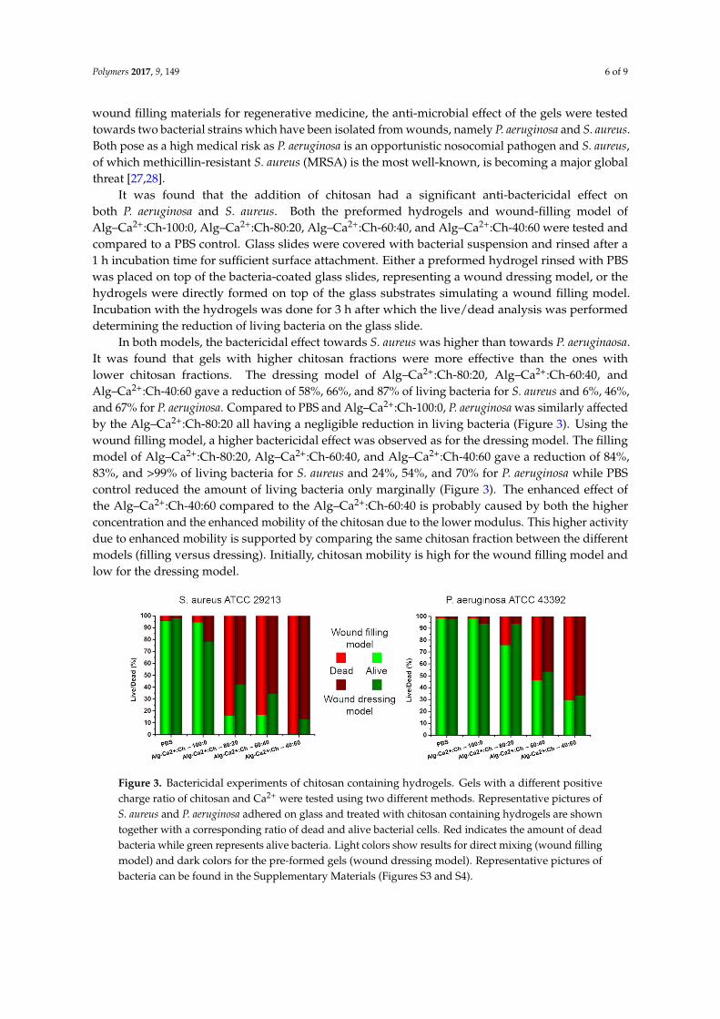
It was found that the addition of chitosan had a significant anti-bactericidal effect onboth P. aeruginosa and S. aureus. Both the preformed hydrogels and wound-filling model ofAlg–Ca2+:Ch-100:0, Alg–Ca2+:Ch-80:20, Alg–Ca2+:Ch-60:40, and Alg–Ca2+:Ch-40:60 were tested andcompared to a PBS control. Glass slides were covered with bacterial suspension and rinsed after a1 h incubation time for sufficient surface attachment. Either a preformed hydrogel rinsed with PBSwas placed on top of the bacteria-coated glass slides, representing a wound dressing model, or thehydrogels were directly formed on top of the glass substrates simulating a wound filling model.Incubation with the hydrogels was done for 3 h after which the live/dead analysis was performeddetermining the reduction of living bacteria on the glass slide.
In both models, the bactericidal effect towards S. aureus was higher than towards P. aeruginaosa.It was found that gels with higher chitosan fractions were more effective than the ones withlower chitosan fractions. The dressing model of Alg–Ca2+:Ch-80:20, Alg–Ca2+:Ch-60:40, andAlg–Ca2+:Ch-40:60 gave a reduction of 58%, 66%, and 87% of living bacteria for S. aureus and 6%, 46%,and 67% for P. aeruginosa. Compared to PBS and Alg–Ca2+:Ch-100:0, P. aeruginosa was similarly affectedby the Alg–Ca2+:Ch-80:20 all having a negligible reduction in living bacteria (Figure 3). Using thewound filling model, a higher bactericidal effect was observed as for the dressing model. The fillingmodel of Alg–Ca2+:Ch-80:20, Alg–Ca2+:Ch-60:40, and Alg–Ca2+:Ch-40:60 gave a reduction of 84%,83%, and >99% of living bacteria for S. aureus and 24%, 54%, and 70% for P. aeruginosa while PBScontrol reduced the amount of living bacteria only marginally (Figure 3). The enhanced effect ofthe Alg–Ca2+:Ch-40:60 compared to the Alg–Ca2+:Ch-60:40 is probably caused by both the higherconcentration and the enhanced mobility of the chitosan due to the lower modulus. This higher activitydue to enhanced mobility is supported by comparing the same chitosan fraction between the differentmodels (filling versus dressing). Initially, chitosan mobility is high for the wound filling model andlow for the dressing model.
Polymers 2017, 9, 149 6 of 9
pathogen and S. aureus, of which methicillin-resistant S. aureus (MRSA) is the most well-known, is becoming a major global threat [27,28].
It was found that the addition of chitosan had a significant anti-bactericidal effect on both P. aeruginosa and S. aureus. Both the preformed hydrogels and wound-filling model of Alg–Ca2+:Ch-100:0, Alg–Ca2+:Ch-80:20, Alg–Ca2+:Ch-60:40, and Alg–Ca2+:Ch-40:60 were tested and compared to a PBS control. Glass slides were covered with bacterial suspension and rinsed after a 1 h incubation time for sufficient surface attachment. Either a preformed hydrogel rinsed with PBS was placed on top of the bacteria-coated glass slides, representing a wound dressing model, or the hydrogels were directly formed on top of the glass substrates simulating a wound filling model. Incubation with the hydrogels was done for 3 h after which the live/dead analysis was performed determining the reduction of living bacteria on the glass slide.
In both models, the bactericidal effect towards S. aureus was higher than towards P. aeruginaosa. It was found that gels with higher chitosan fractions were more effective than the ones with lower chitosan fractions. The dressing model of Alg–Ca2+:Ch-80:20, Alg–Ca2+:Ch-60:40, and Alg–Ca2+:Ch-40:60 gave a reduction of 58%, 66%, and 87% of living bacteria for S. aureus and 6%, 46%, and 67% for P. aeruginosa. Compared to PBS and Alg–Ca2+:Ch-100:0, P. aeruginosa was similarly affected by the Alg–Ca2+:Ch-80:20 all having a negligible reduction in living bacteria (Figure 3). Using the wound filling model, a higher bactericidal effect was observed as for the dressing model. The filling model of Alg–Ca2+:Ch-80:20, Alg–Ca2+:Ch-60:40, and Alg–Ca2+:Ch-40:60 gave a reduction of 84%, 83%, and >99% of living bacteria for S. aureus and 24%, 54%, and 70% for P. aeruginosa while PBS control reduced the amount of living bacteria only marginally (Figure 3). The enhanced effect of the Alg–Ca2+:Ch-40:60 compared to the Alg–Ca2+:Ch-60:40 is probably caused by both the higher concentration and the enhanced mobility of the chitosan due to the lower modulus. This higher activity due to enhanced mobility is supported by comparing the same chitosan fraction between the different models (filling versus dressing). Initially, chitosan mobility is high for the wound filling model and low for the dressing model.
Figure 3. Bactericidal experiments of chitosan containing hydrogels. Gels with a different positive charge ratio of chitosan and Ca2+ were tested using two different methods. Representative pictures of S. aureus and P. aeruginosa adhered on glass and treated with chitosan containing hydrogels are shown together with a corresponding ratio of dead and alive bacterial cells. Red indicates the amount of dead bacteria while green represents alive bacteria. Light colors show results for direct mixing (wound filling model) and dark colors for the pre-formed gels (wound dressing model). Representative pictures of bacteria can be found in the supplementary information (Figures S3 and S4).
4. Conclusions and Discussion
The developed alginate–chitosan system displays a dual functionality important for the purpose of wound healing. It enables encapsulation of hBM-MSCs in an efficient way without reducing their viability, and the chitosan functions as an anti-microbial agent tested against two major strains involved in wound infections. The two-component system offers the possibility of
Figure 3. Bactericidal experiments of chitosan containing hydrogels. Gels with a different positivecharge ratio of chitosan and Ca2+ were tested using two different methods. Representative pictures ofS. aureus and P. aeruginosa adhered on glass and treated with chitosan containing hydrogels are showntogether with a corresponding ratio of dead and alive bacterial cells. Red indicates the amount of deadbacteria while green represents alive bacteria. Light colors show results for direct mixing (wound fillingmodel) and dark colors for the pre-formed gels (wound dressing model). Representative pictures ofbacteria can be found in the Supplementary Materials (Figures S3 and S4).
Polymers 2017, 9, 149 7 of 9
4. Conclusions and Discussion
The developed alginate–chitosan system displays a dual functionality important for the purposeof wound healing. It enables encapsulation of hBM-MSCs in an efficient way without reducing theirviability, and the chitosan functions as an anti-microbial agent tested against two major strains involvedin wound infections. The two-component system offers the possibility of using it either as a preformeddressing or as a wound filling model. The wound filling model offers two advantages: (1) using twoliquid components allows sufficient contact with the whole wound before setting as a gel; (2) it has ahigher bactericidal effect. In particular, wounds that remain non-healing, e.g., diabetic ulcers in needof debridement, would potentially benefit from this system as it is known that stem cells enhancewound healing, and infection is often a major complication [4,9,31–33]. The preformed hydrogelshave the advantage of being stored for long periods of time. Alginate is often used as a storagemedium for frozen (stem) cells and a preformed wound dressing stored in a frozen state would allowmass production and having the dressing ready at hand. There are already commercially availableproducts on the market based on either alginate or chitosan such as Cheerain® alginate patches andHemCon® chitosan-coated bandages for the purpose of wound dressing but not combined and notfor the purpose of additionally containing mesenchymal stem cells. There are systems known wherechitosan and alginate have been combined but often as separated layers, as polyelectrolyte coatingson non-hydrogel substrates or as hydrogel formed at relatively low pH. The latter approach requiresvarious drying and washing steps and is therefore not compatible with encapsulated cells [7,34–36].Hence, translation towards clinical use of the proposed system is regarded as very promising, as thematerials are already used for these specific applications, can include stem cells, and can be applied asboth a preformed dressing and a two-component mixing system.
Supplementary Materials: The following are available online at www.mdpi.com/2073-4360/9/4/149/s1.Figure S1: Atomic Force Microscopy analysis of hydrogels; Figure S2: hBM-MSC live/dead imaging encapsulatedin gels; Figure S3: P. aeruginosa live/dead imaging; Figure S4: S. aureus live/dead imaging.
Acknowledgments: Philipp T. Kühn and Patrick van Rijn kindly thank the graduate school medical sciences(GSMS) of the University of Groningen/University Medical Center Groningen for financial support.
Author Contributions: Philipp T. Kühn and Patrick van Rijn conceived and designed the experiments;Philipp T. Kühn and Estelle Perrels performed the experiments concerning gel formation/characterization andcell inclusion; René T. Rozenbaum and Prashant K. Sharma designed and interpreted the data of the bactericidalexperiments, René T. Rozenbaum performed these; all authors contributed to analysis and interpretation of thedata; Philipp T. Kühn, René T. Rozenbaum, Prashant K. Sharma, and Patrick van Rijn wrote the paper. All authorsread and commented on the manuscript.
Conflicts of Interest: The authors declare no conflict of interest.
References
1. Martin, P. Wound healing—Aiming for perfect skin regeneration. Science 1997, 276, 75–81. [CrossRef][PubMed]
2. Sun, B.K.; Siprashvili, Z.; Khavari, P.A. Advances in skin grafting and treatment of cutaneous wounds.Science 2014, 346, 941–945. [CrossRef] [PubMed]
3. Wynn, T.A.; Ramalingam, T.R. Mechanisms of fibrosis: Therapeutic translation for fibrotic disease. Nat. Med.2012, 18, 1028–1040. [CrossRef] [PubMed]
4. Moura, L.I.F.; Dias, A.M.A.; Carvalho, E.; De Sousa, H.C. Recent advances on the development of wounddressings for diabetic foot ulcer treatment—A review. Acta Biomater. 2013, 9, 7093–7114. [CrossRef] [PubMed]
5. Norouzi, M.; Boroujeni, S.M.; Omidvarkordshouli, N.; Soleimani, M. Advances in skin regeneration:application of electrospun scaffolds. Adv. Healthc. Mater. 2015, 4, 1114–1133. [CrossRef] [PubMed]
6. Drury, J.L.; Mooney, D.J. Hydrogels for tissue engineering: Scaffold design variables and applications.Biomaterials 2003, 24, 4337–4351. [CrossRef]
7. Jayakumar, R.; Prabaharan, M.; Sudheesh Kumar, P.T.; Nair, S.V.; Tamura, H. Biomaterials based on chitinand chitosan in wound dressing applications. Biotechnol. Adv. 2011, 29, 322–337. [CrossRef] [PubMed]
Polymers 2017, 9, 149 8 of 9
8. Altman, A.M.; Matthias, N.; Yan, Y.; Song, Y.-H.; Bai, X.; Chiu, E.S.; Slakey, D.P.; Alt, E.U. Dermal matrix as acarrier for in vivo delivery of human adipose-derived stem cells. Biomaterials 2008, 29, 1431–1442. [CrossRef][PubMed]
9. Reckhenrich, A.K.; Kirsch, B.M.; Wahl, E.A.; Schenck, T.L.; Rezaeian, F.; Harder, Y.; Foehr, P.; Machens, H.-G.;Egaña, J.T. Surgical sutures filled with adipose-derived stem cells promote wound healing. PLoS ONE 2014,9, e91169. [CrossRef] [PubMed]
10. Jiang, D.; Qi, Y.; Walker, N.G.; Sindrilaru, A.; Hainzl, A.; Wlaschek, M.; MacNeil, S.; Scharffetter-Kochanek, K.The effect of adipose tissue derived MSCs delivered by a chemically defined carrier on full-thicknesscutaneous wound healing. Biomaterials 2013, 34, 2501–2515. [CrossRef] [PubMed]
11. Turner, N.J.; Badylak, S.F. The use of biologic scaffolds in the treatment of chronic nonhealing wounds.Adv. Wound Care 2015, 4, 490–500. [CrossRef] [PubMed]
12. Balakrishnan, B.; Mohanty, M.; Umashankar, P.; Jayakrishnan, A. Evaluation of an in situ forming hydrogelwound dressing based on oxidized alginate and gelatin. Biomaterials 2005, 26, 6335–6342. [CrossRef][PubMed]
13. Lee, K.Y.; Mooney, D.J. Alginate: Properties and biomedical applications. Prog. Polym. Sci. 2012, 37, 106–126.[CrossRef] [PubMed]
14. Hillel, A.T.; Unterman, S.; Nahas, Z.; Reid, B.; Coburn, J.M.; Axelman, J.; Chae, J.J.; Guo, Q.; Trow, R.;Thomas, A.; et al. Photoactivated composite biomaterial for soft tissue restoration in rodents and in humans.Sci. Transl. Med. 2011, 3, 93ra67. [CrossRef] [PubMed]
15. Kühn, P.T.; Meijer, T.L.; Schiavon, I.; van Poll, M.; van Aken, J.; Groen, S.; Kuijer, R.; van Kooten, T.G.; vanRijn, P. Non-covalently stabilized alginate hydrogels as functional cell scaffold material. Macromol. Biosci.2016, 16, 1693–1702. [CrossRef] [PubMed]
16. Pawar, S.N.; Edgar, K.J. Alginate derivatization: A review of chemistry, properties and applications.Biomaterials 2012, 33, 3279–3305. [CrossRef] [PubMed]
17. Goy, R.C.; Britto, D.; De Assis, O.B.G. A review of the antimicrobial activity of chitosan. Polímeros 2009, 19,241–247. [CrossRef]
18. Friedman, A.J.; Phan, J.; Schairer, D.O.; Champer, J.; Qin, M.; Pirouz, A.; Blecher-Paz, K.; Oren, A.;Liu, P.T.; Modlin, R.L.; et al. Antimicrobial and anti-inflammatory activity of chitosan-alginate nanoparticles:A targeted therapy for cutaneous pathogens. J. Investig. Dermatol. 2013, 133, 1231–1239. [CrossRef] [PubMed]
19. Ghaffarian, R.; Pérez-herrero, E.; Oh, H.; Raghavan, S.R.; Muro, S. Chitosan-alginate microcapsules providegastric protection and intestinal release of ICAM-1-targeting nanocarriers, enabling GI targeting in vivo.Adv. Funct. Mater. 2016, 26, 3382–3393. [CrossRef] [PubMed]
20. Wang, L.; Khor, E.; Wee, A.; Lim, L.Y. Chitosan-alginate PEC membrane as a wound dressing: Assessment ofincisional wound healing. J. Biomed. Mater. Res. 2002, 63, 610–618. [CrossRef] [PubMed]
21. Meng, X.; Tian, F.; Yang, J.; He, C.N.; Xing, N.; Li, F. Chitosan and alginate polyelectrolyte complexmembranes and their properties for wound dressing application. J. Mater. Sci. Mater. Med. 2010, 21,1751–1759. [CrossRef] [PubMed]
22. Luo, Y.; Wang, Q. Recent development of chitosan-based polyelectrolyte complexes with naturalpolysaccharides for drug delivery. Int. J. Biol. Macromol. 2014, 64, 353–367. [CrossRef] [PubMed]
23. Wang, L.; Khor, E.; Lim, L. Chitosan-alginate-CaCl2 System for membrane coat application. J. Pharm. Sci.2001, 90, 1134–1142. [CrossRef] [PubMed]
24. Caetano, G.F.; Frade, M.A.C.; Andrade, T.A.M.; Leite, M.N.; Bueno, C.Z.; Moraes, Â.M.; Ribeiro-Paes, J.T.Chitosan-alginate membranes accelerate wound healing. J. Biomed. Mater. Res. B 2015, 103, 1013–1022.[CrossRef] [PubMed]
25. Navone, S.E.; Pascucci, L.; Dossena, M.; Ferri, A.; Invernici, G.; Acerbi, F.; Cristini, S.; Bedini, G.; Tosetti, V.;Ceserani, V.; et al. Decellularized silk fibroin scaffold primed with adipose mesenchymal stromal cellsimproves wound healing in diabetic mice. Stem Cell Res. Ther. 2014, 5, 7. [CrossRef] [PubMed]
26. Sorrell, J.M.; Caplan, A.I. Topical delivery of mesenchymal stem cells and their function in wounds. Stem CellRes. Ther. 2010, 1, 30. [CrossRef] [PubMed]
27. Ciofu, O.; Tolker-Nielsen, T.; Jensen, P.Ø.; Wang, H.; Høiby, N. Antimicrobial resistance, respiratory tractinfections and role of biofilms in lung infections in cystic fibrosis patients. Adv. Drug Deliv. Rev. 2015, 85,7–23. [CrossRef] [PubMed]
Polymers 2017, 9, 149 9 of 9
28. McCarthy, H.; Rudkin, J.K.; Black, N.S.; Gallagher, L.; O’Neill, E.; O’Gara, J.P. Methicillin resistance and thebiofilm phenotype in Staphylococcus aureus. Front. Cell. Infect. Microbiol. 2015, 5, 1. [CrossRef] [PubMed]
29. Wang, Q.Z.; Chen, X.G.; Liu, N.; Wang, S.X.; Liu, C.S.; Meng, X.H.; Liu, C.G. Protonation constants of chitosanwith different molecular weight and degree of deacetylation. Carbohydr. Polym. 2006, 65, 194–201. [CrossRef]
30. Lima, I.S.; Airoldi, C. A thermodynamic investigation on chitosan-divalent cation interactions.Thermochim. Acta 2004, 421, 133–139. [CrossRef]
31. Percival, S.L.; Hill, K.E.; Williams, D.W.; Hooper, S.J.; Thomas, D.W.; Costerton, J.W. A review of the scientificevidence for biofilms in wounds. Wound Repair Regen. 2012, 20, 647–657. [CrossRef] [PubMed]
32. Huang, S.-P.; Huang, C.-H.; Shyu, J.-F.; Lee, H.-S.; Chen, S.-G.; Chan, J.Y.-H.; Huang, S.-M. Promotion ofwound healing using adipose-derived stem cells in radiation ulcer of a rat model. J. Biomed. Sci. 2013, 20, 51.[CrossRef] [PubMed]
33. Brower, J.; Student, M.; Blumberg, S.; Fellow, P.; Carroll, E.; Pastar, I.; Brem, H.; Chen, W. Mesenchymal stemcell therapy and delivery systems in nonhealing wounds. Adv. Skin Wound Care 2012, 24, 524–532. [CrossRef][PubMed]
34. Dong, Y.; Liu, H.Z.; Xua, L.; Li, G.; Ma, Z.N.; Han, F.; Yao, H.M.; Sun, Y.H.; Li, S.M. A novel CHS/ALG bi-layercomposite membrane with sustained antimicrobial efficacy used as wound dressing. Chin. Chem. Lett. 2010,21, 1011–1014. [CrossRef]
35. Hong, H.J.; Jin, S.E.; Park, J.S.; Ahn, W.S.; Kim, C.K. Accelerated wound healing by smad3 antisenseoligonucleotides-impregnated chitosan/alginate polyelectrolyte complex. Biomaterials 2008, 29, 4831–4837.[CrossRef] [PubMed]
36. Tsao, C.T.; Chang, C.H.; Lin, Y.Y.; Wu, M.F.; Wang, J.L.; Young, T.H.; Han, J.L.; Hsieh, K.H. Evaluation ofchitosan/γ-poly (glutamic acid) polyelectrolyte complex for wound dressing materials. Carbohydr. Polym.2011, 84, 812–819. [CrossRef]
© 2017 by the authors. Licensee MDPI, Basel, Switzerland. This article is an open accessarticle distributed under the terms and conditions of the Creative Commons Attribution(CC BY) license (http://creativecommons.org/licenses/by/4.0/).