Embed Size (px)
Citation preview

University of Canberra
This thesis is available in print format from the University of Canberra Library. If you are the author of this thesis and wish to have the whole thesis loaded here, please contact the University of Canberra Library at [email protected] Your thesis will then be available on the www providing greater access.
Properties and Dating of Silica Skins associated withRock Art
by
Alan Leslie Watchman
submitted to the University of Canberra
for the Degree of Doctor of Philosophy
February 1996
ACKNOWLEDGEMENTS
I am grateful for the advice, assistance, encouragement and support given by many
people during this work. Professor Colin Pearson AO, MBE formally supervised this
research and I am most grateful for his professional guidance, supervisory comments,
strong personal support and friendship. Professor Graham Taylor and Dr Phillipe Dube
were encouraging and strongly supportive advisers. Jeanette Wigg was my reliable and
extremely efficient administrative assistant at the University of Canberra (UC). Staff at
the National Centre for Cultural Heritage Science Studies gave me their full support.
At Laval University, I received advice and scientific support from many staff and I used
equipment in the Centre d'Optique, Photonique et Laser (COPL), the Centre d'Etudes
Nordiques (CEN), and in the Departments of Geology, Chemistry, Physics and Botany.
Professor Roger Lessard (COPL) gave permission for laser research, Dr Li Song and Dr
Rupak gave specialist advice on lasers and optics, Mr Denis Lessard and Mr Marcel
Denis administered the laser research and Mr Laurent Turgeon, Mr Marc d'Auteuil and
Mr Florent Pouliot furnished technical support. Guillaume LeBrecque provided
assistance during the preparation of samples for accelerator mass spectrometry
radiocarbon dating. Dr Reinhard Pienitz (CEN), identified diatoms and Dr Michel
Allard awarded me collaborative research member status in the CEN, facilitating access
to Laval University equipment. Guy Plante produced high quality photographs in an
extremely efficient and cheerful manner. Geological and technical assistance, and
advisory comments were given by Dr Borque, Dr Laurent (petrography), Dr. Ledoux
(refractive index), Mr Richard (X-ray diffraction; XRD) and Mr Tremblay (scanning
electron microscopy energy dispersive X-ray analyses (SEM/EDXA) and electron
microprobe). Dr LaFontaine made transmission electron microscope (TEM)
preparation, observation and photographic equipment available at no charge.
Mr Charles Gruchy, Canadian Conservation Institute (CCI), gave permission to use
analytical facilities, in particular the Fourier Transform Infrared Spectrometer (FTIR).
Mr Ian Wainwright made samples and analytical information available from Nisula,
Crowrock Inlet and Agawa Rock, advised on the operations of SEM/EDXA and
photographic equipment, and willingly discussed aspects of the research. Technical
assistance and advice were kindly given by Elizabeth Moffatt (FTIR) and Jane Sirois
(XRD). Library staff at the CCI were most helpful with my literature research.
(vii)
Mr Vowles, Mr Ciszewski and Mr Heddy (SEM/EDXA) and Mr Lewellyn (TEM)
provided valuable electron microscope assistance at the Australian National University.
Dr Maite le Gleuher helped with sample preparation for the TEM, willingly discussed
the results and pointed out relevant references on proto-crystalline materials. Mr
Foudoulis conducted XRD analyses and Dr Eggleton gave general advice. Mr Ware,
Research School of Earth Sciences, assisted with electron microprobe analyses. Proton
induced X-ray emission (PIXE) analyses were made at Lucas Heights, Sydney by Dr
Clayton. Dr Laver, Lower Molonglo Water Quality Laboratories, Belconnen, identified
algae and fungi. At the Australian Defence Force Academy, Physics Department,
University of New South Wales, Associate Professor Creagh and Research Officer Dr
Wantenaar provided assistance and advice using a scanning transmission electron
microscope (STEM).
Field support was provided by Dr Kelvin Officer and Bruce Ford (New South Wales),
Percy Trezise, Mary Haginikitas, Noelene Cole and Dr Nicola Horsfall (North
Queensland), Ivan Haskovec, Hilary Sullivan, George Chaloupka and Penny Marshall
(Kakadu National Park), Grahame Walsh and Dr Mike Morwood (Kimberley, Western
Australia), and Dr Daniel Arsenault, Louis Gagnon and Charles Martijn (Nisula,
Quebec). The Bradshaw Foundation made funds available for the dating of samples
from the Kimberley area.
I am grateful for permission to collect samples from sites managed by the Australian
National Parks and Wildlife Service, Queensland Department of Environment and
Heritage, New South Wales National Parks and Wildlife Service, Nowra Aboriginal
Land Council, New South Wales Forestry Commission, Western Australian Museum,
Western Australia Department of Conservation and Land Management, Kalumburu
Community Council, Australian Heritage Commission, Quebec Ministry of Culture and
the Bande de Betsiamites.
The Australian Institute of Aboriginal Studies provided funds for operating the laser at
Laval University and for radiocarbon measurements by accelerator mass spectrometry
(AMS) 14C. Samples for AMS 14C determinations were prepared by Dr Southon,
Lawrence Livermore AMS Center, California, Dr Jacobsen and Dr Tuniz, Australian
Nuclear Science and Technology Organisation, Lucas Heights, Sydney, and Dr Sparkes,
Department of Nuclear Sciences, Lower Hutt, New Zealand, and Dr Jull, Dr Toolin and
Rosemary Maddocks assisted with radiocarbon determinations at the Nuclear Science
Facility, Tucson, Arizona.
(viii)
B. Ford, D. Young and B. Johnson provided valuable comments on the research
methods and results. I wish to thank Darrell Lewis and Dr Debbie Bird Rose in
particular for encouraging me to start my silica skins research and to ignore the
scepticism about my proposed investigations and opposition to my doctoral study
expressed by some Australian academics. Darrell and Debbie gave moral support when
I needed it most, through periods of personal difficulties, Canadian relocation and
administrative frustrations at the Australian National University. Finally, I wish to
thank my wife, Nancy, for her love, support, understanding and patience.
(IX)
Properties and Dating of Silica Skins associated with Rock Art
Summary
Hydrated amorphous silicon dioxide (Si02.nH-,O), or opal-A, is deposited naturally
from seepage and runoff water as white or brown rock surface coatings, called 'skins',
that often partly obscure rock paintings and engravings, but occasionally, a thin
translucent silica skin can form a protective film over rock art. White lustrous silica
skins, less than 1 mm thick, occur where seepage water regularly flows from bedding
and joint planes, whereas much thinner brown skins form on the sides of boulders and
cliffs where runoff water periodically flows. To find the degree of silica skin variability
and to determine how climate and rock type affect the properties of silica skins I
collected samples at seven Australian and two Canadian rock painting sites that were
located in temperate, tropical and sub-arctic regions. The skins had developed on
sandstone, quartzite, schist, gneiss and migmatite. I studied the effects of the skins on
rock art stability, documented their compositions, textures and structures to establish
their common properties, and searched for a way to date the silica which would provide
an indication of the minimum age of the underlying art. 1 also made replication
experiments to determine factors that influence the properties of artificial silica skinsand the rates of their precipitation so that I could propose a mechanism for natural silica
skin formation, and ascertain whether an artificial silica skin could act as a protective
rock art conservation measure.
I was able to subdivide the analysed samples into silica skin Types I, II and III on the
basis of their colour (translucent, white or brown), composition (SiO2, Al2O3 and
absorbed water contents) and texture (smooth vitreous or vermiform). I propose that
silica skins initially begin to form on stable rock surfaces by a process involving a
combination of evaporation- and ionic-induced polymerisation of silicic acid in seepage
and runoff water. Condensation reactions, random clustering of small silica spheres
and deposition of the resulting aggregates eventually produce a thin surficial silica film.
Deposition of silica often traps micro-organisms that live in the damp seepage and
runoff water zones, and these fossils in finely laminated skins enable the radiocarbon
dating of silica deposition, and therefore the dating of rock paintings enclosed by silica.
Micro-excavation of silica layers associated with rock art combined with accelerator
mass spectrometry gave preliminary radiocarbon determinations that were either
consistent with, or contradicted, prevailing opinions about the antiquity of the rock art
at selected sites. Experiments using a laser technique for combusting fossilised micro-
organisms in finely laminated skins were unable to generate sufficient carbon for
dating. Catalysis of a mixture of equal proportions of methyl-trimethoxy silane and
water produces a translucent stable film that may be suitable as a consolidant, whereasother artificial silica skins made from silica glass and tetra-ethoxy silane develop micro-
fractures on drying, and these are unsuitable as rock art consolidants.
( i i )
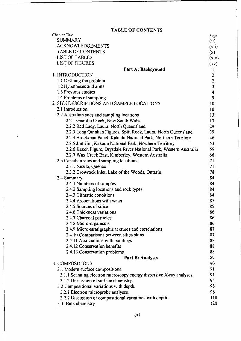
TABLE OF CONTENTSChapter Title Page
SUMMARY (ii)ACKNOWLEDGEMENTS (vii)TABLE OF CONTENTS (x)LIST OF TABLES (xiv)LIST OF FIGURES (xv)
Part A: Background 11. INTRODUCTION 2
1.1 Defining the problem 21.2 Hypotheses and aims 31.3 Previous studies 41.4 Problems of sampling 9
2. SITE DESCRIPTIONS AND SAMPLE LOCATIONS 102.1 Introduction 102.2 Australian sites and sampling locations 13
2.2.1 Gnatilia Creek, New South Wales 132.2.2 Red Lady, Laura, North Queensland 292.2.3 Long Quinkan Figures, Split Rock, Laura, North Queensland 392.2.4 Brockman Panel, Kakadu National Park, Northern Territory 462.2.5 Jim Jim, Kakadu National Park, Northern Territory 532.2.6 Keech Figure, Drysdale River National Park, Western Australia 592.2.7 Wax Creek East, Kimberley, Western Australia 66
2.3 Canadian sites and sampling locations 712.3.1 Nisula, Quebec 712.3.2 Crowrock Inlet, Lake of the Woods, Ontario 78
2.4 Summary 842.4.1 Numbers of samples 842.4.2 Sampling locations and rock types 842.4.3 Climatic conditions 842.4.4 Associations with water 852.4.5 Sources of silica 852.4.6 Thickness variations 862.4.7 Charcoal particles 862.4.8 Micro-organisms 862.4.9 Micro-stratigraphic textures and correlations 872.4.10 Comparisons between silica skins 872.4.11 Associations with paintings 882.4.12 Conservation benefits 882.4.13 Conservation problems 88
Part B: Analyses 893. COMPOSITIONS 90
3.1 Modem surface compositions. 913.1.1 Scanning electron microscopy energy dispersive X-ray analyses. 913.1.2 Discussion of surface chemistry. 95
3.2 Compositional variations with depth. 983.2.1 Electron microprobe analyses. 983.2.2 Discussion of compositional variations with depth. 110
3.3. Bulk chemistry. 120
(x)
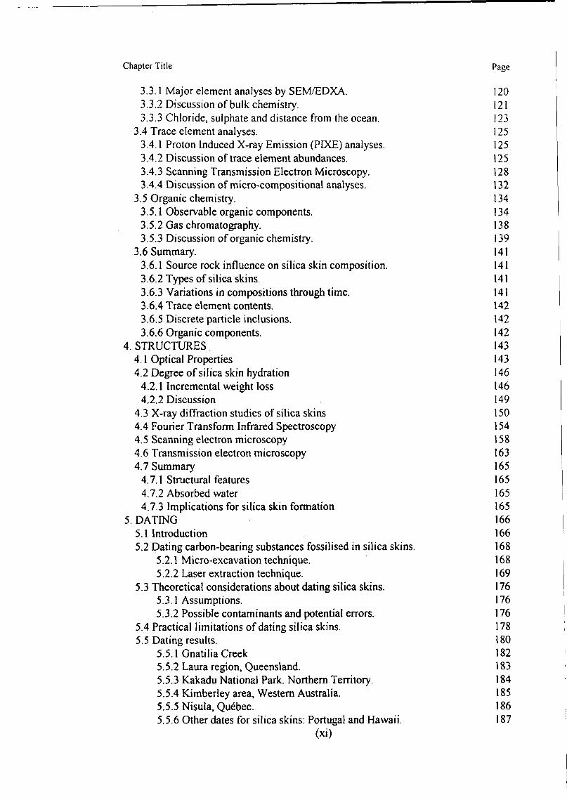
Chapter Title Page
3.3.1 Major element analyses by SEM/EDXA. 1203.3.2 Discussion of bulk chemistry. 1213.3.3 Chloride, sulphate and distance from the ocean. 123
3.4 Trace element analyses. 1253.4.1 Proton Induced X-ray Emission (PIXE) analyses. 1253.4.2 Discussion of trace element abundances. 1253.4.3 Scanning Transmission Electron Microscopy. 1283.4.4 Discussion of micro-compositional analyses. 132
3.5 Organic chemistry. 1343.5.1 Observable organic components. 1343.5.2 Gas chromatography. 1383.5.3 Discussion of organic chemistry. 139
3.6 Summary. 1413.6.1 Source rock influence on silica skin composition. 1413.6.2 Types of silica skins. 1413.6.3 Variations in compositions through time. 1413.6.4 Trace element contents. 1423.6.5 Discrete particle inclusions. 1423.6.6 Organic components. 142
4. STRUCTURES 1434.1 Optical Properties 1434.2 Degree of silica skin hydration 146
4.2.1 Incremental weight loss 1464.2.2 Discussion 149
4.3 X-ray diffraction studies of silica skins 1504.4 Fourier Transform Infrared Spectroscopy 1544.5 Scanning electron microscopy 1584.6 Transmission electron microscopy 1634.7 Summary 165
4.7.1 Structural features 1654.7.2 Absorbed water 1654.7.3 Implications for silica skin formation 165
5. DATING 1665.1 Introduction 1665.2 Dating carbon-bearing substances fossilised in silica skins. 168
5.2.1 Micro-excavation technique. 1685.2.2 Laser extraction technique. 169
5.3 Theoretical considerations about dating silica skins. 1765.3.1 Assumptions. 1765.3.2 Possible contaminants and potential errors. 176
5.4 Practical limitations of dating silica skins. 1785.5 Dating results. 180
5.5.1 Gnatilia Creek 1825.5.2 Laura region, Queensland. 1835.5.3 Kakadu National Park. Northern Territory. 1845.5.4 Kimberley area, Western Australia. 1855.5.5 Nisula, Quebec. 1865.5.6 Other dates for silica skins: Portugal and Hawaii. 187
(xi)
Chapter Title Page
5.5.7 Relationship between thickness of silica skins and radiocarbon age. 1895.6 Discussion 1925.7 Summary 196
5.7.1 Encapsulated carbon. 1965.7.2 Sub-sampling the encapsulated carbon. 1965.7.3 Assumptions. 1965.7.4 Sample size. 1965.7.5 Age for Gnatilia Creek silica skins. 1965.7.6 Age of the Ancestral Figures, Split Rock, Laura. 1975.7.7 Age of a single painting, Kakadu National Park. 1975.7.8 Age of the Bradshaw Figures, Kimberley region, Western Australia. 1975.7.9 Nisula paintings, Quebec. 1975.7.10 Future silica skin dating requirements. 197
Part C: Processes of Formation 1996. REPLICATION EXPERIMENTS 200
6.1 Experimental conditions. 2006.2 Results. 2016.3 Discussion. 2046.4 Summary. 210
6.4.1 Artificial film formation. 2106.4.2 Implications for natural skins. 2106.4.3 Cracking of silica films and skins. 210
7. PROCESSES OF SILICA SKIN FORMATION 2117.1 Sources of silica 213
7.1.1 Dissolution of amorphous silica. 2137.1.2 Dissolution of quartz. 2177.1.3 Dissolution of silica from feldspars. 2207.1.4 Dissolution of silica from micas. 2227.1.5 Role of organic compounds in silica solubility. 223
7.2 Silica in solution. 2247.2.1 Silica in seepage, runoff and river waters. 224
7.3 Precipitation of silica from solution. 2257.3.1 Polymerisation. 2267.3.2 Condensation. 2277.3.3 Aggregation. 227
7.4 Deposition of non-silica skin forms of amorphous silica. 2307.4.1 Opal-A in the regolith. 2307.4.2 Diatoms. 2317.4.3 Why doesn't quartz precipitate instead of amorphous silica? 232
7.5 Proposed process for the genesis of silica skins. 2337.5.1 Liberation of silica by chemical weathering. 2347.5.2 Water volume. 2347.5.3 Climatic effects. 2357.5.4 Silica precipitation. 2367.5.5 Silica skin types. 237
7.5.5.1 Silica skin Type I. 237(xii)
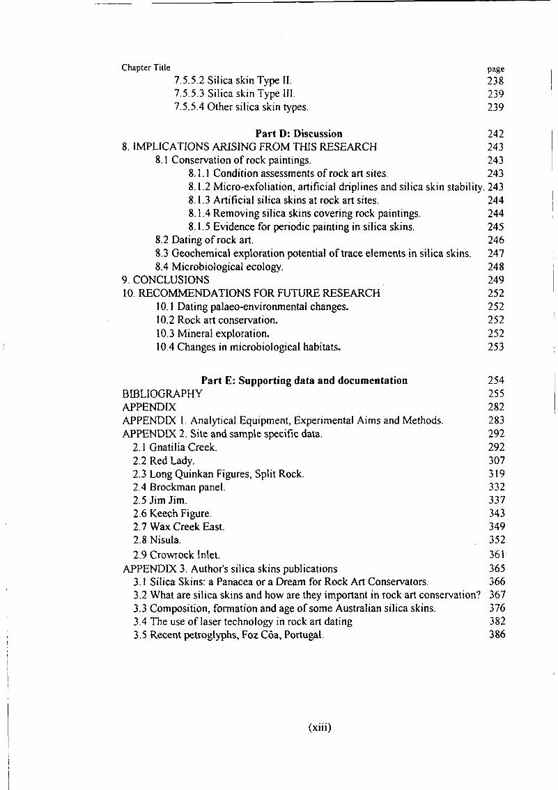
Chapter Title page
7.5.5.2 Silica skin Type II. 2387.5.5.3 Silica skin Type III. 2397.5.5.4 Other silica skin types. 239
Part D: Discussion 2428. IMPLICATIONS ARISING FROM THIS RESEARCH 243
8.1 Conservation of rock paintings. 2438.1.1 Condition assessments of rock art sites 2438.1.2 Micro-exfoliation, artificial driplines and silica skin stability. 2438.1.3 Artificial silica skins at rock art sites. 2448.1.4 Removing silica skins covering rock paintings. 2448.1.5 Evidence for periodic painting in silica skins. 245
8.2 Dating of rock art. 2468.3 Geochemical exploration potential of trace elements in silica skins. 2478.4 Microbiological ecology. 248
9. CONCLUSIONS 24910. RECOMMENDATIONS FOR FUTURE RESEARCH 252
10.1 Dating palaeo-environmental changes. 25210.2 Rock art conservation. 25210.3 Mineral exploration. 25210.4 Changes in microbiological habitats. 253
Part E: Supporting data and documentation 254BIBLIOGRAPHY 255APPENDIX 282APPENDIX 1. Analytical Equipment, Experimental Aims and Methods. 283APPENDIX 2. Site and sample specific data. 292
2.1 Gnatilia Creek. 2922.2 Red Lady. 3072.3 Long Quinkan Figures, Split Rock. 3192.4 Brockman panel. 3322.5 Jim Jim. 3372.6 Keech Figure. 3432.7 Wax Creek East. 3492.8 Nisula. 3522.9 Crowrock Inlet. 361
APPENDIX 3. Author's silica skins publications 3653.1 Silica Skins: a Panacea or a Dream for Rock Art Conservators. 3663.2 What are silica skins and how are they important in rock art conservation? 3673.3 Composition, formation and age of some Australian silica skins. 3763.4 The use of laser technology in rock art dating 3823.5 Recent petroglyphs, Foz Coa, Portugal. 386
(xiii)
LIST OF TABLES
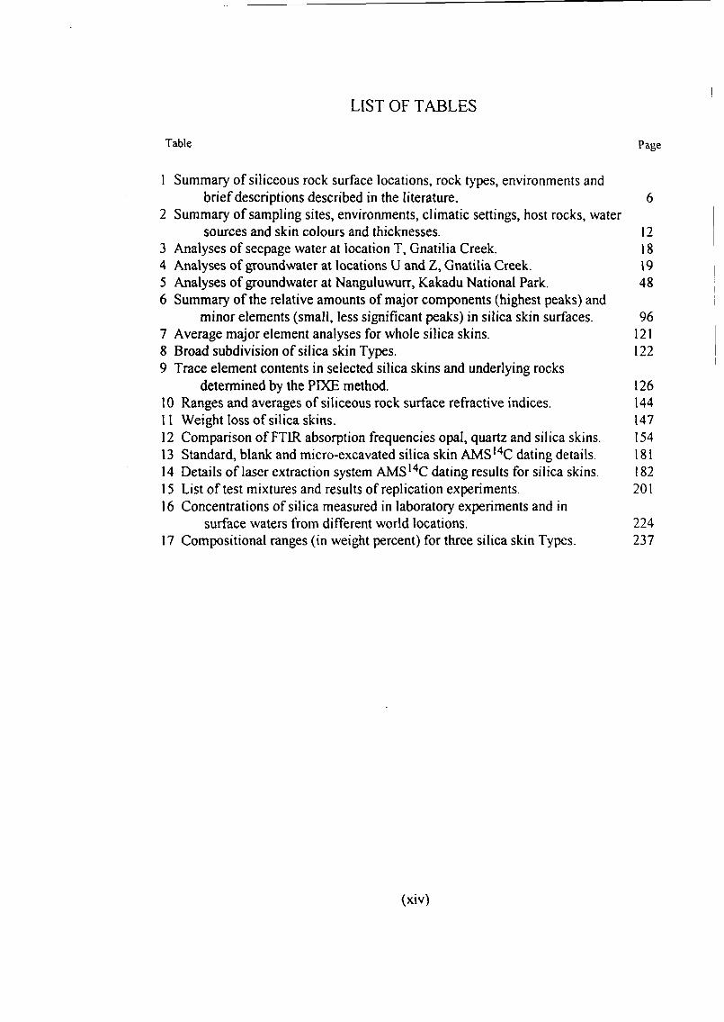
Table Page
1 Summary of siliceous rock surface locations, rock types, environments andbrief descriptions described in the literature. 6
2 Summary of sampling sites, environments, climatic settings, host rocks, watersources and skin colours and thicknesses. 12
3 Analyses of seepage water at location T, Gnatilia Creek. 184 Analyses of groundwater at locations U and Z, Gnatilia Creek. 195 Analyses of groundwater at Nanguluwurr, Kakadu National Park. 486 Summary of the relative amounts of major components (highest peaks) and
minor elements (small, less significant peaks) in silica skin surfaces. 967 Average major element analyses for whole silica skins. 1218 Broad subdivision of silica skin Types. 1229 Trace element contents in selected silica skins and underlying rocks
determined by the PIXE method. 12610 Ranges and averages of siliceous rock surface refractive indices. 14411 Weight loss of silica skins. 14712 Comparison of FTIR absorption frequencies opal, quartz and silica skins. 15413 Standard, blank and micro-excavated silica skin AMSI4C dating details. 18114 Details of laser extraction system AMS14C dating results for silica skins. 18215 List of test mixtures and results of replication experiments. 20116 Concentrations of silica measured in laboratory experiments and in
surface waters from different world locations. 22417 Compositional ranges (in weight percent) for three silica skin Types. 237
(xiv)
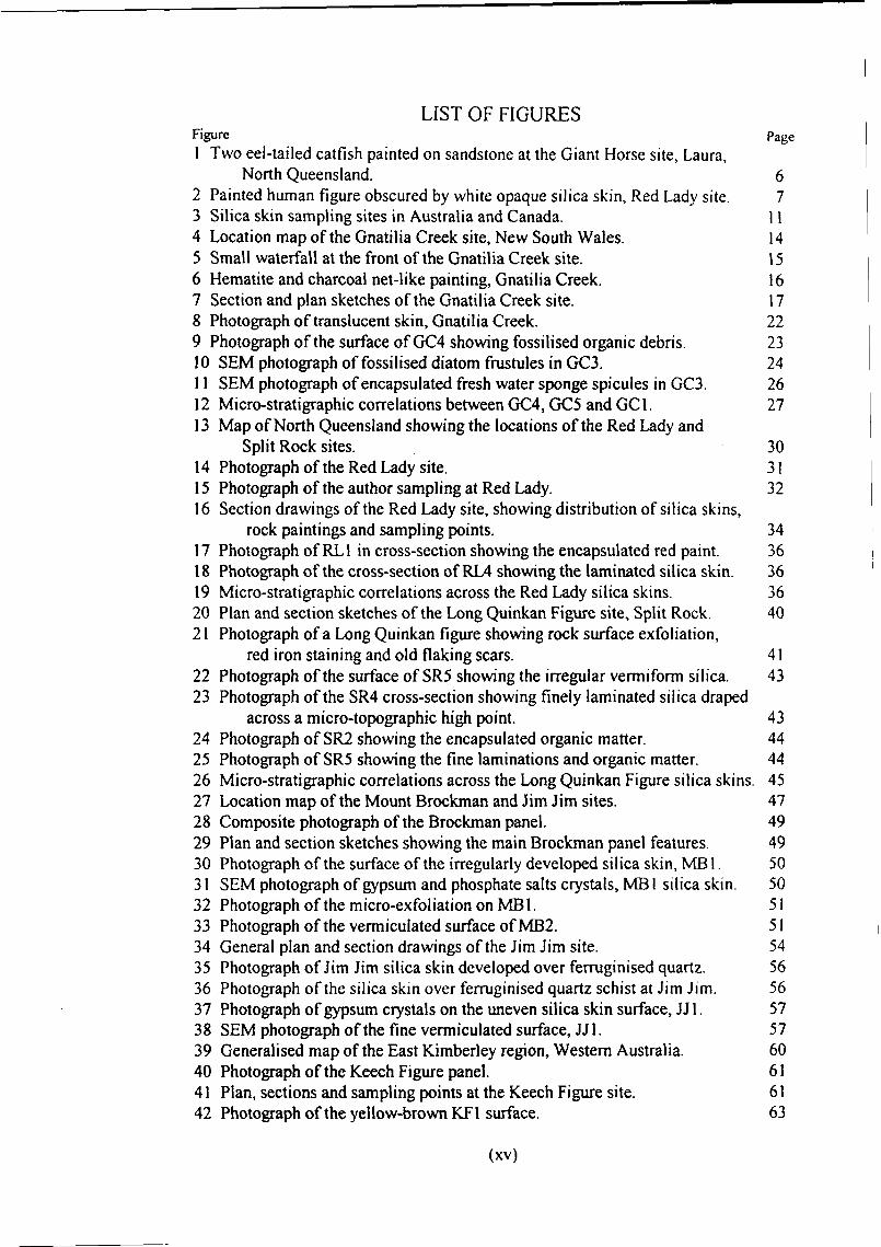
LIST OF FIGURESFigure Page1 Two eel-tailed catfish painted on sandstone at the Giant Horse site, Laura,
North Queensland. 62 Painted human figure obscured by white opaque silica skin, Red Lady site. 73 Silica skin sampling sites in Australia and Canada. 114 Location map of the Gnatilia Creek site, New South Wales. 145 Small waterfall at the front of the Gnatilia Creek site. 156 Hematite and charcoal net-like painting, Gnatilia Creek. 167 Section and plan sketches of the Gnatilia Creek site. 178 Photograph of translucent skin, Gnatilia Creek. 229 Photograph of the surface of GC4 showing fossilised organic debris. 2310 SEM photograph of fossilised diatom frustules in GC3. 2411 SEM photograph of encapsulated fresh water sponge spicules in GC3. 2612 Micro-stratigraphic correlations between GC4, GC5 and GC1. 2713 Map of North Queensland showing the locations of the Red Lady and
Split Rock sites. 3014 Photograph of the Red Lady site. 3115 Photograph of the author sampling at Red Lady. 3216 Section drawings of the Red Lady site, showing distribution of silica skins,
rock paintings and sampling points. 3417 Photograph of RL1 in cross-section showing the encapsulated red paint. 3618 Photograph of the cross-section of RL4 showing the laminated silica skin. 3619 Micro-stratigraphic correlations across the Red Lady silica skins. 3620 Plan and section sketches of the Long Quinkan Figure site, Split Rock. 4021 Photograph of a Long Quinkan figure showing rock surface exfoliation,
red iron staining and old flaking scars. 4122 Photograph of the surface of SR5 showing the irregular vermiform silica. 4323 Photograph of the SR4 cross-section showing finely laminated silica draped
across a micro-topographic high point. 4324 Photograph of SR2 showing the encapsulated organic matter. 4425 Photograph of SR5 showing the fine laminations and organic matter. 4426 Micro-stratigraphic correlations across the Long Quinkan Figure silica skins. 4527 Location map of the Mount Brockman and Jim Jim sites. 4728 Composite photograph of the Brockman panel. 4929 Plan and section sketches showing the main Brockman panel features. 4930 Photograph of the surface of the irregularly developed silica skin, MB 1. 5031 SEM photograph of gypsum and phosphate salts crystals, MB 1 silica skin. 5032 Photograph of the micro-exfoliation on MB1. 5133 Photograph of the vermiculated surface of MB2. 5134 General plan and section drawings of the Jim Jim site. 5435 Photograph of Jim Jim silica skin developed over ferruginised quartz. 5636 Photograph of the silica skin over ferruginised quartz schist at Jim Jim. 5637 Photograph of gypsum crystals on the uneven silica skin surface, JJ1. 5738 SEM photograph of the fine vermiculated surface, JJ1. 5739 Generalised map of the East Kimberley region, Western Australia. 6040 Photograph of the Keech Figure panel. 6141 Plan, sections and sampling points at the Keech Figure site. 6142 Photograph of the yellow-brown KF1 surface. 63
(xv)
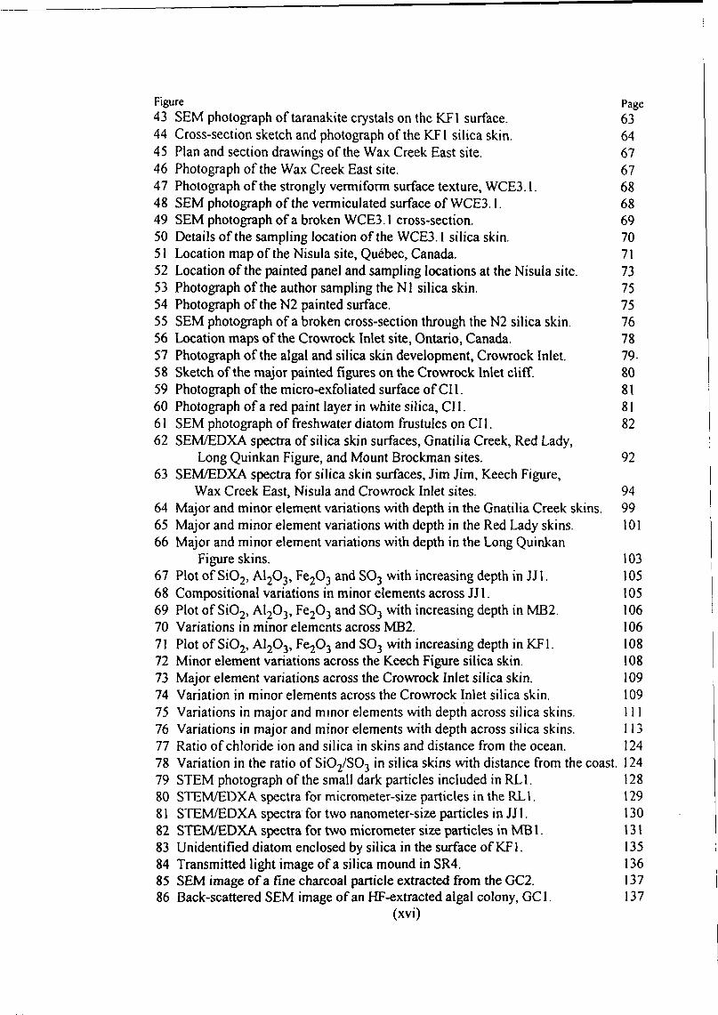
Figure Page43 SEM photograph of taranakite crystals on the KF1 surface. 6344 Cross-section sketch and photograph of the KF1 silica skin. 6445 Plan and section drawings of the Wax Creek East site. 6746 Photograph of the Wax Creek East site. 6747 Photograph of the strongly vermiform surface texture, WCE3.1. 6848 SEM photograph of the vermiculated surface of WCE3.1. 6849 SEM photograph of a broken WCE3.1 cross-section. 6950 Details of the sampling location of the WCE3.1 silica skin. 7051 Location map of the Nisula site, Quebec, Canada. 7152 Location of the painted panel and sampling locations at the Nisula site. 7353 Photograph of the author sampling the Nl silica skin. 7554 Photograph of the N2 painted surface. 7555 SEM photograph of a broken cross-section through the N2 silica skin. 7656 Location maps of the Crowrock Inlet site, Ontario, Canada. 7857 Photograph of the algal and silica skin development, Crowrock Inlet. 7958 Sketch of the major painted figures on the Crowrock Inlet cliff. 8059 Photograph of the micro-exfoliated surface of CI1. 8160 Photograph of a red paint layer in white silica, CII. 8161 SEM photograph of freshwater diatom frustules on CII. 8262 SEM/EDXA spectra of silica skin surfaces, Gnatilia Creek, Red Lady,
Long Quinkan Figure, and Mount Brockman sites. 9263 SEM/EDXA spectra for silica skin surfaces, Jim Jim, Keech Figure,
Wax Creek East, Nisula and Crowrock Inlet sites. 9464 Major and minor element variations with depth in the Gnatilia Creek skins. 9965 Major and minor element variations with depth in the Red Lady skins. 10166 Major and minor element variations with depth in the Long Quinkan
Figure skins. 10367 Plot of SiO2, A12O3, Fe2O3 and SO3 with increasing depth in JJ1. 10568 Compositional variations in minor elements across JJ1. 10569 Plot of SiO2, AI2O3, Fe2O3 and SO3 with increasing depth in MB2. 10670 Variations in minor elements across MB2. 10671 Plot of SiO2, A12O3, Fe2O3 and SO3 with increasing depth in KF1. 10872 Minor element variations across the Keech Figure silica skin. 10873 Major element variations across the Crowrock Inlet silica skin. 10974 Variation in minor elements across the Crowrock Inlet silica skin. 10975 Variations in major and minor elements with depth across silica skins. 11176 Variations in major and minor elements with depth across silica skins. 11377 Ratio of chloride ion and silica in skins and distance from the ocean. 12478 Variation in the ratio of SiO2/SO3 in silica skins with distance from the coast. 12479 STEM photograph of the small dark particles included in RL1. 12880 STEM/EDXA spectra for micrometer-size particles in the RL1. 12981 STEM/EDXA spectra for two nanometer-size particles in JJ 1. 13082 STEM/EDXA spectra for two micrometer size particles in MB 1. 13183 Unidentified diatom enclosed by silica in the surface of KF1. 13584 Transmitted light image of a silica mound in SR4. 13685 SEM image of a fine charcoal particle extracted from the GC2. 13786 Back-scattered SEM image of an HF-extracted algal colony, GC1. 137
(xvi)
Figure page87 Relative proportions of fatty acids in silica skins. 13988 Plot of refractive index against percentage water content. 14589 Incremental weight loss curves for silica skins. 14890 XRD spectra for Gnatilia Creek, Red Lady, Split Rock, Jim Jim and
Mount Brockman silica skins. 15191 XRD patterns for KF1, WCE3.1, Nisula and CI1 silica skins. 15292 FTIR spectra for silica skins from Gnatilia Creek, Red Lady and Split Rock. 15593 FTIR spectra for silica skins from MB2, KF1, WCE3.1, Nisula and C11. 15694 SEM photograph of the surface of a white silica skin (RL2). 15895 Random packing of siliceous aggregates in a white silica skin. 15996 Spiral aggregates of silica particles on the GC1 skin surface. 15997 Extensive network of capillary fractures in a silica skin. 16098 Crowrock Inlet silica skin showing possible in situ dissolution and
reprecipitation of silica from broken diatoms. 16199 TEM photograph showing grey amorphous silica coagulated around dark
nanometer-sized inclusions. 163100 TEM image of a noncrystalline clay or aluminium-rich mineral precursor in
Gnatilia Creek white silica skin. 164101 Assembly of laser components for the FLECS system of extracting carbon
from organic matter fossilised in silica skins. 170102 Carbon dioxide collection arrangement for FLECS. 171103 Schematic diagram illustrating the arrangement of inlet and outlet gas tubes,
micro-combustion chamber, vacuum gauge and laser focusing system. 173104 Glass globules on a silica skin surface following laser treatment. 174105 Magnified view of two spherical soda-magnesia glass beads. 175106 Relationship between thickness and ages of silica skins. 190107 Opalescent artificial silica film produced by dilute ionic catalysis of TEOS. 202108 Pervasive cracking of a transparent artificial silica skin prepared using
concentrated ionic catalyst and TEOS. 203109 Fractured MTMOS polymer film produced from a mixture of 30% v/v water
and silane. 203110 TEM image of transparent film particles produced by catalysis of TEOS
with concentrated ammonia and ethanol. 207111 TEM image of a cluster of silica particles in a film from dilute ionic
catalyst and TEOS. 208112 Un-ionised and ionised silica species as percentages of total silicon in
solution at different pH. 214113 Solubility of amorphous silica as a function of pH. 215114 Solubility of amorphous silica in the presence of A13+ as a function of pH. 216115 Solubility of amorphous silica in the presence of Mg2+ as a function of pH. 217116 Rate of solubility of amorphous silica, opal, cristobalite and quartz. 219117 Experimentally determined release of silicic acid from feldspars as
a function of the square root of time. 221118 Proposed sequence of processes leading to silica skin formation. 233119 Diagram showing the compositions of clear and dark layers in
Hawaiian silica skins compared with three Australian silica skins. 240
(xvii)


![F RADIOCARBON, UNIVERSITY OF TEXAS RADIOCARBON DATES II · F RADIOCARBON, Vor,. 6, 1964, P. 138-159] UNIVERSITY OF TEXAS RADIOCARBON DATES II M. A. TAMERS, F. J. PEARSON, JR., and](https://img.pdfslide.us/doc/110x75/606d59c493119417f12a3a02/f-radiocarbon-university-of-texas-radiocarbon-dates-ii-f-radiocarbon-vor-6.jpg)