Embed Size (px)
Citation preview
University of Dundee
Recognition of substrate degrons by E3 ubiquitin ligases and modulation by small-molecule mimicry strategiesLucas, Xavier; Ciulli, Alessio
Published in:Current Opinion in Structural Biology
DOI:10.1016/j.sbi.2016.12.015
Publication date:2017
Licence:CC BY-NC-ND
Document VersionPeer reviewed version
Link to publication in Discovery Research Portal
Citation for published version (APA):Lucas, X., & Ciulli, A. (2017). Recognition of substrate degrons by E3 ubiquitin ligases and modulation by small-molecule mimicry strategies. Current Opinion in Structural Biology, 44, 101-110.https://doi.org/10.1016/j.sbi.2016.12.015
General rightsCopyright and moral rights for the publications made accessible in Discovery Research Portal are retained by the authors and/or othercopyright owners and it is a condition of accessing publications that users recognise and abide by the legal requirements associated withthese rights.
• Users may download and print one copy of any publication from Discovery Research Portal for the purpose of private study or research. • You may not further distribute the material or use it for any profit-making activity or commercial gain. • You may freely distribute the URL identifying the publication in the public portal.
Take down policyIf you believe that this document breaches copyright please contact us providing details, and we will remove access to the work immediatelyand investigate your claim.
Download date: 18. Feb. 2022

1
Title: Recognition of substrate degrons by E3 ubiquitin ligases and modulation by small-
molecule mimicry strategies
Authors: Xavier Lucas and Alessio Ciulli
Author affiliations: School of Life Sciences, University of Dundee, Division of Biological
Chemistry and Drug Discovery, James Black Centre, Dow Street, Dundee, DD1 5EH, UK.
Corresponding author: Alessio Ciulli; School of Life Sciences, University of Dundee,
Division of Biological Chemistry and Drug Discovery, James Black Centre, Dow Street,
Dundee, DD1 5EH, UK; +44(0)1382386230; [email protected].
Highlights
E3 ligases recruit substrates for proteasomal degradation by recognition of degrons
Crystal structures reveal the structural basis and mechanism of degron recognition
Small-molecule degron mimetics can prevent or re-direct substrate recognition
Small-molecule inducible degrons and PROTACs enable targeted protein degradation
© 2016. This manuscript version is made available under the CC-BY-NC-ND 4.0 license
http://creativecommons.org/licenses/by-nc-nd/4.0/

2
Abstract
The ubiquitin-proteasome system is a master regulator of protein homeostasis, by which
proteins are initially targeted for poly-ubiquitination by E3 ligases and then degraded into
short peptides by the proteasome. Nature evolved diverse peptidic motifs, termed degrons, to
signal substrates for degradation. We discuss degrons of the N-end rule pathway and also
degrons characterized by post-translational modifications, including phosphorylation and
hydroxylation. In each case we detail the structural basis of E3 ligase:degron recognition and
small-molecule mimicry approaches that disrupt those protein-protein interactions. We
present as well genetic and chemical technologies that enable targeted degradation of proteins
of interest, namely small-molecule dependent inducible degrons and chemical degraders, e.g.
proteolysis-targeting chimeras (PROTACs).
3
Introduction
The Nobel Prize in Chemistry 2004 was awarded jointly to Aaron Ciechanover, Avram
Hershko, and Irwin Rose “for the discovery of ubiquitin-mediated protein degradation”.
Since then, many discoveries have paved the way to a better mechanistic and structural
understanding of the protein degradation machinery and have enabled its purposeful
modulation and hijacking.
The ubiquitin-proteasome system (UPS) is a complex cellular pathway by which proteins are
first ubiquitinated and subsequently unfolded and proteolyzed by the proteasome. This
process has direct implications primarily on regulating protein homeostasis and, depending
on the context, can impact many cellular signaling processes, including cell cycle, DNA
repair, apoptosis, inflammation, transcription regulation, stress response, and protein quality
control (PQC) [1]. Three main enzymes are responsible for the specific targeting of proteins
for degradation: E1-activating enzymes, which activate ubiquitin (Ub) in an ATP-dependent
manner; E2-conjugating enzymes, to which the activated Ub is covalently attached to yield an
E2~Ub thioester intermediate; and E3 ubiquitin ligases, which catalyze the transfer of Ub
from the E2 enzyme to form an isopeptide bond with a lysine residue on the protein substrate
(mono-ubiquitination or priming) or its covalently attached Ub (poly-ubiquitination) [2]. To
act as catalyst in the process, E3 ligases typically recruit specific target substrates for
degradation by recognition of peptidic segments termed ‘degrons’ as characterizing signaling
markers [3]. The structural determinants within the degron and the E3 ubiquitin ligase that
confer substrate specificity and dictate protein recognition and fate are of utmost importance
to elucidate and be able to manipulate proteasome-mediated degradation and are the focus of
this review. Recognition of structural protein domains or specific consensus sequences, e.g.
in the case of D-box and KEN-box recognition by anaphase-promoting complex/cyclosome
(APC/C)[4] will not be covered here.
Nature has evolved diverse mechanisms to regulate protein homeostasis. For example,
orchestrated autophagy of misfolded or damaged proteins is intimately linked to the UPS
through the PQC pathway [5]. Those substrates can be targeted for degradation in different
ways, including exposure of hydrophobic degrons that would be otherwise buried inside the
protein or post-translational polyglycosylation of Asn residues. In the latter case, misfolded
proteins are signaled for endoplasmic reticulum-associated protein degradation (ERAD) [6].
Failure to degrade misfolded proteins, consequently favoring their aggregation and eventual

4
collapse, has a major impact in the development of neurological diseases [7, 8]. Other post-
translational modifications (PTMs) apart from Asn glycosylation, such as phosphorylation of
Ser, Tyr, and Thr residues, hydroxylation of Pro, and acetylation of Lys and their interplay
contribute as well to determining a protein’s fate [9]. For example, acetylation competes with
Lys ubiquitination and can prevent target degradation [10]. In other cases, the E3 ligase itself
post-translationally modifies the substrate upon engagement, which in turn allosterically
initiates the ubiquitination cascade of the target protein [11].
In this review we first briefly discuss degradation of proteolytic cleavage products by the N-
end rule pathway. We next examine recruitment to E3 ligases of substrates marked for
degradation by means of recognition of specific PTMs, namely phosphorylation and
hydroxylation. We conclude exploring prominent small molecules from both natural and
unnatural origin capable of modulating or even de novo re-directing substrate specificity of
E3 ligases. In each case we describe related chemical biology tools for targeted protein
degradation.
N-degrons
The pioneering observation of an apparent correlation between the presence of a free α-amino
group in a protein and its ubiquitin-dependent degradation led to the formulation of the ‘N-
end rule’, by which the in vivo half-life of a protein can be determined by the nature of its N-
terminal amino acid, also termed ‘N-degron’ [12]. N-degrons are generated within the cell
when specific residues are exposed at the N terminus by proteolytic cleavage. There are two
classes of destabilizing N-degrons: positively charged amino acids (Arg, Lys, and His) are of
type 1, and bulky hydrophobic ones (Phe, Trp, Tyr, Leu, and Ile) are of type 2 [13].
Conversely, other N-terminal amino acids such as Met and Cys confer stability against
proteosomal degradation [14]. In eukaryotes, N-degrons are recognized by N-recognin, a
UBR box motif present in E3 ligases that targets the substrate for ubiquitin-dependent
proteosomal degradation [15]. For example, endoproteolytic cleavage of Scc1, a subunit of
the cohesion complex in yeast, results in a type 1 Arg N-terminal fragment that is recognized
and targeted for degradation by N-recognin UBR1. Notably, the fragment becomes lethal if
accumulated [16].
The crystallographic structure of the UBR box of S. cerevisiae UBR1 in complex with type 1
N-degrons revealed that specific recognition is achieved by an intricate network of hydrogen-

5
bonds involving as well the amino acid in position 2 of the N-degron, which occupies an
interfacial hydrophobic grove (Fig. 1a) [17]. Conversely, in type 2 N-degrons exquisite
selectivity is accomplished by a highly conserved gatekeeper Tyr residue, which excludes
Val but not the Ile, Leu, Phe, Tyr, and Trp degrons [15]. Interestingly, in bacteria, where Met
instead of Tyr serves as gatekeeper residue, distinct selectivity is achieved by introducing
steric clashes: bacterial UBR1 excludes Ile, Thr, and Val but not Leu, Trp, and Phe (Fig 1b
and 1c) [13, 18, 19]. In eukaryotes, the default N-terminal amino acid is Met (N-
formylmethionine in bacteria). The striking ability of N-recognin to discriminate Met from its
structural cousin Leu with up to 1000-fold selectivity has been deeply investigated. Notably,
only a rare, entropically unfavored Met rotamer can fit in the N-recognin cavity and avoid
large van der Waals steric clashes with the surrounding residues. Moreover, this rotamer
locates the Met’s Cε in a chemically unfavorable environment [18].
Small molecules, e.g. p-Chloroamphetamine, are known to inhibit the N-end rule pathway by
blocking a UBR recognition site [20]. However, broader applicability of such inhibitors to
manipulate the cellular level of specific proteins is dramatically hampered by the lack of
control on which substrate is downstream degraded. This limitation can be overcome by two
distinct chemical biology approaches. In a first strategy, selective proteasome-mediated
degradation of glutathione-S-transferase α1 (GST-α1) was achieved by linking a Boc-
protected Arg (Boc3-Arg) to a potent GST-α1 covalent inhibitor [21]. A clear advantage of
this technology is its intrinsic modularity with respect to which protein can be addressed. For
example, use of a noncovalent inhibitor of dihydrofolate reductase (DHFR) conjugated to
Boc3-Arg led to rapid and robust DHFR degradation in cells. In contrast, linkage of the
inhibitors to non-protected Arg rendered inactive degraders, indicating that Boc3-Arg tagging
works independently of the N-end rule pathway [21]. While the defined biological
mechanism of Boc3-Arg tagging is not fully understood, Long et al. used cycloheximide
blocking to show that reduction of DHFR levels was due to induced degradation and not to
translation inhibition [21]. Very recently it has been shown that Boc3-Arg tagging localizes
the target proteins directly to the 20S proteasome and stimulates its degradation without
requiring ubiquitination [22]. A related approach called hydrophobic tagging (HyT) has been
developed to append hydrophobic moieties to ligands and fusion proteins to induce targeted
degradation [23, 24]. In a different approach, a Trojan horse genetic strategy was conceived
by Taxis et al., who developed tobacco etch virus (TEV) protease-mediated induction of
protein instability (TIPI) [25]. TIPI is a method to genetically control the abundance of a

6
protein of interest (POI) by genetically inserting a dormant destabilizing N-degron. Upon
expression of a site-specific protease, the dormant N-degron becomes exposed and triggers
selective targeting of the POI by UBR and its proteasome-mediated degradation [25].
Phosphodegrons
Phosphorylation at one or several amino acids on proteins is well known to direct formation
of new protein-protein interactions (PPIs). The first protein module identified as a “reader” of
phosphorylated protein modifications was the Src homology 2 (SH2) domain, which belongs
to the protein kinase family and recognizes exclusively phosphorylated Tyr (pTyr) [26]. Later
on pTyr-, as well as pSer- and pThr-binding domains have been identified in other protein
families and their crucial involvement in cell signaling and DNA damage response have
become apparent [27].
Protein phosphorylation in regions so-called ‘phosphodegrons’ is also exploited for effective
substrate recognition by E3 ligases and processive proteasome-mediated degradation [15]. A
well-studied phosphodegron-binding system is the archetypical S-phase kinase-associated
protein 1 (Skp1)-Cul1-F-box (SCF) Cullin RING ligase (CRL) (Fig. 2a), in which the variant
F-box domain dictates substrate recognition. This family can be classified according to the
presence of specific substrate recognition domains into FBWX, containing WD40 repeats,
FBXL, presenting Leu-rich motifs, and the less characterized FBXO subclass [28]. For
example, F-box WD40-containing protein 7 (FBW7) is the substrate recognition module of
the cyclin-dependent kinase (CDK) regulator complex SCFFBW7. Crystallographic studies of
SCFFBW7 and its yeast ortholog, SCFCdc4, revealed that phosphodegron recognition in this
system is driven by three primary features at the PPI interface: electrostatic interactions and a
rich hydrogen-bond network that discriminate and exclusively trap the phosphorylated state
of the target degron; hydrophobic patches that recognize two conserved hydrophobic residues
in the phosphodegron; and positively charged residues that prompt suboptimal binding of
basic phosphodegrons (Fig. 2b and 2c) [29, 30]. In yeast, SCFCdc4 targets for poly-
ubiquitination and proteasome-mediated degradation phosphorylated substrate inhibitor of
CDK1 (SIC1), thereby enabling entry into the cellular S phase. Orlicky et al. carried out a
screening of 50,000 small molecules to identify inhibitors of CDC4 that would prevent
degradation of SCFCdc4 targets. They identified an allosteric modulator of SCFCdc4 that
inhibits recruitment of pSIC1 by intercalating within the β-propeller of Cdc4, ~25Å away
7
from the phosphodegron recognition site of Cdc4 (Fig. 2d) [31]. In mammalian cells,
SCFFBW7 recruits a number of important regulatory factors in cell growth and division
pathways that function as proto-oncogenes in many cancers, such as cyclin E, MYC, and
NOTCH, signaling them for ubiquitination and degradation [32]. Cancer-associated
mutations in Fbxw7 and in the genes encoding SCFFBW7 substrates can weaken binding
affinities of the E3 ligase for its substrate degrons [32]. Small-molecule rescue, as opposed to
disruption, of these PPIs could provide therapeutic benefit against prevalent mutant cancers.
Other examples of phosphodegron reader subunits in E3 CRLs include the Suppressor of
Cytokine Signaling (SOCS) proteins and Cbl, each containing SH2 domains as substrate-
recognition domain [33, 34].
Rational design of small molecules that disrupt the recognition of phosphorylated targets
usually relies on occupying the canonical phosphate-binding site of the reader protein. This
typically involves developing peptidomimetics, i.e. fragments of the native substrate that
retain structural features of the molecular recognition motif while improving specific
physicochemical properties. Alternative approaches involve identifying hits from screening
compound libraries. A limitation of those methods is that resulting molecules often lack
selectivity amongst phosphodegron recognizers. For instance, the SH2-containing
transcription factors signal transducer and activator of transcription (STAT) 5a and STAT5b
have a sequence identity of 93% and recognize the same substrate peptide motifs, despite
tissue-specific expression patterns and a number of non-redundant biological functions [35,
36]. The observation that cathecol bisphosphate is a sub-μM inhibitor of STAT5b with 35-
fold selectivity over STAT5a motivated the development of a series of peptidomimetics using
the cathecol bisphosphate fragment as anchor [37]. The most potent compound, Stafib-1, has
a Ki of 44 nM for STAT5b with over 50-fold selectivity over STAT5a and retains selectivity
in tumor cells when formulated as a prodrug derivative [37]. Structural features that could
shine light on the exquisite selectivity exhibited by Stafib-1 remain elusive.
Oxygen-dependent degrons (ODDs)
The modularity of the CRL architecture enables a dynamic and context-specific recruitment
of substrate-binding proteins [38]. A notable case is the von Hippel-Lindau (VHL) protein,
which forms part of an E3 ligase complex with the adaptor proteins Elongin (Elo) B and
EloC, Cul2, and RBX1 (CRL2VHL) (Fig. 3a). VHL recognizes and targets for degradation

8
hypoxia-inducible factor alpha (HIF-α) subunits, which are efficiently trans-4-prolyl
hydroxylated (Hyp) under normal oxygen levels [39, 40]. In contrast, under hypoxia HIF-α
subunits escape hydroxylation and recognition by VHL, are consequently stabilized inside
cells, and drive transcriptional responses to hypoxia. Crystallographic studies revealed the
structural basis for HIF-1α binding by VHL, and elucidated the exquisite specificity for the
recognition of the C4-exo conformation of Hyp (Fig. 3b) [41-43]. This mechanism of
substrate recognition inspired the structure-guided fragment-based design of non-peptidic
small-molecule Hyp derivatives that mimic binding of the natural substrate (Fig. 3c) [44-47].
By occupying the PPI interface of CRL2VHL:HIF-1α, these molecules could effectively
displace HIF-1α binding with nanomolar potency [47]. Further optimization of this class of
inhibitors led to the discovery of VHL inhibitor VH298 as a novel potent, selective, and cell-
active chemical probe of the VHL-HIF pathway [48]. VHL inhibitors have therapeutic
potential in certain disease conditions where accumulation of HIF-α subunits and subsequent
triggering of hypoxic response could prove beneficial [49].
Small-molecule dependent degrons
Methods to induce conditional and controlled degradation of POIs have substantial potential
as both chemical biology and therapeutic tools. Interestingly, plants have evolved two
analogous induced protein degradation mechanisms by phytohormones auxin and jasmonate
as part of their signalosome [50, 51]. Transport inhibitor response 1 (TIR1) is the F-box
substrate recognition subunit of a SCFTIR1 ubiquitin ligase (Fig. 4a), which targets
transcriptional repressors known as Aux/IAA (indole-3-acetic acid) proteins for proteosomal
degradation. By binding auxin, TIR1 increases affinity for its targets and triggers their rapid
ubiquitination and proteosomal degradation [50, 52]. Crystal structures of Arabidopsis TIR1
in complex with auxin and an Aux/IAA degron peptide derived from the IAA7 protein
elucidated the structural basis of how auxin binding directs TIR1:substrate interactions (Fig.
4b) [50].
Inspired by this natural mechanism, Nishimura et al. developed an auxin-inducible degron
(AID) for the controlled degradation of proteins [53]. First applied to yeast, the method
involves knock-in of the AID at either end of the POI, so that the fusion protein can be
rapidly and efficiently depleted upon addition of auxin to the culture medium and conditional
expression of the plant SCFTIR1 ubiquitin ligase [54]. The auxin degron technology has
9
proven its potential to study the biological function of proteins in higher eukaryotes. For
example, it has been recently applied to induce rapid and conditional depletion of essential
genes, for which knockouts or small-interfering RNAs are not suitable, in human and
embryonic stem cells by introducing the AID-POI fusion using the CRISPR/Cas9-based
method [54].
From a structural point of view, auxin and jasmonate act as “molecular glue” of specific PPIs,
i.e. they stabilize the interaction of the substrate-binding domain of their respective E3 ligase
and specific substrates [55]. Strikingly, phthalimide immunomodulatory drugs (IMiDs)
thalidomide and its second-generation derivatives lenalidomide and pomalidomide act dually
as molecular glues and PPI disruptors in humans by targeting the protein cereblon (CRBN)
[56]. CRBN is the substrate-binding domain of the Rbx1-Cul4-DDB1-CRBN (CRL4CRBN) E3
ubiquitin ligase (Fig. 4c). IMiD-binding by CRL4CRBN prevents engagement of its
endogenous substrate MEIS2; it also re-directs effective recruitment and CRBN-dependent
degradation of the transcription factors Ikaros and Aiolos as well as Casein kinase 1α (CK1α)
[57-60]. Additionally, lenalidomide derivative CC-885 was shown to induce recruitment and
degradation of the translation termination factor GSPT1 [61]. These observations
demonstrate that substrate selectivity of E3 ligases can be effectively modulated by binding
of small molecules, which can act either as stabilizers or disruptors of specific E3
ligase:degron complexes. The structural basis of small-molecule induced recognition of
CK1α and GSPT1 by CRL4CRBN revealed a molecular glue mechanism similar to auxin [61,
62]. Crystallographic data along with site-directed mutagenesis studies on a homology model
of the Ikaros:CRBN complex further demonstrates that a hairpin-loop with low sequence
homology but conserved topology serves as key structural degron for IMiD-induced CRBN
recognition of substrates (Fig. 4d) [61].
PROTACs: Small-molecule directed protein degradation
Small molecules can be designed to recruit proteins into proximity to E3 ligases to induce
target degradation. Proteolysis-targeting chimeras (PROTACs) are heterobifunctional
molecules composed of a ligand for an E3 ligase and a ligand for a POI, connected by a
linker [63]. PROTACs that hijack CRL2VHL and CRL4CRBN using derivatives of the VHL and
CRBN ligands shown in Fig. 3 and 4 have proven very successful in inducing degradation of
the epigenetic regulators BET bromodomain proteins (BRD2, BRD3 and BRD4) and the
10
estrogen-related receptor α (ERRα) in cells and in vivo [64-67]. Crucially, PROTACs can
exhibit higher selectivity for protein degradation than one might anticipate based on the
intrinsic binding selectivity of the warhead target ligand. For example, Zengerle et al. showed
that VHL-targeting PROTACs based on the pan-BET inhibitor JQ1 induced preferential
depletion of BRD4 in cells [64]. Lai et al. later also found that specific PROTACs engaging
VHL or CRBN have distinct degradation preferences for their target kinases [68]. The sub-
stoichiometric catalytic modality of PROTAC’s activity relieves the need to fully occupy a
target binding site, aiding differential efficacy. Furthermore, the nature of the targeted E3
ligase [69], the chemical nature of the ligand and choice of derivatization points from the
ligands, as well as possible cooperativity of ternary complex formation can all influence
PROTAC’s activity and play a role in enhancing target selectivity.
The large number of E3 ligases (> 600) encoded in the human genome [70] and the diversity
and specificity of degron recognition motifs (reviewed recently in ref. [71]) provide
numerous opportunities for PROTAC drug development. To date, only a handful of E3
ligases (including CRBN, VHL, MDM2 and IAP, Table 1) have been effectively hijacked by
all small-molecules PROTACs using the respective E3 ligands. However, drug-like small-
molecule ligands are beginning to emerge for more E3 targets (Table 1), suggesting other
unexplored E3s may prove amenable to structure-based drug design.
PROTACs are an emerging technology that is attracting interest as chemical tool for target
validation due to its simplicity and modularity. Recent improvements in efficacies and
selectivity of PROTACs support development as new therapeutic modality [72]. However,
structural and mechanistic details regarding PROTAC-induced complexes between E3 ligase
and POI, and POI’s processive ubiquitination remain to be elucidated.
Conclusions
We present here a selection of different degrons that E3 ligases recognize to specifically
target substrates for proteasome-mediated degradation. The examples presented highlight
how we are only beginning to scratch the surface of proteasome-mediated protein
degradation, with more mechanisms of degron recognition likely to emerge in future. We
anticipate that unraveling the overall structure and dynamics of E3 ligase:substrate
complexation above and beyond epitope recognition for degron engagement will pave the
way to a more detailed mechanistic understanding of processive ubiquitination. Beyond their
11
relevance to ubiquitin-specific mechanisms, the studies of E3 ligase degron recognition have
contributed more broadly to the field of structural biology and small-molecule druggability
by revealing the structural basis for PTM-dependent and small-molecule induced de novo
formation of PPIs of functional relevance.
Small molecule approaches that enable conditional degradation of POIs, namely small-
molecule dependent inducible degrons and PROTACs, represent complementary
technologies and sophisticated chemical biology tools for post-translational protein
inactivation. Targeted protein degradation is attracting increasing interest at both academic
and pharmaceutical levels because of the potential to address therapeutic areas for which
current methods are not suitable or are inadequate. Indeed, small molecules have been
already used to induce rapid, selective depletion of key oncogenes or aberrant proteins in
cells and in vivo disease models. We anticipate that this new modality of chemical
intervention will impact increasingly relevant and yet un-drugged biological systems in the
future.
Acknowledgements
We are thankful to the organizations that provide funding support to the Ciulli Laboratory at
the University of Dundee, including the European Research Council (ERC-2012-StG-311460
DrugE3CRLs Starting Grant to A.C.); the European Commission (H2020-MSCA-IF-2015-
806323 Marie Skłodowska-Curie Actions Individual Fellowship to X.L.); and the Wellcome
Trust (Strategic Awards 100476/Z/12/Z for biophysics and drug discovery and
094090/Z/10/Z for structural biology and X-ray crystallography to the Division of Biological
Chemistry and Drug Discovery at Dundee).
12
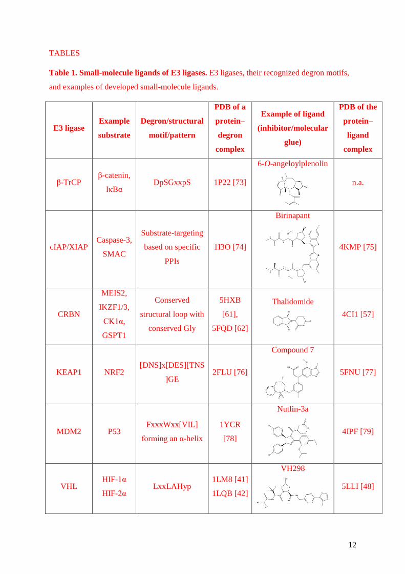
TABLES
Table 1. Small-molecule ligands of E3 ligases. E3 ligases, their recognized degron motifs,
and examples of developed small-molecule ligands.
E3 ligase Example
substrate
Degron/structural
motif/pattern
PDB of a
protein–
degron
complex
Example of ligand
(inhibitor/molecular
glue)
PDB of the
protein–
ligand
complex
β-TrCP β-catenin,
IκBα DpSGxxpS 1P22 [73]
6-O-angeloylplenolin
n.a.
cIAP/XIAP Caspase-3,
SMAC
Substrate-targeting
based on specific
PPIs
1I3O [74]
Birinapant
4KMP [75]
CRBN
MEIS2,
IKZF1/3,
CK1α,
GSPT1
Conserved
structural loop with
conserved Gly
5HXB
[61],
5FQD [62]
Thalidomide
4CI1 [57]
KEAP1 NRF2 [DNS]x[DES][TNS
]GE 2FLU [76]
Compound 7
5FNU [77]
MDM2 P53 FxxxWxx[VIL]
forming an α-helix
1YCR
[78]
Nutlin-3a
4IPF [79]
VHL HIF-1α
HIF-2α LxxLAHyp
1LM8 [41]
1LQB [42]
VH298
5LLI [48]

13
FIGURES
Figure 1. Structural basis of N-degron recognition. a) Crystal structure of ubiquitin ligase
UBR1 from S. cerevisiae in complex with the type 1 N-degron substrate peptide RLGE (PDB
code 3NIN [17]). The electrostatic potential surface of UBR1 is shown. b) Crystal structure
of N-end rule adaptor protein ClpS from C. crescentus in complex with type 2 N-degron
substrates Leu, Phe, and Trp (PDB codes 3G19, 3GQ1, and 3GW1, respectively [18]).
Gatekeeper residue Met53 is highlighted. In a) and b), residues forming hydrogen bonds with
the substrate are labelled. Note that in b), only Trp can interact by hydrogen bond with the
backbone of Met75. c) Sphere representation of amino acid Ile modelled in PyMOL in the
binding site of b) using PDB code 3G19.
14
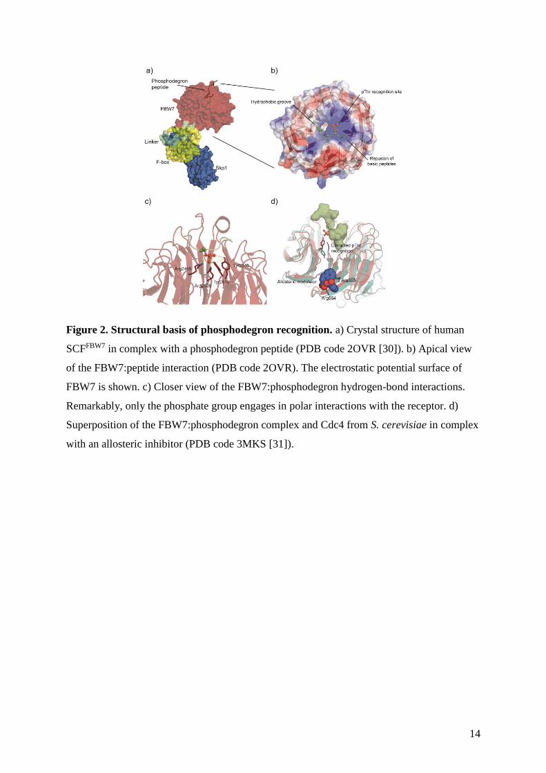
Figure 2. Structural basis of phosphodegron recognition. a) Crystal structure of human
SCFFBW7 in complex with a phosphodegron peptide (PDB code 2OVR [30]). b) Apical view
of the FBW7:peptide interaction (PDB code 2OVR). The electrostatic potential surface of
FBW7 is shown. c) Closer view of the FBW7:phosphodegron hydrogen-bond interactions.
Remarkably, only the phosphate group engages in polar interactions with the receptor. d)
Superposition of the FBW7:phosphodegron complex and Cdc4 from S. cerevisiae in complex
with an allosteric inhibitor (PDB code 3MKS [31]).
15
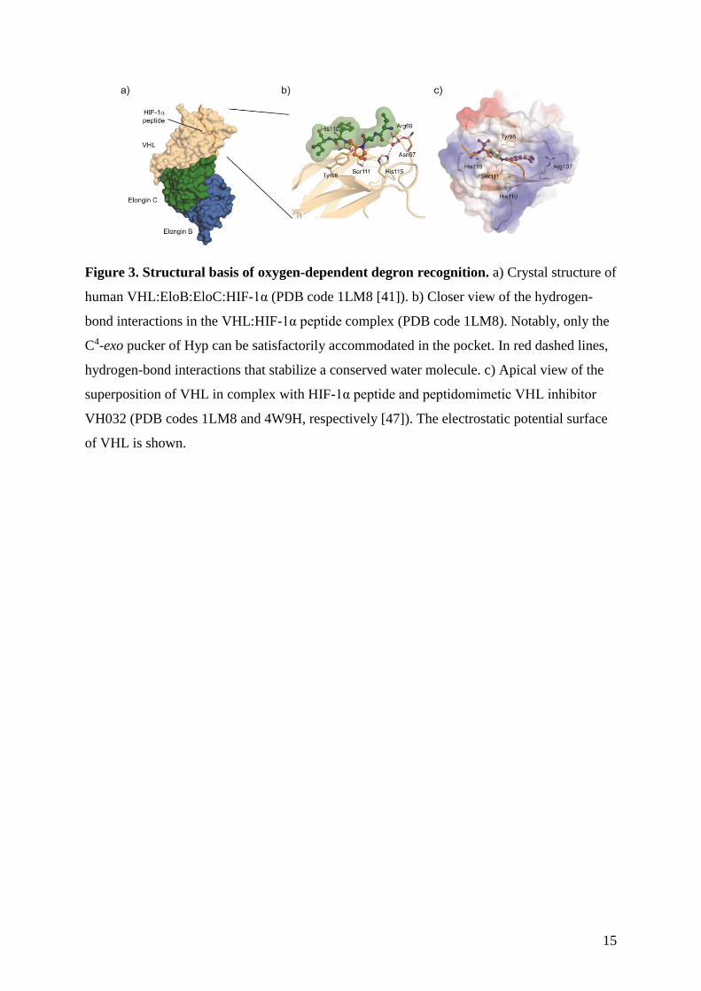
Figure 3. Structural basis of oxygen-dependent degron recognition. a) Crystal structure of
human VHL:EloB:EloC:HIF-1α (PDB code 1LM8 [41]). b) Closer view of the hydrogen-
bond interactions in the VHL:HIF-1α peptide complex (PDB code 1LM8). Notably, only the
C4-exo pucker of Hyp can be satisfactorily accommodated in the pocket. In red dashed lines,
hydrogen-bond interactions that stabilize a conserved water molecule. c) Apical view of the
superposition of VHL in complex with HIF-1α peptide and peptidomimetic VHL inhibitor
VH032 (PDB codes 1LM8 and 4W9H, respectively [47]). The electrostatic potential surface
of VHL is shown.

16
Figure 4. Structural basis of ligand-induced substrate recognition of CRLs. a) Crystal
structure of SCFTIR1 from A. thaliana (PDB code 2P1M [50]). TIR1 binds a molecule of
inositol-6-phosphate (InsP6). b) Closer view of the hydrogen-bond interactions of TIR1 in
complex with auxin and a IAA7 peptide (PDB code 2P1Q [50]). The auxin:IAA7 degron
peptide interaction is primarily driven by van der Waals packing. The hydrogen-bond
network of a stabilized water molecule is shown in red dashed lines. c) Superposition of
crystal structures of human CRL4CRBN:CC-885:GSPT1 and CRL4CRBN:lenalidomide:CK1α
complexes (PDB codes 5HXB and 5FQD, respectively [61, 62]). d) Closer view with
highlighted residues on CRBN that form hydrogen bonds with the ligands (CC-885 in wheat
and lenalidomide in orange). Note that Trp377 interacts only with lenalidomide, whereas CC-
885 extends further reaching His353. Both compounds sit in a hydrophobic cavity of Trp
residues. The topological conservation of the structural degron loop of GSPT1 and CK1α as
recognized by CRBN and the conserved Gly residue are highlighted. The loop interacts with
the small molecules primarily via van der Waals packing. e) Sequence alignment of the
structural degron loops in CK1α, GSPT1, and Ikaros.

17
Figure for Graphical abstract
18
References
1. Finley, D., Recognition and processing of ubiquitin-protein conjugates by the
proteasome. Annu Rev Biochem, 2009. 78: p. 477-513.
2. Hershko, A. and A. Ciechanover, The ubiquitin system. Annu Rev Biochem, 1998. 67:
p. 425-79.
3. Varshavsky, A., Naming a targeting signal. Cell, 1991. 64(1): p. 13-5.
4. Chang, L. and D. Barford, Insights into the anaphase-promoting complex: a
molecular machine that regulates mitosis. Curr Opin Struct Biol, 2014. 29: p. 1-9.
5. Kraft, C., M. Peter, and K. Hofmann, Selective autophagy: ubiquitin-mediated
recognition and beyond. Nat Cell Biol, 2010. 12(9): p. 836-41.
6. Caramelo, J.J. and A.J. Parodi, A sweet code for glycoprotein folding. FEBS Lett,
2015. 589(22): p. 3379-87.
7. Ciechanover, A. and Y.T. Kwon, Degradation of misfolded proteins in
neurodegenerative diseases: therapeutic targets and strategies. Exp Mol Med, 2015.
47: p. e147.
8. Balchin, D., M. Hayer-Hartl, and F.U. Hartl, In vivo aspects of protein folding and
quality control. Science, 2016. 353(6294): p. aac4354.
* This review focuses on the cellular mechanisms involved in protein folding and protein
homeostasis by the protein quality control machinery.
9. Herhaus, L. and I. Dikic, Expanding the ubiquitin code through post-translational
modification. EMBO Rep, 2015. 16(9): p. 1071-83.
10. Ito, A., et al., MDM2-HDAC1-mediated deacetylation of p53 is required for its
degradation. EMBO J, 2002. 21(22): p. 6236-45.
11. DaRosa, P.A., et al., Allosteric activation of the RNF146 ubiquitin ligase by a
poly(ADP-ribosyl)ation signal. Nature, 2015. 517(7533): p. 223-6.
12. Bachmair, A., D. Finley, and A. Varshavsky, In vivo half-life of a protein is a function
of its amino-terminal residue. Science, 1986. 234(4773): p. 179-86.
13. Sriram, S.M., B.Y. Kim, and Y.T. Kwon, The N-end rule pathway: emerging
functions and molecular principles of substrate recognition. Nat Rev Mol Cell Biol,
2011. 12(11): p. 735-47.
14. Tasaki, T., et al., The N-end rule pathway. Annu Rev Biochem, 2012. 81: p. 261-89.
19
15. Ravid, T. and M. Hochstrasser, Diversity of degradation signals in the ubiquitin-
proteasome system. Nat Rev Mol Cell Biol, 2008. 9(9): p. 679-90.
16. Rao, H., et al., Degradation of a cohesin subunit by the N-end rule pathway is
essential for chromosome stability. Nature, 2001. 410(6831): p. 955-9.
17. Choi, W.S., et al., Structural basis for the recognition of N-end rule substrates by the
UBR box of ubiquitin ligases. Nat Struct Mol Biol, 2010. 17(10): p. 1175-81.
18. Roman-Hernandez, G., et al., Molecular basis of substrate selection by the N-end rule
adaptor protein ClpS. Proc Natl Acad Sci U S A, 2009. 106(22): p. 8888-93.
19. Schuenemann, V.J., et al., Structural basis of N-end rule substrate recognition in
Escherichia coli by the ClpAP adaptor protein ClpS. EMBO Rep, 2009. 10(5): p.
508-14.
20. Jiang, Y., et al., A neurostimulant para-chloroamphetamine inhibits the arginylation
branch of the N-end rule pathway. Sci Rep, 2014. 4: p. 6344.
21. Long, M.J., D.R. Gollapalli, and L. Hedstrom, Inhibitor mediated protein
degradation. Chem Biol, 2012. 19(5): p. 629-37.
22. Shi, Y., et al., Boc3Arg-linked ligands induce degradation by localizing target
proteins to the 20S proteasome. ACS Chem Biol, 2016.
* The authors report on the ubiquitin-independent degradation mechanism of Boc3-Arg
compounds.
23. Neklesa, T.K., et al., Small-molecule hydrophobic tagging-induced degradation of
HaloTag fusion proteins. Nat Chem Biol, 2011. 7(8): p. 538-43.
24. Xie, T., et al., Pharmacological targeting of the pseudokinase Her3. Nat Chem Biol,
2014. 10(12): p. 1006-12.
25. Taxis, C., et al., Efficient protein depletion by genetically controlled deprotection of a
dormant N-degron. Mol Syst Biol, 2009. 5: p. 267.
26. Feng, G.S., C.C. Hui, and T. Pawson, SH2-containing phosphotyrosine phosphatase
as a target of protein-tyrosine kinases. Science, 1993. 259(5101): p. 1607-11.
27. Reinhardt, H.C. and M.B. Yaffe, Phospho-Ser/Thr-binding domains: navigating the
cell cycle and DNA damage response. Nat Rev Mol Cell Biol, 2013. 14(9): p. 563-80.
28. Skaar, J.R., J.K. Pagan, and M. Pagano, Mechanisms and function of substrate
recruitment by F-box proteins. Nat Rev Mol Cell Biol, 2013. 14(6): p. 369-81.
20
29. Orlicky, S., et al., Structural basis for phosphodependent substrate selection and
orientation by the SCFCdc4 ubiquitin ligase. Cell, 2003. 112(2): p. 243-56.
30. Hao, B., et al., Structure of a Fbw7-Skp1-cyclin E complex: multisite-phosphorylated
substrate recognition by SCF ubiquitin ligases. Mol Cell, 2007. 26(1): p. 131-43.
31. Orlicky, S., et al., An allosteric inhibitor of substrate recognition by the SCF(Cdc4)
ubiquitin ligase. Nat Biotechnol, 2010. 28(7): p. 733-7.
32. Welcker, M. and B.E. Clurman, FBW7 ubiquitin ligase: a tumour suppressor at the
crossroads of cell division, growth and differentiation. Nat Rev Cancer, 2008. 8(2): p.
83-93.
33. Bullock, A.N., et al., Crystal structure of the SOCS2-elongin C-elongin B complex
defines a prototypical SOCS box ubiquitin ligase. Proc Natl Acad Sci U S A, 2006.
103(20): p. 7637-42.
34. Dou, H., et al., Structural basis for autoinhibition and phosphorylation-dependent
activation of c-Cbl. Nat Struct Mol Biol, 2012. 19(2): p. 184-92.
35. Hennighausen, L. and G.W. Robinson, Interpretation of cytokine signaling through
the transcription factors STAT5A and STAT5B. Genes Dev, 2008. 22(6): p. 711-21.
36. Basham, B., et al., In vivo identification of novel STAT5 target genes. Nucleic Acids
Res, 2008. 36(11): p. 3802-18.
37. Elumalai, N., et al., Nanomolar inhibitors of the transcription factor STAT5b with
high selectivity over STAT5a. Angew Chem Int Ed Engl, 2015. 54(16): p. 4758-63.
* The authors report on potent and selective phosphodegron mimics of the phosphodegron
recognition site of STAT5b.
38. Petroski, M.D. and R.J. Deshaies, Function and regulation of cullin-RING ubiquitin
ligases. Nat Rev Mol Cell Biol, 2005. 6(1): p. 9-20.
39. Ivan, M., et al., HIFalpha targeted for VHL-mediated destruction by proline
hydroxylation: implications for O2 sensing. Science, 2001. 292(5516): p. 464-8.
40. Jaakkola, P., et al., Targeting of HIF-alpha to the von Hippel-Lindau ubiquitylation
complex by O2-regulated prolyl hydroxylation. Science, 2001. 292(5516): p. 468-72.
41. Min, J.H., et al., Structure of an HIF-1alpha -pVHL complex: hydroxyproline
recognition in signaling. Science, 2002. 296(5574): p. 1886-9.
42. Hon, W.C., et al., Structural basis for the recognition of hydroxyproline in HIF-1
alpha by pVHL. Nature, 2002. 417(6892): p. 975-8.

21
43. Loenarz, C., et al., Evidence for a stereoelectronic effect in human oxygen sensing.
Angew Chem Int Ed Engl, 2009. 48(10): p. 1784-7.
44. Buckley, D.L., et al., Small-molecule inhibitors of the interaction between the E3
ligase VHL and HIF1alpha. Angew Chem Int Ed Engl, 2012. 51(46): p. 11463-7.
45. Buckley, D.L., et al., Targeting the von Hippel-Lindau E3 ubiquitin ligase using small
molecules to disrupt the VHL/HIF-1alpha interaction. J Am Chem Soc, 2012.
134(10): p. 4465-8.
46. Van Molle, I., et al., Dissecting fragment-based lead discovery at the von Hippel-
Lindau protein:hypoxia inducible factor 1alpha protein-protein interface. Chem Biol,
2012. 19(10): p. 1300-12.
47. Galdeano, C., et al., Structure-guided design and optimization of small molecules
targeting the protein-protein interaction between the von Hippel-Lindau (VHL) E3
ubiquitin ligase and the hypoxia inducible factor (HIF) alpha subunit with in vitro
nanomolar affinities. J Med Chem, 2014. 57(20): p. 8657-63.
* The authors describe the structure-guided design of peptidomimetic Hyp compounds that
disrupt the VHL:HIF-α interaction.
48. Frost, J., et al., Potent and selective chemical probe of hypoxic signalling downstream
of HIF-alpha hydroxylation via VHL inhibition. Nat Commun, 2016. 7: p. 13312.
49. Wilkins, S.E., et al., Targeting Protein-Protein Interactions in the HIF System.
ChemMedChem, 2016. 11(8): p. 773-86.
50. Tan, X., et al., Mechanism of auxin perception by the TIR1 ubiquitin ligase. Nature,
2007. 446(7136): p. 640-5.
** First structure of an E3 ligase bound to both substrate and small-molecule modulator, that
led to the concept of “molecular glues”.
51. Sheard, L.B., et al., Jasmonate perception by inositol-phosphate-potentiated COI1-
JAZ co-receptor. Nature, 2010. 468(7322): p. 400-5.
52. Calderon Villalobos, L.I., et al., A combinatorial TIR1/AFB-Aux/IAA co-receptor
system for differential sensing of auxin. Nat Chem Biol, 2012. 8(5): p. 477-85.
53. Nishimura, K., et al., An auxin-based degron system for the rapid depletion of
proteins in nonplant cells. Nat Methods, 2009. 6(12): p. 917-22.
54. Natsume, T., et al., Rapid Protein Depletion in Human Cells by Auxin-Inducible
Degron Tagging with Short Homology Donors. Cell Rep, 2016. 15(1): p. 210-8.

22
55. Fischer, E.S., et al., SPLINTS: small-molecule protein ligand interface stabilizers.
Curr Opin Struct Biol, 2016. 37: p. 115-22.
* This review summarizes the use of small molecules to stabilize, rather than disrupt, protein-
protein interactions in several relevant biological systems.
56. Ito, T., et al., Identification of a primary target of thalidomide teratogenicity. Science,
2010. 327(5971): p. 1345-50.
57. Fischer, E.S., et al., Structure of the DDB1-CRBN E3 ubiquitin ligase in complex with
thalidomide. Nature, 2014. 512(7512): p. 49-53.
58. Lu, G., et al., The myeloma drug lenalidomide promotes the cereblon-dependent
destruction of Ikaros proteins. Science, 2014. 343(6168): p. 305-9.
59. Kronke, J., et al., Lenalidomide induces ubiquitination and degradation of CK1alpha
in del(5q) MDS. Nature, 2015. 523(7559): p. 183-8.
60. Chamberlain, P.P., et al., Structure of the human Cereblon-DDB1-lenalidomide
complex reveals basis for responsiveness to thalidomide analogs. Nat Struct Mol
Biol, 2014. 21(9): p. 803-9.
61. Matyskiela, M.E., et al., A novel cereblon modulator recruits GSPT1 to the
CRL4(CRBN) ubiquitin ligase. Nature, 2016. 535(7611): p. 252-7.
** This paper, together with ref 59 below, revealed the structural basis of IMiD-induced
recruitment of substrates by E3 ligase CRL4CRBN.
62. Petzold, G., E.S. Fischer, and N.H. Thoma, Structural basis of lenalidomide-induced
CK1alpha degradation by the CRL4(CRBN) ubiquitin ligase. Nature, 2016.
532(7597): p. 127-30.
** See annotation to ref 58.
63. Toure, M. and C.M. Crews, Small-Molecule PROTACS: New Approaches to Protein
Degradation. Angew Chem Int Ed Engl, 2016. 55(6): p. 1966-73.
* This review focuses on small-molecule induced protein degradation by PROTACs and
highlights recent impressive developments in the field.
64. Zengerle, M., K.H. Chan, and A. Ciulli, Selective Small Molecule Induced
Degradation of the BET Bromodomain Protein BRD4. ACS Chem Biol, 2015. 10(8):
p. 1770-7.
23
65. Bondeson, D.P., et al., Catalytic in vivo protein knockdown by small-molecule
PROTACs. Nat Chem Biol, 2015. 11(8): p. 611-7.
66. Lu, J., et al., Hijacking the E3 Ubiquitin Ligase Cereblon to Efficiently Target BRD4.
Chem Biol, 2015. 22(6): p. 755-63.
67. Winter, G.E., et al., DRUG DEVELOPMENT. Phthalimide conjugation as a strategy
for in vivo target protein degradation. Science, 2015. 348(6241): p. 1376-81.
68. Lai, A.C., et al., Modular PROTAC Design for the Degradation of Oncogenic BCR-
ABL. Angew Chem Int Ed Engl, 2016. 55(2): p. 807-10.
69. Bulatov, E. and A. Ciulli, Targeting Cullin-RING E3 ubiquitin ligases for drug
discovery: structure, assembly and small-molecule modulation. Biochem J, 2015.
467(3): p. 365-86.
* This review comprehensively explores structurally and functionally CRLs and their
modulation by small molecules.
70. Komander, D., The emerging complexity of protein ubiquitination. Biochem Soc
Trans, 2009. 37(Pt 5): p. 937-53.
71. Guharoy, M., et al., Tripartite degrons confer diversity and specificity on regulated
protein degradation in the ubiquitin-proteasome system. Nat Commun, 2016. 7: p.
10239.
72. Deshaies, R.J., Protein degradation: Prime time for PROTACs. Nat Chem Biol, 2015.
11(9): p. 634-5.
73. Wu, G., et al., Structure of a beta-TrCP1-Skp1-beta-catenin complex: destruction
motif binding and lysine specificity of the SCF(beta-TrCP1) ubiquitin ligase. Mol
Cell, 2003. 11(6): p. 1445-56.
74. Riedl, S.J., et al., Structural basis for the inhibition of caspase-3 by XIAP. Cell, 2001.
104(5): p. 791-800.
75. Condon, S.M., et al., Birinapant, a smac-mimetic with improved tolerability for the
treatment of solid tumors and hematological malignancies. J Med Chem, 2014. 57(9):
p. 3666-77.
76. Lo, S.C., et al., Structure of the Keap1:Nrf2 interface provides mechanistic insight
into Nrf2 signaling. EMBO J, 2006. 25(15): p. 3605-17.
77. Davies, T.G., et al., Monoacidic Inhibitors of the Kelch-like ECH-Associated Protein
1: Nuclear Factor Erythroid 2-Related Factor 2 (KEAP1:NRF2) Protein-Protein

24
Interaction with High Cell Potency Identified by Fragment-Based Discovery. J Med
Chem, 2016. 59(8): p. 3991-4006.
78. Kussie, P.H., et al., Structure of the MDM2 oncoprotein bound to the p53 tumor
suppressor transactivation domain. Science, 1996. 274(5289): p. 948-53.
79. Vu, B., et al., Discovery of RG7112: A Small-Molecule MDM2 Inhibitor in Clinical
Development. ACS Med Chem Lett, 2013. 4(5): p. 466-9.