Embed Size (px)
Citation preview
University of Birmingham
Scissor sisters:Matthews, Alexandra; Szyroka, Justyna; Collier, Richard; Noy, Peter; Tomlinson, Michael
DOI:10.1042/BST20160290
License:Creative Commons: Attribution (CC BY)
Document VersionPublisher's PDF, also known as Version of record
Citation for published version (Harvard):Matthews, A, Szyroka, J, Collier, R, Noy, P & Tomlinson, M 2017, 'Scissor sisters: regulation of ADAM10 by theTspanC8 tetraspanins', Biochemical Society Transactions, vol. 45, no. 3, pp. 719-730.https://doi.org/10.1042/BST20160290
Link to publication on Research at Birmingham portal
General rightsUnless a licence is specified above, all rights (including copyright and moral rights) in this document are retained by the authors and/or thecopyright holders. The express permission of the copyright holder must be obtained for any use of this material other than for purposespermitted by law.
•Users may freely distribute the URL that is used to identify this publication.•Users may download and/or print one copy of the publication from the University of Birmingham research portal for the purpose of privatestudy or non-commercial research.•User may use extracts from the document in line with the concept of ‘fair dealing’ under the Copyright, Designs and Patents Act 1988 (?)•Users may not further distribute the material nor use it for the purposes of commercial gain.
Where a licence is displayed above, please note the terms and conditions of the licence govern your use of this document.
When citing, please reference the published version.
Take down policyWhile the University of Birmingham exercises care and attention in making items available there are rare occasions when an item has beenuploaded in error or has been deemed to be commercially or otherwise sensitive.
If you believe that this is the case for this document, please contact [email protected] providing details and we will remove access tothe work immediately and investigate.
Download date: 01. Mar. 2020
brought to you by COREView metadata, citation and similar papers at core.ac.uk
provided by University of Birmingham Research Portal
Review Article
Scissor sisters: regulation of ADAM10 by theTspanC8 tetraspaninsAlexandra L. Matthews, Justyna Szyroka, Richard Collier, Peter J. Noy and Michael G. TomlinsonSchool of Biosciences, College of Life and Environmental Sciences, University of Birmingham, Edgbaston, Birmingham B15 2TT, U.K.
Correspondence: Michael G. Tomlinson ([email protected])
A disintegrin and metalloprotease 10 (ADAM10) is a ubiquitously expressed transmem-brane protein which is essential for embryonic development through activation of Notchproteins. ADAM10 regulates over 40 other transmembrane proteins and acts as a‘molecular scissor’ by removing their extracellular regions. ADAM10 is also a receptor forα-toxin, a major virulence factor of Staphylococcus aureus. Owing to the importance ofits substrates, ADAM10 is a potential therapeutic target for cancer, neurodegenerativediseases such as Alzheimer’s and prion diseases, bacterial infection and inflammatorydiseases such as heart attack, stroke and asthma. However, targetting ADAM10 is likelyto result in toxic side effects. The tetraspanins are a superfamily of 33 four-transmem-brane proteins in mammals which interact with and regulate specific partner proteinswithin membrane nanodomains. Tetraspanins appear to have a cone-shaped structurewith a cholesterol-binding cavity, which may enable tetraspanins to undergo cholesterol-regulated conformational change. An emerging paradigm for tetraspanin function is theregulation of ADAM10 by the TspanC8 subgroup of tetraspanins, namely Tspan5, 10, 14,15, 17 and 33. This review will describe how TspanC8s are required for ADAM10 traffick-ing from the endoplasmic reticulum and its enzymatic maturation. Moreover, differentTspanC8s localise ADAM10 to different subcellular localisations and may cause ADAM10to adopt distinct conformations and cleavage of distinct substrates. We propose thatADAM10 should now be regarded as six different scissor proteins depending on theinteracting TspanC8. Therapeutic targetting of specific TspanC8/ADAM10 complexescould allow ADAM10 targetting in a cell type- or substrate-specific manner, to treatcertain diseases while minimising toxicity.
IntroductionCell development and function are regulated by cell surface receptors and secreted proteins thatco-ordinate intra- and intercellular signalling. Various regulatory mechanisms are required to ensureappropriate spatiotemporal control over these biological processes. Proteolytic cleavage, or ‘shedding’, ofthe ectodomains (extracellular regions) at a juxta-membrane site on transmembrane proteins hasemerged as one such mechanism. Ectodomain shedding directly affects the responsiveness of cells toextracellular signals via activation of intracellular signalling or down-regulation of cell surface receptors,or indirectly through the release of soluble mediators from their membrane-bound precursors. Sheddingtherefore enables cells to rapidly respond to their environment and plays an important role in copious cel-lular processes including cell adhesion, migration, invasion, proliferation and signalling [1,2].
ADAM10: a ubiquitous ‘molecular scissor’The evolutionarily conserved ADAMs (a disintegrin and metalloproteases) are a superfamily of Zn2+-dependent transmembrane metalloproteases that are responsible for a substantial proportion oftransmembrane protein shedding. A total of 22 ADAM genes have been identified in humans, ofwhich 12 encode proteolytically active enzymes. ADAMs share a common multidomain structure
Version of Record published:15 June 2017
Received: 5 January 2017Revised: 26 February 2017Accepted: 28 February 2017
© 2017 The Author(s). This is an open access article published by Portland Press Limited on behalf of the Biochemical Society and distributed under the Creative Commons Attribution License 4.0 (CC BY). 719
Biochemical Society Transactions (2017) 45 719–730DOI: 10.1042/BST20160290
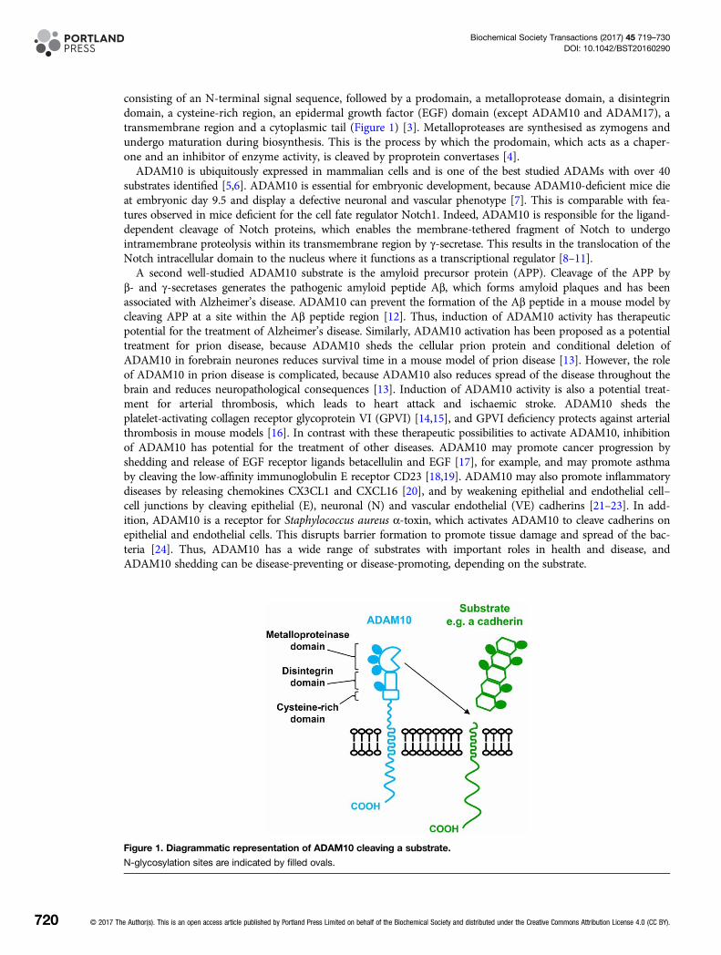
consisting of an N-terminal signal sequence, followed by a prodomain, a metalloprotease domain, a disintegrindomain, a cysteine-rich region, an epidermal growth factor (EGF) domain (except ADAM10 and ADAM17), atransmembrane region and a cytoplasmic tail (Figure 1) [3]. Metalloproteases are synthesised as zymogens andundergo maturation during biosynthesis. This is the process by which the prodomain, which acts as a chaper-one and an inhibitor of enzyme activity, is cleaved by proprotein convertases [4].ADAM10 is ubiquitously expressed in mammalian cells and is one of the best studied ADAMs with over 40
substrates identified [5,6]. ADAM10 is essential for embryonic development, because ADAM10-deficient mice dieat embryonic day 9.5 and display a defective neuronal and vascular phenotype [7]. This is comparable with fea-tures observed in mice deficient for the cell fate regulator Notch1. Indeed, ADAM10 is responsible for the ligand-dependent cleavage of Notch proteins, which enables the membrane-tethered fragment of Notch to undergointramembrane proteolysis within its transmembrane region by γ-secretase. This results in the translocation of theNotch intracellular domain to the nucleus where it functions as a transcriptional regulator [8–11].A second well-studied ADAM10 substrate is the amyloid precursor protein (APP). Cleavage of the APP by
β- and γ-secretases generates the pathogenic amyloid peptide Aβ, which forms amyloid plaques and has beenassociated with Alzheimer’s disease. ADAM10 can prevent the formation of the Aβ peptide in a mouse model bycleaving APP at a site within the Aβ peptide region [12]. Thus, induction of ADAM10 activity has therapeuticpotential for the treatment of Alzheimer’s disease. Similarly, ADAM10 activation has been proposed as a potentialtreatment for prion disease, because ADAM10 sheds the cellular prion protein and conditional deletion ofADAM10 in forebrain neurones reduces survival time in a mouse model of prion disease [13]. However, the roleof ADAM10 in prion disease is complicated, because ADAM10 also reduces spread of the disease throughout thebrain and reduces neuropathological consequences [13]. Induction of ADAM10 activity is also a potential treat-ment for arterial thrombosis, which leads to heart attack and ischaemic stroke. ADAM10 sheds theplatelet-activating collagen receptor glycoprotein VI (GPVI) [14,15], and GPVI deficiency protects against arterialthrombosis in mouse models [16]. In contrast with these therapeutic possibilities to activate ADAM10, inhibitionof ADAM10 has potential for the treatment of other diseases. ADAM10 may promote cancer progression byshedding and release of EGF receptor ligands betacellulin and EGF [17], for example, and may promote asthmaby cleaving the low-affinity immunoglobulin E receptor CD23 [18,19]. ADAM10 may also promote inflammatorydiseases by releasing chemokines CX3CL1 and CXCL16 [20], and by weakening epithelial and endothelial cell–cell junctions by cleaving epithelial (E), neuronal (N) and vascular endothelial (VE) cadherins [21–23]. In add-ition, ADAM10 is a receptor for Staphylococcus aureus α-toxin, which activates ADAM10 to cleave cadherins onepithelial and endothelial cells. This disrupts barrier formation to promote tissue damage and spread of the bac-teria [24]. Thus, ADAM10 has a wide range of substrates with important roles in health and disease, andADAM10 shedding can be disease-preventing or disease-promoting, depending on the substrate.
Figure 1. Diagrammatic representation of ADAM10 cleaving a substrate.
N-glycosylation sites are indicated by filled ovals.
720 © 2017 The Author(s). This is an open access article published by Portland Press Limited on behalf of the Biochemical Society and distributed under the Creative Commons Attribution License 4.0 (CC BY).
Biochemical Society Transactions (2017) 45 719–730DOI: 10.1042/BST20160290
The regulation of ADAM10 activity is not clear. ADAM10 appears to have constitutive activity towardssome substrates, although its activity can be up-regulated by certain stimuli that induce intracellular signals,such as an increase in intracellular Ca2+ concentration. The mechanism underlying the latter is not well under-stood, but is dependent on the transmembrane region of ADAM10 rather than the cytoplasmic tail [25]. Thereis also evidence that ADAM10-mediated shedding may be facilitated by intracellular signals that modify thesubstrates, because phosphorylation of the cytoplasmic tail of the adhesion molecule CD44 may induce dimer-isation and/or a conformational change in the CD44 ectodomain to allow shedding [26,27].ADAM10 is clearly a promising therapeutic target for a range of diseases, but because of the positive and
negative effects of ADAM10 in health and disease processes, substrate-specific ADAM10 targetting may benecessary to avoid toxic side effects. The remainder of this review will describe the emerging role of theTspanC8 tetraspanins as ADAM10-interacting partners that are essential for its exit from the endoplasmicreticulum, and which may dictate substrate specificity.
Tetraspanins regulate partner protein functionThe regulation of transmembrane proteins by compartmentalisation into membrane microdomains is a conceptthat has developed from studies of lipid rafts, caveolae, neuronal and immunological synapses, and tetraspanins.The latter are a superfamily of four-transmembrane proteins that are expressed by animals, plants and somemulticellular fungi. They function by interacting with specific ‘partner proteins’, and regulating their intracellu-lar trafficking and lateral mobility and clustering at the cell surface [28,29]. Notable examples are regulation oflaminin-binding integrin function by tetraspanin CD151 [29], regulation of B-cell co-receptor CD19 traffickingby tetraspanin CD81 [30] and regulation of the Wnt receptor Frizzled-4 by Tspan12 [31]. In each case, tetra-spanin mutations lead to human diseases that are consistent with functional impairment of the partner protein:CD151 deficiency leads to kidney disease due to impaired assembly of the glomerular and tubular basementmembranes [32]; CD81 deficiency leads to impaired antibody generation due to an absence of CD19 on theB-cell surface [33]; and Tspan12 deficiency leads to familial exudative vitreoretinopathy characterised byimpaired vasculature development in the retina [34,35]. A total of 33 tetraspanins are found in humans andeach cell type is estimated to express as many as 20 different tetraspanins. Tetraspanins form dynamicnanoclusters that are distinct from lipid rafts [28,29], and recent evidence using super-resolution microscopysuggests that tetraspanin nanodomains are clusters of ∼10 tetraspanins of just a single type [36]. This is con-sistent with the specific phenotypes that have been observed in many tetraspanin knockout studies, althoughtetraspanin knockouts are typically well tolerated and there is evidence for functional compensation by relatedtetraspanins [28,29].An exciting breakthrough in tetraspanin research is the recent report of the first crystal structure of a full-
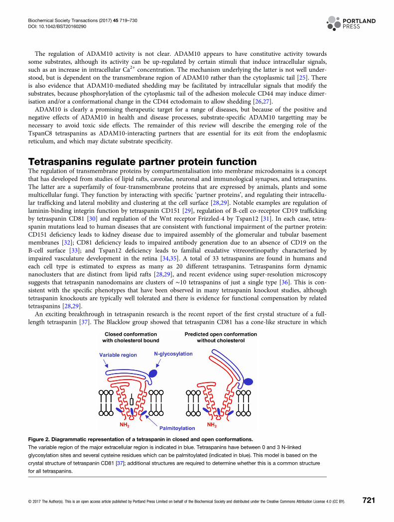
length tetraspanin [37]. The Blacklow group showed that tetraspanin CD81 has a cone-like structure in which
Figure 2. Diagrammatic representation of a tetraspanin in closed and open conformations.
The variable region of the major extracellular region is indicated in blue. Tetraspanins have between 0 and 3 N-linked
glycosylation sites and several cysteine residues which can be palmitoylated (indicated in blue). This model is based on the
crystal structure of tetraspanin CD81 [37]; additional structures are required to determine whether this is a common structure
for all tetraspanins.
© 2017 The Author(s). This is an open access article published by Portland Press Limited on behalf of the Biochemical Society and distributed under the Creative Commons Attribution License 4.0 (CC BY). 721
Biochemical Society Transactions (2017) 45 719–730DOI: 10.1042/BST20160290
transmembranes 1 and 2 are separated at the top of the plasma membrane from transmembranes 3 and 4(Figure 2A). This forms a cavity in which a cholesterol molecule can bind, with its hydroxyl group co-ordinatedby conserved polar amino acids within transmembranes 1 and 4. Molecular dynamics simulations suggest thatremoval of the cholesterol might result in a dramatic conformational change from a ‘closed’ to an ‘open’ con-formation, in which the main extracellular region swings upwards with a ‘switch-blade’-type action (Figure 2B)[37]. Structural determination of additional tetraspanins is now required to discover whether this structure iscommon to the entire superfamily. Nevertheless, this raises the possibility of therapeutic targetting with smallmolecules or antibodies that may lock the tetraspanin into a particular conformation. Hypothetically, this couldthen affect partner protein function, for example, by disrupting the interaction with the tetraspanin, by affectinglocalisation of the complex (e.g. causing internalisation) or by preventing conformational change-induced regu-lation by the tetraspanin.
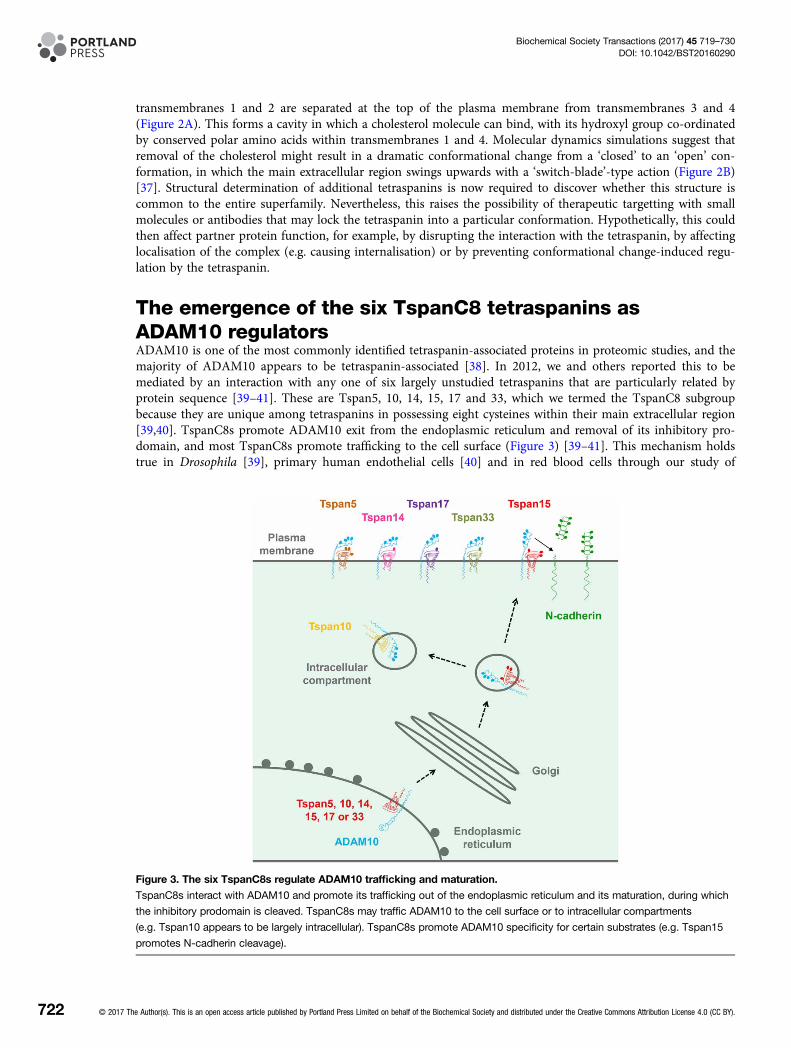
The emergence of the six TspanC8 tetraspanins asADAM10 regulatorsADAM10 is one of the most commonly identified tetraspanin-associated proteins in proteomic studies, and themajority of ADAM10 appears to be tetraspanin-associated [38]. In 2012, we and others reported this to bemediated by an interaction with any one of six largely unstudied tetraspanins that are particularly related byprotein sequence [39–41]. These are Tspan5, 10, 14, 15, 17 and 33, which we termed the TspanC8 subgroupbecause they are unique among tetraspanins in possessing eight cysteines within their main extracellular region[39,40]. TspanC8s promote ADAM10 exit from the endoplasmic reticulum and removal of its inhibitory pro-domain, and most TspanC8s promote trafficking to the cell surface (Figure 3) [39–41]. This mechanism holdstrue in Drosophila [39], primary human endothelial cells [40] and in red blood cells through our study of
Figure 3. The six TspanC8s regulate ADAM10 trafficking and maturation.
TspanC8s interact with ADAM10 and promote its trafficking out of the endoplasmic reticulum and its maturation, during which
the inhibitory prodomain is cleaved. TspanC8s may traffic ADAM10 to the cell surface or to intracellular compartments
(e.g. Tspan10 appears to be largely intracellular). TspanC8s promote ADAM10 specificity for certain substrates (e.g. Tspan15
promotes N-cadherin cleavage).
722 © 2017 The Author(s). This is an open access article published by Portland Press Limited on behalf of the Biochemical Society and distributed under the Creative Commons Attribution License 4.0 (CC BY).
Biochemical Society Transactions (2017) 45 719–730DOI: 10.1042/BST20160290
Tspan33 knockout mice [40], so far the only reported TspanC8 knockout mouse [42], together indicating thatTspanC8s are fundamental to ADAM10 function.Several follow-up studies on TspanC8 regulation of ADAM10 [43–46] can be condensed into the following
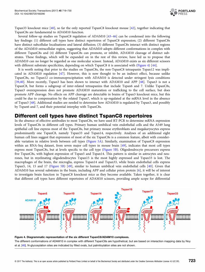
key findings: (1) different cell types have distinct repertoires of TspanC8 expression; (2) different TspanC8shave distinct subcellular localisations and lateral diffusion; (3) different TspanC8s interact with distinct regionsof the ADAM10 extracellular region, suggesting that ADAM10 adopts different conformations in complex withdifferent TspanC8s and (4) different TspanC8s can promote, or inhibit, ADAM10 cleavage of distinct sub-strates. These findings, which will be expanded on in the rest of this review, have led us to propose thatADAM10 can no longer be regarded as one molecular scissor. Instead, ADAM10 exists as six different scissorswith different substrate specificities, depending on which TspanC8 it is associated with (Figure 4) [44].It is worth noting that prior to the studies on TspanC8s, the non-TspanC8 tetraspanin Tspan12 was impli-
cated in ADAM10 regulation [47]. However, this is now thought to be an indirect effect, because unlikeTspanC8s, no Tspan12 co-immunoprecipitation with ADAM10 is detected under stringent lysis conditions[39,40]. More recently, Tspan3 has been shown to interact with ADAM10 and APP [48]. Tspan3 is not aTspanC8, but forms a subgroup of inter-related tetraspanins that include Tspan6 and 7. Unlike TspanC8s,Tspan3 overexpression does not promote ADAM10 maturation or trafficking to the cell surface, but doespromote APP cleavage. No effects on APP cleavage are detectable in brains of Tspan3 knockout mice, but thiscould be due to compensation by the related Tspan7, which is up-regulated at the mRNA level in the absenceof Tspan3 [48]. Additional studies are needed to determine how ADAM10 is regulated by Tspan3, and possiblyby Tspan6 and 7, and their potential interplay with TspanC8s.
Different cell types have distinct TspanC8 repertoiresIn the absence of effective antibodies to most TspanC8s, we have used RT-PCR to determine mRNA expressionlevels of TspanC8s in different cell types. Primary human umbilical vein endothelial cells and the A549 lungepithelial cell line express most of the TspanC8s, but primary mouse erythroblasts and megakaryocytes expresspredominantly one TspanC8, namely Tspan33 and Tspan14, respectively. Analyses of an additional eighthuman cell lines suggest that expression of most of the six TspanC8s is a common feature, albeit with consider-able variation in relative levels between cell types (Figure 5A). Similarly, examination of TspanC8 expressionwithin an RNA-Seq dataset, from seven major cell types in mouse brain [49], indicates that most cell typesexpress most TspanC8s, but at levels specific to the cell type (Figure 5B). Oligodendrocyte precursors expressfive TspanC8s, with highest expression of Tspan5 and Tspan14. This pattern is similar in astrocytes and neu-rones, but in myelinating oligodendrocytes Tspan15 is the most highly expressed and Tspan33 is lost. Themacrophages of the brain, the microglia, express Tspan14 and Tspan33, while brain endothelial cells expressTspan5, 14, 15 and 17 (Figure 5B) [49], similar to human umbilical vein endothelial cells [40]. Given thatADAM10 has several substrates in the brain, including APP and cellular prion protein [6], it will be of interestto investigate brain function in TspanC8 knockout mice as they become available. Taken together, it is clearthat different cell types have different repertoires of ADAM10 scissors, providing ample scope for differential
Figure 4. Diagrammatic representation of the six different TspanC8/ADAM10 complexes.
The different conformations of ADAM10 in complex with different TspanC8s are hypothetical, but are based on interaction mapping data by Noy
et al. [45]. N-glycosylation sites are indicated by filled ovals, but palmitoylation sites are not shown.
© 2017 The Author(s). This is an open access article published by Portland Press Limited on behalf of the Biochemical Society and distributed under the Creative Commons Attribution License 4.0 (CC BY). 723
Biochemical Society Transactions (2017) 45 719–730DOI: 10.1042/BST20160290
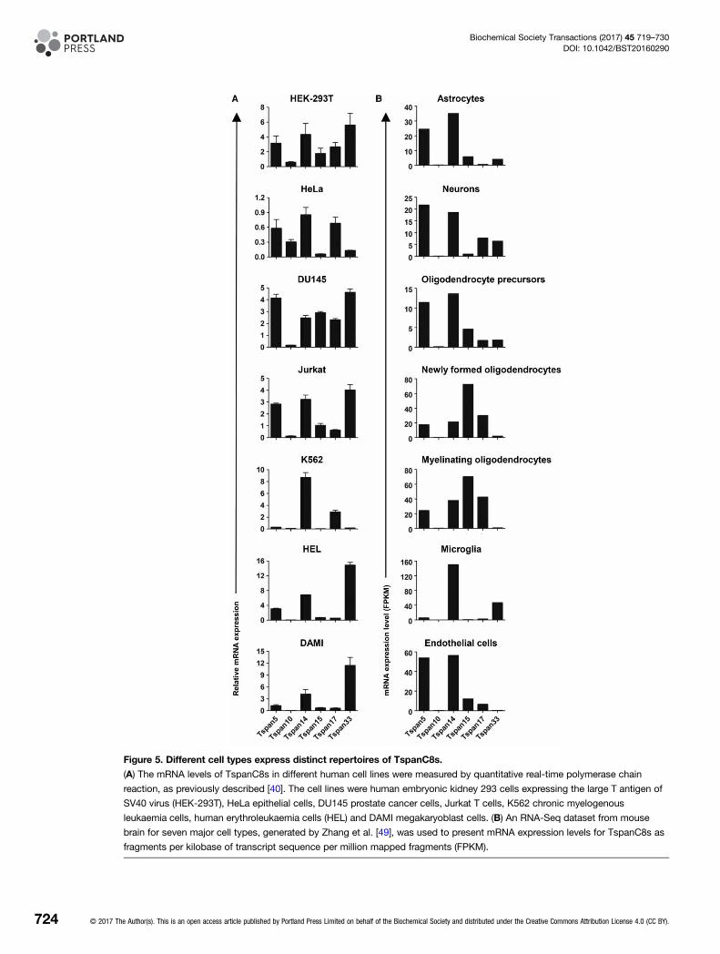
Figure 5. Different cell types express distinct repertoires of TspanC8s.
(A) The mRNA levels of TspanC8s in different human cell lines were measured by quantitative real-time polymerase chain
reaction, as previously described [40]. The cell lines were human embryonic kidney 293 cells expressing the large T antigen of
SV40 virus (HEK-293T), HeLa epithelial cells, DU145 prostate cancer cells, Jurkat T cells, K562 chronic myelogenous
leukaemia cells, human erythroleukaemia cells (HEL) and DAMI megakaryoblast cells. (B) An RNA-Seq dataset from mouse
brain for seven major cell types, generated by Zhang et al. [49], was used to present mRNA expression levels for TspanC8s as
fragments per kilobase of transcript sequence per million mapped fragments (FPKM).
724 © 2017 The Author(s). This is an open access article published by Portland Press Limited on behalf of the Biochemical Society and distributed under the Creative Commons Attribution License 4.0 (CC BY).
Biochemical Society Transactions (2017) 45 719–730DOI: 10.1042/BST20160290
substrate shedding and the potential for therapeutic targetting in a cell type- or substrate-specific manner bytargetting specific TspanC8/ADAM10 complexes.
Different TspanC8s have distinct subcellular localisationsand lateral diffusionThe Rubinstein group have investigated TspanC8 subcellular localisation by transfecting green fluorescentprotein (GFP)-tagged TspanC8s into human cell lines. Tspan5, 14, 15 and 33 promote ADAM10 surface local-isation in the HeLa epithelial cell line, but Tspan10 and 17 do not. This is consistent with substantial localisa-tion of the latter in an intracellular compartment, but cell surface and intracellular localisation of the other fourTspanC8s [39]. In subsequent comparisons between GFP-tagged Tspan5 and 15 expressed in the U2OS osteo-sarcoma cell line, ADAM10 lateral diffusion in the plasma membrane is increased by 55% in the presence ofTspan15, but Tspan5 has no effect [43]. In addition, co-immunoprecipitation followed by mass spectrometry-based proteomics identifies many common interacting proteins, most prominently ADAM10, but some pro-teins are more specific to Tspan5 or Tspan15. Additionally, overexpression of Tspan5 or Tspan15 appears toalter the proteins interacting with ADAM10 [43]. These data suggest that TspanC8s differentially regulateADAM10 localisation, but interpretation is complicated by the fact that the GFP tag and/or overexpressionmight affect localisation. TspanC8 monoclonal antibodies are now required to investigate the localisation ofendogenous proteins.
Different TspanC8s may cause ADAM10 to adopt distinctconformationsWe have demonstrated that the main extracellular region of Tspan14 mediates the interaction with ADAM10and its maturation, and that the variable region is necessary but not sufficient [45]. This study usedco-immunoprecipitation of overexpressed FLAG epitope-tagged constructs chimeric for Tspan14 and thenon-TspanC8 tetraspanin CD9. A similar strategy with ADAM10 truncation constructs and chimeras withADAM17, which does not interact with TspanC8s, showed some interesting differences between TspanC8s.Tspan15 requires only the membrane–proximal stalk region of ADAM10 for its interaction, Tspan17 requiresthe stalk and cysteine-rich domain, whereas Tspan5, 10, 14 and 33 require the stalk, cysteine-rich and disinte-grin domains [45]. These findings suggest that ADAM10 might adopt different conformations in complex withdifferent TspanC8s, as suggested in Figure 4. However, interpretation of these data is complicated by their reli-ance on co-immunoprecipitation of overexpressed mutant constructs, and thus, high-resolution structuralstudies of native TspanC8/ADAM10 complexes are now required.The cleavage sites on ADAM10 substrates share no particular amino acid sequence similarities and differ in
their position relative to the plasma membrane. For example, the GPVI, APP and Notch1 cleavage sites arepositioned at 5, 12 and 15 amino acids from the transmembranes, respectively [11,15,50]. Therefore, if differentTspanC8s are able to ‘lock’ ADAM10 into a distinct conformation, this could position the ADAM10 metallo-protease domain at a position that favours cleavage of certain substrates.
Different TspanC8/ADAM10 complexes cleave distinctsubstratesThe most definitive evidence for cleavage of specific ADAM10 substrates by different TspanC8/ADAM10 com-plexes is the promotion of N-cadherin cleavage by Tspan15/ADAM10. This is because the experiments havecompared Tspan15 with other TspanC8s as controls have been reported by three different groups using differ-ent cell lines and the effects are striking. Indeed, Tspan15 overexpression substantially promotes N-cadherincleavage, but other TspanC8s do not [41,43,45]. Furthermore, Tspan15 knockdown substantially reducesN-cadherin cleavage [43].Notch cleavage is also regulated by TspanC8s [39,43,46,51], but interpretation of these findings is compli-
cated by the positive and negative effects described for different TspanC8s, the different cell types used and thedifferent methods of activating Notch and detecting its cleavage. In the two studies which have compared dif-ferent TspanC8s, ligand-induced Notch reporter activity in HeLa and U2OS cells is shown to be promoted byoverexpression of Tspan5 or 14, whereas Tspan15 or 33 has the opposite effect. Moreover, Tspan15 knockdown
© 2017 The Author(s). This is an open access article published by Portland Press Limited on behalf of the Biochemical Society and distributed under the Creative Commons Attribution License 4.0 (CC BY). 725
Biochemical Society Transactions (2017) 45 719–730DOI: 10.1042/BST20160290
can substantially promote Notch activation [39,43]. In additional studies which focused on just one or twoTspanC8s, it was reported that knockdown of Tspan5 or 10 in primary mouse osteoclasts inhibits Notch cleav-age and target gene expression [46], whereas Tspan33 has a positive role in expression of Notch target genes inthe RAW 264.7 mouse macrophage cell line [51]. Thus, Tspan5, 10 and 14 appear to be Notch-promoting,Tspan15 Notch-inhibiting and Tspan33-dependent on the cell type. A panel of CRISPR/Cas9-generated celllines, expressing single TspanC8s, may ultimately be required to understand the importance of each TspanC8in the activation of Notch and potentially other ADAM10 substrates.Recent additional findings from cell line models suggest that GPVI, CD44 and APP cleavage may also be
regulated by specific TspanC8s. GPVI cleavage is inhibited by overexpression of Tspan14 but not by otherTspanC8s [45], and CD44 cleavage is partially dependent on Tspan5 but not on Tspan15 [43]. Finally, regula-tion of APP cleavage by TspanC8s is less clear, because both positive [41] and negative [43] effects of Tspan15have been reported. This may reflect the different cell lines used and their potentially different endogenousTspanC8 repertoires.Finally, genetic screens in cell lines have recently shown Tspan14 and Tspan33 to be important for cytotox-
icity induced by S. aureus α-toxin; the screens also identified the α-toxin receptor ADAM10 [52,53]. Oneexplanation for their identification would be that these TspanC8s are highly expressed in the cell lines screenedand are required for ADAM10 surface expression, which certainly appears to be the case for Tspan14 in theU937 human monocyte cell line [53]. However, it is possible that TspanC8s might differentially regulateADAM10 function as an α-toxin receptor.The mechanisms by which TspanC8s differentially control ADAM10 substrate specificity are unclear, but the
regulation of ADAM10 subcellular localisation and conformation, proposed in the previous sections, representpotential mechanisms. An additional possibility is direct interaction of the TspanC8 with a particular substrate.However, although at least two ADAM10 substrates are tetraspanin-associated, namely CD44 [54] and GPVI[55,56], there is no current evidence that a TspanC8 can interact with these proteins.
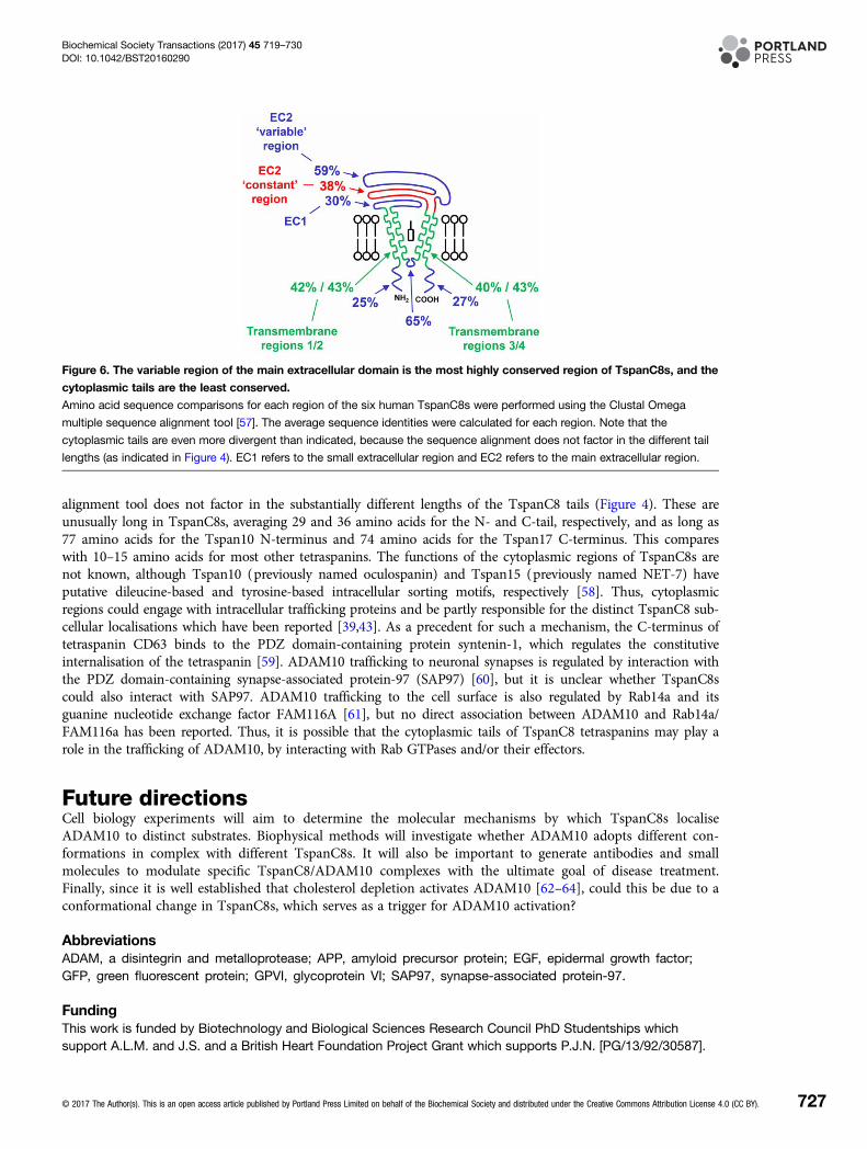
TspanC8 protein sequence analyses: conservedtransmembrane and extracellular regions; divergent tailsIn the current absence of useful tools such as TspanC8 monoclonal antibodies and knockout mice (apart fromTspan33 [40,42]), comparisons of TspanC8 protein sequences may help us to understand how they differen-tially regulate ADAM10. TspanC8s are certainly relatively highly related by amino acid sequence [40], with thehuman homologues ranging from 78% amino acid identity between Tspan5 and 17 to 26% identity betweenthe most distantly related TspanC8s, as determined by Clustal Omega multiple sequence alignment [57](Table 1). The large extracellular region is relatively highly conserved between TspanC8s (average 44% sequenceidentity), with the ‘variable’ region even more conserved (59%) (Figure 6). This contrasts strikingly withsequence comparisons within the tetraspanins as a whole, in which the variable region varies most between tet-raspanins. However, this is consistent with our finding that the interaction with ADAM10 is mediated by themain extracellular region of the TspanC8s and that the variable region is essential [45], suggesting that thisADAM10-interacting region has been conserved during evolution. The transmembrane domains of TspanC8sare also well conserved (40–43%), consistent with their forming the core tetraspanin structure [37].The cytoplasmic tails of TspanC8s are relatively divergent. The 25% and 27% sequence identities of the N- and
C-terminal tails, respectively (Figure 6), actually under-represent the true diversity, because the sequence
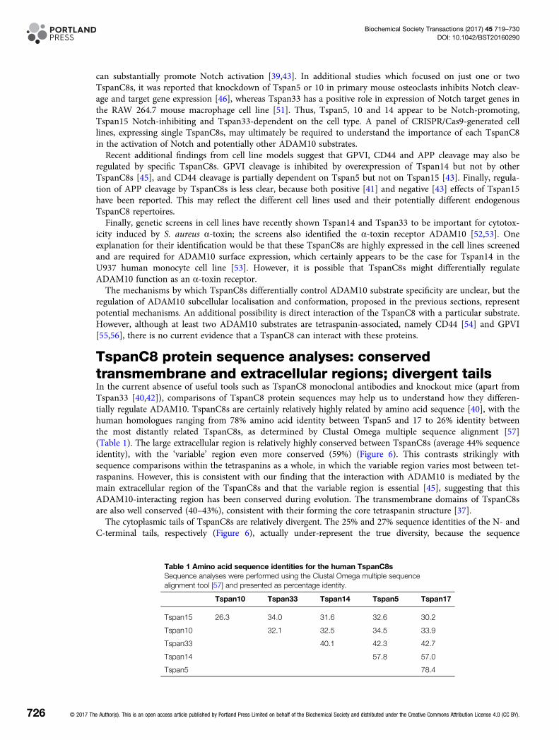
Table 1 Amino acid sequence identities for the human TspanC8sSequence analyses were performed using the Clustal Omega multiple sequencealignment tool [57] and presented as percentage identity.
Tspan10 Tspan33 Tspan14 Tspan5 Tspan17
Tspan15 26.3 34.0 31.6 32.6 30.2
Tspan10 32.1 32.5 34.5 33.9
Tspan33 40.1 42.3 42.7
Tspan14 57.8 57.0
Tspan5 78.4
726 © 2017 The Author(s). This is an open access article published by Portland Press Limited on behalf of the Biochemical Society and distributed under the Creative Commons Attribution License 4.0 (CC BY).
Biochemical Society Transactions (2017) 45 719–730DOI: 10.1042/BST20160290
alignment tool does not factor in the substantially different lengths of the TspanC8 tails (Figure 4). These areunusually long in TspanC8s, averaging 29 and 36 amino acids for the N- and C-tail, respectively, and as long as77 amino acids for the Tspan10 N-terminus and 74 amino acids for the Tspan17 C-terminus. This compareswith 10–15 amino acids for most other tetraspanins. The functions of the cytoplasmic regions of TspanC8s arenot known, although Tspan10 (previously named oculospanin) and Tspan15 (previously named NET-7) haveputative dileucine-based and tyrosine-based intracellular sorting motifs, respectively [58]. Thus, cytoplasmicregions could engage with intracellular trafficking proteins and be partly responsible for the distinct TspanC8 sub-cellular localisations which have been reported [39,43]. As a precedent for such a mechanism, the C-terminus oftetraspanin CD63 binds to the PDZ domain-containing protein syntenin-1, which regulates the constitutiveinternalisation of the tetraspanin [59]. ADAM10 trafficking to neuronal synapses is regulated by interaction withthe PDZ domain-containing synapse-associated protein-97 (SAP97) [60], but it is unclear whether TspanC8scould also interact with SAP97. ADAM10 trafficking to the cell surface is also regulated by Rab14a and itsguanine nucleotide exchange factor FAM116A [61], but no direct association between ADAM10 and Rab14a/FAM116a has been reported. Thus, it is possible that the cytoplasmic tails of TspanC8 tetraspanins may play arole in the trafficking of ADAM10, by interacting with Rab GTPases and/or their effectors.
Future directionsCell biology experiments will aim to determine the molecular mechanisms by which TspanC8s localiseADAM10 to distinct substrates. Biophysical methods will investigate whether ADAM10 adopts different con-formations in complex with different TspanC8s. It will also be important to generate antibodies and smallmolecules to modulate specific TspanC8/ADAM10 complexes with the ultimate goal of disease treatment.Finally, since it is well established that cholesterol depletion activates ADAM10 [62–64], could this be due to aconformational change in TspanC8s, which serves as a trigger for ADAM10 activation?
AbbreviationsADAM, a disintegrin and metalloprotease; APP, amyloid precursor protein; EGF, epidermal growth factor;GFP, green fluorescent protein; GPVI, glycoprotein VI; SAP97, synapse-associated protein-97.
FundingThis work is funded by Biotechnology and Biological Sciences Research Council PhD Studentships whichsupport A.L.M. and J.S. and a British Heart Foundation Project Grant which supports P.J.N. [PG/13/92/30587].
Figure 6. The variable region of the main extracellular domain is the most highly conserved region of TspanC8s, and the
cytoplasmic tails are the least conserved.
Amino acid sequence comparisons for each region of the six human TspanC8s were performed using the Clustal Omega
multiple sequence alignment tool [57]. The average sequence identities were calculated for each region. Note that the
cytoplasmic tails are even more divergent than indicated, because the sequence alignment does not factor in the different tail
lengths (as indicated in Figure 4). EC1 refers to the small extracellular region and EC2 refers to the main extracellular region.
© 2017 The Author(s). This is an open access article published by Portland Press Limited on behalf of the Biochemical Society and distributed under the Creative Commons Attribution License 4.0 (CC BY). 727
Biochemical Society Transactions (2017) 45 719–730DOI: 10.1042/BST20160290
AcknowledgementsWe are grateful to Chek Ziu (Connie) Koo for critically reading the manuscript.
Competing InterestsThe Authors declare that there are no competing interests associated with the manuscript.
References1 Khokha, R., Murthy, A. and Weiss, A. (2013) Metalloproteinases and their natural inhibitors in inflammation and immunity. Nat. Rev. Immunol. 13,
649–665 doi:10.1038/nri34992 Weber, S. and Saftig, P. (2012) Ectodomain shedding and ADAMs in development. Development 139, 3693–3709 doi:10.1242/dev.0763983 Klein, T. and Bischoff, R. (2011) Active metalloproteases of the a disintegrin and metalloprotease (ADAM) family: biological function and structure.
J. Proteome Res. 10, 17–33 doi:10.1021/pr100556z4 Wong, E., Maretzky, T., Peleg, Y., Blobel, C.P. and Sagi, I. (2015) The functional maturation of a disintegrin and metalloproteinase (ADAM) 9, 10, and
17 requires processing at a newly identified proprotein convertase (PC) cleavage site. J. Biol. Chem. 290, 12135–12146 doi:10.1074/jbc.M114.624072
5 Dreymueller, D., Uhlig, S. and Ludwig, A. (2015) ADAM-family metalloproteinases in lung inflammation: potential therapeutic targets. Am. J. Physiol.Lung Cell Mol. Physiol. 308, L325–L343 doi:10.1152/ajplung.00294.2014
6 Saftig, P. and Lichtenthaler, S.F. (2015) The alpha secretase ADAM10: a metalloprotease with multiple functions in the brain. Prog. Neurobiol. 135,1–20 doi:10.1016/j.pneurobio.2015.10.003
7 Hartmann, D., de Strooper, B., Serneels, L., Craessaerts, K., Herreman, A., Annaert, W. et al. (2002) The disintegrin/metalloprotease ADAM 10 isessential for Notch signalling but not for α-secretase activity in fibroblasts. Hum. Mol. Genet. 11, 2615–2624 doi:10.1093/hmg/11.21.2615
8 Bozkulak, E.C. and Weinmaster, G. (2009) Selective use of ADAM10 and ADAM17 in activation of Notch1 signaling. Mol. Cell. Biol. 29, 5679–5695doi:10.1128/MCB.00406-09
9 Groot, A.J., Habets, R., Yahyanejad, S., Hodin, C.M., Reiss, K., Saftig, P. et al. (2014) Regulated proteolysis of NOTCH2 and NOTCH3 receptors byADAM10 and presenilins. Mol. Cell. Biol. 34, 2822–2832 doi:10.1128/MCB.00206-14
10 Rooke, J., Pan, D., Xu, T. and Rubin, G.M. (1996) KUZ, a conserved metalloprotease-disintegrin protein with two roles in Drosophila neurogenesis.Science 273, 1227–1231 doi:10.1126/science.273.5279.1227
11 van Tetering, G., van Diest, P., Verlaan, I., van der Wall, E., Kopan, R. and Vooijs, M. (2009) Metalloprotease ADAM10 is required for Notch1 site 2cleavage. J. Biol. Chem. 284, 31018–31027 doi:10.1074/jbc.M109.006775
12 Postina, R., Schroeder, A., Dewachter, I., Bohl, J., Schmitt, U., Kojro, E. et al. (2004) A disintegrin-metalloproteinase prevents amyloid plaque formationand hippocampal defects in an Alzheimer disease mouse model. J. Clin. Invest. 113, 1456–1464 doi:10.1172/JCI20864
13 Altmeppen, H.C., Prox, J., Krasemann, S., Puig, B., Kruszewski, K., Dohler, F. et al. (2015) The sheddase ADAM10 is a potent modulator of priondisease. eLife 4, e04260 doi:10.7554/eLife.04260
14 Bender, M., Hofmann, S., Stegner, D., Chalaris, A., Bosl, M., Braun, A. et al. (2010) Differentially regulated GPVI ectodomain shedding by multipleplatelet-expressed proteinases. Blood 116, 3347–3355 doi:10.1182/blood-2010-06-289108
15 Gardiner, E.E., Karunakaran, D., Shen, Y., Arthur, J.F., Andrews, R.K. and Berndt, M.C. (2007) Controlled shedding of platelet glycoprotein (GP)VI andGPIb-IX-V by ADAM family metalloproteinases. J. Thromb. Haemost. 5, 1530–1537 doi:10.1111/j.1538-7836.2007.02590.x
16 Induruwa, I., Jung, S.M. and Warburton, E.A. (2016) Beyond antiplatelets: the role of glycoprotein VI in ischemic stroke. Int. J. Stroke 11, 618–625doi:10.1177/1747493016654532
17 Sahin, U., Weskamp, G., Kelly, K., Zhou, H.-M., Higashiyama, S., Peschon, J. et al. (2004) Distinct roles for ADAM10 and ADAM17 in ectodomainshedding of six EGFR ligands. J. Cell Biol. 164, 769–779 doi:10.1083/jcb.200307137
18 Mathews, J.A., Ford, J., Norton, S., Kang, D., Dellinger, A., Gibb, D.R. et al. (2011) A potential new target for asthma therapy: a disintegrin andmetalloprotease 10 (ADAM10) involvement in murine experimental asthma. Allergy 66, 1193–1200 doi:10.1111/j.1398-9995.2011.02614.x
19 Weskamp, G., Ford, J.W., Sturgill, J., Martin, S., Docherty, A.J.P., Swendeman, S. et al. (2006) ADAM10 is a principal ‘sheddase’ of the low-affinityimmunoglobulin E receptor CD23. Nat. Immunol. 7, 1293–1298 doi:10.1038/ni1399
20 Hundhausen, C., Schulte, A., Schulz, B., Andrzejewski, M.G., Schwarz, N., von Hundelshausen, P. et al. (2007) Regulated shedding of transmembranechemokines by the disintegrin and metalloproteinase 10 facilitates detachment of adherent leukocytes. J. Immunol. 178, 8064–8072doi:10.4049/jimmunol.178.12.8064
21 Maretzky, T., Reiss, K., Ludwig, A., Buchholz, J., Scholz, F., Proksch, E. et al. (2005) ADAM10 mediates E-cadherin shedding and regulates epithelialcell-cell adhesion, migration, and β-catenin translocation. Proc. Natl Acad. Sci. U.S.A. 102, 9182–9187 doi:10.1073/pnas.0500918102
22 Reiss, K., Maretzky, T., Ludwig, A., Tousseyn, T., de Strooper, B., Hartmann, D. et al. (2005) ADAM10 cleavage of N-cadherin and regulation of cell-celladhesion and β-catenin nuclear signalling. EMBO J. 24, 742–752 doi:10.1038/sj.emboj.7600548
23 Schulz, B., Pruessmeyer, J., Maretzky, T., Ludwig, A., Blobel, C.P., Saftig, P. et al. (2008) ADAM10 regulates endothelial permeability and T-celltransmigration by proteolysis of vascular endothelial cadherin. Circ. Res. 102, 1192–1201 doi:10.1161/CIRCRESAHA.107.169805
24 Berube, B.J. and Wardenburg, J.B. (2013) Staphylococcus aureus α-toxin: nearly a century of intrigue. Toxins 5, 1140–1166doi:10.3390/toxins5061140
25 Maretzky, T., Evers, A., Le Gall, S., Alabi, R.O., Speck, N., Reiss, K. et al. (2015) The cytoplasmic domain of a disintegrin and metalloproteinase10 (ADAM10) regulates its constitutive activity but is dispensable for stimulated ADAM10-dependent shedding. J. Biol. Chem. 290, 7416–7425doi:10.1074/jbc.M114.603753
26 Hartmann, M., Parra, L.M., Ruschel, A., Lindner, C., Morrison, H., Herrlich, A. et al. (2015) Inside-out regulation of ectodomain cleavage ofcluster-of-differentiation-44 (CD44) and of neuregulin-1 requires substrate dimerization. J. Biol. Chem. 290, 17041–17054doi:10.1074/jbc.M114.610204
728 © 2017 The Author(s). This is an open access article published by Portland Press Limited on behalf of the Biochemical Society and distributed under the Creative Commons Attribution License 4.0 (CC BY).
Biochemical Society Transactions (2017) 45 719–730DOI: 10.1042/BST20160290
27 Parra, L.M., Hartmann, M., Schubach, S., Li, Y., Herrlich, P. and Herrlich, A. (2015) Distinct intracellular domain substrate modifications selectivelyregulate ectodomain cleavage of NRG1 or CD44. Mol. Cell. Biol. 35, 3381–3395 doi:10.1128/MCB.00500-15
28 Charrin, S., Jouannet, S., Boucheix, C. and Rubinstein, E. (2014) Tetraspanins at a glance. J. Cell Sci. 127, 3641–3648 doi:10.1242/jcs.15490629 Hemler, M.E. (2014) Tetraspanin proteins promote multiple cancer stages. Nat. Rev. Cancer 14, 49–60 doi:10.1038/nrc364030 Levy, S. (2014) Function of the tetraspanin molecule CD81 in B and T cells. Immunol. Res. 58, 179–185 doi:10.1007/s12026-014-8490-731 Junge, H.J., Yang, S., Burton, J.B., Paes, K., Shu, X., French, D.M. et al. (2009) TSPAN12 regulates retinal vascular development by promoting Norrin-
but not Wnt-induced FZD4/β-catenin signaling. Cell 139, 299–311 doi:10.1016/j.cell.2009.07.04832 Karamatic Crew, V., Burton, N., Kagan, A., Green, C.A., Levene, C., Flinter, F. et al. (2004) CD151, the first member of the tetraspanin (TM4)
superfamily detected on erythrocytes, is essential for the correct assembly of human basement membranes in kidney and skin. Blood 104, 2217–2223doi:10.1182/blood-2004-04-1512
33 van Zelm, M.C., Smet, J., Adams, B., Mascart, F., Schandené, L., Janssen, F. et al. (2010) CD81 gene defect in humans disrupts CD19 complexformation and leads to antibody deficiency. J. Clin. Invest. 120, 1265–1274 doi:10.1172/JCI39748
34 Nikopoulos, K., Gilissen, C., Hoischen, A., van Nouhuys, C.E., Boonstra, F.N., Blokland, E.A. et al. (2010) Next-generation sequencing of a 40 Mblinkage interval reveals TSPAN12 mutations in patients with familial exudative vitreoretinopathy. Am. J. Hum. Genet. 86, 240–247doi:10.1016/j.ajhg.2009.12.016
35 Poulter, J.A., Ali, M., Gilmour, D.F., Rice, A., Kondo, H., Hayashi, K. et al. (2010) Mutations in TSPAN12 cause autosomal-dominant familial exudativevitreoretinopathy. Am. J. Hum. Genet. 86, 248–253 doi:10.1016/j.ajhg.2010.01.012
36 Zuidscherwoude, M., Göttfert, F., Dunlock, V.M.E., Figdor, C.G., van den Bogaart, G. and van Spriel, A.B. (2015) The tetraspanin web revisited bysuper-resolution microscopy. Sci. Rep. 5, 12201 doi:10.1038/srep12201
37 Zimmerman, B., Kelly, B., McMillan, B.J., Seegar, T.C., Dror, R.O., Kruse, A.C. et al. (2016) Crystal structure of a full-length human tetraspanin revealsa cholesterol-binding pocket. Cell 167, 1041–1051 doi:10.1016/j.cell.2016.09.056
38 Arduise, C., Abache, T., Li, L., Billard, M., Chabanon, A., Ludwig, A. et al. (2008) Tetraspanins regulate ADAM10-mediated cleavage of TNF-α andepidermal growth factor. J. Immunol. 181, 7002–7013 doi:10.4049/jimmunol.181.10.7002
39 Dornier, E., Coumailleau, F., Ottavi, J.-F., Moretti, J., Boucheix, C., Mauduit, P. et al. (2012) Tspanc8 tetraspanins regulate ADAM10/Kuzbaniantrafficking and promote Notch activation in flies and mammals. J. Cell Biol. 199, 481–496 doi:10.1083/jcb.201201133
40 Haining, E.J., Yang, J., Bailey, R.L., Khan, K., Collier, R., Tsai, S. et al. (2012) The TspanC8 subgroup of tetraspanins interacts with A disintegrinand metalloprotease 10 (ADAM10) and regulates its maturation and cell surface expression. J. Biol. Chem. 287, 39753–39765doi:10.1074/jbc.M112.416503
41 Prox, J., Willenbrock, M., Weber, S., Lehmann, T., Schmidt-Arras, D., Schwanbeck, R. et al. (2012) Tetraspanin15 regulates cellular trafficking andactivity of the ectodomain sheddase ADAM10. Cell. Mol. Life Sci. 69, 2919–2932 doi:10.1007/s00018-012-0960-2
42 Heikens, M.J., Cao, T.M., Morita, C., Dehart, S.L. and Tsai, S. (2007) Penumbra encodes a novel tetraspanin that is highly expressed in erythroidprogenitors and promotes effective erythropoiesis. Blood 109, 3244–3252 doi:10.1182/blood-2006-09-046672
43 Jouannet, S., Saint-Pol, J., Fernandez, L., Nguyen, V., Charrin, S., Boucheix, C. et al. (2016) Tspanc8 tetraspanins differentially regulate thecleavage of ADAM10 substrates, Notch activation and ADAM10 membrane compartmentalization. Cell. Mol. Life Sci. 73, 1895–1915doi:10.1007/s00018-015-2111-z
44 Matthews, A.L., Noy, P.J., Reyat, J.S. and Tomlinson, M.G. (2016) Regulation of A disintegrin and metalloproteinase (ADAM) family sheddases ADAM10and ADAM17: The emerging role of tetraspanins and rhomboids. Platelets, 1–9. doi:10.1080/09537104.2016.1184751
45 Noy, P.J., Yang, J., Reyat, J.S., Matthews, A.L., Charlton, A.E., Furmston, J. et al. (2016) TspanC8 tetraspanins and A disintegrin and metalloprotease10 (ADAM10) interact via their extracellular regions: evidence for distinct binding mechanisms for different TspanC8s. J. Biol. Chem. 291, 3145–3157doi:10.1074/jbc.M115.703058
46 Zhou, J., Fujiwara, T., Ye, S., Li, X. and Zhao, H. (2014) Downregulation of notch modulators, tetraspanin 5 and 10, inhibits osteoclastogenesis in vitro.Calcif. Tissue Int. 95, 209–217 doi:10.1007/s00223-014-9883-2
47 Xu, D., Sharma, C. and Hemler, M.E. (2009) Tetraspanin12 regulates ADAM10-dependent cleavage of amyloid precursor protein. FASEB J. 23,3674–3681 doi:10.1096/fj.09-133462
48 Seipold, L., Damme, M., Prox, J., Rabe, B., Kasparek, P., Sedlacek, R. et al. (2017) Tetraspanin 3: A central endocytic membrane componentregulating the expression of ADAM10, presenilin and the amyloid precursor protein. Biochim. Biophys. Acta, Mol. Cell Res. 1864, 217–230doi:10.1016/j.bbamcr.2016.11.003
49 Zhang, Y., Chen, K., Sloan, S.A., Bennett, M.L., Scholze, A.R., O’Keeffe, S. et al. (2014) An RNA-sequencing transcriptome and splicing database ofglia, neurons, and vascular cells of the cerebral cortex. J. Neurosci. 34, 11929–11947 doi:10.1523/JNEUROSCI.1860-14.2014
50 Lammich, S., Kojro, E., Postina, R., Gilbert, S., Pfeiffer, R., Jasionowski, M. et al. (1999) Constitutive and regulated α-secretase cleavage of Alzheimer’samyloid precursor protein by a disintegrin metalloprotease. Proc. Natl Acad. Sci. U.S.A. 96, 3922–3927 doi:10.1073/pnas.96.7.3922
51 Ruiz-García, A., López-López, S., García-Ramírez, J.J., Baladrón, V., Ruiz-Hidalgo, M.J., Lopez-Sanz, L. et al. (2016) The tetraspanin TSPAN33 controlsTLR-triggered macrophage activation through modulation of NOTCH signaling. J. Immunol. 197, 3371–3381 doi:10.4049/jimmunol.1600421
52 Popov, L.M., Marceau, C.D., Starkl, P.M., Lumb, J.H., Shah, J., Guerrera, D. et al. (2015) The adherens junctions control susceptibility toStaphylococcus aureus α-toxin. Proc. Natl Acad. Sci. U.S.A. 112, 14337–14342 doi:10.1073/pnas.1510265112
53 Virreira Winter, S., Zychlinsky, A. and Bardoel, B.W. (2016) Genome-wide CRISPR screen reveals novel host factors required for Staphylococcus aureusα-hemolysin-mediated toxicity. Sci. Rep. 6, 24242 doi:10.1038/srep24242
54 Kuhn, S., Koch, M., Nubel, T., Ladwein, M., Antolovic, D., Klingbeil, P. et al. (2007) A complex of EpCAM, claudin-7, CD44 variant isoforms, andtetraspanins promotes colorectal cancer progression. Mol. Cancer Res. 5, 553–567 doi:10.1158/1541-7786.MCR-06-0384
55 Haining, E.J., Matthews, A.L., Noy, P.J., Romanska, H.M., Harris, H.J., Pike, J. et al. (2016) Tetraspanin Tspan9 regulates platelet collagen receptorGPVI lateral diffusion and activation. Platelets, 1–14 doi:10.1080/09537104.2016.1254175
56 Protty, M.B., Watkins, N.A., Colombo, D., Thomas, S.G., Heath, V.L., Herbert, J.M. et al. (2009) Identification of Tspan9 as a novel platelet tetraspaninand the collagen receptor GPVI as a component of tetraspanin microdomains. Biochem. J. 417, 391–401 doi:10.1042/BJ20081126
© 2017 The Author(s). This is an open access article published by Portland Press Limited on behalf of the Biochemical Society and distributed under the Creative Commons Attribution License 4.0 (CC BY). 729
Biochemical Society Transactions (2017) 45 719–730DOI: 10.1042/BST20160290
57 Sievers, F., Wilm, A., Dineen, D., Gibson, T.J., Karplus, K., Li, W. et al. (2011) Fast, scalable generation of high-quality protein multiple sequencealignments using Clustal Omega. Mol. Syst. Biol. 7, 539 doi:10.1038/msb.2011.75
58 Berditchevski, F. and Odintsova, E. (2007) Tetraspanins as regulators of protein trafficking. Traffic 8, 89–96 doi:10.1111/j.1600-0854.2006.00515.x59 Latysheva, N., Muratov, G., Rajesh, S., Padgett, M., Hotchin, N.A., Overduin, M. et al. (2006) Syntenin-1 is a new component of tetraspanin-enriched
microdomains: mechanisms and consequences of the interaction of syntenin-1 with CD63. Mol. Cell. Biol. 26, 7707–7718doi:10.1128/MCB.00849-06
60 Saraceno, C., Marcello, E., Di Marino, D., Borroni, B., Claeysen, S., Perroy, J. et al. (2014) SAP97-mediated ADAM10 trafficking from Golgi outpostsdepends on PKC phosphorylation. Cell Death Dis. 5, e1547 doi:10.1038/cddis.2014.492
61 Linford, A., Yoshimura, S.-i., Nunes Bastos, R., Langemeyer, L., Gerondopoulos, A., Rigden, D.J. et al. (2012) Rab14 and its exchange factor FAM116link endocytic recycling and adherens junction stability in migrating cells. Dev. Cell 22, 952–966 doi:10.1016/j.devcel.2012.04.010
62 Kojro, E., Gimpl, G., Lammich, S., Marz, W. and Fahrenholz, F. (2001) Low cholesterol stimulates the nonamyloidogenic pathway by its effect on theα-secretase ADAM 10. Proc. Natl Acad. Sci. U.S.A. 98, 5815–5820 doi:10.1073/pnas.081612998
63 Matthews, V., Schuster, B., Schutze, S., Bussmeyer, I., Ludwig, A., Hundhausen, C. et al. (2003) Cellular cholesterol depletion triggers shedding of thehuman interleukin-6 receptor by ADAM10 and ADAM17 (TACE). J. Biol. Chem. 278, 38829–38839 doi:10.1074/jbc.M210584200
64 Murai, T., Maruyama, Y., Mio, K., Nishiyama, H., Suga, M. and Sato, C. (2011) Low cholesterol triggers membrane microdomain-dependent CD44shedding and suppresses tumor cell migration. J. Biol. Chem. 286, 1999–2007 doi:10.1074/jbc.M110.184010
730 © 2017 The Author(s). This is an open access article published by Portland Press Limited on behalf of the Biochemical Society and distributed under the Creative Commons Attribution License 4.0 (CC BY).
Biochemical Society Transactions (2017) 45 719–730DOI: 10.1042/BST20160290