Embed Size (px)
Citation preview

CLONING AND ONTOGENIC EXPRESSION OF SEPIAPTERIN REDUCTASE mRNA IN ZEBRAFISH
EMBRYO: POSSIBLE ROLE IN PTERIDINE BIOSYNTHESIS
KUAH MENG KIAT
UNIVERSITI SAINS MALAYSIA
2008

CLONING AND ONTOGENIC EXPRESSION OF SEPIAPTERIN REDUCTASE mRNA IN ZEBRAFISH EMBRYO: POSSIBLE ROLE
IN PTERIDINE BIOSYNTHESIS
by
KUAH MENG KIAT
Thesis submitted in fulfilment of the requirements for the degree of
Master of Science
APRIL 2008

ii
ACKNOWLEDGEMENTS
First and foremost, I am greatly indebted to both of my supervisors, Assoc. Prof.
Dr. Alexander Chong Shu Chien and Assoc. Prof. Dr. Tengku Sifzizul Tengku
Muhammad for their invaluable guidance and support during the course of this study.
Their constructive ideas and rare insights have benefited and impacted me immensely
in this effort.
My sincere appreciation is also extended to Assoc. Prof. Dr. Chan Woon Khiong
from National University of Singapore for his interest, suggestion and support in this
research. The short term attachment in his laboratory benefited me a lot, and for that, I
am grateful.
I would like to express my deepest gratitude to my mentors, Ms. Lim Chui Hun
and Mr. Loh Chee Keat for their helps and discussions during this study. I am also
grateful to Mr. Patchamuthu, Mr. Johari and other staff of School of Biological Sciences
for their kind assistance and technical support.
With this opportunity, I would also like to acknowledge all my colleagues and
friends from Lab 407, 218 and others. I wish to thank them for everything that they
have done for me and made my study enjoyable.
Last but not least, I would like to dedicate this piece of work to my parents,
sister and girlfriend. They have shown unfailing support in their own ways. I wish to
extend my appreciation to them for being patient with me as I worked towards
achieving my ambition.
Kuah Meng Kiat
May 2007
iii
TABLE OF CONTENTS
Page
ACKNOWLEDGEMENTS ii
TABLE OF CONTENTS iii
LIST OF TABLES vi
LIST OF FIGURES vii
LIST OF ABBREVIATIONS viii
ABSTRAK xi
ABSTRACT xii
CHAPTER 1: INTRODUCTION 1
1.1 General 1
1.2 Objectives 3
CHAPTER 2: LITERATURE REVIEW 4
2.1 Colouration 4
2.2 Chromatophores 4
2.2.1 Melanophores 5
2.2.2 Cyanophores 5
2.2.3 Iridophores 6
2.2.4 Leucophores 7
2.2.5 Xanthophores and erythrophores 7
2.3 Pteridines 8
2.4 Zebrafish 10
2.5 Strengths of the zebrafish as a model organism 12
2.5.1 Mutagenesis screens 12
2.5.2 Reverse genetics 13
2.5.3 Genome sequencing 13
2.6 Pigmentation of zebrafish 14
2.7 The development of zebrafish pigment pattern 16
2.8 The biosynthesis pathway of pteridines in zebrafish 18
2.9 Sepiapterin reductase (SR) 20
2.9.1 Structure, chromosomal localisation and expression of SR gene
20
2.9.2 Structure and localisation of SR enzyme 20
2.9.3 Functional roles of SR in the biosynthesis of tetrahydropteridines
21

iv
CHAPTER 3: MATERIALS AND METHODS 24
3.1 Stock solutions and reagents 24
3.2 Culture media and antibiotic 24
3.2.1 LB medium and LB-ampicillin plate 24
3.2.2 Antibiotic 24
3.3 Host strain and cloning vector 24
3.4 Animals 26
3.5 Isolation of total RNA 26
3.6 Quantification of total RNA 26
3.7 DNase treatment of total RNA 27
3.8 5’ and 3’ RACE 27
3.9 Cloning of PCR products 29
3.9.1 Gel extraction of PCR products 29
3.9.2 Ligation of PCR products 29
3.9.3 Preparation of competent cells 30
3.9.4 Transformation of competent cells 30
3.9.5 PCR-screen of recombinant colonies 31
3.9.6 Purification of recombinant plasmids 31
3.10 Sequencing of PCR products 32
3.11 Computational sequence analysis and phylogenetic analysis 32
3.12 Semi-quantitative RT-PCR analysis 32
3.12.1 Primer design 32
3.12.2 RT-PCR 33
3.12.3 Validation of semi-quantitative RT-PCR analysis 35
3.12.4 Data analysis 35
3.13 RNA probe synthesis 35
3.13.1 Linearisation of cDNA clone 35
3.13.2 DNA cleanup 36
3.13.3 In vitro transcription 36
3.13.4 Ethanol precipitation of RNA transcripts 37
3.14 Whole-mount in situ hybridisation 37
CHAPTER 4: RESULTS 40
4.1 Isolation and characterisation of zebrafish SR cDNA 40
4.1.1 Isolation of total RNA from adult zebrafish 40
4.1.2 5’ and 3’ RACE 40
4.1.3 Nucleotide sequence of the full-length zebrafish SR cDNA 43

v
4.1.4 Analysis of the deduced amino acid sequence of zebrafish SR 43
4.1.5 Phylogenetic analysis of zebrafish SR with other SR proteins 46
4.2 Comparative ontogenic expression of zebrafish SR, GCH and XDH 49
4.2.1 Total RNA isolation from zebrafish embryos 49
4.2.2 Validation of semi-quantitative RT-PCR analysis 49
4.2.3 Semi-quantitative RT-PCR analysis of zebrafish SR, GCH and XDH
54
4.3 Spatio-temporal expression of zebrafish SR in embryo 57
CHAPTER 5: DISCUSSION 60
CHAPTER 6: CONCLUSION 66
BIBLIOGRAPHY 67
APPENDICES
Appendix A Stock solutions and reagents used in this study 77
Appendix B Nucleotide and deduced amino acid sequences of the 5’ and 3’ end of zebrafish SR together with the result of BLASTP analysis showed at the bottom
78
Appendix C Theoretical molecular weight and isoelectric point predicted from zebrafish SR
80
Appendix D Motifs identified in the deduced amino acid sequence of zebrafish SR
81
Appendix E One-way ANOVA analysis and Tukey’s HSD Post Hoc Test for the data from semi-quantitative RT-PCR analysis of zebrafish SR, GCH and XDH mRNA expression in embryo
82

vi
LIST OF TABLES
Page
Table 3.1 Genotype of Escherichia coli strain DH5α 25
Table 3.2 Nucleotide sequences of the primers used in RT-PCR analysis
34
Table 3.3 Duration of proteinase K digestion for zebrafish embryos 38
Table 4.1 Amino acid identities (%) of the deduced amino acid sequences encoding SR of zebrafish and other species
45

vii
LIST OF FIGURES
Page
Figure 2.1 Ring structures of pteridines and pterins (Ziegler, 2003) 9
Figure 2.2 Adult male and female zebrafish 11
Figure 2.3 Chromatophores present in the lateral trunk of an adult zebrafish (Parichy et al., 2000)
15
Figure 2.4 The distribution of pigment cells, in relation to the stripe pattern in zebrafish (Hirata et al., 2003)
17
Figure 2.5 Proposed biosynthesis pathway of pteridines in zebrafish (Ziegler, 2003)
19
Figure 2.6 A proposed BH4 salvage pathway (Blau et al., 2001) 22
Figure 3.1 Circle map and sequence reference points of the pGEM®-T Easy Vector from Promega, USA (Technical manual of pGEM®-T and pGEM®-T Easy Vector Systems)
25
Figure 4.1 Total RNA isolated from adult zebrafish 41
Figure 4.2 Amplification of 5’ cDNA end of zebrafish SR 42
Figure 4.3 Amplification of 3’ cDNA end of zebrafish SR 42
Figure 4.4 Nucleotide and deduced amino acid sequences of the full-length cDNA of zebrafish SR
44
Figure 4.5 Comparison of the deduced zebrafish SR protein with other known SR proteins
47
Figure 4.6 Phylogenetic analysis of zebrafish SR and other SR proteins
48
Figure 4.7 Total RNA isolated from different stages of zebrafish embryos during development
50
Figure 4.8 Comparison between the sequences of PCR products from RT-PCR analysis against zebrafish SR (DQ336569), GCH (AJ311846), XDH (XM_685717) and β-actin (BC045846)
51
Figure 4.9 Kinetics of PCR amplifications with the representatives of the electrophoretic images shown at the bottom
53
Figure 4.10 Ontogenic expression profiles of SR, GCH and XDH during the embryogenesis of zebrafish
55
Figure 4.11 Spatio-temporal expression of SR mRNA during the development of zebrafish
58
viii
LIST OF ABBREVIATIONS
ANOVA analysis of variance
BAC bacterial artificial chromosome
BCIP 5-bromo-4-chloro-3-indolyl phosphate
BH4 L-erythro-tetrahydrobiopterin
BLAST basic local alignment search tool
BSA bovine serum albumin
CaCl2 calcium chloride
cDNA complementary deoxyribonucleic acid
dH2O distilled water
DH4 D-threo-tetrahydrobiopterin
DNA deoxyribonucleic acid
DNase deoxyribonuclease
dNTP deoxyribonucleoside triphosphate
EC Enzyme Commission
EDTA ethylenediaminetetraacetic acid
EST expressed sequence tag
GC guanine-cytosine
GCH GTP cyclohydrolase I
GFP green fluorescence protein
GTP guanosine triphosphate
HSD honestly significantly different
IPTG isopropyl β-D-thiogalactopyranoside
KCl potassium chloride
KH2PO4 potassium dihydrogen phosphate
LB Luria-Bertani
LiCl lithium chloride
mCR2 mouse carbonyl reductase 2
ix
MgCl2 magnesium chloride
M-MLV Moloney murine leukemia virus
MOPS 4-morpholinepropanesulfonic acid
mRNA messenger RNA
Na2HPO4.2H2O di-sodium hydrogen phosphate dihydrate
NaCl sodium chloride
NADP+ nicotinamide adenine dinucleotide phosphate, oxidised form
NADPH nicotinamide adenine dinucleotide phosphate, reduced form
NaOH sodium hydroxide
NBT nitro blue tetrazolium
NCBI National Centre for Biotechnology Information
NO nitric oxide
ORF open reading frame
PAC P1-derived artificial chromosome
PBS phosphate buffered saline
PCR polymerase chain reaction
PKU phenylketonuria
PTPS 6-pyruvoyl tetrahydropterin synthase
PTU 1-phenyl-2-thiourea
RACE rapid amplification of cDNA ends
RE restriction enzyme
RNA ribonucleic acid
RNAi RNA interference
RNase ribonuclease
rRNA ribosomal RNA
RT-PCR reverse transcription-polymerase chain reaction
SDR short-chain dehydrogenase/reductase
SEM standard error of the mean
SR sepiapterin reductase
x
SSC saline-sodium citrate
TBE Tris-Borate-EDTA
TILLING targeting induced local lesions in genomes
Tris tris(hydroxymethyl)aminomethane
TTS transcription start site
Tween® 20 polyoxyethylenesorbitan monolaurate
UTP uridine 5’-triphosphate
UTR untranslated region
XDH xanthine dehydrogenase
X-Gal 5-bromo-4-chloro-3-indolyl-β-D-galactopyranoside
xi
PENGKLONAN DAN PENGEKSPRESAN ONTOGENIK mRNA SEPIAPTERIN REDUCTASE DI DALAM EMBRIO ZEBRAFISH: PERANAN
YANG MUNGKIN DI DALAM BIOSINTESIS PTERIDIN
ABSTRAK
Sepiapterin reductase (SR, EC1.1.1.153) memangkinkan langkah terakhir di
dalam laluan biosintesis de novo bagi tetrahidrobiopterin (BH4), yang mana merupakan
kofaktor sangat penting untuk penghasilan pteridin di dalam kromatofor teleost.
Walaupun demikian, pemahaman pada peringkat transkripsi gen SR, khususnya ketika
perkembangan embrio adalah terhad. Dalam penyelidikan ini, jujukan penuh cDNA
zebrafish yang mengekodkan SR telah dipencilkan dan dicirikan. Protein sepanjang
261 asid amino tersebut mempunyai homologi sebanyak 29 hingga 51% dengan
jujukan asid amino protein SR lain yang telah diketahui. Analisis jujukan menunjukkan
residu-residu tapak fungsi yang tipikal bagi SR adalah terpelihara di dalam zebrafish.
Analisis RT-PCR separa-kuantitatif telah dilakukan untuk menentukan corak
pengekspresan ontogenik bagi gen ini di dalam embrio yang sedang berkembang.
Keputusan menunjukkan tahap pengekspresan SR adalah meningkat secara
beransuran, bersama-sama dengan penampilan melanofor dan xantofor yang
mengandungi pteridin. Tambahan lagi, “whole-mount in situ hybridisation” telah
dilakukan untuk menentukan pengekpresan SR zebrafish secara lebih mendalam.
Pengkespresan SR yang sederhana telah dikesan kebanyakannya di bahagian kepala,
badan dan ekor pada 36 hpf yang terawal, berkolerasi dengan corak perkembangan
melanofor. Dari 48 hpf seterusnya, pengekspresan SR telah meningkat terutamanya
pada bahagian dorso-anterior kepala, dan di seluruh kawasan lateral badan, selari
dengan corak perkembangan xantofor. Kesimpulannya, penyelidikan ini telah
menunjukkan corak pengekspresan SR zebrafish semasa perkembangan embrio dan
konsistensinya dengan penampilan melanofor dan xantofor yang berasal daripada
puncak saraf.
xii
CLONING AND ONTOGENIC EXPRESSION OF SEPIAPTERIN REDUCTASE mRNA IN ZEBRAFISH EMBRYO: POSSIBLE ROLE IN PTERIDINE
BIOSYNTHESIS
ABSTRACT
Sepiapterin reductase (SR, EC 1.1.1.153) catalyses the final step in the de novo
biosynthesis pathway of tetrahydrobiopterin (BH4), which is the essential cofactor for
the pteridine production in teleost chromatophores. However, the understanding on the
gene transcriptional level of SR, especially during the embryonic development is
limited. In this present study, full-length zebrafish cDNA encoding SR was isolated and
characterised. The deduced 261 amino acid protein shares 29 to 51% homology with
amino acid sequences of other known SR proteins. Sequence analysis revealed that
typical functional site residues of SR are well conserved in zebrafish. Semi-quantitative
RT-PCR analysis was performed to determine the ontogenic expression pattern of this
gene in the developing embryo. The result demonstrated that the expression levels of
SR increased gradually, in tandem with the appearance of melanophores and
pteridine-contained xanthophores. In addition, whole-mount in situ hybridisation of
embryo was also carried out to further determine the spatio-temporal expression of
zebrafish SR. Moderate expression of SR was mostly detected at the head, trunk and
tail regions during the first 36 hpf, correlating with the development pattern of
melanophores. From 48 hpf onwards, the expression of SR intensified especially at
the dorso-anterior part of head, and throughout the lateral trunk region, parallel to the
development pattern of xanthophores. In summary, this study revealed the expression
pattern of zebrafish SR during the development of embryo and its consistency with the
appearance of the neural crest-derived melanophores and xanthophores.
1
CHAPTER 1
INTRODUCTION
1.1 General
In vertebrates, chromatophores are pigmented cells derived from neural crest
during embryonic development at the neurulation stage. Based on differences in
interaction, distribution and density, chromatophores associated within the integuments
give rise to the attractive body hues and striking patterns in brightly coloured
vertebrates such as fish and amphibians. Pigments within chromatophores play an
integral role in colouration as they adsorb, scatter or reflect lights from different
wavelengths to form the skin colouration. In parallel, chromatophores are classified
into six distinct colour types based on the presence of different pigments.
Pteridines, the major pigments contained in bright-coloured chromatophores,
are enclosed in the pterinosomes, which are presumably derived from the smooth
endoplasmic reticulum. Pteridines are chemical compounds comprising fused
pyrimidine and pyrazine rings. In fact, the capacity to generate pteridines from
guanosine triphosphate (GTP) is a common feature to most of the chromatophores.
Among pteridines, tetrahydrobiopterin (BH4) is well known for its functions in many
physiological pathways.
Danio rerio, commonly known as zebrafish, is a popular ornamental fish
originated from the Ganges region in Northern India as well as Pakistan, Bangladesh,
Nepal and Myanmar. However, this humble tiny fish has gained popularity among
scientists during the past decade, being one of the model organisms especially in
vertebrate developmental and gene functional studies. It is easy to maintain in
laboratory environment, has a short and rapid generation time, breeds readily, and
produces large numbers of transparent embryos, which are accessible for all the
embryonic stages due to the ex utero development. In addition, the availability of
various types of mutants, sequence information of an almost completed genome
2
project and the growing armamentarium of techniques have strengthened the
usefulness of zebrafish as a model organism in the modern biological research.
In zebrafish, some of the pigments of chromatophores are believed to be
pteridines which are synthesised de novo in the fish. The biosynthesis of pteridines
begins with GTP, through the formation of BH4 as intermediate, and finally to the
production of pteridines in the chromatophores. During the BH4 synthesis process,
enzymes involved are GTP cyclohydrolase I (GCH), pyruvoyl tetrahydropterin synthase
(PTPS) and sepiapterin reductase (SR). However, the origin and regulation of these
enzymes especially at the molecular level is still not well studied. Furthermore, the
exact pathway leading to the formation of pteridines from BH4 is still getting to be fully
understood.
Melanophores, iridophores and xanthophores are the three types of
chromatophores found in zebrafish. They contain melanins, purines and pteridines as
pigments or reflective platelets, respectively in the pigment organelles. In some way,
the synthesis of melanin from tyrosine in the melanophores depends on the availability
of BH4, which its biosynthesis process involves SR as one of the key enzymes. On the
other hand, the coloured pteridines found within the pterinisomes of xanthophores were
demonstrated to be sepiapterin, which is also believed to be synthesised by SR from
BH4.
These facts indicate that SR plays a crucial role in the biosynthesis of pteridines
within chromatophores. However, the understanding on the transcriptional level of this
gene during the development of pteridines biosynthesis pathway remains unknown. By
using zebrafish as a model, the ontogenic expression levels of SR were analysed by
semi-quantitative RT-PCR assay. Concurrently, the spatio-temporal expression
patterns of SR were determined by whole-mount in situ hybridisation in the developing
embryos.

3
1.2 Objectives
In order to unlock the mechanism of pteridine biosynthesis within different types
of chromatophores in teleosts during development, the gene regulation and functional
role of the key enzymes involved in this biosynthesis pathway must be fully understood.
As the pre-requisite of this, the ontogenic expression pattern of one of these key
enzymes, sepiapterin reductase (SR) was determined in this study using the embryo of
zebrafish. The objectives of this study were listed as below:
1. To clone and characterise the full-length cDNA of SR from zebrafish.
2. To determine the expression changes of zebrafish SR during its embryonic
development, in comparison to other pteridine-synthesised enzymes.
3. To determine the spatio-temporal expression pattern of zebrafish SR in the
embryo.

4
CHAPTER 2
LITERATURE REVIEW
2.1 Colouration
Among vertebrates, fish probably possess the most diversified and fascinating
body hues and patterns due to the differentiation, proliferation and migration of different
pigmented cells known as chromatophores within the integument (Quigley & Parichy,
2002; Kelsh, 2004). Under neuronal and hormonal influences, chromatophores are
able to undergo changes in colour patterns almost instantaneously via cellular motile
activities and changes in morphology or density of chromatophores (Burton, 1981; Fujii,
2000; Sugimoto, 2002). Fish body colouration and background adaptation are critically
important and essential for protection and predation. Further importance of colouration
is seen in social interactions through signal aggression or submission (Höglund et al.,
2002), signal of sexual interest by development of nuptial colouration (Kodric-Brown,
1998; Amundsen & Forsgren, 2001; Bourne et al., 2003), and explosive speciation
under sexual selection (Seehausen et al., 1999; Allender et al., 2003). These
extraordinary sophisticated colouration systems that we observed now are certainly
developed during the long run of evolution in fish.
2.2 Chromatophores
Like other lower vertebrates, fish form their bodily patterns through the
interaction, density and distribution of different types of chromatophores (Hirata et al.,
2003; Kelsh, 2004). Generally, chromatophores are characterised based on the
content of the pigmentary substances, which are enclosed in the organelles termed
chromatosomes. These pigment granules absorb, scatter or reflect lights from different
wavelengths of the visual light spectrum to the colours of chromatophores. Hitherto,
melanophores, xanthophores, erythrophores and cyanophores have been identified as
the light-absorbing pigment cells, while iridophores and leucophores are light-reflecting
5
chromatophores. In total, six types of chromatophores are now known in poikilothermal
vertebrates (Fujii, 2000).
2.2.1 Melanophores
Melanophores, a group of dendritic cells that contain eumelanins as the black or
dark brown pigments enclosed in specific organelles termed eumelanosomes, are one
of the most common chromatophores that present in poikilothermal vertebrates. In
teleosts, melanophores are usually present in the dermis with relatively limited
distribution in the epidermis. In contrast, the black pigment cells in homeothermal
vertebrates, melanocytes are mostly abundant in epidermis (Fujii, 2000).
Teleost melanophores usually display high motile activities and play principal
roles in the physiological colour changes. Under the neuronal and hormonal control,
numerous melanosomes associated with the cytoskeletal filaments such as
microtubules and actins are aggregated rapidly or dispersed throughout the cytoplasm
slowly in response to various stimuli (Murphy & Tilney, 1974; Schliwa et al., 1979;
Rodionov et al., 1998; Rogers & Gelfand, 1998; Kimler & Taylor, 2002). Due to the flat
and discoidal shape of cell, and the size, colour and rapid rate of pigment granule
transportation, melanophores have been considered as one of the most useful models
in cellular motility studies (McNiven & Porter, 1984; Stearns, 1984).
2.2.2 Cyanophores
Cyanophores are the latest discovered chromatophore and hence, not much is
known for this type of chromatophore. They were found in two species of callionymid
fish, the mandarin fish (Synchiropus splendidis) and psychedelic fish (S. picturatus) by
Goda and Fujii (1995). Like other chromatophores, the blue pigments are enclosed in
the pigmentary organelles designated cyanosomes within the dendritic
chromatophores. These granules are approximately 500 nm in diameter and are
composed of fibrous material enclosed by a limiting membrane. Cyanophores respond
6
to various stimuli by the aggregation and dispersion of cyanosomes (Bagnara et al.,
2007). However, further investigations are still needed on this chromatophore to reveal
its importance in fish colouration.
2.2.3 Iridophores
Unlike other chromatophores aforementioned, iridophores (also known as
guanophores) are light-reflecting chromatophores which are located in the dermis of
fish. They reflect light through the reflective platelets. In guppy, these platelets vary
from 0.2 to 0.7 µm in length with the diameter approximately 35 µm (Takeuchi, 1976).
They are flat-appearing organelles that contain stacks of empty spaces occupied by
purine crystals especially guanine, although hypoxanthine, adenine or uric acid may be
employed in small quantity. These purines do not serve as true pigment but they are
involved in imparting structural colours. The iridescent colours arise from the
interference of light scattered from precisely ordered crystalline purines in the
cytoplasm of iridophores (Ziegler, 2003; Lynn Lamoreux et al., 2005). Usually,
iridophores are immotile and inactive physiologically, meaning the distance between
the adjacent platelets in a stack does not change.
However, teleost iridophores can change colours by varying the distance
between adjacent platelets in response to stimuli. This phenomenon was first
observed in the blue spots of the male killifish, Fundulus heteroclitus by Foster (1933).
The motile iridophores were able to change their colour from blue to red within a few
seconds and took less than a minute to regain the original colour. Furthermore, similar
colour changes of iridophores were also observed in Neon tetra (Clothier & Lythgoe,
1987; Nagashi & Oshima, 1989) and blue damselfish (Kasukawa et al., 1986). In
addition, aggregation and dispersion of reflecting platelets were demonstrated by
dendritic iridophores of freshwater goby (Iga & Matsuno, 1986; Matsuno & Iga, 1989).
7
2.2.4 Leucophores
Similar to iridophores, leucophores are white or cream light-reflecting
chromatophores that found only in certain groups within the Osteichthyes among
vertebrates. Fishes that possess leucophores are killifish, guppy and medaka (Fries,
1942; Takeuchi, 1976; Lynn Lamoreux et al., 2005). In guppy, Lebistes reticulatus,
leucophores are highly dentritic chromatophores that contain spherical or ellipsoidal
pigment granules measuring 500 to 800 nm in diameter. Furthermore, these pigment
granules showed degenerative changes during melamine treatment which are similar
to xanthophores in some mutants of the goldfish (Takeuchi, 1976). On the other hand,
pteridine derivatives were found within the drosopterinosomes, membranous
pigmentary organelles of leucophores in medaka (Lynn Lamoreux et al., 2005).
2.2.5 Xanthophores and erythrophores
Xanthophores and erythrophores are bright coloured-chromatophores present
mainly in dermis although some xanthophores also known to be present in epidermis
(Obika & Meyer-Rochow, 1990; Ferrer et al., 1999). They contain yellow and red
pigments within their pigment granules termed pterinosomes. Generally, pterinosomes
are spherical or ellipsoidal, about 500 to 700 nm in diameter and contain internal series
of concentric lamellae (Matsumoto, 1965; Matsumoto & Obika, 1968). Similar to
melanosomes, pterinosomes have been demonstrated to involve in the pigment
translocation activities in chromatophores (Kotz & McNiven, 1994; Kimler & Taylor,
2002; Sato et al., 2004). In tandem, this feature has made xanthophores and
erythrophores play a critical role in physiological colour changes in teleosts (Fujii,
2000). In cyprinid fishes, sepiapterin has been identified as one of the major pteridines
in their pterinosomes (Matsumoto & Obika, 1968; Le Guyader & Jesuthasan, 2002).
On the other hand, pterinosomes for poeciliid fishes have been proven to contain
drosopterin as major pteridines (Matsumoto, 1965; Grether et al., 2001). Furthermore,
8
sepiapterin and three drosopterins were also abundant in bright coloured-
chromatophores from amphibians (Obika & Bagnara, 1964).
Pteridines are present within the chromatophores, either alone or together with
another distinctive pigmentary compound, carotenoids (Goodrich et al., 1941).
However, animals have lost the ability to de novo biosynthesise carotenoids due to the
lack of enzymes involved in the biosynthesis pathway, but they obtain carotenoids
exclusively from their food sources (Olson & Owens, 1998). Carotenoids are present in
xanthophores and erythrophores as lipid droplets within vesicles (Takeuchi &
Kajishima, 1972; Ferrer et al., 1999).
2.3 Pteridines
Pteridines, including pterins, are derivatives from purines with addition of one
more carbon. They are chemical compounds consisting of a pyrimidine ring and a
pyrazine ring. Pteridines are heterocyclic compounds containing different substitutions
on their ring structure as shown in Figure 2.1. The first pterin compound was
discovered in the pigments of Brimstone yellow butterfly by Hopkins (Smith, 1962).
In contrast to mammals, teleosts are one of the vertebrates that de novo
synthesise and deposit various pteridines into the pterinosomes for colouration
purposes. Among chromatophores, sepiapterin and drosopterin are the major coloured
pteridines abundant in xanthophores and erythrophores (Grether et al., 2001; Le
Guyader & Jesuthasan, 2002). Besides, other colourless pteridines including 7-
oxobiopterin, biopterin, isoxanthopterin, xanthopterin, 2,4,7-trioxopteridine and
tetrahydrobiopterin (BH4) are also present in chromatophores (Matsumoto, 1965;
Ziegler et al., 2000).
Among these pteridines, BH4 is well studied due to its various functions in many
other cell-types. BH4 serves as the cofactor for aromatic amino acid hydroxylation,
synthesis of all three forms of nitric oxide (NO), and glyceryl-ether hydroxylation (Thöny
et al., 2000). BH4 is also involved in proliferation of murine erythroid cells (Tanaka et

9
Figure 2.1 Ring structures of pteridines and pterins (Ziegler, 2003).
10
al., 1989), promotion of neurotransmitter release in brain (Mataga et al., 1991) and
regulation of melanogenesis in human epidermis (Schallreuter et al., 1994). In a rare
variant of hyperphenylalaninaemia (atypical phenylketonuria, PKU), BH4 deficiency can
result in the deficit of neurotransmitter dopamine and serotonin. Reduced levels of
BH4 in the brain and cerebrospinal fluid have also been documented to associate with
several neurological diseases presenting phenotypically without
hyperphenylalaninaemia, such as Parkinson’s disease, Alzheimer’s disease,
depression and autism (Thöny et al., 2000).
2.4 Zebrafish
Zebrafish (Danio rerio), belongs to the minnow family (Cyprinidae), is a common
ornamental fish throughout the world (Figure 2.2). It was first described by the British
surgeon Francis Hamilton in 1822. This small freshwater fish, with full-grown body
length approximately 4 to 5 cm is native to streams, canals, ditches, ponds and even
rice fields in the Ganges region in Northern India, as well as Pakistan, Nepal and
Myanmar. Zebrafish is named for its bodily pattern comprising of longitudinal blue-
black and silvery-yellow stripes on the side of body. After years in the aquarium trade,
two other forms, ‘long finned’ and ‘leopard’ were developed through selective breeding
from the typical ‘short finned’ form. Zebrafish prefers to live in water with a temperature
range of 18 to 24°C, pH range of 6.0 to 8.0 and hardness of 5.0 to 19.0 dGH.
Normally, zebrafish mates and spawns at dawn (Axelrod, 1995). It is considered to be
one of the easiest aquarium fish to breed. A female fish is able to produce up to 300
eggs in a single spawning. The larvae take about two days to hatch and grow rapidly.
Subsequently, zebrafish grows through larval and juvenile stages, and continue to the
adult stages in 90 days. However, zebrafish is able to achieve reproductive maturity in
just six weeks (Quigley & Parichy, 2002).

11
Figure 2.2 Adult male and female zebrafish.
♂
♀

12
2.5 Strengths of the zebrafish as a model organism
Zebrafish has emerged as one of the most versatile model organism in life
sciences since the pioneering work of the late George Streisinger and colleagues
(Streisinger et al., 1981). A particular field that has recognised the potential of
zebrafish as a model organism is developmental biology. Several factors contributing
to this are:
i) Zebrafish is easy to maintain and breed under laboratory conditions in large
scale (Brand et al., 2002).
ii) The production of relatively large and transparent embryos. In addition, the ex
utero development of embryos facilitates visual observation of their rapid
developing anatomical structures within 48 h (Nüsslein-Volhard et al., 2002).
iii) The existence of several techniques such as in situ hybridisation, green
fluorescence protein (GFP) labelling coupled with advances in microscopy
imaging has enabled the rapid and precise detection of gene expressions in
developing embryos (Gilmour et al., 2002; Schulte-Merker, 2002).
Nevertheless, the importance of zebrafish as a model organism is also being
recognised in other fields, including gerontology (Gerhard & Cheng, 2002), toxicology
(Spitsbergen & Kent, 2003), cancer (Stern & Zon, 2003), behaviour (Gerlai, 2003),
pigmentation (Pickart et al., 2004), aquaculture (Dahm & Geisler, 2006) and cell death
research (Pyati et al., 2007).
2.5.1 Mutagenesis screens
While the visual accessibility of developing embryos is an essential feature, the
existence of zebrafish mutants also provides researchers with valuable insights in the
determination of gene functions. Over the past decade, large numbers of mutants with
various developmental defects, ranging from neural degeneration to organ

13
development and pigmentation patterns were identified either from large-scale
chemical mutagenesis screens (Driever et al., 1996; Haffter et al., 1996) or through
insertional mutagenesis screens (Amsterdam et al., 1999, 2004; Golling et al., 2002).
Both of these methods randomly induce mutation without prior knowledge of targeted
gene and thus, allowing the identification of novel genes. However, they can only
identify genes with unique or partially irredundant functions based on morphological
defects. Genes with redundant functions will go undetected.
2.5.2 Reverse genetics
In order to determine the function of a particular gene during development, it is
necessary to generate organisms carrying loss-of-function mutations. This can be
accomplished in zebrafish through reverse genetic approaches, either by specific
knockout or knockdown of genes of interest. These methods include RNAi-mediated
knockdown (Wargelius et al., 1999; Li et al., 2000), morpholino-mediated knockdown
(Nasevicius & Ekker, 2000) and TILLING (Wienholds et al., 2003). Compared to the
other two methods, TILLING seems to be more efficient and easier to be performed for
target-selected mutagenesis in zebrafish. However, morpholino-mediated knockdown
method is still widely used in zebrafish studies due to its various advantages despite its
major drawback. The effect of gene knockdown by morpholino is transient and only
stable for 3 to 4 days (Dahm & Geisler, 2006).
2.5.3 Genome sequencing
The current existence of an array of molecular tools needs to be backed by
availability of genome sequence information. The existing zebrafish genome
sequencing project was started in February, 2001. The genome sequencing strategies
comprised of mapping and sequencing clones from BAC and PAC libraries. Secondly,
whole genome shotgun sequencing is carried out to fill in the gaps between clone
contigs. The assemblies are then automatically annotated and accessible in Ensembl
14
(http://www.ensembl.org/Danio_rerio/index.html; Vogel, 2000). At the time of writing of
this thesis, the latest assembly is version 6 (Zv6), which was released on 30 March
2006, meaning an approximate 1.2 Gb of the zebrafish genome sequence is released
to the public from the estimated whole genome of 1.7 Gb.
2.6 Pigmentation of zebrafish
Zebrafish is famously characterised by its horizontal-stripe bodily pattern
comprising different distribution of three types of pigment cells: melanophores,
xanthophores and iridophores (Figure 2.2). Hirata et al. (2003) reported that these
three types of chromatophores in zebrafish are generally similar to the respective
chromatophores in other teleosts under the transmission electron microscopic study.
Furthermore, they classified two types of iridophores from zebrafish based on the size
of the reflecting platelets contained therein. Type L iridophores contain relatively large
(~10 µm) but small number (<20) of platelets. On the other hand, type S iridophores
contain uniform-sized (2 µm) and large number (>50) of platelets.
The adult pigmentation pattern of zebrafish consists of five alternating dark and
light stripes (Schilling, 2002). In general, the dark stripes comprising principally
melanophores and iridophores, although occasionally xanthophores can be found
within this region. The light interstripes lack of melanophores but consist of
xanthophores and iridophores only (Parichy et al., 2000; Parichy & Turner, 2003;
Figure 2.3). However, a study by Hirata et al. (2003) using transmission electron
microscopy had showed the details of the interaction and distribution of these three
chromatophores in the stripe and interstripe regions of zebrafish. In the interstripe
region, xanthophores were found lying between the dermis and type S iridophore layer,
which is just above the muscular layer. On the other hand, xanthophores were located
immediately below the dermis at the stripe region, but the density of xanthophores in
stripe region is lower compared to the interstripe region. A layer of type S iridophores
was found below the xanthophores. Just below these two layers of xanthophores and
15
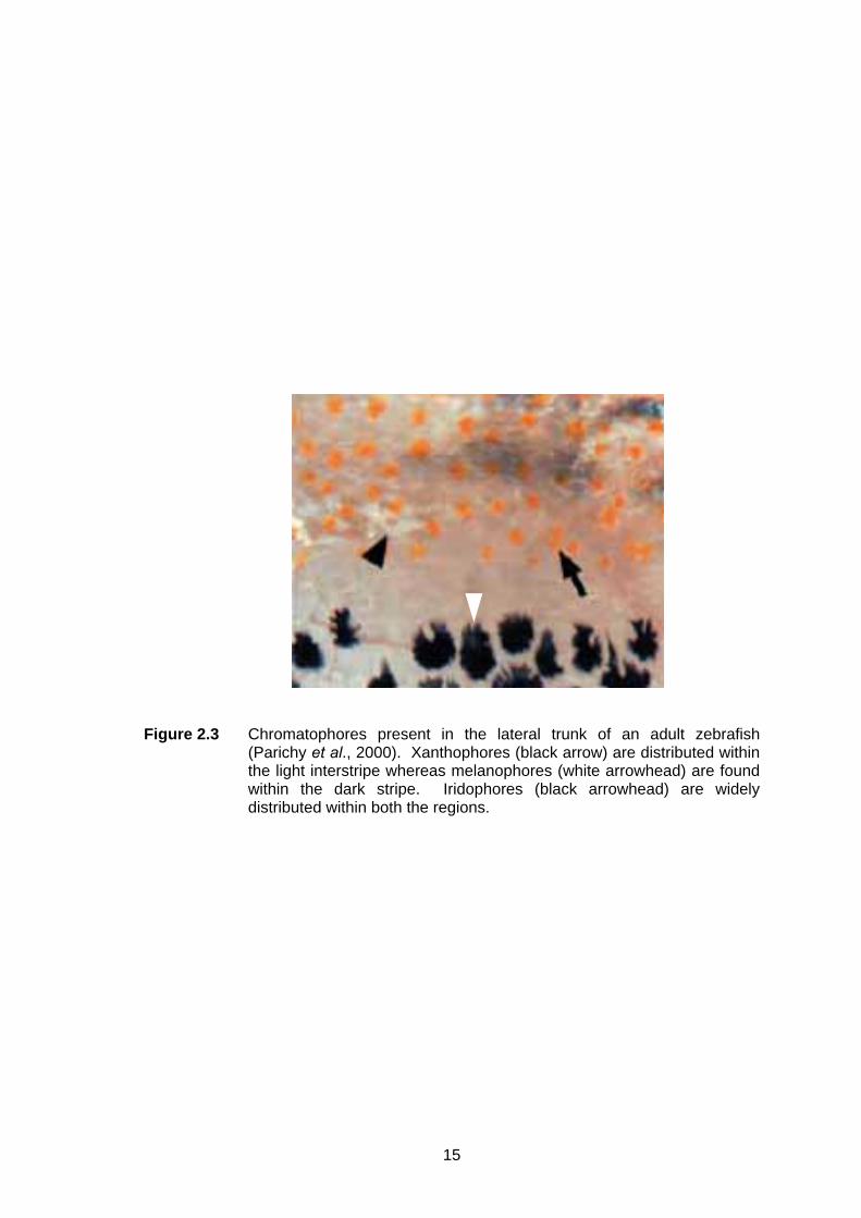
Figure 2.3 Chromatophores present in the lateral trunk of an adult zebrafish
(Parichy et al., 2000). Xanthophores (black arrow) are distributed within the light interstripe whereas melanophores (white arrowhead) are found within the dark stripe. Iridophores (black arrowhead) are widely distributed within both the regions.
16
iridophores, there is a layer of melanophores and they showed ubiquitous distribution
throughout the whole stripe region. Additionally, there is another layer of type L
iridophores present just above the muscular layer. No type L iridophores have been
observed in contact with xanthophores (Figure 2.4).
2.7 The development of zebrafish pigment pattern
In zebrafish, pigment cells are derived from the neural crest cells during the
embryogenesis and the chromatoblasts (melanoblasts, xanthoblasts and iridoblasts)
are derived from two migration pathways, lateral and medial pathway. The former
pathway is below the developing epidermis and the latter is in between the somites and
neural tube. The melanophores of zebrafish migrate on both the lateral and medial
pathways while xanthophores and iridophores are restricted to the lateral pathway.
These chromatoblasts are later proliferated and differentiated into specific
chromatophores forming the early larval pattern (Kelsh, 2004).
The early larval pattern is clearly observed at 5 days post-fertilisation (dpf),
comprises of four longitudinal stripes of melanophores, which include dorsal, lateral,
ventral and yolk-sac stripes. In addition, the melanophore stripes are accompanied by
iridophores formed in overlapping sequence essentially in order of their distance from
the neural tube. In contrast, xanthophores are distributed homogeneously over the
lateral face of the underlying myotome, in a slightly graded fashion with the density is
higher at dorsal part of body (Schilling, 2002; Kelsh, 2004). Melanophores begin to be
visible around 24 hours post-fertilisation (hpf) in the eye and then extended from the
hindbrain to posterior and laterally. The first appearance of xanthophores is around 42
hpf as a very pale tinge at the dorsal of the head. Iridophores only started to scatter in
the eye around 43 hpf and formed lateral patches at the swim bladder at 48 hpf (Lister,
2002; Ziegler, 2003).

17
Figure 2.4 The distribution of pigment cells, in relation to the stripe pattern in zebrafish (Hirata et al., 2003).
18
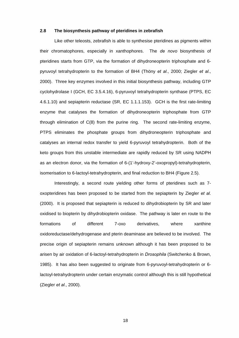
2.8 The biosynthesis pathway of pteridines in zebrafish
Like other teleosts, zebrafish is able to synthesise pteridines as pigments within
their chromatophores, especially in xanthophores. The de novo biosynthesis of
pteridines starts from GTP, via the formation of dihydroneopterin triphosphate and 6-
pyruvoyl tetrahydropterin to the formation of BH4 (Thöny et al., 2000; Ziegler et al.,
2000). Three key enzymes involved in this initial biosynthesis pathway, including GTP
cyclohydrolase I (GCH, EC 3.5.4.16), 6-pyruvoyl tetrahydropterin synthase (PTPS, EC
4.6.1.10) and sepiapterin reductase (SR, EC 1.1.1.153). GCH is the first rate-limiting
enzyme that catalyses the formation of dihydroneopterin triphosphate from GTP
through elimination of C(8) from the purine ring. The second rate-limiting enzyme,
PTPS eliminates the phosphate groups from dihydroneopterin triphosphate and
catalyses an internal redox transfer to yield 6-pyruvoyl tetrahydropterin. Both of the
keto groups from this unstable intermediate are rapidly reduced by SR using NADPH
as an electron donor, via the formation of 6-(1’-hydroxy-2’-oxopropyl)-tetrahydropterin,
isomerisation to 6-lactoyl-tetrahydropterin, and final reduction to BH4 (Figure 2.5).
Interestingly, a second route yielding other forms of pteridines such as 7-
oxopteridines has been proposed to be started from the sepiapterin by Ziegler et al.
(2000). It is proposed that sepiapterin is reduced to dihydrobiopterin by SR and later
oxidised to biopterin by dihydrobiopterin oxidase. The pathway is later en route to the
formations of different 7-oxo derivatives, where xanthine
oxidoreductase/dehydrogenase and pterin deaminase are believed to be involved. The
precise origin of sepiapterin remains unknown although it has been proposed to be
arisen by air oxidation of 6-lactoyl-tetrahydropterin in Drosophila (Switchenko & Brown,
1985). It has also been suggested to originate from 6-pyruvoyl-tetrahydropterin or 6-
lactoyl-tetrahydropterin under certain enzymatic control although this is still hypothetical
(Ziegler et al., 2000).
19
Figure 2.5 Proposed biosynthesis pathway of pteridines in zebrafish (Ziegler, 2003). GTP, guanosine triphosphate; BH4,
tetrahydrobiopterin; GCH, GTP cyclohydrolase I; PTPS, 6-pyruvoyl tetrahydropterin synthase; SR, sepiapterin reductase; XOD, xanthine oxidase; XDH, xanthine dehydrogenase; hypothetical pathway. Pteridines identified in zebrafish was boxed.
dihydroneopterin triphosphate
6-pyruvoyl tetrahydropterin
6-(1’-hydroxy-2-oxopropyl)-tetrahydropterin
6-lactoyl-tetrahydropterin
GCH PTPS SR SR SR BH4 GTP
sepiapterin
dihydrobiopterin
biopterin
7-oxobiopterin
NADPH NADP+ NADPH NADP+
O2
H2O2
O2
H2O2
NADPH
NADP+
dihydropterin
SR
dihydrobiopterin oxidase
isoxanthopterin 2,4,7-trioxopteridine
PTPS orthologue
pterin 2,4-dioxopteridine pterin
deaminase XOD variant
dihydrobiopterin oxidase
O2
H2O2
XOD/XDH XOD/XDH
O2 / NAD+
H2O2 / NADH
O2 / NAD+
H2O2 / NADH
20
2.9 Sepiapterin reductase (SR)
2.9.1 Structure, chromosomal localisation and expression of SR gene
In mammals, the genomic organisation of SR genes is highly conserved
between human and mouse (Ohye et al., 1998; Lee et al., 1999). They both consist of
three exons, span a region of 4 to 5 kb and no splice variants have been observed so
far. However, a highly homologue pseudogene (Sprp) was isolated and characterised
from the mouse genome. It is composed of exon 1 and exon 2 region of the authentic
SR gene with overall homology of 82%. Precisely, human SR gene was mapped to the
p13 region of chromosome 2, while mouse SR gene was located in the central region
of chromosome 6, which is also the known syntenic region of human chromosome 2
(Kim et al., 1997). In contrast to mammalian SR genes, Drosophila SR gene does not
have any intron and produce a single transcript of 1.4 kb (Seong et al., 2000). It was
mapped to 15A on the X chromosome.
The transcription start sites (TSS) of mammalian and Drosophila SR genes
have been determined, which are located within 100 bp upstream from the ATG codon.
The promoter region studies revealed that there is devoid of distinctive TATA- and
CAAT-box motifs in all three SR genes. Furthermore, sequence between -83 and -51
at the promoter region was shown to be essential for the mouse SR gene expression
(Lee et al., 1999). Previous works done by Maier et al. (1993) have shown that rat SR
mRNA was expressed in different types of tissue including liver, kidney, spleen and
brain. Moreover, SR mRNA was also detected throughout different developmental
stages, and in both heads and bodies of Drosophila (Seong et al., 2000).
2.9.2 Structure and localisation of SR enzyme
The 1.25 Å crystal structure of mouse SR in its ternary complex with
oxaloacetate and NADP+ has been demonstrated by Auerbach et al. (1997). SR is a
homodimer comprising 2 monomers with 261 amino acids each. The monomers form a
single-domain α/β-fold with a central four-helix bundle connecting two seven-stranded
21
parallel β-sheets, each sandwiched between two arrays of three α-helices. However,
the two parallel β-sheets of the dimer are in an anti-parallel orientation enclosing an
angle of 90°. At the C-terminal end of the β-sheets contains the active site of SR, a 15
Å-deep pocket that is suitable to receive pterin and small carbonyl substrates. The
substrate is later anchored by the guanidine moiety of a specific Asp residue (Asp-258
in mouse SR).
Recently, Ikemoto et al. (2002) has demonstrated that the wide distribution
patterns of SR enzyme in different parts of human brain, such as substantia nigra (SN),
caudate nucleus (CN), grey and white matters of the cerebral cortex (CTX), and dorsal
and ventral parts of the medulla oblongata (MO). On the other hand, researchers from
Japan revealed that the distributions of SR enzyme are mainly in the xanthophores but
very little in the melanophores of medaka fish (Oryzias latipes), although SR is very
likely to be included in melanophores due to the biosynthesis of BH4 within the cells
(Negishi et al., 2003).
2.9.3 Functional roles of SR in the biosynthesis of tetrahydropteridines
Commonly, SR is well known to be the enzyme involved in the terminal step of
the de novo biosynthesis of L-erythro-tetrahydrobiopterin (BH4), which is a well-known
cofactor important for various physiological processes in higher mammals. Under the
presence of NADPH, SR first reduces the keto group at the side-chain C-1 and forms
6-(1’-hydroxy-2’-oxopropyl)-tetrahydropterin. Following this, the internal rearrangement
of the keto group takes place via side-chain isomerisation, and leads to the formation of
6-lactoyl-tetrahydropterin. Immediately, the intermediate is then converted into BH4 in
a second NADPH-dependent reduction step (Thöny et al., 2000).
Beside this, Blau et al. (2001) suggested that SR may be involved in a BH4
salvage pathway by catalysing the conversion of sepiapterin into dihydrobiopterin
(Figure 2.6). However, Sawada et al. (2005) showed that the proposed pathway in
which the non-enzymatic conversion of 6-lactoyl-tetrahydropterin to sepiapterin is

22
Figure 2.6 A proposed BH4 salvage pathway (Blau et al., 2001). AR, aldose reductase; CR, carbonyl reductase; DHFR, dihydrofolate
reductase; non-enzymatic conversion.
6-pyruvoyl tetrahydropterin
6-lactoyl- tetrahydropterin
AR sepiapterin dihydrobiopterin tetrahydrobiopterin
CR DHFR

23
difficult or unlikely to proceed in human. Recently in Dictyostelium discoideum, SR has
also being demonstrated that its novel capability to convert 1’-oxo-2’-D-hydroxypropyl-
tetrahydropterin into D-threo-tetrahydrobiopterin (DH4). Moreover, Dictyostelium SR
prefers 1’-oxo-2’-D-hydroxypropyl-tetrahydropterin as substrate than 6-pyruvoyl
tetrahydopterin to maintain the dominant production of DH4 over BH4 in this species
(Choi et al., 2006).
24
CHAPTER 3
MATERIALS AND METHODS
3.1 Stock solutions and reagents
All the stock solutions and reagents used in this study were prepared according
to Appendix A.
3.2 Culture media and antibiotic
3.2.1 LB medium and LB-ampicillin plate
LB medium was prepared according to Maniatis et al. (1982). A total of 10 g of
tryptone, 5 g of yeast extract and 10 g of NaCl were dissolved in a final volume of 1000
ml dH2O. The pH of medium was adjusted to 7.5 with NaOH, and sterilised by
autoclaving at 121°C for 20 min.
To prepare LB-ampicillin plate, 15 g of agar was added to 1000 ml of LB
medium. The medium was sterilised by autoclaving and allowed to cool to 50°C,
before adding ampicillin to a final concentration of 100 µg/ml. Approximately 15 to 20
ml of the medium was poured into an 85 mm petri dish and allowed to solidify. LB-
ampicillin plate was then stored at 4°C for up to 1 month. For blue-white colony
selection, 100 µl of 100 mM IPTG and 20 µl of 50 mg/ml X-Gal were spread over the
surface of an LB-ampicillin plate and allowed to absorb for 30 min at 37°C prior to use.
3.2.2 Antibiotic
Sodium salt of ampicillin was dissolved in dH2O to a final concentration of 100
mg/ml, followed by filter-sterilisation and stored in aliquots at -20°C.
3.3 Host strain and cloning vector
The genotype of the bacteria host strain used in this study is presented in Table
3.1 and the map of pGEM®-T Easy Vector (Promega, USA) is shown in Figure 3.1.