Embed Size (px)
Citation preview
UNIVERSITI PUTRA MALAYSIA
THE ROLE OF THE RESPIRATORY MUCOSAL IMMUNITY IN PROTECTION AGAINST PASTEURELLA HAEMOLYTICA
A2 INFECTION IN GOATS
MOHD. EFFENDY ABD. WAHID
FPV 1998 4

THE ROLE OF TH E RESPIRATORY MUCOSAL IMMUNITY I N PROTECTION AGAIN ST PASTEURELLA HAEMOL YTICA A2 I N F ECTION IN GOATS
By
MOHD. EFFENDY ABD. WAHI D
A Dissertation Submitted in Fu lfilment of the Requirements for the Degree of Doctor of Phi losophy i n the
Faculty of Veterinary Medicine and Animal Science U niversiti Putra Malaysia
June 1 998

AC KNOWLEDGEMENTS
I would l ike to express my heartiest appreciation especial ly to my
supervisor, Associate Professor Dr. Mohd Zamri Saad, who have devoted a lot
of his time for inva luable guidance especia l ly on the planning of the study,
constructive suggestions and consistent motivation during the course of this
study. His sacrifices and unreserved assistance wil l forever last to be
remembered . Sincere thanks are also due to my co-supervisors , Dr. Daud
Ahmad Israf Al i and Dr. Mohd Azmi Mohd Li la for their ideas and helpful
discussion.
I deeply indebted to Mr. Mohd. Jami l Samad, Dr. Md . Sabri Mohd . Yusoff,
Mr. R. Kumar and Mr. Zakaria Razal i , who were most helpful and generous with
their time. I would l ike to record my sincere appreciation to Dr. Mohammad
Mustafa (MARDI Serdang) and Mr. Omar Hassan (MARDI Sg. Sum un) for
making the vaccination trials at MARDI Sg . Sumun a success. The same
appreciation is a lso due to Dr. Maswati Mat Amin (Department of Veterinary
Services) , her col league Mr. Nutman as the Supervisor of Goat Farm in Kuala
Pah, Klawang , N egeri Sembilan and the staff members who have devoted their
valuable efforts in the vaccination trials conducted at the farm.
ii

Thanks are a lso recorded to Dr. Saleha Abdul Aziz, Professor Dr. Abd
Rani Bahaman and Associate Professor Dr. Rehana Abdul lah Sani for a l lowing
me to use their laboratory faci l ities during the course of this study. I am very
grateful to Miss Hartina Khan, Mr. Fauzi Che Yusof, Mr. Kamarzaman Ahmad,
Mr. Ismai l Md . Shairi , Mr. Zainuddin Ibrahim , Mr. Md. Noh Manaf, Mr. Mohd .
Isnain Al i , Miss Isma Suzyta I smai l , Dr. Siti Khairani Bejo, Dr. Muthafar AI
Haddawi and those who were involved either directly or indirectly in sharing their
knowledge, ski l l and assistance during this study.
A specia l dedication is a lso due to my wife, Shahnaz Ismai l , my son
Ahmad Irfan Azfar and my daughter Nur Irdina Danisyah, my father Abdul Wahid
Ismai l , my mother Nyonya Yoon Abdul lah and my brothers for their love,
patience, sacrifices, understanding and continuous encouragement during the
long period of this study.
iii

TABLE OF CONTENTS
ACKNOWLEDGEMENTS . . . . . . . . . . . . . . . . . . . . . . . . . . . . . . . . . . . . . . . . . . . . . . . . . . . . . . . . . . . . i i
LIST OF TABLES vii
LIST OF PLATES ix
LIST OF FIGURES . . . . . . . . . . . . . . . . . . . . . . . . . . . . . . . . . . . . . . . . . . . . . . . . . . . . . . . . . . . . . . .. . . . . . xi
ABSTRACT ... . .. . . . . . . . . . . . . . . . . . . . . . . . . . . . . . . . . . . . . . . . . . . . . . . . . . . . . . . . . . . . . . . . . . . . . . . . . . xii
ABSTRAK . . . . . . . . . . . . . . . . . . . . . . . . . . . . . . . . . . . . . . . . . . . . . . . . . . . . . . . . . . . . . . . . . . . . . . . . . . . . . . . . . xv
CHAPTER
1
2
INTRODUCTION
LITERATURE REVIEW
Pasteurella haemolytica
Pneumonic Pasteurellosis . . . . . . . . . . . . . . . . . . .. . . . . . . . . . . . . . . . . . . . ' " . . . . . . . .
Pneumonic Pasteurellosis Status in Malaysia . . . . . . . . . . . . . . . . . . . . . . . . .
Background of Pasteurella Vaccination in Malaysia . . . . . . . . . . . . . . . . .
Mucosal Immunity in Respiratory Tract . . . . . . . . . . . . . . . . . . . . . . . . . . . . . . . . .
1
5
5
6
9
1 0
1 3
3 ATTEMPTS TO STIMULATE THE BRONCHUS-ASSOCIATED LYMPHOID TISSUE IN THE LUNGS OF GOATS VIA ORAL ADMIN ISTRATION OF PASTEURELLA HAEMOL YTICA A2 I ntroduction . . , . . . . . . . . . . . . . . . . . . . . . . . . . . . . . . . . . . . , . . . . . . . . . . . . . . . . . . . . . . . . . . . . 24
Materials and Methods . . . . . . . . . . . . . . . . . . . . . . . . . . . . . . . . . . . , . . . . . . . . . . . . . . . . 25
Animals . . . . . . . . . . . . . . . . . . . . . . .. . . . . . . . . . . . . . . . . . ... . . . . . . . . . ... . . . . . . . . 25 Inoculum . . . . . . . . . . . . . . . '" . . . . . . . . . . . . . . . . . . . . . . . . . . . '" . . . . . . . . . . . . . . 26
Experimental Design .. . . . . . . . . . . . . . . . . . . . . . . . . . . . . . . . . . . . . . . . . . . . . . . 26
Sample Collection and Processing .. . . . . . . . . . . . . . . . . . . . . . . . . . . 27
Immunoperoxidase Procedure . . . . . . . . . . . . . . . . . . . . . . . . . . . . . . . . . . 28
Results . . . . . . . . , . . . . . . . . . . . , . . . . . . . . . . . , . . . . . . . . . . . . . . . . . . . . . . . , . . . . . . '" . . , . . . . 29
Responses by the BAL T. . . . . . . . . . . . . . . . . . . . . . . . . . . . . . . . . . . . . . . . . . . . . 29
Responses by the IgA-Producing Cells in the BAL T . . . . . . . 30
Responses by the Mesenteric Lymph Nodes . . . . . . . . . . . . . . . . 31
iv

Discussion . . . . .. . . . . . . . . . . . . . . . . . . ... . . . ... . . . . . . . . . . . . . . . . . . . . . . . . .. . . . . . . . . . 32
Summary . . . . . . . . . . . . . . . . . . . . .. . . . . . . . . . . .. . . . . . . . . . . .... . . . . . . . . . . . . . . . . . . . . . 34
4 INTRANASAL STIMULATION OF THE BRONCHUS-ASSOCIATED LYMPHOID TISSUE OF GOATS AND ITS EFFECT ON IN VITRO COLONISATION OF PASTEURELLA HAEMOL YTICA A2 I ntroduction . . . . . . . .. ... . . . . . . . . . . .. .. . . .. . . . . . . . . . . . . . . . . . . . . . . . . . . . . . . . . . . . 36
Materials and Methods .. . . ... . . . . . . . . . . . . . . . , . . . . . . . . . . . , . . . . . . . '" . , . . . 37
Animals . . . . . . . . . . ... . . . . . . . . . . . . . . . . . . .. . . . . . . . . . . . . . . ... . . . . . . . . . . . . 37
I noculum . . . . . . . .. .. . ... . . . . . . . . . ... . . . . . , . . . . . . . . . . . . . . .. . . . . . . . , . . . . 38
Experimental Design ...... .... . . . .. . . , . . . . . . . .... . . . . . . . . . . . . . . . . 38
The Study of BAL T Reactions . . . . . . .. . . . . . . . . . . . . . . . . . . . . . . . . . . . . 39
In Vitro Colonisation Technique .. . . , . ' " . . . ..... . .. . . . . . . . . , . . . . 40
Statistical Analysis . . . . . . . . , . . . ... . . . . . , . . . . . . .. . .. , . . . . . . . . . .. . . . . 4 1
Results . . . . . . . . . . . . . . . . . . . . . . . . . . . . . . . . . . . . . . . . . . . . . . . . . . . . . . . . . . . . . . . . . . . . . .. 42
Responses in the Number and Size of BAL T ............ .. 42
In Vitro Colonisation by Pasteurella haemolytica A2 . . ... 46
Discussion . . , . . . . . . . . . . ... . . . . . . . . . . , . . . . . . . . .. . , . . . . . . . . . . . . . . . . . . . . . . . , . . . 47
Summary ... . .. . . . . . . . . . . . . . . .. . . . . . .. . . . . . . . . . . . . . . . . . . . . .. . .. . . . . . . . . . . . . . . 49
5 CELLULAR AND HUMORAL RESPONSES I N THE RESPIRATORY TRACT OF GOATS FOLLOWING INTRANASAL STIMULATION USING FORMALlNKILLED PASTEURELLA HAEMOL YTICA A2 Introduction .. . . . . . . . . . . .. . . . . . . . . . . . . . . . . . . . . . . . . . . .. . . . . . . . . . . . . . . . . . . . . . . . . . 5 1
Materials and Methods . . . . . . . ... . . . . . . .. . . . . . . . . . . . . . . . . . ... . . . . . . . . . . . . . . . 52
Animals .. . . . . . . .... . . . .. . . . . . . . . . . . . . . . . . . . . . . . . . . . . . . . . . .. . . . . . .. . . . . . . . 52
I noculum . . . . . . . . ... . . . . . . . . . . . . . . . . . . . .. . . . . . . . . . . . . . . . . . . . . . . . . . . . . . . . . . 53
Experimental Design ... .. . . .. ... ... ... .. . ... ... ... ... ... .. . ... ... . . . . 53 Serology . . . . , . . . . . . . . . . . . . . . . . . . . . . . . . . . . ... . . . . . . . . . . . . . . . . . . . . . . . . . . . .. 54
Studies on BAL T Response .. . . . . . . . . . . . . . . . . . . . .. . . . . . . . . . . . . .. . . . . 55
Statistical Analysis . . . . . . . . . . . . . . . . .. . . . . . . ... . . . .. . . . . .. . . . . . . . . . . . . . . 56
Results . .. . . . . . . . . . . . . . . , . . .. . . . . . . . . . . . . . . . ... . . .. . .. , . . . . . . . . . . .. . . . . . . . . . . . . . 56
Antibody Responses in Lung Lavage Fluid . . . . . . . , . . . . . . . . . . .. . 56
Serum Antibody Responses . . . . . . . . . . . . . . . . . . . . . . . . . . , . . . . . . . . . . . . . 57
Responses by the BAL T .. . . . . . . . . . . . . .. . . . . . . . . . . . . . . . . . . . . . . . . . . . . . . 59
Discussion . . . . . . . . . . .. . . . . . . . , . . . . . .. . . . . , . ... . . . . . . . . . . . . . . . . . . . , . . . . . . . . . . . , . 63
Sum mary . . . . . . . . . . . . . .. .... . . . . . . . . . . . . .. . .. .. . . . . . . . . . . . . . . .. . . . . . . . . . . ... . . . 65
v
6 THE EFFECTS OF DEXAMETHASONE ON THE RESPONSE BY BRONCHUS ASSOCIATED LYMPHOI D TISSUE TO I NTRANASAL ADMIN ISTRATION OF FORMALIN-KI LLED PASTEURELLA HAEMOL YTiCA A2 I N GOATS I ntroduction . . . . . . . . . . . . . . . . . . . . . . . . . . . . . . . . . . . . . . . . . . . . . . . . . . . . . . . . . . . . . . . . . . . . . . 67
Materials and Methods . . . . . . . . . . . . . . . . .. . . . . . . . . . . . . . . . . . . . . . . . . . . . . . . . . . . . . 69
Animals . . . . . . . . . . . . . . . . . . . . . . . . . . . . . . . . . . . . .. . . . . . . . . . . . . . . . . . . . . . . . . . . . . . 69
I noculum . .. . . . . . . . . . . . . . . . . . . . . . . . . . . . . .. . . . . . . . . . . . . . . . . . . . . . . . . . . . . . 69
Experimental Design . . . . . . . . . . . . . . . . . . . . . . . . . . . . . . . . . . . . . . . . . . . . . . . 70
Microscopic Study on BALT Responses . . . . . . . . . . . . . . . . . . . . . 7 1
Immunoperoxidase Staining . . . . . . . . . . . . . . . . . . . . . . . . . . . . . . . . . . . 72
Results . . . . . . . .... . . . . . . . . . . .. . . . . . . . . . . . . . . . . .. . . . . . . . . .. . . . . . . . . . . . . . . . . . . . . 73
Responses by the BAL T . . . . .. . . . . . . . . . . . . . . . . . . . .. . . . . . . . . . . . . . . . . 73
Responses by the IgA-Producing Cells in BALT. . . . . . . . . . . . . 75
Discussion.. . . .. . . . . . . . . . . . . . . . . . . . . . . . . . . . . . . . . . . . . . . . . . . . . . . . . . . . . . . . . . . . . . . 78
Summary 80
7 EFFICACY OF I NTRANASAL ADMIN ISTRATION OF FORMALIN-KILLED PASTEURELLA HAEMOL YTiCA A2 AGAINST INTRATRACHEAL CHALLENGE EXPOSURE I N GOATS Introduction . . . . . . . . . .. . . . . . . . . . . . . . . . . .. . . . . . . . . . . . . . . . . . . . . . . . . . . . . . . . . . . . . .
Materials and Methods . . . . . . . . . . . . . . . . . . . . . . . . . . . . . . . . . . . . . . . . . . . . . . . . . . .
Animals . . . . ..... . . . ..... . . . .. . . . . . . ...... . . . . ... . . . . . . . . .. . . .. . . . . . .
Preparation of the I noculum . . . . . . . . . . . . . . . . . . . . . . . . . . . . . . . . . . . .
Experimental Design .. . . . . . . . . . . . .. . . . . . . . . . . . . . . . . . . . . . . . . . . . . .
Serology . . . . . . . . . . . . . . . . . . . .. . . . . . .. . . . . . . . . . . . . .. . . . . . . . . . . . . . . . . . .
Bacterial Isolations
Statistical Analysis
Results . . . . . . . . . . . . . . . . . . . . . . . . . . . . . . . . . . . . . . . . . . . . . . . . . . . . . . . . . . . . . . . . . . . . . . .
Clinical Observations . . . . . . . . . . . . . . . . . . . . . . . . . . . . . . . . . . . . . . . . . . . .
Pathological Observations . . . . . . . . . . . . . . . . . . . . . . . . . . . . . . . . . . . . .
Serology . . . . . . . . . . . . . . . . . . . . . . . . . . . . . . . . . . . . . . . . . . . . . . . . . . . . . . . . . . .
Microbial Observation . . . . . . . . . . . . . . . . . . . . . . . . . . . . . . . . . . . . . . . . . .
Correlation Between Clinical Score, Lung Lesions
and Serological Responses. . . . . . . . . . . .. . . . . . . . . . . . . . . . . . . . . . . .
Discussion . . . . . . . . . . . . .. . . . . . . . . . . . . . . . . . . . . . . . . . . . . . . . . . . . .. . . . . . . . . . . . . . . .
Summary . . . . . . . . . . . . . . . . . . . . . . . . . . . . . . . . . . . . . . . . . . . . . . . . . . . . . . . . . . . . . . . . . . . .
vi
82
84
84
84
85
86
87
87
88
88
89
92
93
93
95
97

9
8 VACCI NATION TRIAL AGAINST PNEUMONIC PASTEURELLOSIS IN SHEEP AND GOATS USING A NEWLY DEVELOPED PASTEURELLA SPRAY VACCINE Introduction ... . . . . . . . . . . . . . . . . . .. . . . . . . . . . . . .. , . . . . . . . . . . . . . . . . . . . . . . . . . . . . 99
Materials and Methods . . . . . , ...................................... , . .. . 100
Sheep . . . . . . ........................................................ 100
Goats . . . . . . ............ ... ......... ... ............ ...... ... ...... ... 100
Vaccine Preparation 101
Experimental Design . . . ...... ... ...... ... ...... ...... ... ... ..... 102
Statistical Analysis . . . . . . . . . .. . . . . . . . . ... ... . , ... '" .,. . .. . .. . . . . 103
Results . . . . . . . . . . , ............... , .............. , ....... , . . .. ... .. . . .. ... . . . 104
Serological Response by Sheep . . . . . . . . . . . . . . . . ,. ... ... ..... 104
Serological Response by Goats . , . ... ...... ........ ... ....... 105
I ncidences of Pneumonic Pasteurellosis in
S heep Farm ... . . . . . . . . , ....... ,. '" ........... , .............. , .... 107
I ncidences of Pneumonic Pasteurellosis in
Goat Farm . . . . . . . . . . . . .. , .............................. , ... ... ... ... 108
Bacteriology . . . . . . . . . . . . . .. . . . . . . . . . . . . . . . . . . . . . . . . . . . . . . . . . . . . . . . . 108
Discussion .. . . . . . . . . . . . . , ............ '" ....................... , .. . ... . .. .. 108
Summary . . . . . . . . . . . . . . . . . . . . . . . . . . . . . . . . . . . . . . . . . . . . . . . . . . . '" .,. ...... .... 112
GENERAL D ISCUSSION . . . . . . . . . . . . . . . . . . . . . . . . . . . . . . . . . .. . . . . . . . . . . . . . 114
BIBLIOGRAPHy .. . . . . . . . . . . . . . . . . ... . . . . . . . . . . . . . . . . . . . . . . . . . . . . . . . . . . . . . . . . . . . . .. . . . 123
APPENDICES . .. . . . . . . . . .. . . . . . . . .. . . . ... . . . . .. . . . . . . .. . . . . . . . . . . ..................... 134
VITA . . . . . . . . . . . . . . . . . . . . . . . . . . . . . . . . . . . . . . . . . . . . . . . . . . . . . . . . . . . . . . . . . . . . . . . . . . . . . . . . . . . . . . 141
PUBLICATIONS . . . . . . . . . . . . . . . . . . . ,. '" .......... ,. '" ...... '" ....... ... ... ... ... ... 142
vii

LIST OF TABLES
Table
3 .1 The number of bronchus associated lymphoid tissue (SALT) and their average size in the l ungs of goats fol lowing different
Page
treatments . . . . . . . . . . . . . . . . . . . . . . . . . . . . . . . . . . . . . . . . . . . . . . . . . . . . . . . . . . . . . . . . . . . . . 30
3 .2 The effects of oral administration of formalin-ki l led Pasteurella haemolytica A2 on the total number of cel ls and the percentage of IgA-producing cel ls in SALT of goats . . , . . . . . . . . . . . . . . . . . . . . . . . . . . . . . 31
3.3 The effects of oral administrations of formal in-kil led Pasteurella haemolytica A2 on the size and cortex:medul lar ratio of mesenteric lymph nodes of goats . . . . . . ' " . , . . . . . . . . . . . . . . . . ' " . . . . . . . . . . 32
4. 1 Average size and number of bronchus-associated lymphoid tissue (SALT) in the lungs of goats fol lowing either a single intranasal exposure to l ive (group 1) or dead Pasteurella haemolytica A2 (group 2) or a double intranasal exposures to l ive (group 3) or dead Pasteurella haemolytica A2 (group 4) . . . . . . . . . . . . 42
5 . 1 The average number of bronchus associated lymphoid tissue SALT) in lungs of goats exposed twice to formal in-ki l led Pasteurella haemolytica A2 . . . . . . . . . . . . . . . . . . . , . . . . . . . . . , . . . . . . . . . . . . . . . . . . . . 60
6 .1 The average number and size of bronchus-associated lymphoid tissue (SALT) in the l ungs of goats fol lowing intranasal exposure to formal in-ki l led Pasteurella haemolytica . . . . . . . . . . . . . . . . . . . . . . . . . . . . . . . 74
6 .2 The effects of dexamethasone on the average number of cel ls and the average percentage of IgA-producing cel ls in SALT of goats exposed to formal in-kil led Pasteurella haemolytica A2. . . . . . 75
7. 1 Average clinical score and extent of lung lesions after chal lenge with Pasteurella haemolytica A2 . . . . . . . . . . . . . , . . . . . . . . . , . . . . . . . . . . . . . . . . . . . . 88
viii
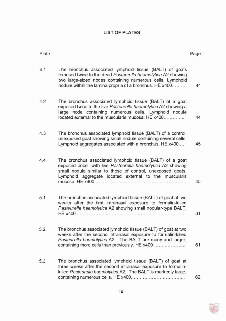
Plate
4. 1
4. 2
4 .3
4.4
5.1
5. 2
5 .3
LIST OF PLATES
The bronchus associated lymphoid tissue (SALT) of goats exposed twice to the dead Pasteurella haemolytica A2 showing two large-sized nodes containing numerous cel ls . Lymphoid nodule within the lamina propria of a bronchus. HE x400 . . . . . . . . .
The bronchus associated lymphoid tissue (SALT) of a goat exposed twice to the l ive Pasteurella haemolytica A2 showing a large node containing numerous cel ls . Lymphoid nodule located external to the muscularis mucosa . HE x400 . . . . . . . . . . . . . .
The bronchus associated lymphoid tissue (SALT) of a control , unexposed goat showing smal l nodule containing several cel ls . Lymphoid aggregates associated with a bronchus . HE x400 . . . .
The bronchus associated lymphoid tissue (SALT) of a goat exposed once with l ive Pasteurella haemolytica A2 showing smal l nodule simi lar to those of control , unexposed goats. Lymphoid aggregate located external to the muscularis mucosa. HE x400 . . . . . . . . . . . . . . . . . . . . . . . . . . . . . , . . . . . . . . . . . . . . . . . . . . . . . . . . . . .
The bronchus associated lymphoid tissue (SALT) of goat at two weeks after the first intranasal exposure to formal in-ki l led Pasteurella haemolytica A2 showing smal l nodular-type SALT. HE x400 . . . . . . . . . . . . . . . .. . . . . . . . . . . . . . . . . . . . . . . . . . . .. . . . . . . . . . . . . . . . . . . . . . . . . . .
The bronchus associated lymphoid tissue (SALT) of goat at two weeks after the second intranasal exposure to formal in-kil led Pasteurella haemolytica A2. The SALT are many and larger, containing more cel ls than previously. HE x400 . . . . . . . . . . . . . . . . . . . . .
The bronchus associated lymphoid tissue (SALT) of goat at three weeks after the second intranasal exposure to formal inki l led Pasteurella haemolytica A2. The SALT is markedly large, containing numerous cel ls . HE x400 . . . . . . . . . . . . . . . . . . ' " . . . . . . . . . . . . . . .
ix
Page
44
44
45
45
61
61
62

5.4
6 .1
6 .2
6 .3
6.4
7 .1
7 .1
7 .2
7. 3
The bronchus associated lymphoid tissue (SALT) of goat at four weeks after the second intranasal exposure to formal inki l led Pasteurella haemolytica A2 showing large nodular-type SALT. HE x400 . . . . . . .. . . . . . . . . . . . . . . . . . . . . . . . . . . . . . . . . . . . . . . . . . . . . . . . . . . . . .. .
The bronchus associated lymphoid tissue (SALT) of a goat ki l led at 1 day post-dexamethasone treatment showing a smal l aggregate with few cel ls . HE x400 . . . . . . . . . . . . . . . . . . . . . . . . . . . . . . . . . . . . . . .
The bronchus associated lymphoid tissue (SALT) of a goat ki l led at 21 days post-dexamethasone treatment showing a markedly smal l aggregate with few cel ls . HE x400 . . . . . . .. . . . . . . . . . .
The bronchus associated lymphoid tissue (SALT) of a goat exposed to formal in-ki l led Pasteurella haemolytica A2 without the dexamethasone treatment showing a large nodule with numerous cel ls . HE x400 . . . . . . . . . . . . . . . . . . . . . . . . . . . . . . . . . . . . . . . . . . . . . . . .
The bronchus associated lymphoid tissue (SALT) of a control untreated goat showing a smal l nodule. HE x400 . . . . . . . . . . . . . . . . . .
Normal lungs of goat without any lesions or vaccination history . . . . . . . . . . . . . . . . . . . . . . . . . . . . . . . .. . . . . . . . . . . . . . . . . . . . . . . . . . . . . . . . . . . . . . . . . .
Lungs of goat vaccinated with the intranasal spray vaccine and chal lenged with l ive Pasteurella haemolytica A2 showing relatively no pneumonic lesions . . . . . . . . . . . . . . . . . . . . . . . . ' " . . . . . . . . . . . . .
Lungs of goat vaccinated with the oi l adjuvant vaccine and chal lenged with l ive Pasteurella haemolytica A2 showing smal l area of pneumonia of approximately 10% . . . . . . . . . . . . . . . . . . . . . . . . . . . .
Lungs of a control unvaccinated goat chal lenged with l ive Pasteurella haemolytica A2 showing extensive area of pneumonia . Approximately 40% of the lung area were affected . . . . . . . . . . . . . . . ' " . . . . . . . . . . . . . . . . . . . . . . . . . . . . . . . . . . . . . . . . . . . . . . . . . . . . . .
x
62
76
76
77
77
90
90
91
91

LIST OF FIGURES
Figure
4. 1 The colonisation of Pasteurella haemolytica A2 in the lung tissues of goat with different treatments. Group 1 and 2 were intranasally exposed once to live or dead P. haemolytica A2 respectively while groups 3 and 4 were intranasally exposed
Page
twice to live or dead P. haemolytica A2 respectively. . . . . . . . . . . . . . . 47
5. 1 The serum antibody (lgM, IgA, IgG) responses in the lung lavage fluid of goats following intranasal exposures to formalin-killed Pasteurella haemolytica A2. . . . . . . . . . . . . . . . . . . . . . . . . . . 57
5 .2 The serum antibody (lgM, IgA, IgG) responses following intranasal exposures of goats to formalin-killed Pasteurella haemolytica A2. . . . . . . . . . . . . . . . . . . . . . . . . . . . . . . . . . . . . . . . . . . . . . . . . . . . . . . . . . . . . . . 58
5 .3 The size of bronchus associated lymphoid tissue (SALT) and the number of lymphocytes in the SALT responses by goats exposed twice to formalin-killed Pasteurella haemolytica A2. . . . 59
7.1 Serum antibody levels of goats following intranasal vaccination (group 1 ) , intramuscular vaccination (group 2) and mixed vaccination (group 3) . Group 4 was the unvaccinated controL. . 93
8 . 1 IgG antibody response in serum of sheep following intranasal spray vaccination of ki lled Pasteurella haemolytica A2. Note the drop in IgG level below the cut-off point on week 4 after second vaccination due to Haemonchus contortus. The incidence of pneumonic pasteurellosis also increase during that period of time. . . . . . . . . . . . . . . . . . . . . . . . . . . . . . . . . . . . . . . . . . . . . . . . . . . . . . . . . 1 05
8 .2 IgG antibody responses in serum goats following intranasal spray of killed Pasteurella haemolytica A2 and alum precipitate vaccination subcutaneously. Note the incidence of pasteurellosis in the farm before vaccination (week -2 and 0) and the increase in IgG level during outbreak of pasteurellosis in the farm at week 6 . . . . . . . . . . . . . . . . . . . . . . . . . . . . . . . . . . . . . . . . . . . . . . . . . . . . . . 1 06
xi

Abstract of the dissertation presented to the Senate of Universiti Putra Malaysia in fulfi lment of the requirements for the degree of Doctor of Phi losophy
THE ROLE OF THE RESPIRATORY MUCOSAL IMMUNITY I N P ROTECTION AGAI NST PASTEURELLA HAEMOL YTICA A2 I NFECTION IN GOATS
By
MOHO. EFFENOY ABO. WAH IO
June 1 998
Chairman: Assoc Prof Dr Mohd Zamri Saad
Faculty: Veterinary Medicine and Animal Science
Pneumonic pasteurel losis is one of the most important and devastating
d iseases i n sheep and goats , causing great economic losses to smal l ruminant
industry worldwide. The d isease is caused either by Pasteurella haemolytica or
Pasteurella multocida and Pasteurella haemolytica A2 is the most common
isolate from sheep and goats in Malaysia, comprising approximately 38 per cent
of the isolates from pneumonic lung lesions.
The d isease is best control led by vaccination, and systemic vaccination
has been used for years with l imited success. Stress, improper vaccination
program and unpopularity of the vaccine among the farmers are some of the
xii

reasons that have been associated with the persistence of the disease. S ince
the systemic vaccination fai led to give promising results , studies on the role of
mucosal immunity of the respiratory tract in control l ing pneumonic pasteurel losis
should t imely be reviewed.
In this study, the bronchus-associated lymphoid tissue (SALT) in the
lungs 9 been successful ly stimulated by double intranasal exposures to either
l ive or formal in-kil led Pasteurella haemolytica A2 at two weeks interva l . The
size of SALT and number of lymphocytes in the SALT were significantly
increased as early as week 2 post-exposure and remained h igh unti l week 4
post-exposure. At the same time, the level of IgA against Pasteurella
haemolytica A2 increased significantly as early as week 1 post-first exposure
and reached a peak level at week 6 post-exposure. The IgM appeared to be
present for a short wh i le, at week 3 post-exposure before the levels started to
decline in the fol lowing week. Initially, the IgG increased gradual ly and
insignificantly before it reached significantly h igh level at week 4 post-exposure,
and remained h igh at weeks 5 and 6 at the time when the numbers of SALT
continued to increase.
Th is study a lso revealed that intranasal stimulation of SALT was ab le to
protect the lungs from colonization by Pasteurella haemolytica A2 during an in
vitro study, thus prevent the lung surface from being adhered and invaded by
xiii

the organism. However, dexamethasone treatment which is similar to the effect
of steroid released under stressful conditions, significantly reduced the number
and size of the SALT, thus significantly reduced the percentage of IgA-producing
cel ls .
Vaccination tria l on goat farm using the pasteurel la spray vaccine
intranasal ly showed good protection towards pneumonic pasteurel losis.
Significant high levels of systemic antibody responses were also noted during
the period of vaccination tria l . The incidence of pneumonic pasteurel losis in the
farm was markedly reduced.
xiv

Abstrak d isertasi yang dikemukakan kepada Senat Universiti Putra Malaysia bagi memenuhi keperluan Ijazah Doktor Falsafah
PERANAN KEIMU NAN MU KOSA PERNAFASAN OALAM MELINOU NGI KAMBING TERHAOAP JANGKITAN PASTEURELLA HAEMOL YTICA A2
Oleh
MOHO EFFENOY ABO. WAHIO
Jun 1 998
Pengerusi: Prof M adya Dr Mohd Zamri Saad
Fakulti : Kedoktoran Veterinar dan Sains Peternakan
Pasteurel losis pneumonia adalah salah satu penyakit terpenting pada
kambing dan bebiri , mengakibatkan kerugian ekonomi yang nyata pada industri
ruminan keci l serata dunia. Penyakit ini disebabkan oleh sama ada Pasteurella
haemolytica atau Pasteurella multocida, dan Pasteurella haemolytica A2
merupakan isolat yang pal ing kerap diasingkan daripada kambing dan bebiri d i
Malaysia , merangkumi kira-kira 38 peratus isolat yang d iperolehi daripada lesi
pneumonia.
xv

Penyakit ini boleh dikawal dengan baik melalui pemvaksinan, dan
pemvaksinan sistemik telah digunakan selama bertahun-tahun dengan
kejayaan yang terhad. Ketegasan , program pemvaksinan yang tidak sesuai dan
ketidakpopularan vaksin di kalangan penternak adalah antara sebab-sebab
yang dikenalpasti menyebabkan penyakit ini berterusan. Memandangkan
selama ini pemvaksinan sistemik gagal untuk membuahkan hasil yang
memuaskan, sudah sampai masanya kajian ke atas peranan keimunan
mukosal pada trakus pernafasan dalam mengawal radang peparu pasteurel losis
dipertimbangkan semula .
D alam kajian ini, tisu l imfoid berkait bronkus (BAL T) dalam peparu telah
berjaya dirangsangkan melalui dedahan berganda kepada Pasteurella
haemolytica A2 yang hidup atau dimatikan dengan formalin mela lui intranasal .
Dedahan berganda dibuat dalam jangkamasa selang 2 minggu. Saiz BAL T dan
bilangan sel l imfosit dalam BAL T bertambah dengan ketara seawal 2 minggu
selepas dedahan, dan mengekalkan ketinggiannya sehingga minggu ke 4
selepas dedahan. Pada masa yang sama, tahap IgA terhadap Pasteurella
haemolytica A2 meningkat dengan ketara seawal minggu pertama selepas
dedahan, dan mencapai kemuncaknya pada minggu ke 6 selepas dedahan.
IgM hadir cuma sebentar, iaitu pada minggu ke 3 selepas dedahan, sebelum
menurun pada minggu berikutnya. Pada peringkat awal , IgG meningkat
beransur-ansur sebelum mencapai peningkatan bermakna pad a minggu ke 4,
xvi

dan mengekalkan ketinggiannya pada minggu ke 5 dan 6, iaitu masa di mana
bilangan SALT turut meningkat.
Kajian ini juga menunjukkan bahawa rangsangan melalui intranasal ke
atas SALT mampu melindungi peparu daripada dikoloni oleh Pasteurella
haemolytica A2, seterusnya menghalang permukaan peparu daripada dilekati
dan dicerobohi organisma berkenaan.
Walau bagaimanapun , rawatan oleh dexamethasone yang mempunyai
kesan yang sama seperti steroid sewaktu tegasan, mengurangkan bilangan dan
saiz SALT dan turut mengurangkan peratusan sel yang menghasilkan IgA.
Pemvaksinan yang dilakukan di ladang ternakan kambing menggunakan vaksin
semburan intranasal menghasilkan perlindungan yang baik terhadap penyakit
pasteure l losis pneumonia. Gerakbalas antibodi sistemik yang tinggi dan ketara
di dapati berlaku sewaktu kajian dija lankan Pengurangan kadar penyakit
pasteurel losis pneumonia d i ladang berkenaan juga dapat dil ihat dengan jelas.
xvi i

CHAPTER 1
I NTRODUCTION
Pneumonic pasteure l losis is one of the most important and
devastating d iseases affecting smal l ruminants worldwide. The disease is
caused by either Pasteurella haemolytica or Pasteurella multocida
(Donach ie, 1 993), although the former is frequently isolated from such cases
in goats (Gi lmour, 1 992).
There are two biotypes of Pasteurella haemolytica , namely biotype A
and T. C l inical ly, biotype A is associated with pneumonic pasteurel losis and
comprises serotypes 1 , 2 , 5 , 6 , 7 , 8 , 9 , 1 1 , 1 2 , 1 3 , 1 4 and 1 6. B iotype T, that
comprises serotypes 3, 4, 1 0 and 1 5 , is associated with systemic d isease in
young lambs. About 1 0 percent of the Pasteurella haemolytica serotypes
remain untypable (Donachie, 1 993) . The development of the disease has
been known to be associated with various stress factors particularly the
environmental factors such as cl imate, transportation , nutrition and housing
(Jubb ef a/. , 1 985; Zamri-Saad ef a/. , 1 989; Jasni ef a/. , 1 99 1 ) . Concurrent
infections with parainfluenza type I I I virus, herpesvirus and Haemonchus
contortus have also been shown to predispose animals to pneumonic
pasteure l losis (Gi lmour et aI. , 1 993; Zamri-Saad et aI. , 1 993a).
1

2
Various studies have been conducted regarding the use of vaccines to
control pneumonic pasteurel losis in Malaysia (Zamri-Saad et al. , 1 989;
Chandrasekaran et al. , 1 99 1 ; Zamri-Saad et al., 1 993b) however the results
were inconclusive and the disease remains a major threat to the goat and
sheep industry. Imported vaccines, such as HEPTAVAC-P (Hoechst, UK) ,
Ovipast (Hoechst (M) Sdn. Bhd.) and Carovax (Wellcome, UK) have been
used to control the disease with l imited success rate (Wan Mohamed et al.,
1 988). A locally produced formalin-killed oil adjuvant vaccine, incorporating
Pasteurella haemolytica and Pasteurella multocida of unknown serotypes
was also used unsuccessful in control ling the disease (Zamri-Saad et aI. ,
1 989). However, an improved oil adjuvant vaccine that incorporating the
locally isolated Pasteurella haemolytica A 7 and Pasteurella multocida types
A and 0 showed ability to protect the lambs when challenged with either
Pasteurella haemolytica A2 or A7 (Zamri-Saad et al. , 1 993b). Although the
local ly produced oil adjuvant vaccine gave better protection , its thick
viscousity lead to difficulties in administration and caused swel l ing at the site
of administration and lameness with uneventful recovery in approximately ten
percent of vaccinated animals (Jamaludin, 1 993).
The locally produced oil adjuvant vaccine has been shown to be able
to stimulate good antibody response (Zamri-Saad et aI. , 1 993c). However,
most sheep in various farms in Malaysia showed low antibody titre against
pasteurel losis (Zamri-Saad et al. 1 993d) and does not protect the animal
from being contacted with pneumonic pasteurel losis , thus the disease

3
incidence remained h igh in many farms. This is probably due to the
unpopularity of the oil adjuvant vaccine among farmers (Jamaludin, 1 993)
and the improper vaccination program provided by the vaccine manufacturer,
which has been recognised as one of the contributing factors in vaccination
fai lure (Zamri-Saad , 1 996).
The respiratory tract, wh ich is the route of infection for Pasteurella
haemolytica (Gi lmour et al. , 1 991 ) , is one of the mucosal tissues in the body
which armed with mucosal immunity (Kaltrieder, 1 976) . Anderson et al.
( 1 986) stated the importance of stimulating the pulmonary lymphoid tissue
with effective methods that wi l l provide substantia l local cel lular and antibody
mediated immune responses (Kaltrieder, 1 976).
Other factors that contribute to the success of vaccination include the
method of vaccine administration (Mosier, 1 993) . Thus, with the uncertainties
and d ifficulties encountered in systemic vaccination of animals against
pneumonic pasteurel losis, other methods for the control of pneumonic
pasteure l losis should perhaps be explored .
The objectives of this study are :
1 . to stimulate the mucosal immunity in the respiratory tract of goats
fol lowing either with intranasal or oral exposures to Pasteurella
haemolytica A2.

4
2. to determine the mucosal immune responses fol lowing intranasal
exposures to formalin-ki l led Pasteurella haemolytica A2.
3. to verify the immunosuppresive effect of dexamethasone on mucosal
immunity of the respiratory tract fol lowing intranasal exposure to
formal in-ki l led Pasteurella haemolytica A2.
4. to determine the protective role of mucosal immunity against
intratracheal cha l lenge with l ive Pasteurella haemolytica A2.

CHAPTER 2
LITERATURE REVIEW
Pasteurella haemolytica
Pasteurella haemolytica is a Gram-negative, smal l , non-motile , cocco
bacil lus bacteria with slight pleomorphism and occasional bipolar staining
(Adlam , 1 989). The organism can easily be recognised upon cultivation on blood
agar and show various sizes of beta zone haemolysis. Bovine blood appears to
be superior for the demonstration of the organism when compared with ovine or
horse blood (Soltys , 1 979).
The organism can be separated into two biotypes, A and T (Donachie,
1 993). Biotypes A and T from cases of ovine pneumonia can be distinguished
based on different cul tura l , biochemical and pathological characteristics (Soltys ,
1 979) . De Alwis ( 1 993) recorded that the differences between biotypes A and T
depend on their abil ity to ferment arabinose and not trehalose and vice versa.
Both serotypes are heterologus in nature and sixteen serotypes can be
differentiated by an indirect haemagg lutination ( IHA) test which distinguishes
5

6
sixteen different capsular antigens in the two biotypes (Donachie, 1 993) .
Biotype A comprises serotypes 1 , 2, 5, 6, 7, 8, 9, 1 1 , 1 2, 1 3 , 1 4 and 1 6, while
the biotype T contains serotypes 3, 4, 1 0 and 1 5. Ten percent remain untypable
(Donachie, 1 993).
Al l serotypes are pathogenic and serotype A2 is the most prevalent,
comprises around 38% of a l l Pasteurella haemolytica isolates (Donachie, 1 993).
The most common serotypes isolated from pneumonic pasteurel losis of sheep
and goats in Malaysia are serotypes A2, fol lowed by A7 and A9 (Bahaman et al. ,
1 991 ) . Pasteurella haemolytica serotypes A 1 1 and A 1 2 were also isolated from
cases in Malaysia (Bahaman et al. , 1 99 1 ) .
Pneumonic Pasteurel losis
Pasteurella haemolytica can be found in nasopharynx of healthy animals
as normal flora (Gilmour, et al. , 1 99 1 ; De Alwis, 1 993). Although the organism
are present in the nasopharynx area, the animal are not clinical ly affected with
pneumonic pasteurellosis (Mosier, 1 993). Colostral immunity in lambs can last
for approximately 4 to 5 weeks (Gilmour and Gilmour, 1 989), but the duration of
immunity fol lowing natura l infection has not been ful ly investigated adequately
(Mosier, 1 993). The prevalence of respiratory infections and associated
economic losses continues to be very high although various attempts have been

7
made to control the d isease. It is difficult to control respiratory disease since
respiratory infections often occur as a resul t of synergistic interactions between
various pathogens (Busse, 1 99 1 ) . Many carriers of Pasteurella haemolytica wi l l
not a lways develop pasteurel losis and remained cl inical ly healthy (Gi lmour and
Gi lmour, 1 989) . This is because the exposure of respiratory pathogenic
organism per se is not sufficient enough for the development of disease.
Furthermore, the respiratory tract provides a variety of integrated defence
mechanisms including mechanical non-immunological and immunologica l , which
function in concert to prevent development of respiratory tract infections
(Stratton, 1 986) . However, fai lure of any one or more of the lung defence
mechanisms d isrupts th is stage of homeostasis resulting in pulmonary infection
(Babiuk and Campos, 1 993).
Various stress factors such as transportation, cl imate and housing stress
(Jubb, et aI. , 1 985; Zamri-Saad , et al. , 1 989; Jasni , et al., 1 991 ) could alter the
normal homeostasis of the host. This favour the organism in such a way that i t
repl icates much h igher levels and migrates to the lower lung without being
cleared by the normal defence mechanisms. The prevalence and numbers of
Pasteurella haemolytica in the nasopharynx increase during stressful conditions,
predisposing the h ost to pneumonic pasteurel losis (Gi lmour, 1 993). Outbreaks
of the d isease usually are sporadic and unpred ictable (De Alwis, 1 993; Gi lmour,
1 989) and is unl ikely to develop a long lasting flock resistance in the farm.