Embed Size (px)
Citation preview
UNIVERSITI PUTRA MALAYSIA
STUDIES ON THE PATHOGENESIS OF CONTAGIOUS ECTHYMA IN GOATS AND SHEEP
ROSHIDAH BINTI ISMAIL
FPV 1994 2
STUDIES ON THE PATHOGENESIS OF CONTAGIOUS ECTHYMA IN GOATS AND SHEEP
By
ROSHIDAH BINTI ISMAIL
A Thesis Submitted in Fulfilment of the Requirements for the Degree of Master of Science in the Faculty of
Veterinary Medicine and Animal Science, Universiti Pertanian Malaysia
April 1994
ACKNOWLEDGEMENT
First and foremost, the author wishes to extend her deepest appreciation and '
heartfelt thanks to chairman, Associate Professor Dr Mohd. Zamri Saad for his
invaluable encouragement, guidance, constructive comments, advice and suggestion
that led to the completion of the study. The author is also greatly indebted to Dr
Karim for his guidance and advice. The author also wishes to express her sincere
thanks to Dr Nadzri Salim and Dr Ungku Chulan Ungku Mohsin for their help in the
statistical analysis of the project's data.
Special thanks are also extended to the personnel in the Department of
Pathology and Microbiology for the permission to use the facilities available. A
special thanks to Fazal, Apparou and Kumar who have worked very hard to maintain
the experimental animals. Thanks also to all who had in one way or another
contributed to the successful completion of this study especially Yeen, Zalina and
Rahman.
Lastly to her parent , the author would like to expre s s her heartfelt
appreciation for their love.
11
TABLE OF CONTENTS
Page
ACKNOWLEDGEMENT ........................ .......................................... 11
LIST OF TABLES .............................................................................. ix
LIST OF FIGURES .. .......................................................................... x
LIST OF PLATES . ..... ........................................................ ................ Xl
LIST OF ABBREVIATIONS ............................................................. xii
ABSTRACT ....... ............................................................................ .... xiv
ABSTRAK .......................................................................................... xvi
CHAPTER
1 INTRODUCTION ........................... ........................................ 1
2 LITERATURE REVIEW ............................ . . .......................... 3
History of Contagious Ecthyma..................... .... .... .................. 4
Contagious Ecthyma in Malaysia ...... .................. .... ................ 5
Aetiological Agent ..... .......................................................... .... 6
Age Susceptibility ......................... ......................................... .. 6
Species Susceptibility ............................................................. . 7
Affected Site ......................................................... ................... 8
Mode of Transmission ....... . ................................................ ..... 10
Clinical and Pathological ObseIVation ................................ .... 10
Histopathological Changes .. . . ..... ............................................. 12
iii
A · .
L .
ntigenlc ocation ................................................................. .
Complicated Fonn of Contagious Ecthyma ........................... .
Diagnosis ................................................................................ .
Animal Inoculation ..................................................... .
Isolation in Embryonated Eggs ................................... .
Cell Lines .................................................................... .
Electron Microscopic Examination ........................... ..
Indirect Fluorescence .................................................. .
Prevention and Treatment ....................................................... .
Page
14
15
17
18
18
18
19
20
20
3 MATERIALS AND METHODS ....... ...... ........ .. ..... ....... ..... ... . 22
Isolates of Contagious Ecthyma Viruses .. .......... ............ .. .... ... 22
Caprine GV 1 Isolate .. ......... .. ... ........ ...................... .. ... 22
Caprine GV 2 Isolate .... ......... ............. .... .. ...... ............. 22
Ovine Lb Isolate .. .. .......... ........ .................................... 22
Ovine OR F II Isolate ......... ...... .... ............. ...... ............. 23
Corynebacterium pyogenes ..... ........ ... ..... .... .. ....... ...... ........ ..... 23
Media and Solutions for Bacteriological Works ........ ... .... ..... . 23
Peptone Water ....... .......... .......... ................................... 23
Nutrient Broth ....... ........... .............. .............................. 23
Blood Agar Base ... ....... ..... ... ..... ... .... ......... ........... ........ 23
Solution and Solvent .. ........................................................ ...... 24
Antibiotic and Antimycotic....... . .. . .............................. 24
Methanol .... .. .......... ........... .. ............. .......... .. . .......... ..... 24
Hydrogen Peroxide ............ ........... ....... ..... ... ............ .... 24
Ethanol......................................................................... 24
iv
Page
Xylene ......................... . ....... . ........................................ 25
DPX ... . . . . . . . . . . . . . . . . . . . . . . .. . . . . . . . . . . . . . . . . . .. . . . . . . . . . . . . . . . . . . . . . . . . . . . .. . . 25
Modified Alcoholic Bouin's Solution ...................... ... 25
Preparation of Antigen . ........... ................................................. 25
Sodium Dodecyl Sulphate ........................................... 25
Tris-EDTA Buffer (TE Buffer) ................................... 26
Bio-Rad Kits ................................................................ 26
Preparation of Antisera .. . . . . . . . . . . . ............................................... 26
Freund's Complete Adjuvant ....................................... 26
Experimental Animals .. ...... ...... ............ .... ............................... 27
Animal Facilities ......................... ............................................. 27
Instruments for Viral Inoculation ................ . . . .... . . . . ......... . . . ..... 27
Steel Wire Gauze ......................................................... 27
Sharp Needle Pricker ................................................... 28
Samples Collection .................................... .. .................. ... . . . .... 28
Skin Biopsies .... . . . . .................... ......... .......................... 28
Serum Sampling ........................................................... 28
Processing of Skin Biopsies ............. . ................. ...... ................ 29
Historesin ..................................................................... 29
Microtome ...................... .............................................. 29
Transmission Electron Microscope .. . . . . . . . . . . . . . . . . . . . . . . . . . . . 29
Reagents and Solutions for Serological Tests ................. ... . .... 30
Phosphate Buffered Saline (PBS) ............................. ... 30
Carbonate Buffer (Coating Buffer) ............. . . . ... . . ......... 3 1
Diluent Buffer ...... . ........ ............................................... 3 1
v
Page
Washing Buffer (PBS Tween) .... ................................. 31
Stopping Solution . ...... . . . . . . . . . ... . ... . . . . . . . . . ... . .. .... . . . . . . . .. ... . . 31
Glycerol .. . . . . . .. . . . . .. . . . . . . . . .. . . . . . . . . . . .. . . . . . . . . . . . . . . . . . . . . . . . . . . . . . . . . . . 32
Hydroxymethylamine (TRIS) . . ... . . . . . ....... ... . . . . . . . ... . . . . . . . . 32
Substrate Solution . . . . . . . . ... . . . . . .... . . . . . . . . . . . . . . . . . . . . . . . . . . ... . . . . . . . 32
Microplates ... . . . . . . . . . . . . ... . . . . . . . ... . . . .... . . . . . . . . . . . . . . . . . . .. . . .. . . . .... 33
ELISA Reader . . . ... .... . . . . . . . . . . . ... . . ... . . . . . . . . . . . ... . . . . . . . . . . . . . . . . . . . 34
Veronal Buffered Agar ... . . . ... . . . . . . . . . . . . . . .... . . . . . . . . . . . . ... .. . . . . 34
Preparation of Viral Inoculum .. . . . ... . . . . ... . . . . . . . . . ... . . . . . . . . . . . . ..... . . ... 34
Collection and Storage of Scabs . ... . . . . . ... . . ... ................. 34
Processing of Scabs for Virus Isolation . . ........... . . . . . . . . . . 35
Preparation of Media for Bacterial Inoculum .. . . . . . . . . . . . . . . . ... . . . . .. 35
Peptone Water . . . .... . . ..................................................... 35
Nutrient Broth .. . . ... . . . . . . . . . . . . . . . . . . .. ... . . . . ... . . . ...................... 36
Blood Agar Base ....... . . . . . . . . . . . . ... . . . . . . ...... . .. . . . . . . . . . . . . . . . . . ... . 36
Preparation of Antigen ... . . . . . . . . . . . . . . . . . . ... .. . . . ....... . . . . . . . . . . . . . . . ... . . . ... . 36
Purification .. . . .... . . . . . . . . . . . . . ... . . . . . . . . . . . . . . . . . . . . . . . . . . . . . . . . . . . . . . . . . . 36
Antigen Production . . . . . . . . . . . . . . ... . . . . ... . . . . . . ... . ... . . . . . . . . . . . . . . . . . 37
Preparation of Antisera ... . . . . . . . . . ... ... .... . . . . ......... . .... . . . . .. .. . . . . . . . . . . .. 38
Preparation of Bacterial Inoculum . .. . . . . . . . . . ........... ....... . .. .......... 39
Preparation of Animals for Inoculation . . . . ........ ... . . . ... . . . . . . . . . . . . .. 39
General Experimental Procedure ... . . . . . . . ... . . . . . . . . . . . . . . ..... . . . . . . ... . . . . 40
Inoculation Technique . . . . . . . ....... . . . ......... . ... . . . . . . . . . . . . . . . . . .. 40
Collection of Samples .. . . . . . . . . . . . . . . . . . . . . . . . . . . . . . . . . . . . . . . . . .... ... . 40
Lesions Scoring ...... . ... . . . . .... . . . . . ........ . . ... . . . . . . . . . ... . . . . . . ..... 41
VI
Page
Processing of Samples ................................ . ................ 41
Statistical Analysis ................................................................... 48
4 PATHOGENESIS OF EXPERIMENTAL CONTAGIOUS ECTHYMA IN KIDS AND LAMBS .......... 49
Introduction .... .............. ............ ........................................ ....... 49
Materials and Methods .......... .......................... .................... .... 50
Viral Inoculum ... . . ...................... .................... .............. 50
Experimental Animals .............................................. ... 50
Experimental Procedure ............... .... ...... ......... ............. 50
Samples Collection and Processing ............................. 5 1
Results ...................................................................................... 52
Gross Changes .............................. . .............................. 52
Histopathology ............................................................. 54
Electron Microscopy ....... ........................ ..................... 57
Discussion ........ .................................. .............................. ........ 60
Conclusions ................................................ .......... .................... 61
5 PATHOGENESIS OF EXPERIMENTAL CONTAGIOUS ECTHYMA FOLLOWING SECONDARY INOCULATION WITH CORYNEBACTERIUM PYOGENES IN KIDS... .. . . . . . . . . 63
Introduction .............................................................................. 63
Materials and Methods ............................................................ 64
Viral Inoculum ... . . . . . . . . . . . . . . ............................................ 64
Experimental Animals ................................................. 64
Bacterial Inoculum ...................................... . . . .............. 64
Experimental Procedure ............................................... 65
Sample Collection and Processing .............. ................. 66
Vll
Page
Results .......... . . . . . ..... . . . . . .. . . . . . ..... . . ........... ... ............ ....... . . .. .. ........ 67
Gross Changes .. . ....... . . . . . . ...... . . ..... . . . . . . . ................... . . . . . . 67
Histopathology . .. . . ...... . ....... . . ... . . . . . . . . . . . . . . . . . . . ..... . . . . . .... . . .. 69
Changes in the Body Weight . . . . ... ... ................. .. .. . . . .... . 7 1
Microbiology . . . . .... . . . . . . . ........... . .. . . . . . ... ...... . . . . . ... . . ..... . . ... 74
Discussion .... ..... . . . ... . . . .... . . ....... . . ... . ............... ..... ..... .................. 74
Conclusions . . . . . ... ........ . . . ...... . .... ....... . . . . ....... . ... . . ... . . . . . ... . . . ... . . . .... 76
6 EXPERIMENTAL CROSS-INFECTION OF KIDS AND LAMBS WITH OVINE AND CAPRINE CONTAGIOUS ECTHYMA VIRUS ISOLATES . . . . . .......... . . .... . . . . . ........ . . . ... . ........ . ... . . . . . ....... 77
Introduction ..... . . . .................................. .................................... 77
Materials and Methods ........ . ................................. . . . ............... 79
Viruses . . . . . .... . . . . .......... . . .. ... . . ... . . . ....... . . . . .... . . ... ... . . . . ....... . 79
Experimental Animals . . . . .. ....... . . . ................. ..... ... . . . ... . . 79
Experimental Procedure .... . . . . . ....... . . ... . . . . . ..... . . . . . . . . . . ...... 79
Sample Collection and Processing ........ . . . . ......... . . . . . . . . .. 82
Results .... . ... . . . . ... . ..... . . . . . . . . . . . . . . ..... . . . .... ........ . . . .... . . . . . . ... . . . . . . ......... 82
Gross Changes . . .... .... . ........ . . . .. ........ . ... . . . . .. ..... . . . . . ...... . . . 82
Histopathology .. . . . . . . . . . . . . . . . . . . . . . . . . . . . . . . . ................. . ..... . .... 85
Serology .. . . . . . . . . . . ..... . . . .. . . . . . . . . . ...... . . . . . ... . . ........................ 87
Discussion ...... ....... . . . . . .... . . . . . . . . ..... . . . . . . . . . .... . . . . . . . . . . . . . . . . .... .. ... ... . . .. 90
Conclusions .............. .. . . . ....... . . . . . ........... . ... ........... . . . . ............ . . . . . 93
7 GENERAL DISCUSSION AND CONCLUSION .................. 94
BIBLIOGRAPHy ... . . . . . . . . . . . . . . . . . . . . . . . . . . . . . . . . . . . . . . . . . . . . . . . . . . . . . . . . . . . . . . . . 100
LIST OF PUBLICATIONS .................................................... 107
VITA ........................................................................................ 108
viii

LIST OF TABLES
Table Page
1 Treatment Regime of Animals in All Groups . . . . . . . . . . . . . . . . . . . . . . . . . . 5 1
2 Healing Time of the Contagious Ecthyma Lesions in Kids and Lambs Following Several Inoculations . . . . . . . . . . . . . . . . 54
3 Inoculation of a Various Group of Kids with The Respective Agents ............................................................ 66
4 Cross-Infection Trial of Caprine and Ovine Contagious Ecthyma Virus Isolates in Kids and Lambs . . . . . . . . . . . . . . . . . . . . . . . . . . . . . 80
5 Gross Lesion Scores for Experimental Contagious Ecthyma .. . . . . . . . . . . . . . . . . . . . . . . . . . . . . . . . . . . . . . . .. 80
6 Response of Kids and Lambs to Different Isolates of Contagious Ecthyma Virus . . . . . . . . . . . . . . . . . . . . . . . . . . . . . . . . . . . . . . . . . . . . . . . . . . . . . . . 85
ix

LIST OF FIGURES
Figure Page
1 Mean Body Weight of Kids Infected with Contagious Ecthyma Virus ......................................................................... 73
2 Mean Gross Lesion Scores of Kids and Lambs Infected with Contagious Ecthyma Virus .................................... .......... 83
3 Mean Histopathological Scores of Kids and Lambs Infected with Contagious Ecthyma Virus .. ...... ........................ 86
4 Mean Antibody Level of Kids and Lambs Infected with Contagious Ecthyma Virus ...................................................... 89
x

Abstrak tesis yang dikemukakan kepada Senat Universiti Pertanian Malaysia bagi memenuhi syarat untuk Ijazah Master Sains.
KAJIAN KE ATAS PATOGENESIS PENYAKIT EKTIMA MENULAR DI DALAM KAMBING DAN BEBIRI
Pengerusi
Fakulti
Oleh
ROSHIDAH HINTI ISMAIL
April 1994
Professor Madya Dr. Mohd Zamri Saad, D.V.M.; Ph. D
Kedoktoran Veterinar dan Sains Peternakan
Virus penyakit ektima menular tempatan yang diasingkan daripada kambing
dan bebiri digunakan dalam beberapa siri eksperimen ke atas kambing dan bebiri
untuk membandingkan corak perkembangan penyakit, kevirulenan virus dan
ketahanan hos.
Virus ektima menular kaprin (GV 1) boleh menjangkiti kambing dan bebiri,
menghasilkan lesi tipikal. Penginokulatan kali kedua ke atas haiwan yang sama
menghasilkan lesi yang cepat sembuh daripada lesi pertama. Perubahan matakasar
dan histopatologi yang dihasilkan dalam jangkitan pertama dan kedua adalah sama
walaupun lesi dalam jangkitan pertama adalah lebih teruk. Walau bagaimanapun,
penginokulatan kali ketiga ke atas haiwan yang sama gagal untuk menghasilkan
sebarang lesi.
Jangkitan oleh bakteria sekunder ke atas lesi penyakit ektima menular
menurunkan berat badan dan kadang-kala menyebabkan kematian. Penginokulatan
XVI
LIST OF PLATES
Plate Page
1 Scabby Lesions Typical of Contagious Ecthyma on the Lips of a Kid . . . . . . . . . . . . . . . . . . . .... . . . .. ... ................ ... . . ... . ........... . . . 53
2 Swollen Epidermal Cells with Necrotic Cellular Debris Forming a Scab (x 250) ........................................................... 55
3 Inflammatory Cells Accumulation in the Dermis and Epidermis, Separating the Necrotic Epidermis ......... . ... . ... 55
4 Scabs Formed on the Surface of the Affected Skin which were Made up of Necrotic Cells and Inflammatory Cells (x 100) . . . . . . . . . . . . . . . . . . . . . . . . . . . . . . . . . . . . . . . . . . . . . . . . . . . . . 56
5 Swollen Epidermal Cells with Infiltration of Inflammatory Cells mainly Neutrophils and Lymphocytes (x 100) . . . . . . . . . . . . . . . . . . . . . . . . 58
6 The Affected Skin which has Returned to Almost Normal with Scattered Inflammatory Cells (x 100) . . . . . . . . . . . . . . . . . . . . . . . . . . . . . 58
7 Electron Micrograph Showing Numerous Virions (arrow) in the Cytoplasm of the Affected Skin Cells ........ . .. . .. . . ... ........ 59
8 Severe Lip Lesions in a Kid in a Complicated Contagious Ecthyma ... . . . . . . . . .. . . . . . . . . . . . . . . . . . . . . . . . . . . . . . 68
9 Necrosis of the Dermis and Epidermis with Inflammatory Cells Surrounding the Affected Areas ...... . . . . . . . . . . . . ... ........ ........ 70
10 Bacterial Colonies (arrow) Among the Inflammatory Cells in the Dermis and Epidermis . . . .................... . .... . . . ... ........ 70
1 1 Affected Skin Stained by FAT ........ . . ......... . . . . . . ... . ... ... . . . . . . . . . . . . . 72
12 Accumulation of Inflammatory Cells in the Affected Skin . . . . . 8 1
13 Immunoperoxidase Staining Showing Viral Antigen in the Affected Skin (arrow) ........ . ........... .......... ................... ... 88
Xl
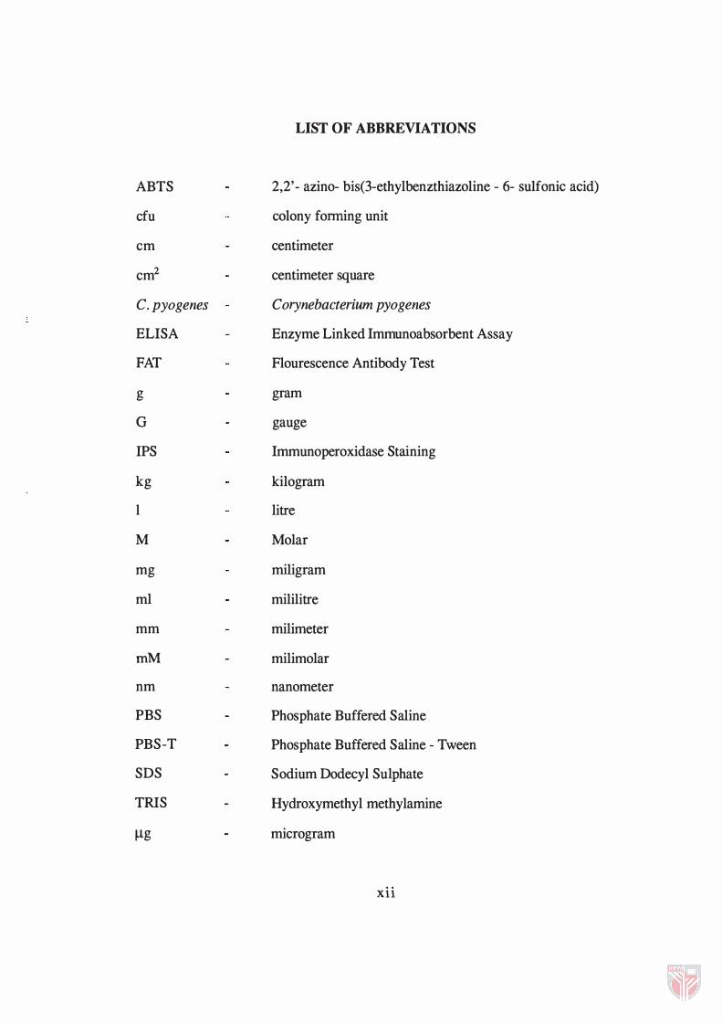
ABTS
cfu
cm
cm2
c. pyogenes
ELISA
FAT
g
G
IPS
kg
I
M
mg
ml
mm
mM
nm
PBS
PBS-T
SDS
TRIS
Ilg
LIST OF ABBREVIATIONS
2,2' - azino- bis(3-ethylbenzthiazoline - 6- sulfonic acid)
colony fonning unit
centimeter
centimeter square
Corynebacterium pyogenes
Enzyme Linked Immunoabsorbent Assay
Flourescence Antibody Test
gram
gauge
Immunoperoxidase Staining
kilogram
litre
Molar
miligram
mililitre
milimeter
milimolar
nanometer
Phosphate Buffered Saline
Phosphate Buffered Saline - Tween
Sodium Dodecyl Sulphate
Hydroxymethyl methylamine
microgram
xu

�l
%
>
<
microlitre
per cent
more than
less than
Xlll
Abstract of thesis submitted to the Senate of Universiti Pertanian Malaysia in fulfilment of the requirements for the degree of Master of Science.
STUDIES ON THE PATHOGENESIS OF CONTAGIOUS ECTHYMA IN GOATS AND SHEEP
Chairman
Faculty
By
ROSHIDAH BINTI ISMAIL
April 1994
Associate Professor Dr Mohd Zamri Saad, D.V.M.; Ph. D
Veterinary Medicine and Animal Science
Local contagious ecthyma viruses isolated from sheep (Lb) and goats (GV 1
and GV 2) were used in a series of infection trials in kids and lambs to compare the
pathogenesis, virus virulence and host susceptibility.
Caprine contagious ecthyma isolate (GV 1) was able to establish infection in
kids and lambs, producing typical lesions. When rechallenged, the infected kids and
lambs developed rapid but milder lesions followed by rapid resolution. The clinical
and histopathological changes in the primary and secondary infections were similar
but were more severe in the former. The third infection, however, failed to establish
any lesions.
Secondary bacterial infection appeared to have complicated the contagious
ecthyma lesions resulting in a marked reduction in body weight and occasional
death. The inoculation of Corynebacterium pyogenes into the contagious ecthyma
lesions resulted in much more severe lesions resembling those observed in field cases
XIV
of complicated contagious ecthyma. This finding suggests that in natural cases,
contagious ecthyma virus probably acts synergistically with other agents to produce
severe and generalised lesions.
Although the kids and lambs appeared to have similar disease, the kids
developed more severe lesions. Severity of the lesions produced were compared
statistically between the viral isolates and between the animal species. The caprine
GV 2 isolate was found to produce lesions in both kids and lambs with similar
severity whereas the ovine isolate (Lb) produced milder lesions in lambs but severe
lesions in kids. These observations correlated well with the development of antibody
response. In general, the lambs showed better antibody response than the kids,
reaching significantly high level on day twenty-two, coinciding with recovery from
the disease. The kids infected with Lb virus responded poorly leading to the severe
disease and longer recovery period.
xv
bakteria Corynebacterium pyogenes ke dalam lesi ektima menular menghasilkan lesi
teruk yang menyerupai lesi semulajadi ektima menular berkomplikasi. Kajian ini
mencadangkan bahawa dalam keadaan semulajadi, virus ektima menular mungkin
bekerjasama dengan agen-agen lain dalam menghasilkan lesi penyakit ektima
menular yang lebih teruk.
Walaupun kambing dan bebiri berupaya menghasilkan corak penyakit yang
sama setelah diinokulat dengan virus penyakit ektima menular, kajian menunjukkan
bahawa kambing adalah spesis yang akan menghasilkan lesi yang lebih teruk
dibandingkan dengan bebiri. Isolat kambing GV 2 menghasilkan lesi yang sama
darj ah keterukannya ke atas kambing dan bebiri manakala isolat bebiri Lb
menghasilkan lesi yang kurang teruk ke atas bebiri berbanding dengan lesi pada
kambing. Ini berkaitan rapat dengan corak perkembangan antibodi. Secara umum,
perkembangan antibodi adalah lebih baik dalam bebiri daripada kambing, dan boleh
mencapai tahap tinggi dalam masa dua puluh dua hari, iaitu bersamaan dengan mas a
sembuh. Kambing yang dijangkiti isolat bebiri Lb amat kurang menunjukkan
gerakbalas antibodi sehingga menghasilkan lesi yang teruk dan tempoh sembuh yang
lama.
XVll
CHAPTER!
INTRODUCTION
In Malaysia, contagious ecthyma is known to be a common disease of sheep
and goats (Peters et al., 1979). The disease is caused by a parapoxvirus, and is
believed to be more severe in goats than in sheep. Goats of all ages are known to be
susceptible to the infection although many adult animals are less likely to be infected
due to immunity as a result of an earlier infection (Reid, 1991).
The disease is characterised by the formation of vesicular and scabby lesions
on the lips which usually develop following minor damage to the skin caused by dry
and prickly pasture (McKeever et al., 1988). Following the infection, progressive
epidermal lesions develop, beginning from the formation of macule, papule, vesicle,
pustule to scab formation.
The incidence of the disease in different flocks is extremely variable; it may
attack all members of a flock irrespective of age, it may affect most or all of the
youngs, or it may affect only a few animals in the flock.
Infection with a highly contagious parapoxvirus results in considerable
losses. In 1976, the disease was rated as a top health priority problem by the
United State Sheep Industry Development Programme (Morison, 1976). It is an
important disease of economic significance to the animal industry; in severe and
moderately severe cases it causes marked loss of body condition particularly in the
1
2
feedlots due to the difficulty or inability of the affected animals to suckle or prehend
food due to the deformation of the lips. If the lesions become secondarily infected
with screw-worms, bacteria or parasites, it may contribute to the death of of the
affected animals as high as 50 % (Robinson and Balassu, 1981).
Outbreaks of contagious ecthyma have been reported in Peninsular Malaysia
especially in the states of Perak, Kedah, Pahang and Johor. The first reported
outbreak was in 1935, and since then several cases were reported to occured until
1960 (Asiah, 1990). No further studies on contagious ecthyma in Malaysia were
carried out following these reported outbreaks until recently when a study was
conducted by Zamri-Saad et aI., (1989). The lack of detailed studies on contagious
ecthyma in Malaysia could be due to the benign nature of the disease and the
familiarity with the infection. These lead to those involved with sheep and goat
rearing opting not to seek professional advice while those involved with research
unaware of the importance of this disease (peters et al., 1979).
Studies on the disease have been carried out in sheep and goats overseas,
mostly using the viruses isolated from the same animal species . Reports of
experimental cross-infection such as infection in sheep using isolates from goats are
less common although a considerable heterogeneity based on the restriction
endonuclease analysis was observed between the different isolates of contagious
ecthyma virus (Robinson et al., 1982; Rafii and Burder, 1985).
The aims of these studies are:
1 ) to determine the pathogenesis of complicated and uncomplicated
contagious ecthyma in sheep and goats.
2) to determine the differences in virulence between caprine and ovine
isolates on contagious ecthyma virus and the differences in susceptibility to
the infection between sheep and goat hosts.
CHAPTER 2
LITERATURE REVIEW
Livestock is an important and integral component of the agricultural sector.
The Malaysian livestock industry can be classified into the ruminant and the non
ruminant sub-sectors. Cattle, buffalo, goat and sheep constitute the ruminant sub
sectors. In Malaysia, most goats and sheep belong to smallholders who keep the
animals in small flocks (Peters et al., 1979).
There are about 288,5 16 and 234,901 heads of goats and sheep in Malaysia
respectively (Zamri-Saad et aI., 1990). The goat population is made up of mostly the
indigenous kambing kacang, several exotic purebreds and the resulting crossbreds.
The sheep population is also made up of the indigenous breed Malin (from the words
Malaysian indigenous), exotic purebreds and the resulting crossbreds. The overall
development of the sheep and goat industry in this country is not very encouraging
although the sheep industry has been targeted to be one of the most prominent
ruminant subsector in Malaysia in the next century (Babjee, 1988).
One of the constraints for rapid development of ruminant industry in Malaysia is
their small population that is widely distributed in small groups (Babjee, 1988) .
Furthermore, the rearing system which minimises planned breeding and the fact that
rearing ruminants is only a subsidiary activity of most smallholder farmers
contributed significantly to the slow growth of this sub-sector (Peters et aI. , 1979;
Babjee, 1988). Thus, two aproaches have been considered to improve the industry.
3

4
The first approach is to select suitable local animals to be crossbred with improved
temperate breeds. The second approach is to import productive temperate or 'tailor
made' breed deemed adaptable to the Malaysian environment (Babjee, 1988). Both
approaches have been carried out on sheep to improve the industry and with this,
many more diseases such as contagious ecthyma will be encountered.
Contagious ecthyma has been reported as one of the most common diseases
of sheep and goats in Malaysia (peters et aI., 1979; Babjee, 1980) although currently
there are very few published data on the incidence of the disease. A review of the
literature shows that the disease occurs in many parts of the world including
Malaysia and there were several reports describing this condition (Zamri-Saad et al. ,
1989; Martin and Aitken, 1991).
History of Contagious Ecthyma
Contagious ecthyma was first described by Walley (1890) who referred the
disease as orf. Later, Glover (1928) reported an extensive investigation of the disease
in England using the name contagious pustular dermatitis. Howarth ( 1929) in
California and Schmidt and Hardy (1932) in Texas, described the disease as sore
mouth, which they consider identical with the condition reported earlier by Glover
( 1928). The term contagious ecthyma was first used by Moussu ( 1923) who
described the lesions on the lips. He refused the use of the term stomatitis because
the lesions were often confined to the skin of the lips without involving the
epithelium of the mouth. At the same time in France, Aynaud (1923) described the
same condition as contagious pustular stomatitis.
In Western Texas, the disease was observed to occur during the spring and
summer months as a mild form among short-yearling lambs (Boughton and Hardy,
5
1934). The disease had earlier been reported in Greece (Blanc et al., 1922), Colorado
(Newsom and Cross, 1934) and South Africa (Theiler, 1928).
In Malaysia, the disease was first reported in 1935 in a herd of Government
goats kept at Raub, Pahang (Babjee, 1980). The term ecthyma contagiousm was
used to described the disease (Asiah, 1990). The disease was again reported in 1938
and since then the disease was noted to be of common occurrence in goats and was
encountered in practically every state.
Contagious Ecthyma in Malaysia
In 1 937, detailed observations of contagious ecthyma were made by the
Veterinary Officer of Perak, Mr. W. Orr. He reported a high incidence of the disease
in northern part of Perak. A severe case of ecthyma contagiousm occurred in a
young experimental goats from which the description of the disease in Malaysia from
its commencement to a fatal termination was made (Asiah, 1990).
Further detail observations were made in 1 938, where experiments were
undertaken to confirm the pathological identity of contagious ecthyma, to ascertain
the susceptibility of local goats to reinfection, to determine the susceptibility of
sheep to artificial infection and the possibility of transmitting infection artificially
through a series of goats.
In a recent study in Malaysia, Zamri-Saad et aI. , ( 1 989) monitored 1 5
smallholder goat farms for naturally occuring caprine contagious ecthyma in the
local kacang goats. A total of 260 goats were confirmed to suffer from contagious
ecthyma and did not succumb again to the disease within a period of one year
following the infection. Another study described the oral lesions of contagious

6
ecthyma following several outbreaks in farms which had a persistent problem of
contagious ecthyma (Zamri-Saad et al., 1992).
Aetiological Agent
Contagious ecthyma virus is a member of the genus parapoxvirus in the
family of Poxviridae. According to Mathews ( 1 982) , the genus consists of
contagious ecthyma virus, bovine papular stomatitis virus and pseudocowpox virus
with which it shares at least one common antigen (Papadopoulos et al., 1968).
The virus of contagious ecthyma resembles the viruses of sheep pox and goat
pox (Bennet et aI. , 1944, Sharma and Bhatia, 1 959). The virus particle is smaller
than other pox viruses and appeared as a monomorphic short rod with rounded ends.
The most striking morphological feature is a crisscross pattern produced by 8- 10 mil
wide tubular threads (Nagington et al., 1964).
The virus is relatively thermostable and resistant to dessication. It is
completely inactivated at 60°C for thirty minutes but retains some infectivity when
held at 55°C for thirty minutes (Sawhney, 1972; Buxton and Fraser, 1977).
Mazur and Machado (1989), claimed that the virus had been poorly studied
throughout the world and noted that the virus was low in pathogenicity in herds
which were kept extensively but extremely pathogenic in closed herds.
Age Susceptibility
Sheep and goats of all ages were susceptible. However the incidence is
usually high in young animals particularly within a few weeks of birth. In a study by
Zamri-Saad et ai. ( 1989), out of 260 goats examined, 54 % were kids of less than
three months old and the youngest affected kids were twenty-two days old.

7
Experiments by Boughton and Hardy (1934) demonstrated that aged sheep
and goats remained susceptible if they had not undergone a previous attack of the
disease during their early life. If the animals has been exposed to the infection, they
will develop an immunity that protects them from the reinfection for a period of
about as long as eight months. Moreover older animals seldom show the severe type
of infection which is the typical form encountered in lambs and kids.
Species Susceptibility
It had been noted that local and crossbred sheep showed less severe lesions
compared to goats but imported sheep tended to show much more severe lesions
than local sheep (Zamri-Saad et aI. , 1989). Though the degree of severity was
comparable between goats and sheep, the disease was identical within each species
(Blanc et at, 1922).
The disease has been observed to be able to transmit to cattle, dog and man
(Leavell et al. , 1968; Erickson et at, 1975; Kim and Tarrier, 1977; Wilkinson, 1977;
Guss, 1980; Robinson and Balassu, 1981 ; PhiliP, 1983). In man, the lesions usually
develop after accidental inoculation from infedted animals or contaminated objects.
Direct human to human infection has not been reported (Wilkinson, 1 977) .
Generally, the infections in man are benign and appear to be clinically identical with
those of contagious ecthyma of sheep and goats.
No characteristic lesions were observed in experimentally inoculated rabbits,
mice, chickens and chick embryos (Sinha et aI., 1986). In a similar attempt, Aynaud
(1923), Greing (1956) and Plowright et aI. (1959) also failed to cultivate the virus in
such animals and birds. However, Abdussalam (1957) succeeded in the cultivation
of contagious ecthyma virus in rabbits but failed to grow the rabbit-adapted virus in