Embed Size (px)
Citation preview
UNIVERSITI PUTRA MALAYSIA
EFFECTS OF ZERUMBONE FROM ZINGIBER ZERUMBET ON CERVICALCANCER-INDUCED FEMALE BALB/C MICE
NIRMALA DEVI TAILAN
FPSK(M) 2007 9
EFFECTS OF ZERUMBONE FROM ZZNGIBER ZERUMBET ON CERVICAL CANCER-INDUCED FEMALE BALBIC MICE
BY
NIRMALA DEVI TAILAN
Thesis Submitted to the School of Graduate Studies, Universiti Putra Malaysia, in Fulfilment of the Requirements for the Degree of Master of Science
MARCH 2007

This thesis is especrbly dedicated to:
My loving grandmo ther & Aun ty, who are infinitely precious to me,
Kav/j.arasu and Vasanth, who have filled my life with joy and happiness,
&
My frieods, who were there for me!
Abstract of thesis presented to the Senate of Universiti Putra Malaysia in fulfilment of the requirement for the degree of Master of Science
EFFECTS OF ZERUMBONE FROM ZINGZBER ZERUMBET ON CERVICAL CANCER INDUCED FEMALE BALBIC MICE
BY
NIRMALA DEVI TAILAN
MARCH 2007
Chairman: Ahmad Bustamam Hj Abdul, PhD
Faculty : Medicine and Health Sciences
In the present study, the chemotherapeutic potential of zerumbone towards HeLa cell
line and cervical cancer induced in female Balblc mice were investigated. The
chemotherapeutic potential of zerumbone was compared with cisplatin, a commercial
drug used to treat cervical cancer. The cytotoxicity of both zerumbone and cisplatin
towards HeLa cell line were determined using MTT assay. The findings showed that the
ICso value (* S.E.M) of zerumbone towards HeLa cell line was 11.3 * 0.2 pM, whilst
the ICSO value of cisplatin was 7.5 * 0.3 pM. Both IC50 values for zerumbone and
cisplatin fall within the very significant group based on the National Cancer Institute
Standard. All the values are significant (P < 0.01). The HeLa cells were treated with
ICso concentration of zerumbone and cisplatin respectively for morphological analysis
using inverted microscopy. The results showed significant growth retardation in HeLa
iii
cells exposed to zerumbone and cisplatin at 24,48 and 72 hours, whilst the control cells
are well spread and confluent. Pregnant female Balblc mice were exposed to
Diethylstilbestrol (DES) at 13 '~ to 1 8 ' ~ day of gestation. The progeny of the DES-
exposed mothers developed cervical intra-epithelial neoplasia. These progenies were
divided into 4 groups and were either given treatment with normal saline, 8 mglkg
zerumbone, 16 mg/kg zerumbone and 10 mg/kg cisplatin. The mice were sacrificed
following the treatments and their cervical tissues subjected to histological examination,
TUNEL Assay and immunohistochemistry. The histological examination revealed that
both zerumbone and cisplatin treatments were able to inhibit the progression of cervical
dysplasia fiom becoming more severe dysplasia (CIN 3). In the mice treated with
normal saline, the dysplasia had progressed to CIN 3 (severe dysplasia). The TUNEL
assay micrographs showed that there was no apoptosis in the cervical tissue of the
normal saline treated mice compared to the cervical tissue of mice treated with
zerumbone and cisplatin, where abundant apoptotic cells were noticed. The levels of
serum IL-6 were suppressed in mice treated with zerumbone and cisplatin. In contrast,
mice treated with normal saline showed elevated level of serum IL-6.
Immunohistochemistry study demonstrated that the production level of membrane
bound IL-6 receptor had been suppressed by the treatment of zerumbone and cisplatin
compared to mice treated with normal saline which had a higher concentration of
membrane bound IL-6 receptors. This showed that both zerumbone and cisplatin act in
a similar manner in vivo and in vitro. In conclusion, it is suggested that zerumbone, a
plant derived compound, could be explored as a new anti cancer agent for treating
cervical cancer in the future.
Abstrak tesis yang dikemukakan kepada Senat Universiti Putra Malaysia sebagai memenuhi keperluan untuk ijazah Master Sains
KESAN ZERUMBONE DARIPADA ZZNGZBER ZERUMBET KE ATAS TIKUS MENCIT YANG TELAH DIINDUKSI DENGAN KANSER SERVIKS
Oleh
NIRMALA DEVI TAILAN
MAC 2007
Pengerusi: Ahmad Bustamam Hj Abdul, PhD
Fakulti : Perubatan dan Sains Kesihatan
Dalam kajian ini, keupayaan kemoterapeutik zerwnbone terhadap sel selanjar kanser
serviks (HeLa) dan tikus mencit Balb/c telah dikaji. Keupayaan terapeutik zerurnbone
dibandingkan dengan cisplatin, dadah komersial bagi merawat kanser serviks. Kesan
sitotoksik zerurnbone dan cisplatin terhadap sel HeLa dikaji dengan menggunakan asai
MTT. Keputusan menunjukkan bahawa nilai ICso (A S.E.M) bagi zerumbone ialah 11.3
k 0.2 pM, manakala bagi cisplatin ialah 7.5 A 0.3 pM. Nilai ICSo bagi zerumbone dan
cisplatin adalah sangat signifikan berdasarkan garis panduan Institut Kanser Nasional.
Semua nilai ICso adalah signifikan (P < 0.01). Sel HeLa dirawat dengan zerumbone dan
cisplatin dengan nilai ICso masing-masing dan kajian morfologi dijalankan dengan
bantuan mikroskopi inverted. Keputusan menunjukkan pertumbuhan sel pada jam ke-
24, 48 dan 72 rawatan zerumbone dan cisplatin telah terbantut, manakala sel kawalan
tumbuh dengan sihat dan tersebar luas. Dalam kajian in vivo, tikus.betina yang hamil
diberi suntikan DES (Diethylstibestrol) pada hari gestasi 13 hingga 18. Progeni betina
bagi ibu-ibu yang terdedah kepada DES, telah menggalakan pertumbuhan CIN
(Cervical Intra-epithelial Neoplasia). Tikus- tikus betina yang telah terdedah kepada
DES dalam rahim ibu dibahagikan kepada 4 kumpulan dan diberi rawatan dengan
normal saline, 8 rnglkg zerumbone, 16 mglkg zerumbone dan 10 mgkg cisplatin.
Tikus-tikus tersebut dibunuh dan tisu serviksnya dianalisis melalui kaedah histologi,
asai TUNEL dan kaedah immunohistokimia. Kajian histologi menunjukkan zerumbone
dan cisplatin dapat menghalang pertumbuhan CIN 1 (displasia awal) kepada CIN 3
(displasia teruk) pada tisu serviks tikus. Manakala bagi tikus yang hanya diberi saline
normal, penyakitnya (displasia) berkembang dan mencapai CW 3. Hasil kajian TUNEL
asai menunjukkan tiada sel apoptosis diperhatikan dalam tisu serviks tikus yang dirawat
dengan normal saline, manakala tikus yang dirawat dengan zerumbone dan cisplatin
menunjukkan sel apoptosis yang banyak. Paras IL-6 darah bagi tikus yang dirawat
dengan zerumbone dan cisplatin adalah sangat rendah berbanding tikus yang dirawat
dengan normal saline dimana paras IL-6 adalah amat tinggi. Kajian immunohistokimia
menunjukkan penghasilan reseptor IL-6 menurun pada tikus yang dirawat dengan
zerumbone dan cisplatin, manakala tikus yang dirawat dengan normal saline
menunjukkan peningkatan dalam penghasilan reseptor IL-6 secara mendadak. Kedua-
dua zerumbone dan cisplatin bertindak pada kadar yang sama dalam kajian in vitro dan
in vivo. Kesimpulannya zerumbone, sebatian semulajadi daripada tumbuhan
dicadangkan untuk digunakan bagi menhasilkan ubatan alternatif untuk merawat kanser
serviks pada masa hadapan.
ACKNOWLEDGEMENTS
I would like to take this opportunity to thank all those who gave great support to me
while doing the project. First of all, I would like to express my sincere gratitude and
special appreciation to my supewisor, Dr. Ahmad Bustamam Hj. Abdul and co-
supewisor, Associate Professor Dr. Muhd. Nazrul Hakim Abdullah for their endless
support, advice, guidance and encouragement throughout the completion of this thesis
as partial fuljZlment of the requirement for the degree of Master's Science
(Pharmacology and Toxicology)
I would also like to express my deepest appreciation and sincere gratitude to Prof:
Nordin Lajis for allowing me to use the phytochemical laboratory, Institute oj
Bioscience, Universiti Putra Malaysia for the extraction and isolation of the compound
used in this research. A special thanks is owed to Mr. Shahrin for his endless help and
guidance throughout the extraction and isolation of the compound. I would also like to
thank Mrs. Zurina for her priceless guidance and support in completing the HPLC and
LCMS analysis of the isolated compound.
vii
My heartfelt appreciation goes to Mrs. Siti Muskinah, who have helped and guided me
regardless of time, throughout the completion of animal model studies. I would also like
to acknowledge Miss Mohanambal, Miss Uma Nanthini and Mr. Zulfahmi for their
invaluable support and help while carrying out the animal model studies.
My sincere appreciation are extended to my colleagues, Miss Ajantha Sinniah and Miss
Zetty Nadia for their priceless and invaluable guidance, support, advice and help from
the beginning until the end of the research project. Not forgetting my other lab mates,
Mrs. Normah and Miss Ooi Suek Chin who gave me a hand in completing my lab work.
I am also grateful to Dr. Hairuszah Ithnin for allowing me to use the medical
laboratory (Histology Laboratory) for my research work. Special thanks are also
extended to Mrs. Juita, Mrs. Normah and Mr. Yip for their kind help and guidance
during my histological and immunohistochemistry staining.
I would also like to extend my sincere appreciation to Associate Professor Dr. Fauziah
Othman for giving me permission to use the Laser Scanning Confocal Microscopy in
Micro electron and Micro Analysis Unit , Institute of Bioscience, Universiti Putra
Malaysia. A special appreciation was extended to Mr. RaJiq and Miss Huey Fern who
had helped me in TUNEL Assay staining.
. . . Vll l
My heartfelt gratitude goes to my jianck Mr. Kaviyarasu Yellappan, for his endless
guidance, support, advice and help throughout the completion of this research project.
I would also like to express my deepest and warmest appreciation to my family members
especially my grandmother, Madam Vally Palany, my aunty, Miss Visalachee Suppiah
and my cousin Vasanth Palanivelu for their patience, concern and kindness in helping
me in every part of this thesis.
Above all, I would like to extend my utmost and deepest gratitude to GOD for blessing
me with patience and persistence to complete this research.
I certify that an Examination Committee has met on 26' March 2007 to conduct the final examination of Nirmala Devi dlo Tailan on her Master of Science thesis entitled "Effects of Zerumbone from Zingiber zertrmhct on Cervical Cancer-Induced Female BalbIC Mice" in accordance with Universiti Pertanian Malaysia (Higher Degree) Act 1980 and Universiti Pertanian Malaysia (Higher Degree) Regulations 198 1. The Committee recommends that the candidate be awarded the relevant degree. Members of the Examination Committee are as follows:
Elizabeth George, PhD Professor Faculty of Medicine and Health Sciences Universiti Putra Malaysia (Chairman)
Mohd Zamri Saad, PhD Professor Faculty of Veterinary Universiti Putra Malaysia (Internal Examiner)
Sabrina Sukardi, PhD Associate Professor Faculty of Medicine and Health Sciences Universiti Putra Malaysia (Internal Examiner)
Mustafa Ali Mohd, PhD Professor Faculty of Medicine Universiti Malaya (External Examiner)
School of Graduate Studies Universiti Putra Malaysia
Date: 2 1 JUNE 2007
This thesis submitted to the Senate of Universiti Putra Malaysia and has been accepted as fulfillment of the requirement for the degree of Master of Science. The members of the Supervisory Committee are as follows:
Ahmad Bustamam Hj Abdul, PhD Lecturer Institute of Bioscience Universiti Putra Malaysia (Chairman)
Muhammad Nazrul Hakim Abdullah, D.V.M. PhD Associate Professor Faculty of Medicine and Health Sciences Universiti Putra Malaysia (Member)
AINI IDERIS, PhD ProfessorIDean School of Graduate Studies Universiti Putra Malaysia
Date: 17 JULY 2007

DECLARATION
I hereby declare that the thesis is based on my original work except for the quotations and citations which have been duly acknowledged. I also declare that it has not been previously or concurrently submitted for any other degree at UPM or other institutions.
NIRMALA DEVI A/P TAILAN
Date: 05 .TUN 2007
xii
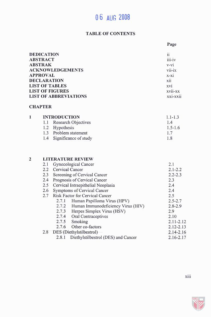
DEDICATION ABSTRACT ABSTRAK ACKNOWLEDGEMENTS APPROVAL DECLARATION LIST OF TABLES LIST OF FIGURES LIST OF ABBREVIATIONS
TABLE OF CONTENTS
Page
CHAPTER
INTRODUCTION 1.1 Research Objectives 1.2 Hypothesis 1.3 Problem statement 1.4 Significance of study
LITERATURE REVIEW 2.1 Gynecological Cancer 2.2 Cervical Cancer 2.3 Screening of Cervical Cancer 2.4 Prognosis of Cervical Cancer 2.5 Cervical Intraepithelial Neoplasia 2.6 Symptoms of Cervical Cancer 2.7 Risk Factor for Cervical Cancer
2.7.1 Human Papilloma Virus (HPV) 2.7.2 Human Immunodeficiency Virus (HIV) 2.7.3 Herpes Simplex Virus (HSV) 2.7.4 Oral Contraceptives 2.7.5 Smoking 2.7.6 Other co-factors
2.8 DES (Diethyistilbtstml) 2.8.1 Diethylstilbestrol (DES) and Cancer
v-vi vii-ix x-xi xii xvi xvii-xx xxi-xxii
xiii
2.9 Natural Products 2.10 Zingiber Zerumbet from Zingiberaceae
2.10.1 Zerumbone from Zingiber Zerumbet 2.1 1 Cisplatin Overview
2.1 1.1 Basic Information 2.1 1.2 Toxicity 2.1 1.3 Modes of Action of Cisplatin
2.12 Apoptosis 2.12.1 Induction of Apoptosis
2.13 Overview of Interleukin-6 2.13.1 Interleukin 6 and Cervical Cancer
2.14 Overview of Interleukin-6 Receptor 2.14.1 Trans signaling via IL-61sIL-6R
METHODOLOGY 3.1 Materials and Equipments
3.1.1 Extraction of zerumbone Assay 3.1.2 Cell culture and MTT Cytotoxicity 3.1.3 High Performance Liquid Chromatography 3.1.4 Liquid Chromatography Mass Spectrometry 3.1.5 Induction of DES in female balblc mice 3.1.6 Hematoxylin and Eosin staining 3.1.7 TUNEL Assay 3.1.8 ELISA IL-6 3.1.9 Immunohistochemistry staining
3.2 Extraction and Isolation of zerumbone from zingiber zerumbet 3.2.1 Crude Extraction 3.6-3.7 3.2.2 Separation of non-polar compound from
polar compound 3.8-3.9 3.2.3 Fractionation of non-polar compound 3.10 3.2.4 Recrystallization and purification of zerumbone 3.11-3.12 3.2.5 High Performance Liquid Chromatography
(HPLC) of zerumbone. 3.2.6 Liquid Chromatography Mass Spectrometry
(LCMS) of zerumbone. 3.3 Cell culture and maintenance
3.3.1 Cryopreservation of cell line 3.3.2 Thawing cryopreserved cell line 3.3.3 Cytotoxicity Assay 3.3.4 Morphological studies
xiv
3.4 Induction of cervical dysplasia in female balblc mice 3.18 3.5 Treatment of offspring exposed to DES in utero 3.19-3.20 3.6 Hematoxylin and Eosin staining 3.2 1-3.23 3.7 Detection of apoptosis using TUNEL Assay system 3.24-3.27 3.8 Serum IL-6 detection using anti-mouse IL-6 ELISA kit 3.28-3.29 3.9 ImmunoHistoChemistry (IHC) staining to detect
IL-6 receptor 3.30-3.3 1
RESULTS 4.1 High Performance Liquid Chromatography and Liquid
Chromatography Mass Spectrometry analysis of zerumbone.
4.2 Effects of zerumbone on cell viability using MTT cytotoxicity Assay.
4.3 Effects of zerumbone of cell morphology using inverted microscopy. 4.8-4.1 1
4.4 Effects of zerumbone on progression of dysplasia using histological procedure. 4.12-4.17
4.5 Effects of zerumbone on apoptosis using TUNEL Assay. 4.1 8-4.25 4.6 Effects of zerumbone on secretion level of IL-6 using
IL-6 ELISA Assay. 4.26-4.28 4.7 Effects of zerumbone on membrane and cytoplasmic
bound IL-6 receptors using immunohistochemistry. 4.29-4.34
DISCUSSION
CONCLUSION AND RECOMMENDATIONS 6.1 Conclusion 6.2 Future work and Recommendation
REFERENCES APPENDICES BIODATA OF THE AUTHOR
LIST OF TABLES
Table
Treatment groups.
Hematoxylin and eosin staining method.
Grading of Cervical Intra-epithelial Neoplasia (abnormalities of cervix).
Preparation of rTdT Incubation Buffer for experimental reactions.
4.6.1 The table shows various concentrations of serum IL-6 with Standard Error Mean (* S.E.M), according to treatment groups in mice.
Treatment concentration of zerumbone and cisplatin towards HeLa cells.
Page
xvi
LIST OF FIGURES
Figure Page
Zingiber zerumbet plant (A) Whole plant and (B) Rhizome
Molecular structure of zerumbone
Molecular structure of cisplatin
Mode of action of cisplatin
Apoptotic cell death in the nematode worm C. elegans
A caspase cascade.
Extraction of crude extract from zingiber zerumbet
Separation of non-polar compounds from crude extract.
Fractionation of non-polar compounds and recrystallization and purification of zerumbone
4.1.1 HPLC analysis graph of the isolated compound showed only one single peak at retention time of 16 minutes.
4.1.2 The LCMS analysis graph of zerumbone.
4.13 The LCMS analysis graph of zerumbone showed only single high fragment.
4.2.1 The effects of zerumbone to inhibit growth proliferation of human cancer cells, HeLa after 72-hours post-treatment of the compound at concentrations, 5pM to 35pM.
4.2.2 The effects of cisplatin to inhibit growth proliferation of human cancer cells, HeLa after 72-hours of post-treatment with the compound at concentrations, 2.5pM - 100pM.
4.3.1 Micrographs showing morphological changes towards human cancer cells, HeLa, as viewed under light contrasting inverted microscope after 24-hours post-treatment with compounds, zerumbone and cisplatin.
xvii
Micrographs showing morphological changes towards human cancer cells, HeLa, as viewed under light contrasting inverted microscope after 48-hours post-treatment with compounds, zerumbone and cisplatin.
Micrographs showing morphological changes towards human cancer cells, HeLa, as viewed under light contrasting inverted microscope after 72-hours post-treatment with compounds, zerumbone and cisplatin.
Histological micrograph showing normal cervical epithelial cells with low nuclear: cytoplasmic ratio, without noticeable angiogenesis.
Histological micrograph showing normal cervical epithelial cells with low nuclear: cytoplasmic ratio, with no apparent angiogenesis.
Histological micrograph showing treatment of induced cervical cancer in female Balblc mice with IOmglkg cisplatin.
Histological micrograph showing treatment of induced cervical cancer in female Balbh c mice with 10mgkg cisplatin.
Histological micrograph showing treatment of induced cervical cancer in female Balblc mice with 16mgkg zerumbone.
Histological micrograph showing treatment of induced cervical cancer in female Balblc mice with 16mglkg zerumbone.
4.4.7 Histological micrograph showing treatment of induced cervical cancer in female Balblc mice with 8mgkg zerumbone.
4.4.8 Histological micrograph showing treatment of induced cervical cancer in femaleBbalbh c mice with 8mgkg zerumbone.
4.4.9 Histological micrograph showing treatment of induced cervical cancer in female Balblc mice with normal saline.
xviii
4.4.10 Histological micrograph showing treatment of induced cervical cancer in female Balblc mice with normal saline.
4.5.1 TUNEL micrograph showing cervix tissue of normal female mice.
4.5.2 TUNEL micrograph of cervical tissue of normal female mice exhibiting less apoptotic cells.
4.5.3 TUNEL micrograph of cervical tissue of female mice induced with cervix cancer, using normal saline for treatment.
4.5.4 TUNEL micrograph of cervical tissue of female mice induced cervix cancer, using normal saline for treatment.
4.5.5 TUNEL micrograph of the cervix tissue of female mice induced cervical cancer with follow-up treatment of zerumbone at 8 mgkg dosage.
4.5.6 TUNEL micrograph of the cervical tissue of female mice induced cervical cancer with follow-up treatment of zerumbone at 8 mgkg dosage.
4.5.7 TUNEL micrograph of the cervical tissue of female mice induced cervical cancer with follow-up treatment of zerumbone at 16 mgkg dosage.
4.5.8 TUNEL micrograph of the cervical tissue of female mice induced cervical cancer with follow-up treatment of zerumbone at 16 mgkg dosage.
4.5.9 TUNEL micrograph of cervical tissue of female mice induced cervical cancer with follow-up treatment of cisplatin at 10 mglkg dosage.
4.5.10 TUNEL micrograph of cervical tissue of female mice induced cervical cancer with follow-up treatment of cisplatin at 10 mglkg dosage.
4.5.11 The mean percentage of apoptotic cells in cervical tissue sectioning of female balb/c mice induced with cervical cancer after treatments with zerumbone, cisplatin and normal saline.
xix
4.6.1 A standard curve of absorbance versus known concentrations of IL-6. A linear graph shows that the absorbance increases proportionately as concentration of IL-6 increases.
4.7.1 Imrnunohistochemistry micrograph of cervical tissue of normal female mice.
4.7.2 Imrnunohistochemistry micrograph of cervical cancer tissue of female balblc mice treated with cisplatin at 10 mglkg dosage.
4.7.3 Imrnunohistochemistry micrograph of cervical cancer tissue of female balblc mice treated with cisplatin at 10 mgkg dosage.
4.7.4 Imrnunohistochemistry micrograph of cervical cancer tissue of female balblc mice treated with zerumbone at 10 mgkg dosage.
4.7.5 Irnmunohistochemistry micrograph of cervical cancer tissue of female balblc mice treated with zerumbone at 16 mglkg dosage.
4.7.6 Immunohistochemistry micrograph of cervical cancer tissue of female balblc mice treated with zerumbone at 8 mgkg dosage.
4.7.7 Imrnunohistochemistry micrograph of cervical cancer tissue of female balblc mice treated with zerumbone at 8 mglkg dosage.
4.7.8 Imrnunohistochemistry micrograph of cervical cancer tissue of female balblc mice treated with normal saline.
4.7.9 Imrnunohistochemistry micrograph of cervical cancer tissue of female balblc mice treated with normal saline.
A. 1. Serial dilutions for standard preparation for IL-6 ELISA.
LIST OF ABBREVIATIONS
AIDS
ATCC
CIN I
CIN I1
CIN I11
CIN
CIS
CNTF
COX-2
DDP
DES
DESAD
DESAD
DMSO
DNA
ELISA
FCS
FDA
FIG0
HIV
HPLC
Acquired Immuno Deficiency Syndrome
American Type Culture Collection
Mild dysplasia
Moderate dysplasia
Severe dysplasia
Cervical Intra-epithelial Neoplasia
Carcinoma in situ
Ciliary Neurotrophic Factor
Cyclooxygenase-2
Diamminedichloroplatinum (cisplatinum)
Diethylstilbestrol
National Cooperative Diethylstilbestrol Adenosis
National Cooperative Diethylstilbestrol Adenosis
Dimethylsulphoxide
Deoxyribonecleic Acid
Enzyme Link Immuno Sorbent Assay
Feotal Calf Serum
Food and Drug Administration
Federation of Gynecology and Obstetrics
Human Immunodeficiency Virus
High Performance Liquid Chromatography
xxi
HPV
HSV-2
HTC
IL-6
IL-6R
LCB
LCMS
LIF
MT
MTT
NCI
OC
OSM
PBS
RNA
TdT
TNF-a
TPA
TUNEL
uv
VEGF
Human Papilloma Virus
Herpes Simplex Virus type-2
Hepatome Tissue Culture
Interleukin-6
Interleukin-6 Receptor
Liquid Base Cytology
Liquid Chromatography Mass Spectrometry
Leukemia Inhibitory Factor
Metallothionein
Micro Tetrazolium
National Cancer Institute
Oral Contraceptives
Oncostatin M
Phosphate Buffer Saline
Ribonucleic Acid
Terminal Deoxynucleotidyl Transferase
Tumour Necrosis Factor-Alpha
Tetradecanoylphorbol- 13-Acetate
TdT-mediated dUTP Nick-End-Labeling
Ultra Violet
Vascular endothelial Growth Factor
xxii
CHAPTER 1
INTRODUCTION
Cervical cancer remains the second common cancer among women worldwide. It is,
however, the most common cancer in women in developing countries accounting for
80% of all cases. Majority of tumors in these women are diagnosed at advanced stages
with a resulting high mortality (Rowlands and de Barros Lopez, 2001). Human
papilloma virus (HPV) has been implicated as the primary causative agent for cervical
squamous cancer, whereby 99.7% of all cervical cancers had been identified to have
HPV DNA (Rowlands and de Barros Lopez, 2001).
In 1943, Papanicolaou and Traut published their experience on the role of cervical
cytology in identifLing cancer of the cervix. The technique known as Pap smear, was
rapidly incorporated as a screening test for invasive and pre-invasive lesions (Rowlands
and de Barros Lopez, 2001). Despite the success of the Papanicolou Smear (Pap smear),
cervical carcinoma still causes high mortality but is treatable and curable if diagnosed at
early stage. Moreover, the malignant transformation of cervical epithelial cells has been
reported to have a long latency period of up to 10 years (Ho et al., 1998).
To date, the most effective single chemotherapeutic agent for cervical carcinoma is
cisplatin, which normally achieves a response rate of between 23% and 50% (Ozols,
2003). Even though chemotherapy has been successful for treatment of some tumors
such as testicular cancer and certain leukemia, its success rate for treatment of common
epithelial tumors such as breast, colon, cervical, ovarian and lung has been less
impressive (Johnstone et al., 2002). A chemotherapeutic drug used as an anti-cancer
drug destroys the cancerous cells by interfering with the ability of the cancer cells to
divide and reproduce. A drawback to this chemotherapeutic drug, however, is that these
drugs also affect normal cells.
Apoptosis is also known as programmed cell death and is a mechanism for eliminating
unwanted cells or damaged cells. The cells initiate a suicide sequence which results in a
quick and systematic approach in triggering cell death. It is widely believed that tumor
cells treated with anticancer agents, including radiation, will die by apoptosis and the
tumors that do not readily undergo apoptosis are resilient towards treatment (Lowe and
Lin, 2000). Furthermore, response to tumor treatment that employs plant derived-
bioactive substances in cancer patients have been implicated by apoptosis induction in
tumor cells (Smets, 1994; Paschka et al., 1998).
Most of the earliest pharmaceuticals were plant-derived compounds whereby plants
have been used initially to treat diseases (Peter et al., 1998). The plant-derived drugs
that are useful in clinical oncology include those of flavonoids, coumarins, cinnamates
or phenolics.