Embed Size (px)
Citation preview


Université de Montréal
CEP78, a Novel Centrosomal Protein
by
Yalda Javadi Esfehani
Department of Pathology and Cell Biology
Faculty of Medicine
Memoire presented to Faculty of Medicine for the Degree of
Master of Science (M.Sc)
March, 2014
© Yalda Javadi Esfehani, 2014

Université de Montréal
Ce mémoire intitulé:
CEP78, a Novel Centrosomal Protein
Présenté par:
Yalda Javadi Esfehani
Département de pathologie et biologie cellulaire, Faculté de
médecine
Mémoire présenté à la Faculté de médecine en vue de
l’o te tio du grade de aîtrise e pathologie et biologie
cellulaire
Mars, 2014
© Yalda Javadi Esfehani, 2014

iv
Abstract
Background: The centrosome is a tiny organelle well-known for its
role in establishing the bipolar spindle during cell division. Defects
in centrosome function often give rise to human diseases
including cancer and kidney cyst formation. We are interested in
studying the function of one novel centrosomal protein named
CEP78, identified in a proteomic screen for novel centrosomal
components. Methods and results: Treatment of cells with
nocodazole, a microtubule-depolymerizing agent that specifically
depolymerizes cytoplasmic microtubules but not the stabilized
centrosome microtubules, showed that CEP78 is a stable
centrosomal component. Colocalization of this protein with other
centrosomal markers such as CEP164, SAS6, Centrin,
Polyglutamylated tubulin and POC5 at different phases of the cell
cycle indicated that CEP78 specifically localizes to the distal end of
the mother and daughter centrioles. There are 2 CEP78 dots
during the interphase and as the cells go through mitosis,
procentrioles mature, and the number of CEP78 dots increases to
4 dots per cell and by the end of telophase each daughter cell has
2 CEP78 dots. Characterization of CEP78 functional domains
showed that Leucine-rich repeats are necessary for centrosomal
localization of the protein. In addition, we found that
overexpression of CEP78 did not change the number of centrioles
and centrosomes but decreased the number and intensity of
CEP170 dots (sub-distal appendage protein) without a decrease in
the expression level of this protein. Further studies showed that
there is no interaction between these 2 proteins. Finally,

v
overexpression of CEP78 protects microtubules from
depolymerization in the presence of nocodazole, suggesting its
ability to bind microtubules. Conclusion: Our findings suggest that
CEP78 is targeted to the distal end of mature centrioles via its
lecuine-rich repeats, where it could be involved in centriolar
maturation or regulation of sub-distal appendage assembly
and/or remodeling, a structure known to nucleate and anchor
microtubules. Understanding the function of CEP78 will shed light
on the role of the centrosome in cell cycle.
Key words: CEP78, Centrosome, CEP170, Microtubules

vi
Resumé
Contexte: Le centrosome est un petit organite bien connu pour
son rôle dans l'établissement du fuseau bipolaire pendant la
division cellulaire. Les déficiences de la fonction du centrosome
donnent souvent lieu à des maladies humaines, y compris le
cancer et la formation de kystes rénaux. Nous sommes intéressés
à étudier la fonction d'une nouvelle protéine centrosomale
nommée CEP78, identifiée dans un criblage protéomique pour de
nouveaux composants centrosomaux. Méthodes et résultats : Le
traitement des cellules avec le nocodazole, un agent qui
dépolymérise spécifiquement les microtubules cytoplasmiques
mais pas les microtubules stabilisés du centrosome, a montré que
CEP78 est un composant centrosomal stable. La colocalisation de
cette protéine avec d'autres marqueurs centrosomaux tels que
CEP164, SAS6, Centrine, tubuline polyglutamylée et POC5, à
différentes phases du cycle cellulaire a indiqué que CEP78 est
précisément à l'extrémité distale des centrioles, mères et filles. Il
e iste deu poi ts CEP au ours de l’i terphase et les ellules passent par la mitose, procentrioles maturent, et le nombre de
points de CEP78 augmente à 4 par cellule et, à la fin de la
télophase chaque cellule fille possède 2 points CEP78. La
caractérisation des domaines fonctionnels de CEP78 a montré que
des répétitions riches en leucine sont nécessaires pour la
localisation centrosomale de la protéine. En outre, nous avons
constaté que la surexpression de CEP78 ne change pas le nombre
de mères/procentrioles mais diminue le nombre et l'intensité des
points de CEP170 (protéine d'appendice sous-distal) sans

vii
diminution du niveau d'expression de cette protéine. D'autres
études ont montré qu'il n'y a pas d'interaction entre ces deux
protéines. Enfin, la surexpression de CEP78 protège des
microtubules contre la dépolymérisation en présence de
nocodazole, ce qui suggère qu'il possède la capacité de lier les
microtubules. Conclusion : Nos résultats suggèrent que CEP78 est
destiné à l'extrémité distale des centrioles matures par ses
répétitions riche en lecuine, où il pourrait être impliqué dans la
maturation ou la régulation de l'assemblage ou de la rénovation
de l'appendice sous-distal centriolaire, une structure connue dans
la nucléation des microtubules et d'ancrage. Comprendre la
fonction de Cep78 contribuera à éclaircir le rôle du centrosome
dans le cycle cellulaire.
Mots clés: CEP78, Centrosome, CEP170, Microtubules
viii
Table of Contents
Abstract ................................................................................... IV
Table of Contents ................................................................... VIII
List of Figures and Tables .......................................................... XI
List of Abbreviations .............................................................. XIII
Acknowledgments .................................................................. XV
Chapter 1 Introduction ............................................................. 16
1.1 Centrosome Structure ......................................................... 16
1.2 Centrosomal Proteins and their functions ........................... 20
1.3 Cell Cycle ............................................................................. 23
1.3.1 The Cycle ...................................................................... 23
1.3.2 Control of Cell Cycle ...................................................... 24
1.3.3 Centrosome and Cell Cycle ............................................ 25
1.4 Centrosome Cycle ................................................................ 27
1.5 Cilia ..................................................................................... 31
1.6 Cytoskeleton ....................................................................... 34
1.7 Diseases ............................................................................... 37
1.8 CEP78 .................................................................................. 41
Chapter 2 Material and Methods .............................................. 43
2.1 Material .............................................................................. 43
2.1.1 Chemicals ...................................................................... 43
2.1.2 Solutions, Buffers and Media ........................................ 43

ix
2.2 Methods .............................................................................. 46
2.2.1 Ba terial ethods ……………………………………………………… 46
2.2.1.1 Purification of GST- tagged CEP78 and GST proteins . 46
2.2.1.2 CEP78 Antibody Purification ...................................... 47
2.2.1.3 Transformation of Competent Cells .......................... 48
2.2.2 Cellular ethods ………………………………………………………. 49
2.2.2.1 Immunofluorescence Assay ...................................... 49
2.2.2.2 Western Blotting ....................................................... 50
2.2.2.3 Knock down with siRNA ............................................ 50
2.2.2.4 Plasmid Transfection with Transit Reagent ................51
2.2.2.5 Plasmid Transfection with CaCl2 ................................51
2.2.2.6 Immunoprecipitation .................................................52
2.2.2.7 Centrosomal Localization Study .................................52
2.2.2.8 Microtubule Assay .....................................................53
Chapter 3 Results ..................................................................... 54
3.1 CEP78 is an intrinsic component of Centrosome ................. 54
3.2 CEP78 is a centriolar protein present at the distal end ......... 57
3.3 CEP78 localizes to mature centrioles ................................... 59
3.4 Leucin Rich Repeats are responsible for centrosomal
localization of CEP78 ..................................................................62
3.5 CEP78 overexpression does not result in centriole duplication
or accumulation .........................................................................67

x
3.6 CEP78 overexpression does not affect or bind POC5 and
PLK1, proteins involved in centriole maturation ....................... 69
3.7 CEP78 overexpression does not have a significant effect on
other centrosomal proteins ....................................................... 72
3.8 CEP78 overexpression reduces the number and intensity of
CEP170, a sub-distal appendage protein .................................... 74
3.9 CEP78 overexpression stabilizes microtubules after
nocodazole treatment ............................................................... 79
Chapter 4 Discussion and Conclusion ........................................ 83
Chapter 5 Reference ................................................................. 93

xi
Table of Figures and Tables
Figure 1 The centrosome of human cells
Figure 2 The centrosome cycle of animal cells
Figure 3 Diagram of ciliary structure
Figure 4 CEP78 is an intrinsic component of centrosomes and
does not require microtubule for its centrosomal localization.
Figure 5 CEP78 localizes to the distal end of centrioles.
Figure 6 CEP78 localization pattern at different stages of cell cycle
Figure 7 LRRs are necessary for centrosomal localization.
Figure 8 Some fragments show a) microtubule binding b)
aggregate formation.
Figure 9 CEP78 overexpression does not cause centriole
overduplication or accumulation.
Figure 10 a) CEP78 overexpression does not affect POC5 dots b)
CEP78 does not interact with POC5 or PLK1.
Figure 11 CEP78 overexpression does not affect Gamma-tubulin
and Pericentrin dots.

xii
Figure 12 CEP78 overexpression a) decreases the number of
CEP170 dots b) does not decrease CEP170 expression level c)
CEP170 does not interact with CEP78.
Figure 13 a) CEP78 overexpression does not decrease the number
of ninein dots b) CEP78 does not interact with CEP170, Ninein
and α- tubulin.
Figure 14 CEP78 overexpression stabilizes microtubules after
nocodazole treatment in RPE cells.
Figure 15 a) Some CEP78 fragments stabilize microtubules after
nocodazole treatment in RPE cells b) ARPE cells overexpressing
CEP78 also keep their microtubules after nocodazole treatment.
Table 1 Percentage of RPE cells transfected with CEP78 fragments
showing centrosomal localization or microtubule binding pattern

xiii
List of Abbreviations
γ-TuRCs: γ-Tubulin Ring Complexes
ARJP: Autosomal Recessive Juvenile Parkinsonism
BBS: Bardet-Biedle Syndrome
CDKs: Cyclin-Dependent Kinases
CIN: Chromosomal INstability
CLS: Centrosome Localization Signal
CPAP: Centrosome P4.1 Associated Protein
DMP: DiMethyl Pimelimidate
HAP1: Huntington-Associated Protein 1
IFT: IntraFlagellar Transport
LB: Luria broth
LRRs: Leucine-Rich Repeats
M: Mitosis
MAPs: Microtubule-Associated Proteins
MCPH: MicroCePHalies
MTOCs: Microtubule Organizing Centers

xiv
NuMA: Nuclear Mitotic Apparatus
PBS: Phosphate Buffered Saline
PCD: Primary Cilia Dyskinesia
PCM: PeriCentriolar Material
PKD: Polycystic Kidney Disease
PLK1: Polo-Like Kinase 1
RPM: Revolutions Per Minute
STIL: SCL-Interrupting Locus

xv
Acknowledgments
The work of my master could not have been accomplished
without the help of many others, who also made my experience
at Institut de Recherches Cliniques de Montréal and Université de
Montréal a memorable one.
First, I want to thank Dr. William Y. Tsang who was always ready
to answer my questions in spite of his busy schedule. This
research in his lab helped me learn new techniques and improve
my analytical thinking. I also want to thank all the members of our
lab for their cooperation and help.
I also thank my committee and jury members, Dr. Éric Lecuyer, Dr.
Jean-François Côté, Dr. Jean-Philippe Gratton, Dr. David R. Hipfner
and Dr. Guy Doucet for all their support and help.
Finally, I want to dedicate my work to my family who have always
supported and encouraged me in my life.

16
Chapter 1 Introduction
Cell cycle deregulation is a common feature of several cancers.
This cycle is controlled by different pathways and organelles. Due
to its microtubule nucleation ability and the role it plays in mitotic
spindle assembly, centrosome is a key player in this regulation.
Therefore, understanding the biology of centrosomes is key to
better understanding of cell cycle.
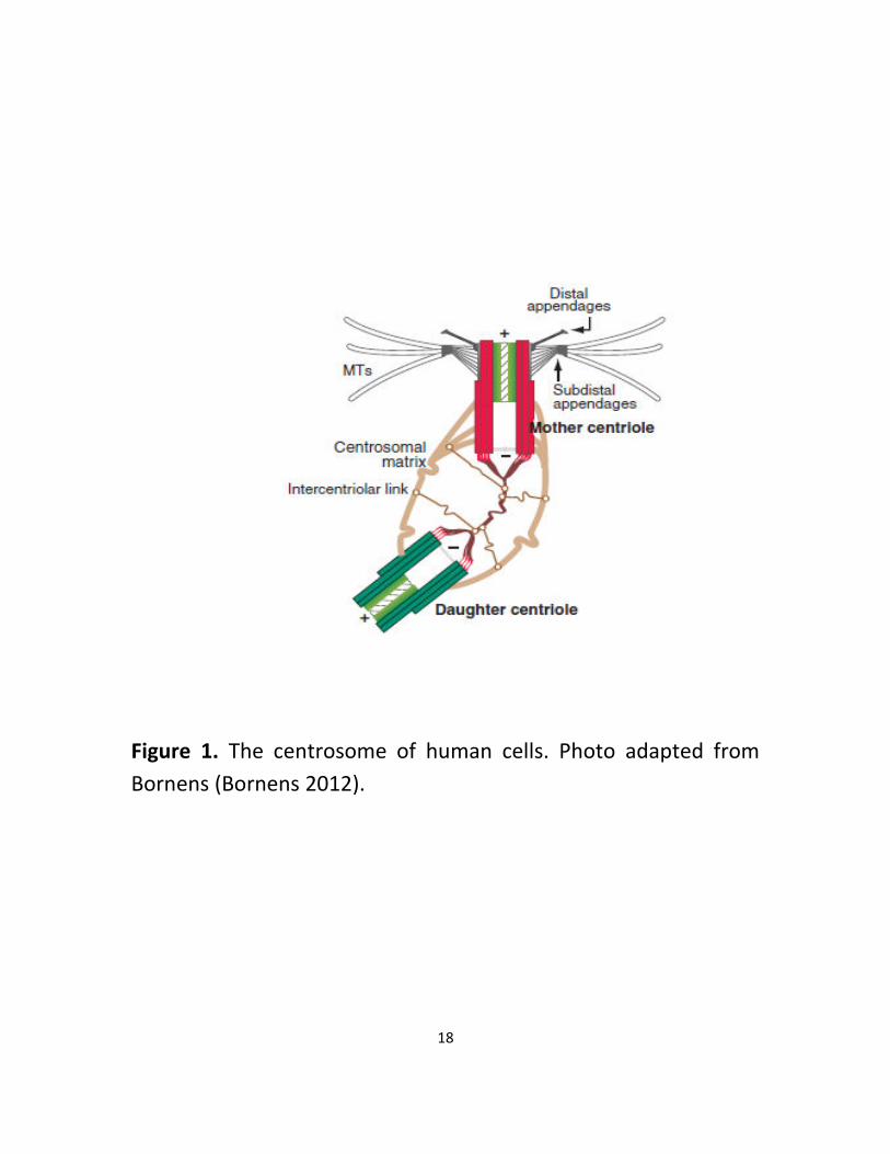
1.1 Centrosome structure
The centrosome is a non-membranous organelle in the periphery
of the nucleus during interphase (Fukasawa 2005, Schatten 2008).
It consists of 2 centrioles (mother and daughter) embedded in an
electron dense pericentriolar material (PCM) (Figure 1). The PCM
contains proteins that regulate centrosome functions and is also
involved in microtubule nucleation and anchoring (Dammermann,
Muller-Reichert et al. 2004, Korzeniewski, Hohenfellner et al.
2013). The centrioles are microtubule-based cylinders that are
arranged orthogonally and are characterized by a 9-fold radial
symmetry. The distal and proximal ends of centrioles have
different functions. While the distal end of mother centriole is
involved in ciliogenesis, its proximal end is the site of centriole
duplication and that is where procentrioles, the centrioles in early

17
stage of development, start to form. Centrioles have polarity in
terms of structure and composition and due to generational
difference; the two centrioles are structurally and functionally
different (Ou, Zhang et al. 2004, Kitagawa, Vakonakis et al. 2011,
Bornens 2012). Structurally, the mother centriole carries two sets
of appendages at the distal and sub-distal end. Two main distal
appendage proteins include CEP164 and Odf2 and some
important sub-distal appendage proteins are CEP170, Cenexin,
Ninein, EB1 and ε-tubulin. Studies on sub-distal appendages
indicate that these proteins are acquired during G1 and unlike
distal appendages, disappear at the onset of mitosis
(Guarguaglini, Duncan et al. 2005). Distal appendages are
important for docking of the basal body to the cell membrane
whereas sub-distal appendages participate in anchoring
microtubules, endosome recycling and forming basal body, a
structure at the base of cilia which promotes microtubule
nucleation (Dammermann, Muller-Reichert et al. 2004, Tsang and
Dynlacht 2013). Mother and daughter centrioles are also different
functionally. Although both centrioles can nucleate microtubules
and accumulate PCM, microtubule anchoring is only done by
mother centriole through its sub-distal appendages (Bornens
2012).
18
Figure 1. The centrosome of human cells. Photo adapted from
Bornens (Bornens 2012).

19
So far 500 proteins have been identified as centrosomal by mass
spe tros op . “o e of these protei s su h as γ-tubulin are
permanently associated with centrosome and remain in
centrosome even after treatment with microtubule
depolymerizing agents such as cold, nocodazole, colchicine
derivatives. These proteins do not need microtubules for their
centrosomal localization. However, some other centrosomal
proteins such as Nuclear Mitotic Apparatus protein (NuMA) are
cell-cycle-specific and temporarily associated with centrosome
(Schatten 2008).
Centriole biogenesis happens through two pathways,canonical
and de novo. In the first and most common pathway, canonical,
procentrioles form in association with the old centrioles whereas
the de novo pathway is activated in the absence of centrioles and
occurs in multiciliated cells. The second pathway is also thought
to take place primarily at deuterosomes (Brito, Gouveia et al.
2012).
During centriole assembly, a disc of fibrous material forms first
adjacent to the proximal end of the parental centriole. Next, a set
of 9-fold symmetric spokes connected to a central axis form the
cart wheel within this material. As the assembly of centriolar
microtubules begins at the tips of the spokes, the structure
elongates to form the mature centriole. Although in most dividing
cells, mother and daughter centrioles template the formation of
only one centriole per cell cycle, in ciliated tissues, up to 8
centrioles can form simultaneously around the parent centriole.
During differentiation, multiciliated cells assemble multiple basal

20
bodies around structures of unknown composition called
deuterosomes (Dammermann, Muller-Reichert et al. 2004,
Azimzadeh and Marshall 2010, Korzeniewski, Hohenfellner et al.
2013).
1.2 Centrosomal proteins and their functions
The centrosome is involved in cell shape, cell division, and
transport of vesicles, cell polarity and motility through
microtubule organization. Centrosomal proteins can be divided
into 2 categories:
1. “tru tural protei s su h as γ-tu uli , α-tu li , β-tublin,
centrin, pericentrin, Ninein, C-Nap1, centriolin, CP110,
cenexin, ODF2, CEP170 and PCM1
2. Regulatory molecules such as Cdc2, Cdk1, PLK1, Nek2 and
Dynactin
Due to the importance of the structural proteins, some of them
are further discussed here.
Gamma-tubulin: One well-studied structural centrosomal protein
is Gamma-tubulin which is localized in PCM. This protein is
conserved in eukaryotes and is a component of tubulin ring
complex γ-TuRC . γ-TuRC plays a role in microtubule nucleation
by covering the minus ends of microtubules. This helps facilitate

21
the growth of protofilaments, the microtubule subunits (Schatten
2008).
Pericentrin: Another well-known centrosomal protein is
pericentrin which forms a o ple ith γ- tubulin and needs the
motor protein dynein for its centrosome localization. This protein
acts as a scaffold for anchoring numerous proteins and is essential
for centrosome and spindle organization (Schatten 2008, Delaval
and Doxsey 2010).
Centrins: These proteins are conserved Ca2+ binding centrosomal
proteins that are associated with centrioles and are important for
centriole duplication (Schatten 2008).
NuMA (Nuclear Mitotic Apparatus protein): NuMA is a regulatory
centrosomal protein involved in the organization of mitotic
apparatus during mitosis. It has microtubule binding capacity and
converges spindle microtubule ends to poles. It also acts as
nuclear matrix protein during interphase (Zeng 2000, Schatten
2008)
CEP170: This is a sub-distal appendage protein which gets
phosphorylated by Polo-like kinase 1 (PLK1). It associates with
spindle apparatus during mitosis. This protein has several
microtubule binding domains and possibly plays a role in
microtubule organization (Guarguaglini, Duncan et al. 2005).
Ninein: This is another sub-distal appendage protein which acts as
a do ki g site for γ-tubulin complex. It also participates in the
anchorage of microtubule minus-ends (Moss, Bellett et al. 2007).

22
C-Nap1: C-Nap1 is important for centriolar cohesion and is
regulated through phosphorylation by NEK2. It is involved in
establishing link between the pair of basal bodies/centrioles
through the protein rootletin which is a physical linker between
the centrioles and binds to C-Nap1 (Yang, Adamian et al. 2006).
CP110: This protein is a substrate of Cdk2 and is involved in
centriole duplication. It also acts as a cap for the distal end of
centrioles and in this way controls their length. Another important
function of this protein is the negative modulation of cilia
assembly through cooperation with CEP97 (Schmidt, Kleylein-
Sohn et al. 2009, Tsang and Dynlacht 2013).
Centriolin: This centriolar protein localizes to the mother centriole
and induces the assembly of primary cilia (Hinchcliffe 2003).
CEP164: This component of distal appendage is indispensable for
primary cilia formation and localizes to the mother centriole
(Graser, Stierhof et al. 2007).
SAS6: SAS6 is one of the several proteins involved in the early
stage of procentriole assembly and is essential for the nine-fold
symmetry of the centriole (Nakazawa, Hiraki et al. 2007).
POC5: This protein localizes to the distal end of the centrioles and
is important for centriole elongation and hence full maturation of
procentrioles (Azimzadeh, Hergert et al. 2009).
The various functions of centrosomal proteins underline the
importance of the centrosome in cellular function and the role it
plays in regulation of several proteins.

23
1.3 Cell Cycle
1.3.1 The cycle
Cell cycle is a crucial cellular event which takes place in order to
divide and duplicate cells. It consists of 2 distinct stages:
interphase (G1, S and G2) and mitosis (prophase, metaphase,
anaphase and telophase). The landmark of interphase is DNA
replication which occurs during the S phase. G1 and G2 are the
gap phases of interphase that prepare the cell for DNA synthesis
and mitosis (Schafer 1998). Also, we must remember that
sometimes cells enter a resting phase called G0 which means no
proliferation and no DNA replication. Following DNA synthesis in
interphase, mitosis (M) begins during which the replicated
chromosomes get segregated into two cells. The 4 phases of
mitosis are prophase, metaphase, anaphase and telophase
(Vermeulen, Van Bockstaele et al. 2003). During prophase,
chromatin becomes condensed to form chromosomes and the
nucleolus disappears. In early prometaphase, the nuclear
membrane dissolves and kinetochores are formed around
centromeres where microtubules attach to move the
chromosomes. During metaphase, spindle fibers align the
chromosomes ensuring that only one copy of each chromosome is
received by each new nucleus. During anaphase, the paired
chromosomes separate and move to opposite sides of the cell.
Finally, during telophase new membrane surrounds the

24
chromatids at the opposite poles and chromosomes go back to
their chromatin form. Following mitosis, the spindle fibers
disperse and the cytokinesis begins. During this stage, actin
contracts around the cell center and divides the cell into two new
daughter cells. Cell division is controlled and regulated by
different pathways and cell organelles including centrosomes
(Morgan 2007). In the next section, these regulations will be
further discussed.
1.3.2 Control of cell cycle
The control of cell cycle is vital for cell survival. The main players
in the regulation of cell division are cyclin-dependent kinases
(CDKs) which act by phosphorylating their target proteins. CDKs
have a stable expression level throughout the cell cycle and are
activated by cyclins required for different stages of cell cycle.
During G1, CDK2 already activated by cyclin E, phosphorylates
Histone H1. This helps regulate the progression from G1 to S and
is important for chromosome condensation and DNA replication.
Next, cyclin A participates in both G2 and G2/M transition through
the activation of CDK2 and CDK1. Furthermore, CDK4/CDK6/cyclin
D phosphorylates Rb, which in turn release E2F, allowing E2F to
activate transcription. Same is true for CDK2/ cyclin E. Finally,
mitosis is regulated by CDK2 and cyclin B (Vermeulen, Van
Bockstaele et al. 2003). In order for the cell cycle to progress

25
properly, there are several checkpoints which work through
regulating the CDK activity. When there is a defect in DNA
synthesis or chromosome segregation, the checkpoints become
active and arrest the cell cycle for the repair to be done
(Malumbres and Barbacid 2009).
It seems that the events in the cell cycle are tightly coordinated
with the centrosome cycle.
1.3.3 Centrosome and cell cycle
The centrosome has several functions during cell division. During
interphase, it serves by nucleating microtubules, organizing
cytoplasmic organelles and forming primary cilia. During mitosis,
the centrosome plays an important role in bipolar spindle
assembly and this is controlled by a checkpoint monitoring
microtubule defects and their attachments to kinetochores
(Schwartz and Shah 2005).
For these functions, the centrosome cooperates with CDKs and
cyclins. For instance it modulates G1 progression and entry into S
phase through cyclins A/E. It has been shown that cyclin E has a
centrosome localization signal (CLS) motif which is necessary to
target cyclin E to the centrosome and controls the S phase
initiation. Also, cyclin A binding to the centrosome might control
the entry into S phase. Centrosome might control the interphase

26
through other pathways as well. For instance, studies have shown
that removing the core centrosomal components such as
centriolin, a mother centriole protein, delay cytokinesis and
induces G1 arrest. Another example of such studies indicates that
the overexpression of AKAP450, a PCM protein, induces
cytokinesis defect and G1 arrest through p53 or p38. Finally,
G2/M transition could be arrested by disruption of the interaction
et ee γ-Tu uli ri g o ple es γ-TuRCs) and pericentrin
which anchors this complex at centrosomes. These findings imply
the significance of centrosomes in the regulation of interphase
events during the cell cycle (Matsumoto and Maller 2004, Doxsey,
McCollum et al. 2005, Sluder 2005, Loffler, Lukas et al. 2006).
Centrosome can also regulate mitosis. During prophase of mitosis,
the activation of cyclinB/CDK1 occurs in centrosome. Also, the
activation of cyclinB/CDK2 by cdc25 is centrosomal dependent.
First, cdc25 gets phosphorylated by Aurora-A which localizes to
centrosome during mitosis and then the activated cdc25 removes
the inhibitory phosphate residues from CDK2 to control mitotic
progression. Furthermore, the centrosome participates in DNA
damage repair. This is done through negatively regulating cdc25
by Chk1 which accumulates at centrosomes in response to the
DNA damage caused by ultraviolet radiation or Hydroxyurea
treatment (Doxsey, McCollum et al. 2005, Sluder 2005, Loffler,
Lukas et al. 2006).

27
1.4 Centrosome Cycle
Centrosomes need to be duplicated and segregated in synchrony
with chromosomes. There are four phases in centrosome cycle:
centriole disengagement, centriole duplication, centriole
maturation and centriole separation. In summary, at the end of
mitosis, the two centrioles of each centrosome disengage but
remain in close proximity. During S phase, each centriole
nucleates a procentriole along its wall, and in G2 phase, the
centriole pairs accumulate more PCM required for microtubule
nucleation and anchoring to mature into two centrosomes
required for mitosis (Figure 2).
a. Centriole disengagement
This phase starts in prophase and ends at the end of telophase.
During centriole disengagement, the tight orthogonal positioning
of the two centrioles in each centrosome pair is released and they
move to a near parallel position. This stage is mainly controlled by
PLK1 and Separase. First, PLK1 promotes the removal of Cohesin
from centrosomes. Next, Separase cleaves Cohesin at the
centriole to complete this process. Centriole disengagement is
important for centriole duplication and for limiting it to once per
cell cycle (Azimzadeh, Hergert et al. 2009, Bettencourt-Dias,
Hildebrandt et al. 2011, Nigg and Stearns 2011, Korzeniewski,
Hohenfellner et al. 2013).

28
b. Centriole duplication
Since each daughter cell inherits one centrosome upon
cytokinesis, it is essential that the centrosome duplicates before
mitosis so that it can establish bipolarity and correct mitotic
spindles. Centriole duplication starts early G1 and continues till
G2. During this phase, PLK4 is first recruited to the wall of the
mother and daughter centrioles by CEP152. The recruited PLK4
then phosphorylates E3-ubiquitin ligase which in turn stabilizes its
substrate SAS-6. Finally, SAS-6 plus SCL-interrupting locus protein
(STIL) and CEP135 form a cartwheel that helps define the centriole
nine-fold symmetry of procentrioles (Azimzadeh, Hergert et al.
2009, Bettencourt-Dias, Hildebrandt et al. 2011, Nigg and Stearns
2011, Korzeniewski, Hohenfellner et al. 2013).
c. Centriole elongation and maturation
The new formed procentrioles elongate during S and G2 phase.
SAS-4 promotes this process and CP110 acts as a cap for the distal
end of centrioles to limit microtubule extension. The proteins
POC5, OFD1, CEP120 and SPICE1 help this process as well.
Following the elongation, the daughter centriole acquires distal
and sub-distal appendage components such as Ninein, CEP170
and ODF2 and becomes fully mature. This phase is called
maturation and is important for microtubule anchoring and
ciliogenesis. Mature centrioles also accumulate more PCM
proteins such as CEP152 and CEP192 which are involved in the
recruitment of centriole duplication factors. CEP215 is also the

29
PCM protein essential for PCM assembly in the maturation
process. Another important event of this stage is the significant
increase of microtubule nucleation activity in centrosomes due to
the proteins Aurora A and PLK1. For this, PLK1 first recruits Aurora
A to the centrosome which co-lo alizes ith γ-tubulin and then
this protein in turn recruits the proteins necessary for
microtubule stabilization, such as NDEL1 (Azimzadeh, Hergert et
al. 2009, Bettencourt-Dias, Hildebrandt et al. 2011, Nigg and
Stearns 2011, Korzeniewski, Hohenfellner et al. 2013).
d. Centriole separation
During most of the cell cycle, the mother and daughter centrioles
are connected to each other by Rootletin and C-Nap1, the
components of the centrosomal linker. This link needs to be
broken at the G2/M transition so the two new centrosomes can
separate and move to the opposite sides of the cell and form the
bipolar mitotic spindles. The proteins participating in this process
are NEK2A, MST1/2, PLK1 and Eg5. First, MST1/2 kinases
phosphorylates the protein kinase NEK2A which in turn
phosphorylates C-Nap1 and rootletin to promote centrosome
separation. Eg5 compliments this process by compensating for
NEK2A activity if reduced and its recruitment to centrosome is
done by PLK1 phosphorylation (Azimzadeh, Hergert et al. 2009,
Bettencourt-Dias, Hildebrandt et al. 2011, Nigg and Stearns 2011,
Korzeniewski, Hohenfellner et al. 2013).

30
Figure 2. The centrosome cycle. Photo adapted from Mardin et al
(Mardin and Schiebel 2012).

31
1.5 Cilia
Although eukaryotic cilia are conserved, they come in different
sizes and functional roles (Quarmby and Parker 2005) . These
structures are centriole-derived protrusions on the cell surface
that contain microtubules and consist of axoneme and basal body.
Axoneme is the microtubule structure of cilium and grows from
ciliary basal body. Basal body which is at the base of eukaryotic
cilia, is the same as mother centriole and participates in axoneme
assembly (Bettencourt-Dias and Glover 2007). Similar to centriolar
i rotu ules, a o e e i rotu ules are ade of αβ tu uli heterodimers and are surrounded by ciliary membrane which is
different from the cell membrane. There are 2 types of Cilia: 1.
Primary or non-motile cilia which consist of 9 doublet
microtubules and lack molecular motors. These cilia are usually
one per cell and are specialized sensory structures. 2. Motile cilia
which consist of 9 doublet microtubules surrounding a central pair
of singlet microtubules and may be several hundred per cell
(figure 3). These cilia need the motor protein dynein for their
motility (Satir and Christensen 2007).
Cilia grow at their distal tips and motor proteins transport ciliary
precursors for assembly and maintenance (Quarmby and Parker
2005). Signaling molecules, receptors and tubulins are
transported to primary cilia by intraflagellar transport (IFT) and
motor proteins such as dynein and kinesin-2 (Tsang, Bossard et al.
2008). Since cilia do not have protein synthesis machinery, they

32
depend on IFTs for their assembly. IFTs perform in 2 directions
due to the protein complexes, IFT-A and IFT-B. IFT-A is involved in
both anterograde and retrograde transport of molecules, whereas
IFT-B is only involved in transport from cell body to cilia and
directs anterograde transport (Tsang and Dynlacht 2013).
Most ciliated cells are in G0 of the cell cycle. For these cells to
enter the mitosis stage, first the cilia need to be resorbed and
when mitosis is complete, the cilia will be reassembled (Quarmby
and Parker 2005). There are 3 distinct stages in cilia assembly.
First, a Golgi-derived vesicle containing membrane proteins
destined to the ciliary compartment binds the distal end of the
mother centriole and the axoneme starts to form. This vesicle
accumulates the essential structures inside the centriole to form
the basal body. Next, vesicles create a sheath around the
axoneme in which the microtubules are assembled. Finally, the
axoneme reaches the cell surface and its membrane fuses to the
plasma membrane to form the ciliary necklace (Pedersen, Veland
et al. 2008).

33
Figure 3. Diagram of ciliary structure. Photo adapted from
Ainsworth (Ainsworth 2007).

34
1.6 Cytoskeleton
The centrosome is involved in cytoskeleton regulation by its active
participation in the assembly of microtubules, a cytoskeleton
component that plays important roles in transport of proteins and
organelles, cell polarity and mitotic spindles (Luders and Stearns
2007). The cytoskeleton plays an important role in 3 cellular
functions. First, it organizes the cell content and components.
Second, it helps connect the cells with the external environment
physically and biochemically. Finally, it is implicated in cellular
movement (Fletcher and Mullins 2010).
Three main polymers of cytoskeleton are actin filaments,
microtubules and intermediate filaments. The polymerization and
depolymerization of actin filaments and microtubules lead to
changes in cell shape and with the help of motor proteins, cellular
components are organized. The differences between the 3
cytoskeletal subunits go back to their mechanical stiffness,
dynamics of their assembly, their polarity and molecular motors
associated with them (Fletcher and Mullins 2010).
Microtubules are the stiffest subunit and have a very complicated
assembly dynamic. Their stiffness is beneficial in the interphase
stage of cell cycle by assembling the radial array of microtubules
that help the intracellular traffic. During mitosis, microtubules
form mitotic spindles which enable chromosome alignment
through dynamic instability of microtubules (Fletcher and Mullins
2010).

35
Although actin filaments are less rigid than microtubules by
themselves, high concentration of the crosslinkers binding them
make stiff isotropic, bundled and branched networks. These
networks are involved in chemotaxis, cell-cell communication and
phagocytosis. Unlike microtubules, actin filaments elongate
steadily in the presence of nucleotide-bound monomers and their
assembly is in response to the local activity of signaling systems.
The intermediate filaments are the least stiff subunit and are not
polarized. They interact with both microtubules and actin
filaments through plectins and are usually assembled in response
of mechanical stress (Fletcher and Mullins 2010).
Mi rotu ules are tu ular pol ers o posed of α a d β tu uli s that asso iate to for protofila e ts ith the β-tubulin subunit
o the plus e d of i rotu ules a d α-tubulin subunit on the
i us e d. A third e er of tu uli fa il , γ-tubulin is
important for microtubule nucleation and assembly. Microtubule
assembly needs GTP hydrolysis so the GDP-tubulin is stabilized at
the plus end by a short cap (Luders and Stearns 2007, Wade
. The α a d β o o ers are kDa a d oth o sist of amino acid residues. Tubulin is subject to several post-
translational modifications like acetylation, detyronization and
polyglutamylation. These modifications determine the stability of
microtubules (Wade 2009).
When tubulin concentrations are low, the microtubule nucleation
process is kinetically limiting. Therefore, nucleation takes place in
specific structures called microtubule organizing centers (MTOCs)
such as centrosome (Wiese and Zheng 2006). During interphase,

36
microtu ules get u leated i MTOCs ri h i γ-TuRC whereas
during mitosis they nucleate on centrosomes which are located at
spindle poles and the astral microtubules are formed dynamically
(Wade 2009).
A large number of proteins interact with microtubules and are
referred to as microtubule-associated proteins (MAPs). Two
classical types of MAPs isolated from brain are the high-
molecular-weight MAPs (200-300 kDa) and the lower molecular
weight tau proteins which is 55 kDa. The main role of these
proteins is microtubule stabilization against dynamic instability
(Wade 2009). The motor proteins, kinesin and dynein, are
important microtubule partners during cell division in eukaryotes.
Kinesins have 2 conserved regions which are responsible for ATP-
binding and microtubule-bindng. Conventional kinesins move
to ard plus e d of i rotu ules at μ /s i itro. D ei s also use ATP energy to move but they move towards the minus end of
microtubules. Dyneins can move laterally and reverse direction as
well. They have 1-3 heavy chains plus several intermediate and
light chains. Their important function is in orientation of mitotic
spindle and in nuclear migration (Wade 2009).

37
1.7 Diseases
There are 2 types of centrosome abnormalities: 1. structural
defect and 2. numerical aberrations. The structural defects are
largely due to changes in the expression levels of different
centrosomal proteins or altered posttranslational modifications
that would lead to an enlarged centrosome or reduction in MT
nucleation. Also, a reduction of centrosome size reduces spindle
length. Structural defects are common in tumors. As for numerical
aberrations, overduplication of centrosome is a good example of
these kinds of defects and is widely found in tumors. Both these
aberrations could cause diseases (Greenan, Brangwynne et al.
2010, Bettencourt-Dias, Hildebrandt et al. 2011). Some common
ones are discussed below.
1. Aneuploidy
Centrosomal deregulation usually leads in chromosomal instability
(CIN) and aneuploidy. Aneuploidy is the result of chromosome
missegregation and is caused by abnormal mitotic spindle
assembly. This is mostly a numerical defect (Kumar, Rajendran et
al. 2013).

38
2. Cancer
Important evidence of the role of centrosomal defects in
tumorigenesis came from the fact that p53 knock down resulted
in centrosome amplification in mouse fibroblasts and skin tumors.
Centrosome abnormalities are often observed in breast, prostate,
lung, colon and brain cancers. There are several pathways leading
to centrosome overduplication. First, the overexpression of PLK4
or mutation in oncogenes or tumor suppressors will cause
centriole over-duplication. Another pathway is through cell
division failure and cell-cell fusion which causes tetraploidisation
(Nigg 2006, Bettencourt-Dias, Hildebrandt et al. 2011).
Centrosomal amplifications and defects usually occur very early in
tumorigenesis and are associated with initiation of chromosomal
changes. These defects get more severe with tumor progression.
In a study on cervical carcinoma, centrosomal amplification
increased 20% in epithelia of grade 1 tumors, 5o% in grade 2
tumors and finally in grade 3 tumors, this increase was 70%. In
tumor cell lines, centrosome overduplication is mainly caused by
the reduced activity of p53 and the overexpression of its
inactivating protein, Mdm2, which allows polyploid cells to
proliferate rather than undergoing apoptosis (Saunders 2005).
Some studies have suggested the link between DNA damage and
centrosome numerical abberations. For instance, DNA damage
could lead to centrosomal splitting in Drosophila and mammalian
cells. Furthermore, the overexpression of ATM/ATR could result in

39
this amplification. The consequences could be cell cycle arrest or
errors in mitosis (Saunders, 2005).
3. Brain development
The most common phenotypes in this category are neural
migration disorders such as lissencephaly, disorders of brain
growth such as microcephalic osteodysplastic primordial dwarfism
and primary microcephalies (MCPH) in which the size of brain is
significantly reduced. The genes affected by primary
microcephalies are either involved in centriole duplication or
centrosome maturation. Centrosome P4.1 associated protein
(CPAP) and CEP152 are MCPH proteins essential in both of these
processes. Also, MCPH mutations could lead in a reduction of the
whole body including the size of the brain (Bettencourt-Dias,
Hildebrandt et al. 2011).
4. Ciliopathies
Defects in motile cilia cause pathologies referred to as primary
cilia dyskinesia (PCD). Patients with PCD show body asymmetry
which is an indication of the importance of ciliary motility in
directional flow in early embryos and initiation of normal left-right
developmental program. Mutations sometimes happen in the
primary cilia and cause defects in its structure or function which
would lead in diseases such as polycystic kidney disease (PKD),

40
nephronophthisis, retinitis pigmentosa, Bardet-Biedle (BBS) and
Joubert and Meckel syndrome. Although cilia structure might not
be altered in these disorders, its sensory function might have
defects and therefore affects multiple organs such as kidney,
retina, brain, bones and liver (Bettencourt-Dias, Hildebrandt et al.
2011).
6. Defects in intracellular transport
Because of its microtubule organizing ability, centrosome plays a
crucial role in intracellular transport and spatial organization of
cellular organelles. Huntington disease is one of the
neurodegenerative disorders that is a consequence of defects in
microtubule-dependent vesicular transport. This disease is
characterized by loss of cognitive function and motor defects.
Huntington-associated protein (HAP1) binds to dynactin and
pericentriolar material 1 protein (PCM1) which is involved in
centrosome and basal body function. Studies in fibroblast cultures
of patients with Huntington disease exhibit aberrant centrosome
numbers, a reduction in mitotic index, an increase in aneuploidy
and finally persistence of midbody (Badano, Teslovich et al. 2005).
Since centrosome deregulation is the cause of several diseases, it
is important to study novel centrosomal proteins and their
functions as potential therapeutic targets.

41
1.8 CEP78
CEP78 is a novel centrosomal protein first identified in 2003
through proteomic characterization of human centrosome. In this
study, a mass-spectrometry analysis of human centrosomes in
interphase was performed and 23 new components were
discovered. CEP78 was one of them (Andersen, Wilkinson et al.
2003). The CEP78 gene is located on chromosome 9q21. Human
CEP78 protein has several isoforms, the biggest one a 78 kDa
protein consisting of 722 amino acids. This protein has orthologs
in mouse, chicken, lizard, tropical clawed frog, zebrafish and fruit
fly. As for the structure of this protein, it consists of 4-6 Leucin-
rich Repeats (LRRs) and one coiled-coil domain. Very few papers
have discussed possible CEP78 functions. In one such studies, the
possible role of CEP78 in centriole anchoring and ciliogenesis was
discussed (Azimzadeh, Wong et al. 2012). Also, in a study carried
out on the effect of standard treatments on immune responses in
prostate cancer patients, CEP78 was one of the proteins
recognized for its treatment associated autoantigen reactivity
(Nesslinger, Sahota et al. 2007). In a study carried out in 2012,
CEP78 expression upregulated 5 fold by noise stress in rat
cochleae (Han, Hong et al. 2012). Another study on the genes
altered by ethanol treatment during neurodevelopment showed
that CEP78 expression decreased on E 14/16 and P 4/7 (Kleiber,
Mantha et al. 2013). Finally, a study in 2013 claimed the
interaction between CEP78 and PLK4, CP110 and CEP97 (Baffet,
Martin et al. 2013). Since all these three proteins are involved in

42
centriole duplication, it is necessary to look at the possible role of
CEP78 in this process. The objective of my project was to further
study CEP78 localization, function and interactions.

43
Chapter 2 Materials and Methods
2.1 Materials
2.1.1 Chemicals
β-gl erophosphate, β-mercaptoethanol, AEBSF, Ampicillin,
Aprotinin, CaCl2, DAPI, DMP, DTT, EDTA, Ethanolamine,
Glutathione, Glycerol, Glycine, HCl, Hepes, IPTG, KCl, Leupeptin,
Methanol, MgCl2, Tris, Na Borate, NaCl, Nocodazole, NP-40,
Paraformaldehyde, PBS, SDS, Triton
2.1.2 Solutions, Buffers and media
2.1.2.1 Coomassie
(50% Methanol; 10% Acetic Acid; 0.2% Coomassie Blue; dH2O)
2.1.2.2 ELB+ Buffer
(1M Hepes pH 7; 5M NaCl; 0.5M EDTA pH 8; 10% NP-40; 1mM
DTT; . M AEB“F; Leupepti μg/ l; Aproti i μg/ l; M NaF; M β-glycerophosphate; dH2O)

44
2.1.2.3 Glutathione elution buffer
(100mM Tris pH 7.9; 120mM NaCl; 20mM Glutathione; 1mM DTT;
0.2mM AEBSF, dH2O)
2.1.2.4 4X Lower Gel Buffer
(1.5mM Tris-HCl pH 8.8; 0.4% SDS; dH2O)
2.1.2.5 0.1 HEMGN
(100mM KCl; 25mM Hepes pH 7.6; 0.2mM EDTA pH 8; 12.5mM
MgCl2; 10% Glycerol; 0.1% NP-40; 1mM DTT; 0.2mM AEBSF;
Leupeptin 2 μg/ml ; Aprotinin 2 μg/ml, dH2O)
2.1.2.6 Maniatis 5x SDS Page Running Buffer
(25mM Tris; 250mM glycine; 0.1% SDS; dH2O)
2.1.2.7 Stripping buffer
% “D“, . M β-mercaptoethanol; 1M Tris; dH2O)

45
2.1.2.8 4X Upper Gel Buffer
(0.5M Tris-HCl pH 6.8; 0.4% SDS; dH2O)
2.1.2.9 Western Transfer Buffer
(50mM Tris; 380mM Glycine; 0.1% SDS; 20% Methanol; dH2O)
46
2.2 Methods
2.2.1 Bacterial methods
2.2.1.1 Purification of GST- tagged CEP78 and GST proteins
The bacteria E.coli DH α strai o tai i g tru ated C a i o acids 590-722) and N (amino acids 1-146) terminal CEP78
plasmids were inoculated from glycerol stock in Luria broth (LB)
edia o tai i g μg/ l A pi illi a d gre o er ight. The
protein expression was induced by adding 1M IPTG to the cultures
and incubating them at 20°C for 16 hours. Next, the bacteria were
pelleted at 4000 rpm. After the pellets were washed with 1X PBS
(Phosphate Buffered Saline), they were resuspended in 0.1
HEMGN buffer. Then, the bacterial suspensions were sonicated 3
times with 15 second bursts at the microtip limit. The lysates were
centrifuged at 10000 rpm(Revolutions Per Minute) and the
supernatants were transferred to Eppendorf tubes. Next, the 50%
slurry Glutathione agarose beads were prepared. To do this, the
beads were first resuspended in 0.1 HEMGN buffer and rocked for
1 hour at room temperature. Next, they were equilibrated in 0.1
HEMGN buffer and finally resuspended in 0.1 HEMGN buffer to
make 50% slurry. The beads then were added to the extracts and
the samples got incubated at 4°C for 1 hour. After the incubation,
the samples were spun at 3000 rpm and the supernatants were

47
aspirated. The beads were washed with 0.1M HEMGN and then
eluted with 1ml glutathione elution buffer for 20 minutes and
spinned at 3000 rpm. Finally, the eluates were dialyzed against
0.1M HEMGN at 4°C overnight. The dialyzed proteins were stored
at -80°C or run on a 10% SDS-PAGE gel and coomassie stained.
2.2.1.2 CEP78 Antibody purification
2.2.1.2.1 Making Columns
At first, the concentration of dialyzed GST and GST-CEP78 proteins
was measured by running them on SDS-PAGE, doing a Coomassie
staining and comparing the intensity of their bands to the ones of
different BSA concentrations. Next, the GST-agarose beads were
added to the proteins and incubated at 4°C for 2 hours. After the
binding, the samples were spinned down at 1000 rpm and washed
with 1X PBS. Next, the beads were washed and resuspended in
0.2M Na Borate pH=9. For crosslinking, solid DMP(Dimethyl
pimelimidate) was added to beads and they were incubated at
room temperature for 30 minutes. Next, the samples were spun
at 1000 rpm and the beads were washed, resuspended in 0.2M
Ethanolamine pH=8 and incubated at room temperature. After 2
hours, the samples were spun at 1000 rpm and the beads were
washed with 1X PBS and 0.1 Glycine pH=2.5. Following another

48
round of washing with 1X PBS, the beads were transferred to the
columns.
2.2.1.2.2 Purifying antibodies
First, rabbits were immunized against CEP78 truncated proteins
and their serums were collected by Cocalico Biologicals company.
Then the serums were loaded on the GST column and incubated
at room temperature. After an hour, the flowthrough was
collected from the GST column and added to the GST-CEP78
column and incubated in room temperature. One hour later, the
beads were washed with 1X PBS and the antibody elution was
carried out with fractions of 0.1M Glycine pH=2.5. The eluates
were then collected in the Eppendorf tubes already containing 1M
Tris HCl pH=8.
2.2.1.3 Transformation of competent cells
First, the bacteria E.coli DH α strai o pete t ells ere thawed. Next, 10 ng DNA was added to the competent cells and
they were incubated on ice. Then, the samples were heat
shocked first at 42°C for 45 seconds and then back on ice for 5
minutes. Later, LB media was added to the cells and the samples

49
were incubated at 37°C. After an hour, the cells were spinned
down at 9000 rpm and the supernatant was aspirated. Finally, the
pellet was resuspended in LB media and plated on LB plates
containing appropriate antibiotic which were incubated at 37°C
overnight.
2.2.2 Cellular methods
2.2.2.1 Immunofluorescence Assay
First, the cells were washed with 1X PBS. Next, they were fixed
with 100% iced Methanol or 4% Paraformaldehyde and washed
with 1X PBS. Following permeabilization with PBS-1% Triton, cells
were washed with 1% PBS and blocked with PBS-3% BSA-0.1%
Triton. Then they were incubated with the primary antibody. After
one hour incubation, the cells were washed with PBS-0.1% Triton.
Next, they were incubated in dark with the secondary antibody-
fluorochrome-labeled. One hour later, the cells were washed with
PBS-0.1% Triton and incubated with DAPI in dark for 7 minutes.
Then, they were washed with 1X PBS and H2O. Once dry, the
coverslips were mounted on slides using mounting media. Finally,
they were sealed with nail polish.

50
2.2.2.2 Western Blotting
First, the cells were harvested by spinning at 1000 rpm for conical
tubes or at 3000 rpm for microcentrifuge tubes. The supernatant
was aspirated and the pellet was washed with 1X PBS. Next, the
sample was lysed with ELB+ buffer and its protein concentration
was measured via Biorad protein assay and Spectrophotometry.
Then, the sample and loading dye were loaded on 10% gel and
run at 150V. Once the running step was complete, the transfer to
Nitrocellulose membrane was carried out at 60V for one hour.
Next, the membrane was blocked in 3% milk and incubated with
the primary antibody. After 1 hour incubation at room
temperature, the membrane was washed with H2O and incubated
with the secondary antibody at room temperature. One hour
later, the membrane was washed with H2O. Finally, ECL was
added on the membrane and developing was carried out.
2.2.2.3 Knock Down with siRNA
First, the cells were plated in 6 well plates so that at the time of
transfection, they were 40%-50% confluent. For transfection, the
siIMPORTER reagent from Millipore company was diluted with

51
serum-free medium in one microcentrifuge tube and in another
tube siRNA oligo, siRNA diluent and serum-free medium were
mixed so that the final concentration of the oligo was 100 nM.
Next, the content of both tubes were mixed and incubated at
room temperature for 5 minutes. Finally, the mixture was added
to the cells and the cells were incubated at 37°C.
2.2.2.4 Plasmid Transfection with Transit reagent
First, the cells were plated in 6 well plates so that at the time of
transfection, they were 60%-70% confluent. For transfection, the
Transit reagent was first diluted with serum-free medium and
e t μg plas id as added to it. The i ture as the incubated at room temperature for 20 minute. Finally, the
mixture was added to the cells and the cells were incubated at
37°C.
2.2.2.5 Plasmid transfection with CaCl2
First, the cells were plated so that at the time of transfection, they
were 60%-70% confluent. For transfection, μg DNA, . M CaCl2, 1ml 2X HEPES and H2O were mixed and incubated at room

52
temperature for 20 minutes. Finally, the mixture was added to the
cells and the cells were incubated at 37°C.
2.2.2.6 Immunoprecipitation
First, the cells were lysed in ELB+ buffer for 30 minutes. Next, the
lysate was spinned down at 14000 rpm and the supernatant was
tra sferred i to e Eppe dorf tu es. The , μg a ti od as added to the supernatant and the sample was incubated at 4°C
for 1 hour. Following the incubation, 50% slurry protein A/G
beads were added to the sample and once again the beads were
incubated at 4°C for one hour. Next, the sample was spinned
down at 3000 rpm in the cold and washed 3 times with ELB+
buffer. Finally, loading dye was added to the sample for western
blotting.
2.2.2.7 Centrosomal localization study
First, the ells ere treated ith μM No odazole a d i u ated at °C. After a hour, the ells ere fi ed a d stai ed ith α-
tubulin.

53
2.2.2.8 Microtubule Assay
First, the ells ere treated ith μM No odazole a d i u ated at 4°C. After an hour, Nocodazole was aspirated from plates, the
media was replaced and the cells were incubated at 37°C for 2, 5
and 20 minutes. Fi all , the ells ere fi ed a d stai ed ith α-
tubulin antibody.

54
Chapter 3 Results
CEP 78 is a novel protein first identified through a mass-
spectrometry analysis of human centrosome(Andersen, Wilkinson
et al. 2003). It consists of 722 amino acids and structurally has 4-6
Leucine-rich repeats and a coiled coil domain. The objective of
this project was to study the localization and function of this
protein. For the localization study, experiments were planned to
look at the cell cycle pattern of this protein and its centrosomal
localization. As for the functions of CEP78, its possible role in
different stages of centrosome cycle and its effect on some other
centrosomal proteins were studied.
3.1 CEP78 is an intrinsic component of Centrosome.
It had already been shown that CEP78 is a centrosomal protein. In
order to study whether CEP78 is a permanent component of
centrosome or it requires microtubules for its centrosomal
localization, retinal pigment epithelial (hTERT-RPE or RPE) cells
were treated with nocodazole which is a microtubule
depolymerizing agent. RPE cells were used because they are

55
normal diploid, have normal centrosome number and
morphology, and undergo normal cell division. These traits makes
them good candidates for studying the effect of a new
e troso al protei o other e troso al o po e ts. γ-tubulin
a d α-tubulin were used as control proteins. In fact, what we
expected was that the net pattern of alpha-tubulin disappeared
due to microtubule depolymerization whereas the genuine
centrosomal proteins would remain. Following an
i u ofluores e e assa a d stai i g the ells ith α-tubulin,
γ-tubulin (a permanent centrosomal protein) and CEP78
a ti odies, it as o ser ed that the et patter of α-tubulin
disappeared hereas γ-tubulin and CEP78 proteins remained at
the centrosome. These results indicated that CEP78 is indeed a
stable centrosomal component and does not require microtubules
for its centrosomal localization [Figure 4].

56
Figure 4: CEP78 is an intrinsic component of centrosomes and does not require microtubule for
its centrosomal localization. RPE cells are treated with nocodazole for 1 hour, fixed and stained
ith α-tu uli ,γ-tubulin and CEP78.
α- tubulin
Cep78
γ- tubulin
Cep78

57
3.2 CEP78 is a centriolar protein present at the distal end.
To see whether CEP78 is present at the distal or proximal end of
centrioles, co-localization of this protein with other centrosomal
proteins was studied in RPE cells by immunofluorescence assay
and fluorescence microscopy. Several proximal (C-Nap1,
Polyglutamylated Tubulin) and distal (CEP170, POC5) proteins
were studied at this step. The results indicated that CEP78 did not
colocalize with the proximal proteins C-Nap1 and
Polyglutamylated Tubulin. However, the distal proteins CEP170,
Centrin and POC5 showed a close co-localization with CEP78. In
conclusion, CEP78 localizes to distal end of centrioles [Figure 5].

58
Figure 5: CEP78 localizes to the distal end of centrioles. RPE cells are fixed and stained with
CEP170, Polyglutamylated tubulin, C-Nap1, POC5 and CEP78.
Cep170
Cep78
C-Nap1
Cep78
Polyglutamylated
tubulin
Cep78
POC5
Cep78

59
3.3 CEP78 localizes to mature centrioles.
In order to study the cell cycle pattern of CEP78, co-localization
with centrin (centriolar marker on mother, daughter and
procentrioles), was carried out in RPE cells at different stages of
cell cycle by immunofluorescence assay and fluorescence
microscopy. Different stages of the cell cycle were identified
based on centrin and DAPI staining in asynchonzied cells and
Polyglutamylated tubulin for G0 cells. Centrin is a centriolar
marker which appears as 2 dots during G1 (mother and daughter
centrioles) and 4 dots during S, G2 and mitosis (mother, daughter
and procentrioles). During the G0, G1, S and early G2 phases of
interphase, there were 2 CEP78 dots in the cells with the intensity
of one dot stronger than the other one. My previous
colocalization study with CEP170 (a sub-distal appendage protein
on mother centriole) had indicated that the stronger dot belongs
to the mother centriole. Measuring the intensity of these dots
using the software Velocity showed that the mother centriole dot
was 1.9 times stronger than the daughter centriole dot. In order
to calculate the above number, first a number of images were
taken by the microscope camera and each image was analyzed
separately by the Velocity software to measure the intensity of
the CEP78 dots on mother and daughter centrioles. Finally, an
average was taken of the intensity differences. During late G2
phase of interphase, the intensity of the mother and daughter

60
centrioles became quite equal implicating that daughter has
matured into a mother. Also, 2 new weak CEP78 dots started to
appear on the procentrioles evolving to the daughter centrioles.
As the cells went through mitosis, the intensity of the new dots
increased gradually. In prophase the CEP78 dots have a quite
diffused staining but by the end of telophase, 2 obvious CEP78
dots could be observed in each daughter cell. The co-localization
study with Centrin indicated that CEP78 dots only localized to the
mother and daughter centrioles but not procentrioles and that
CEP78 is stronger on the mother compared to the daughter
centriole[Figure 6].

61
Figure 6: CEP78 localization pattern at different stages of cell cycle. RPE cells are fixed
and stained with Centrin and CEP78.
G1 S
G2 Prophase
Metaphase Anaphase
Telophase Centrin
Cep78

62
3.4 Leucin rich repeats are responsible for centrosomal
localization of CEP78.
The CEP78 protein consists of 4-6 Leucin rich repeats (LRRs),
amino acids 147-308, and one coiled coil domain, amino acids
450-497. To address the importance of these domains, several
CEP78 fragments with deletion in one or some of these domains
were expressed in RPE cells and their expression pattern was
studied by immunofluorescence assay and fluorescence
microscopy. The results showed three distinct phenotypes:
centrosomal localization, microtubule binding and aggregate
formation [Table 1]. All these three patterns were observed after
overexpression of full length CEP78 as well. While the fragments
with deletion in any LRRs could not localize to ce troso e Δ -
, Δ - , Δ - a d Δ -308), the fragments that
o tai ed all the LRRs lo alized to e troso e learl Δ -497,
1-445). About 70% of the cells expressing full length CEP78 (1-
722) also showed centrosomal localization [Figure 7].
Furthermore, some of the fragments including the fragment 221-
445 as well as 40% of the cells expressing full length CEP78
showed microtubule binding pattern. In fact, 70% of the cells
expressing fragment 221-445, showed the net pattern of
microtubules. This fragment contains the three middle LRRs.
Although the microtubule binding pattern is observed, further
studies are required to confirm this binding. Finally, the

63
expression of some of the fragments including 1-220 resulted in
aggregate formation. This pattern was only observed in 15% of
the cells expressing the full length CEP78 [Figure 8].The protein
aggregates could be indicative of a malfunction of the normal
process of protein turnover or a problem in the recruitment of the
protein to centrosome. These results show that LRRs are crucial
for centrosomal localization and probably MT binding of CEP78.
They can function by interacting with other proteins that help
recruit CEP78 to centrosome or microtubules.

64
TABLE 1: Percentage of RPE cells transfected with CEP78 fragments showing
centrosomal localization or microtubule binding pattern
Construct Centrosomal
localization
Microtubule binding
Full Length Cep78 74% 42%
Δ -174) 2% 0%
Δ -254) 0% 0%
Δ -282) 0% 0%
Δ -308) 0% 0%
Δ -497) 83% 10%
1-220 0% 0%
221-722 0% 33%
1-445 42% 32%
221-445 7% 70%
446-722 0% 0%

65
Figure 7: LRRs are necessary for centrosomal localization. RPE cells are transfected with
flag tagged CEP78 fragments, fixed and stained with flag and Centrin.
Centrin
Cep78
Δ 255-282) Δ 283-308)
Δ 450-497) 1-445
Full Length
Cep78
Δ -174) Δ -254)

66
Figure 8: Some fragments show a) microtubule binding b) aggregate formation. RPE cells
are transfected with flag tagged CEP78 fragments, fixed and stained with flag and
Centrin.
Centrin
Cep78
221-445
1-220
Full Length
Cep78

67
3.5 CEP78 overexpression does not result in centriole duplication
or accumulation.
Since CEP78 is a centriolar protein, I speculated that modulation
of its protein levels could affect the number of centrioles within a
cell. In order to study the possible role of CEP78 in centriole
duplication or accumulation, the full length protein was expressed
in RPE cells and its effect on CEP164 (mother centriole marker),
SAS-6 (procentriole marker) and centrin (marker of mother,
daughter and procentrioles) was studied. Since most cells were in
G1 phase and only had one mother centriole, most control cells
had only one CEP164 dot. Similar to control, most transfected
cells had 1 CEP164 dot and there was no increase/decrease in the
number of mother centrioles. Studying SAS-6 showed no
significant difference between the control and transfected cells
either and most cells had zero (no procentriole in G1 phase) or 2
(2 procentrioles from G2) SAS-6 dots based on the stage of cell
cycle. This meant that there was no change in the number of
procentrioles. Also, looking at centrin confirmed the results of
CEP164 and SAS-6 since the number of centrioles did not change.
So these experiments indicate that CEP78 overexpression does
not result in centriole duplication or accumulation [Figure 9].

68
Cep164
-20
0
20
40
60
80
100
0 dot 1 dot 2 dots >2 dots
Cep164 dots
% C
ell
sControl
Cep78
SAS6
-20
0
20
40
60
80
100
0 dot 1 dot 2 dots >2 dots
SAS6 dots
% C
ell
s
Control
Cep78
Centrin
0
10
20
30
40
50
60
70
80
90
100
0 dot 2 dot 4 dots >4 dots
Centrin dots
% C
ell
s
Control
Cep78
Figure 9: CEP78 overexpression does not cause centriole overduplication or
accumulation. RPE cells are transfected with flag tagged full length CEP78 and CAIP
(control), fixed and stained with flag, CEP164, SAS6 and Centrin.

69
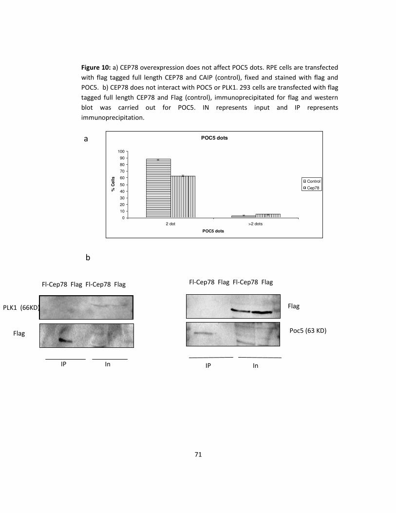
3.6 CEP78 overexpression does not affect or bind POC5 and
PLK1, proteins involved in centriole maturation.
As mentioned before, two new endogenous CEP78 dots start to
appear at late G2 and also the intensity of mother and daughter
dots becomes equal at this point. Since this phase coincides with
centriole maturation in centrosome cycle, there is the possibility
that CEP78 is involved in this process. For this purpose, the full
length CEP78 was expressed in RPE cells and its effect on POC5
was studied by immunofluorescence assay and fluorescence
microscopy. POC5 is a protein involved in centriole elongation and
has a cell cycle pattern similar to CEP78, that is there are 2 POC5
dots during G1 and S phase and late G2, 2 new weak POC5 dots
start to appear that become strong gradually. The results
indicated that similar to the control, the transfected cells had
mostly 2 dots and there was no significant difference in the
number of POC5 dots [Figure 10a]. Also since POC5 is a distal
centriolar protein, its interaction with CEP78 was studied by
expressing full length flag-CEP78 and flag (control) in 293 cells and
doing a flag immunoprecipitation to pull down CEP78 protein and
its interacting proteins.This was followed by Western blotting of
POC5. There was no interaction between the 2 proteins [Figure
10b]. The interaction between CEP78 and PLK1, another protein
involved in centriole maturation, was studied with the same
method discussed for POC5 as well. No interaction was observed

70
between CEP78 and PLK1 either [Figure 10b]. Therefore, CEP78
does not interact with POC5 or PLK1, proteins involved in
centriole maturation and its overexpression does not have an
effect on the number of POC5 dots. It would be interesting in the
future to study the effect of overexpressing POC5 on the number
of CEP78 dots and also the effect of CEP78 overexpression on
centriole elongation.
71
Figure 10: a) CEP78 overexpression does not affect POC5 dots. RPE cells are transfected
with flag tagged full length CEP78 and CAIP (control), fixed and stained with flag and
POC5. b) CEP78 does not interact with POC5 or PLK1. 293 cells are transfected with flag
tagged full length CEP78 and Flag (control), immunoprecipitated for flag and western
blot was carried out for POC5. IN represents input and IP represents
immunoprecipitation.
POC5 dots
0
10
20
30
40
50
60
70
80
90
100
2 dot >2 dots
POC5 dots
% C
ell
s
Control
Cep78
Fl-Cep78 Flag Fl-Cep78 Flag
Flag
Poc5 (63 KD)
IP In
PLK1 (66KD)
Flag
In IP
b
a
Fl-Cep78 Flag Fl-Cep78 Flag

72
3.7 CEP78 overexpression does not have a significant effect on
other centrosomal proteins.
In order to study the effect of CEP78 overexpression on other
centrosomal proteins and the PCM integrity, the full length
protein was expressed in RPE cells and the cells were stained for
differe t e troso al arkers i ludi g γ-tubulin and Pericentrin
by immunofluorescence assay. No significant difference was
observed in the number of any of the above centrosomal
proteins. These results indicate that CEP78 overexpression does
no affect PCM integrity and other centrosomal components
[Figure 11].

73
Gamma-tubulin dots
0
20
40
60
80
100
0 dot 1 dot 2 dots >2 dots
Gamma-tubulin dots
% C
ell
s
Control
Cep78
Pericentrin dots
0
10
20
30
40
50
60
70
80
90
100
0 dot 1 dot 2 dots >2 dots
Pericentrin dots
% C
ell
s
Control
Cep78
Figure 11: CEP78 overexpression does not affect Gamma-tubulin and Pericentrin dots.
RPE cells are transfected with flag tagged full length CEP78 and CAIP (control), fixed and
stained with flag and Gamma-tubulin amd Pericentrin.

74
3.8 CEP78 overexpression reduces the number and intensity of
CEP170, a sub-distal appendage protein.
The only protein that showed a difference after CEP78
overexpression was CEP170 which is a sub-distal appendage
marker. Following the overexpression of the full length CEP78 in
RPE cells, an immunofluorescence assay was carried out and the
cells were stained for CEP170. Comparing control and transfected
cells showed that the number of the transfected cells not having
CEP170 dots increased. In fact 22% of the transfected cells did not
have CEP170 dot compared to 4% in control [Figure 12a]. Also, the
intensity of CEP170 dots decreased significantly in the transfected
cells. Measuring the intensity of CEP170 dots by fluorescence
microscopy and the software Velocity, indicated a decrease of
about 3.7 times in the transfected cells. A similar procedure had
already been used to compare the intensity of CEP78 dots on
mother and daughter centrioles. Next, the expression level of
CEP170 was checked in transfected cells. For this purpose, the full
length CEP78 was expressed in 293 cells and a western blot was
carried out. The comparison between the control sample
expressing Flag and CEP78 overexpressing samples did not show a
decrease in the expression level of CEP170 [Figure 12b]. Finally,
the interaction between CEP78 and CEP170 was studied by
expressing the full length CEP78 in 293 cells, doing flag
immunoprecipitation and western blotting for CEP170. No

75
interaction between the 2 proteins was observed [Figure 10b].
Overexpressing GFP-CEP170 in 293 cells, immunoprecipitation
and western blotting for CEP78 did not show an interaction either
[Figure 12c]. Since CEP170 is a sub-distal appendage protein, it
was necessary to check the effect of CEP78 overexpression on
other sub-distal appendage proteins. One of these proteins is
Ninein. Once again, the full length CEP78 was expressed in RPE
cells and an immunofluorescence assay was carried out to stain
them for Ninein. The results indicated that unlike CEP170, the
number of Ninein dots did not decrease and their intensity did not
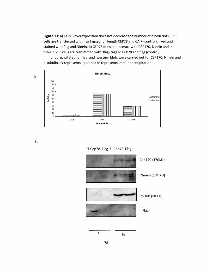
change either [Figure 13A]. Also the possible interaction between
CEP78 and Ninein was studied by the same method used for
CEP170. No interaction was observed between the two proteins
[Figure 13b]. So the results indicate that CEP78 overexpression
decreases both the number and intensity of CEP170 dots but does
not decrease its expression level. This implies that CEP78 does not
regulate the expression level of CEP170 but it might affect the
recruitment of CEP170 to centrosome. This result was not
confirmed for Ninein, another sub-distal appendage protein. Also,
there was no interaction between CEP78 and CEP170 or Ninein.

76
Figure 12: CEP78 overexpression a) decreases the number of CEP170 dots. RPE cells are
transfected with flag tagged full length CEP78 and CAIP (control), fixed and stained with
flag and CEP170. b) does not decrease CEP170 expression level. RPE cells are transfected
with flag tagged full length CEP78 and flag (control) and western blots were carried out
for CEP170. c) CEP170 does not interact with CEP78. 293 cells are transfected with GFP-
tagged CEP170 and GFP (control), immunoprecipitated for GFP and western blots were
carried out for CEP78. IN represents input and IP represents immunoprecipitation.
Cep170
0
10
20
30
40
50
60
70
80
90
100
0 dot 1 dot 2 dots >2 dots
Cep170 dots
% C
ell
s
Control
Cep78
Flag Flag Cep78
Cep170
α- tub
Cep170 (170 KD)
α- tub (50 KD)
Flag Cep78 Flag
a
b

77
GFP GFPCep170 GFP GFPCep170
Cep78 (78 KD)
Cep170 (170 KD)
GFP
IP In
c
78
Figure 13: a) CEP78 overexpression does not decrease the number of ninein dots. RPE
cells are transfected with flag tagged full length CEP78 and CAIP (control), fixed and
stained with flag and Ninein. b) CEP78 does not interact with CEP170, Ninein and α-
tubulin.293 cells are transfected with flag- tagged CEP78 and flag (control),
immunoprecipitated for flag and western blots were carried out for CEP170, Ninein and
α-tubulin. IN represents input and IP represents immunoprecipitation.
Ninein dots
0
10
20
30
40
50
60
70
80
90
100
0 dot 1 dot 2 dots
Ninein dots
% C
ell
s
Control
Cep78
α- tub (50 KD)
In IP
Cep170 (170KD)
Ninein (184 KD)
Flag
Fl-Cep78 Flag Fl-Cep78 Flag
a
b

79
3.9 CEP78 overexpression stabilizes microtubules after
nocodazole treatment.
Since RPE cells overexpressing full length CEP78 and some CEP78
fragments showed microtubule binding pattern, a microtubule
assay was carried out to see the effect of nocodazole on
transfected cells. For this assay, cells were treated with
nocodazole and after the removal of the nocodazole from the
wells and replacing the media, they were incubated at 37°C for 2,
5 and 20 minutes. The purpose of this experiment was to study
the effect of CEP78 overexpression on microtubule stability and
repolymerization following nocodazole treatment. The cells that
were not transfected and also the cells transfected for full length
NPHP5 were used as a control for this experiment. The reason
NPHP5 was used as control is that its overexpression does not
have any effect on microtubule stability. Finally, the cells were
stai ed for α-tubulin by immunofluorescence assay and studied
by fluorescent microscopy. Before the treatment, both the
o trols a d CEP o ere pressi g ells had a et α-tubulin
pattern due to intact microtubules. The results showed that
following nocodazole removal, the controls had depolymerized
microtubules and their net pattern was not observed anymore
whereas 86% of the cells overexpressing full length CEP78, still
had their microtubules. This could be because CEP78 binds to
microtubules and prevents their depolymerization. After 2 and 5

80
minute incubation at 37°C, some filaments started to appear in all
samples but these filaments were significantly more in CEP78
overexpressing cells. Finally, after 20 minutes, asters started to
form in controls whereas in cells overexpressing full length CEP78,
these asters were not as clear. This could be because the majority
of microtubules did not depolymerize after the treatment [Figure
14]. In order to study the possible interaction between CEP78 and
alpha-tublin, full length CEP78 was expressed in 293 cells, a flag
immunoprecipitation was carried out and the membrane was
stained for alpha-tubulin. No interaction was observed (Figure
13b). Next, to make sure the observed pattern is not exclusive to
RPE cells, nocodazole treatment was also carried out on ARPE
cells expressing full length CEP78. ARPE cells are derived from RPE
cells and contain a spontaneous mutation. The results indicated
that the microtubule binding pattern was observed in 74% of the
transfected ARPE cells as well. Next, CEP78 fragments were
studied after nocodazole treatment. The fragments that already
showed microtubule binding pattern (1-445, 221-445, 221-722,
Δ -497), were able to stabilize microtubules after nocodazole
treatment as well [Figure 15]. This could help identify the region
of CEP78 protein involved in microtubule binding and
stabilization.

81
Figure 14: CEP78 overexpression stabilizes microtubules after nocodazole treatment in RPE cells.
RPE cells are transfected with full length CEP78 and full length NPHP5 (control), treated with
nocodazole for 1 hour, incubated back at 37°C for 0, 2 and 20 minutes, fixed and stained with α-
tubulin and flag.
Non-transfectant Fl-NPHP5 Fl-Cep78
α- tubulin
Flag
No
treatment
0 min
2 min
20 min

82
221-445
1-445
221-722
-(450-497)
Figure 15: a) Some CEP78 fragments stabilize microtubules after nocodazole treatment in RPE
cells. RPE cells are transfected with CEP78 fragments (221-445, 221-722, 1- a d Δ -497)),
treated with nocodazole for 1 hour, fi ed a d stai ed ith α-tubulin and flag b) ARPE cells
overexpressing CEP78 also keep their microtubules after nocodazole treatment. ARPE cells are
tra sfe ted ith full le gth CEP , treated ith o odazole for hour, fi ed a d stai ed ith α-
tubulin and flag
a
b
α- tubulin
Flag
α- tubulin
Flag

83
Chapter 4 Discussion
The cell cycle is an important cellular process modulated and
regulated by different molecules, pathways and organelles and its
misregulation could result in several diseases. One cellular
organelle crucial in the cell cycle regulation is the centrosome
which consists of two centrioles embedded in an amorphous
proteinacious material called pericentriolar material (PCM).
Around 500 centrosomal proteins have been identified so far and
any abberations in these proteins can cause defect in the number
or structure of centrosome and lead to several diseases including
cancer, ciliopathies and brain disorder syndromes. Therefore, it is
vital to study the function of novel centrosomal proteins for
potential therapeutic applications.
The objective of this research was to study the localization and
function of CEP78 as a novel centrosomal protein. CEP78 was first
identified by Anderson et al. in 2003 (Andersen, Wilkinson et al.
2003). They isolated centrosome from the cells in the interphase
and did a mass-spectrometry analysis to identify any new
centrosomal proteins. Following this analysis, they studied the
discovered proteins by correlation with already known
centrosomal proteins and also their in vivo localization. They
identified 23 new centrosomal components including CEP78. This

84
78kDa protein has several isoforms with the longest one
consisting of 722 amino acids. It also has orthologs in mouse,
chicken, lizard, tropical clawed frog, zebrafish and fruit fly. The
human cep78 gene is located on chromosome 9.
As it was mentioned in the previous paragraph, CEP78 had already
been discovered as a centrosomal protein but further studies
were necessary to confirm whether it is a permanent component
of centrosomes and also where in centrosome it localizes.
Depolymerization of RPE microtubules by nocodazole indicated
that CEP78 is a stable centrosomal protein that does not require
microtubules for its centrosomal localization. Also, colocalization
studies between CEP78 and other centrosomal proteins showed
that this protein does not colocalize with the proximal centriolar
proteins such as C-Nap1 or Polyglutamylated tubulin. On the
other hand, the proteins localizing to the distal end of centrioles
such as CEP170, Centrin and POC5 showed a close colocalization
with CEP78. These results suggest that CEP78 is a permanent
component of the distal end of centrioles. For further studies,
electron microscopy can be carried out to determine whether
CEP78 is a distal or sub-distal protein.
Centrosomal proteins show different patterns during the cell cycle
based on their functions and studying this pattern could provide
85
us with more information regarding the function of a new protein.
Thus CEP78 was studied during different stages of the cell cycle by
colocalization with the centriolar marker, Centrin, and using DAPI
to identify the nuclei cycle. The results indicated that there are
two CEP78 dots on the mother and daughter centrioles in G1, S
and early G2 phases of interphase, with the intensity of the one
on the mother 1.9 times stronger than the one on the daughter.
Late G2, the intensity of the two existing dots becomes equal and
two new weak CEP78 start to appear. The new dots get stronger
as the cell goes through mitosis and by the end of the telophase
each daughter cell has two CEP78 dots. The colocalization studies
with Centrin showed that CEP78 only localizes to the mature
centrioles, mother and daughter, and not the procentrioles. Since
late G2 coincides with centriole maturation phase of centrosome
cycle during which the daughter centriole acquires appendages to
become mother and also procentrioles elongate, the increase in
the intensity of the daughter centriole and appearance of the two
new CEP78 dots suggest the possible role of CEP78 in centriole
maturation process. To further study this hypothesis, more
experiments were planned and carried out. First, the effect of the
overexpression of CEP78 on another protein involved in centriole
maturation, POC5, was studied. Next, the possible interaction
between CEP78 and the two proteins POC5 and PLK1 was studied.
It has been shown that POC5 gets recruited to centrosome during
G2/M and is involved in centriole elongation (Azimzadeh, Hergert
et al. 2009). This protein has a similar cell cycle pattern to CEP78.

86
So we first studied the effect of CEP78 overexpression on the
number of POC5 dots. Our results showed that the overexpression
had no significant effect on the number of POC5 dots and most
transfected cells showed similar results to the control. One
possible explanation could be that CEP78 is downstream to POC5
hence does not affect POC5. Therefore, it would be a good idea to
study the effect of POC5 overexpression/depletion on the number
of CEP78 dots as well. Moreover, in the research on POC5, the
distance between the distal ends of the mother and daughter
centrioles was measured to study the effect of POC5 depletion on
centriole elongation by high resolution microscopy. It would be
interesting to measure this distance following CEP78
overexpression/knockdown as well. POC5 is a distal centriolar
protein which colocalizes quite well with CEP78. So for the next
step, the interaction between CEP78 and POC5 was studied in 293
cells using immunoprecipitation. Despite what we expected, no
interaction was observed between the two proteins. There are
other proteins involved in centriole maturation as well including
PLK1. This protein plays a role in centriole maturation by
recruitment of Aurora A to the centrosome (Korzeniewski,
Hohenfellner et al. 2013). This persuaded us to study the possible
interaction between this protein and CEP78 in 293 cells as well.
However, our studies did not show an interaction between CEP78
and PLK1. So far, our results do not show a role for CEP78 in
centriole maturation but more experiments need to be done to

87
confirm this result. For instance, it would be interesting to study
the effect of PLK1 inhibition on the number of CEP78 dots.
Structurally, CEP78 consists of 4-6 Leucin rich repeats (LRRs),
amino acids 147-308, and one coiled coil domain, amino acids
450-497. In order to study the importance and function of each of
these domains, fragments with deletions in one or some of these
domains were expressed in RPE cells and their expression
patterns were more closely studied. Our results indicated that the
LRRs are essential for CEP78 localization to centrosome and any
deletions in them would prevent the protein from its centrosomal
localization. This could be due to an unidentified protein that
binds to the LRRs of Cep78 and brings Cep78 to the centrosome.
In order to identify this protein a mass-spectrometry analysis can
be done to study the possible proteins interacting with CEP78. As
for the coiled-coil domain, since these domains are usually
involved in the regulation of gene expression, CEP78 coiled-coil
domain might play a similar role in its expression level as well.
Overexpression of full length CEP78 and the CEP78 fragments
containing the three middle LRRs in RPE cells showed a
microtubule binding pattern. To further study this phenotype and
the effect of CEP78 overexpression on microtubule stability and
nucleation, both RPE and ARPE cells were transfected with full

88
length CEP78 and then treated with Nocodazole. Contrary to what
we expected, the transfected cells maintained their microtubule
network even after the depolymerizing treatment. One
explanation for this phenotype could be that CEP78 when
overexpressed, covers microtubules and does not allow
nocodazole to depolymerize them. Also, carrying out this
experiment for CEP78 fragments showed that the fragments
containing the three middle LRRs, amino acids 221-445, had the
ability to stabilize microtubules as well. It seems that the region
responsible for this phenotype is somewhere in the three middle
LRRs but still more fragments are necessary to identify the exact
region involved in this phenotype. The next question to answer
as hether CEP sta ilized i rotu ules i di g α-tubulin
directly. For this purpose, full length CEP78 was expressed in 293
cells and immunoprecipitation assay was carried out. Western for
α-tu uli did ot sho a i tera tio et ee CEP a d α-
tubulin. However, it is possible that CEP78 interacts with
microtubules that are polymers of tubulins instead and therefore
further studies are necessary to confirm the microtubule binding
activity of CEP78 by an in vitro microtubule binding assay.
I a eeti g held o Buildi g a Ce troso e i , it as suggested that CEP78 interacts with PLK4, CP110 and CEP97
(Baffet, Martin et al. 2013). These proteins are involved in
centriole duplication and cooperate to regulate this process. In

89
order to study the possible role of CEP78 in centriole duplication
or accumulation, several centriolar proteins including CEP164
(mother centriole marker), SA6 (procentriole marker) and Centrin
(mother, daughter and procentriole marker) were studied after
overexpressing CEP78 in RPE cells. Our results indicated no
increase/ decrease in the number of centrioles and the number of
mother, daughter or procentrioles did not change. This suggests
that CEP78 overexpression does not result in centriole
overduplication or accumulation. Also, there is no significant
change in the number or intensity of CEP78 dots at S phase
during which the centriole duplication occurs.
Studying the effect of CEP78 overexpression on various
centrosomal components in RPE cells showed that the only
affected protein was CEP170 which is a sub-distal appendage
protein. This protein localizes only to the mother centriole and its
overexpression shows a microtubule binding pattern similar to
CEP78 (Guarguaglini, Duncan et al. 2005). The results of our
overexpression studies indicated that the RPE cells expressing full
length CEP78 did not have CEP170 or the intensity of this protein
was too weak in them. Despite the observed phenotype, the
expression level of CEP170 did not decrease in 293 cells
overexpressing full length CEP78. This could mean that CEP78
does not regulate the expression of CEP170 but instead might
play a role in its recruitment to the centrosome. This could be due

90
to cell line-specific differences as well so it would be a good idea
to study the effect of CEP78 overexpression on the number and
expression level of CEP170 in other cell lines. We were also
curious to see if this phenotype is caused by the direct interaction
of CEP78 and CEP170. For this purpose, the interaction between
CEP78 and CEP170 was studied by co-immunoprecipitation and
contrary to what we expected no interaction was observed
between the two proteins. Next, we looked at the effect of CEP78
overexpression on another sub-distal appendage marker, Ninein.
Unlike CEP170, there was no decrease in the number of Ninein
dots in RPE cells overexpressing full length CEP78. Furthermore,
there was no interaction between CEP78 and Ninein in 293 cells
overexpressing full length CEP78. For future direction, it would be
interesting to study more sub-distal appendage proteins as well as
the effect of their overexpression on the number of CEP78 dots
and determine whether Cep78 plays a role in the
formation/maintenance of sub-distal appendages, a hallmark of
centrosome maturation.
As it was mentioned before, it is important to study the function
of novel centrosomal proteins in order to use this information for
possible therapeutic applications in the future. As for the clinical
studies on CEP78, there have been very few papers published so
far. In 2007, a study was carried out by Nesslinger et al. on the
effect of standard treatments, hormone and radiation therapy, on

91
immune responses in prostate cancer patients (Nesslinger, Sahota
et al. 2007). They used SEREX immunoscreening of a prostate
cancer cDNA expression library and discovered several treatment
associated autoanitigens including CEP78. In their SEREX antigen
array analysis, CEP78 was negative in the pretreatment sample
but was seroreactive following hormone therapy. This could imply
the possible importance of CEP78 in prostate cancer. All our
studies were carried in RPE, ARPE and 293 cells. For future
direction, it would be a good idea to use a prostate cancer cell line
as well to study CEP78 overexpression pattern and its possible
phenotype. Also, comparing the mRNA/protein levels of Cep78 in
a prostate cell line and other cell lines would be interesting.
Finally, it is a good idea to study the effect of CEP78 knockdown in
a prostate cell line and see if these cells stop growing.
Finally, there are two studies suggesting a possible role for CEP78
in ciliogenesis. In a study on the components of the human
centrosome for which homologs are still present in planarians,
Azimzadeh et al. observed a locomotion defect in planarians
knocked down for CEP78 (Azimzadeh, Wong et al. 2012). Since
planarians lack centrosomes but still own cilia, it is suggested that
CEP78 is rather involved in ciliogenesis. In another study, it was
observed that CEP78 expression is upregulated 5 fold by noise
stress in rat cochleae (Han, Hong et al. 2012). A possible
explanation could be the effect it has on the cochleae cilia.
Although our preliminary experiments did not show any change in
the number of cilia following CEP78 overexpression, further

92
studies including knocking down CEP78 are still necessary to
confirm these results.
In order to confirm the results of the overexpression studies
including the observed decrease in CEP170, the knockdown of
CEP78 was required. For this, several siRNA oligos, transfection
reagents and incubation times were used but none was efficient.
Previously, Azimzadeh et al. used a double knockdown procedure
to knock down CEP78 (Azimzadeh, Wong et al. 2012). Although
we used the same oligos and procedure, the knockdown was not
as good as we expected. For future direction, it would be a good
option to use shRNA for CEP78 knockdown. The advantage of
using shRNA is that its effect can be more specific and last longer
than siRNA.
In summary, I found that CEP78 decreases the number and
intensity of CEP170 and it helps stabilize the microtubule network
in RPE cells treated with nocodazole. My findings contribute to
our understanding of the role of centrosome in the cell cycle and
cytoskeleton.

93
Chapter 5 Reference
Ainsworth, C. (2007). "Cilia: tails of the unexpected." Nature
448(7154): 638-641.
Andersen, J. S., C. J. Wilkinson, T. Mayor, P. Mortensen, E. A. Nigg
and M. Mann (2003). "Proteomic characterization of the human
centrosome by protein correlation profiling." Nature 426(6966):
570-574.
Azimzadeh, J., P. Hergert, A. Delouvee, U. Euteneuer, E.
Formstecher, A. Khodjakov and M. Bornens (2009). "hPOC5 is a
centrin-binding protein required for assembly of full-length
centrioles." J Cell Biol 185(1): 101-114.
Azimzadeh, J. and W. F. Marshall (2010). "Building the centriole."
Curr Biol 20(18): R816-825.
Azimzadeh, J., M. L. Wong, D. M. Downhour, A. Sanchez Alvarado
and W. F. Marshall (2012). "Centrosome loss in the evolution of
planarians." Science 335(6067): 461-463.

94
Badano, J. L., T. M. Teslovich and N. Katsanis (2005). "The
centrosome in human genetic disease." Nat Rev Genet 6(3): 194-
205.
Baffet, A. D., C. A. Martin, I. Scarfone, O. M. Daly, A. David, A.
Tibelius, R. Lattao, M. S. Hussain and J. B. Woodruff (2013).
"Meeting report - building a centrosome." J Cell Sci 126(Pt 15):
3259-3262.
Bettencourt-Dias, M. and D. M. Glover (2007). "Centrosome
biogenesis and function: centrosomics brings new
understanding." Nat Rev Mol Cell Biol 8(6): 451-463.
Bettencourt-Dias, M., F. Hildebrandt, D. Pellman, G. Woods and S.
A. Godinho (2011). "Centrosomes and cilia in human disease."
Trends Genet 27(8): 307-315.
Bornens, M. (2012). "The centrosome in cells and organisms."
Science 335(6067): 422-426.

95
Brito, D. A., S. M. Gouveia and M. Bettencourt-Dias (2012).
"Deconstructing the centriole: structure and number control."
Curr Opin Cell Biol 24(1): 4-13.
Dammermann, A., T. Muller-Reichert, L. Pelletier, B. Habermann,
A. Desai and K. Oegema (2004). "Centriole assembly requires both
centriolar and pericentriolar material proteins." Dev Cell 7(6):
815-829.
Delaval, B. and S. J. Doxsey (2010). "Pericentrin in cellular function
and disease." J Cell Biol 188(2): 181-190.
Doxsey, S., D. McCollum and W. Theurkauf (2005). "Centrosomes
in cellular regulation." Annu Rev Cell Dev Biol 21: 411-434.
Fletcher, D. A. and R. D. Mullins (2010). "Cell mechanics and the
cytoskeleton." Nature 463(7280): 485-492.
Fukasawa, K. (2005). "Centrosome amplification, chromosome
instability and cancer development." Cancer Lett 230(1): 6-19.

96
Graser, S., Y. D. Stierhof, S. B. Lavoie, O. S. Gassner, S. Lamla, M.
Le Clech and E. A. Nigg (2007). "Cep164, a novel centriole
appendage protein required for primary cilium formation." J Cell
Biol 179(2): 321-330.
Greenan, G., C. P. Brangwynne, S. Jaensch, J. Gharakhani, F.
Julicher and A. A. Hyman (2010). "Centrosome size sets mitotic
spindle length in Caenorhabditis elegans embryos." Curr Biol
20(4): 353-358.
Guarguaglini, G., P. I. Duncan, Y. D. Stierhof, T. Holmstrom, S.
Duensing and E. A. Nigg (2005). "The forkhead-associated domain
protein Cep170 interacts with Polo-like kinase 1 and serves as a
marker for mature centrioles." Mol Biol Cell 16(3): 1095-1107.
Han, Y., L. Hong, C. Zhong, Y. Chen, Y. Wang, X. Mao, D. Zhao and
J. Qiu (2012). "Identification of new altered genes in rat cochleae
with noise-induced hearing loss." Gene 499(2): 318-322.
Hinchcliffe, E. H. (2003). "Cell cycle: seeking permission from the
mother centriole." Curr Biol 13(16): R646-648.

97
Kitagawa, D., I. Vakonakis, N. Olieric, M. Hilbert, D. Keller, V.
Olieric, M. Bortfeld, M. C. Erat, I. Fluckiger, P. Gonczy and M. O.
Steinmetz (2011). "Structural basis of the 9-fold symmetry of
centrioles." Cell 144(3): 364-375.
Kleiber, M. L., K. Mantha, R. L. Stringer and S. M. Singh (2013).
"Neurodevelopmental alcohol exposure elicits long-term changes
to gene expression that alter distinct molecular pathways
dependent on timing of exposure." J Neurodev Disord 5(1): 6.
Korzeniewski, N., M. Hohenfellner and S. Duensing (2013). "The
centrosome as potential target for cancer therapy and
prevention." Expert Opin Ther Targets 17(1): 43-52.
Kumar, A., V. Rajendran, R. Sethumadhavan and R. Purohit (2013).
"CEP proteins: the knights of centrosome dynasty." Protoplasma
250(5): 965-983.
Loffler, H., J. Lukas, J. Bartek and A. Kramer (2006). "Structure
meets function--centrosomes, genome maintenance and the DNA
damage response." Exp Cell Res 312(14): 2633-2640.

98
Luders, J. and T. Stearns (2007). "Microtubule-organizing centres:
a re-evaluation." Nat Rev Mol Cell Biol 8(2): 161-167.
Malumbres, M. and M. Barbacid (2009). "Cell cycle, CDKs and
cancer: a changing paradigm." Nat Rev Cancer 9(3): 153-166.
Mardin, B. R. and E. Schiebel (2012). "Breaking the ties that bind:
new advances in centrosome biology." J Cell Biol 197(1): 11-18.
Matsumoto, Y. and J. L. Maller (2004). "A centrosomal localization
signal in cyclin E required for Cdk2-independent S phase entry."
Science 306(5697): 885-888.
Morgan, D. O. (2007). The cell cycle : principles of control. London
Sunderland, MA, Published by New Science Press in association
with Oxford University Press ;Distributed inside North America by
Sinauer Associates, Publishers.
Moss, D. K., G. Bellett, J. M. Carter, M. Liovic, J. Keynton, A. R.
Prescott, E. B. Lane and M. M. Mogensen (2007). "Ninein is
released from the centrosome and moves bi-directionally along
microtubules." J Cell Sci 120(Pt 17): 3064-3074.

99
Nakazawa, Y., M. Hiraki, R. Kamiya and M. Hirono (2007). "SAS-6 is
a cartwheel protein that establishes the 9-fold symmetry of the
centriole." Curr Biol 17(24): 2169-2174.
Nesslinger, N. J., R. A. Sahota, B. Stone, K. Johnson, N. Chima, C.
King, D. Rasmussen, D. Bishop, P. S. Rennie, M. Gleave, P. Blood,
H. Pai, C. Ludgate and B. H. Nelson (2007). "Standard treatments
induce antigen-specific immune responses in prostate cancer."
Clin Cancer Res 13(5): 1493-1502.
Nigg, E. A. (2006). "Origins and consequences of centrosome
aberrations in human cancers." Int J Cancer 119(12): 2717-2723.
Nigg, E. A. and T. Stearns (2011). "The centrosome cycle: Centriole
biogenesis, duplication and inherent asymmetries." Nat Cell Biol
13(10): 1154-1160.
Ou, Y., M. Zhang and J. B. Rattner (2004). "The centrosome: The
centriole-PCM coalition." Cell Motil Cytoskeleton 57(1): 1-7.
Pedersen, L. B., I. R. Veland, J. M. Schroder and S. T. Christensen
(2008). "Assembly of primary cilia." Dev Dyn 237(8): 1993-2006.
100
Quarmby, L. M. and J. D. Parker (2005). "Cilia and the cell cycle?" J
Cell Biol 169(5): 707-710.
Satir, P. and S. T. Christensen (2007). "Overview of structure and
function of mammalian cilia." Annu Rev Physiol 69: 377-400.
Saunders, W. (2005). "Centrosomal amplification and spindle
multipolarity in cancer cells." Semin Cancer Biol 15(1): 25-32.
Schafer, K. A. (1998). "The cell cycle: a review." Vet Pathol 35(6):
461-478.
Schatten, H. (2008). "The mammalian centrosome and its
functional significance." Histochem Cell Biol 129(6): 667-686.
Schmidt, T. I., J. Kleylein-Sohn, J. Westendorf, M. Le Clech, S. B.
Lavoie, Y. D. Stierhof and E. A. Nigg (2009). "Control of centriole
length by CPAP and CP110." Curr Biol 19(12): 1005-1011.
Schwartz, G. K. and M. A. Shah (2005). "Targeting the cell cycle: a
new approach to cancer therapy." J Clin Oncol 23(36): 9408-9421.

101
Sluder, G. (2005). "Two-way traffic: centrosomes and the cell
cycle." Nat Rev Mol Cell Biol 6(9): 743-748.
Tsang, W. Y., C. Bossard, H. Khanna, J. Peranen, A. Swaroop, V.
Malhotra and B. D. Dynlacht (2008). "CP110 suppresses primary
cilia formation through its interaction with CEP290, a protein
deficient in human ciliary disease." Dev Cell 15(2): 187-197.
Tsang, W. Y. and B. D. Dynlacht (2013). "CP110 and its network of
partners coordinately regulate cilia assembly." Cilia 2(1): 9.
Vermeulen, K., D. R. Van Bockstaele and Z. N. Berneman (2003).
"The cell cycle: a review of regulation, deregulation and
therapeutic targets in cancer." Cell Prolif 36(3): 131-149.
Wade, R. H. (2009). "On and around microtubules: an overview."
Mol Biotechnol 43(2): 177-191.
Wiese, C. and Y. Zheng (2006). "Microtubule nucleation: gamma-
tubulin and beyond." J Cell Sci 119(Pt 20): 4143-4153.

102
Yang, J., M. Adamian and T. Li (2006). "Rootletin interacts with C-
Nap1 and may function as a physical linker between the pair of
centrioles/basal bodies in cells." Mol Biol Cell 17(2): 1033-1040.
Zeng, C. (2000). "NuMA: a nuclear protein involved in mitotic
centrosome function." Microsc Res Tech 49(5): 467-477.

103


![Transistor Bipolaire [Mode de compatibilité]users.polytech.unice.fr/~lorenz/Transistor_Bipolaire.pdf · Références: 2 • H. Mathieu, « Physique des semi-conducteurs et des composants](https://img.pdfslide.us/doc/110x75/5b58d4497f8b9a655d8c837a/transistor-bipolaire-mode-de-compatibiliteusers-lorenztransistorbipolairepdf.jpg)


![LP5 - Organite Celulare [Compatibility Mode]](https://img.pdfslide.us/doc/110x75/577cb1141a28aba7118b7f3d/lp5-organite-celulare-compatibility-mode.jpg)
