Embed Size (px)
Citation preview
UNIVERSIDADE DO ALGARVE
Faculdade de Engenharia de Recursos Naturais
Biotechnology in Aquaculture: polyploidy versus
transgenic technology
Dissertação de Mestrado Integrado em Engenharia Biológica
Ângela Alexandra Martinho Ramos
Faro 2009
UNIVERSIDADE DO ALGARVEFaculdade de Engenharia de
Recursos Naturais
Biotechnology in Aquaculture: polyploidy versus
transgenic technology
Dissertação de Mestrado Integrado em Engenharia Biológica
Ângela Alexandra Martinho Ramos
NIVERSIDADE DO ALGARVE Faculdade de Engenharia de
ecursos Naturais
Biotechnology in Aquaculture: polyploidy versus
transgenic technology
Dissertação de Mestrado Integrado em Engenharia Biológica
Ângela Alexandra Martinho Ramos
Orientadora: Prof Dr. Deborah M
2009
Biotechnology in Aquaculture: polyploidy versus
Dissertação de Mestrado Integrado em Engenharia Biológica
Prof Dr. Deborah M. Power
Declaração: O conteúdo, e execução do
trabalho experimental e interpretação de
resultados é da exclusiva responsabilidade
da autora
Ângela Alexandra Martinho Ramos
________________________________________
ACKNOLEDGMENTS
I would like to acknowledge and give a special thanks to my supervisor, Professor Deborah Power,
for her patience throughout this long process and above all for her enormous support with this
work, thank you very much for all Professor. It is a privilege to learn with you!
I also would like to thank Professor Adelino Canário for accepting me in his team.
To Dr.Stephanie Fontagné a special thanks, because she is the responsible for providing all of the
samples and also for resolving some doubts concerning the experimental trial and also for the
encouragement and understanding.
A special thanks to Dr. António Abrantes for all the x-rays, and for providing knowledge in a
different study area. Also a special acknowledgment to Dr. Patricia Pinto, who tutored me on the
quantitative real time qPCR technique, to Alexandra Filipe, Dr. Pedro Guerreiro, Elsa Couto,
Teresa Sancho and Mar Huertas for helping me in some parts of this work.
For all the people in the CME group a big thanks, in special for Nádia Silva (that shared most of
this work) and Rita Costa that accompanied me and supported me throughout all of this process,
and above all for your friendship. Also very special thanks to Silvia Gregório for her precious help
with the accomplishment of innumerous tasks allowing an increase in rhythm of work. To all the
people in lab 3.32 I am very pleased to work with you all.
To some special friends that where available to listen and support in innumerous situations to
Dulce Estevão, Anabela Nobre, Ana Evaristo, Monika Grunner and Madalena Bentes, you are all
very special to me and thanks for all. Nonetheless acknowledge Mestre Jorge Veiga e Castro for
the friendship and the permanent scientific encouragement.
To Leonel a special appreciation for all the love and also for believing in my challenges, and for all
support in every shared moment throughout this thesis. And finally I would like to thank my
family, my parents and sister, that give always a tremendous strength giving a renewal power, and
for all their love and support in every moment of my life.
LIST OF ABREVIATIONS
CA – Caudal
Ca – Calcium
DA – Diploid diet A
DC – Diploid diet C
GH – Growth Hormone
GMO’s – Genetic modified organisms
OSC - Osteocalcin
OSN - Osteonectin
OSP- Osteopontin
P- Phosphorous
TA – Triploid diet A
TC – Triploid diet C
TCR-Trunco Cranial
TCA – Trunco caudal
VCR – Vizcero cranial
ABSTRACT
Triploidy is the condition in which somatic cells contain three sets of
chromosomes which might be a consequence of environmental changes or hybrid
stabilization, though in the present work the ploidy induction was done by
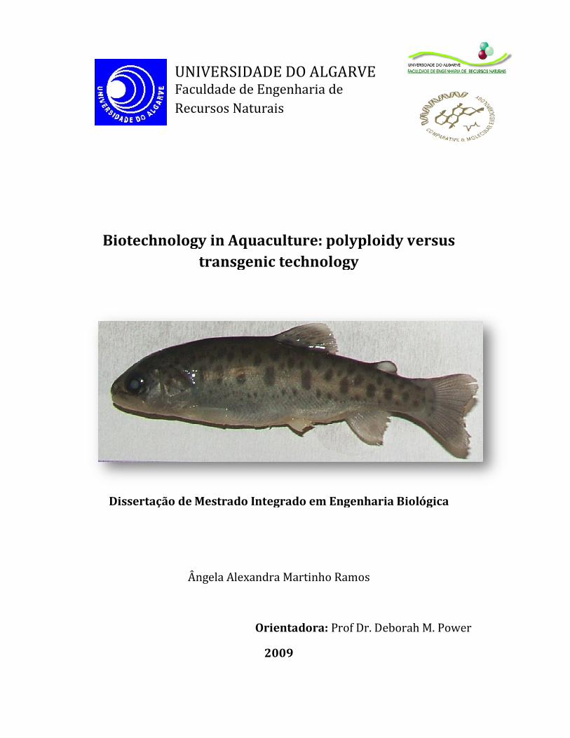
hyperbare pressure. Diploid and triploid Rainbow trout, Onchorynchus mykiss
(Walbaum, 1792) were the species characterized in this thesis. Besides ploidy a
modification in the mineral availability of phosphorous, was applied influencing
the whole body mineral homeostasis in diploid and triploid trout.
The skeletogenesis in triploids animal is different from the diploid, having a
general delay, the mineral deficient diet has also an impact on the ossification
retreating. It allowed studying the development through triploids, which at the end
of this trial they grew less than diploids. Vertebra area is also affected varying
acconding to their regions, total number of vertebra differs, and in average
triploids have less one vertebra than diploid groups. And there are also
histological differences at a muscle level, having triploid Diet A less myotomes in
average.
Animals present a high capacity in recovering, once they start eating a normal diet
in mineral composition, the restore their mineral content in bone structures in
some situation an over calcification is observe (Diploid A). In a molecular level,
there were any molecular differences observed only a high variability at the OSN
levels.
Key words: Diploids, Triploids, Skeletogenesis, Ontogeny, mineral content.
RESUMO
Triploidia é um estado, em que as células somáticas contém três conjuntos de
cromossomas (3n). Pode ocorre naturalmente ou ser induzido experimentalmente
com este estudo em que foi usada uma câmara hiperbárica. A espécie modelo
utilizada foi a truta (Onchorynchus mykiss Walbaum, 1792) Diplóides e
Triplóides. Para além da manipulação na ploidia efectuou-se uma alteração na
disponibilidade do mineral fósforo e foi avaliado o seu efeito no desenvolvimento
do sistema musculo-esquelético, usando parametros morfométricos, biométricos,
bioquímicos e moleculares.
Verifica-se uma diferença na esqueletogénese em trutas triplóides
comparativamente às diplóides, denotando-se um atraso na ossificação em ambos
os grupos triplóides (dieta controlo e dieta restrita em fósforo). A acumulação
corporal total de cálcio em ambos os grupos diploides e triploides de alevinos
alimentados com dieta pobre em fósforo, é significativamente mais baixa (P<0,05)
comparativamente a alevinos diplóides ou triplóides alimentados com dieta
controlo. Constata-se uma alteração significativa nas vertebras de trutas cujo
esqueleto estava completamente formado, este efeito varia consoante a região da
coluna vertebral. De um modo geral trutas triploides têm menos vertebras. Nas
quatro regiões identificadas e estudadas da coluna vertebral, encontraram-se
diferenças significativas (p<0,01-0,001) na área das vertebras. Os marcadores
moleculares avaliados por PCR em tempo real, Osteocalcina, Osteonectina e
Osteopontina mostraram baixa variabilidade entre nos indivíduos analisados, e não
se obtiveram diferenças conclusivas entre os grupos experimentais a 64 dpf de
idade.
Em conclusão, a triploidização afecta o esqueleto de truta. Um fornecimento
deficiente em fósforo afecta significativamente a ontogenia esquelética,
morfometria e parâmetros bioquímicos de trutas diplóides e triplóides.
Palavras Chave: Diplóide, Triplóide, esqueletogénese, conteúdo mineral.
TABLE OF CONTENTS
1. Introduction…………….……………………..………………………………………….1
1.1. Development………...………………………………………………..…..1
1.1.1.General overview…………………………………………………1
1.1.2.Ectoderm,endoderm, and mesoderme………………………….2
1.2. Skeletal System…….……….……………………………….………………3
1.2.1 Skeleton origin and function……..………………………………….3
1.2.2. Skeletal tissue metabolism…..……….……………………………….4
1.2.3. Skeletal tissue composition.……..……………………………………5
1.2.4. Skeletal cell differentiation………….….……………………….……7
1.2.5. Skeletal tissue endocrine regulation.…….…………………………...8
1.3. Muscle………………..…………........………………………………...………9
1.3.1. General……………………………………………………………....9
1.3.2. Organization of muscle structure………………………….…...……11
1.4. Mineral requirement……...………..…..…………………………….………..12
1.4.1. Calcium and phosphorus…………………….…………..…………12
1.4.2. Phosphorus deficiency deformities…………………….…..………12
1.5. Biotecnology applied to aquaculture and lifesciences……...…………………13
1.5.1. Ploidy vs Transgenics………………………..…………..………….14
1.5.2. Triploid fish production..………………………………..…………..15
1.5.3. Morpho-anatomical changes in triploids..…………………...……...17
1.5.4. Growth performances in triploids……..…………………..….……..18
1.6. The experimental model - Rainbow trout……….……………………...……..19
2. Thesis Objectives………….……………………………………….…….……………..20
3. Methodology……………………………………..…………………….……………….21
3.1. Biological trials and sampling………………………………………..……….22
3.1.1. Experimental fish and dietary trial conditions……………...……….22
3.1.2. Sample collection…….…………………………………………...…23
3.1.3. Experimental diets…..………………………………………………24
3.1.4. Diets composition……………………..………………………….…24
3.2. Biochemical analysis…………………………………………………….……26
3.2.1 Calcium and phosphorus quantification…..........................................26
3.3. Morphological and biometric analysis……………………….……………….27
3.3.1 Whole-mount cartilage and bone differential staining………………28
3.3.2 X-rays analysis……………………………………………………….30
3.3.3 Histological and stereological analysis….…………………………...31
3.3.3.1 Sample preparation………………………………………...31
3.3.3.2 Tissue processing and wax embedding………………….....31
3.3.3.3 Histological sectioning………………….……………….....32
3.3.3.4 Heamatoxilyn eosin staining……..…….…….………….…33
3.3.3.5 Von kossa staining................................................................33
3.3.4 Muscle myotomes total counts………………………………………34
3.4. Molecular Analysis- gene expression………………………………………....35
3.4.1 RNA extraction.……………………………………………………...36
3.4.2 Quantification and RNA integrity analysis…………………………..36
3.4.3 cDNA synthesis and amplification………………………………......37
3.4.4 Real Time Reverse Transcription Polimerase Chain reaction (qRT-
PCR)….........................................................................................................38
3.4.4.1 General Technique…………….…………………………...38
3.4.4.2 Genes of interest…..…………………………………...…..39
3.4.4.3 Real-Time PCR preparation and pre-run tests......................39
3.4.4.4 Real-Time PCR.....................................................................40
3.4.4.5 Data Processing.....................................................................40
3.4.4.6 Data Normalization...............................................................40
3.5. Statistical analysis…………………………………………………………….41
4. Results…………………………………………………………….…………………….42
4.1 Survival rate………………………….………………...…………………...….42
4.2 Biochemical analysis……………………………………………………….….42
4.2.1 Whole alevin Ca and P content………………………………….…..42
4.2.2 Body region calcium and phosphorous quantification results..….…..44
4.3 Morphological and Biometric Analysis……………...................………46
4.3.1. Ontogenic Evolution…………………………………………….......46
4.3.2 X-rays analysis…...………………………………………………..…52
4.4 Histology - muscle myotome total counts………………………...………..…58
4.5 Molecular Analysis- Gene expression…………………………………..60
4.5.1 β-actin………………………………………………………………..60
4.5.2 Osteocalcin…………………………………………………………..61
4.5.3 Osteonectin…………………………………………………………..62
4.5.4 Osteopontin…………………………………………………………..62
5. Discussion………………….…………………………………………………………...64
6. Final conclusions…………………………………………………………………..……70
7. Future work......................................................................................................................71
8. References………………………..…………………………….…………………….....72
LIST OF FIGURES
Figure 1.1 _ Squematic representation of four vertebrate model organisms........................2
Figure 1.2 - Overview of zebrafish embryogenesis.............................................................10
Figure 1.3 – General representation of the process of triploidy induction..........................17
Figure 1.4 – Schematic presentation and photograph of Onchorynchus mykiss.................19
Figure 3.1 – Fluxogram representing the experimental design and analysis performed….21
Figure 3.2 – Oncorhynchus mykiss radiography scheme………………………………....27
Figure 3.3 - Photograph of a trout (79 dpf) stained with Alcian Blue and Alizarin Red…29
Figure 3.4 – Photograph of a rainbow trout and a schematic drawing…………………....31
Figure 3.5 – Schematic representation of the tissue segments….………………………...33
Figure 3.6 – Photograph of a typical section used to count myotomes………………..….35
Figure 4.1 –Survival rate (%) in each experimental group during the feeding trial ……...40
Figure 4.2 – Determination of whole body calcium (A) and phosphorus (B)………...…..40
Figure 4.3 –Radiography of a rainbow trout diploid control of 219 dpf………………….44
Figure 4.4 – A. Total mean Ca concentration (µmolCa2+
/mg Ash) and the SEM…......…45
Figure 4.5 - Diagram representing the ontogenic development of rainbow trout skeleton.47
Figure 4.6 – Cumulative counts + SEM, sum of all analyzed…………...………………..48
Figure 4.7 – Cumulative counts+ SEM, sum of all analyzed structures…...…………......49
Figure 4.8 - Total number of vertebra counted in individuals…………………………….51
Figure 4.9 – Mean area + SEM (cm2) of vertebra from each vertebral region…...……….52
Figure 4.10 – Standard length measurements mean (cm2)…………….………………….55
Figure 4.11 – Ratio of head length: body length presented as mean + SEM (cm2) ……...56
Figure 4.12 – Representation of the Ct value for the sample expression of β-actin…..…..59
Figure 4.13 – Logarithm of the ratio of the square means of the expression of OSC….…60
Figure 4.14 – Logarithm of the ratio of the square means of the expression of OSN…….61
Figure 4.15 – Logarithm of the ratio of the square means of the expression of OSP…….62
LIST OF TABLES
Table 1.1 – Components of mammalian bone………………………….………….6
Table 1.2 – Description of methods applied for inducing triploidy……………...…….…16
Table 3.1 – Age and development stage of the biological samples analyzed……………....23
Table 3.2 - Formulation and composition of experimental diets (g/100g dry weight)……25
Table 3.3 – Number of specimens analyzed (n=100) by alcian blue/alizarin red S ……...28
Table 3.4 – Skeletal structures observed in each skeletal area evaluated…………………29
Table 3.5 – Number of specimens.......................................................................................31
Table 3.6 – Description of the solutions used the duration and the number.........32
Table 3.7 – Description of the treatment group and their age….........................................37
Table 3.8 – Q PCR primer designation, sequence, annealing temperature (Ta)……….....39
Table 4.1 - Results of the statistical analysis applied .........................................................……..43
Table 4.2 – Results of the t-student test results and each hypothesis tested……………....46
Table 4.3 – Results presented for the endochondral and dermal ossification………….....51
Table 4.4 - Results of t-student and each hypothesis tested for vertebra area …………………...55
Table 4.5 - Summary table with the t-student test results and each hypothesis…….……………57
Table 4.6 – Mean, standard deviation, standard error of the mean (SEM) ………….…....58
Table 4.7 – CT values results, mean value Standard error and SEM…………………..…60