Embed Size (px)
Citation preview

1
UNIVERSIDAD CATÓLICA SANTO TORIBIO DE MOGROVEJO
FACULTAD DE MEDICINA
ESCUELA DE ENFERMERÍA
REVISIÓN CRÍTICA: CUIDADOS DE ENFERMERÍA PARA EL
MANTENIMIENTO DEL CATÉTER VENOSO CENTRAL
PERCUTÁNEO EN EL SERVICIO DE EMERGENCIA
INVESTIGACIÓN PARA OPTAR EL TÍTULO DE:
SEGUNDA ESPECIALIDAD PROFESIONAL DE ENFERMERÍA EN
EMERGENCIAS Y DESASTRES
AUTOR: Lic. Verónica Catherine Ripalda Pérez
Chiclayo, 07 de Setiembre del 2017

2
REVISIÓN CRÍTICA: CUIDADOS DE ENFERMERÍA PARA EL
MANTENIMIENTO DEL CATÉTER VENOSO CENTRAL
PERCUTÁNEO EN EL SERVICIO DE EMERGENCIA
POR:
Lic. Verónica Catherine Ripalda Pérez
Presentada a la Facultad de Medicina de la Universidad Católica Santo Toribio
de Mogrovejo, para optar el Título de:
SEGUNDA ESPECIALIDAD PROFESIONAL DE ENFERMERÍA EN
EMERGENCIAS Y DESASTRES
APROBADO POR:
_____________________________
Lic. María Ysabel Villena Campos
Presidente de Jurado
_____________________________
Lic. Cinthya Quiroz Vigil
Secretaria de Jurado
_____________________________
Mgtr. Nancy Sánchez Merino
Vocal/Asesor de Jurado
Chiclayo, 07 de Setiembre del 2017
ii

3
ÍNDICE
Pág
.
DEDICATORIA iv
AGRADECIMIENTO v
RESUMEN vi
ABSTRACT vii
INTRODUCCIÓN 8
CAPÍTULO I: MARCO METODOLÓGICO 12
1.1 Tipo de investigación 12
1.2 Metodología EBE 12
1.3 Formulación de la Pregunta según esquema PICOT 14
1.4 Viabilidad y pertinencia de la pregunta 15
1.5 Met Metodología de Búsqueda de Información 15
1.6 Síntesis de la Evidencia encontrada a través de la Guía de Validez y
utilidad aparentes de Gálvez Toro
21
1.7 Listas de chequeo específicas a emplear para los trabajos
seleccionados
24
CAPÍTULO II: DESARROLLO DEL COMENTARIO CRÍTICO 26
2.1 Fi Artículo para revisión 26
2.2 Comentario crítico 30
2.3 Importancia de los resultados 41
2.4 Nivel de evidencia 41
2.5 Respuesta a la pregunta 42
2.6 Recomendaciones 44
CAPÍTULO III: PROPUESTA 45
REFERENCIA BIBLIOGRÁFICA 49
ANEXOS 51
iii

4
DEDICATORIA
Dedico este trabajo de investigación de manera muy especial a mis
padres Alfredo y Olinda, por ser las fuentes de mi inspiración y motivo de
progreso día a día; a mis hermanos María y Gerardo, por su gran ejemplo de
superación y valioso apoyo en todo momento; a mi hermana Máryorie y a mis
sobrinos Geraldine, Nicolás y Gerard, a quienes exhorto a tener una visión de
éxito en su vida mediante el estudio continuo.
La autora.
iv

5
AGRADECIMIENTO
A Dios por iluminarme y guiarme incondicionalmente, y por haberme
dado fuerza y valor para concluir mis estudios de segunda especialidad.
A mi familia por su confianza, apoyo, ayuda, aliento y sacrificio para
llevar a cabo esta tarea difícil, por celebrar conmigo mis triunfos y darme la
fuerza necesaria para superar cada obstáculo.
A mi asesora Nancy Sánchez Merino por su tiempo, consejos y por
compartir desinteresadamente su amplio conocimiento y experiencia.
Y finalmente gracias a todas las personas que ayudaron directa e
indirectamente en la realización de este trabajo de investigación.
La autora.
v

6
RESUMEN
La revisión crítica titulada Cuidados de enfermería para el
mantenimiento del catéter venoso central (CVC) percutáneo en el servicio de
emergencia, tuvo como objetivo identificar los mejores cuidados de
enfermería frente al CVC percutáneo en el servicio de emergencia, puerta de
entrada de pacientes críticos, en los que el uso del CVC percutáneo es
frecuente, quedando así expuestos a complicaciones. La enfermera es la
responsable directa sobre el mantenimiento del catéter, por lo que este
procedimiento se debería realizar en base a protocolos que permitan actuar de
una manera uniforme a todos los profesionales enfermeros. La metodología
utilizada fue enfermería basada en evidencia, con la interrogante ¿Cuáles son
los mejores cuidados de enfermería para el mantenimiento del CVC
percutáneo en los pacientes que se encuentran en el área de shock trauma del
servicio de emergencia? Se realizó búsqueda en Google académico, Lilacs,
BVS; seleccionándose 10 investigaciones, de las cuales 6 pasaron el filtro de
Gálvez Toro (2 cuantitativas, 1 observacional prospectivo, 1 ensayo controlado
aleatorizado (ECA), 1 descriptivo-longitudinal y 1 guía de práctica clínica (GPC)
eligiéndose la guía Guidelines for the Prevention of Intravascular Catheter-
Related Infections, 2011 por su nivel de evidencia y grado de recomendación:
B-I, concluyendo que los cuidados de enfermería frente al CVC se centran en:
evaluar la zona de inserción del CVC, mantener una buena técnica aséptica,
realizar la curación con antisépticos adecuados y cambio de apósito de catéter
cada vez que sea necesario y sustituir los sistemas de administración en el
tiempo adecuado, con el mínimo de puertos esenciales.
Palabras claves: cuidados de enfermería, catéter venoso central,
clorhexidina e infección.
vi

7
ABSTRACT
The critical review titled Nursing Care for the maintenance of
percutaneous central venous catheter (CVC) in the emergency department,
aimed to identify the best nursing care against percutaneous CVC in the
emergency service, critical patients' entrance door, in which the use of
percutaneous CVC is frequent, thus being exposed to complications. The nurse
is directly responsible for the maintenance of the catheter, so this procedure
should be performed based on protocols that allow all nurses to act in a
uniform way. The methodology used was evidence-based nursing, with the
question What are the best nursing care for the maintenance of percutaneous
CVC in patients who are in the area of shock emergency service trauma?
Search was done in Google academic, Lilacs, VHL; (1 quantitative, 1
observational prospective, 1 randomized controlled trial (RCT), 1 descriptive-
longitudinal study and 1 clinical practice guide (GPC) were selected, and the
Guidelines for the Prevention Of Intravascular Catheter-Related Infections,
2011 by level of evidence and degree of recommendation: B-I, concluding that
CVC nursing care focuses on: evaluating the area of CVC insertion, maintaining
good aseptic technique, performing healing with adequate antiseptics, and
changing catheter dressings whenever necessary and replacing delivery
systems In the right time, with the minimum of essential ports.
Keywords: care nursing, central venous catheters, clorhexidine and
infection.
vii
8
INTRODUCCIÓN
El objetivo de las emergencias es proporcionar atención médica y de
enfermería especializadas de forma personalizada, a pacientes que presentan
graves afecciones funcionales y/o estructurales de órganos o sistemas. La
vigilancia o tratamiento de dichos pacientes es un continuo desafío para el
equipo de trabajo asistencial. La mayoría de ellos requieren procedimientos
diagnósticos o terapéuticos que se realizan de forma urgente o programada.
Muchas de esas intervenciones son de alto riesgo; pues, por una parte, los
pacientes se encuentran sometidos a la severidad e inestabilidad de su salud
propia de las afecciones que padecen, y las complicaciones sobreañadidas que
pudieran producir las intervenciones agravarían su estado.1
Uno de estos procedimientos importantes de naturaleza frecuente
utilizados especialmente en el área de shock trauma es la colocación del CVC
percutáneo, el cual es un dispositivo radiopaco cuyo extremo distal llega a la
vena cava superior o inferior, justo en la entrada de la aurícula derecha, el cual
se implanta con fines diagnósticos o terapéuticos y se realiza únicamente
cuando los beneficios potenciales superan claramente los riesgos inherentes al
procedimiento. 2
El CVC percutáneo permite realizar diferentes actividades entre las que
destacan la medición de la presión venosa central (PVC), administración de
drogas vaso-activas, administración de nutrición parenteral, administración de
retos de fluidos para hidratar, administración de soluciones hipertónicas,
monitoreo hemodinámico; dichos catéteres se usan en el paciente por un
tiempo de 7 a 10 días o hasta que el paciente lo requiera, es decir hasta su
mejoría clínica.
Sin embargo, pese a los grandes beneficios que el uso del CVC
percutáneo aporta para los pacientes críticos, es necesario acotar que su
utilización no está exenta de riesgos derivados de un inadecuado cuidado en
9
su mantenimiento, que evite sobre todo la aparición de complicaciones,
siendo una de las más frecuentes la infección por bacterias gram-positivas. En
estados unidos se instalan más de 5000000 de CVC por año, y alrededor del 15%
de los pacientes presentan alguna complicación, entre las cuales están las
mecánicas, trombóticas e infecciosas. 3
Se estima que ocurren unos 250000
casos anuales de Bacteriemia relacionada con catéter venoso central (BCVC).
La mortalidad atribuible ronda el 12 – 25% y el coste marginal para el sistema
de salud es de unos $25000 por episodio. 4
La enfermera como responsable directa del cuidado del CVC
percutáneo, tiene un protagonismo importante en la prevención de este tipo
de complicaciones, por lo que es necesario que sus intervenciones estén
sustentadas en sólidas evidencias científicas que garanticen un cuidado seguro
y libre de riesgos para sus pacientes críticos.
En mi realidad hospitalaria el cuidado del CVC percutáneo adolece de
ciertas deficiencias y variabilidad clínica en la ejecución de su cuidado, ya que
se puede apreciar que para el mantenimiento del mismo algunos profesionales
de enfermería lo realizan sin guantes quirúrgicos, utilizando para ello gasas
estériles impregnadas de alcohol yodado realizando la limpieza en círculos
rotatorios del punto de inserción del CVC percutáneo hacia afuera, luego de
ello esperan unos segundos a que seque dicha zona o en algunos casos
ventilan con la mano para secar más rápidamente, posterior a ello colocan un
apósito transparente para cubrir dicho catéter, cabe resaltar que este
procedimiento lo realizan cada vez que el apósito se encuentra despegado en
las puntas o en mal estado de higiene, dependiendo en la mayoría de los casos
del criterio individual de cada licenciado en enfermería.
Por otro lado, en la manipulación del CVC percutáneo, para la
administración de medicamentos, tampoco se da la utilización de guantes
estériles ni manoplas, y en lejanas ocasiones se puede apreciar que las llaves de
3 vías sin extensión se encuentran sin tapa; esto puede acarrear en el paciente
complicaciones como la infección, pues como profesionales de salud, sabemos
10
que las manos son un medio de propagación importante de microrganismos
causantes de múltiples enfermedades.
Cabe resaltar que esta situación (el uso poco frecuente de guantes
estériles) se observa porque el servicio presenta escases de materiales
necesarios para la utilización correcta en la manipulación del CVC percutáneo,
asimismo, la utilización de clorhexidina al 2% para la limpieza del catéter no se
da en nuestra realidad a pesar de que se ha comprobado que es un buen
antiséptico local. Son preocupantes las cifras de complicaciones por infección,
por la estancia hospitalaria sobre-agregada que genera, aumentando así los
costes hospitalarios, la morbi-mortalidad en el paciente, así como también la
insatisfacción del usuario.
Finalmente podemos manifestar que los profesionales de enfermería
buscan brindar un cuidado que logre la restauración de salud del paciente,
pero debido a diferentes criterios, las medidas que toman al aplicar su cuidado
no son las mismas y esa no uniformidad en la mantención del CVC percutáneo
puede deberse a la falta de aplicación de protocolos específicos en el cuidado
de dicho catéter, o a que simplemente no lo realicen por el tiempo corto que
emplean con cada usuario debido a la demanda de pacientes en el servicio de
emergencia, o a la falta de materiales que aquejan a la institución,
ocasionando así que cada personal de salud cuide a su manera causando
probablemente complicaciones en el paciente, la más común BCVC.
La enfermera representa actualmente en la medicina crítica el eslabón
necesario para mantener la atención continua de alta calidad de la que tanto
se habla; el paciente críticamente enfermo necesita de cuidados integrados,
que le ayuden a recuperarse, en su efecto el CVC percutáneo se convierte en
una herramienta primordial pues permite administrar soluciones,
medicamentos, nutrición parenteral entre otros, para ayudar a la recuperación
del paciente.
La inserción del CVC percutáneo es realizado por el médico, apoyado
por la enfermera, siendo ella la encargada de su mantenimiento, algo que no

11
puede tomarse a la ligera ya que si se realizan acciones no presentes en los
protocolos establecidos podrían desencadenar en el paciente complicaciones
entre las que se encuentran la BCVC, que si bien, su visibilidad se da en la
unidad de cuidado intensivo (UCI), podríamos decir que su mantenimiento
hasta llegar a dicha unidad se realiza en las salas de shock trauma de los
servicios de emergencia convirtiéndose en un factor predisponente para la
infección, si los licenciados de enfermería no toman las precauciones
necesarias para su mantenimiento.
La importancia radica en que el paciente enfermo que llega a los
servicios de emergencia, busca restaurar su salud, y si el profesional realiza
acciones que aumentan el riesgo de complicaciones, genera en los pacientes
una larga estancia hospitalaria, acarreando problemas económicos, ya que el
paciente no solo se tratará de la enfermedad inicial sino también por la
enfermedad agregada, sumado a esto se aumenta la insatisfacción del usuario
y familiar, fracasando así en nuestro actuar.
El objetivo de la revisión fue identificar los mejores cuidados de
enfermería en el mantenimiento del CVC percutáneo en el servicio de
emergencia; teniendo como objetivos específicos el de identificar la eficacia
del uso de la clorhexidina al 2% vs alcohol yodado para la curación del CVC
percutáneo e identificar la secuencia correcta de intervenciones para la
curación del CVC percutáneo.

12
CAPÍTULO I: MARCO METODOLOGICO
1.1.- Tipo de Investigación:
Uno de los tipos de investigación es la investigación secundaria
que es una serie de compilaciones, resúmenes y listados de referencias
publicadas sobre un tema (listado de fuentes primarias). 5
1.2.- Metodología:
En los últimos años, el desarrollo de la práctica clínica basada en
la evidencia ha revolucionado el mundo sanitario con sus
planteamientos. Su aplicación se basa en la utilización de la evidencia
científica disponible para la toma de decisiones en el cuidado de los
pacientes. Como señala Alberdi, casi todas las enfermeras aceptamos que
la función fundamental de nuestra profesión es cuidar, por lo que es
evidente que la disciplina enfermera sólo se puede desarrollar
adecuadamente con métodos de investigación que generen
conocimiento acorde con la visión holística del cuidado y que apoyen la
comprensión de la disciplina enfermera como una ciencia humana.
Así, pensamos que la enfermería basada en la evidencia (EBE) se
puede definir como la aplicación consciente, explícita y juiciosa de la
mejor evidencia científica disponible relativa al conocimiento enfermero
para tomar decisiones sobre el cuidado de los pacientes, teniendo en
cuenta sus preferencias y valores, e incorporando la pericia profesional
en esta toma de decisiones. 6
La metodología que se usó en la
investigación es EBE.
Metodológicamente este modelo nos propone una forma de
actuar en base a cinco etapas: Formulación de preguntas clínicas: esta
pregunta surge en el día a día del profesional y tiene una naturaleza
práctica. Pone en duda lo que se hace y cómo se hace; durante las
prácticas profesionales se apreciaron diferentes formas de cuidado
enfermero en el mantenimiento del CVC percutáneo, debido a diferentes
13
motivos asistenciales y/o personales, lo que conllevó a la formulación de
la siguiente pregunta: ¿Cuáles son los mejores cuidados de enfermería
para el mantenimiento del CVC percutáneo en los pacientes que se
encuentran en el área de shock trauma del servicio de emergencia?
La localización de la información, mediante un procedimiento
sistemático y estructurado de búsqueda de información científica, se trata
de localizar las mejores recomendaciones basadas en la investigación
para dar respuesta a la pregunta clínica; se usó los buscadores como
LILACS, BVS, SciELO y Google académico, encontrando artículos de
investigación, tesis, ensayos, revisiones de las cuales se evaluó cada una
presentando dificultad en algunas ya que estas investigaciones no son
totalmente completas para su análisis, otro inconveniente fue el manejo
del idioma inglés teniendo que traducir todas las investigaciones al
idioma castellano para su lectura.
Se seleccionaron 10 investigaciones relacionadas con el tema, de
los cuales 6 pasaron la lista de validación que fue según Gálvez Toro: 2
investigaciones cuantitativas, 1 estudio observacional prospectivo, 1
ensayo controlado aleatorizado (ECA), 1 estudio descriptivo-longitudinal
y 1 guía de práctica clínica (GPC).
En la lectura crítica: es importante no aceptar como válido todo lo
que leemos porque no siempre es así. Mediante la lectura crítica
conseguimos seleccionar la información válida. Se trata de un proceso en
el que se evalúa el diseño y la metodología de un estudio, la calidad de
los datos y se analizan e interpretan los resultados; después de la
selección se consiguió una investigación adecuada para el análisis: la
guía Guidelines for the Prevention of Intravascular Catheter-Related
Infections, 2011 por su nivel de evidencia y grado de recomendación.
En la implementación, una vez que disponemos de la información
necesaria que responda a la pregunta clínica, es el momento de llevar a
cabo esa nueva intervención o cambio de procedimiento y evaluación:

14
paralelamente a la fase anterior, trataremos de comprobar si ese cambio
que hemos introducido en la práctica es efectivo y proporciona mejores
resultados que la intervención que realizábamos previamente. 7
1.3.- Formulación de la pregunta según esquema PICOT:
Cuadro Nº 02: Formulación de la Pregunta y Viabilidad.
P Paciente o Problema. Paciente crítico portador de CVC percutáneo
con riesgo de complicación infecciosa.
I Intervención. Cuidado y mantenimiento inadecuado del
CVC, por parte del personal enfermero, sin
considerar las recomendaciones basadas en
GPC; justificando su actuar debido a la
ausencia de protocolos en el servicio, la
escasez de materiales, la gran demanda de
pacientes y el cuidado rutinario.
C Comparación o
Control.
Brindar cuidado y mantenimiento del CVC de
manera eficiente, aplicando las
recomendaciones indicados por la GPC
como el lavado de manos, el uso de
antisépticos, el uso de apósito transparente,
el calzado de guantes estériles, entre otros.
O Outcomes o
Resultados.
Se espera el menor número de
complicaciones infecciosas, debido a un
cuidado eficiente y de calidad; disminución
de la estancia hospitalaria prolongada y con
ello la disminución del coste hospitalario; y
satisfacción del paciente y enfermero.
T Tipo de Diseño de
Investigación.
Cuantitativa.
Oxford-Centre of Evidence Based Medicine.
15
Pregunta: ¿Cuáles son los mejores cuidados de enfermería para el
mantenimiento del CVC percutáneo en los pacientes que se encuentran
en el área de shock trauma del servicio de emergencia?
1.4.- Viabilidad y pertinencia de la pregunta:
Durante las prácticas hospitalarias, en los diferentes servicios de
emergencia de la ciudad de Chiclayo, se pudo observar a diferentes
licenciados de enfermería impartiendo cuidados en el paciente crítico,
con la intención de restablecer su salud. En el mantenimiento del CVC
percutáneo, responsabilidad exclusiva del profesional de enfermería, se
aprecia diferencias en el cuidado, debido a la alta demanda de pacientes
en los servicios de emergencia o a los diferentes criterios de cada persona
o también a la escasez de material en dicho servicio. Actualmente, de los
aproximadamente 14 millones de CVC colocados en todo el mundo cada
año, el 3 a 5% resulta en infecciones del torrente sanguíneo relacionadas
al catéter (ITSRC).
Las prácticas de mantenimiento inadecuadas del CVC en un
paciente pueden contribuir al aumento de las mismas. La pregunta
formulada en la presente investigación se torna factible de responder en
el límite de tiempo establecido y tiene una relevancia significativa ya que
es posible cambiar la práctica habitual y contribuye a que los cuidadores
potencien su cuidado, elevando la calidad de atención, disminuyendo la
prolongada estancia hospitalaria, reduciendo los costes de salud y la
morbi-mortalidad en el paciente y además aumentando la satisfacción
del paciente y familiar.
1.5.- Metodología de Búsqueda de Información:
El proceso de la búsqueda de información se realizó mediante el
taller de búsqueda, proceso estructurado cuyo objetivo es la localización
de información relevante 8
, se consultaron páginas de internet como:
LILACS, BVS, SciELO y Google académico, se utilizaron palabras claves

16
como infección relacionada a CVC, cuidados del CVC, clorhexidina y
CVC, cuidados de enfermería en el CVC, se tuvo como criterio de
inclusión artículos en español, inglés y portugués con una antigüedad
menor a 5 años realizados en pacientes críticos, se obtuvieron criterios de
exclusión los trabajos que no mencionaban a pacientes críticos, y la
duración de la búsqueda fue alrededor de 2 meses.
Cuadro N°03: Paso 1: Elección de las palabras claves.
Palabra Clave. Inglés. Portugués. Sinónimo.
Cuidados de
enfermería.
Nursing care. A assistência de
enfermagem.
Atención de
enfermería.
Catéter venoso
central.
Central venous
catheter.
Catéter venoso
central.
Dispositivo
venoso.
Infección. Infection. Infección. Contaminación.
Clorhexidina. Chlorhexidine. Clorhexidina. Antiséptico.
Cuadro N° 04: Paso 2: Registro escrito de la búsqueda.
Base de
datos
consultada
.
Fecha de la
búsqueda.
Estrategia para la
búsqueda o
Ecuación de
búsqueda.
Nº de
artículos
encontrados
.
Nº de
artículos
seleccionados.
BVS. 09/01/2016 Catéter venoso
central.
613 4
BVS. 23/01/2016 Clorhexidina y
catéter venoso
central.
7 3
BVS. 06/02/2016 Catéter venoso
central y cuidados
de enfermería.
44 1

17
académico
.
20/02/2016 Infección de
catéter venoso
central.
11,900 2
Cuadro N° 05: Paso 3: Ficha para recolección Bibliográfica
Autor(es). Título Artículo. Revista
(Volume
n, año,
número)
.
Link. Idioma. Método.
Johanna
Osorio,
Diana
Álvarez,
Robinson
Pacheco,
Carlos A.
Gómez y
Abner
Lozano.
Implementación
de un manojo
de medidas
(bundle) de
inserción para
prevenir la
infección del
torrente
sanguíneo
asociada a
dispositivo
intravascular
central en
Cuidado
Intensivo en
Colombia.
Rev.
chil.
infectol.
vol.30
no.5
Santiago
oct.
2013.
http://w
ww.sciel
o.cl/
scielo.p
hp?scrip
t=sci_
arttext&
pid=S07
16-
1018201
3000500
001
Español. Cuantitativa.
Ángela
Liliana
Londoño
Epidemiología
de la infección
asociada a
Rev.
chil.
pediatr.
http://w
ww.sciel
o.cl/
Español. Cuantitativa.

18
F.,
Margarita
Ardila F.,
David
Ossa P.
catéter venoso
central.
vol.82
no.6
Santiago
dic.
2011.
scielo.p
hp?pid=
S0370-
4106201
1000600
003
&script
=sci_art
text
M. Espiau,
M. Pujol,
M.
Campins-
Marti,
A.M.
Planes, Y.
Peña, J.
Balcells, J.
Roqueta.
Incidencia de
bacteriemia
asociada a
catéter venoso
central en una
unidad de
cuidados
intensivos.
An
Pediatr
(Barc).
2011; 75
(3):188-
193.
http://w
ww.scie
ncedirec
t.com
/science/
article/pi
i/
S169540
3311001
627
Español. Cuantitativa.
Andrea
Iroa,
María
José Da
Rosa
Héctor
Telechea,
Amanda
Menchac
a.
Prevención de
bacteriemia
asociada a
catéteres
intravenosos en
unidad de
cuidados
intensivos
mediante la
implementación
de un protocolo
Arch.
Pediatr.
Urug.
vol.86
no.2
Montevi
deo jun.
2015.
http://w
ww.sciel
o.edu.uy
/
scielo.p
hp?scrip
t=sci_
arttext&
pid=S16
88-
1249201
Español. Cualitativa.

19
de trabajo. 5000200
004
Ferreira,
María
Verónica
Ferrareze;
Andrade,
Denise de
and
Ferreira,
Adriano
Menis.
Control de
infección
relacionada con
catéter venoso
central
impregnado con
antisépticos:
revisión
integradora.
Revista
da
Escola
de
Enferma
gem da
USP
version
ISSN
0080-
6234.
http://w
ww.sciel
o.br/
scielo.p
hp?pid=
S0080-
6234201
1000400
030
&script
=sci_
abstract
&tlng=e
s
Portugu
és.
Enfermería
basada en
evidencia.
Kaur R,
Mathai
AS,
Abraham
J.
Las
complicaciones
mecánicas e
infecciosas del
cateterismo
venoso central
en una unidad
de cuidados
intensivos de
nivel terciario
en el norte de la
India.
Indian J
Anaesth.
2012 Jul;
56(4):376
-81. doi:
10.4103/
0019-
5049.100
823.
http://w
ww.ncbi
.nlm.nih
.gov/pub
med/230
87461
Inglés. Estudio
observación
al
prospectivo.
Ayten
Bilir,
Clorhexidina,
octenidina o
J Res
Med Sci.
http://w
ww.ncbi
Inglés. Ensayo
controlado

20
Birgül
Yelken,
and Ayse
Erkan.
povidona
yodada para las
infecciones
relacionadas
con el catéter:
un ensayo
controlado
aleatorizado.
2013
Jun;
18(6):
510–512.
.nlm.nih
.gov/pm
c/article
s/PMC38
18623/
aleatorizado.
Ennid
Margarita
Chamorr
o, Luz
Dary
Plaza,
Claudia
Patricia
Valencia,
Yolanda
Caicedo.
Fortalezas y
debilidades en el
manejo del
catéter venoso
central en una
unidad de
cuidados
intensivos
neonatales.
Vol. 36
Nº 3
(Supl 1),
2005
(Julio-
Septiem
bre).
http://w
ww.bioli
ne.org.b
r/pdf?rc
05049
Español. Estudio
descriptivo-
longitudinal.
Gema Mª
Soria
Carrión,
María
Isabel
López
Medina.
Cuidados de
enfermería para
la inserción y
mantenimiento
del catéter
venoso central.
Seminar
io 221-
B3 el 29
Mayo de
2014.
http://ta
uja.ujae
n.es/bitst
ream/10
953.1/13
07/1/TFG
_SoriaC
arrion,G
emaMari
a.pdf
Español. Revisión
sistemática.
CDC. Guidelines for 2011. http://w Inglés. Guía de

21
1.6.- Síntesis de la Evidencia encontrada a través de la Guía de Validez y
utilidad aparentes de Gálvez Toro:
Cuadro N° 06: Síntesis de la Evidencia.
Título del Artículo. Tipo de
Investigación-
Metodología.
Resultado. Decisión.
1.-Implementación de
un manojo de
medidas (bundle) de
inserción para
prevenir la infección
del torrente
sanguíneo asociada a
dispositivo
intravascular central
en Cuidado Intensivo
en Colombia.
Cuantitativa. Responde todas
las preguntas.
Para pasar
lista.
2.-Epidemiología de
la infección asociada
a catéter venoso
central.
Cuantitativa. Responde todas
las preguntas.
Para pasar
lista.
the Prevention
of Intravascular
Catheter-
Related
Infections.
ww.cdc.
gov/hicp
ac/pdf/g
uideline
s/bsi-
guidelin
es-
2011.pdf
práctica
clínica.

22
3.-Incidencia de
bacteriemia asociada
a catéter venoso
central en una unidad
de cuidados
intensivos.
Cuantitativa. Sólo responde 4
de las 5.
No se puede
emplear.
4.-Prevención de
bacteriemia asociada
a catéteres
intravenosos en
unidad de cuidados
intensivos mediante
la implementación de
un protocolo de
trabajo.
Cualitativa. Sólo responde 4
de las 5.
No se puede
emplear.
5.-Control de
infección relacionada
con catéter venoso
central impregnado
con antisépticos:
revisión integradora.
Enfermería basada
en evidencia.
Sólo responde 4
de las 5.
No se puede
emplear.
6.-Las complicaciones
mecánicas e
infecciosas del
cateterismo venoso
central en una unidad
de cuidados
intensivos de nivel
terciario en el norte
de la India.
Estudio
observacional
prospectivo.
Responde todas
las preguntas.
Para pasar
lista.

23
7.-Clorhexidina,
octenidina o
povidona yodada
para las infecciones
relacionadas con el
catéter: un ensayo
controlado
aleatorizado.
Ensayo
controlado
aleatorizado.
Responde todas
las preguntas.
Para pasar
lista.
8.-Fortalezas y
debilidades en el
manejo del catéter
venoso central en una
unidad de cuidados
intensivos neonatales.
Estudio
descriptivo-
longitudinal.
Responde todas
las preguntas.
Para pasar
lista.
9.-Cuidados de
enfermería para la
inserción y
mantenimiento del
catéter venoso
central.
Revisión
sistemática.
Sólo responde 4
de las 5.
No se puede
emplear.
10.-Guidelines for the
Prevention of
Intravascular
Catheter-Related
Infections, 2011.
Guía. Responde todas
las preguntas.
Para pasar
lista.

24
1.7.- Listas de chequeo específicas a emplear para los trabajos seleccionados:
Cuadro N° 07 : Listas de chequeo según artículo y su nivel de evidencia
Título del Artículo. Tipo de
Investigación-
Metodología.
Lista a
emplear.
Nivel de
evidencia y
grado de
recomendació
n.
1.-Implementación de un
manojo de medidas
(bundle) de inserción para
prevenir la infección del
torrente sanguíneo
asociada a dispositivo
intravascular central en
Cuidado Intensivo en
Colombia.
Cuantitativa. MANUAL
RAPID (JBI)
B – II-3
2.-Epidemiología de la
infección asociada a
catéter venoso central.
Cuantitativa. MANUAL
RAPID (JBI)
C – II-3
3.-Las complicaciones
mecánicas e infecciosas
del cateterismo venoso
central en una unidad de
cuidados intensivos de
nivel terciario en el norte
de la India.
Cuantitativa:
observacional
-prospectivo.
STROBE C - III
4.-Clorhexidina,
octenidina o povidona
yodada para las
infecciones relacionadas
Cuantitativa:
Ensayo
controlado
aleatorizado.
CONSORT C – I

25
con el catéter: un ensayo
controlado aleatorizado.
5.-Fortalezas y debilidades
en el manejo del catéter
venoso central en una
unidad de cuidados
intensivos neonatales.
Cuantitativa:
descriptivo-
longitudinal.
MANUAL
RAPID (JBI)
C – III
6.-Guidelines for the
Prevention of
Intravascular Catheter-
Related Infections, 2011.
Guía. AGREE II B – I

26
CAPÍTULO II: DESARROLLO DEL COMENTARIO CRÍTICO
2.1.- Artículo para revisión: se compone de las siguientes partes:
a. Título de la Investigación secundaria que desarrollará:
“Cuidados de enfermería para el mantenimiento del CVC percutáneo en
el servicio de emergencia”.
b. Revisor:
Licenciada en enfermería Verónica Catherine Ripalda Pérez.
c. Institución:
Universidad Católica Santo Toribio de Mogrovejo - Especialización de
enfermería en Emergencias y Desastres - Chiclayo - Perú.
d. Dirección para correspondencia:
Dirección postal: Calle El Olimpo # 113 - El Paraíso - Chiclayo - Perú.
E-mail: [email protected]
e. Referencia completa del artículo seleccionado para revisión:
Centers for Disease Control and Prevention (CDC). Guidelines for the
Prevention of Intravascular Catheter-Related Infections, 2011. Disponible
en: http://www.cdc.gov/hicpac/pdf/guidelines/bsi-guidelines-2011.pdf
f. Resumen del artículo original:
En Estados Unidos, se registran 15 millones días de CVC en la UCI
cada año. Los estudios han abordado diversas ITSRC. Estas infecciones
aumentan los costes de hospital y alargan la estancia, sin aumentar la
mortalidad. El coste de estas infecciones es sustancial, tanto en términos
de morbilidad como de recursos financieros empleados. Para reducir las
estancias hospitalarias y los costes sanitarios, existe un considerable
interés en los gestores sanitarios, las aseguradoras, las agencias
reguladoras y los defensores de los pacientes, que abogan por reducir la
incidencia de estas infecciones.

27
Este debe ser un esfuerzo multidisciplinar, incluyendo a los
profesionales de la sanidad que prescriben la implantación y retirada de
los CVC, el personal que implanta y mantiene los catéteres intra-
vasculares, el personal de control de infecciones, los responsables
sanitarios y todos aquellos que asignan recursos, así como los pacientes
capaces de ayudar en el cuidado de los catéteres.
El objetivo de un programa efectivo de prevención debe ser la
eliminación de las ITSRC de todas las áreas de cuidados del paciente.
Aunque éste es el reto y los programas han tenido éxito, la eliminación
sostenida requiere un esfuerzo continuo. El objetivo de las medidas
debatidas en el documento es reducir la incidencia tanto como sea
posible, dada la población de pacientes específicos atendida, la presencia
universal de microorganismos en el entorno humano, y las limitaciones
de las actuales estrategias y las tecnologías.
En cuanto al diseño, la Guidelines for the Prevention of
Intravascular Catheter-Related Infections, 2011, que es una GPC que
sustituye a la publicada en el año 2002, emitida por CDC y Healthcare
Infection Control Practices Advisory Committee (HICPAC) de los Estados
Unidos, los cuales designan a un grupo de trabajo (miembros HICPAC,
miembros del personal CDC y expertos externos de la metodología) para
la elaboración de la guía. Se seleccionaron ECAs, investigaciones
analíticas primarias, revisiones sistemáticas o meta-análisis, utilizando
criterios de inclusión y exclusión; los desacuerdos con respecto a si un
estudio cumple con los criterios de inclusión / exclusión se resolvieron
por consenso de los colaboradores.
El grupo de trabajo llevo a cabo la búsqueda de base de datos de
literatura médica (MEDLINE y la National Guideline Clearinghouse) y
sitios web (programas de evaluación de tecnologías de gobierno, como el
Instituto Nacional de Salud y Excelencia Clínica (NICE) en el Reino Unido,
los pagadores comerciales como BlueCross / BlueShield en los Estados

28
Unidos, o sitios web estatales o federales en los Estados Unidos) para
llegar así a las preguntas claves, que a su vez fueron revisadas e
investigadas por los expertos en el tema. Se utilizó para proporcionar
vínculos explícitos entre las pruebas disponibles y las recomendaciones
resultantes a GRADE (The Grading of Recommendations Assessment,
Development and Evaluation).
Todas las recomendaciones se formularon con las políticas de la
Food and Drug Administration (FDA) y Environmental Protection Agency
(EPA). Todas las recomendaciones son aprobadas por los miembros
HICPAC y enfoca la eficacia, efectividad y seguridad. El grupo de trabajo,
hace revisiones del proyecto basado en los comentarios de los expertos
en el tema, y lo presenta a la directiva de HICPAC para su revisión; luego,
CDC presenta la guía en su Registro Federal para comentario público;
después de un período de comentarios, la directriz revisa la guía y
finalmente se publica en el sitio web.
Dentro de los resultados observamos que la guía proporciona
recomendaciones, cada una con su grado de evidencia: categoría IA,
categoría IB, categoría IC, categoría II y punto no resuelto, claras y
específicas, basadas en la experiencia, las cuales se agrupan en secciones
para una mejor comprensión, destacando los siguientes aspectos: 1)
educación y formación del personal sanitario que implanta y mantiene
los catéteres; 2) utilización de las máximas precauciones de barrera
durante la inserción de CVC; 3) utilización de una preparación de
clorhexidina superior al 0,5% para la antisepsia de la piel; 4) evitar la
sustitución sistemática de CVC como estrategia para prevenir las infeccio-
nes, y 5) utilización de CVC de corta duración impregnados en
antiséptico/antibiótico si el grado de infección que existe no disminuye a
pesar de la aplicación de otras estrategias.
La guía recomienda educar y evaluar periódicamente al personal
sanitario en cuanto a la inserción y mantenimiento de los catéteres intra-

29
vasculares; utilizar el CVC solo hasta cuando sea necesario, usar
preferentemente el punto subclavio, a excepción de los pacientes en
hemodiálisis, minimizar el número de puertos del CVC; efectuar higiene
de manos (con agua y jabón convencional y/o antiséptico) y uso de
guantes estériles antes y después de manipulación de sitios de inserción
de catéteres.
Asimismo recomienda utilizar barreras estériles para la inserción
de CVC; limpiar la piel con clorhexidina con alcohol > 0,5%, tintura de
yodo, yodoforo o alcohol al 70% antes de la inserción y durante los
cambios de apósitos del CVC; utilizar apósito estéril o de poliuretano
transparente para cubrir el sitio del CVC sustituyéndolo cada 2 o 7 días
respectivamente o cuando este húmedo, flojo o sucio; sustituir los
sistemas de administración con una frecuencia no superior a las 96 horas,
y cada 24 horas los sistemas de sangre, hemoderivados y emulsiones
lipídicas; y finalmente usar iniciativas de mejora con prácticas basadas en
la evidencia.
En conclusión, la guía manifiesta que, informes que abarcan las
cuatro décadas anteriores han demostrado con consistencia que el riesgo
de infección se reduce siguiendo la normalización de cuidados asépticos.
Las intervenciones educativas fundamentalmente indicadas son: higiene
de manos, uso de precaución de máxima barrera estéril durante la
inserción, elección apropiada del punto de inserción, cuidado local
adecuado usando gluconato de clorhexidina y extracción rápida de los
CVC innecesarios.
Toda la financiación fue proporcionada por el CDC. Los autores
declaran no haber conflictos reales o potenciales de intereses durante la
elaboración de la guía.
E-mail de correspondencia de los autores del artículo original:
http://www.cdc.gov/

30
Palabras claves:
Fueron infecciones asociadas a la asistencia sanitaria, infecciones
adquiridas en la asistencia sanitaria e infecciones del torrente sanguíneo.
2.2.- Comentario crítico:
Calidad metodológica:
El artículo original es una GPC para la prevención de infecciones
relacionadas con el catéter intra-vascular, para ello se utilizó como
instrumento de evaluación el AGREE II 9
, el cual examina el tema de la
variabilidad en la calidad de las guías, evaluando el rigor metodológico y
la transparencia con la cual se elaboran (esto incluye versiones originales
y actualizaciones de guías existentes). El Instrumento AGREE ha sido
refinado, de lo cual ha resultado el AGREE II, convirtiéndose en una
herramienta que puede utilizarse como parte de las estrategias generales
de calidad destinadas a mejorar los cuidados en salud. Este instrumento
contiene 23 ítems incluidos en 6 dominios de calidad, los cuales se
comentan a continuación:
En relación al dominio 1 sobre alcance y objetivo, es fácil
encontrar en la GPC el objetivo planteado y el resultado esperado, siendo
éste el de reducir la incidencia tanto como sea posible de las ITSRC. Las
acciones y recomendaciones están basadas en la experiencia, en función
de datos científicos existentes, del razonamiento teórico, de la
aplicabilidad y del impacto económico, haciendo hincapié en la
educación y formación del personal sanitario que implanta y mantiene
los catéteres, la utilización de las máximas precauciones de barrera
durante la inserción de CVC, utilización de una preparación de
clorhexidina superior al 0,5% para la antisepsia de la piel entre otros,
buscando minimizar las complicaciones, permitiendo la recuperación
rápida y oportuna del paciente.

31
La población diana son todas las personas que necesitan de un
CVC en las diferentes etapas de la vida, en las unidades de cuidado
intensivo de los hospitales de Estados Unidos, por el elevado riesgo de
infección del torrente sanguíneo que presentan, sin embargo, las
recomendaciones de la GPC pueden aplicarse también a la población que
se encuentra en las unidades de shock trauma de los servicios de
emergencias, por ser personas que necesitan de la administración y
manipulación del CVC, por su mismo estado crítico, buscando la
restauración de su salud.
En cuanto al dominio 2 sobre participación de los implicados, la
elaboración de la GPC estuvo a cargo de un grupo de profesionales de la
salud de las diferentes especialidades, entre las que encontramos expertos
en el campo de las enfermedades infecciosas, enfermería, cirugía,
epidemiología, salud pública, y otros campos afines a la pericia,
mencionando sus nombres, su localización geográfica y la institución a la
que representan, las cuales contribuyen con el desarrollo de la salud.
Los usuarios diana son apropiados para el alcance de la guía, ya
que se ha desarrollado para los facultativos que implantan catéteres, así
como para las personas responsables de vigilar y controlar las infecciones
en hospitales, servicios de consulta externa y servicios de cuidados
domiciliarios, buscando asegurar la calidad y la mejora del rendimiento
en el actuar.
En el dominio 3 sobre rigor en la elaboración, la revisión
sistemática de las cuestiones planteadas durante el proceso de la directriz
fue realizado por Center for Evidence-based Practice, University of
Pennsylvania Health System Philadelphia. Los criterios de inclusión y
exclusión manifestados en la GPC se han utilizado para las diferentes
investigaciones analíticas primarias, revisiones sistemáticas o meta-
análisis, y escritos en inglés; los desacuerdos con respecto a si un estudio

32
individual cumple con los criterios de inclusión/exclusión se resolvieron
por consenso de los colaboradores.
La calidad del estudio se midió a través de escalas adaptativas,
para luego evaluarse mediante el método de GRADE, en donde la
calificación inicial de pruebas se consideró alta si incluye un ECA, baja si
incluye estudios observacionales, o muy baja si incluye estudios
descriptivos o experto opinión.
La guía fue desarrollada por un grupo de trabajo en consulta con
un panel de expertos en el tema; cada grupo de trabajo incluye un
miembro de HICPAC, un miembro de CDC, y expertos externos en la
metodología; el panel de expertos se compone de tres o más expertos en
el tema tanto interna y externa, estos expertos son elegidos a discreción
por HICPAC.
A pesar de la gran variabilidad encontrada en los procesos
utilizados para la actualización de GPC a nivel internacional, parece
haber consenso general sobre la necesidad de evaluar la actualización de
las GPC en un período que puede estar entre 3 y 5 años posteriores a su
publicación. 10
A diferencia de los que nos manifiesta la literatura, la
actualización de la guía se produce a solicitud de HICPAC.
En el dominio 4 sobre claridad de presentación, las
recomendaciones basadas en la evidencia fueron acordadas por
consenso de expertos; describiendo las acciones a realizar a través de las
categorías I (evidencia científica proporciona un fuerte apoyo o rechazo),
II (prácticas en la que solo hay evidencia sugestiva o menos definitiva) y
la categoría de “no hay recomendación / no resuelto (no hay evidencia
publicada sobre los resultados).
La mayoría de las recomendaciones de esta GPC tiene un grado de
recomendación IB o II (muy recomendada y sugerida para su
implantación, respectivamente); y estas las podemos encontrar en la GPC

33
detalladas paso a paso, ordenadamente, agrupadas en cada sección o
ítem, lo que permite identificarlas rápidamente, individualizándose según
el público objetivo, modificándose entre la población pediátrica y adulta;
lo que permite la comprensión completa de la persona que lo lee.
Además, por ser una guía de amplio alcance, sus recomendaciones
se aplican a una serie de situaciones en las que se basan los catéteres
intravenosos, entre los cuales encontramos: educación, formación y
dotación del personal y selección de catéteres y lugares de inserción,
entre los cuales apreciamos: CVC, higiene de las manos y técnica
aséptica, precauciones de máxima barrera estéril, preparación de la piel,
regímenes de apósitos en el sitio de inserción del catéter, sustitución de
CVC, mejora de la actuación, entre otros.
En el dominio 5 sobre aplicabilidad, la guía establece una serie de
recomendaciones para aplicarlas en nuestro quehacer diario,
describiendo acciones claras y definidas en cuanto a los cuidados para
prevenir las ITSRC como: garantizar unos niveles adecuados para el
personal de enfermería - estudios de observación sugieren que una
proporción elevada de pacientes por enfermera se asocian a ITSRC; en
nuestra realidad se aprecia el colapso de los hospitales públicos,
brindando la enfermera cuidado a un número de pacientes mayor que el
establecido.
Otra acción específicamente definida es la de cambiar los apósitos
utilizados en los sitios de inserción de CVC de corta duración cada 2 días
en caso de apósitos de gasa y al menos cada 7 días los apósitos de
poliuretano transparentes o cuando estos estén húmedos, flojos o
visiblemente sucios; sin embargo, en nuestra realidad esta práctica
depende mucho de la institución hospitalaria.
El seguro social de salud (Essalud), cubre los costos en su totalidad,
teniéndose a diario los insumos necesarios para la aplicación de los
cuidados, específicamente hablando de los parches de poliuretano,

34
utilizándolo el personal de manera efectiva; frente a ello apreciamos la
otra realidad del Ministerio de Salud (Minsa), en donde el Seguro Integral
de Salud (SIS) cubre los insumos más básicos, teniendo el familiar que
comprar algunos materiales necesarios, cabe resaltar que las personas
que acceden a estos hospitales muchas veces no cuentan con los medios
económicos suficientes.
Por otro lado, existen algunas prácticas preventivas basadas en la
evidencia que no han sido desarrolladas a pesar de su amplia
recomendación en las directrices publicadas en el 2002 como el uso de
gluconato de clorhexidina como antiséptico local que no se usa
habitualmente. Debido a que las ITSRC alargan la estancia hospitalaria,
aumentando la morbilidad y los recursos financieros empleados, la guía
se centra en investigación de costo beneficio.
En el dominio 6 sobre independencia editorial, el comité fue
organizado a petición del CDC, el cual proporcionó un entorno de
desarrollo de la guía que estuviera libre de toda influencia política o
económica, además los autores manifestaron que no hubo conflicto de
intereses durante la elaboración de la guía.
Resultados:
Los resultados obtenidos en relación a la pregunta de
investigación ¿Cuáles son los mejores cuidados de enfermería para el
mantenimiento del CVC percutáneo en los pacientes que se encuentran
en el área de shock trauma del servicio de emergencia?, se pueden
encontrar en la guía Guidelines for the Prevention of Intravascular
Catheter-Related Infections, 2011, en la sección de recomendaciones en
donde no solo nos muestra el nivel de evidencia si no también los
materiales a usar en el cuidado brindado, contrastado con la literatura
plasmada:

35
Educación, formación y dotación del personal:
1.- Evaluar el conocimiento y el cumplimiento de las instrucciones
periódicamente en todas aquellas personas que insertan y manejan
catéteres intra-vasculares. (Categoría IA).
2.- Garantizar unos niveles adecuados para el personal de enfermería.
Estudios de observación sugieren que muchas enfermeras no
especializadas o una proporción elevada de pacientes por enfermera se
asocian a ITSRC en las UCIs donde las enfermeras tratan pacientes con
CVC. (Categoría IB).
Informes que abarcan las cuatro décadas anteriores han demostrado con
consistencia que el riesgo de infección se reduce siguiendo la
normalización de cuidados asépticos, y que la inserción y el
mantenimiento de catéteres intra-vasculares por personal inexperto
podrían incrementar el riesgo de colonización del catéter e ITSRC.
Adicionalmente, el riesgo de infección aumenta cuando el personal de
enfermería se reduce por debajo del nivel crítico.
Selección de CVC y lugar de inserción:
3.- Evaluar el sitio de inserción del catéter diariamente, mediante
inspección, si se utiliza un apósito transparente. Los apósitos de gasa y
opacos no se deben quitar si el paciente no presenta signos clínicos de
infección. Si el paciente presenta sensibilidad local u otros signos de
posible ITSRC, se deberá quitar el apósito opaco para efectuar una
inspección visual de la zona. (Categoría II).
4.- Utilizar un CVC con el número mínimo de puertos o luces/aberturas
esenciales para el tratamiento del paciente. (Categoría IB).
La influencia del lugar en el riesgo de infecciones por catéter está
relacionada en parte con el riesgo de tromboflebitis y la densidad de
micro-flora local de la piel. En estudios retrospectivos de observación, los
catéteres insertados en la vena yugular interna normalmente se han

36
asociado a mayores riesgos de colonización e ITSRC que los insertados en
una subclavia. Hallazgos similares se lograron en neonatos en un único
estudio retrospectivo.
Higiene de las manos y técnica aséptica:
5.- Seguir procedimientos de higiene de las manos lavando éstas con
jabón convencional y/o antiséptico y agua, o con desinfectantes para
manos a base de alcohol. Efectuar la higiene de manos antes y después
de manipular sitios de inserción de catéteres, y también antes y después
de insertar, reemplazar, acceder, reparar o colocar un apósito en un
catéter intra-vascular. (Categoría IB).
6.- Mantener la técnica aséptica para la inserción y el cuidado de
catéteres intra-vasculares. (Categoría IB).
7.- Llevar guantes limpios o estériles cuando se cambie el apósito de
catéteres intra-vasculares. (Categoría IC).
La higiene de las manos antes de la inserción o el mantenimiento de un
catéter, combinada con una técnica aséptica adecuada durante la
manipulación del mismo ofrecen protección contra la infección.
Preparación de la piel:
8.- Preparar la piel limpia con una preparación de clorhexidina con
alcohol a >0,5% antes de la inserción de un CVC y durante los cambios
de apósito. Si existe alguna contraindicación para la clorhexidina,
pueden usarse como alternativas: tintura de yodo, yodóforo o alcohol al
70%. (Categoría IA).
9.- No se han realizado comparaciones entre el uso de preparados de
clorhexidina con alcohol y povidona yodada en alcohol para preparar la
piel limpia. (Punto no resuelto).

37
10.- No se puede hacer ninguna recomendación sobre la seguridad o
eficacia de la clorhexidina en lactantes menores de 2 meses. (Punto no
resuelto).
En dos estudios bien diseñados que evaluaron el régimen de antisépticos
cutáneos con clorhexidina en comparación con otro de povidona
yodada o alcohol, para el cuidado del sitio de inserción de un catéter
intra-vascular, se han demostrado tasas más bajas de colonización del
catéter o de ITSRC asociadas al preparado de clorhexidina.
Un meta-análisis de 4.143 catéteres indicó que la preparación de
clorhexidina reducía el riesgo de infección debida al catéter en un 49% en
relación con la povidona yodada.
Regímenes de apósitos en el sitio de inserción del catéter:
11.- Utilizar una gasa estéril o un apósito estéril, transparente y
semipermeable, para cubrir el sitio del catéter. (Categoría IA).
12.- Si el paciente suda o si el sitio presenta hemorragia o supura, usar un
apósito de gasa hasta que se resuelva. (Categoría II).
13.- Sustituir el apósito del sitio de inserción del catéter si se humedece,
se afloja o está visiblemente sucio. (Categoría IB).
14.- No usar ungüentos o cremas antibióticas tópicas en los sitios de
inserción, salvo para catéteres de diálisis, por su posibilidad de promover
infecciones fúngicas y resistencia antimicrobiana. (Categoría IB).
15.- No sumergir el catéter ni la zona del catéter en agua. Se permitirá
una ducha si se pueden tomar precauciones para reducir la probabilidad
de introducir microorganismos en el catéter. (Categoría IB).
16.- Cambiar los apósitos utilizados en los sitios de inserción de CVC de
corta duración cada 2 días si son apósitos de gasa. (Categoría II).
17.- Cambiar los apósitos transparentes utilizados en los sitios de
inserción de CVC de corta duración al menos cada 7 días, excepto en

38
aquellos pacientes pediátricos en los que el riesgo de mover el catéter sea
mayor que las ventajas derivadas del cambio de apósito. (Categoría IB).
18.- No cambiar los apósitos transparentes utilizados en los sitios de
inserción de CVC tunelizados o implantados más de una vez a la semana
(salvo que esté sucio o flojo), hasta que el sitio de inserción haya
cicatrizado. (Categoría II).
19.- Usar un apósito de esponja de clorhexidina para catéteres temporales
de corta duración en pacientes de más de 2 meses de edad si la tasa de
ITSRC no decrece a pesar de cumplir con las medidas básicas de
prevención, incluyendo la educación y formación, el uso adecuado de
clorhexidina para la antisepsia cutánea. (Categoría IB).
20.- No se hace recomendación de otros tipos de apósitos con
clorhexidina. (Punto no resuelto).
21.- Vigilar visualmente los sitios de inserción de los catéteres cuando se
cambie el apósito, o al tacto a través del apósito intacto, de forma
periódica, dependiendo de la situación clínica de cada paciente. Si los
pacientes presentan alguna sensibilidad en el sitio de inserción, fiebre sin
origen evidente u otras manifestaciones que pudieran sugerir una
infección local o una infección del torrente sanguíneo, debe retirarse el
apósito para permitir el examen de la zona. (Categoría IB).
Los apósitos de poliuretanos semipermeables y transparentes permiten la
inspección visual continua del sitio del catéter, y requieren cambios
menos frecuentes que la gasa estándar y el esparadrapo.
En un meta-análisis se han valorado estudios que comparaban el riesgo
de ITSRC usando apósitos transparentes frente a apósitos con gasa no
difiriendo entre los grupos. La elección del apósito puede ser una
cuestión de preferencia.
En el mayor estudio clínico publicado hasta la fecha, controlado,
aleatorizado y multicéntrico, comparando apósitos con esponjas
39
impregnadas de clorhexidina frente a apósitos normalizados en pacientes
de UCI, las tasas de ITSRC se redujeron incluso cuando las tasas de
escenario de infección ya eran bajas.
Los apósitos con esponja impregnada de clorhexidina se asociaban a
dermatitis de contacto localizada en niños de muy bajo peso al nacer.
Higiene del paciente:
22.- Usar un lavatorio de clorhexidina al 2% para la limpieza diaria de la
piel para reducir las ITSRC. (Categoría II).
En un solo centro de estudio de 836 pacientes de UCI, los pacientes
tratados con clorhexidina tuvieron significativamente un menor riesgo de
adquirir una infección del torrente sanguíneo primaria que los lavados
con agua y jabón.
Cambio de los sistemas de administración:
23.- En los pacientes que no están recibiendo sangre, hemoderivados ni
emulsiones lipídicas, sustituir los sistemas de administración usados
continuamente, incluyendo los secundarios y los dispositivos adicionales,
con una frecuencia superior a la de intervalos de 96 horas, pero al menos
cada 7 días. (Categoría IA).
24.- Cambiar los sistemas utilizados para administrar sangre,
hemoderivados o emulsiones lipídicas a las 24 horas del inicio de la
infusión. (Categoría IB).
El intervalo óptimo para el reemplazo sistemático de los sistemas de
administración intravenosa ha sido examinado en varios estudios bien
controlados y meta- análisis revelando que reemplazar los sistemas de
administración con una frecuencia no superior a 72 - 96 horas tras el
inicio de su uso resulta seguro y es rentable. Estudios más recientes
indican que los sistemas de administración pueden usarse con seguridad
40
hasta durante 7 días si se usan junto con catéteres antisépticos o si no se
han usado líquidos que aumentan el crecimiento microbiano.
Sistemas de catéter intra-vascular sin aguja:
25.- Minimizar los riesgos de contaminación limpiando el puerto de
acceso con un antiséptico apropiado (clorhexidina, povidona yodada, un
yodóforo o alcohol al 70%) y accediendo al puerto sólo con dispositivos
estériles. (Categoría IA).
Las válvulas para la inyección de medicamentos, administración de
infusiones intravenosas y recolección de muestras de sangre representan
una posible puerta de entrada para los microorganismos en el acceso
vascular de catéteres y líquidos intravenosos. Sin embargo, las válvulas
deben taparse cuando no se usan. En general, los sistemas de acceso de
catéter cerrado se asocian a menos ITSRC que los sistemas abiertos, y
debieran usarse preferentemente.
Algunos estudios demuestran que la desinfección de los dispositivos con
soluciones de clorhexidina/alcohol parece ser lo más efectivo para
reducir la colonización. Se ha estudiado en el laboratorio una tapa de
barrera antiséptica para conectores sin agujas, y parece efectiva en la
prevención de la entrada de microorganismos, pero no ha sido todavía
estudiada mediante un ensayo clínico.
Se registraron datos de 151 CVC en 106 pacientes. En conjunto, se
identificaron 323 incumplimientos de cuidados, con diferencias
significativas entre la UCI y las salas no UCI. Se identificaron los apósitos
(no intactos) y las tapas (incorrectamente puestas) como fallos más
importantes en el cuidado de CVC.
Discusión:
La guía de práctica clínica analizada fue desarrollada por un grupo
de expertos del CDC el cual trabaja en conjunto con el gobierno de los
estados unidos, país en el que la investigación en el campo de la salud es
41
bastante apoyada y fomentada, logrando emitir sus resultados a los
diferentes países del mundo, por ello es que se refuerza las
recomendaciones reflejadas en la GPC, sumado a ello se observa que la
revisión sistemática fue completa contando con estudios clínicos
aleatorios, descriptivos, observaciones y de opinión de expertos
asegurando la calidad de las recomendaciones basadas en la evidencia.
2.3.- Importancia de los resultados:
Las recomendaciones plasmadas en Guidelines for the Prevention
of Intravascular Catheter-Related Infections, 2011, son confiadas
ampliamente para su aplicación, pues estadísticas emitidas por el
gobierno de los Estados Unidos refieren que las ITSRC han disminuido
considerablemente. 11
La organización mundial de la salud (OMS) en referencia a ello, ha
puesto en marcha en todo el mundo el proyecto de bacteremia zero en
las UCIs 12
, usando como medidas de acción las recomendaciones de la
guía emitida por el CDC.
Cabe resaltar que estas acciones deben realizarse también en las
áreas de shock trauma del servicio de emergencia de los hospitales, por
darse aquí la inserción y el mantenimiento inicial del CVC hasta que el
paciente sea derivado a otro servicio, comúnmente la UCI, y con ello
disminuiría las complicaciones que conllevan a la prolongación de la
estancia del paciente, reduciendo los costos hospitalarios, causando
satisfacción en el paciente y mejorando la calidad de atención del
profesional de enfermería.
2.4.- Nivel de evidencia:
El nivel de evidencia de la GPC “Guidelines for the Prevention of
Intravascular Catheter-Related Infections, 2011”, se prescribe como B1
según la Canadian Task Force on the Periodic Health Examination
(CTFPHC) 13
, pues se aprecia en la guía que la selección de estudios

42
consistieron en ensayos clínicos aleatorios, meta-análisis, revisiones
sistemáticas, estudios observacionales, estudios descriptivos o no
controlados y expertos de opinión, debido a que la mayoría de las
recomendaciones dadas por la guía son IB y II, que representan una
recomendación basada en prácticas aceptadas y una sugerencia basada
en un beneficio neto probable a pesar de la evidencia limitada
respectivamente. Sin embargo, los resultados obtenidos por el uso de la
guía en algunos países realzan la evidencia.
2.5.- Respuesta a la pregunta:
Los mejores cuidados de enfermería para el mantenimiento del
CVC percutáneo en los pacientes que se encuentran en el área de shock
trauma del servicio de emergencia son:
1.- Evaluar el conocimiento y el cumplimiento de las instrucciones
periódicamente en todas aquellas personas que manejan CVC.
2.- Disminuir la sobrecarga laboral.
3.- Evaluar el sitio de inserción del CVC diariamente, mediante
inspección, si se utiliza un apósito de poliuretano transparente.
4.- Si el paciente presenta sensibilidad local u otros signos de inflamación,
se deberá quitar el apósito de gasa para efectuar una inspección visual de
la zona.
5.- Utilizar un CVC con el número mínimo de puertos esenciales para el
tratamiento del paciente.
6.- Mantener la técnica aséptica para el cuidado de CVC.
7.- Realizar higiene de las manos con agua y jabón convencional y/o
antiséptico, o frotación de manos con alcohol, antes y después de colocar
un apósito en un CVC.
8.- Llevar guantes limpios o estériles cuando se cambie el apósito del
CVC.

43
9.- Limpiar la piel con una preparación de clorhexidina con alcohol a >
0,5% durante los cambios de apósito, en personas mayores de 2 meses de
edad. Si existe alguna contraindicación para la clorhexidina, pueden
usarse como alternativas: tintura de yodo, yodoforo o alcohol al 70%.
10.- Utilizar un apósito de poliuretano estéril, transparente y
semipermeable, para cubrir el sitio del CVC.
11.- Si el paciente suda o el sitio del CVC presenta filtración, usar un
apósito de gasa hasta que se resuelva.
12.- Cambiar los apósitos utilizados en los sitios de inserción del CVC cada
2 días si son apósitos de gasa o al menos cada 7 días, si son apósitos de
poliuretano transparentes, excepto en aquellos pacientes pediátricos en
los que el riesgo de mover el catéter sea mayor que las ventajas derivadas
del cambio de apósito.
13.- Sustituir inmediatamente el apósito del sitio de inserción del CVC si
se humedece, se afloja o está visiblemente sucio.
14.- Usar un apósito de esponja de clorhexidina para CVC en pacientes
mayores de 2 meses de edad si la tasa de ITSRC no disminuye a pesar de
cumplir con las medidas básicas de prevención y el uso adecuado de
clorhexidina para la antisepsia cutánea.
15.- No usar ungüentos o cremas antibióticas tópicas en los sitios de
inserción del CVC, por su posibilidad de promover infecciones fúngicas y
resistencia antimicrobiana.
16.- Vigilar visualmente el sitio de inserción del CVC cuando se cambie el
apósito, de forma periódica.
17.- No sumergir la zona del CVC en agua durante el aseo.
18.- Usar un preparado de clorhexidina al 2% para la limpieza diaria de la
piel.

44
19.- Sustituir los sistemas de administración usados continuamente, con
una frecuencia superior a la de intervalos de 96 horas.
20.- Cambiar los sistemas utilizados para administrar sangre,
hemoderivados o emulsiones lipídicas a las 24 horas del inicio de la
infusión.
21.- Limpiar el puerto de acceso del CVC con un antiséptico apropiado
(clorhexidina, povidona yodada, un yodoforo o alcohol al 70%) y acceder
a éste sólo con dispositivos estériles.
2.6.- Recomendaciones:
Se recomienda en la realidad del estudio la aplicación de
protocolos específicos en el mantenimiento del CVC percutáneo basados
en esta revisión crítica.
45
CAPITULO III: PROPUESTA
Posterior a la revisión crítica se propone la capacitación al personal de
salud que labora en las áreas de shock trauma de los servicios de emergencia
de los diferentes hospitales de nuestra región, con la finalidad de refrescar y
actualizar los conocimientos sobre los cuidados del CVC, buscando el
cumplimiento de las recomendaciones formuladas por la guía.
Propuesta educativa
Datos informativos:
Institución investigadora: Universidad Católica Santo Toribio de
Mogrovejo.
Ámbito de la investigación: Servicio de Emergencia.
Responsable: Lic. Verónica Catherine Ripalda Pérez.
Asesora: Mgtr. Nancy Sánchez Merino.
Introducción:
El objetivo de las emergencias es proporcionar atención médica y
de enfermería especializada a pacientes que presentan graves afecciones
funcionales y/o estructurales de órganos o sistemas. La mayoría de ellos
requieren procedimientos terapéuticos como colocación del CVC
percutáneo, el cual permite realizar diferentes actividades entre las que
destacan el monitoreo hemodinámico (PVC), administración de drogas
vaso-activas, administración de nutrición parenteral, entre otros; dichos
catéteres se usan en el paciente por un tiempo de 7 a 10 días o hasta que
el paciente lo requiera, es decir hasta su mejoría clínica y se realiza
únicamente cuando los beneficios potenciales superan claramente los
riesgos inherentes al procedimiento. 2
Sin embargo, pese a los grandes beneficios que el uso del CVC
percutáneo aporta para los pacientes críticos, es necesario acotar que su
utilización no está exenta de riesgos derivados de un inadecuado cuidado
46
en su mantenimiento, que evite sobre todo la aparición de
complicaciones, siendo una de las más frecuentes la infección por
bacterias gram-positivas. En estados unidos se instalan más de 5000000 de
CVC por año, y alrededor del 15% de los pacientes presentan alguna
complicación, entre las cuales están las mecánicas, trombóticas e
infecciosas. 3
Se estima que ocurren unos 250000 casos anuales de
Bacteriemia relacionada con catéter venoso central (BCVC).4
Los Procedimientos de Enfermería tienen como propósito unificar
los criterios y la secuencia de los pasos que deben seguirse, el personal y
material necesario para llevarlo a cabo, impulsar la calidad asistencial y
científica de los cuidados de enfermería; Además, ofrecer el
conocimiento de las actividades que se debe realizar y garantizar la
correcta ejecución de las mismas ayudar a los profesionales a decidir las
medidas más eficaces y satisfactorias frente a problemas de salud y a la
toma de decisiones8
.
La labor de la Enfermera del Servicio de Emergencia dedicado a la
atención de pacientes críticos, portadores de CVC es la de brindar
atención adecuada y disminuir las complicaciones que puedan
acarrearse; es por eso que es necesario tener de forma estandarizada los
mejores cuidados de enfermería en el mantenimiento del CVC
percutáneo en el servicio de emergencia a través de una secuencia
correcta de intervenciones para la curación del mismo; esta inquietud
nace debido a la no uniformidad en la mantención del CVC percutáneo,
debido en algunos casos a la falta de protocolos específicos en el cuidado
de dicho catéter.
La enfermera como responsable directa del cuidado del CVC
percutáneo, tiene un protagonismo importante en la prevención de este
tipo de complicaciones, por lo que es necesario que sus intervenciones
estén sustentadas en sólidas evidencias científicas que garanticen un
cuidado seguro y libre de riesgos para sus pacientes críticos.

47
Objetivo del programa educativo:
Mejorar y actualizar los conocimientos del mantenimiento del CVC
percutáneo.
Plan metodológico:
Posterior a la revisión crítica se propone un protocolo para el
personal de enfermería donde indique la correcta mantención del CVC
percutáneo en el área de emergencia, para ello se emplearán las
siguientes estrategias:
Reunión con la Jefatura de Enfermería lo que posibilite acuerdos
en las áreas dentro del ambiente de emergencia.
La creación de protocolos de enfermería facilitara la atención de
la misma en pacientes críticos con CVC percutáneo, promoviendo
la uniformidad en el cuidado del mismo.
Promover las capacitaciones en el servicio de emergencia sobre
mantenimiento del CVC percutáneo en el paciente portador del
mismo.
Cronograma:
ACTIVIDAD CRONOGRAMA RESPONSABLE
Capacitación dirigida a
enfermeras que laboran
en el área de emergencia
Lic. Verónica Catherine
Ripalda Pérez.
Publicación de los
resultados en la página
oficial del hospital.
Lic. Verónica Catherine
Ripalda Pérez.

48
Presupuesto:
Cantidad Descripción Precio unitario Precio total
200 Papel bond 0.05 10.00
200 hojas Impresión 0.15 30.00
100 Invitaciones 2.00 200.00
1 Gigantografía 30.00 30.00
100 Refrigerios 4.00 400.00
Pasajes 40.00 40.00
Subtotal 76.2 710.00
Imprevistos (10 %) 71
Total 781
Financiamiento:
La capacitación que se brindará y que se encuentra dirigida a todo el
personal asistencial del servicio de emergencia será autofinanciada.

49
REFERENCIAS BIBLIOGRÁFICAS
1.- Díaz Águila H. Manual de procedimientos invasivos en medicina intensiva y
emergencias [Internet]. 2014. [Citado 16 enero 2016]. Disponible en:
http://www.intramed.net/userfiles/ebook/Manual_medicina_intensiva.pdf
2.- Pastor Martínez I, Muñoz Jiménez A, Cebrián Camíns M. Protocolo de
enfermería en vía central [Internet]. 2011. [Citado 25 enero 2016]. Disponible
en:http://www.chospab.es/publicaciones/protocolosEnfermeria/documentos/f6
3ed57d6820c010ad54f94260cb1089.pdf
3.- Felipe Imigo G, Alvaro Elgueta C, Erick Castillo F, Eduardo Celedón L,
Carlos Fonfach Z, Jorge Lavanderos F, et al. Accesos venosos centrales
[Internet]. 2011. [Citado 22 abril 2016]. Disponible en:
http://mingaonline.uach.cl/pdf/cuadcir/v25n1/art08.pdf
4.- Pablo Casas M, Penas Ríos JL. Guía para la prevención de complicaciones
infecciosas relacionadas con catéteres intravenosos [Internet]. 2004. [Citado 22
abril 2016]. Disponible en: http://www.meiga.info/guias/Cateteres.pdf
5.- Huamán Calderón D. Fuentes de información [Internet]. 2011. [Citado 08
marzo 2016]. Disponible en:
http://servicios2.abc.gov.ar/lainstitucion/caj/descargas/documentos/edu.ambien
tal/21.Fuentes_informacion.pdf
6.- Alonso Coello P, Ezquerro Rodríguez O, Fargues García I, García Alamino J,
Marzo Castillejo M, Navarra Llorens M, et al. Enfermería basada en la evidencia.
Hacia la excelencia en los cuidados [Internet]. 2004. [Citado 08 marzo 2016].
Disponible en:
http://www.secpal.com/%5CDocumentos%5CBlog%5Carchivo_301.pdf
7.- Gomez Urquiza JL, Hueso Montoro E. ¿Qué es la EBE? Un resumen para un
primer acercamiento [Internet]. Blog del observatorio Enfermería Basada en la
Evidencia. [Citado 08 marzo 2016]. Disponible en:
http://www.ciberindex.com/blog_oebe/?page_id=41
8.- Rodríguez Campo VA, Paravic Klijn TM. Enfermería basada en la evidencia y
gestión del cuidado [Internet]. 2011. [Citado 08 marzo 2016]. Disponible en:
http://scielo.isciii.es/scielo.php?pid=S1695-
61412011000400020&script=sci_arttext
9.- AGREE Next Steps Consortium. El Instrumento AGREE II Versión electrónica
[Internet]. 2009. [Citado 18 junio 2016]. Disponible en:
http://www.agreetrust.org

50
10.- MinSalud. Guía metodológica para la elaboración de guías de práctica
clínica con evaluación económica en el sistema general de seguridad social en
salud colombiano [Internet]. 2014. [Citado 29 junio 2016]. Disponible en:
http://gpc.minsalud.gov.co/recursos/Documents/Gu%C3%ADa%20Metodol%C3%
B3gica_Web.pdf
11.- Centers for Disease Control and Prevention (CDC). National and state
healthcare associated infections progress report [Internet]. 2016. [Citado 03
julio 2016]. Disponible en: http://www.cdc.gov/HAI/pdfs/progress-report/hai-
progress-report.pdf
12.- Minsa. Prevención de las infecciones del torrente sanguíneo asociadas a
catéter venoso central en las unidades de cuidados intensivos de 07
establecimientos de salud de Lima y Callao [Internet]. 2010. [Citado 03 julio
2016]. Disponible en: http://bvs.minsa.gob.pe/wp-
content/uploads/local/MINSA/1352_MINSA1519.pdf
13.- Gálvez Toro A. Enfermería Basada en la Evidencia. Cómo incorporar la
Investigación a la práctica de los cuidados. Fundación Index: Granada. 2007.
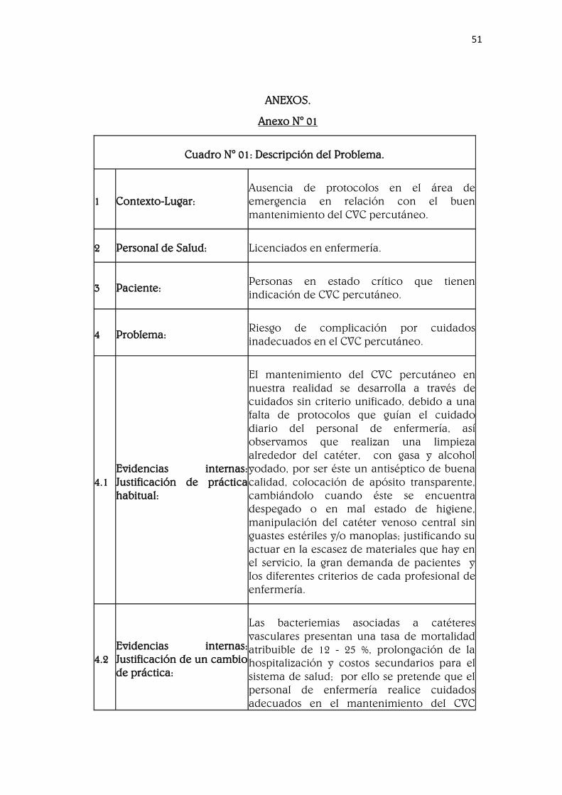
51
ANEXOS.
Anexo N° 01
Cuadro N° 01: Descripción del Problema.
1 Contexto-Lugar:
Ausencia de protocolos en el área de
emergencia en relación con el buen
mantenimiento del CVC percutáneo.
2 Personal de Salud: Licenciados en enfermería.
3 Paciente: Personas en estado crítico que tienen
indicación de CVC percutáneo.
4 Problema: Riesgo de complicación por cuidados
inadecuados en el CVC percutáneo.
4.1
Evidencias internas:
Justificación de práctica
habitual:
El mantenimiento del CVC percutáneo en
nuestra realidad se desarrolla a través de
cuidados sin criterio unificado, debido a una
falta de protocolos que guían el cuidado
diario del personal de enfermería, así
observamos que realizan una limpieza
alrededor del catéter, con gasa y alcohol
yodado, por ser éste un antiséptico de buena
calidad, colocación de apósito transparente,
cambiándolo cuando éste se encuentra
despegado o en mal estado de higiene,
manipulación del catéter venoso central sin
guastes estériles y/o manoplas; justificando su
actuar en la escasez de materiales que hay en
el servicio, la gran demanda de pacientes y
los diferentes criterios de cada profesional de
enfermería.
4.2
Evidencias internas:
Justificación de un cambio
de práctica:
Las bacteriemias asociadas a catéteres
vasculares presentan una tasa de mortalidad
atribuible de 12 - 25 %, prolongación de la
hospitalización y costos secundarios para el
sistema de salud; por ello se pretende que el
personal de enfermería realice cuidados
adecuados en el mantenimiento del CVC
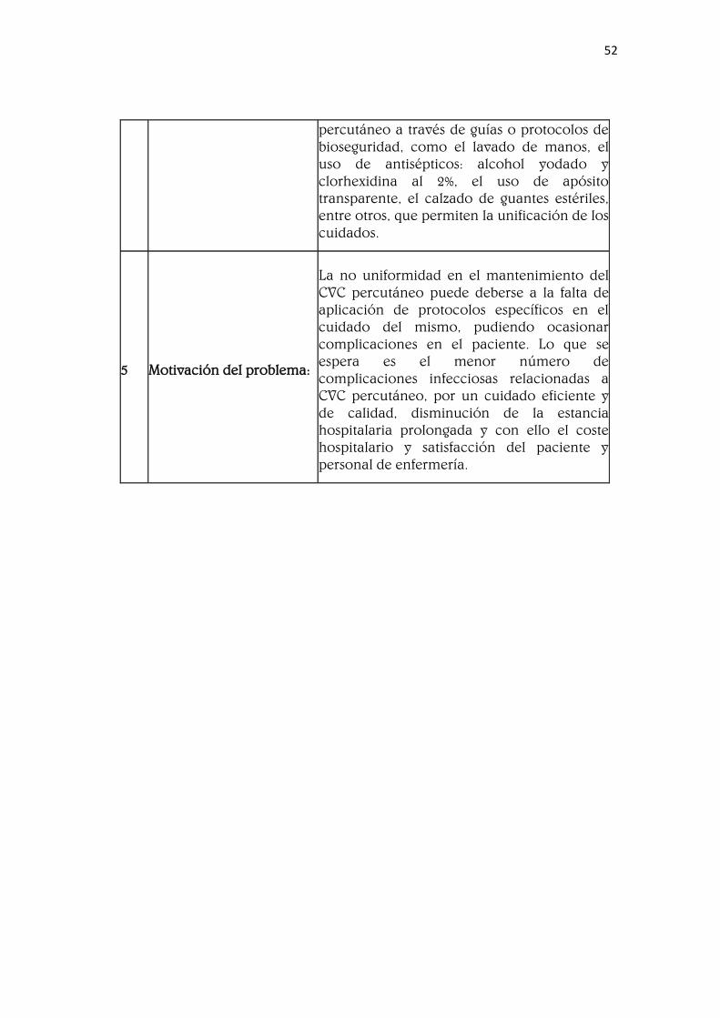
52
percutáneo a través de guías o protocolos de
bioseguridad, como el lavado de manos, el
uso de antisépticos: alcohol yodado y
clorhexidina al 2%, el uso de apósito
transparente, el calzado de guantes estériles,
entre otros, que permiten la unificación de los
cuidados.
5 Motivación del problema:
La no uniformidad en el mantenimiento del
CVC percutáneo puede deberse a la falta de
aplicación de protocolos específicos en el
cuidado del mismo, pudiendo ocasionar
complicaciones en el paciente. Lo que se
espera es el menor número de
complicaciones infecciosas relacionadas a
CVC percutáneo, por un cuidado eficiente y
de calidad, disminución de la estancia
hospitalaria prolongada y con ello el coste
hospitalario y satisfacción del paciente y
personal de enfermería.

53
Anexo N° 02
Cuadro de validez de Gálvez Toro: ARTÍCULO N° 01
Título de la investigación a validar: Implementación de un manojo de
medidas (bundle) de inserción para prevenir la infección del torrente
sanguíneo asociada a dispositivo intravascular central en Cuidado Intensivo
en Colombia.
Metodología: Cuantitativa.
Año: 2010 – 2011.
Pregunta Descripción Respuesta*
¿Cuáles son los
resultados o los
hallazgos?
El empleo de manojo de
medidas para la inserción de
líneas centrales tiene un
impacto positivo en la
disminución de
complicaciones infecciosas
derivadas de dichos
procedimientos.
El manojo de medidas
constituye una buena
alternativa para la
prevención de
infecciones asociadas al
uso de dispositivos
intravasculares, con
relativo bajo costo y
mínimo riesgo para el
paciente.
¿Parecen útiles los
hallazgos para mi
problema?
Un manojo de medidas
contribuye positivamente a la
disminución de la infección en
el torrente sanguíneo por CVC.
Lo resuelve
parcialmente.
¿Son aplicables los
resultados para la
resolución del
problema en tu
medio?
Una infección al torrente
sanguíneo causa mucho
mayor presupuesto y mano de
obra, que la prevención a
través de un manojo de
medidas aplicables, aunado a
esto el profesional siempre
busca cuidar de manera
integral al paciente.
Puede aplicarse bajo
ciertas condiciones.
¿Son seguras las
evidencias para el
paciente?
El desarrollo del estudio fue
aprobado por el Comité de
Ética, el Comité de Infecciones
y el Comité de Docencia e
Investigación de dicho
hospital.
Si.
¿Son válidos los
resultados y los
hallazgos?
La metodología utilizada fue
cuantitativa con diseño de
estudio de intervención tipo
antes y después, entre los
participantes fueron mayores
de 500 personas, pero no tuvo
Se puede considerar.

54
significancia estadística.
Cuadro de validez de Gálvez Toro: ARTÍCULO N° 02
Título de la investigación a validar: Epidemiología de la infección asociada a
catéter venoso central.
Metodología: Semi - cuantitativa.
Año: 2009 – 2010.
Pregunta Descripción Respuesta*
¿Cuáles son los
resultados o los
hallazgos?
Se encontró una tasa de
infecciones alta; se resalta la
importancia de la colonización
de la conexión dentro de la
patogénesis de las infecciones
encontradas. El perfil
microbiológico predominante fue
por contaminantes comunes de
la piel y gérmenes hospitalarios
Gram negativos con altos índices
de resistencia.
Se recomienda la
implementación del
Bundle para
prevención de
infecciones; así mismo
enfatizar en la
uniformidad de
criterios para el
cambio de sistemas de
infusión y la
utilización de
antisépticos.
¿Parecen útiles los
hallazgos para mi
problema?
El catéter venoso central
predispone al paciente que lo
lleva, a una alta probabilidad de
infección, por ello se deben
utilizar medidas de prevención:
bundle.
Lo resuelve
parcialmente.
¿Son aplicables los
resultados para la
resolución del
problema en tu
medio?
El costo por tratar una infección
causada por catéter venoso
central es más alto que las
acciones que se tomen para
prevenir dicha infección.
Puede aplicarse bajo
ciertas condiciones.
¿Son seguras las
evidencias para el
paciente?
Se cumplieron los aspectos
bioéticos según lo establecido por
el Ministerio de Salud de
Colombia.
Es necesaria la
formación previa del
profesional.
¿Son válidos los
resultados y los
hallazgos?
Se hizo un estudio descriptivo
longitudinal en 109 catéteres
venosos centrales, en 82
pacientes pediátricos; se detectó
colonización en sitio de entrada y
conexiones de los catéteres con
método semi-cuantitativo. Se
Se puede considerar.
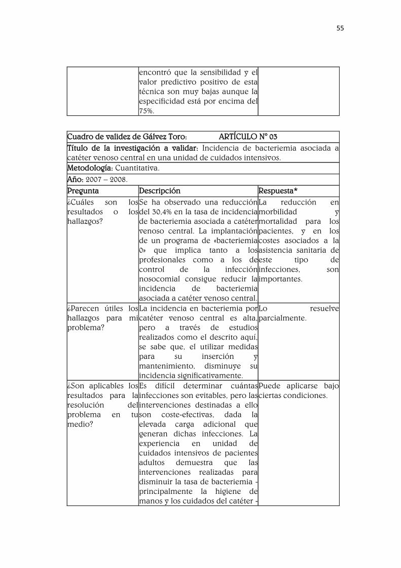
55
encontró que la sensibilidad y el
valor predictivo positivo de esta
técnica son muy bajas aunque la
especificidad está por encima del
75%.
Cuadro de validez de Gálvez Toro: ARTÍCULO N° 03
Título de la investigación a validar: Incidencia de bacteriemia asociada a
catéter venoso central en una unidad de cuidados intensivos.
Metodología: Cuantitativa.
Año: 2007 – 2008.
Pregunta Descripción Respuesta*
¿Cuáles son los
resultados o los
hallazgos?
Se ha observado una reducción
del 30,4% en la tasa de incidencia
de bacteriemia asociada a catéter
venoso central. La implantación
de un programa de «bacteriemia
0» que implica tanto a los
profesionales como a los de
control de la infección
nosocomial consigue reducir la
incidencia de bacteriemia
asociada a catéter venoso central.
La reducción en
morbilidad y
mortalidad para los
pacientes, y en los
costes asociados a la
asistencia sanitaria de
este tipo de
infecciones, son
importantes.
¿Parecen útiles los
hallazgos para mi
problema?
La incidencia en bacteriemia por
catéter venoso central es alta,
pero a través de estudios
realizados como el descrito aquí,
se sabe que, el utilizar medidas
para su inserción y
mantenimiento, disminuye su
incidencia significativamente.
Lo resuelve
parcialmente.
¿Son aplicables los
resultados para la
resolución del
problema en tu
medio?
Es difícil determinar cuántas
infecciones son evitables, pero las
intervenciones destinadas a ello
son coste-efectivas, dada la
elevada carga adicional que
generan dichas infecciones. La
experiencia en unidad de
cuidados intensivos de pacientes
adultos demuestra que las
intervenciones realizadas para
disminuir la tasa de bacteriemia -
principalmente la higiene de
manos y los cuidados del catéter -
Puede aplicarse bajo
ciertas condiciones.
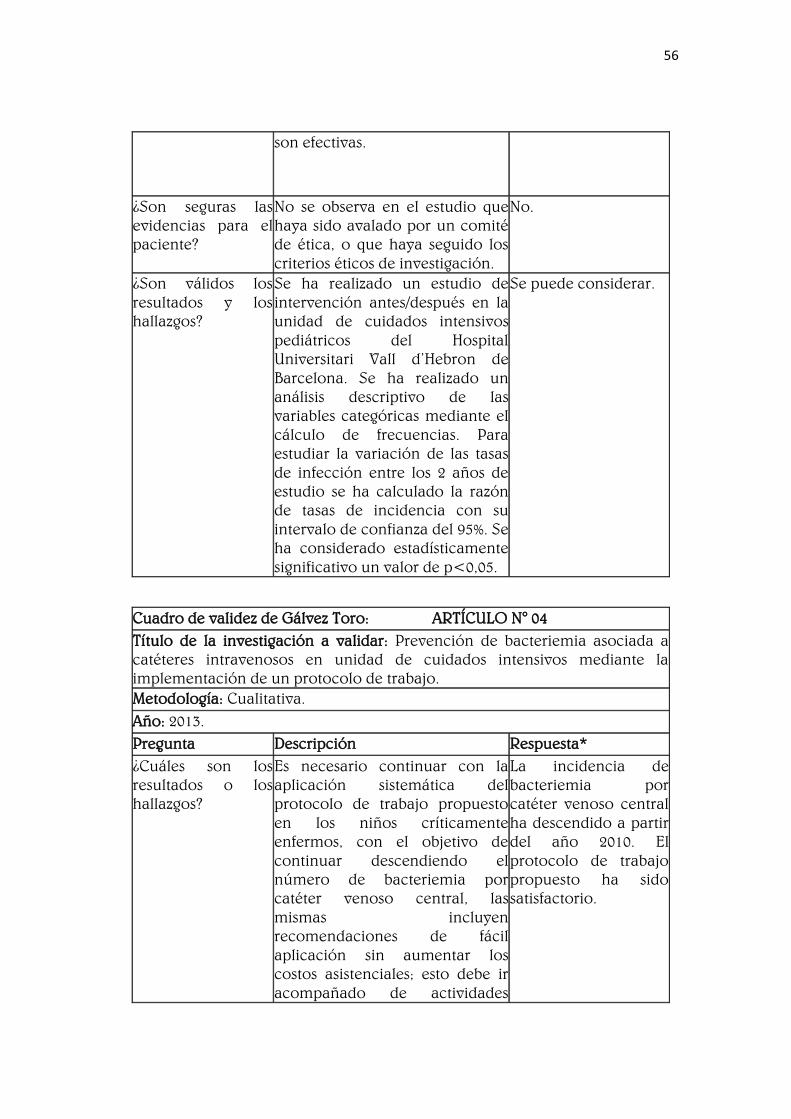
56
son efectivas.
¿Son seguras las
evidencias para el
paciente?
No se observa en el estudio que
haya sido avalado por un comité
de ética, o que haya seguido los
criterios éticos de investigación.
No.
¿Son válidos los
resultados y los
hallazgos?
Se ha realizado un estudio de
intervención antes/después en la
unidad de cuidados intensivos
pediátricos del Hospital
Universitari Vall d’Hebron de
Barcelona. Se ha realizado un
análisis descriptivo de las
variables categóricas mediante el
cálculo de frecuencias. Para
estudiar la variación de las tasas
de infección entre los 2 años de
estudio se ha calculado la razón
de tasas de incidencia con su
intervalo de confianza del 95%. Se
ha considerado estadísticamente
significativo un valor de p<0,05.
Se puede considerar.
Cuadro de validez de Gálvez Toro: ARTÍCULO N° 04
Título de la investigación a validar: Prevención de bacteriemia asociada a
catéteres intravenosos en unidad de cuidados intensivos mediante la
implementación de un protocolo de trabajo.
Metodología: Cualitativa.
Año: 2013.
Pregunta Descripción Respuesta*
¿Cuáles son los
resultados o los
hallazgos?
Es necesario continuar con la
aplicación sistemática del
protocolo de trabajo propuesto
en los niños críticamente
enfermos, con el objetivo de
continuar descendiendo el
número de bacteriemia por
catéter venoso central, las
mismas incluyen
recomendaciones de fácil
aplicación sin aumentar los
costos asistenciales; esto debe ir
acompañado de actividades
La incidencia de
bacteriemia por
catéter venoso central
ha descendido a partir
del año 2010. El
protocolo de trabajo
propuesto ha sido
satisfactorio.

57
educativas y sensibilización del
equipo de salud.
¿Parecen útiles los
hallazgos para mi
problema?
La incidencia en bacteriemia por
catéter venoso central es alta,
pero a través de estudios
realizados como el descrito aquí,
se sabe que disminuye la
incidencia significativamente, al
aplicar medidas de prevención.
Lo resuelve
parcialmente.
¿Son aplicables los
resultados para la
resolución del
problema en tu
medio?
La seguridad del paciente
consiste en reducir a un nivel
mínimo el riesgo (infecciones
intrahospitalarias) y evitar al
máximo la producción de un
evento adverso. Los pacientes
críticamente enfermos presentan
mayor riesgo de desarrollar
infecciones intrahospitalarias por
el uso de dispositivos invasivos,
sin embargo, las medidas de un
protocolo de trabajo reducen
significativamente la
epidemiologia.
Puede aplicarse bajo
ciertas condiciones.
¿Son seguras las
evidencias para el
paciente?
No se observa en el estudio que
haya sido avalado por un comité
de ética, o que haya seguido los
criterios éticos de investigación.
No.
¿Son válidos los
resultados y los
hallazgos?
Se realizó un estudio descriptivo
intervencionista desde el 1 de
mayo de 2013 al 31 de octubre
del mismo año, se incluyeron 45
niños con catéter venoso central
colocados en unidad de cuidados
intensivos pediátricos, con un
total de 51 catéteres venosos
centrales y 419 días de
cateterización. Se identificó un
caso de bacteriemia por catéter
venoso central. La densidad de
incidencia fue 2,38 casos/1.000
días de catéter venoso central.
Se puede considerar.

58
Cuadro de validez de Gálvez Toro: ARTÍCULO N° 05
Título de la investigación a validar: Control de infección relacionada con
catéter venoso central impregnado con antisépticos: revisión integradora.
Metodología: Enfermería basada en evidencia.
Año: 1996 – 2006.
Pregunta Descripción Respuesta*
¿Cuáles son los
resultados o los
hallazgos?
Las publicaciones referidas a
utilización de catéteres
impregnados con antisépticos
mostraron diferencias
estadísticamente significantes en
lo referente a reducción de
colonización microbiana,
mientras apenas un estudio
demostró reducción de
ocurrencia de infección.
El uso de catéteres
venosos centrales
impregnados con los
antisépticos redujo la
colonización
microbiana.
¿Parecen útiles los
hallazgos para mi
problema?
La aplicación de catéteres
venosos centrales impregnados
de antisépticos, muestran una
significativa reducción en cuanto
a las infecciones que puede
presentarse por catéter venoso
central.
Resuelve el problema.
¿Son aplicables los
resultados para la
resolución del
problema en tu
medio?
Son aplicables, puesto que en
cuanto a la relación coste
beneficio, el hospital no quedara
muy afectado en cuanto a la
parte económica, ya que la
mayor demanda de trabajo es por
parte de la enfermera.
Puedo aplicarlo.
¿Son seguras las
evidencias para el
paciente?
No se observa en el estudio que
haya sido avalado por un comité
de ética, o que haya seguido los
criterios éticos de investigación.
No.
¿Son válidos los
resultados y los
hallazgos?
El presente estudio utilizó la
Práctica Basada en la Evidencia,
ya que hace posible la utilización
sistemática de la mejor evidencia
disponible para la evaluación de
opciones y toma de decisiones en
la atención integral del paciente.
Se trata de un estudio de revisión
integradora de la literatura que
permite la investigación anterior
para resumir y sacar conclusiones
Se puede considerar.

59
basadas en el análisis cuidadoso
del diseño metodológico y los
resultados sobre el tema
investigado. Ascendió a nueve
publicaciones sobre el catéter
venoso central impregnados con
un antiséptico, de los cuales siete
(77,8%) correspondieron a los
ensayos controlados aleatorios
(nivel II), y dos (22,2%) de los
meta-análisis (Nivel I pruebas).
Cuadro de validez de Gálvez Toro: ARTÍCULO N° 06
Título de la investigación a validar: Las complicaciones mecánicas e
infecciosas del cateterismo venoso central en una unidad de cuidados
intensivos de nivel terciario en el norte de la India.
Metodología: Estudio observacional prospectivo.
Año: 2008 - 2009.
Pregunta Descripción Respuesta*
¿Cuáles son los
resultados o los
hallazgos?
Entre las complicaciones
mecánicas se hallaron:
complicaciones hemorrágicas
(hematomas y sangrado);
complicaciones relacionadas con
el catéter (retorcimiento de
alambre guía y malas posiciones
de la punta del catéter) y
neumotórax. Dentro de las
complicaciones infecciosas
encontramos las infecciones del
sitio (CVC yugular mayor
probabilidad de infección por la
cercanía a la boca y la secreción
de la oro-faringe, la mayor
densidad de la flora local en la
piel debido a la temperatura de la
piel y las dificultades para
mantener los apósitos oclusivos);
las infecciones de la punta del
catéter (colonización); las
infecciones del torrente
sanguíneo y las infecciones del
sitio de salida.
Las tasas de
complicaciones
mecánicas e
infecciosas asociadas
con CVC son altas. La
ruta de CVC en
yugular, en paralelo
con la ruta del CVC en
subclavia, se asocia
con un mayor número
de complicaciones
hemorrágicas
mecánicas y de
complicaciones
infecciosas como
infecciones de la
punta del catéter
(colonizaciones)
especialmente cuando
se usan durante
períodos de más de 7
días.

60
¿Parecen útiles los
hallazgos para mi
problema?
Los CVC son una parte integral de
la atención del paciente crítico;
sin embargo, las complicaciones
mecánicas e infecciosas
asociadas con estos siguen siendo
altas. Tales complicaciones
plantean enormes cargas en
términos de morbilidad,
mortalidad y gasto sanitario.
Datos como este son útiles para
la planificación e
implementación de la prevención
de las infecciones y las estrategias
de control en cualquier
población a través de normas
básicas de atención, al igual que
los protocolos de higiene de las
manos y el uso de sistemas de
infusión cerrados.
Lo resuelve
parcialmente.
¿Son aplicables los
resultados para la
resolución del
problema en tu
medio?
De una manera u otra, la persona
que se encuentra en estado
crítico, se encuentra más
susceptible a las complicaciones
infecciosas, debido a que se
encuentran invadidos con
dispositivos como el CVC. En los
hospitales de nuestro país
muchas veces no se cuenta con
implementos médicos que nos
permitan un correcto cuidado
para con el paciente, pero existen
acciones que no demandan de
equipos sofisticados como el
lavado de manos y a través de
ello se pueden evitar
complicaciones como las
infecciones.
Puede aplicarse bajo
ciertas condiciones.
¿Son seguras las
evidencias para el
paciente?
El artículo que se valida, expresa
que fue aprobado por el comité
de ética del hospital y el comité
de investigación, permisos
escritos se obtuvieron de los
propios pacientes o de los
familiares o jurídica sustituto.
Es necesaria la
formación previa del
profesional.

61
¿Son válidos los
resultados y los
hallazgos?
Se estudió prospectivamente un
total de 480 colocaciones de CVC,
de éstos, 239 eran en subclavia y
241 en yugular. Se cumplieron
criterios de inclusión y exclusión.
Las precauciones de barrera
estéril fueron utilizadas para la
aplicación del CVC. La elección
del sitio de inserción se dejó a
discreción del médico. La fecha,
lugar y lado de la canulación y el
número de punciones se
observaron para cada
procedimiento. Se realizó
radiografía de tórax en todos los
pacientes para detectar
complicaciones como
neumotórax o hemotórax. En los
pacientes que se sospechó de
infección se realizó análisis
microbiológico. El software de
SPSS para Windows, versión
SPSS 16.0 y el software Epi Info se
utilizó para procesar los datos y
generar las estadísticas media y
desviación estándar. El análisis
univariante se realizó para
comparar la supervivencia con
los grupos no sobrevivientes, y P
<0,05 fue considerado
significativo.
Se puede considerar.
Cuadro de validez de Gálvez Toro: ARTÍCULO N° 07
Título de la investigación a validar: Clorhexidina, octenidina o povidona
yodada para las infecciones relacionadas con el catéter: un ensayo
controlado aleatorizado.
Metodología: Ensayo controlado aleatorizado.
Año:
Pregunta Descripción Respuesta*
¿Cuáles son los
resultados o los
hallazgos?
Hubo una diferencia estadística
significativa entre los grupos: la
tasa de sepsis relacionada con el
catéter fue del 10,5% en el yodo
povidona y grupos de clorhidrato
La clorhexidina es un
agente desinfectante
eficaz en la UCI de
adultos. El uso de 4%
de clorhexidina en

62
de octenidina, y la tasa de sepsis
relacionada con la colonización
fue de 26,3% en el grupo de la
povidona yodada y el 21,5% en el
grupo de clorhidrato de
octenidina. En el grupo de
clorhexidina, no hubo sepsis o
colonización relacionada con el
catéter. Ni reacciones de
hipersensibilidad local ni
sistémica se observaron con el
uso de cualquiera de solución
antiséptica.
lugar de 10% de yodo
povidona o
clorhidrato de
octenidina para la
desinfección cutánea
antes de la inserción
de un dispositivo
intravascular y para el
cuidado del sitio
después de la
inserción puede
reducir
sustancialmente la
incidencia de la
infección relacionada
con el catéter.
¿Parecen útiles los
hallazgos para mi
problema?
A pesar de la importancia de los
catéteres, el uso de estos pone a
los pacientes en riesgo de
complicaciones tales como
infecciones locales, infección del
torrente sanguíneo,
tromboflebitis séptica,
endocarditis entre otras. Además,
estas condiciones contribuyen al
aumento de la morbilidad, la
mortalidad, la duración de la
estancia y el excesivo costo de la
atención. La protección del sitio
del catéter por los agentes
antimicrobianos es uno de los
factores más importantes en la
prevención de la infección.
Apósitos estériles apropiados
deben cubrir el sitio de inserción
y ser sustituidos de manera
regular, povidona yodada,
gluconato de clorhexidina y
clorhidrato de octenidina son los
agentes más comunes utilizados
para vestirse.
Resuelve el problema.
¿Son aplicables los
resultados para la
resolución del
problema en tu
medio?
Actualmente en nuestro país, se
ha dado un pequeño paso
adelante en cuanto a salud, ya
que contamos con muchos
hospitales nuevos, debidamente
Puedo aplicarlo.

63
implementados, en los cuales a
través de la práctica diaria se
puede observar que se usan
antisépticos en el lavado de
manos diario del personal de
salud, medida importante en la
prevención de infecciones. El
costo que acarrea una infección
por catéter venoso central en el
paciente demanda mucho factor
económico, tiempo y riesgo para
la vida del paciente a diferencia
de la prevención que no
demanda tanto dinero ni
esfuerzo, beneficiando al
paciente y al seguro de salud, por
tanto existe una alta posibilidad
de la aplicación del mismo.
¿Son seguras las
evidencias para el
paciente?
El estudio se realizó en
anestesiología y cuidados
intensivos de la unidad de
reanimación de la Universidad de
Eskisehir Osmangazi Facultad de
Medicina, y se realizó tras la
aprobación del comité de ética
del hospital, se obtuvieron los
permisos por escrito, ya sea del
propio paciente o del
representante legal.
Es necesaria la
formación previa del
profesional.
¿Son válidos los
resultados y los
hallazgos?
Los pacientes fueron asignados
aleatoriamente; 57 pacientes se
incluyeron en este ensayo, en
total fueron evaluados 109
catéteres (57 catéteres arteriales y
52 CVC), los cultivos fueron
tomados en el sitio de inserción
del catéter y en el cubo del
catéter después de la retirada
para ayudar a identificar la fuente
de los microorganismos. Todos
los análisis estadísticos se
realizaron con el programa SPSS
13.0 para Windows. Los datos se
expresan como mediana de
características de los pacientes y
los catéteres o porcentajes para
Se puede considerar.

64
las variables categóricas. Un valor
de p <0,05 fue considerado
estadísticamente significativo.
Cuadro de validez de Gálvez Toro: ARTÍCULO N° 08
Título de la investigación a validar: Fortalezas y debilidades en el manejo del
catéter venoso central en una unidad de cuidados intensivos neonatales.
Metodología: Estudio descriptivo-longitudinal.
Año: 2003.
Pregunta Descripción Respuesta*
¿Cuáles son los
resultados o los
hallazgos?
Entre las fortalezas de los
catéteres percutáneos y
umbilicales observados en el
estudio se pueden mencionar: la
limpieza del sitio antes de la
inserción de catéteres y en las
curaciones se realizó con alcohol
yodado, hubo una asepsia
correcta en la preparación de los
medicamentos a infundir y entre
las debilidades se observó una
gran falta del lavado de manos
quirúrgico antes de la inserción
del catéter y la toma de rayos x
post inserción de catéteres, la
manipulación de los catéteres se
realizó sin guantes.
En este estudio el
lavado de las manos
fue la principal
deficiencia del
personal médico y de
enfermería, al ejecutar
los distintos
procedimientos, así
como también la
ausencia de guantes
limpios para las
curaciones de los
catéteres percutáneos.
¿Parecen útiles los
hallazgos para mi
problema?
Así como en los adultos, en los
pacientes pediátricos las
bacteriemias se relacionan con el
uso de catéteres intra-vasculares;
dependiendo del tipo de catéter,
el sitio de inserción, la frecuencia
de manipulación del catéter, la
severidad de la enfermedad y las
condiciones de inserción:
electivas o de urgencias. Por este
motivo se presume que el
aumento en las infecciones
asociadas con catéteres se puede
presentar por deficiencias en su
inserción o en su manejo.
Lo resuelve
parcialmente.
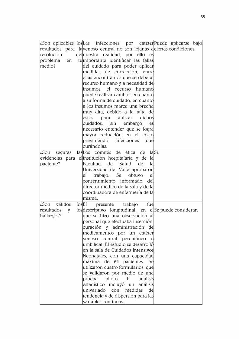
65
¿Son aplicables los
resultados para la
resolución del
problema en tu
medio?
Las infecciones por catéter
venoso central no son lejanas a
nuestra realidad, por ello es
importante identificar las fallas
del cuidado para poder aplicar
medidas de corrección, entre
ellas encontramos que se debe al
recurso humano y a necesidad de
insumos, el recurso humano
puede realizar cambios en cuanto
a su forma de cuidado, en cuanto
a los insumos marca una brecha
muy alta, debido a la falta de
estos para aplicar dichos
cuidados, sin embargo es
necesario entender que se logra
mayor reducción en el costo
previniendo infecciones que
curándolas.
Puede aplicarse bajo
ciertas condiciones.
¿Son seguras las
evidencias para el
paciente?
Los comités de ética de la
institución hospitalaria y de la
Facultad de Salud de la
Universidad del Valle aprobaron
el trabajo. Se obtuvo el
consentimiento informado del
director médico de la sala y de la
coordinadora de enfermería de la
misma.
Si.
¿Son válidos los
resultados y los
hallazgos?
El presente trabajo fue
descriptivo longitudinal, en el
que se hizo una observación al
personal que efectuaba inserción,
curación y administración de
medicamentos por un catéter
venoso central percutáneo o
umbilical. El estudio se desarrolló
en la sala de Cuidados Intensivos
Neonatales, con una capacidad
máxima de 62 pacientes. Se
utilizaron cuatro formularios, que
se validaron por medio de una
prueba piloto. El análisis
estadístico incluyó un análisis
univariado con medidas de
tendencia y de dispersión para las
variables continuas.
Se puede considerar.
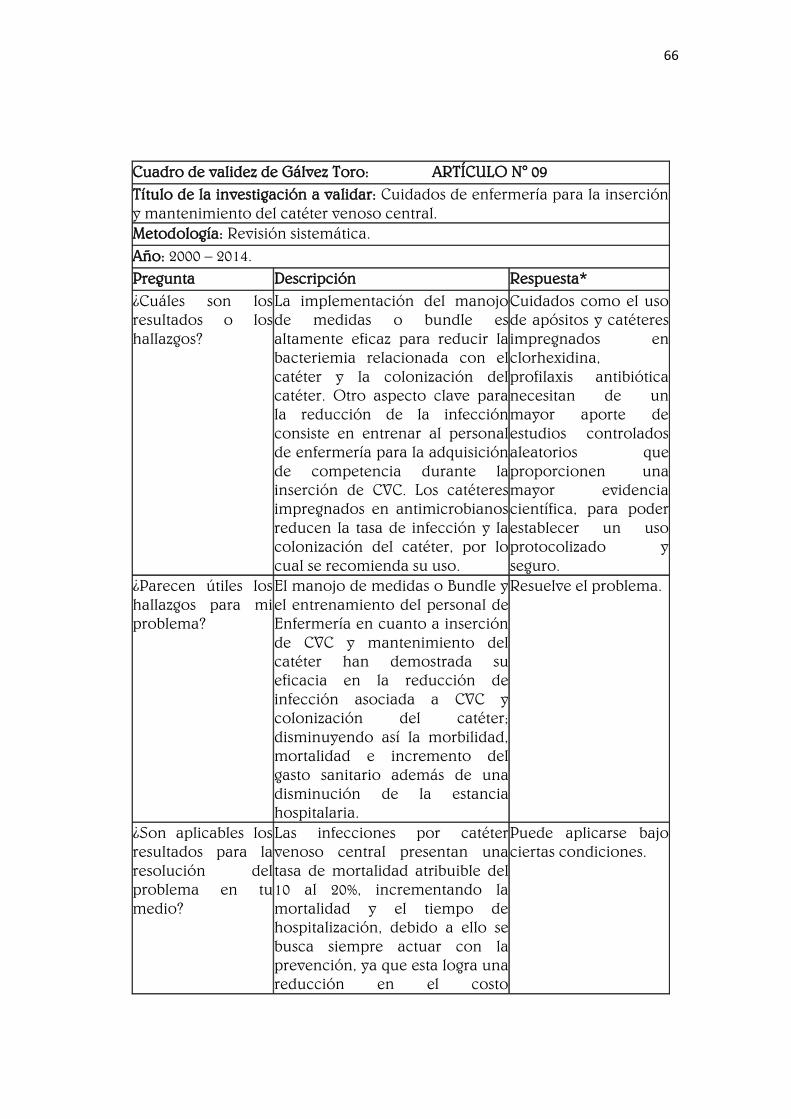
66
Cuadro de validez de Gálvez Toro: ARTÍCULO N° 09
Título de la investigación a validar: Cuidados de enfermería para la inserción
y mantenimiento del catéter venoso central.
Metodología: Revisión sistemática.
Año: 2000 – 2014.
Pregunta Descripción Respuesta*
¿Cuáles son los
resultados o los
hallazgos?
La implementación del manojo
de medidas o bundle es
altamente eficaz para reducir la
bacteriemia relacionada con el
catéter y la colonización del
catéter. Otro aspecto clave para
la reducción de la infección
consiste en entrenar al personal
de enfermería para la adquisición
de competencia durante la
inserción de CVC. Los catéteres
impregnados en antimicrobianos
reducen la tasa de infección y la
colonización del catéter, por lo
cual se recomienda su uso.
Cuidados como el uso
de apósitos y catéteres
impregnados en
clorhexidina,
profilaxis antibiótica
necesitan de un
mayor aporte de
estudios controlados
aleatorios que
proporcionen una
mayor evidencia
científica, para poder
establecer un uso
protocolizado y
seguro.
¿Parecen útiles los
hallazgos para mi
problema?
El manojo de medidas o Bundle y
el entrenamiento del personal de
Enfermería en cuanto a inserción
de CVC y mantenimiento del
catéter han demostrada su
eficacia en la reducción de
infección asociada a CVC y
colonización del catéter;
disminuyendo así la morbilidad,
mortalidad e incremento del
gasto sanitario además de una
disminución de la estancia
hospitalaria.
Resuelve el problema.
¿Son aplicables los
resultados para la
resolución del
problema en tu
medio?
Las infecciones por catéter
venoso central presentan una
tasa de mortalidad atribuible del
10 al 20%, incrementando la
mortalidad y el tiempo de
hospitalización, debido a ello se
busca siempre actuar con la
prevención, ya que esta logra una
reducción en el costo
Puede aplicarse bajo
ciertas condiciones.

67
económico, pero sobre todo una
atención de calidad al paciente
logrando la prevención de
infecciones, para ello
necesitamos de más
capacitaciones para el recurso
humano y materiales, que hoy en
día creo que si es posible su
gestión.
¿Son seguras las
evidencias para el
paciente?
No se observa en el estudio que
haya sido avalado por un comité
de ética, o que haya seguido los
criterios éticos de investigación.
No.
¿Son válidos los
resultados y los
hallazgos?
La búsqueda es seleccionada a
partir de una serie de criterios de
inclusión como: revisiones
sistemáticas mayoritariamente de
ensayos controlados aleatorios y
artículos primarios sobre
pacientes adultos en unidades
como UCI y pacientes
oncológicos, artículos primarios
publicados desde el año 2000
hasta abril de 2014, artículos y
revisiones escritas en castellano,
inglés y/o portugués, cumplen
con excepción las guías de
práctica clínica. De los 28
artículos revisados, 16 son
revisiones sistemáticas y 12 son
artículos primarios.
Se puede considerar.
Cuadro de validez de Gálvez Toro: ARTÍCULO N° 10
Título de la investigación a validar: Guidelines for the Prevention of
Intravascular Catheter-Related Infections
Metodología: Guía clínica.
Año: 2011.
Pregunta Descripción Respuesta*
¿Cuáles son los
resultados o los
hallazgos?
Los resultados son las diversas
actividades específicas que se
registran para prevenir las
infecciones relacionadas con los
diversos tipos de catéteres
intravenosos.
La guía proporciona
recomendaciones
basadas en la
experiencia para
prevenir las
infecciones
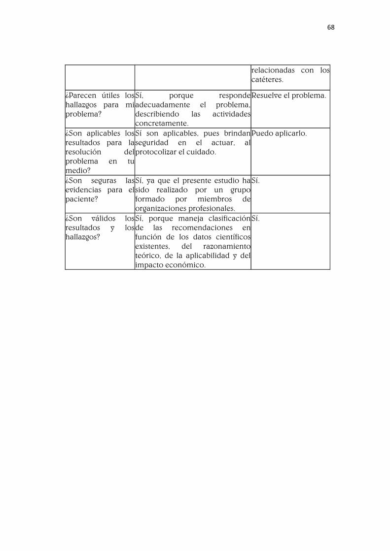
68
relacionadas con los
catéteres.
¿Parecen útiles los
hallazgos para mi
problema?
Sí, porque responde
adecuadamente el problema,
describiendo las actividades
concretamente.
Resuelve el problema.
¿Son aplicables los
resultados para la
resolución del
problema en tu
medio?
Sí son aplicables, pues brindan
seguridad en el actuar, al
protocolizar el cuidado.
Puedo aplicarlo.
¿Son seguras las
evidencias para el
paciente?
Sí, ya que el presente estudio ha
sido realizado por un grupo
formado por miembros de
organizaciones profesionales.
Sí.
¿Son válidos los
resultados y los
hallazgos?
Sí, porque maneja clasificación
de las recomendaciones en
función de los datos científicos
existentes, del razonamiento
teórico, de la aplicabilidad y del
impacto económico.
Sí.

69
Anexo N° 03
Guidelines for the Prevention of
Intravascular Catheter-Related
Infections, 2011
Naomi P. O'Grady, M.D.1, Mary Alexander, R.N.
2, Lillian A. Burns, M.T., M.P.H.,
C.I.C.3,
E. Patchen Dellinger, M.D.4,
Jeffery Garland, M.D., S.M.5,
Stephen O.
Heard, M.D.6,
Pamela A. Lipsett, M.D.7,
Henry Masur, M.D.1,
Leonard A. Mermel,
D.O., Sc.M.8,
Michele L. Pearson, M.D.9,
Issam I. Raad, M.D.10,
Adrienne
Randolph, M.D., M.Sc.11,
Mark E. Rupp, M.D.12,
Sanjay Saint, M.D., M.P.H.13
and the Healthcare Infection Control Practices Advisory Committee
(HICPAC)14
.
1 National Institutes of Health, Bethesda, Maryland
2 Infusion Nurses Society, Norwood, Massachusetts
3 Greenich Hospital, Greenwich, Connecticut
4 University of Washington, Seattle, Washington
5 Wheaton Franciscan Healthcare-St. Joseph, Milwaukee, Wisconsin
6 University of Massachusetts Medical School, Worcester, Massachusetts
7 Johns Hopkins University School of Medicine, Baltimore, Maryland
8 Warren Alpert Medical School of Brown University and Rhode Island
Hospital, Providence, Rhode Island
9 Office of Infectious Diseases, CDC, Atlanta, Georgia 10 MD Anderson Cancer
Center, Houston, Texas
11 The Children's Hospital, Boston, Massachusetts
12 University of Nebraska Medical Center, Omaha, Nebraska
13 Ann Arbor VA Medical Center and University of Michigan, Ann Arbor,
Michigan
Available from: https://www.cdc.gov/infectioncontrol/guidelines/bsi/
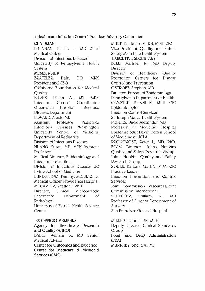
70
4 Healthcare Infection Control Practices Advisory Committee
CHAIRMAN
BRENNAN, Patrick J., MD Chief
Medical Officer
Division of Infectious Diseases
University of Pennsylvania Health
System
MEMBERSHIP
BRATZLER, Dale, DO, MPH
President and CEO
Oklahoma Foundation for Medical
Quality
BURNS, Lillian A., MT, MPH
Infection Control Coordinator
Greenwich Hospital, Infectious
Diseases Department
ELWARD, Alexis, MD
Assistant Professor, Pediatrics
Infectious Diseases Washington
University School of Medicine
Department of Pediatrics
Division of Infectious Diseases
HUANG, Susan, MD, MPH Assistant
Professor
Medical Director, Epidemiology and
Infection Prevention
Division of Infectious Diseases UC
Irvine School of Medicine
LUNDSTROM, Tammy, MD, JD Chief
Medical Officer Providence Hospital
MCCARTER, Yvette S., PhD
Director, Clinical Microbiology
Laboratory Department of
Pathology
University of Florida Health Science
Center
MURPHY, Denise M. RN, MPH, CIC
Vice President, Quality and Patient
Safety Main Line Health System
EXECUTIVE SECRETARY
BELL, Michael R., MD Deputy
Director
Division of Healthcare Quality
Promotion Centers for Disease
Control and Prevention
OSTROFF, Stephen, MD
Director, Bureau of Epidemiology
Pennsylvania Department of Health
OLMSTED, Russell N., MPH, CIC
Epidemiologist
Infection Control Services
St. Joseph Mercy Health System
PEGUES, David Alexander, MD
Professor of Medicine, Hospital
Epidemiologist David Geffen School
of Medicine at UCLA
PRONOVOST, Peter J., MD, PhD,
FCCM Director, Johns Hopkins
Quality and Safety Research Group
Johns Hopkins Quality and Safety
Research Group
SOULE, Barbara M., RN, MPA, CIC
Practice Leader
Infection Prevention and Control
Services
Joint Commission Resources/Joint
Commission International
SCHECTER, William, P., MD
Professor of Surgery Department of
Surgery
San Francisco General Hospital
EX-OFFICIO MEMBERS
Agency for Healthcare Research
and Quality (AHRQ)
BAINE, William B., MD Senior
Medical Advisor
Center for Outcomes and Evidence
Center for Medicare & Medicaid
Services (CMS)
MILLER, Jeannie, RN, MPH
Deputy Director, Clinical Standards
Group
Food and Drug Administration
(FDA)
MURPHEY, Sheila A., MD

71
Division of Anesthesiology, General
Hospital Infection Control Dental
Devices
Center for Devices and Radiology
Health
LIAISONS
Advisory Council for the
Elimination of Tuberculosis (ACET)
STRICOF, Rachel L., MPH
American College of Occupational
and Environmental Medicine
RUSSI, Mark, MD, MPH Professor of
Medicine
Yale University School of Medicine
Director, Occupational Health
Yale-New Haven Hospital
American Health Care Assn (AHCA)
FITZLER, Sandra L., RN
Senior Director of Clinical Services
American Hospital Association
(AHA)
SCHULMAN, Roslyne, MHA, MBA
Director, Policy Development
Association of Professionals of
Infection Control and
Epidemiology, Inc. (APIC)
DeBAUN, Barbara, MSN, RN, CIC
Association of periOperative
Registered Nursed (AORN)
BLANCHARD, Joan C., RN, BSN
Council of State and Territorial
Epidemiologists (CSTE)
KAINER, Marion, MD, MPH
Director, Hospital Infections and
Antimicrobial Resistance Program
Tennessee Department Health
National Institute of Health (NIH)
HENDERSON, David, MD
Deputy Director for Clinical Care
Associate Director for Hospital
Epidemiology and
Quality Improvement NIH Clinical
Center
Department of Veterans Affairs (VA)
ROSELLE, Gary A., MD
National Program Director,
Infectious Diseases VA Central
Office
Cincinnati VA Medical Center
Consumers Union
MCGIFFERT, Lisa
Senior Policy Analyst on Health
Issues Project Director Stop Hospital
Infections Organization
Infectious Disease Society of
America (IDSA)
HUSKINS, W. Charles MD, MSc
Division of Pediatric Infectious
Diseases Assistant Professor of
Pediatrics
Mayo Clinic
Public Health Agency of Canada
PATON, Shirley, RN, MN
Senior Advisor Healthcare Acquired
Infections
Center for Communicable Diseases
and Infection Control
Society for Healthcare
Epidemiology of America (SHEA)
MARAGAKIS, Lisa, MD
Assistant Professor of Medicine John
Hopkins Medical Institutions
Society of Hospital Medicine
SAINT, Sanjay, MD, MPH
Director, Ann Arbor VA Medical
Center/University of Michigan
Patient Safety Enhancement
Program
The Joint Commission
WISE, Robert A., MD Vice President
Division of Standards & Survey
Methods

72
Use of trade names and commercial sources is for identification only and does
not imply endorsement by the U.S. Department of Health and Human Services.
Acknowledgments
We wish to acknowledge Ingi Lee, MD, MSCE and Craig A. Umscheid, MD,
MSCE from Center for Evidence-based Practice, University of Pennsylvania
Health System Philadelphia, PA who performed a systematic review for issues
raised the during the guideline process.
Disclosure of potential conflicts of interests. N.P.O.’G. served as a board
member for the ABIM Subspecialty Board for Critical Care Medicine. M.A. is an
employee of the Infusion Nurses Society, Honoraria from 3M, Becton
Dickinson, Smiths Medical. L.A.B. is a consultant for Institute of Healthcare
Improvement, Board membership for Theradoc, Medline. Honoraria from
APIC, Clorox. E.P.D. consulting from Merck, Baxter, Ortho-McNeil, Targanta,
Schering- Plough, Optimer, Cadence, Cardinal, BDGeneOhm, WebEx, Cerebrio,
and Tyco. Grant support through the NIH. Payment for lecture from Merck.
Payment for development of educational presentation from Medscape. Travel
and meeting expenses paid for by ASHP, IDSA, ASM, American College of
Surgeons, NQF, SHEA/CDC, HHS, Trauma Shock Inflammation and Sepsis
Meeting (Munich), University of Minnesota. J.G. Honoria from Ethicon. S.O.H.
provides research support from Angiotech; Honoraria from Angiotech, Merck.
L.A.M provides research support from Astellas, Theravance, Pfizer; Consulting
for Ash Access, Cadence, CorMedix, Catheter Connections, Carefusion, Sage,
Bard, Teleflex; Payment for manuscript preparation from Catheter
Connections. I.I.R. provides research support from Cubist, Enzon, and Basilea;
Consulting for Clorox; Stock Equity or Options in Great Lakes
Pharmaceuticalsand Inventive Protocol; Speakers Bureau for Cook, Inc.;
Royalty income (patents owned by MD Anderson on which Dr. Raad in an
inventor: American Medical Systems, Cook, Inc., Cook urological, Teleflex,
TyRx, Medtronic, Biomet, Great Lakes Pharmaceuticals. A.R. consulting income
from Eisai Pharmaceuticals, Discovery Laboratories. M.E.R. provides research
support from Molnlycke, Cardinal Healthcare Foundation, Sanofi-Pasteur, 3M,
and Cubist; Consulting from Semprus; Honorarium for lectures from 3M,
Carefusion, Baxter and Becton Dickinson. Previously served on Board of
Directors for Society for Healthcare Epidemiology of America. All other
authors: no conflicts.

73
Table of Contents
Notice to Readers 7
Introduction 9
Summary of Recommendations 9
1. Education, Training and Staffing 9
2. Selection of Catheters and Sites 10
2.1. Peripheral Catheters and Midline Catheters 10
2. Central Venous Catheters 10
3. Hand Hygiene and Aseptic Technique 12
4. Maximal Sterile Barrier Precautions 12
5. Skin Preparation 13
6. Catheter Site Dressing Regimens 13
7. Patient Cleansing 15
8. Catheter Securement Devices 15
9. Antimicrobial/Antiseptic Impregnated Catheters and Cuffs 15
10. Systemic Antibiotic Prophylaxis 15
11. Antibiotic/Antiseptic Ointments 15
12. Antibiotic Lock Prophylaxis, Antimicrobial Catheter Flush and
Catheter Lock Prophylaxis
16
13. Anticoagulants 16
14. Replacement of Peripheral and Midline Catheters 16
15. Replacement of CVCs, Including PICCs and Hemodialysis Catheters 16
16. Umbilical Catheters 17
17. Peripheral Arterial Catheters and Pressure Monitoring Devices for
Adult and Pediatric Patients
18
18. Replacement of Administration Sets 19
19. Needleless Intravascular Catheter Systems 19
20. Performance Improvement 20
Background Information 20
Terminology and Estimates of Risk 20
Table 1. Catheters Used for Venous and Arterial Access 22
Epidemiology and Microbiology in Adult and Pediatric Patients 23
Pathogenesis 24
Strategies for Prevention of Catheter-Related Infections in Adult and
Pediatric Patients
25
1. Education, Training and Staffing 25
2. Selection of Catheters and Sites 26
3. Hand Hygiene and Aseptic Technique 29
4. Maximal Sterile Barrier Precautions 30
5. Skin Preparation 31
6. Catheter Site Dressing Regimens 32
7. Patient Cleansing 35
8. Catheter Securement Devices 36
9. Antimicrobial/Antiseptic Impregnated Catheters and Cuffs 36
10. Systemic Antibiotic Prophylaxis 39

74
Notice to Readers:
In 2009, the Centers for Disease Control and Prevention (CDC) and Healthcare
Infection Control Practices Advisory Committee (HICPAC) integrated current
advances in guideline production and implementation into its development
process ([This link is no longer active:
http://www.cdc.gov/hicpac/guidelineMethod/guidelineMethod.html. Similar
information may be found at Umscheid CA, Agarwal RK, Brennan PJ,
Healthcare Infection Control Practices Advisory Committee. Updating the
guideline development methodology of the Healthcare Infection Control
Practices Advisory Committee (HICPAC). American journal of infection control.
2010;38(4):264-273.]). The new methodology enables CDC and HICPAC to
improve the validity and usability of its guidelines while also addressing
emerging challenges in guideline development in the area of infection
prevention and control. However, the Guidelines for the Prevention of
Intravascular Catheter-Related Infections were initiated before the
methodology was revised. Therefore, this guideline reflects the development
methods that were used for guidelines produced prior to 2009. Future revisions
will be performed using the updated methodology.
These guidelines have been developed for healthcare personnel who insert
intravascular catheters and for persons responsible for surveillance and control
of infections in hospital, outpatient, and home healthcare settings. This report
was prepared by a working group comprising members from professional
organizations representing the disciplines of critical care medicine, infectious
diseases, healthcare infection control, surgery, anesthesiology, interventional
radiology, pulmonary medicine, pediatric medicine, and nursing. The working
group was led by the Society of Critical Care Medicine (SCCM), in collaboration
with the Infectious Diseases Society of America (IDSA), Society for Healthcare
Epidemiology of America (SHEA), Surgical Infection Society (SIS), American
College of Chest Physicians (ACCP), American Thoracic Society (ATS),
American Society of Critical Care Anesthesiologists (ASCCA), Association for
Professionals in Infection Control and Epidemiology (APIC), Infusion Nurses
11. Antibiotic/Antiseptic Ointments 40
12. Antibiotic Lock Prophylaxis, Antimicrobial Catheter Flush and
Catheter Lock Prophylaxis
41
13. Anticoagulants 43
14. Replacement of Peripheral and Midline Catheters 45
15. Replacement of CVCs, Including PICCs and Hemodialysis Catheters 46
16. Umbilical Catheters 49
17. Peripheral Arterial Catheters and Pressure Monitoring Devices for
Adult and Pediatric Patients
51
18. Replacement of Administration Sets 53
19. Needleless Intravascular Catheter Systems 54
20. Performance Improvement 57
References 59

75
Society (INS), Oncology Nursing Society (ONS), American Society for
Parenteral and Enteral Nutrition (ASPEN), Society of Interventional Radiology
(SIR), American Academy of Pediatrics (AAP), Pediatric Infectious Diseases
Society (PIDS), and the Healthcare Infection Control Practices Advisory
Committee (HICPAC) of the Centers for Disease Control and Prevention (CDC)
and is intended to replace the Guideline for Prevention of Intravascular
Catheter-Related Infections published in 2002. These guidelines are intended to
provide evidence-based Recommendations for preventing intravascular
catheter-related infections. Major areas of emphasis include
1. Educating and training healthcare personnel who insert and maintain
catheters;
2. Using maximal sterile barrier precautions during central venous catheter
insertion;
3. Using a > 0.5% chlorhexidine skin preparation with alcohol for antisepsis;
4. Avoiding routine replacement of central venous catheters as a strategy to
prevent infection; and
5. Asing antiseptic/antibiotic impregnated short-term central venous catheters
and chlorhexidine impregnated sponge dressings if the rate of infection is not
decreasing despite adherence to other strategies (i.e., education and training,
maximal sterile barrier precautions, and >0.5% chlorhexidine preparations
with alcohol for skin antisepsis).
These guidelines also emphasize performance improvement by implementing
bundled strategies, and documenting and reporting rates of compliance with
all components of the bundle as benchmarks for quality assurance and
performance improvement.
As in previous guidelines issued by CDC and HICPAC, each recommendation is
categorized on the basis of existing scientific data, theoretical rationale,
applicability, and economic impact. The system for categorizing
Recommendations in this guideline is as follows:
Category IA. Strongly recommended for implementation and strongly
supported by well- designed experimental, clinical, or epidemiologic studies.
Category IB. Strongly recommended for implementation and supported by
some experimental, clinical, or epidemiologic studies and a strong theoretical
rationale; or an accepted practice (e.g., aseptic technique) supported by limited
evidence.
Category IC. Required by state or federal regulations, rules, or standards.
Category II. Suggested for implementation and supported by suggestive clinical
or epidemiologic studies or a theoretical rationale.
Unresolved issue. Represents an unresolved issue for which evidence is
insufficient or no consensus regarding efficacy exists.

76
Introduction
In the United States, 15 million central vascular catheter (CVC) days (i.e., the
total number of days of exposure to CVCs among all patients in the selected
population during the selected time period) occur in intensive care units (ICUs)
each year [1]. Studies have variously addressed catheter- related bloodstream
infections (CRBSI). These infections independently increase hospital costs and
length of stay [2-5], but have not generally been shown to independently
increase mortality. While 80,000 CRBSIs occur in ICUs each year [1], a total of
250,000 cases of BSIs have been estimated to occur annually, if entire hospitals
are assessed [6]. By several analyses, the cost of these infections is substantial,
both in terms of morbidity and financial resources expended. To improve
patient outcome and to reduce healthcare costs, there is considerable interest
by healthcare providers, insurers, regulators, and patient advocates in reducing
the incidence of these infections. This effort should be multidisciplinary,
involving healthcare professionals who order the insertion and removal of
CVCs, those personnel who insert and maintain intravascular catheters,
infection control personnel, healthcare managers including the chief executive
officer (CEO) and those who allocate resources, and patients who are capable
of assisting in the care of their catheters.
The goal of an effective prevention program should be the elimination of
CRBSI from all patient- care areas. Although this is challenging, programs have
demonstrated success, but sustained elimination requires continued effort. The
goal of the measures discussed in this document is to reduce the rate to as low
as feasible given the specific patient population being served, the universal
presence of microorganisms in the human environment, and the limitations of
current strategies and technologies.
Summary of Recommendations
Edit [February 2017]: An * indicates recommendations that were renumbered
for clarity. The renumbering does not constitute change to the intent of the
recommendations.
1. * Education, Training and Staffing
1. Educate healthcare personnel regarding the indications for intravascular
catheter use, proper procedures for the insertion and maintenance of
intravascular catheters, and appropriate infection control measures to prevent
intravascular catheter-related infections [7–15]. Category IA
2. Periodically assess knowledge of and adherence to guidelines for all
personnel involved in the insertion and maintenance of intravascular catheters
[7–15]. Category IA
3. Designate only trained personnel who demonstrate competence for the
insertion and maintenance of peripheral and central intravascular catheters.
[14–28]. Category IA
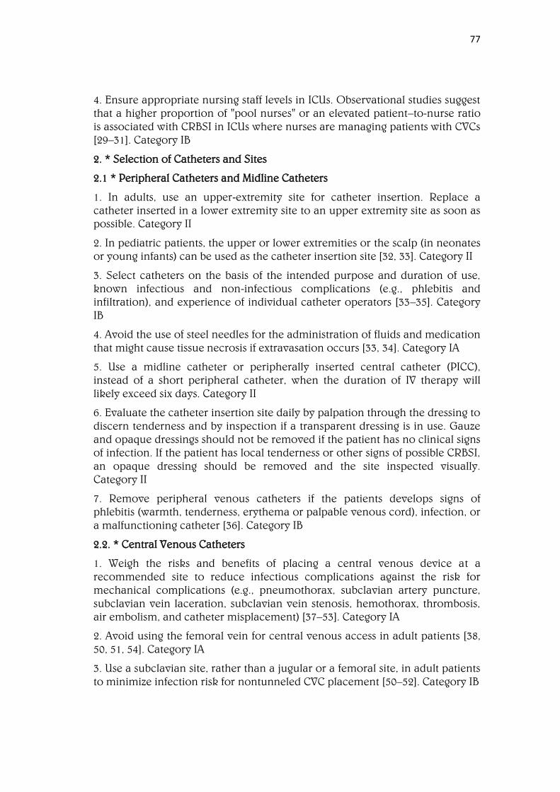
77
4. Ensure appropriate nursing staff levels in ICUs. Observational studies suggest
that a higher proportion of "pool nurses" or an elevated patient–to-nurse ratio
is associated with CRBSI in ICUs where nurses are managing patients with CVCs
[29–31]. Category IB
2. * Selection of Catheters and Sites
2.1 * Peripheral Catheters and Midline Catheters
1. In adults, use an upper-extremity site for catheter insertion. Replace a
catheter inserted in a lower extremity site to an upper extremity site as soon as
possible. Category II
2. In pediatric patients, the upper or lower extremities or the scalp (in neonates
or young infants) can be used as the catheter insertion site [32, 33]. Category II
3. Select catheters on the basis of the intended purpose and duration of use,
known infectious and non-infectious complications (e.g., phlebitis and
infiltration), and experience of individual catheter operators [33–35]. Category
IB
4. Avoid the use of steel needles for the administration of fluids and medication
that might cause tissue necrosis if extravasation occurs [33, 34]. Category IA
5. Use a midline catheter or peripherally inserted central catheter (PICC),
instead of a short peripheral catheter, when the duration of IV therapy will
likely exceed six days. Category II
6. Evaluate the catheter insertion site daily by palpation through the dressing to
discern tenderness and by inspection if a transparent dressing is in use. Gauze
and opaque dressings should not be removed if the patient has no clinical signs
of infection. If the patient has local tenderness or other signs of possible CRBSI,
an opaque dressing should be removed and the site inspected visually.
Category II
7. Remove peripheral venous catheters if the patients develops signs of
phlebitis (warmth, tenderness, erythema or palpable venous cord), infection, or
a malfunctioning catheter [36]. Category IB
2.2. * Central Venous Catheters
1. Weigh the risks and benefits of placing a central venous device at a
recommended site to reduce infectious complications against the risk for
mechanical complications (e.g., pneumothorax, subclavian artery puncture,
subclavian vein laceration, subclavian vein stenosis, hemothorax, thrombosis,
air embolism, and catheter misplacement) [37–53]. Category IA
2. Avoid using the femoral vein for central venous access in adult patients [38,
50, 51, 54]. Category IA
3. Use a subclavian site, rather than a jugular or a femoral site, in adult patients
to minimize infection risk for nontunneled CVC placement [50–52]. Category IB

78
4. No recommendation can be made for a preferred site of insertion to
minimize infection risk for a tunneled CVC. Unresolved issue
5. Avoid the subclavian site in hemodialysis patients and patients with
advanced kidney disease, to avoid subclavian vein stenosis [53,55–58].
Category IA
6. Use a fistula or graft in patients with chronic renal failure instead of a CVC
for permanent access for dialysis [59]. Category IA
7. Use ultrasound guidance to place central venous catheters (if this technology
is available) to reduce the number of cannulation attempts and mechanical
complications. Ultrasound guidance should only be used by those fully trained
in its technique. [60–64]. Category IB
8. Use a CVC with the minimum number of ports or lumens essential for the
management of the patient [65–68]. Category IB
9. No recommendation can be made regarding the use of a designated lumen
for parenteral nutrition. Unresolved issue
10. Promptly remove any intravascular catheter that is no longer essential [69–
72]. Category IA
11. When adherence to aseptic technique cannot be ensured (i.e., catheters
inserted during a medical emergency), replace the catheter as soon as possible,
i.e., within 48 hours [37,73– 76]. Category IB
3. * Hand Hygiene and Aseptic Technique
1. Perform hand hygiene procedures, either by washing hands with
conventional soap and water or with alcohol-based hand rubs (ABHR). Hand
hygiene should be performed before and after palpating catheter insertion sites
as well as before and after inserting, replacing, accessing, repairing, or dressing
an intravascular catheter. Palpation of the insertion site should not be
performed after the application of antiseptic, unless aseptic technique is
maintained [12, 77–79]. Category IB
2. Maintain aseptic technique for the insertion and care of intravascular
catheters [37, 73, 74, 76]. Category IB
3. Wear clean gloves, rather than sterile gloves, for the insertion of peripheral
intravascular catheters, if the access site is not touched after the application of
skin antiseptics. Category IC
4. Sterile gloves should be worn for the insertion of arterial, central, and
midline catheters [37, 73, 74, 76]. Category IA
5. Use new sterile gloves before handling the new catheter when guidewire
exchanges are performed. Category II
6. Wear either clean or sterile gloves when changing the dressing on
intravascular catheters. Category IC

79
4. * Maximal Sterile Barrier Precautions
1. Use maximal sterile barrier precautions, including the use of a cap, mask,
sterile gown, sterile gloves, and a sterile full body drape, for the insertion of
CVCs, PICCs, or guidewire exchange [14, 75, 76, 80]. Category IB
2. Use a sterile sleeve to protect pulmonary artery catheters during insertion
[81]. Category IB
5. * Skin Preparation
1. Prepare clean skin with an antiseptic (70% alcohol, tincture of iodine, or
alcoholic chlorhexidine gluconate solution) before peripheral venous catheter
insertion [82]. Category IB
2. Prepare clean skin with a >0.5% chlorhexidine preparation with alcohol
before central venous catheter and peripheral arterial catheter insertion and
during dressing changes. If there is a contraindication to chlorhexidine,
tincture of iodine, an iodophor, or 70% alcohol can be used as alternatives [82,
83]. Category IA
3. No comparison has been made between using chlorhexidine preparations
with alcohol and povidone-iodine in alcohol to prepare clean skin. Unresolved
issue.
4. No recommendation can be made for the safety or efficacy of chlorhexidine
in infants aged <2 months. Unresolved issue
5. Antiseptics should be allowed to dry according to the manufacturer’s
recommendation prior to placing the catheter [82, 83]. Category IB
6. * Catheter Site Dressing Regimens
1. Use either sterile gauze or sterile, transparent, semipermeable dressing to
cover the catheter site [84–87]. Category IA
2. If the patient is diaphoretic or if the site is bleeding or oozing, use a gauze
dressing until this is resolved [84–87]. Category II
3. Replace catheter site dressing if the dressing becomes damp, loosened, or
visibly soiled [84, 85]. Category IB
4. Do not use topical antibiotic ointment or creams on insertion sites, except
for dialysis catheters, because of their potential to promote fungal infections
and antimicrobial resistance [88, 89]. Category IB
5. Do not submerge the catheter or catheter site in water. Showering should be
permitted if precautions can be taken to reduce the likelihood of introducing
organisms into the catheter (e.g., if the catheter and connecting device are
protected with an impermeable cover during the shower) [90–92]. Category IB
6. Replace dressings used on short-term CVC sites every 2 days for gauze
dressings. Category II

80
7. Replace dressings used on short-term CVC sites at least every 7 days for
transparent dressings, except in those pediatric patients in which the risk for
dislodging the catheter may outweigh the benefit of changing the dressing [87,
93]. Category IB
8. Replace transparent dressings used on tunneled or implanted CVC sites no
more than once per week (unless the dressing is soiled or loose), until the
insertion site has healed. Category II
9. No recommendation can be made regarding the necessity for any dressing
on well- healed exit sites of long-term cuffed and tunneled CVCs. Unresolved
issue
10. Ensure that catheter site care is compatible with the catheter material [94,
95]. Category IB
11. Use a sterile sleeve for all pulmonary artery catheters [81]. Category IB
12. Use a chlorhexidine-impregnated sponge dressing for temporary short-term
catheters in patients older than 2 months of age if the CLABSI rate is not
decreasing despite adherence to basic prevention measures, including
education and training, appropriate use of chlorhexidine for skin antisepsis,
and MSB [93, 96–98]. Category IB
13. No recommendation is made for other types of chlorhexidine dressings.
Unresolved issue
14. Monitor the catheter sites visually when changing the dressing or by
palpation through an intact dressing on a regular basis, depending on the
clinical situation of the individual patient. If patients have tenderness at the
insertion site, fever without obvious source, or other manifestations suggesting
local or bloodstream infection, the dressing should be removed to allow
thorough examination of the site [99–101]. Category IB
15. Encourage patients to report any changes in their catheter site or any new
discomfort to their provider. Category II
7. * Patient Cleansing
1.* Use a 2% chlorhexidine wash for daily skin cleansing to reduce CRBSI [102–
104]. Category II
8. * Catheter Securement Devices
1. * Use a sutureless securement device to reduce the risk of infection for
intravascular catheters [105]. Category II
9. * Antimicrobial/Antiseptic Impregnated Catheters and Cuffs
1. * Use a chlorhexidine/silver sulfadiazine or minocycline/rifampin -
impregnated CVC in patients whose catheter is expected to remain in place >5
days if, after successful implementation of a comprehensive strategy to reduce
rates of CLABSI, the CLABSI rate is not decreasing. The comprehensive strategy
should include at least the following three components: educating persons who
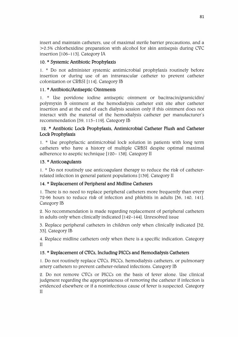
81
insert and maintain catheters, use of maximal sterile barrier precautions, and a
>0.5% chlorhexidine preparation with alcohol for skin antisepsis during CVC
insertion [106–113]. Category IA
10. * Systemic Antibiotic Prophylaxis
1. * Do not administer systemic antimicrobial prophylaxis routinely before
insertion or during use of an intravascular catheter to prevent catheter
colonization or CRBSI [114]. Category IB
11. * Antibiotic/Antiseptic Ointments
1. * Use povidone iodine antiseptic ointment or bacitracin/gramicidin/
polymyxin B ointment at the hemodialysis catheter exit site after catheter
insertion and at the end of each dialysis session only if this ointment does not
interact with the material of the hemodialysis catheter per manufacturer’s
recommendation [59, 115–119]. Category IB
12. * Antibiotic Lock Prophylaxis, Antimicrobial Catheter Flush and Catheter
Lock Prophylaxis
1. * Use prophylactic antimicrobial lock solution in patients with long term
catheters who have a history of multiple CRBSI despite optimal maximal
adherence to aseptic technique [120– 138]. Category II
13. * Anticoagulants
1. * Do not routinely use anticoagulant therapy to reduce the risk of catheter-
related infection in general patient populations [139]. Category II
14. * Replacement of Peripheral and Midline Catheters
1. There is no need to replace peripheral catheters more frequently than every
72-96 hours to reduce risk of infection and phlebitis in adults [36, 140, 141].
Category IB
2. No recommendation is made regarding replacement of peripheral catheters
in adults only when clinically indicated [142–144]. Unresolved issue
3. Replace peripheral catheters in children only when clinically indicated [32,
33]. Category IB
4. Replace midline catheters only when there is a specific indication. Category
II
15. * Replacement of CVCs, Including PICCs and Hemodialysis Catheters
1. Do not routinely replace CVCs, PICCs, hemodialysis catheters, or pulmonary
artery catheters to prevent catheter-related infections. Category IB
2. Do not remove CVCs or PICCs on the basis of fever alone. Use clinical
judgment regarding the appropriateness of removing the catheter if infection is
evidenced elsewhere or if a noninfectious cause of fever is suspected. Category
II

82
3. Do not use guidewire exchanges routinely for non-tunneled catheters to
prevent infection. Category IB
4. Do not use guidewire exchanges to replace a non-tunneled catheter
suspected of infection. Category IB
5. Use a guidewire exchange to replace a malfunctioning non-tunneled
catheter if no evidence of infection is present. Category IB
6. Use new sterile gloves before handling the new catheter when guidewire
exchanges are performed. Category II
16. * Umbilical Catheters
1. Remove and do not replace umbilical artery catheters if any signs of CRBSI,
vascular insufficiency in the lower extremities, or thrombosis are present [145].
Category II
2. Remove and do not replace umbilical venous catheters if any signs of CRBSI
or thrombosis are present [145]. Category II
3. No recommendation can be made regarding attempts to salvage an
umbilical catheter by administering antibiotic treatment through the catheter.
Unresolved issue
4. Cleanse the umbilical insertion site with an antiseptic before catheter
insertion. Avoid tincture of iodine because of the potential effect on the
neonatal thyroid. Other iodine- containing products (e.g., povidone iodine)
can be used [146– 150]. Category IB
5. Do not use topical antibiotic ointment or creams on umbilical catheter
insertion sites because of the potential to promote fungal infections and
antimicrobial resistance [88, 89]. Category IA
6. Add low-doses of heparin (0.25—1.0 U/ml) to the fluid infused through
umbilical arterial catheters [151–153]. Category IB
7. Remove umbilical catheters as soon as possible when no longer needed or
when any sign of vascular insufficiency to the lower extremities is observed.
Optimally, umbilical artery catheters should not be left in place >5 days [145,
154]. Category II
8. Umbilical venous catheters should be removed as soon as possible when no
longer needed, but can be used up to 14 days if managed aseptically [155, 156].
Category II
9. An umbilical catheter may be replaced if it is malfunctioning, and there is no
other indication for catheter removal, and the total duration of catheterization
has not exceeded 5 days for an umbilical artery catheter or 14 days for an
umbilical vein catheter. Category II
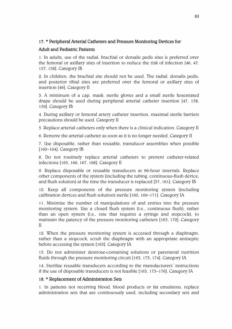
83
17. * Peripheral Arterial Catheters and Pressure Monitoring Devices for
Adult and Pediatric Patients
1. In adults, use of the radial, brachial or dorsalis pedis sites is preferred over
the femoral or axillary sites of insertion to reduce the risk of infection [46, 47,
157, 158]. Category IB
2. In children, the brachial site should not be used. The radial, dorsalis pedis,
and posterior tibial sites are preferred over the femoral or axillary sites of
insertion [46]. Category II
3. A minimum of a cap, mask, sterile gloves and a small sterile fenestrated
drape should be used during peripheral arterial catheter insertion [47, 158,
159]. Category IB
4. During axillary or femoral artery catheter insertion, maximal sterile barriers
precautions should be used. Category II
5. Replace arterial catheters only when there is a clinical indication. Category II
6. Remove the arterial catheter as soon as it is no longer needed. Category II
7. Use disposable, rather than reusable, transducer assemblies when possible
[160–164]. Category IB
8. Do not routinely replace arterial catheters to prevent catheter-related
infections [165, 166, 167, 168]. Category II
9. Replace disposable or reusable transducers at 96-hour intervals. Replace
other components of the system (including the tubing, continuous-flush device,
and flush solution) at the time the transducer is replaced [37, 161]. Category IB
10. Keep all components of the pressure monitoring system (including
calibration devices and flush solution) sterile [160, 169–171]. Category IA
11. Minimize the number of manipulations of and entries into the pressure
monitoring system. Use a closed flush system (i.e., continuous flush), rather
than an open system (i.e., one that requires a syringe and stopcock), to
maintain the patency of the pressure monitoring catheters [163, 172]. Category
II
12. When the pressure monitoring system is accessed through a diaphragm,
rather than a stopcock, scrub the diaphragm with an appropriate antiseptic
before accessing the system [163]. Category IA
13. Do not administer dextrose-containing solutions or parenteral nutrition
fluids through the pressure monitoring circuit [163, 173, 174]. Category IA
14. Sterilize reusable transducers according to the manufacturers’ instructions
if the use of disposable transducers is not feasible [163, 173–176]. Category IA
18. * Replacement of Administration Sets
1. In patients not receiving blood, blood products or fat emulsions, replace
administration sets that are continuously used, including secondary sets and
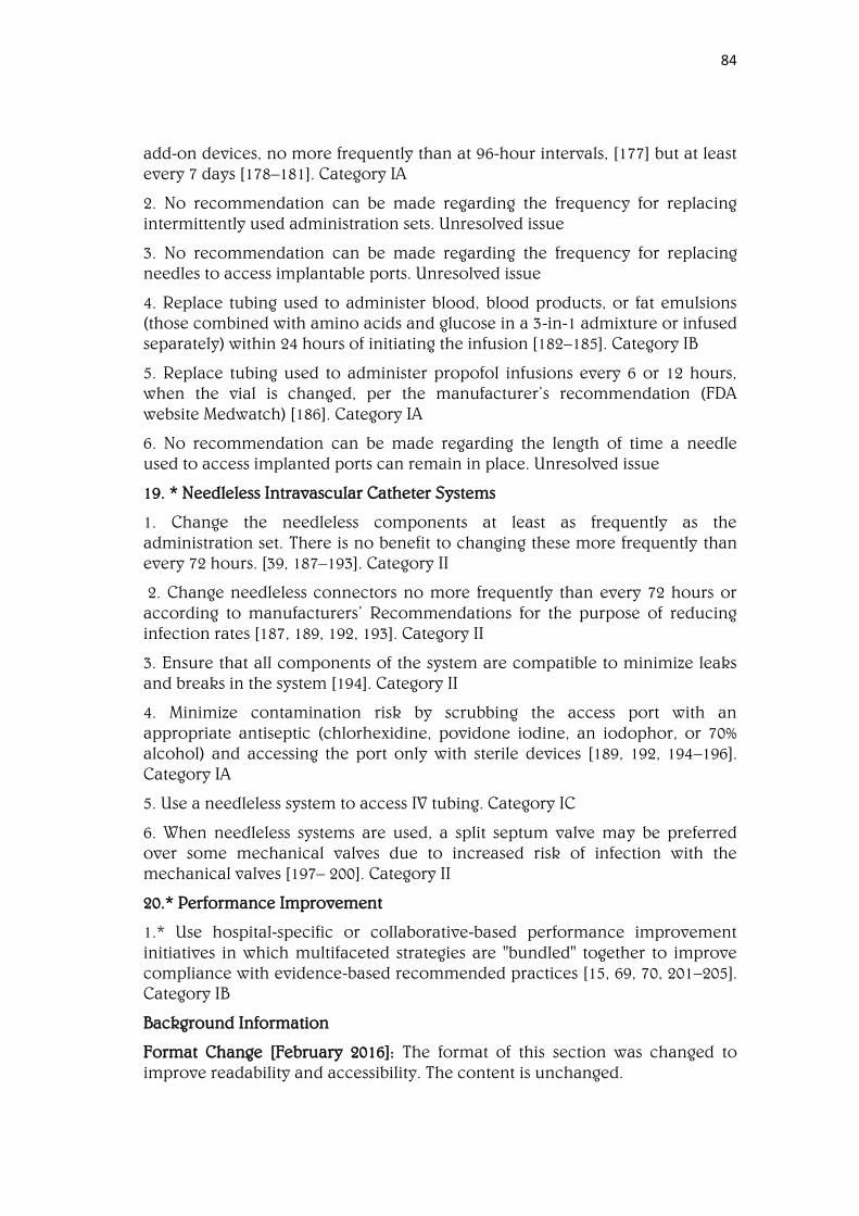
84
add-on devices, no more frequently than at 96-hour intervals, [177] but at least
every 7 days [178–181]. Category IA
2. No recommendation can be made regarding the frequency for replacing
intermittently used administration sets. Unresolved issue
3. No recommendation can be made regarding the frequency for replacing
needles to access implantable ports. Unresolved issue
4. Replace tubing used to administer blood, blood products, or fat emulsions
(those combined with amino acids and glucose in a 3-in-1 admixture or infused
separately) within 24 hours of initiating the infusion [182–185]. Category IB
5. Replace tubing used to administer propofol infusions every 6 or 12 hours,
when the vial is changed, per the manufacturer’s recommendation (FDA
website Medwatch) [186]. Category IA
6. No recommendation can be made regarding the length of time a needle
used to access implanted ports can remain in place. Unresolved issue
19. * Needleless Intravascular Catheter Systems
1. Change the needleless components at least as frequently as the
administration set. There is no benefit to changing these more frequently than
every 72 hours. [39, 187–193]. Category II
2. Change needleless connectors no more frequently than every 72 hours or
according to manufacturers’ Recommendations for the purpose of reducing
infection rates [187, 189, 192, 193]. Category II
3. Ensure that all components of the system are compatible to minimize leaks
and breaks in the system [194]. Category II
4. Minimize contamination risk by scrubbing the access port with an
appropriate antiseptic (chlorhexidine, povidone iodine, an iodophor, or 70%
alcohol) and accessing the port only with sterile devices [189, 192, 194–196].
Category IA
5. Use a needleless system to access IV tubing. Category IC
6. When needleless systems are used, a split septum valve may be preferred
over some mechanical valves due to increased risk of infection with the
mechanical valves [197– 200]. Category II
20.* Performance Improvement
1.* Use hospital-specific or collaborative-based performance improvement
initiatives in which multifaceted strategies are "bundled" together to improve
compliance with evidence-based recommended practices [15, 69, 70, 201–205].
Category IB
Background Information
Format Change [February 2016]: The format of this section was changed to
improve readability and accessibility. The content is unchanged.
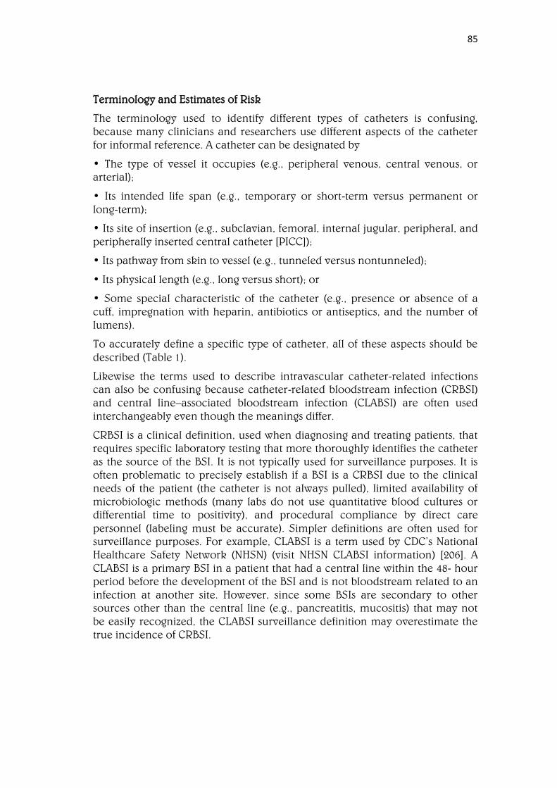
85
Terminology and Estimates of Risk
The terminology used to identify different types of catheters is confusing,
because many clinicians and researchers use different aspects of the catheter
for informal reference. A catheter can be designated by
• The type of vessel it occupies (e.g., peripheral venous, central venous, or
arterial);
• Its intended life span (e.g., temporary or short-term versus permanent or
long-term);
• Its site of insertion (e.g., subclavian, femoral, internal jugular, peripheral, and
peripherally inserted central catheter [PICC]);
• Its pathway from skin to vessel (e.g., tunneled versus nontunneled);
• Its physical length (e.g., long versus short); or
• Some special characteristic of the catheter (e.g., presence or absence of a
cuff, impregnation with heparin, antibiotics or antiseptics, and the number of
lumens).
To accurately define a specific type of catheter, all of these aspects should be
described (Table 1).
Likewise the terms used to describe intravascular catheter-related infections
can also be confusing because catheter-related bloodstream infection (CRBSI)
and central line–associated bloodstream infection (CLABSI) are often used
interchangeably even though the meanings differ.
CRBSI is a clinical definition, used when diagnosing and treating patients, that
requires specific laboratory testing that more thoroughly identifies the catheter
as the source of the BSI. It is not typically used for surveillance purposes. It is
often problematic to precisely establish if a BSI is a CRBSI due to the clinical
needs of the patient (the catheter is not always pulled), limited availability of
microbiologic methods (many labs do not use quantitative blood cultures or
differential time to positivity), and procedural compliance by direct care
personnel (labeling must be accurate). Simpler definitions are often used for
surveillance purposes. For example, CLABSI is a term used by CDC’s National
Healthcare Safety Network (NHSN) (visit NHSN CLABSI information) [206]. A
CLABSI is a primary BSI in a patient that had a central line within the 48 hour
period before the development of the BSI and is not bloodstream related to an
infection at another site. However, since some BSIs are secondary to other
sources other than the central line (e.g., pancreatitis, mucositis) that may not
be easily recognized, the CLABSI surveillance definition may overestimate the
true incidence of CRBSI.

86
Table 1. Catheters Used for Venous and Arterial Access
Catheter type Entry Site Length Comments
Peripheral
venous
catheters
Usually inserted
in veins of
forearm or hand
<3 inches Phlebitis with
prolonged use;
rarely
associated with
bloodstream
infection
Peripheral
arterial
catheters
Usually inserted
in radial artery;
can be placed in
femoral, axillary,
brachial,
posterior tibial
arteries
<3 inches Low infection
risk; rarely
associated with
bloodstream
infection
Midline
catheters
Inserted via the
antecubital fossa
into the proximal
basilic or cephalic
veins; does not
enter central
veins, peripheral
catheters
3 to 8 inches Anaphylactoid
reactions have
been reported
with catheters
made of
elastomeric
hydrogel;lower
rates of phlebitis
than short
peripheral
catheters
Nontunneled
central
venouscatheters
Percutaneously
inserted into
central veins
(subclavian,
internal jugular,
or femoral)
≥8 cm depending
on patient size
Account for
majority of
CRBSI
Pulmonary
artery catheters
Inserted through
a Teflon®
introducer in a
central vein
(subclavian,
internal jugular,
or femoral)
≥30 cm
depending on
patient size
Usually heparin
bonded; similar
rates of
bloodstream
infection as
CVCs;
subclavian site
preferred to
reduce
infection risk
Peripherally
inserted
centralvenous
Inserted into
basilic, cephalic,
or brachial veins
≥20 cm
depending on
patient size Lower
Lower rate of
infection than
nontunneled

87
catheters (PICC) and enter the
superior vena
cava ≥20 cm
depending on
patient size Lower
rate of infection
than nontunneled
CVCs
rate of infection
than nontunneled
CVCs
CVCs
Tunneled
central venous
catheters
Implanted into
subclavian,
internal jugular,
or femoral veins
≥8 cm depending
on patient size
Cuff inhibits
migration of
organisms into
catheter tract;
lower rate of
infection than
nontunneled
CVC
Totally
implantable
Tunneled
beneath skin and
have
subcutaneous
port accessed
with a needle;
implanted in
subclavian or
internal jugular
vein
≥8 cm depending
on patient size
Lowest risk for
CRBSI;
improved
patient
selfimage; no
need for local
catheter-site
care; surgery
required for
catheter
removal
Umbilical
catheters
Inserted into
either umbilical
vein or umbilical
artery
≤6 cm depending
on patient size
Risk for CRBSI
similar with
catheters placed
in umbilical
vein versus
artery
Epidemiology and Microbiology in Adult and Pediatric Patients
National estimates of CLABSI rates are available through CDC’s NHSN, a
surveillance system for healthcare-associated infections, and are available on
CDC’s website. A recent report highlights data from 1,545 hospitals in 48 States
and the District of Columbia that monitor infections in one or more ICUs
and/or non-ICUs (e.g., patient care areas, wards) [207]. Because BSI rates are
influenced by patient-related factors, such as severity of illness and type of
illness (e.g., third-degree burns versus post-cardiac surgery), by catheter-related
factors, (such as the condition under which the catheter was placed and
catheter type), and by institutional factors (e.g., bed-size, academic affiliation),

88
these aggregate, risk-adjusted rates can be used as benchmarks against which
hospitals can make intra-and inter-facility comparisons.
The most commonly reported causative pathogens remain coagulase-negative
staphylococci, Staphylococcus aureus, enterococci, and Candida spp [208].
Gram negative bacilli accounted for 19% and 21% of CLABSIs reported to CDC
[209] and the Surveillance and Control of Pathogens of Epidemiological
Importance (SCOPE) database, respectively [208].
For all common pathogens causing CLABSIs, antimicrobial resistance is a
problem, particularly in ICUs. Although methicillin-resistant Staphylococcus
aureus (MRSA) now account for more than 50% of all Staphylococcus aureus
isolates obtained in ICUs, the incidence of MRSA CLABSIs has decreased in
recent years, perhaps as a result of prevention efforts [210]. For gram negative
rods, antimicrobial resistance to third generation cephalosporins among
Klebsiella pneumoniae and E. coli has increased significantly as has imipenem
and ceftazidine resistance among Pseudomonas aeruginosa [209]. Candida spp.
are increasingly noted to be fluconazole resistant.
Pathogenesis
There are four recognized routes for contamination of catheters:
1. Migration of skin organisms at the insertion site into the cutaneous catheter
tract and along the surface of the catheter with colonization of the catheter tip;
this is the most common route of infection for short-term catheters [37, 211,
212];
2. Direct contamination of the catheter or catheter hub by contact with hands
or contaminated fluids or devices [213, 214];
3. Less commonly, catheters might become hematogenously seeded from
another focus of infection [215]; and
4. Rarely, infusate contamination might lead to CRBSI [216].
Important pathogenic determinants of CRBSI are
1. The material of which the device is made;
2. The host factors consisting of protein adhesions, such as fibrin and
fibronectin, that form a sheath around the catheter [217]; and
3. The intrinsic virulence factors of the infecting organism, including the
extracellular polymeric substance (EPS) produced by the adherent organisms
[218].
Some catheter materials also have surface irregularities that enhance the
microbial adherence of certain species (e.g., S. epidermidis and C. albicans)
[219, 220]. Catheters made of these materials are especially vulnerable to
microbial colonization and subsequent infection. Due to the formation of the
fibrin sheath, silastic catheters are associated with higher risk of catheter
infections than polyurethane catheters [217]. On the other hand, biofilm

89
formation by C. albicans occurs more readily on silicone elastomer
catheter surfaces than polyurethane catheters [219]. Modification of the
biomaterial surface properties has been shown to influence the ability of C.
albicans to form biofilm [220]. Additionally, certain catheter materials are more
thrombogenic than others, a characteristic that also might predispose to
catheter colonization and infection [221, 222]. This association has led to
emphasis on preventing catheter-related thrombus as an additional
mechanism for reducing CRBSI [223, 224].
The adherence properties of a given microorganism in relationship to host
factors are also important in the pathogenesis of CRBSI. For example, S. aureus
can adhere to host proteins (e.g., fibrinogen, fibronectin) commonly present on
catheters by expressing clumping factors (ClfA and ClfB) that bind to the
protein adhesins [217, 222, 225, 226]. Furthermore, adherence is enhanced
through the production by microbial organisms, such as coagulase negative
staphylococci [227, 228], S. aureus [229], Pseudomonas aeruginosa [230], and
Candida species [231] of an extracellular polymeric substance (EPS) consisting
mostly of an exopolysaccharide that forms a microbial biofilm layer [218, 232].
This biofilm matrix is enriched by divalent metallic cations, such as calcium,
magnesium and iron, which make it a solid enclave in which microbial
organisms can embed themselves [233–235]. Such a biofilm potentiates the
pathogenicity of various microbes by allowing them to withstand host defense
mechanisms (e.g., acting as a barrier to engulfment and killing by
polymorphonuclear leukocytes) or by making them less susceptible to
antimicrobial agents (e.g., forming a matrix that binds antimicrobials before
their contact with the organism cell wall or providing for a population of
metabolically quiescent, antimicrobial tolerant "persister" cells) [228, 236, 237].
Some Candida spp., in the presence of dextrose-containing fluids, produce
slime similar to that of their bacterial counterparts, potentially explaining the
increased proportion of BSIs caused by fungal pathogens among patients
receiving parenteral nutrition fluids [238].
Strategies for Prevention of Catheter-Related Infections in Adult and Pediatric
Patients
Education, Training and Staffing Recommendations
1. Educate healthcare personnel regarding the indications for intravascular
catheter use, proper procedures for the insertion and maintenance of
intravascular catheters, and appropriate infection control measures to prevent
intravascular catheter-related infections [7–15]. Category IA
2. Periodically assess knowledge of and adherence to guidelines for all
personnel involved in the insertion and maintenance of intravascular catheters
[7–15]. Category IA
3. Designate only trained personnel who demonstrate competence for the
insertion and maintenance of peripheral and central intravascular catheters.
[14–28]. Category IA

90
4. Ensure appropriate nursing staff levels in ICUs. Observational studies suggest
that a higher proportion of "pool nurses" or an elevated patient–to-nurse ratio
is associated with CRBSI in ICUs where nurses are managing patients with CVCs
[29–31]. Category IB
Background
Well-organized programs that enable healthcare providers to become educated
and to provide, monitor, and evaluate care are critical to the success of this
effort. Reports spanning the past four decades have consistently demonstrated
that risk for infection declines following standardization of aseptic care [7, 12,
14, 15, 239–241] and that insertion and maintenance of intravascular catheters
by inexperienced staff might increase the risk for catheter colonization and
CRBSI [15, 242]. Specialized "IV teams" have shown unequivocal effectiveness
in reducing the incidence of CRBSI, associated complications, and costs [16–
26]. Additionally, infection risk increases with nursing staff reductions below a
critical level [30].
Selection of Catheters and Sites
Peripheral and Midline Catheter Recommendations
1. In adults, use an upper-extremity site for catheter insertion. Replace a
catheter inserted in a lower extremity site to an upper extremity site as soon as
possible. Category II
2. In pediatric patients, the upper or lower extremities or the scalp (in neonates
or young infants) can be used as the catheter insertion site [32, 33]. Category II
3. Select catheters on the basis of the intended purpose and duration of use,
known infectious and non-infectious complications (e.g., phlebitis and
infiltration), and experience of individual catheter operators [33–35]. Category
IB
4. Avoid the use of steel needles for the administration of fluids and medication
that might cause tissue necrosis if extravasation occurs [33, 34]. Category IA
5. Use a midline catheter or peripherally inserted central catheter (PICC),
instead of a short peripheral catheter, when the duration of IV therapy will
likely exceed six days. Category II
6. Evaluate the catheter insertion site daily by palpation through the dressing to
discern tenderness and by inspection if a transparent dressing is in use. Gauze
and opaque dressings should not be removed if the patient has no clinical signs
of infection. If the patient has local tenderness or other signs of possible CRBSI,
an opaque dressing should be removed and the site inspected visually.
Category II
7. Remove peripheral venous catheters if the patients develops signs of
phlebitis (warmth, tenderness, erythema or palpable venous cord), infection, or
a malfunctioning catheter [36]. Category IB

91
Central Venous Catheters Recommendations
1. Weigh the risks and benefits of placing a central venous device at a
recommended site to reduce infectious complications against the risk for
mechanical complications (e.g., pneumothorax, subclavian artery puncture,
subclavian vein laceration, subclavian vein stenosis, hemothorax, thrombosis,
air embolism, and catheter misplacement) [37–53]. Category IA
2. Avoid using the femoral vein for central venous access in adult patients [38,
50, 51, 54]. Category IA
3. Use a subclavian site, rather than a jugular or a femoral site, in adult patients
to minimize infection risk for nontunneled CVC placement [50–52]. Category IB
4. No recommendation can be made for a preferred site of insertion to
minimize infection risk for a tunneled CVC. Unresolved issue
5. Avoid the subclavian site in hemodialysis patients and patients with
advanced kidney disease, to avoid subclavian vein stenosis [53, 55–58].
Category IA
6. Use a fistula or graft in patients with chronic renal failure instead of a CVC
for permanent access for dialysis [59]. Category IA
7. Use ultrasound guidance to place central venous catheters (if this technology
is available) to reduce the number of cannulation attempts and mechanical
complications. Ultrasound guidance should only be used by those fully trained
in its technique. [60–64]. Category IB
8. Use a CVC with the minimum number of ports or lumens essential for the
management of the patient [65–68]. Category IB
9. No recommendation can be made regarding the use of a designated lumen
for parenteral nutrition. Unresolved issue
10. Promptly remove any intravascular catheter that is no longer essential [69–
72]. Category IA
11. When adherence to aseptic technique cannot be ensured (i.e., catheters
inserted during a medical emergency), replace the catheter as soon as possible,
i.e., within 48 hours [37, 73–76]. Category IB
Background
The site at which a catheter is placed influences the subsequent risk for
catheter-related infection and phlebitis. The influence of site on the risk for
catheter infections is related in part to the risk for thrombophlebitis and density
of local skin flora.
As in adults, the use of peripheral venous catheters in pediatric patients might
be complicated by phlebitis, infusion extravasation, and catheter infection
[243]. Catheter location, infusion of parenteral nutritional fluids with
continuous IV fat emulsions, and length of ICU stay before catheter insertion,
have all increased pediatric patients’ risk for phlebitis. However, contrary to the

92
risk in adults, the risk for phlebitis in children has not increased with the
duration of catheterization [243, 244].
The density of skin flora at the catheter insertion site is a major risk factor for
CRBSI. No single trial has satisfactorily compared infection rates for catheters
placed in jugular, subclavian, and femoral veins. In retrospective observational
studies, catheters inserted into an internal jugular vein have usually been
associated with higher risk for colonization and/or CRBSI than those inserted
into a subclavian [37–47]. Similar findings were noted in neonates in a single
retrospective study [245]. Femoral catheters have been demonstrated to have
high colonization rates compared with subclavian and internal jugular sites
when used in adults and, in some studies, higher rates of CLABSIs [40, 45–47,
50, 51, 246]. Femoral catheters should also be avoided, when possible, because
they are associated with a higher risk for deep venous thrombosis than are
internal jugular or subclavian catheters [48–50, 53, 247]. One study [38] found
that the risk of infection associated with catheters placed in the femoral vein is
accentuated in obese patients. In contrast to adults, studies in pediatric patients
have demonstrated that femoral catheters have a low incidence of mechanical
complications and might have an equivalent infection rate to that of non-
femoral catheters [248–251]. Thus, in adult patients, a subclavian site is
preferred for infection control purposes, although other factors (e.g., the
potential for mechanical complications, risk for subclavian vein stenosis, and
catheter-operator skill) should be considered when deciding where to place the
catheter.
In two meta-analyses, the use of real-time two-dimensional ultrasound for the
placement of CVCs substantially decreased mechanical complications and
reduced the number of attempts at required cannulation and failed attempts at
cannulation compared with the standard landmark placement [60, 61].
Evidence favors the use of two-dimensional ultrasound guidance over Doppler
ultrasound guidance [60]. Site selection should be guided by patient comfort,
ability to secure the catheter, and maintenance of asepsis as well as patient-
specific factors (e.g., preexisting catheters, anatomic deformity, and bleeding
diathesis), relative risk of mechanical complications (e.g., bleeding and
pneumothorax), the availability of bedside ultrasound, the experience of the
person inserting the catheter, and the risk for infection.
Catheters should be inserted as great a distance as possible from open wounds.
In one study, catheters inserted close to open burn wounds (i.e., 25 cm2
overlapped a wound) were times more likely to be colonized and 5.12 times
more likely to be associated with bacteremia than catheters inserted farther
from the wounds [252].
Type of Catheter Material. Polytetrafluoroethylene (Teflon ®) or polyurethane
catheters have been associated with fewer infectious complications than
catheters made of polyvinyl chloride or polyethylene [36, 253, 254]. Steel
needles used as an alternative to catheters for peripheral venous access have
the same rate of infectious complications as do Teflon® catheters [33, 34].

93
However, the use of steel needles frequently is complicated by infiltration of
intravenous (IV) fluids into the subcutaneous tissues, a potentially serious
complication if the infused fluid is a vesicant [34].
Hand Hygiene and Aseptic Technique
Recommendations
1. Perform hand hygiene procedures, either by washing hands with
conventional soap and water or with alcohol-based hand rubs (ABHR). Hand
hygiene should be performed before and after palpating catheter insertion sites
as well as before and after inserting, replacing, accessing, repairing, or dressing
an intravascular catheter. Palpation of the insertion site should not be
performed after the application of antiseptic, unless aseptic technique is
maintained [12, 77–79]. Category IB
2. Maintain aseptic technique for the insertion and care of intravascular
catheters [37, 73, 74, 76]. Category IB
3. Wear clean gloves, rather than sterile gloves, for the insertion of peripheral
intravascular catheters, if the access site is not touched after the application of
skin antiseptics. Category IC
4. Sterile gloves should be worn for the insertion of arterial, central, and
midline catheters [37, 73, 74, 76]. Category IA
5. Use new sterile gloves before handling the new catheter when guidewire
exchanges are performed. Category II
6. Wear either clean or sterile gloves when changing the dressing on
intravascular catheters. Category IC
Background
Hand hygiene before catheter insertion or maintenance, combined with proper
aseptic technique during catheter manipulation, provides protection against
infection [12]. Proper hand hygiene can be achieved through the use of either
an al-cohol-based product [255] or with soap and water with adequate rinsing
[77]. Appropriate aseptic technique does not necessarily require sterile gloves
for insertion of peripheral catheters; a new pair of disposable nonsterile gloves
can be used in conjunction with a "no-touch" technique for the insertion of
peripheral venous catheters. Sterile gloves must be worn for placement of
central catheters since a "no-touch" technique is not possible.
Maximal Sterile Barrier Precautions
Recommendations
1. Use maximal sterile barrier precautions, including the use of a cap, mask,
sterile gown, sterile gloves, and a sterile full body drape, for the insertion of
CVCs, PICCs, or guidewire exchange [14, 75, 76, 80]. Category IB
2. Use a sterile sleeve to protect pulmonary artery catheters during insertion
[81]. Category IB

94
Background
Maximum sterile barrier (MSB) precautions are defined as wearing a sterile
gown, sterile gloves, and cap and using a full body drape (similar to the drapes
used in the operating room) during the placement of CVC. Maximal sterile
barrier precautions during insertion of CVC were compared with sterile gloves
and a small drape in a randomized controlled trial. The MSB group had fewer
episodes of both catheter colonization (RR = .32, 95% CI, .10–.96, P = .04) and
CR-BSI (RR = .16, 95% CI, .02–1.30, P = .06). In addition, the group using MSB
precautions had infections that occurred much later and contained gram
negative, rather than gram positive, organisms [76]. A study of pulmonary
artery catheters also secondarily demonstrated that use of MSB precautions
lowered risk of infection [37]. Another study evaluated an educational program
directed at improving infection control practices, especially MSB precautions.
In this study, MSB precautions use increased and CRBSI decreased [14]. A small
trial demonstrated a reduced risk of skin colonization at the insertion site when
MSB precautions were used [OR 3.40, 95%CI 1.32 to 3.67] [80].
Skin Preparation
Recommendations
1. Prepare clean skin with an antiseptic (70% alcohol, tincture of iodine, an
iodophor or chlorhexidine gluconate) before peripheral venous catheter
insertion [82]. Category IB
2. Prepare clean skin with a >0.5% chlorhexidine preparation with alcohol
before central venous catheter and peripheral arterial catheter insertion and
during dressing changes. If there is a contraindication to chlorhexidine,
tincture of iodine, an iodophor, or 70% alcohol can be used as alternatives [82,
83]. Category IA
3. No comparison has been made between using chlorhexidine preparations
with alcohol and povidone-iodine in alcohol to prepare clean skin. Unresolved
issue.
4. No recommendation can be made for the safety or efficacy of chlorhexidine
in infants aged <2 months. Unresolved issue
5. Antiseptics should be allowed to dry according to the manufacturer’s
recommendation prior to placing the catheter [82, 83]. Category IB
Background
Two well-designed studies evaluating the chlorhexidine-containing cutaneous
antiseptic regimen in comparison with either povidone iodine or alcohol for
the care of an intravascular catheter insertion site have shown lower rates of
catheter colonization or CRBSI associated with the chlorhexidine preparation
[82, 83]. (The comparison of chlorhexidine gluconate alcohol to povidone
iodine alcohol has not been done.) When 0.5% tincture of chlorhexidine was
compared with 10% povidone iodine, no differences were seen in central
venous catheter (CVC) colonization or in CRBSI [256]. In a three-armed study

95
(2% aqueous chlorhexidine gluconate vs 10% povidone-iodine vs 70% alcohol),
2% aqueous chlorhexidine gluconate tended to decrease CRBSI compared with
10% povidone iodine or 70% alcohol [82]. A meta-analysis of 4,143 catheters
suggested that chlorhexidine preparation reduced the risk of catheter related
infection by 49% (95% CI .28 to .88) relative to povidone iodine [257]. An
economic decision analysis based on available evidence suggested that the use
of chlorhexidine, rather than povidone iodine, for CVC care would result in a
1.6% decrease in the incidence of CRBSI, a 0.23% decrease in the incidence of
death, and a savings of $113 per catheter used [258]. While chlorhexidine has
become a standard antiseptic for skin preparation for the insertion of both
central and peripheral venous catheters, 5% povidone iodine solution in 70%
ethanol was associated with a substantial reduction of CVC-related colonization
and infection compared with 10% aqueous povidone iodine [259].
Catheter Site Dressing Regimens
Recommendations
1. Use either sterile gauze or sterile, transparent, semipermeable dressing to
cover the catheter site [84–87]. Category IA
2. If the patient is diaphoretic or if the site is bleeding or oozing, use gauze
dressing until this is resolved [84–87]. Category II
3. Replace catheter site dressing if the dressing becomes damp, loosened, or
visibly soiled [84, 85]. Category IB
4. Do not use topical antibiotic ointment or creams on insertion sites, except
for dialysis catheters, because of their potential to promote fungal infections
and antimicrobial resistance [88, 89]. Category IB
5. Do not submerge the catheter or catheter site in water. Showering should be
permitted if precautions can be taken to reduce the likelihood of introducing
organisms into the catheter (e.g., if the catheter and connecting device are
protected with an impermeable cover during the shower) [90–92]. Category IB
6. Replace dressings used on short-term CVC sites every 2 days for gauze
dressings. Category II
7. Replace dressings used on short-term CVC sites at least every 7 days for
transparent dressings, except in those pediatric patients in which the risk for
dislodging the catheter may outweigh the benefit of changing the dressing [87,
93]. Category IB
8. Replace transparent dressings used on tunneled or implanted CVC sites no
more than once per week (unless the dressing is soiled or loose), until the
insertion site has healed. Category II
9. No recommendation can be made regarding the necessity for any dressing
on well-healed exit sites of long-term cuffed and tunneled CVCs. Unresolved
issue
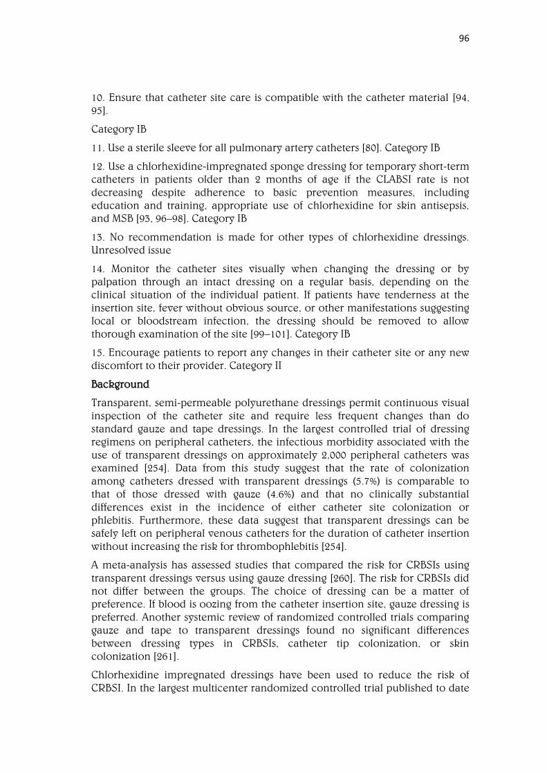
96
10. Ensure that catheter site care is compatible with the catheter material [94,
95].
Category IB
11. Use a sterile sleeve for all pulmonary artery catheters [80]. Category IB
12. Use a chlorhexidine-impregnated sponge dressing for temporary short-term
catheters in patients older than 2 months of age if the CLABSI rate is not
decreasing despite adherence to basic prevention measures, including
education and training, appropriate use of chlorhexidine for skin antisepsis,
and MSB [93, 96–98]. Category IB
13. No recommendation is made for other types of chlorhexidine dressings.
Unresolved issue
14. Monitor the catheter sites visually when changing the dressing or by
palpation through an intact dressing on a regular basis, depending on the
clinical situation of the individual patient. If patients have tenderness at the
insertion site, fever without obvious source, or other manifestations suggesting
local or bloodstream infection, the dressing should be removed to allow
thorough examination of the site [99–101]. Category IB
15. Encourage patients to report any changes in their catheter site or any new
discomfort to their provider. Category II
Background
Transparent, semi-permeable polyurethane dressings permit continuous visual
inspection of the catheter site and require less frequent changes than do
standard gauze and tape dressings. In the largest controlled trial of dressing
regimens on peripheral catheters, the infectious morbidity associated with the
use of transparent dressings on approximately 2,000 peripheral catheters was
examined [254]. Data from this study suggest that the rate of colonization
among catheters dressed with transparent dressings (5.7%) is comparable to
that of those dressed with gauze (4.6%) and that no clinically substantial
differences exist in the incidence of either catheter site colonization or
phlebitis. Furthermore, these data suggest that transparent dressings can be
safely left on peripheral venous catheters for the duration of catheter insertion
without increasing the risk for thrombophlebitis [254].
A meta-analysis has assessed studies that compared the risk for CRBSIs using
transparent dressings versus using gauze dressing [260]. The risk for CRBSIs did
not differ between the groups. The choice of dressing can be a matter of
preference. If blood is oozing from the catheter insertion site, gauze dressing is
preferred. Another systemic review of randomized controlled trials comparing
gauze and tape to transparent dressings found no significant differences
between dressing types in CRBSIs, catheter tip colonization, or skin
colonization [261].
Chlorhexidine impregnated dressings have been used to reduce the risk of
CRBSI. In the largest multicenter randomized controlled trial published to date

97
comparing chlorhexidine impregnated sponge dressings vs standard dressings
in ICU patients, rates of CRBSIs were reduced even when background rates of
infection were low. In this study, 1636 patients (3778 catheters, 28 931 catheter-
days) were evaluated. The chlorhexidine- impregnated sponge dressings
decreased the rates of major CRBSIs (10/1953 [0.5%], 0.6 per 1,000 catheter-days
vs 19/1825 [1.1%], 1.4 per 1,000 catheter-days; hazard ratio [HR], 0.39 [95%
confidence interval {CI}, .17–.93]; P = .03) and CRBSIs (6/1953 catheters, 0.40
per 1,000 catheter-days vs 17/1825 catheters, 1.3 per 1,000 catheter-days; HR,
0.24 [95% CI, .09–.65]) [93]. A randomized controlled study of polyurethane or a
chlorhexidine impregnated sponge dressing in 140 children showed no
statistical difference in BSIs; however, the chlorhexidine group had lower rates
of CVC colonization [98]. In 601 cancer patients receiving chemotherapy, the
incidence of CRBSI was reduced in patients receiving the chlorhexidine
impregnated sponge dressing compared with standard dressings (P = .016,
relative risk 0.54; confidence interval 0.31–.94) [262]. A meta-analysis that
included eight randomized controlled trials demonstrated that chlorhexidine
impregnated sponge dressings are associated with a reduction of vascular and
epidural catheter exit site colonization but no significant reduction in CRBSI
(2.2% versus 3.8%, OR 0.58, 95% CI: .29–1.14, p= .11) [97].
Although data regarding the use of a chlorhexidine impregnated sponge
dressing in children are limited, one randomized, controlled study involving
705 neonates reported a substantial decrease in colonized catheters in infants
in the chlorhexidine impregnated sponge dressing group compared with the
group that had standard dressings (15% versus 24%; RR = .6; 95% CI 5 0.5–.9),
but no difference in the rates of CRBSI or BSI without a source. Chlorhexidine
impregnated sponge dressings were associated with localized contact
dermatitis in infants of very low birth weight. In 98 neonates with very low birth
weight, 15 (15%) developed localized contact dermatitis; four (1.5%) of 237
neonates weighing >1,000 g developed this reaction (P
< .0001). Infants with gestational age <26 weeks who had CVCs placed at age
<8 days were at increased risk for having localized contact dermatitis, whereas
no infants in the control group developed this local reaction [96].
Patient Cleansing
Recommendation
Use a 2% chlorhexidine wash for daily skin cleansing to reduce CRBSI [102–
104]. Category II
Background
Daily cleansing of ICU patients with a 2% chlorhexidine impregnated washcloth
may be a simple, effective strategy to decrease the rate of primary BSIs. In a
single center study of 836 ICU patients, patients receiving the chlorhexidine
intervention were significantly less likely to acquire a primary BSI (4.1 vs 10.4
infections per 1,000 patient days; incidence difference, 6.3 [95% confidence
interval, 1.2–11.0]) than those bathed with soap and water [102].

98
Catheter Securement Devices
Recommendation
Use a sutureless securement device to reduce the risk of infection for
intravascular catheters [105]. Category II
Background
Catheter stabilization is recognized as an intervention to decrease the risk for
phlebitis, catheter migration and dislodgement, and may be advantageous in
preventing CRBSIs. Pathogenesis of CRBSI occurs via migration of skin flora
through the percutaneous entry site. Sutureless securement devices avoid
disruption around the catheter entry site and may decrease the degree of
bacterial colonization. [105]. Using a sutureless securement device also
mitigates the risk of sharps injury to the healthcare provider from inadvertent
needlestick injury.
Antimicrobial/Antiseptic Impregnated Catheters and Cuffs
Recommendation
Use a chlorhexidine/silver sulfadiazine or minocycline/ rifampin -impregnated
CVC in patients whose catheter is expected to remain in place >5 days if, after
successful implementation of a comprehensive strategy to reduce rates of
CLABSI, the CLABSI rate is not decreasing. The comprehensive strategy should
include at least the following three components: educating persons who insert
and maintain catheters, use of maximal sterile barrier precautions, and a >0.5%
chlorhexidine preparation with alcohol for skin antisepsis during CVC insertion
[106–113]. Category IA
Background
Certain catheters and cuffs that are coated or impregnated with antimicrobial
or antiseptic agents can decrease the risk for CRBSI and potentially decrease
hospital costs associated with treating CRBSIs, despite the additional
acquisition cost of an antimicrobial/antiseptic impregnated catheter [110].
Nearly all of the studies involving antimicrobial/antiseptic impregnated
catheters have been conducted using triple-lumen, uncuffed catheters in adult
patients whose catheters remained in place <30 days. While most of the studies
have been conducted in adults, these catheters have been approved by FDA for
use in patients weighing >3 kg. Two non-randomized studies [112, 113] in
pediatric ICU patients suggest that these catheters might reduce risk of
catheter-associated infection. No antiseptic or antimicrobial impregnated
catheters currently are available for use in infants weighing <3kg.
Chlorhexidine/Silver Sulfadiazine. Catheters coated with chlorhexidine/silver
sulfadiazine only on the external luminal surface have been studied as a means
to reduce CRBSI. Two meta- analyses of first-generation catheters [1, 263]
demonstrated that such catheters reduced the risk for CRBSI compared with
standard non-coated catheters. The duration of catheter placement in one
study ranged from 5.1 to 11.2 days [264]. A second-generation catheter is now
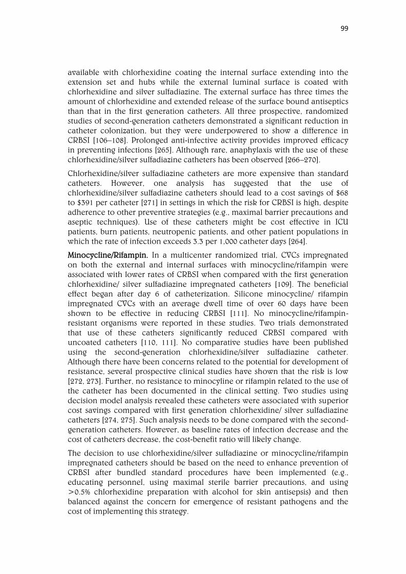
99
available with chlorhexidine coating the internal surface extending into the
extension set and hubs while the external luminal surface is coated with
chlorhexidine and silver sulfadiazine. The external surface has three times the
amount of chlorhexidine and extended release of the surface bound antiseptics
than that in the first generation catheters. All three prospective, randomized
studies of second-generation catheters demonstrated a significant reduction in
catheter colonization, but they were underpowered to show a difference in
CRBSI [106–108]. Prolonged anti-infective activity provides improved efficacy
in preventing infections [265]. Although rare, anaphylaxis with the use of these
chlorhexidine/silver sulfadiazine catheters has been observed [266–270].
Chlorhexidine/silver sulfadiazine catheters are more expensive than standard
catheters. However, one analysis has suggested that the use of
chlorhexidine/silver sulfadiazine catheters should lead to a cost savings of $68
to $391 per catheter [271] in settings in which the risk for CRBSI is high, despite
adherence to other preventive strategies (e.g., maximal barrier precautions and
aseptic techniques). Use of these catheters might be cost effective in ICU
patients, burn patients, neutropenic patients, and other patient populations in
which the rate of infection exceeds 3.3 per 1,000 catheter days [264].
Minocycline/Rifampin. In a multicenter randomized trial, CVCs impregnated
on both the external and internal surfaces with minocycline/rifampin were
associated with lower rates of CRBSI when compared with the first generation
chlorhexidine/ silver sulfadiazine impregnated catheters [109]. The beneficial
effect began after day 6 of catheterization. Silicone minocycline/ rifampin
impregnated CVCs with an average dwell time of over 60 days have been
shown to be effective in reducing CRBSI [111]. No minocycline/rifampin-
resistant organisms were reported in these studies. Two trials demonstrated
that use of these catheters significantly reduced CRBSI compared with
uncoated catheters [110, 111]. No comparative studies have been published
using the second-generation chlorhexidine/silver sulfadiazine catheter.
Although there have been concerns related to the potential for development of
resistance, several prospective clinical studies have shown that the risk is low
[272, 273]. Further, no resistance to minocyline or rifampin related to the use of
the catheter has been documented in the clinical setting. Two studies using
decision model analysis revealed these catheters were associated with superior
cost savings compared with first generation chlorhexidine/ silver sulfadiazine
catheters [274, 275]. Such analysis needs to be done compared with the second-
generation catheters. However, as baseline rates of infection decrease and the
cost of catheters decrease, the cost-benefit ratio will likely change.
The decision to use chlorhexidine/silver sulfadiazine or minocycline/rifampin
impregnated catheters should be based on the need to enhance prevention of
CRBSI after bundled standard procedures have been implemented (e.g.,
educating personnel, using maximal sterile barrier precautions, and using
>0.5% chlorhexidine preparation with alcohol for skin antisepsis) and then
balanced against the concern for emergence of resistant pathogens and the
cost of implementing this strategy.

100
Platinum/Silver. A combination platinum/silver impregnated catheter (i.e., a
silver iontophoretic catheter) is available for use in the United States. Several
prospective, randomized studies have been published comparing these
catheters to uncoated catheters [276–279]. One study showed a reduction in
the incidence density of catheter colonization and CRBSI [278], but the other
studies found no difference in catheter colonization or CRBSI between the
impregnated catheter and a non-impregnated catheter [39, 276, 277]. In light of
this, a firm recommendation for or against the use of these catheters cannot be
made.
Systemic Antibiotic Prophylaxis
Recommendation
Do not administer systemic antimicrobial prophylaxis routinely before
insertion or during use of an intravascular catheter to prevent catheter
colonization or CRBSI [114]. Category IB
Background
Several studies have examined the role of systemic antibiotic prophylaxis in
prevention of catheter-related infection. A recent meta-analysis reviewed these
studies in oncology patients [114]. Four studies used a prophylactic
glycopeptide prior to catheter insertion. However, heterogeneity in these
studies precludes making any conclusion regarding efficacy.
In a study examining the effect of ongoing oral prophylaxis with rifampin and
novobiocin on catheter-related infection in cancer patients treated with
interleukin-2 [280], a reduction in CRBSI was observed, even though 9 of 26
subjects (35%) discontinued the prophylactic antibiotics due to side effects or
toxicity. In non-oncology patients, no benefit was associated with vancomycin
administration prior to catheter insertion in 55 patients undergoing
catheterization for parenteral nutrition [281]. Similarly, extending perioperative
prophylactic antibiotics in cardiovascular surgery patients did not reduce
central venous catheter colonization [282]. A recent Cochrane review of
prophylactic antibiotics in neonates with umbilical venous catheters concluded
that there is insufficient evidence from randomized trials to support or refute
the use of prophylactic antibiotics [283].
Late onset neonatal sepsis is often due to coagulase negative staphylococci and
is thought to frequently stem from infected central venous catheters. Five trials
involved a total of 371 neonates comparing vancomycin by continuous
infusion via parenteral nutrition or intermittent dosing, and placebo. The
infants treated with vancomycin experienced less sepsis (RR .11; 95% CI .05-.24)
and less sepsis due to coagulase negative staphylococci (RR .33; 95% CI .19–.59)
[284]. However, mortality and length of stay were not significantly different
between the two groups. There were insufficient data to evaluate the risk of
selection for vancomycin resistant organisms.

101
Antibiotic/Antiseptic Ointments
Recommendation
Use povidone iodine antiseptic ointment or bacitracin/ gramicidin/polymyxin B
ointment at the hemodialysis catheter exit site after catheter insertion and at
the end of each dialysis session only if this ointment does not interact with the
material of the hemodialysis catheter per manufacturer’s recommendation [59,
115–119]. Category IB
Background
A variety of topical antibiotic or antiseptic ointments have been utilized in
attempts to lower the antimicrobial burden at the catheter insertion site and
thus prevent infection. A number of older studies, examining primarily
peripheral venous catheters, yielded varying conclusions [82, 285, 286]. In
addition, the use of antibiotic ointments that have limited antifungal activity
may serve to increase colonization and/or infection due to Candida species
[89].
More recent studies have examined this approach in high-risk patients,
particularly those undergoing hemodialysis [116–119]. Three randomized,
controlled trials have evaluated the use of 10% povidone iodine [117–119]. A
significant decrease in colonization, exit-site infection, or bloodstream
infection was observed. The beneficial effect was most prominent in subjects
with nasal colonization by Staphylococcus aureus [117–119].
Nasal carriers of S. aureus are more likely to experience a CRBSI than non-
colonized persons [287–289]. This has prompted investigators to assess the
utility of topical mupirocin, a potent anti-staphylococcal agent. Several studies
have demonstrated a reduced risk of CRBSI when mupirocin ointment was
applied at the catheter insertion site [117, 290–292]. Others have shown similar
benefits when mupirocin was applied nasally [288, 289, 293]. However,
enthusiasm for this measure has been dampened by the rapid emergence of
mupirocin resistance observed at some centers [88, 294, 295], and the potential
degrading effect that mupirocin has on polyurethane catheters [94, 95].
In the only study demonstrating a significant effect on mortality, the
application of bacitracin/gramicidin/polymyxin B ointment at the catheter
insertion site was compared with placebo in 169 hemodialysis patients [296].
Infections were observed in more patients in the placebo group than in the
bacitracin/gramicidin/polymyxin B group (34 versus 12%; relative risk, 0.35; 95%
CI, .18 to .68). The number of infections per 1,000 catheter days (4.10 versus
1.02; P < .0001) and the number of bacteremias per 1,000 catheter days (2.48
versus .63; P = .0004) were also greater in the placebo group. Within the 6-
month study period, there were 13 deaths in the placebo group as compared
with three deaths in the bacitracin/gramicidin/ polymyxin B group (P = .004).
Thus, there is evidence from one study in hemodialysis patients that
bacitracin/gramicidin/ polymyxin B ointment can improve outcome, but no
similar data exist for use in other patient populations [296]. It should be noted

102
that the gramicidin containing ointment is not currently available in the
United States.
Antibiotic Lock Prophylaxis, Antimicrobial Catheter Flush and Catheter Lock
Prophylaxis
Recommendation
Use prophylactic antimicrobial lock solution in patients with long term
catheters who have a history of multiple CRBSI despite optimal maximal
adherence to aseptic technique [120– 138]. Category II
Background
To prevent CRBSI, a wide variety of antibiotic and antiseptic solutions have
been used to flush or lock catheter lumens [120– 138]. Catheter lock is a
technique by which an antimicrobial solution is used to fill a catheter lumen
and then allowed to dwell for a period of time while the catheter is idle.
Antibiotics of various concentrations that have been used either alone (when
directed at a specific organism) or in combination (to achieve broad empiric
coverage) to prophylactically flush or lock central venous catheters include
vancomycin, gentamicin, ciprofloxacin, minocycline, amikacin, cefazolin,
cefotaxime, and ceftazidime; while antiseptics have included alcohol,
taurolidine, trisodium citrate. (Taurolidine and trisodium citrate are not
approved for this use in the United States). These agents are usually combined
with a compound acting as an anticoagulant, such as heparin or EDTA. Most of
these studies have been conducted in relatively small numbers of high-risk
patients, such as hemodialysis patients, neonates, or neutropenic oncology
patients. Although most studies indicate a beneficial effect of the antimicrobial
flush or lock solution in terms of prevention of catheter-related infection, this
must be balanced by the potential for side effects, toxicity, allergic reactions, or
emergence of resistance associated with the antimicrobial agent. The wide
variety of compounds used, the heterogeneity of the patient populations
studied, and limitations in the size or design of studies preclude a general
recommendation for use. In addition, there are no FDA approved formulations
approved for marketing, and most formulations have been prepared in hospital
pharmacies. A brief overview of some of the studies follows.
At least 10 studies regarding catheter flush or lock solutions have been
performed in hemodialysis patients [128, 129, 131– 138]. Three meta-analyses
have all demonstrated that catheter lock solutions reduce risk of CRBSI in
hemodialysis patients [297–299]. In the largest of these studies, 291 subjects
were enrolled in a prospective randomized comparison of 30% trisodium
citrate versus heparin [133]. The rate of CRBSI was significantly lower in the
group whose catheters were locked with trisodium citrate (4.1 BSI/1,000 CVC
days vs. 1.1 BSI/1,000 CVC days, P< .001), and no significant difference in
thrombosis or occlusion of the catheter was noted. However, if infused rapidly,
concentrated citrate can result in serious hypocalcaemia, cardiac dysrhythmia,
and death. The second largest study in hemodialysis subjects examined the

103
effect of a catheter lock solution containing cefazolin, gentamicin, and heparin
compared with control patients receiving only heparin [135]. In 120 subjects,
the rate of CRBSI was significantly lower in those receiving the antibiotic lock
solution (0.44 BSI/1,000 CVC days vs. BSI/1,000 CVC days, P = .03) [135]. Other
trials in hemodialysis patients have studied minocycline, gentamicin, EDTA,
heparin, taurolidine, vancomycin, and cefotaxime.
At least five studies have been conducted in pediatric oncology patients [120,
121, 124, 126, 127]. In the largest trial, 126 subjects were enrolled in a
prospective, randomized, double blind study comparing
vancomycin/ciprofloxacin/heparin (VCH) to vancomycin/heparin (VH) to
heparin (H) alone [124]. The time to CVC-related infection was significantly
longer in the VCH or VH arms of the study compared with heparin, and the rate
of infection was significantly lower with either of the antibiotic containing
solutions compared with heparin alone (1.72/1,000 CVC days [H] vs. 0.55/1,000
CVC days [VCH] vs. 0.37/1,000 CVC days [VH]).
In a meta-analysis of seven randomized, controlled trials examining the utility
of vancomycin containing lock or flush solutions compared with heparin
alone, the risk ratio for vancomycin/heparin solutions was 0.49 (95% CI .26–.95,
P = .03) [300]. Use of the catheter lock technique appeared to have greater
benefit than simply flushing vancomycin through the catheter.
Recently, a prospective, double blind, randomized trial compared the utility of
70% ethanol lock versus heparinized saline for the prevention of primary CRBSI
in oncology patients. Patients receiving the ethanol lock preventive therapy
were significantly less likely to experience a primary CRBSI (0.60/ 1,000 CVC
days vs. 3.11/1,000 CVC days; OR 0.18, 95% CI .05.65, P5 .008) [301].
Anticoagulants
Recommendation
Do not routinely use anticoagulant therapy to reduce the risk of catheter-
related infection in general patient populations [139]. Category II
Background
Shortly after insertion, intravascular catheters are coated with a conditioning
film, consisting of fibrin, plasma proteins, and cellular elements, such as
platelets and red blood cells [213, 302]. Microbes interact with the conditioning
film, resulting in colonization of the catheter [303]. There is a close association
between thrombosis of central venous catheters and infection [221, 304, 305].
Therefore, anticoagulants have been used to prevent catheter thrombosis and
presumably reduce the risk of infection.
In a meta-analysis evaluating the benefit of heparin prophylaxis (3 units/mL in
parenteral nutrition, 5,000 units every 6 or 12 hours flush or 2,500 units low
molecular weight heparin subcutaneously) in patients with short-term CVCs,
the risk for catheter-related central venous thrombosis was reduced with the
use of prophylactic heparin [139]. However, no substantial difference in the
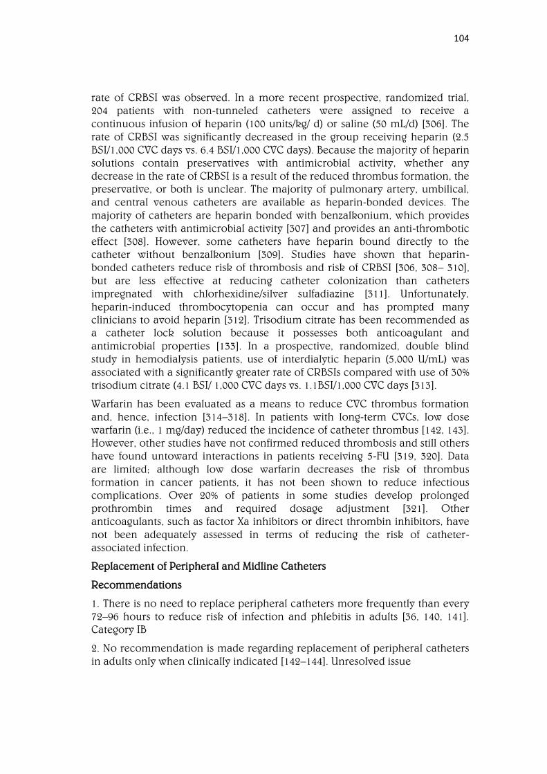
104
rate of CRBSI was observed. In a more recent prospective, randomized trial,
204 patients with non-tunneled catheters were assigned to receive a
continuous infusion of heparin (100 units/kg/ d) or saline (50 mL/d) [306]. The
rate of CRBSI was significantly decreased in the group receiving heparin (2.5
BSI/1,000 CVC days vs. 6.4 BSI/1,000 CVC days). Because the majority of heparin
solutions contain preservatives with antimicrobial activity, whether any
decrease in the rate of CRBSI is a result of the reduced thrombus formation, the
preservative, or both is unclear. The majority of pulmonary artery, umbilical,
and central venous catheters are available as heparin-bonded devices. The
majority of catheters are heparin bonded with benzalkonium, which provides
the catheters with antimicrobial activity [307] and provides an anti-thrombotic
effect [308]. However, some catheters have heparin bound directly to the
catheter without benzalkonium [309]. Studies have shown that heparin-
bonded catheters reduce risk of thrombosis and risk of CRBSI [306, 308– 310],
but are less effective at reducing catheter colonization than catheters
impregnated with chlorhexidine/silver sulfadiazine [311]. Unfortunately,
heparin-induced thrombocytopenia can occur and has prompted many
clinicians to avoid heparin [312]. Trisodium citrate has been recommended as
a catheter lock solution because it possesses both anticoagulant and
antimicrobial properties [133]. In a prospective, randomized, double blind
study in hemodialysis patients, use of interdialytic heparin (5,000 U/mL) was
associated with a significantly greater rate of CRBSIs compared with use of 30%
trisodium citrate (4.1 BSI/ 1,000 CVC days vs. 1.1BSI/1,000 CVC days [313].
Warfarin has been evaluated as a means to reduce CVC thrombus formation
and, hence, infection [314–318]. In patients with long-term CVCs, low dose
warfarin (i.e., 1 mg/day) reduced the incidence of catheter thrombus [142, 143].
However, other studies have not confirmed reduced thrombosis and still others
have found untoward interactions in patients receiving 5-FU [319, 320]. Data
are limited; although low dose warfarin decreases the risk of thrombus
formation in cancer patients, it has not been shown to reduce infectious
complications. Over 20% of patients in some studies develop prolonged
prothrombin times and required dosage adjustment [321]. Other
anticoagulants, such as factor Xa inhibitors or direct thrombin inhibitors, have
not been adequately assessed in terms of reducing the risk of catheter-
associated infection.
Replacement of Peripheral and Midline Catheters
Recommendations
1. There is no need to replace peripheral catheters more frequently than every
72–96 hours to reduce risk of infection and phlebitis in adults [36, 140, 141].
Category IB
2. No recommendation is made regarding replacement of peripheral catheters
in adults only when clinically indicated [142–144]. Unresolved issue

105
3. Replace peripheral catheters in children only when clinically indicated [32,
33]. Category IB
4. Replace midline catheters only when there is a specific indication. Category
II
Background
Scheduled replacement of intravascular catheters has been proposed as a
method to prevent phlebitis and catheter-related infections. Studies of short
peripheral venous catheters indicate that the incidence of thrombophlebitis
and bacterial colonization of catheters increases when catheters are left in
place >72 hours [258]. However, rates of phlebitis are not substantially different
in peripheral catheters left in place 72 hours compared with 96 hours [141].
Because phlebitis and catheter colonization have been associated with an
increased risk for catheter-related infection, short peripheral catheter sites
commonly are replaced at 72–96 hour intervals to reduce both the risk for
infection and patient discomfort associated with phlebitis.
Some studies have suggested that planned removal at 72 hours vs. removing as
needed resulted in similar rates of phlebitis and catheter failure [142–144].
However, these studies did not address the issue of CRBSI, and the risk of
CRBSIs with this strategy is not well studied.
Midline catheters are associated with lower rates of phlebitis than short
peripheral catheters and with lower rates of infection than CVCs [322–324]. In
one prospective study of 140 midline catheters, their use was associated with a
BSI rate of 0.8 per 1,000 catheter days [324]. No specific risk factors, including
duration of catheterization, were associated with infection. Midline catheters
were in place a median of 7 days, but for as long as 49 days. Although the
findings of this study suggested that midline catheters could be changed only
when there is a specific indication, no prospective, randomized studies have
assessed the benefit of routine replacement as a strategy to prevent CRBSI
associated with midline catheters.
Replacement of CVCs, Including PICCs and Hemodialysis Catheters
Recommendations
1. Do not routinely replace CVCs, PICCs, hemodialysis catheters, or pulmonary
artery catheters to prevent catheter-related infections. Category IB
2. Do not remove CVCs or PICCs on the basis of fever alone. Use clinical
judgment regarding the appropriateness of removing the catheter if infection is
evidenced elsewhere or if a noninfectious cause of fever is suspected. Category
II
3. Do not use guidewire exchanges routinely for non-tunneled catheters to
prevent infection. Category IB
4. Do not use guidewire exchanges to replace a non-tunneled catheter
suspected of infection. Category IB

106
5. Use a guidewire exchange to replace a malfunctioning non-tunneled
catheter if no evidence of infection is present. Category IB
6. Use new sterile gloves before handling the new catheter when guidewire
exchanges are performed. Category II
Background
Catheter replacement at scheduled time intervals as a method to reduce CRBSI
has not lowered rates. Two trials have assessed a strategy of changing the
catheter every 7 days compared with a strategy of changing catheters as
needed [165, 325]. One of these studies involved 112 surgical ICU patients
needing CVCs, pulmonary artery catheters, or peripheral arterial catheters
[165], whereas the other study involved only subclavian hemodialysis catheters
[325]. In both studies, no difference in CRBSI was observed in patients
undergoing scheduled catheter replacement every 7 days compared with
patients whose catheters were replaced as needed.
Scheduled guidewire exchange of CVCs is another proposed strategy for
preventing CRBSI. The results of a meta-analysis of 12 randomized, controlled
trials assessing CVC management failed to demonstrate any reduction of CRBSI
rates through routine replacement of CVCs by guidewire exchange compared
with catheter replacement on an as needed basis [326]. Thus, routine
replacement of CVCs is not necessary for catheters that are functioning and
have no evidence of causing local or systemic complications.
Catheter replacement over a guidewire has become an accepted technique for
replacing a malfunctioning catheter or exchanging a pulmonary artery
catheter for a CVC when invasive monitoring no longer is needed. Catheter
insertion over a guidewire is associated with less discomfort and a significantly
lower rate of mechanical complications than are those percutaneously inserted
at a new site [327]. In addition, this technique provides a means of preserving
limited venous access in some patients. Replacement of temporary catheters
over a guidewire in the presence of bacteremia is not an acceptable
replacement strategy because the source of infection is usually colonization of
the skin tract from the insertion site to the vein [37, 327]. However, in selected
patients with tunneled hemodialysis catheters and bacteremia, catheter
exchange over a guidewire, in combination with antibiotic therapy, is an
alternative as a salvage strategy in patients with limited venous access [328–
331].
Because of the increased difficulty obtaining vascular access in children,
attention should be given to the frequency with which catheters are replaced in
these patients. In a study in which survival analysis techniques were used to
examine the relation between the duration of central venous catheterization
and complications in pediatric ICU patients, all of the patients studied (n = 397)
remained uninfected for a median of 23.7 days [250]. In addition, no relation
was found between duration of catheterization and the daily probability of

107
infection (r = 0.21; P > .1), suggesting that routine replacement of CVCs likely
does not reduce the incidence of catheter-related infection [250].
Vascular access sites can be even more limited among neonates. Four
randomized trials (n = 368) summarized in a recent Cochrane Database
Systemic Review compared the effects of giving parenteral nutrition through
percutaneous central venous catheters vs. peripheral intravenous catheters.
Fewer painful procedures (venipunctures) were required in neonates
randomized to percutaneously placed CVCs, and there was no evidence for
increased risk of BSIs [332]. CVC occlusion due to thrombus formation is one of
the most common reasons for CVC removal in neonates. Various methods
have been tried to prevent catheter occlusion. Recently, a randomized trial (n
= 201) evaluated whether a continuous heparin infusion (0.5 units/kg/hour)
could effectively prolong the duration of catheterization when compared with
a placebo infusion. The rate of catheter occlusion requiring catheter removal
was lower in the heparin group (6% vs. 31%, P = .001: NNT = 4). Rates of CRBSI
were similar, although the study was not powered to evaluate CRBSI rate
differences. Heparin associated antibody levels were not routinely measured
[333].
Hemodialysis Catheters. The use of catheters for hemodialysis is the most
common factor contributing to bacteremia in dialysis patients [334, 335]. The
relative risk for bacteremia in patients with dialysis catheters is sevenfold the
risk for patients with arteriovenous (AV) fistulas [336]. AV fistulas and grafts are
preferred over hemodialysis catheters in patients with chronic renal failure,
due to their lower associated risk of infection. If temporary access is needed for
dialysis, a tunneled cuffed catheter is preferable to a non-cuffed catheter, even
in the ICU setting, if the catheter is expected to stay in place for >3weeks [59].
Pulmonary Artery Catheters. Pulmonary artery catheters are inserted through a
Teflon® introducer and typically remain in place an average of 3 days. The
majority of pulmonary artery catheters are heparin bonded, which reduces not
only catheter thrombosis but also microbial adherence to the catheter [307].
Meta-analysis indicates that the CRBSI rate associated with pulmonary artery
catheterization is 3.7 per 1,000 catheter days and somewhat higher than the
rate observed for unmedicated and non-tunnelled CVCs (2.7 per 1,000 catheter
days)[6, 45].
Data from prospective studies indicate that the risk of significant catheter
colonization and CRBSI increases the longer the catheter remains in place. In
general, the risk of significant catheter colonization increases after 4 days of
catheterization [75, 337, 338], whereas the risk of CRBSI increases beyond 5-7
days of catheterization [75, 84, 166]. Efforts must be made to differentiate
between infection related to the introducer and that related to the pulmonary
artery catheter. Significant colonization of the introducer occurs earlier than
that of the pulmonary artery catheter [337, 339]. However, no studies indicate
that catheter replacement at scheduled time intervals is an effective method to
reduce risk of CRBSI [165, 327, 339]. In patients who continue to require

108
hemodynamic monitoring, pulmonary artery catheters do not need to be
changed more frequently than every 7 days [339]. No specific recommendation
can be made regarding routine replacement of catheters that need to be in
place for >7 days.
Pulmonary artery catheters are usually packaged with a thin plastic sleeve that
prevents touch contamination when placed over the catheter. In a study of 166
catheters, patients who were randomly assigned to have their catheters self-
contained within this sleeve had a reduced risk for CRBSI compared with those
who had a pulmonary artery catheter placed without the sleeve (P = .002) [81].
Umbilical Catheters
Recommendations
1. Remove and do not replace umbilical artery catheters if any signs of CRBSI,
vascular insufficiency in the lower extremities, or thrombosis are present [145].
Category II
2. Remove and do not replace umbilical venous catheters if any signs of CRBSI
or thrombosis are present [145]. Category II
3. No recommendation can be made regarding attempts to salvage an
umbilical catheter by administering antibiotic treatment through the catheter.
Unresolved issue
4. Cleanse the umbilical insertion site with an antiseptic before catheter
insertion. Avoid tincture of iodine because of the potential effect on the
neonatal thyroid. Other iodine- containing products (e.g., povidone iodine)
can be used [146– 150]. Category IB
5. Do not use topical antibiotic ointment or creams on umbilical catheter
insertion sites because of the potential to promote fungal infections and
antimicrobial resistance [88, 89]. Category IA
6. Add low-doses of heparin (0.25–1.0 U/ml) to the fluid infused through
umbilical arterial catheters [151–153]. Category IB
7. Remove umbilical catheters as soon as possible when no longer needed or
when any sign of vascular insufficiency to the lower extremities is observed.
Optimally, umbilical artery catheters should not be left in place >5 days [145,
154]. Category II
8. Umbilical venous catheters should be removed as soon as possible when no
longer needed, but can be used up to 14 days if managed aseptically [155, 156].
Category II
9. An umbilical catheter may be replaced if it is malfunctioning, and there is no
other indication for catheter removal, and the total duration of catheterization
has not exceeded 5 days for an umbilical artery catheter or 14 days for an
umbilical vein catheter. Category II

109
Background
Although the umbilical stump becomes heavily colonized soon after birth,
umbilical vessel catheterization often is used for vascular access in newborn
infants. Umbilical vessels can be cannulated easily and permit both collection
of blood samples and measurement of hemodynamic status. The incidences of
catheter colonization and BSI are similar for umbilical vein catheters and
umbilical artery catheters. In several studies, an estimated 40%–55% of
umbilical artery catheters were colonized and 5% resulted in CRBSI; umbilical
vein catheters were associated with colonization in 22%–59% of cases [147, 148,
340] and with CRBSI in 3%– 8% of cases [148]. Although CRBSI rates are similar
for umbilical catheters in the high position (i.e., above the diaphragm)
compared with the low position (i.e., below the diaphragm and above the
aortic bifurcation), catheters placed in the high position result in a lower
incidence of vascular complications without an increase in adverse sequelae
[148].
Risk factors for infection differ for umbilical artery and umbilical vein
catheters. In one study, neonates with very low birth weight who also received
antibiotics for >10 days were at increased risk for umbilical artery CRBSIs
[148]. In comparison, those with higher birth weight and receipt of parenteral
nutrition fluids were at increased risk for umbilical vein CRBSI. Duration of
catheterization was not an independent risk factor for infection of either type
of umbilical catheter.
A recent randomized trial (n = 210) evaluated whether long-term umbilical
venous catheterization (up to 28 days) would result in the same or fewer
CRBSIs when compared with neonates who were randomized to short-term
umbilical venous catheterization for 7–10 days followed by percutaneous
central venous catheterization. CRBSI rate was higher (20%) among long term
catheterized neonates when compared with short term catheterized neonates
(13%). The difference was not statistically significant (P = .17), although the
study was underpowered. The study was not powered to evaluate differences in
venous thrombosis rates [341].
Peripheral Arterial Catheters and Pressure Monitoring Devices for Adult and
Pediatric Patients
Recommendations
1. In adults, use of the radial, brachial or dorsalis pedis sites is preferred over
the femoral or axillary sites of insertion to reduce the risk of infection [46, 47,
157, 158]. Category IB
2. In children, the brachial site should not be used. The radial, dorsalis pedis,
and posterior tibial sites are preferred over the femoral or axillary sites of
insertion [46]. Category II

110
3. A minimum of a cap, mask, sterile gloves and a small sterile fenestrated
drape should be used during peripheral arterial catheter insertion [47, 158,
159]. Category IB
4. During axillary or femoral artery catheter insertion, maximal sterile barriers
precautions should be used. Category II
5. Replace arterial catheters only when there is a clinical indication. Category II
6. Remove the arterial catheter as soon as it is no longer needed. Category II
7. Use disposable, rather than reusable, transducer assemblies when possible
[160–164]. Category IB
8. Do not routinely replace arterial catheters to prevent catheter-related
infections [165, 166, 167, 168]. Category II
9. Replace disposable or reusable transducers at 96-hour intervals. Replace
other components of the system (including the tubing, continuous-flush device,
and flush solution) at the time the transducer is replaced [37, 161]. Category IB
10. Keep all components of the pressure monitoring system (including
calibration devices and flush solution) sterile [160, 169–171]. Category IA
11. Minimize the number of manipulations of and entries into the pressure
monitoring system. Use a closed flush system (i.e., continuous flush), rather
than an open system (i.e., one that requires a syringe and stopcock), to
maintain the patency of the pressure monitoring catheters [163, 172]. Category
II
12. When the pressure monitoring system is accessed through a diaphragm,
rather than a stopcock, scrub the diaphragm with an appropriate antiseptic
before accessing the system [163]. Category IA
13. Do not administer dextrose-containing solutions or parenteral nutrition
fluids through the pressure monitoring circuit [163, 173, 174]. Category IA
14. Sterilize reusable transducers according to the manufacturers’ instructions
if the use of disposable transducers is not feasible [163, 173–176]. Category IA
Background
Arterial catheters are usually inserted into the radial or femoral artery and
permit continuous blood pressure monitoring and blood gas measurements.
The risk of CRBSI for arterial catheters is lower than that associated with non-
coated, uncuffed, non-tunneled short term CVCs (1.7 versus 2.7 per 1,000
catheter days) [6]. However, risk of CRBSI rates are comparable between
arterial catheters and coated, uncuffed, non-tunneled short term CVCs [6].
Unlike CVCs, use of full barrier precautions during arterial cannulaton does not
appear to reduce the risk of arterial CRBSI [158, 159]. Nonetheless, when
arterial catheters are inserted using a protocol which includes maximum
barrier precautions, a very low risk of CRBSI (0.41/1,000 catheter days) can be
achieved [47]. Although a meta-analysis failed to discern a difference in rates of
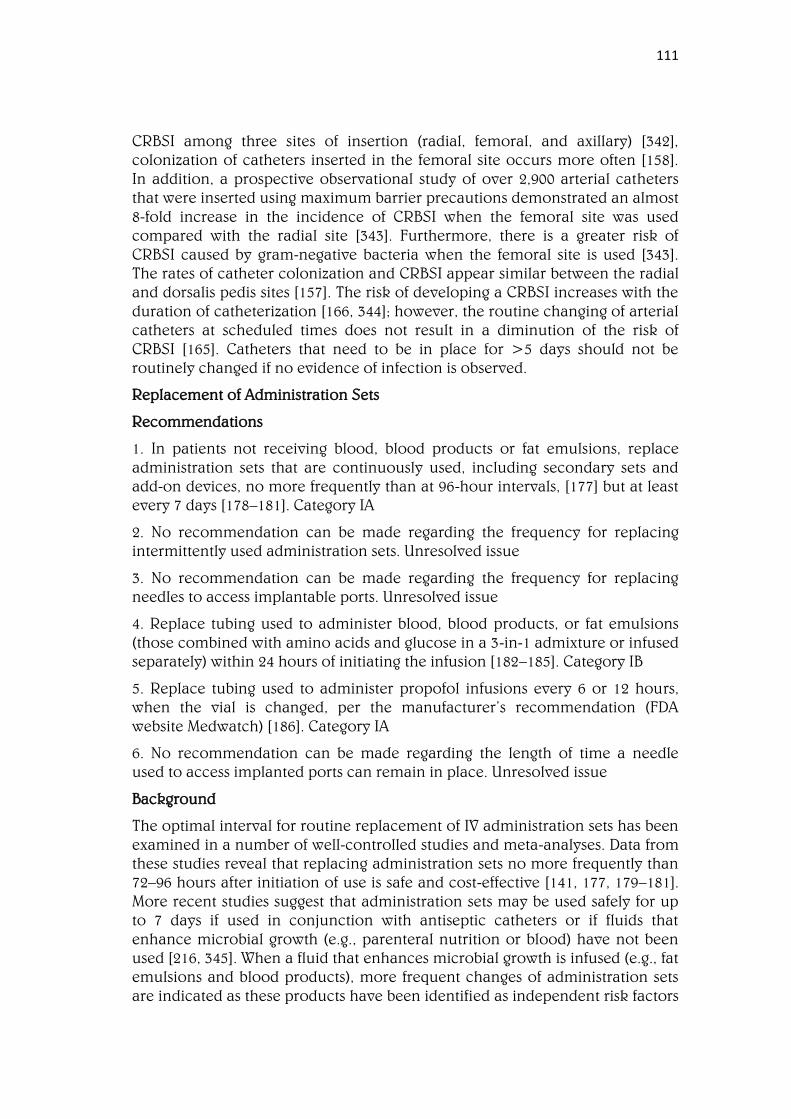
111
CRBSI among three sites of insertion (radial, femoral, and axillary) [342],
colonization of catheters inserted in the femoral site occurs more often [158].
In addition, a prospective observational study of over 2,900 arterial catheters
that were inserted using maximum barrier precautions demonstrated an almost
8-fold increase in the incidence of CRBSI when the femoral site was used
compared with the radial site [343]. Furthermore, there is a greater risk of
CRBSI caused by gram-negative bacteria when the femoral site is used [343].
The rates of catheter colonization and CRBSI appear similar between the radial
and dorsalis pedis sites [157]. The risk of developing a CRBSI increases with the
duration of catheterization [166, 344]; however, the routine changing of arterial
catheters at scheduled times does not result in a diminution of the risk of
CRBSI [165]. Catheters that need to be in place for >5 days should not be
routinely changed if no evidence of infection is observed.
Replacement of Administration Sets
Recommendations
1. In patients not receiving blood, blood products or fat emulsions, replace
administration sets that are continuously used, including secondary sets and
add-on devices, no more frequently than at 96-hour intervals, [177] but at least
every 7 days [178–181]. Category IA
2. No recommendation can be made regarding the frequency for replacing
intermittently used administration sets. Unresolved issue
3. No recommendation can be made regarding the frequency for replacing
needles to access implantable ports. Unresolved issue
4. Replace tubing used to administer blood, blood products, or fat emulsions
(those combined with amino acids and glucose in a 3-in-1 admixture or infused
separately) within 24 hours of initiating the infusion [182–185]. Category IB
5. Replace tubing used to administer propofol infusions every 6 or 12 hours,
when the vial is changed, per the manufacturer’s recommendation (FDA
website Medwatch) [186]. Category IA
6. No recommendation can be made regarding the length of time a needle
used to access implanted ports can remain in place. Unresolved issue
Background
The optimal interval for routine replacement of IV administration sets has been
examined in a number of well-controlled studies and meta-analyses. Data from
these studies reveal that replacing administration sets no more frequently than
72–96 hours after initiation of use is safe and cost-effective [141, 177, 179–181].
More recent studies suggest that administration sets may be used safely for up
to 7 days if used in conjunction with antiseptic catheters or if fluids that
enhance microbial growth (e.g., parenteral nutrition or blood) have not been
used [216, 345]. When a fluid that enhances microbial growth is infused (e.g., fat
emulsions and blood products), more frequent changes of administration sets
are indicated as these products have been identified as independent risk factors

112
for CRBSI [182, 216, 346–350]. Little data exist regarding the length of time a
needle used to access implanted ports can remain in place and the risk of
CRBSI. While some centers have left them in place for several weeks without
CRBSI, [351], this practice has not been adequately studied.
Needleless Intravascular Catheter Systems
Recommendations
1. Change the needleless components at least as frequently as the
administration set. There is no benefit to changing these more frequently than
every 72 hours. [39, 187–193]. Category II
2. Change needleless connectors no more frequently than every 72 hours or
according to manufacturers’ Recommendations for the purpose of reducing
infection rates [187, 189, 192, 193]. Category II
3. Ensure that all components of the system are compatible to minimize leaks
and breaks in the system [194]. Category II
4. Minimize contamination risk by scrubbing the access port with an
appropriate antiseptic (chlorhexidine, povidone iodine, an iodophor, or 70%
alcohol) and accessing the port only with sterile devices [189, 192, 194–196].
Category IA
5. Use a needleless system to access IV tubing. Category IC
6. When needleless systems are used, a split septum valve may be preferred
over some mechanical valves due to increased risk of infection with the
mechanical valves [197–200]. Category II
Background
Stopcocks used for injection of medications, administration of IV infusions,
and collection of blood samples represent a potential portal of entry for
microorganisms into vascular access catheters and IV fluids. Whether such
contamination is a substantial entry point of microorganisms that cause CRBSI
has not been demonstrated. Nonetheless, stopcocks should be capped when
not being used. In general, closed catheter access systems are associated with
fewer CRBSIs than open systems and should be used preferentially [352].
"Piggyback" systems (secondary intermittent infusions delivered through a port
on a primary infusion set) are used as an alternative to stopcocks. However,
they also pose a risk for contamination of the intravascular fluid if the device
entering the rubber membrane of an injection port is exposed to air or if it
comes into direct contact with nonsterile tape used to fix the needle to the
port. Modified piggyback systems have the potential to prevent contamination
at these sites [353].
Attempts to reduce the incidence of sharps injuries and the resultant risk for
transmission of bloodborne infections to healthcare personnel have led to the
introduction and mandating of needleless infusion systems. There are several
types of needleless connectors on the market.

113
The first type of needleless system connectors consisted of a split septum
connector, which is accessed with a blunt cannula instead of a needle (external
cannulae activated split septums). Because of the large amount of space in the
connector to accommodate the cannula, when the cannula is removed it may
result in the creation of negative pressure which may cause blood to be
aspirated into the distal lumen, possibly increasing the risk of catheter
occlusion or thrombosis. A luer-activated device, which incorporates a valve
preventing the outflow of fluid through the connector, was designed to
eliminate this problem. Some luer devices require a cap to be attached to the
valve when not in use, which can be difficult to maintain aseptically, and
therefore they may be prone to contamination.
Another type of second-generation needleless system addressed the occlusion
issue by incorporating positive or neutral fluid displacement to either flush out
aspirated blood or prevent its aspiration into infusion catheters.
Use of needleless connectors or mechanical valves appear to be effective in
reducing connector colonization in some [196, 354, 355], but not all studies
[356] when compared with stopcocks and caps. In one study [354], the
incidence of CRBSI was reduced when the needleless connector was compared
with standard stopcocks. Appropriate disinfectants must be used to prevent
transmission of microbes through connectors [357]. Some studies have shown
that disinfection of the devices with chlorhexidine/alcohol solutions appears to
be most effective in reducing colonization [195, 196]. In addition, the time
spent applying the disinfectant may be important. One study found that
swiping the luer-activated device with 70% alcohol for only 3 to 5 seconds did
not adequately disinfect the septal surface [358]. However, a number of
outbreak investigations have reported increases in CRBSIs associated with a
switch from external cannulae activated split septum needleless devices to
mechanical valve devices [197, 198, 200, 359]. The reasons for these
associations are not known and it is also not known if this is a device-specific
or class association, particularly as physical and mechanical properties of
needleless connectors vary from device to device. In addition, one
investigation found CRBSIs increased with the switch from a luer-activated
negative displacement mechanical valve to a luer-activated positive fluid
displacement mechanical valve [199]. However in an observational study, a
switch from a luer-activated negative displacement mechanical valve to a
different luer-activated positive displacement mechanical valve as part of a
bundled intervention resulted in a significant decrease in CRBSIs [201].
Potential explanations for outbreaks associated with these devices include
difficulty encountered in adequate disinfection of the surface of the connector
due to physical characteristics of the plastic housing diaphragm interface, fluid
flow properties (laminar vs. turbulent), internal surface area, potential fluid
dead space, inadequate flushing of the device due to poor visualization of the
fluid flow pathway in opaque devices, and the presence of internal
corrugations that could harbor organisms, particularly if the catheters are used
to withdraw blood [199]. Some studies have shown that the increase in CRBSIs

114
with the change to lueractivated devices may be related to improper cleaning
and infection control practices such as infrequently changing the devices [192,
194]. Additionally, silver-coated connector valves have been FDA approved;
however, there are no published randomized trials with this device and no
recommendation can be made regarding its use. Likewise, an antiseptic-barrier
cap for needleless connectors has been studied in a laboratory setting and
appears to be effective in preventing the entry of microorganisms [360], but has
not yet been studied in a clinical trial.
Performance Improvement
Recommendation
Use hospital-specific or collaborative-based performance improvement
initiatives in which multifaceted strategies are "bundled" together to improve
compliance with evidence-based recommended practices [15, 69, 70, 201–205].
Category IB
Background
Clinical decision makers, healthcare payers, and patient safety advocates
emphasize the importance of translating research findings into everyday
practice. Rigorous evaluations of CRBSI preventive practices using study
designs with high internal validity and including study populations that
optimize external validity remain necessary. Once practices have been
determined to be effective and economically efficient, the next step is to
implement these evidence-based practices so they become part of routine
clinical care. Unfortunately, implementation of evidence- based CRBSI
preventive practices in U.S. hospitals has been suboptimal [361, 362]. In a
national survey conducted in March 2005 of over 700 U.S. hospitals,
approximately one quarter of U.S. hospitals indicated that either maximal
sterile barrier precautions during central line insertion or chlorhexidine
gluconate as site disinfectant, two practices widely recommended in the
guidelines published in 2002 [363], were not being used routinely [364].
Approximately 15% of U.S. hospitals reported routinely changing CVCs to
prevent infection despite evidence that this practice should no longer be used
[362, 364].
Accordingly, investigators have attempted various approaches to better
translate research findings and evidence-based Recommendations into clinical
practice. Numerous quality improvement studies have been published during
the past several years that have used various methods, such as education of
healthcare personnel, audit and feedback, organizational change, and clinical
reminders [8–11, 69, 70, 202, 365–367]. The educational interventions primarily
targeted hand hygiene, use of maximal sterile barriers during insertion,
appropriate insertion site selection, proper site care using chlorhexidine
gluconate, and prompt removal of unnecessary catheters. While a large
number of before-and-after studies with a few using concurrent control groups
[15, 70] have been published, no randomized, controlled trial evaluating a

115
quality improvement strategy to prevent CRBSI has been reported [368]. The
vast majority of before-and-after studies reported statistically significant
decreases in CRBSI rates after a quality improvement strategy was
implemented [368]. Additionally, both controlled trials also found statistically
significant reductions of CRBSI in the intervention units compared with control
units [15, 70].
Investigators have also employed multifaceted approaches in which several
strategies are bundled together to improve compliance with evidence-based
guidelines [15, 69, 70]. One such collaborative cohort study [69] of 108 ICUs in
Michigan targeted clinicians’ use of five evidence- based practices: hand
hygiene, maximum barrier precautions, chlorhexidine site disinfection,
avoiding the femoral site, and promptly removing unnecessary central venous
catheters. In addition to educating clinicians about CRBSI prevention,
interventions used included:
1. A central venous catheter cart that contained all the necessary supplies;
2. A checklist to ensure adherence to proper practices;
3. Stoppage of procedures in non-emergent situations, if evidence- based
practices were not being followed;
4. Prompt removal of unnecessary central catheters identified during daily
patient rounds;
5. Feedback to the clinical teams regarding the number of CRBSI episodes and
overall rates; and
6. Buy-in from the chief executive officers of the participating hospitals that
chlorhexidine gluconate products/solutions would be stocked prior to study
initiation.
Using an interrupted time series analysis and multivariable regression, the
investigators reported a statistically significant 66% decrease in CRBSI rates
approximately 18 months after the intervention began [69] and sustained
reductions over time [369]. Specific process and outcome measures for
tracking and feedback (i.e., rate of central line infections, proportion of central
lines placed with all or individual bundle elements performed AND
documented) should be identified in individual institutions based on areas that
have been identified for performance improvement.
Finally, emphasis on the care and maintenance of catheters once they are in
place should be a focus of performance improvement and quality assurance in
all programs. A study to assess practice and staff knowledge of CVC post-
insertion care and identify aspects of CVC care with potential for improvement
revealed several areas of opportunity to improve post-insertion care [370]. Data
were recorded on 151 CVCs in 106 patients giving a total of 721 catheter days.
In all, 323 breaches in care were identified giving a failure rate of 44.8%, with
significant differences between intensive care unit (ICU) and non-ICU wards.
Dressings (not intact) and caps (incorrectly placed) were identified as the major
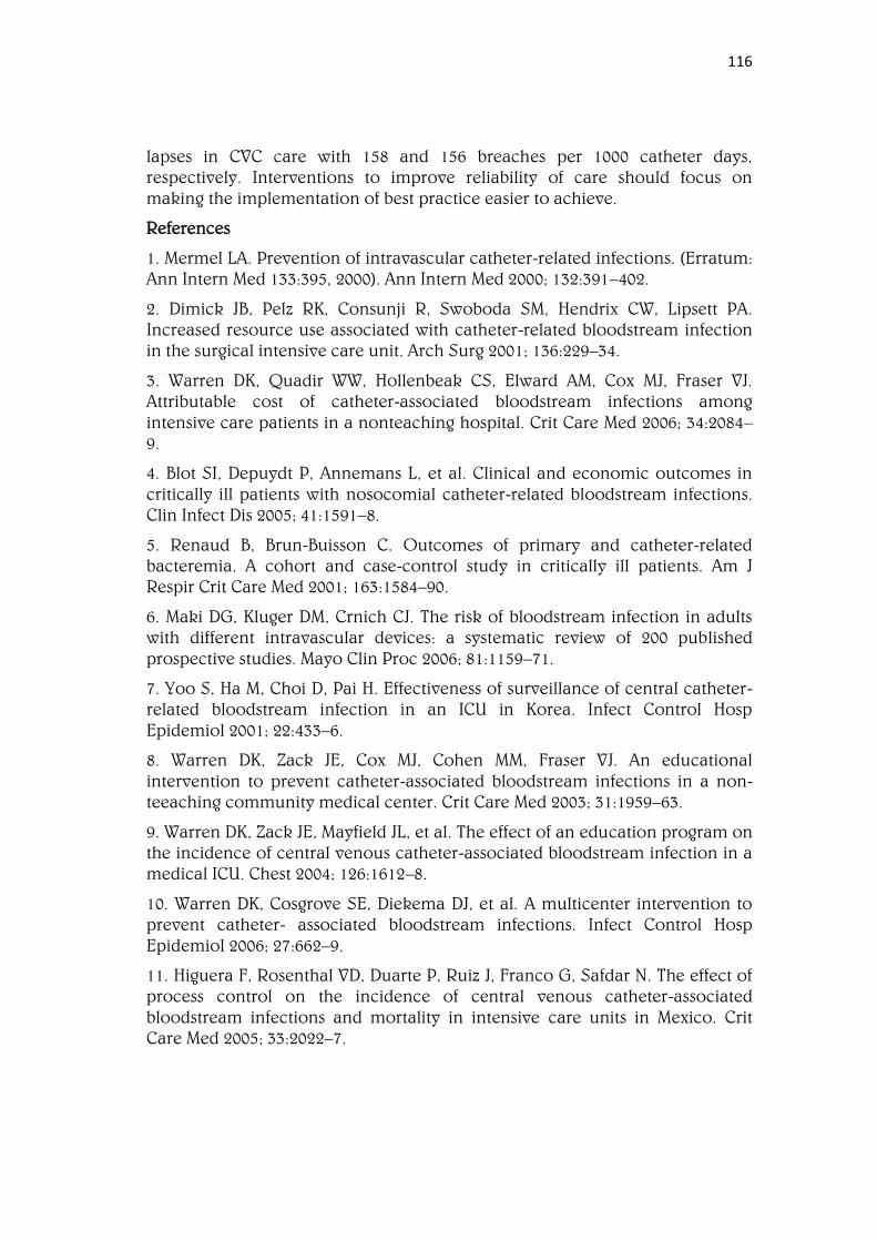
116
lapses in CVC care with 158 and 156 breaches per 1000 catheter days,
respectively. Interventions to improve reliability of care should focus on
making the implementation of best practice easier to achieve.
References
1. Mermel LA. Prevention of intravascular catheter-related infections. (Erratum:
Ann Intern Med 133:395, 2000). Ann Intern Med 2000; 132:391–402.
2. Dimick JB, Pelz RK, Consunji R, Swoboda SM, Hendrix CW, Lipsett PA.
Increased resource use associated with catheter-related bloodstream infection
in the surgical intensive care unit. Arch Surg 2001; 136:229–34.
3. Warren DK, Quadir WW, Hollenbeak CS, Elward AM, Cox MJ, Fraser VJ.
Attributable cost of catheter-associated bloodstream infections among
intensive care patients in a nonteaching hospital. Crit Care Med 2006; 34:2084–
9.
4. Blot SI, Depuydt P, Annemans L, et al. Clinical and economic outcomes in
critically ill patients with nosocomial catheter-related bloodstream infections.
Clin Infect Dis 2005; 41:1591–8.
5. Renaud B, Brun-Buisson C. Outcomes of primary and catheter-related
bacteremia. A cohort and case-control study in critically ill patients. Am J
Respir Crit Care Med 2001; 163:1584–90.
6. Maki DG, Kluger DM, Crnich CJ. The risk of bloodstream infection in adults
with different intravascular devices: a systematic review of 200 published
prospective studies. Mayo Clin Proc 2006; 81:1159–71.
7. Yoo S, Ha M, Choi D, Pai H. Effectiveness of surveillance of central catheter-
related bloodstream infection in an ICU in Korea. Infect Control Hosp
Epidemiol 2001; 22:433–6.
8. Warren DK, Zack JE, Cox MJ, Cohen MM, Fraser VJ. An educational
intervention to prevent catheter-associated bloodstream infections in a non-
teeaching community medical center. Crit Care Med 2003; 31:1959–63.
9. Warren DK, Zack JE, Mayfield JL, et al. The effect of an education program on
the incidence of central venous catheter-associated bloodstream infection in a
medical ICU. Chest 2004; 126:1612–8.
10. Warren DK, Cosgrove SE, Diekema DJ, et al. A multicenter intervention to
prevent catheter- associated bloodstream infections. Infect Control Hosp
Epidemiol 2006; 27:662–9.
11. Higuera F, Rosenthal VD, Duarte P, Ruiz J, Franco G, Safdar N. The effect of
process control on the incidence of central venous catheter-associated
bloodstream infections and mortality in intensive care units in Mexico. Crit
Care Med 2005; 33:2022–7.

117
12. Coopersmith CM, Rebmann TL, Zack JE, et al. Effect of an education
program on decreasing catheter-related bloodstream infections in the surgical
intensive care unit. Crit Care Med 2002; 30:59–64.
13. Coopersmith CM, Zack JE, Ward MR, et al. The impact of bedside behavior
on catheter-related bacteremia in the intensive care unit. Arch Surg 2004;
139:131–6.
14. Sherertz RJ, Ely EW, Westbrook DM, et al. Education of physiciansin-training
can decrease the risk for vascular catheter infection. Ann Intern Med 2000;
132:641–8.
15. Eggimann P, Harbarth S, Constantin MN, Touveneau S, Chevrolet JC, Pittet
D. Impact of a prevention strategy targeted at vascular-access care on
incidence of infections acquired in intensive care. Lancet 2000; 355:1864–8.
16. Nehme AE. Nutritional support of the hospitalized patient. The team
concept. JAMA 1980; 243:1906–8.
17. Soifer NE, Borzak S, Edlin BR, Weinstein RA. Prevention of peripheral
venous catheter complications with an intravenous therapy team: a
randomized controlled trial. Arch Intern Med 1998; 158:473–7.
18. Tomford JW, Hershey CO, McLaren CE, Porter DK, Cohen DI. Intravenous
therapy team and peripheral venous catheter-associated complications. A
prospective controlled study. Arch Intern Med 1984; 144:1191–4.
19. Scalley RD, Van CS, Cochran RS. The impact of an i.v. team on the
occurrence of intravenous- related phlebitis. A 30-month study. J Intraven Nurs
1992; 15:100–9.
20. Palefski SS, Stoddard GJ. The infusion nurse and patient complication rates
of peripheral-short catheters. A prospective evaluation. J Intraven Nurs 2001;
24:113–23.
21. Miller JM, Goetz AM, Squier C, Muder RR. Reduction in nosocomial
intravenous device-related bacteremias after institution of an intravenous
therapy team. J Intraven Nurs 1996; 19:103–6.
22. Hunter MR. Development of a Vascular Access Team in an acute care
setting. J Infus Nurs 2003; 26:86–91.
23. Hawes ML. A proactive approach to combating venous depletion in the
hospital setting. J Infus Nurs 2007; 30:33–44.
24. Brunelle D. Impact of a dedicated infusion therapy team on the reduction of
catheter-related nosocomial infections. J Infus Nurs 2003; 26:362–6.
25. Bosma TL, Jewesson PJ. An infusion program resource nurse consult
service: our experience in a major Canadian teaching hospital. J Infus Nurs
2002; 25:310–5.
26. Pierce CA, Baker JJ. A nursing process model: quantifying infusion therapy
resource consumption. J Infus Nurs 2004; 27:232–44.

118
27. Tomford JW, Hershey CO. The i.v. therapy team: impact on patient care and
costs of hospitalization. NITA 1985; 8:387–9.
28. Davis D, O’Brien MA, Freemantle N, Wolf FM, Mazmanian P, Taylor-Vaisey
A. Impact of formal continuing medical education: do conferences, workshops,
rounds, and other traditional continuing education activities change physician
behavior or health care outcomes? JAMA 1999; 282:867–74.
29. Alonso-Echanove J, Edwards JR, Richards MJ, et al. Effect of nurse staffing
and antimicrobial- impregnated central venous catheters on the risk for
bloodstream infections in intensive care units. Infect Control Hosp Epidemiol
2003; 24:916–25.
30. Fridkin SK, Pear SM, Williamson TH, Galgiani JN, Jarvis WR. The role of
understaffing in central venous catheter-associated bloodstream infections.
Infect Control Hosp Epidemiol 1996; 17: 150–8.
31. Robert J, Fridkin SK, Blumberg HM, et al. The influence of the composition
of the nursing staff on primary bloodstream infection rates in a surgical
intensive care unit. Infect Control Hosp Epidemiol 2000; 21:12–7.
32. Maki DG, Goldman DA, Rhame FS. Infection control in intravenous therapy.
Ann Intern Med 1973; 79:867–87.
33. Band JD, Maki DG. Steel needles used for intravenous therapy. Morbidity in
patients with hematologic malignancy. Arch Intern Med 1980; 140:31–4.
34. Tully JL, Friedland GH, Baldini LM, Goldmann DA. Complications of
intravenous therapy with steel needles and Teflon catheters. A comparative
study. Am J Med 1981; 70:702–6.
35. Ryder MA. Peripheral access options. Surg Oncol Clin N Am 1995; 4:395–
427.
36. Maki DG, Ringer M. Risk factors for infusion-related phlebitis with small
peripheral venous catheters. A randomized controlled trial. Ann Intern Med
1991; 114:845–54.
37. Mermel LA, McCormick RD, Springman SR, Maki DG. The pathogenesis and
epidemiology of catheter-related infection with pulmonary artery Swan-Ganz
catheters: a prospective study utilizing molecular subtyping. Am J Med 1991;
91:197S–205.
38. Parienti JJ, Thirion M, Megarbane B, et al. Femoral vs jugular venous
catheterization and risk of nosocomial events in adults requiring acute renal
replacement therapy: a randomized controlled trial. JAMA 2008; 299:2413–22.
39. Moretti EW, Ofstead CL, Kristy RM, Wetzler HP. Impact of central venous
catheter type and methods on catheter-related colonization and bacteraemia. J
Hosp Infect 2005; 61:139–45.

119
40. Nagashima G, Kikuchi T, Tsuyuzaki H, et al. To reduce catheter-related
bloodstream infections: is the subclavian route better than the jugular route for
central venous catheterization? J Infect Chemother 2006; 12:363–5.
41. Ruesch S, Walder B, Tramer MR. Complications of central venous catheters:
internal jugular versus subclavian access–a systematic review. Crit Care Med
2002; 30:454–60.
42. Sadoyama G, Gontijo Filho PP. Comparison between the jugular and
subclavian vein as insertion site for central venous catheters: microbiological
aspects and risk factors for colonization and infection. Braz J Infect Dis 2003;
7:142–8.
43. Heard SO, Wagle M, Vijayakumar E, et al. Influence of triple-lumen central
venous catheters coated with chlorhexidine and silver sulfadiazine on the
incidence of catheter-related bacteremia. Arch Intern Med 1998; 158:81–7.
44. Richet H, Hubert B, Nitemberg G, et al. Prospective multicenter study of
vascular-catheter related complications and risk factors for positive central-
catheter cultures in intensive care unit patients. J Clin Microbiol 1990; 28:2520–
5.
45. Safdar N, Kluger DM, Maki DG. A review of risk factors for catheter-related
bloodstream infection caused by percutaneously inserted, noncuffed central
venous catheters: implications for preventive strategies. Medicine (Baltimore)
2002; 81:466–79.
46. Lorente L, Jimenez A, Iribarren JL, Jimenez JJ, Martin MM, Mora ML. The
micro-organism responsible for central venous catheter related bloodstream
infection depends on catheter site. Intensive Care Med 2006; 32:1449–50.
47. Traore O, Liotier J, Souweine B. Prospective study of arterial and central
venous catheter colonization and of arterial-and central venous catheter-
related bacteremia in intensive care units. Crit Care Med 2005; 33:1276–80.
48. Joynt GM, Kew J, Gomersall CD, Leung VY, Liu EK. Deep venous thrombosis
caused by femoral venous catheters in critically ill adult patients. Chest 2000;
117:178–83.
49. Mian NZ, Bayly R, Schreck DM, Besserman EB, Richmand D. Incidence of
deep venous thrombosis associated with femoral venous catheterization. Acad
Emerg Med 1997; 4:1118– 21.
50. Merrer J, De Jonghe B, Golliot F, et al. Complications of femoral and
subclavian venous catheterization in critically ill patients: a randomized
controlled trial. JAMA 2001; 286:700–7.
51. Goetz AM, Wagener MM, Miller JM, Muder RR. Risk of infection due to
central venous catheters: effect of site of placement and catheter type. Infect
Control Hosp Epidemiol 1998; 19:842–5.

120
52. Robinson JF, Robinson WA, Cohn A, Garg K, Armstrong JD, 2nd. Perforation
of the great vessels during central venous line placement. Arch Intern Med
1995; 155:1225–8.
53. Trottier SJ, Veremakis C, O’Brien J, Auer AI. Femoral deep vein thrombosis
associated with central venous catheterization: results from a prospective,
randomized trial. Crit Care Med 1995; 23:52–9.
54. Lorente L, Henry C, Martin MM, Jimenez A, Mora ML. Central venous
catheter-related infection in a prospective and observational study of 2,595
catheters. Crit Care 2005; 9:R631–5.
55. Schillinger F, Schillinger D, Montagnac R, Milcent T. Post catheterisation
vein stenosis in haemodialysis: comparative angiographic study of 50
subclavian and 50 internal jugular accesses. Nephrol Dial Transplant 1991;
6:722–4.
56. Cimochowski GE, Worley E, Rutherford WE, Sartain J, Blondin J, Harter H.
Superiority of the internal jugular over the subclavian access for temporary
dialysis. Nephron 1990; 54:154–61.
57. Barrett N, Spencer S, McIvor J, Brown EA. Subclavian stenosis: a major
complication of subclavian dialysis catheters. Nephrol Dial Transplant 1988;
3:423–5.
58. Trerotola SO, Kuhn-Fulton J, Johnson MS, Shah H, Ambrosius WT,
Kneebone PH. Tunneled infusion catheters: increased incidence of
symptomatic venous thrombosis after subclavian versus internal jugular
venous access. Radiology 2000; 217:89–93.
59. National Kidney Foundation. III. NKF-K/DOQI Clinical Practice Guidelines
for Vascular Access: update 2000. Am J Kidney Dis 2001; 37:S137–81.
60. Hind D, Calvert N, McWilliams R, et al. Ultrasonic locating devices for
central venous cannulation: meta-analysis. BMJ 2003; 327:361.
61. Randolph AG, Cook DJ, Gonzales CA, Pribble CG. Ultrasound guidance for
placement of central venous catheters: a meta-analysis of the literature. Crit
Care Med 1996; 24:2053–8.
62. Froehlich CD, Rigby MR, Rosenberg ES, et al. Ultrasound-guided central
venous catheter placement decreases complications and decreases placement
attempts compared with the landmark technique in patients in a pediatric
intensive care unit. Crit Care Med 2009; 37:1090–6.
63. Lamperti M, Caldiroli D, Cortellazzi P, et al. Safety and efficacy of
ultrasound assistance during internal jugular vein cannulation in neurosurgical
infants. Intensive Care Med 2008; 34:2100– 5.
64. Schweickert WD, Herlitz J, Pohlman AS, Gehlbach BK, Hall JB, Kress JP. A
randomized, controlled trial evaluating postinsertion neck ultrasound in
peripherally inserted central catheter procedures. Crit Care Med 2009; 37:1217–
21.

121
65. Clark-Christoff N, Watters VA, Sparks W, Snyder P, Grant JP. Use of triple-
lumen subclavian catheters for administration of total parenteral nutrition.
JPEN J Parenter Enteral Nutr 1992; 16:403–7.
66. Early TF, Gregory RT, Wheeler JR, Snyder SO Jr., Gayle RG. Increased
infection rate in double- lumen versus single-lumen Hickman catheters in
cancer patients. South Med J 1990; 83:34–6.
67. Hilton E, Haslett TM, Borenstein MT, Tucci V, Isenberg HD, Singer C. Central
catheter infections: single-versus triple-lumen catheters. Influence of guide
wires on infection rates when used for replacement of catheters. Am J Med
1988; 84:667–72.
68. Yeung C, May J, Hughes R. Infection rate for single lumen v triple lumen
subclavian catheters. Infect Control Hosp Epidemiol 1988; 9:154–8.
69. Pronovost P, Needham D, Berenholtz S, et al. An intervention to decrease
catheter-related bloodstream infections in the ICU. N Engl J Med 2006;
355:2725–32.
70. Berenholtz SM, Pronovost PJ, Lipsett PA, et al. Eliminating catheter-related
bloodstream infections in the intensive care unit. Crit Care Med 2004; 32:2014–
20.
71. Lederle FA, Parenti CM, Berskow LC, Ellingson KJ. The idle intravenous
catheter. Ann Intern Med 1992; 116:737–8.
72. Parenti CM, Lederle FA, Impola CL, Peterson LR. Reduction of unnecessary
intravenous catheter use. Internal medicine house staff participate in a
successful quality improvement project. Arch Intern Med 1994; 154:1829–32.
73. Abi-Said D, Raad I, Umphrey J, et al. Infusion therapy team and dressing
changes of central venous catheters. Infect Control Hosp Epidemiol 1999;
20(2):101–5.
74. Capdevila JA, Segarra A, Pahissa A. Catheter-related bacteremia in patients
undergoing hemodialysis. Ann Intern Med 1998; 128 (7):600.
75. Mermel LA, Maki DG. Infectious complications of Swan-Ganz pulmonary
artery catheters. Pathogenesis, epidemiology, prevention, and management.
Am J Respir Crit Care Med 1994; 149:1020–36.
76. Raad II, Hohn DC, Gilbreath BJ, et al. Prevention of central venous catheter-
related infections by using maximal sterile barrier precautions during insertion.
Infect Control Hosp Epidemiol 1994; 15:231–8.
77. Boyce JM, Pittet D. Guideline for hand hygiene in health-care settings:
Recommendations of the Healthcare Infection Control Practices Advisory
Committee and the HICPAC/SHEA/APIC/IDSA Hand Hygiene Task Force. Infect
Control Hosp Epidemiol 2002; 23:S3–40.

122
78. Bischoff WE, Reynolds TM, Sessler CN, Edmond MB, Wenzel RP.
Handwashing compliance by health care workers: the impact of introducing an
accessible, alcohol-based hand antiseptic. Arch Intern Med 2000; 160:1017–21.
79. Pittet D, Dharan S, Touveneau S, Sauvan V, Perneger TV. Bacterial
contamination of the hands of hospital staff during routine patient care. Arch
Intern Med 1999; 159:821–6.
80. Carrer S, Bocchi A, Bortolotti M, et al. Effect of different sterile barrier
precautions and central venous catheter dressing on the skin colonization
around the insertion site. Minerva Anestesiol 2005; 71:197–206.
81. Cohen Y, Fosse JP, Karoubi P, et al. The "hands-off" catheter and the
prevention of systemic infections associated with pulmonary artery catheter: a
prospective study. Am J Respir Crit Care Med 1998; 157:284–7.
82. Maki DG, Ringer M, Alvarado CJ. Prospective randomised trial of povidone-
iodine, alcohol, and chlorhexidine for prevention of infection associated with
central venous and arterial catheters. Lancet 1991; 338:339–43.
83. Mimoz O, Pieroni L, Lawrence C, et al. Prospective, randomized trial of two
antiseptic solutions for prevention of central venous or arterial catheter
colonization and infection in intensive care unit patients. Crit Care Med 1996;
24:1818–23.
84. Maki DG, Stolz SS, Wheeler S, Mermel LA. A prospective, randomized trial
of gauze and two polyurethane dressings for site care of pulmonary artery
catheters: implications for catheter management. Crit Care Med 1994; 22:1729–
37.
85. Bijma R, Girbes AR, Kleijer DJ, Zwaveling JH. Preventing central venous
catheter-related infection in a surgical intensive-care unit. Infect Control Hosp
Epidemiol 1999; 20:618–20.
86. Madeo M, Martin CR, Turner C, Kirkby V, Thompson DR. A randomized trial
comparing Arglaes (a transparent dressing containing silver ions) to Tegaderm
(a transparent polyurethane dressing) for dressing peripheral arterial catheters
and central vascular catheters. Intensive Crit Care Nurs 1998; 14:187–91.
87. Rasero L, Degl’Innocenti M, Mocali M, et al. Comparison of two different
time interval protocols for central venous catheter dressing in bone marrow
transplant patients: results of a randomized, multicenter study. The Italian
Nurse Bone Marrow Transplant Group (GITMO). Haematologica 2000; 85:275–9.
88. Zakrzewska-Bode A, Muytjens HL, Liem KD, Hoogkamp-Korstanje JA.
Mupirocin resistance in coagulase-negative staphylococci, after topical
prophylaxis for the reduction of colonization of central venous catheters. J
Hosp Infect 1995; 31:189–93.
89. Flowers RH, Schwenzer KJ, Kopel RF, Fisch MJ, Tucker SI, Farr BM. Efficacy
of an attachable subcutaneous cuff for the prevention of intravascular catheter-
related infection. A randomized, controlled trial. JAMA 1989; 261:878–83.

123
90. Robbins J, Cromwell P, Korones DN. Swimming and central venous
catheter-related infections in the child with cancer. J Pediatr Oncol Nurs 1999;
16:51–6.
91. Howell PB, Walters PE, Donowitz GR, Farr BM. Risk factors for infection of
adult patients with cancer who have tunnelled central venous catheters.
Cancer 1995; 75:1367–75.
92. Ivy DD, Calderbank M, Wagner BD, et al. Closed-hub systems with protected
connections and the reduction of risk of catheter-related bloodstream infection
in pediatric patients receiving intravenous prostanoid therapy for pulmonary
hypertension. Infect Control Hosp Epidemiol 2009; 30:823–9.
93. Timsit JF, Schwebel C, Bouadma L, et al. Chlorhexidine-impregnated
sponges and less frequent dressing changes for prevention of catheter-related
infections in critically ill adults: a randomized controlled trial. JAMA 2009;
301:1231–41.
94. Rao SP, Oreopoulos DG. Unusual complications of a polyurethane PD
catheter. Perit Dial Int 1997; 17:410–2.
95. Riu S, Ruiz CG, Martinez-Vea A, Peralta C, Oliver JA. Spontaneous rupture
of polyurethane peritoneal catheter. A possible deleterious effect of mupirocin
ointment. Nephrol Dial Transplant 1998; 13:1870–1.
96. Garland JS, Alex CP, Mueller CD, et al. A randomized trial comparing
povidone-iodine to a chlorhexidine gluconate-impregnated dressing for
prevention of central venous catheter infections in neonates. Pediatrics 2001;
107:1431–6.
97. Ho KM, Litton E. Use of chlorhexidine-impregnated dressing to prevent
vascular and epidural catheter colonization and infection: a meta-analysis. J
Antimicrob Chemother 2006; 58:281–7.
98. Levy I, Katz J, Solter E, et al. Chlorhexidine-impregnated dressing for
prevention of colonization of central venous catheters in infants and children:
a randomized controlled study. Pediatr Infect Dis J 2005; 24:676–9.
99. Lorenzen AN, Itkin DJ. Surveillance of infection in home care. Am J Infect
Control 1992; 20:326–9.
100. White MC. Infections and infection risks in home care settings. Infect
Control Hosp Epidemiol 1992; 13:535–9.
101. White MC, Ragland KE. Surveillance of intravenous catheter-related
infections among home care clients. Am J Infect Control 1994; 22:231–5.
102. Bleasdale SC, Trick WE, Gonzalez IM, Lyles RD, Hayden MK, Weinstein RA.
Effectiveness of chlorhexidine bathing to reduce catheter-associated
bloodstream infections in medical intensive care unit patients. Arch Intern Med
2007; 167:2073–9.

124
103. Munoz-Price LS, Hota B, Stemer A, Weinstein RA. Prevention of
bloodstream infections by use of daily chlorhexidine baths for patients at a
long-term acute care hospital. Infect Control Hosp Epidemiol 2009; 30:1031–5.
104. Popovich KJ, Hota B, Hayes R, Weinstein RA, Hayden MK. Effectiveness of
routine patient cleansing with chlorhexidine gluconate for infection prevention
in the medical intensive care unit. Infect Control Hosp Epidemiol 2009; 30:959–
63.
105. Yamamoto AJ, Solomon JA, Soulen MC, et al. Sutureless securement
device reduces complications of peripherally inserted central venous catheters.
J Vasc Interv Radiol 2002; 13:77–81.
106. Brun-Buisson C, Doyon F, Sollet JP, Cochard JF, Cohen Y, Nitenberg G.
Prevention of intravascular catheter-related infection with newer
chlorhexidine-silver sulfadiazine-coated catheters: a randomized controlled
trial. Intensive Care Med 2004; 30:837–43.
107. Ostendorf T, Meinhold A, Harter C, et al. Chlorhexidine and silver-
sulfadiazine coated central venous catheters in haematological patients–a
double-blind, randomised, prospective, controlled trial. Support Care Cancer
2005; 13:993–1000.
108. Rupp ME, Lisco SJ, Lipsett PA, et al. Effect of a second-generation venous
catheter impregnated with chlorhexidine and silver sulfadiazine on central
catheter-related infections: a randomized, controlled trial. Ann Intern Med
2005; 143:570–80.
109. Darouiche RO, Raad II, Heard SO, et al. A comparison of two
antimicrobial-impregnated central venous catheters. Catheter Study Group. N
Engl J Med 1999; 340:1–8.
110. Raad I, Darouiche R, Dupuis J, et al. Central venous catheters coated with
minocycline and rifampin for the prevention of catheter-related colonization
and bloodstream infections. A randomized, double-blind trial. The Texas
Medical Center Catheter Study Group. Ann Intern Med 1997; 127:267–74.
111. Hanna H, Benjamin R, Chatzinikolaou I, et al. Long-term silicone central
venous catheters impregnated with minocycline and rifampin decrease rates of
catheter-related bloodstream infection in cancer patients: a prospective
randomized clinical trial. J Clin Oncol 2004; 22:3163–71.
112. Bhutta A, Gilliam C, Honeycutt M, et al. Reduction of bloodstream
infections associated with catheters in paediatric intensive care unit: stepwise
approach. BMJ 2007; 334:362–5.
113. Chelliah A, Heydon KH, Zaoutis TE, et al. Observational trial of antibiotic-
coated central venous catheters in critically ill pediatric patients. Pediatr Infect
Dis J 2007; 26:816–20.
114. van de Wetering MD, van Woensel JBM. Prophylactic antibiotics for
preventing early central venous catheter Gram positive infections in oncology
125
patients. Cochrane Database of Systematic Reviews 2007; Issue 1. Art. No.:
CD003295. DOI: 10.1002/ 14651858.CD003295.pub2.
115. Maki DG, Band JD. A comparative study of polyantibiotic and
iodophorointments in prevention of vascular catheter-related infection. Am J
Med 1981; 70:739–44.
116. Fukunaga A, Naritaka H, Fukaya R, Tabuse M, Nakamura T. Povidone-
iodine ointment and gauze dressings associated with reduced catheter-related
infection in seriously ill neurosurgical patients. Infect Control Hosp Epidemiol
2004; 25:696–8.
117. Johnson DW, MacGinley R, Kay TD, et al. A randomized controlled trial of
topical exit site mupirocin application in patients with tunnelled, cuffed
haemodialysis catheters. Nephrol Dial Transplant 2002; 17:1802–7.
118. Fong IW. Prevention of haemodialysis and peritoneal dialysis catheter
related infection by topical povidone-iodine. Postgrad Med J 1993; 69(Suppl
3):S15–7.
119. Levin A, Mason AJ, Jindal KK, Fong IW, Goldstein MB. Prevention of
hemodialysis subclavian vein catheter infections by topical povidone-iodine.
Kidney Int 1991; 40:934–8.
120. Schwartz C, Henrickson KJ, Roghmann K, Powell K. Prevention of
bacteremia attributed to luminal colonization of tunneled central venous
catheters with vancomycin-susceptible organisms. J Clin Oncol 1990; 8:1591–7.
121. Rackoff WR, Weiman M, Jakobowski D, et al. A randomized, controlled
trial of the efficacy of a heparin and vancomycin solution in preventing central
venous catheter infections in children.
J Pediatr 1995; 127:147–51.
122. Carratala J, Niubo J, Fernandez-Sevilla A, et al. Randomized, double-blind
trial of an antibiotic- lock technique for prevention of gram-positive central
venous catheter-related infection in neutropenic patients with cancer.
Antimicrob Agents Chemother 1999; 43:2200–4.
123. Jurewitsch B, Lee T, Park J, Jeejeebhoy K. Taurolidine 2% as an
antimicrobial lock solution for prevention of recurrent catheter-related
bloodstream infections. J Parenter Enteral Nutr 1998; 22:242–4.
124. Henrickson KJ, Axtell RA, Hoover SM, et al. Prevention of central venous
catheter-related infections and thrombotic events in immunocompromised
children by the use of vancomycin/ciprofloxacin/ heparin flush solution: a
randomized, multicenter, double-blind trial. J Clin Oncol 2000; 18:1269–78.
125. Garland JS, Alex CP, Henrickson KJ, McAuliffe TL, Maki DG. A vancomycin-
heparin lock solution for prevention of nosocomial bloodstream infection in
critically ill neonates with peripherally inserted central venous catheters: a
prospective, randomized trial. Pediatrics 2005; 116:e198– 205.

126
126. Daghistani D, Horn M, Rodriguez Z, Schoenike S, Toledano S. Prevention
of indwelling central venous catheter sepsis. Med Pediatr Oncol 1996; 26:405–8.
127. Barriga FJ, Varas M, Potin M, et al. Efficacy of a vancomycin solution to
prevent bacteremia associated with an indwelling central venous catheter in
neutropenic and non-neutropenic cancer patients. Med Pediatr Oncol 1997;
28:196–200.
128. Dogra GK, Herson H, Hutchison B, et al. Prevention of tunneled
hemodialysis catheter-related infections using catheter-restricted filling with
gentamicin and citrate: a randomized controlled study. J Am Soc Nephrol 2002;
13:2133–9.
129. Allon M. Prophylaxis against dialysis catheter-related bacteremia with a
novel antimicrobial lock solution. Clin Infect Dis 2003; 36:1539–44.
130. Elhassan NO, Stevens TP, Gigliotti F, Hardy DJ, Cole CA, Sinkin RA.
Vancomycin usage in central venous catheters in a neonatal intensive care
unit. Pediatr Infect Dis J 2004; 23:201–6.
131. McIntyre CW, Hulme LJ, Taal M, Fluck RJ. Locking of tunneled
hemodialysis catheters with gentamicin and heparin. Kidney Int 2004; 66:801–5.
132. Betjes MG, van Agteren M. Prevention of dialysis catheter-related sepsis
with a citrate taurolidine-containing lock solution. Nephrol Dial Transplant
2004; 19:1546–1.
133. Weijmer MC, van den Dorpel MA, Van de Ven PJ, et al. Randomized,
clinical trial comparison of trisodium citrate 30% and heparin as catheter-
locking solution in hemodialysis patients. J Am Soc Nephrol 2005; 16:2769–77.
134. Bleyer AJ, Mason L, Russell G, Raad II, Sherertz RJ. A randomized,
controlled trial of a new vascular catheter flush solution (minocycline-EDTA) in
temporary hemodialysis access. Infect Control Hosp Epidemiol 2005; 26:520–4.
135. Kim SH, Song KI, Chang JW, et al. Prevention of uncuffed hemodialysis
catheter-related bacteremia using an antibiotic lock technique: a prospective,
randomized clinical trial. Kidney Int 2006; 69:161–4.
136. Al-Hwiesh AK, Abdul-Rahman IS. Successful prevention of tunneled,
central catheter infection by antibiotic lock therapy using vancomycin and
gentamycin. Saudi J Kidney Dis Transpl 2007; 18:239–47.
137. Nori US, Manoharan A, Yee J, Besarab A. Comparison of low-dose
gentamicin with minocycline as catheter lock solutions in the prevention of
catheter-related bacteremia. Am J Kidney Dis 2006; 48:596–605.
138. Saxena AK, Panhotra BR, Sundaram DS, et al. Tunneled catheters’
outcome optimization among diabetics on dialysis through antibiotic-lock
placement. Kidney Int 2006; 70:1629–35.

127
139. Randolph AG, Cook DJ, Gonzales CA, Andrew M. Benefit of heparin in
central venous and pulmonary artery catheters: a meta-analysis of randomized
controlled trials. Chest 1998; 113:165–71.
140. Tager IB, Ginsberg MB, Ellis SE, et al. An epidemiologic study of the risks
associated with peripheral intravenous catheters. Am J Epidemiol 1983;
118:839–51.
141. Lai KK. Safety of prolonging peripheral cannula and i.v. tubing use from 72
hours to 96 hours. Am J Infect Control 1998; 26:66–70.
142. Van Donk P, Rickard CM, McGrail MR, Doolan G. Routine replacement
versus clinical monitoring of peripheral intravenous catheters in a regional
hospital in the home program: a randomized controlled trial. Infect Control
Hosp Epidemiol 2009; 30:915–7.
143. Webster J, Clarke S, Paterson D, et al. Routine care of peripheral
intravenous catheters versus clinically indicated replacement: randomised
controlled trial. BMJ 2008; 337:a339.
144. Webster J, Osborne S, Rickard C, Hall J. Clinically-indicated replacement
versus routine replacement of peripheral venous catheters. Cochrane Database
Syst Rev 2010; 3:CD007798.
145. Boo NY, Wong NC, Zulkifli SS, Lye MS. Risk factors associated with
umbilical vascular catheter- associated thrombosis in newborn infants. J
Paediatr Child Health 1999; 35:460–5.
146. Garland JS, Buck RK, Maloney P, et al. Comparison of 10% povidone-
iodine and 0.5% chlorhexidine gluconate for the prevention of peripheral
intravenous catheter colonization in neonates: a prospective trial. Pediatr
Infect Dis J 1995; 14:510–6.
147. Krauss AN, Albert RF, Kannan MM. Contamination of umbilical catheters in
the newborn infant. J Pediatr 1970; 77:965–9.
148. Landers S, Moise AA, Fraley JK, Smith EO, Baker CJ. Factors associated
with umbilical catheter- related sepsis in neonates. Am J Dis Child 1991;
145:675–80.
149. Cronin WA, Germanson TP, Donowitz LG. Intravascular catheter
colonization and related bloodstream infection in critically ill neonates. Infect
Control Hosp Epidemiol 1990; 11:301–8.
150. Miller KL, Coen PE, White WJ, Hurst WJ, Achey BE, Lang CM. Effectiveness
of skin absorption of tincture of I in blocking radioiodine from the human
thyroid gland. Health Phys 1989; 56:911–4.
151. Ankola PA, Atakent YS. Effect of adding heparin in very low concentration
to the infusate to prolong the patency of umbilical artery catheters. Am J
Perinatol 1993; 10:229–32.

128
152. David RJ, Merten DF, Anderson JC, Gross S. Prevention of umbilical artery
catheter clots with heparinized infusates. Dev Pharmacol Ther 1981; 2:117–26.
153. Horgan MJ, Bartoletti A, Polansky S, Peters JC, Manning TJ, Lamont BM.
Effect of heparin infusates in umbilicalarterialcatheters on frequencyof
thrombotic complications. J Pediatr 1987; 111:774–8.
154. Fletcher MA, Brown DR, Landers S, Seguin J. Umbilical arterial catheter
use: report of an audit conducted by the Study Group for Complications of
Perinatal Care. Am J Perinatol 1994; 11:94–9.
155. Seguin J, Fletcher MA, Landers S, Brown D, Macpherson T. Umbilical
venous catheterizations: audit by the Study Group for Complications of
Perinatal Care. Am J Perinatol 1994; 11:67–70.
156. Loisel DB, Smith MM, MacDonald MG, Martin GR. Intravenous access in
newborn infants: impact of extended umbilical venous catheter use on
requirement for peripheral venous lines. J Perinatol 1996; 16:461–6.
157. Martin C, Saux P, Papazian L, Gouin F. Long-term arterial cannulation in
ICU patients using the radial artery or dorsalis pedis artery. Chest 2001;
119:901–6.
158. Koh DB, Gowardman JR, Rickard CM, Robertson IK, Brown A. Prospective
study of peripheral arterial catheter infection and comparison with
concurrently sited central venous catheters. Crit Care Med 2008; 36:397–402.
159. Rijnders BJ, Van Wijngaerden E, Wilmer A, Peetermans WE. Use of full
sterile barrier precautions during insertion of arterial catheters: a randomized
trial. Clin Infect Dis 2003; 36:743–8.
160. Donowitz LG, Marsik FJ, Hoyt JW, Wenzel RP. Serratia marcescens
bacteremia from contaminated pressure transducers. JAMA 1979; 242:1749–51.
161. Luskin RL, Weinstein RA, Nathan C, Chamberlin WH, Kabins SA. Extended
use of disposable pressure transducers. A bacteriologic evaluation. JAMA 1986;
255:916–20.
162. Maki DG, Hassemer CA. Endemic rate of fluid contamination and related
septicemia in arterial pressure monitoring. Am J Med 1981; 70:733–8.
163. Mermel LA, Maki DG. Epidemic bloodstream infections from
hemodynamic pressure monitoring: signs of the times. Infect Control Hosp
Epidemiol 1989; 10:47–53.
164. Tenold R, Priano L, Kim K, Rourke B, Marrone T. Infection potential of
nondisposable pressure transducers prepared prior to use. Crit Care Med 1987;
15:582–3.
165. Eyer S, Brummitt C, Crossley K, Siegel R, Cerra F. Catheter-related sepsis:
prospective, randomized study of three methods of long-term catheter
maintenance. Crit Care Med 1990; 18:1073–9.
129
166. Raad I, Umphrey J, Khan A, Truett LJ, Bodey GP. The duration of
placement as a predictor of peripheral and pulmonary arterial catheter
infections. J Hosp Infect 1993; 23:17–26.
167. Thomas F, Burke JP, Parker J, et al. The risk of infection related to radial vs
femoral sites for arterial catheterization. Crit Care Med 1983; 11:807–12.
168. Leroy O, Billiau V, Beuscart C, et al. Nosocomial infections associated with
long-term radial artery cannulation. Intensive Care Med 1989; 15:241–6.
169. Fisher MC, Long SS, Roberts EM, Dunn JM, Balsara RK. Pseudomonas
maltophilia bacteremia in children undergoing open heart surgery. JAMA 1981;
246:1571–4.
170. Stamm WE, Colella JJ, Anderson RL, Dixon RE. Indwelling arterial
catheters as a source of nosocomial bacteremia. An outbreak caused by
Flavobacterium Species. N Engl J Med 1975; 292:1099–102.
171. Weinstein RA, Emori TG, Anderson RL, Stamm WE. Pressure transducers as
a source of bacteremia after open heart surgery. Report of an outbreak and
guidelines for prevention. Chest 1976; 69:338–44.
172. Shinozaki T, Deane RS, Mazuzan JE Jr., Hamel AJ, Hazelton D. Bacterial
contamination of arterial lines. A prospective study. JAMA 1983; 249:223–5.
173. Solomon SL, Alexander H, Eley JW, et al. Nosocomial fungemia in
neonates associated with intravascular pressure-monitoring devices. Pediatr
Infect Dis 1986; 5:680–5.
174. Weems JJ Jr., Chamberland ME, Ward J, Willy M, Padhye AA, Solomon SL.
Candida parapsilosis fungemia associated with parenteral nutrition and
contaminated blood pressure transducers. J Clin Microbiol 1987; 25:1029–32.
175. Villarino ME, Jarvis WR, O’Hara C, Bresnahan J, Clark N. Epidemic of
Serratia marcescens bacteremia in a cardiac intensive care unit. J Clin
Microbiol 1989; 27:2433–6.
176. Beck-Sague CM, Jarvis WR, Brook JH, et al. Epidemic bacteremia due to
Acinetobacter baumannii in five intensive care units. Am J Epidemiol 1990;
132:723–33.
177. Gillies D, O'Riordan L, Wallen MM, Morrison AL, Rankin K, Nagy SA,
O’Riordan L. Optimal timing for intravenous administration set replacement.
Cochrane Database of Syst Rev 2005; (4). CD003588.
178. Sitges-Serra A, Linares J, Perez JL, Jaurrieta E, Lorente L. A randomized trial
on the effect of tubing changes on hub contamination and catheter sepsis
during parenteral nutrition. JPEN J Parenter Enteral Nutr 1985; 9(3):322–5.
179. Snydman DR, Donnelly-Reidy M, Perry LK, Martin WJ. Intravenous tubing
containing burettes can be safely changed at 72 hour intervals. Infect Control
1987; 8:113–6.
130
180. Maki DG, Botticelli JT, LeRoy ML, Thielke TS. Prospective study of
replacing administration sets for intravenous therapy at 48-vs 72hour intervals.
72 hours is safe and cost-effective. JAMA 1987; 258:1777–81.
181. Josephson A, Gombert ME, Sierra MF, Karanfil LV, Tansino GF. The
relationship between intravenous fluid contamination and the frequency of
tubing replacement. Infect Control 1985; 6:367–70.
182. Melly MA, Meng HC, Schaffner W. Microbiol growth in lipid emulsions used
in parenteral nutrition. Arch Surg 1975; 110:1479–81.
183. Mershon J, Nogami W, Williams JM, Yoder C, Eitzen HE, Lemons JA.
Bacterial/fungal growth in a combined parenteral nutrition solution. JPEN J
Parenter Enteral Nutr 1986; 10:498–502.
184. Gilbert M, Gallagher SC, Eads M, Elmore MF. Microbial growth patterns in
a total parenteral nutrition formulation containing lipid emulsion. JPEN J
Parenter Enteral Nutr 1986; 10:494–7.
185. Maki DG, Martin WT. Nationwide epidemic of septicemia caused by
contaminated infusion products. IV. Growth of microbial pathogens in fluids
for intravenous infusions. J Infect Dis 1975; 131:267–72.
186. Bennett SN, McNeil MM, Bland LA, et al. Postoperative infections traced to
contamination of an intravenous anesthetic, propofol. N Engl J Med 1995;
333:147–54.
187. Arduino MJ, Bland LA, Danzig LE, McAllister SK, Aguero SM. Microbiologic
evaluation of needleless and needle-access devices. Am J Infect Control 1997;
25:377–80.
188. Brown JD, Moss HA, Elliott TS. The potential for catheter microbial
contamination from a needleless connector. J Hosp Infect 1997; 36:181–9.
189. Cookson ST, Ihrig M, O’Mara EM, et al. Increased bloodstream infection
rates in surgical patients associated with variation from recommended use and
care following implementation of a needleless device. Infect Control Hosp
Epidemiol 1998; 19:23–7.
190. Seymour VM, Dhallu TS, Moss HA, Tebbs SE, Elliot TS. A prospective
clinical study to investigate the microbial contamination of a needle-less
connector. J Hosp Infect 2000; 45:165–8.
191. Luebke MA, Arduino MJ, Duda DL, et al. Comparison of the microbial
barrier properties of a needleless and a conventional needle-based intravenous
access system. Am J Infect Control 1998; 26:437–41.
192. McDonald LC, Banerjee SN, Jarvis WR. Line-associated bloodstream
infections in pediatric intensive-care-unit patients associated with a needleless
device and intermittent intravenous therapy. Infect Control Hosp Epidemiol
1998; 19:772–7.

131
193. Mendelson MH, Short LJ, Schechter CB, et al. Study of a needleless
intermittent intravenous- access system for peripheral infusions: analysis of
staff, patient, and institutional outcomes. Infect Control Hosp Epidemiol 1998;
19:401–6.
194. Do AN, Ray BJ, Banerjee SN, et al. Bloodstream infection associated with
needleless device use and the importance of infection-control practices in the
home health care setting. J Infect Dis 1999; 179:442–8.
195. Soothill JS, Bravery K, Ho A, Macqueen S, Collins J, Lock P. A fall in
bloodstream infections followed a change to 2% chlorhexidine in 70%
isopropanol for catheter connection antisepsis: a pediatric single center
before/after study on a hemopoietic stem cell transplant ward. Am J Infect
Control 2009; 37:626–30.
196. Casey AL, Burnell S, Whinn H, Worthington T, Faroqui MH, Elliott TS. A
prospective clinical trial to evaluate the microbial barrier of a needleless
connector. J Hosp Infect 2007; 65:212–8.
197. Rupp ME, Sholtz LA, Jourdan DR, et al. Outbreak of bloodstream infection
temporally associated with the use of an intravascular needleless valve. Clin
Infect Dis 2007; 44:1408–14.
198. Salgado CD, Chinnes L, Paczesny TH, Cantey JR. Increased rate of catheter-
related bloodstream infection associated with use of a needleless mechanical
valve device at a long-term acute care hospital. Infect Control Hosp Epidemiol
2007; 28:684–8.
199. Maragakis LL, Bradley KL, Song X, et al. Increased catheter-related
bloodstream infection rates after the introduction of a new mechanical valve
intravenous access port. Infect Control Hosp Epidemiol 2006; 27:67–70.
200. Field K, McFarlane C, Cheng AC, et al. Incidence of catheter-related
bloodstream infection among patients with a needleless, mechanical valve-
based intravenous connector in an Australian hematology-oncology unit.
Infect Control Hosp Epidemiol 2007; 28:610–3.
201. Costello JM, Morrow DF, Graham DA, Potter-Bynoe G, Sandora TJ, Laussen
PC. Systematic intervention to reduce central line-associated bloodstream
infection rates in a pediatric cardiac intensive care unit. Pediatrics 2008;
121:915–23.
202. Frankel HL, Crede WB, Topal JE, Roumanis SA, Devlin MW, Foley AB. Use
of corporate Six Sigma performance-improvement strategies to reduce
incidence of catheter-related bloodstream infections in a surgical ICU. J Am
Coll Surg 2005; 201:349–58.
203. Galpern D, Guerrero A, Tu A, Fahoum B, Wise L. Effectiveness of a central
line bundle campaign on line-associated infections in the intensive care unit.
Surgery 2008; 144:492–5; discussion 495.

132
204. McKee C, Berkowitz I, Cosgrove SE, et al. Reduction of catheter-associated
bloodstream infections in pediatric patients: experimentation and reality.
Pediatr Crit Care Med 2008; 9:40–6.
205. Pronovost PJ, Berenholtz SM, Goeschel CA. Improving the quality of
measurement and evaluation in quality improvement efforts. Am J Med Qual
2008; 23:143–6.
206. Horan TC, Andrus M, Dudeck MA. CDC/NHSN surveillance definition of
health care-associated infection and criteria for specific types of infections in
the acute care setting. Am J Infect Control 2008; 36:309–32.
207. Edwards JR, Peterson KD, Mu Y, et al. National Healthcare Safety Network
(NHSN) report: data summary for 2006 through 2008, issued December 2009.
Am J Infect Control 2009; 37:783– 805.
208. Wisplinghoff H, Bischoff T, Tallent SM, Seifert H, Wenzel RP, Edmond MB.
Nosocomial bloodstream infections in US hospitals: analysis of 24,179 cases
from a prospective nationwide surveillance study. Clin Infect Dis 2004; 39:309–
17.
209. Gaynes R, Edwards JR. Overview of nosocomial infections caused by
gram-negative bacilli. Clin Infect Dis 2005; 41:848–54.
210. Burton DC, Edwards JR, Horan TC, Jernigan JA, Fridkin SK. Methicillin-
resistant Staphylococcus aureus central line-associated bloodstream infections
in US intensive care units, 1997–2007. JAMA 2009; 301:727–36.
211. Safdar N, Maki DG. The pathogenesis of catheter-related bloodstream
infection with noncuffed short-term central venous catheters. Intensive Care
Med 2004; 30:62–7.
212. Maki DG, Weise CE, Sarafin HW. A semiquantitative culture method for
identifying intravenous-catheter-related infection. N Engl J Med 1977; 296:1305–
9.
213. Raad I, Costerton W, Sabharwal U, Sacilowski M, Anaissie E, Bodey GP.
Ultrastructural analysis of indwelling vascular catheters: a quantitative
relationship between luminal colonization and duration of placement. JInfect
Dis 1993; 168:400–7.
214. Dobbins BM, Kite P, Kindon A, McMahon MJ, Wilcox MH. DNA
fingerprinting analysis of coagulase negative staphylococci implicated in
catheter related bloodstream infections. J Clin Pathol 2002; 55:824–8.
215. Anaissie E, Samonis G, Kontoyiannis D, et al. Role of catheter colonization
and infrequent hematogenous seeding in catheter-related infections. Eur J Clin
Microbiol Infect Dis 1995; 14:134–7.
216. Raad I, Hanna HA, Awad A, et al. Optimal frequency of changing
intravenous administration sets: is it safe to prolong use beyond 72 hours?
Infect Control Hosp Epidemiol 2001; 22:136–9.
133
217. Mehall JR, Saltzman DA, Jackson RJ, Smith SD. Fibrin sheath enhances
central venous catheter infection. Crit Care Med 2002; 30:908–12.
218. Donlan RM, Costerton JW. Biofilms: survival mechanisms of clinically
relevant microorganisms. Clin Microbiol Rev 2002; 15:167–93.
219. Hawser SP, Douglas LJ. Biofilm formation by Candida species on the
surface of catheter materials in vitro. Infect Immun 1994; 62: 915–21.
220. Stillman RM, Soliman F, Garcia L, Sawyer PN. Etiology of catheter-
associated sepsis. Correlation with thrombogenicity. Arch Surg 1977; 112:1497–
9.
221. Raad II, Luna M, Khalil SA, Costerton JW, Lam C, Bodey GP. The
relationship between the thrombotic and infectious complications of central
venous catheters. JAMA 1994; 271:1014–6.
222. Herrmann M, Suchard SJ, Boxer LA, Waldvogel FA, Lew PD.
Thrombospondin binds to Staphylococcus aureus and promotes
staphylococcal adherence to surfaces. Infect Immun 1991; 59:279–88.
223. Shanks RM, Sargent JL, Martinez RM, Graber ML, O’Toole GA. Catheter
lock solutions influence staphylococcal biofilm formation on abiotic surfaces.
Nephrol Dial Transplant 2006; 21:2247–55.
224. Chatzinikolaou I, Zipf TF, Hanna H, et al. Minocycline-
ethylenediaminetetraacetate lock solution for the prevention of implantable
port infections in children with cancer. Clin Infect Dis 2003; 36:116–9.
225. McDevitt D, Francois P, Vaudaux P, Foster TJ. Identification of the ligand-
binding domain of the surface-located fibrinogen receptor (clumping factor) of
Staphylococcus aureus. Mol Microbiol 1995; 16:895–907.
226. Ni Eidhin D, Perkins S, Francois P, Vaudaux P, Hook M, Foster TJ.
Clumping factor B (ClfB), a new surface-located fibrinogen-binding adhesin of
Staphylococcus aureus. Mol Microbiol 1998; 30:245–57.
227. Mack D, Davies AP, Harris LG, Rohde H, Horstkotte MA, Knobloch JK.
Microbial interactions in Staphylococcus epidermidis biofilms. Anal Bioanal
Chem 2007; 387:399–408.
228. von Eiff C, Peters G, Heilmann C. Pathogenesis of infections due to
coagulase-negative staphylococci. Lancet Infect Dis 2002; 2:677–85.
229. Zhu Y, Weiss EC, Otto M, Fey PD, Smeltzer MS, Somerville GA.
Staphylococcus aureus metabolism in a biofilm: the influence of arginine on
polysaccharide intercellular adhesin synthesis, biofilm formation, and
pathogenesis. Infect Immun 2007; 75:4219–26.
230. Murga R, Miller JM, Donlan RM. Biofilm formation by gram-negative
bacteria on central
venous catheter connectors: effect of conditioning films in a laboratory model.
J Clin Microbiol 2001; 39:2294–7.
134
231. Douglas LJ. Candida biofilms and their role in infection. Trends Microbiol
2003; 11:30–6.
232. Donlan RM. Biofilms: microbial life on surfaces. Emerg Infect Dis 2002;
8:881–90.
233. Dunne WM Jr., Burd EM. The effects of magnesium, calcium, EDTA, and pH
on the in vitro adhesion of Staphylococcus epidermidis to plastic. Microbiol
Immunol 1992; 36:1019–27.
234. Ozerdem Akpolat N, Elci S, Atmaca S, Akbayin H, Gul K. The effects of
magnesium, calcium and EDTA on slime production by Staphylococcus
epidermidis strains. Folia Microbiol (Praha) 2003; 48:649–53.
235. Banin E, Brady KM, Greenberg EP. Chelator-induced dispersal and killing
of Pseudomonas aeruginosa cells in a biofilm. Appl Environ Microbiol 2006;
72:2064–9.
236. Donlan RM. Role of biofilms in antimicrobial resistance. ASAIO J 2000;
46:S47–52.
237. Farber BF, Kaplan MH, Clogston AG. Staphylococcus epidermidis extracted
slime inhibits the antimicrobial action of glycopeptide antibiotics. J Infect Dis
1990; 161:37–40.
238. Branchini ML, Pfaller MA, Rhine-Chalberg J, Frempong T, Isenberg HD.
Genotypic variation and slime production among blood and catheter isolates
of Candida parapsilosis. J Clin Microbiol 1994; 32:452–6.
239. Sanders RA, Sheldon GF. Septic complications of total parenteral
nutrition. A five year experience. Am J Surg 1976; 132:214–20.
240. Ryan JA Jr., Abel RM, Abbott WM, et al. Catheter complications in total
parenteral nutrition. A prospective study of 200 consecutive patients. N Engl J
Med 1974; 290:757–61.
241. Murphy LM, Lipman TO. Central venous catheter care in parenteral
nutrition: a review. JPEN J Parenter Enteral Nutr 1987; 11: 190–201.
242. Armstrong CW, Mayhall CG, Miller KB, et al. Prospective study of catheter
replacement and other risk factors for infection of hyperalimentation catheters.
J Infect Dis 1986; 154:808–16.
243. Garland JS, Dunne WM Jr., Havens P, et al. Peripheral intravenous catheter
complications in critically ill children: a prospective study. Pediatrics 1992;
89:1145–50.
244. Garland JS, Nelson DB, Cheah TE, Hennes HH, Johnson TM. Infectious
complications during peripheral intravenous therapy with Teflon catheters: a
prospective study. Pediatr Infect Dis J 1987; 6:918–21.
245. Breschan C, Platzer M, Jost R, Schaumberger F, Stettner H, Likar R.
Comparison of catheter- related infection and tip colonization between

135
internal jugular and subclavian central venous catheters in surgical neonates.
Anesthesiology 2007; 107:946–53.
246. Deshpande KS, Hatem C, Ulrich HL, et al. The incidence of infectious
complications of central venous catheters at the subclavian, internal jugular,
and femoral sites in an intensive care unit population. Crit Care Med 2005;
33:13–20; discussion 234–5.
247. Durbec O, Viviand X, Potie F, Vialet R, Albanese J, Martin C. A prospective
evaluation of the use of femoral venous catheters in critically ill adults. Crit
Care Med 1997; 25:1986–9.
248. Venkataraman ST, Thompson AE, Orr RA. Femoral vascular
catheterization in critically ill infants and children. Clin Pediatr (Phila) 1997;
36:311–9.
249. Sheridan RL, Weber JM. Mechanical and infectious complications of
central venous cannulation in children: lessons learned from a 10-year
experience placing more than 1000 catheters. J Burn Care Res 2006; 27:713–8.
250. Stenzel JP, Green TP, Fuhrman BP, Carlson PE, Marchessault RP.
Percutaneous central venous catheterization in a pediatric intensive care unit:
a survival analysis of complications. Crit Care Med 1989; 17:984–8.
251. Goldstein AM, Weber JM, Sheridan RL. Femoral venous access is safe in
burned children: an analysis of 224 catheters. J Pediatr 1997; 130:442–6.
252. Ramos GE, Bolgiani AN, Patino O, et al. Catheter infection risk related to
the distance between insertion site and burned area. J Burn Care Rehabil 2002;
23:266–71.
253. Sheth NK, Franson TR, Rose HD, Buckmire FL, Cooper JA, Sohnle PG.
Colonization of bacteria on polyvinyl chloride and Teflon intravascular
catheters in hospitalized patients. J Clin Microbiol 1983; 18:1061–3.
254. Maki DG, Ringer M. Evaluation of dressing regimens for prevention of
infection with peripheral intravenous catheters. Gauze, a transparent
polyurethane dressing, and an iodophor-transparent dressing. JAMA 1987;
258:2396–403.
255. Pittet D, Hugonnet S, Harbath S, et al. Effectiveness of a hospital-wide
prgramme to improve compliance with hand hygiene. Lancet 2000; 356:1307–
9.
256. Humar A, Ostromecki A, Direnfeld J, et al. Prospective randomized trial of
10% povidone iodine versus 0.5% tincture of chlorhexidine as cutaneous
antisepsis for prevention of central venous catheter infection. Clin Infect Dis
2000; 31:1001–7.
257. Chaiyakunapruk N, Veenstra DL, Lipsky BA, Saint S. Chlorhexidine
compared with povidone iodine solution for vascular catheter-site care: a
meta-analysis. Ann Intern Med 2002; 136:792–801.

136
258. Chaiyakunapruk N, Veenstra DL, Lipsky BA, Sullivan SD, Saint S. Vascular
catheter site care: the clinical and economic benefits of chlorhexidine
gluconate compared with povidone iodine. Clin Infect Dis 2003; 37:764–71.
259. Parienti JJ, du Cheyron D, Ramakers M, et al. Alcoholic povidone-iodine to
prevent central venous catheter colonization: a randomized unit-crossover
study. Crit Care Med 2004; 32:708–13.
260. Hoffmann KK, Weber DJ, Samsa GP, Rutala WA. Transparent polyurethane
film as an intravenous catheter dressing. A meta-analysis of the infection risks.
JAMA 1992; 267:2072–6.
261. Gillies D, O’Riordan E, Carr D, O’Brien I, Frost J, Gunning R. Central
venous catheter dressings: a systematic review. J Adv Nurs 2003; 44:623–32.
262. Ruschulte H, Franke M, Gastmeier P, et al. Prevention of central venous
catheter related infections with chlorhexidine gluconate impregnated wound
dressings: a randomized controlled trial. Ann Hematol 2009; 88:267–72.
263. Veenstra DL, Saint S, Saha S, Lumley T, Sullivan SD. Efficacy of antiseptic-
impregnated central venous catheters in preventing catheter-related
bloodstream infection: a meta-analysis. JAMA 1999; 281:261–7.
264. Maki DG, Stolz SM, Wheeler S, Mermel LA. Prevention of central venous
catheter-related bloodstream infection by use of an antiseptic-impregnated
catheter. A randomized, controlled trial. Ann Intern Med 1997; 127:257–66.
265. Bassetti S, Hu J, D’Agostino RB Jr., and Sherertz RJ. Prolonged
antimicrobial activity of a catheter containing chlorhexidine-silver sulfadiazine
extends protection against catheter infections in vivo. Antimicrob Agents
Chemother 2001; 45:1535–8.
266. Oda T, Hamasaki J, Kanda N, Mikami K. Anaphylactic shock induced by an
antiseptic-coated central venous [correction of nervous] catheter.
Anesthesiology 1997; 87:1242–4.
267. Pittaway A, Ford S. Allergy to chlorhexidine-coated central venous
catheters revisited. Br J Anaesth 2002; 88:304–5; author reply 305.
268. Stephens R, Mythen M, Kallis P, Davies DW, Egner W, Rickards A. Two
episodes of life- threatening anaphylaxis in the same patient to a chlorhexidine-
sulphadiazine-coated central venous catheter. Br J Anaesth 2001; 87:306–8.
269. Terazawa E, Shimonaka H, Nagase K, Masue T, Dohi S. Severe
anaphylactic reaction due to a chlorhexidine-impregnated central venous
catheter. Anesthesiology 1998; 89:1296–8.
270. Jee R, Nel L, Gnanakumaran G, Williams A, Eren E. Four cases of
anaphylaxis to chlorhexidine impregnated central venous catheters: a case
cluster or the tip of the iceberg? Br J Anaesth 2009; 103:614–5.

137
271. Veenstra DL, Saint S, Sullivan SD. Cost-effectiveness of antiseptic-
impregnated central venous catheters for the prevention of catheter-related
bloodstream infection. JAMA 1999; 282:554– 60.
272. Tambe SM, Sampath L, Modak SM. In vitro evaluation of the risk of
developing bacterial resistance to antiseptics and antibiotics used in medical
devices. J Antimicrob Chemother 2001; 47:589–98.
273. Sampath LA, Tambe SM, Modak SM. In vitro and in vivo efficacy of
catheters impregnated with antiseptics or antibiotics: evaluation of the risk of
bacterial resistance to the antimicrobials in the catheters. Infect Control Hosp
Epidemiol 2001; 22:640–6.
274. Marciante KD, Veenstra DL, Lipsky BA, Saint S. Which antimicrobial
impregnated central venous catheter should we use? Modeling the costs and
outcomes of antimicrobial catheter use. Am J Infect Control 2003; 31:1–8.
275. Shorr AF, Humphreys CW, Helman DL. New choices for central venous
catheters: potential financial implications. Chest 2003; 124:275–84.
276. Hagau N, Studnicska D, Gavrus RL, Csipak G, Hagau R, Slavcovici AV.
Central venous catheter colonization and catheter-related bloodstream
infections in critically ill patients: a comparison between standard and silver-
integrated catheters. Eur J Anaesthesiol 2009; 26:752–8.
277. Bong JJ, Kite P, Wilco MH, McMahon MJ. Prevention of catheter related
bloodstream infection by silver iontophoretic central venous catheters: a
randomisedcontrolledtrial.J Clin Pathol 2003; 56:731–5.
278. Corral L, Nolla-Salas M, Ibanez-Nolla J, et al. A prospective, randomized
study in critically ill patients using the Oligon Vantex catheter. J Hosp Infect
2003; 55:212–9.
279. Ranucci M, Isgro G, Giomarelli PP, et al. Impact of oligon central venous
catheters on catheter colonization and catheter-related bloodstream infection.
Crit Care Med 2003; 31:52–9.
280. Raad II, Hachem RY, Abi-Said D, et al. A prospective crossover randomized
trial of novobiocin and rifampin prophylaxis for the prevention of intravascular
catheter infections in cancer patients treated with interleukin-2. Cancer 1998;
82:403–11.
281. McKee R, Dunsmuir R, Whitby M, Garden OJ. Does antibiotic prophylaxis
at the time of catheter insertion reduce the incidence of catheter-related sepsis
in intravenous nutrition? J Hosp Infect 1985; 6:419–25.
282. Sandoe JA, Kumar B, Stoddart B, et al. Effect of extended perioperative
antibiotic prophylaxis on intravascular catheter colonization and infection in
cardiothoracic surgery patients. J Antimicrob Chemother 2003; 52:877–9.
283. Inglis GDT, Jardine LA, Davies MW. Prophylactic antibiotics to reduce
morbidity and mortality in neonates with umbilical artery catheters. Cochrane

138
Database of Systematic Reviews 2007; Issue 4. Art. No.: CD004697. DOI:
10.1002/14651858.CD004697.pub3.
284. Craft AP, Finer N, Barrington KJ. Vancomycin for prophylaxis against sepsis
in preterm neonates. Cochrane Database of Systematic Reviews 2000; Issue 1.
Art. No.: CD001971. DOI: 10.1002/14651858. CD001971.
285. Norden CW. Application of antibiotic ointment to the site of venous
catheterization–a controlled trial. J Infect Dis 1969; 120:611–5.
286. Zinner SH, Denny-Brown BC, Braun P, Burke JP, Toala P, Kass EH. Risk of
infection with intravenous indwelling catheters: effect of application of
antibiotic ointment. J Infect Dis 1969; 120:616–9.
287. von Eiff C, Becker K, Machka K, Stammer H, Peters G. Nasal carriage as a
source of Staphylococcus aureus bacteremia. N Engl J Med 2001; 344:11–6.
288. Chow JW, Yu VL. Staphylococcus aureus nasal carriage in hemodialysis
patients. Its role in infection and approaches to prophylaxis. Arch Intern Med
1989; 149:1258–62.
289. Yu VL, Goetz A, Wagener M, et al. Staphylococcus aureus nasal carriage
and infection in patients on hemodialysis. Efficacy of antibiotic prophylaxis. N
Engl J Med 1986; 315:91–6.
290. Casewell MW. The nose: an underestimated source of Staphylococcus
aureus causing wound infection. J Hosp Infect 1998; 40:S3–11.
291. Hill RL, Fisher AP, Ware RJ, Wilson S, Casewell MW. Mupirocin for the
reduction of colonization of internal jugular cannulae–a randomized
controlled trial. J Hosp Infect 1990; 15:311–21.
292. Sesso R, Barbosa D, Leme IL, et al. Staphylococcus aureus prophylaxis in
hemodialysis patients using central venous catheter: effect of mupirocin
ointment. J Am Soc Nephrol 1998; 9:1085– 92.
293. Boelaert JR, Van Landuyt HW, Godard CA, et al. Nasal mupirocin ointment
decreases the incidence of Staphylococcus aureus bacteraemias in
haemodialysis patients. Nephrol Dial Transplant 1993; 8:235–9.
294. Netto dos Santos KR, de Souza Fonseca L, Gontijo Filho PP. Emergence of
high-level mupirocin resistance in methicillin-resistant Staphylococcus aureus
isolated from Brazilian university hospitals. Infect Control Hosp Epidemiol
1996; 17:813–6.
295. Miller MA, Dascal A, Portnoy J, Mendelson J. Development of mupirocin
resistance among methicillin-resistant Staphylococcus aureus after widespread
use of nasal mupirocin ointment. Infect Control Hosp Epidemiol 1996; 17:811–
3.
296. Lok CE, Stanley KE, Hux JE, Richardson R, Tobe SW, Conly J. Hemodialysis
infection prevention with polysporin ointment. J Am Soc Nephrol 2003; 14:169–
79.

139
297. Yahav D, Rozen-Zvi B, Gafter-Gvili A, Leibovici L, Gafter U, Paul M.
Antimicrobial lock solutions for the prevention of infections associated with
intravascular catheters in patients undergoing hemodialysis: systematic review
and meta-analysis of randomized, controlled trials. Clin Infect Dis 2008; 47:83–
93.
298. Labriola L, Crott R, Jadoul M. Preventing haemodialysis catheter-related
bacteraemia with an antimicrobial lock solution: a metaanalysis of prospective
randomized trials. Nephrol Dial Transplant 2008; 23:1666–72.
299. Jaffer Y, Selby NM, Taal MW, Fluck RJ, McIntyre CW. A meta-analysis of
hemodialysis catheter locking solutions in the prevention of catheter-related
infection. Am J Kidney Dis 2008; 51:233–41.
300. Safdar N, Maki DG. Use of vancomycin-containing lock or flush solutions
for prevention of bloodstream infection associated with central venous access
devices: a meta-analysis of prospective, randomized trials. Clin Infect Dis 2006;
43:474–84.
301. Sanders J, Pithie A, Ganly P, et al. A prospective double-blind randomized
trial comparing intraluminal ethanol with heparinized saline for the prevention
of catheter-associated bloodstream infection in immunosuppressed
haematology patients. J Antimicrob Chemother 2008; 62:809–15.
302. Schinabeck MK, Ghannoum MA. Biofilm-related indwelling medical device
infections. In: Pace JL, Rupp ME, Finch RG, eds. Biofilms, Infection, and
Antimicrobial Therapy. Boca Raton, FL: CRC Press, 2006: 39–50.
303. Gristina AG. Biomaterial-centered infection: microbial adhesion versus
tissue integration. Science 1987; 237:1588–95.
304. Timsit JF, Farkas JC, Boyer JM, et al. Central vein catheter-related
thrombosis in intensive care patients: incidence, risks factors, and relationship
with catheter-related sepsis. Chest 1998; 114:207–13.
305. Eastman ME, Khorsand M, Maki DG, et al. Central venous device-related
infection and thrombosis in patients treated with moderate dose continuous-
infusion interleukin-2. Cancer 2001; 91:806–14.
306. Abdelkefi A, Torjman L, Ladeb S, et al. Randomized trial of prevention of
catheter-related bloodstream infection by continuous infusion of low-dose
unfractionated heparin in patients with hematologic and oncologic disease. J
Clin Oncol 2005; 23:7864–70.
307. Mermel LA, Stolz SM, Maki DG. Surface antimicrobial activity of heparin-
bonded and antiseptic-impregnated vascular catheters. J Infect Dis 1993;
167:920–4.
308. Pierce CM, Wade A, Mok Q. Heparin-bonded central venous lines reduce
thrombotic and infective complications in critically ill children. Intensive Care
Med 2000; 26:967–72.

140
309. Appelgren P, Ransjo U, Bindslev L, Espersen F, Larm O. Surface
heparinization of central venous catheters reduces microbial colonization in
vitro and in vivo: results from a prospective, randomized trial. Crit Care Med
1996; 24:1482–9.
310. Abdelkefi A, Achour W, Ben Othman T, et al. Use of heparin-coated central
venous lines to prevent catheter-related bloodstream infection. J Support
Oncol 2007; 5:273–8.
311. Carrasco MN, Bueno A, de las Cuevas C, et al. Evaluation of a triple-lumen
central venous heparin-coated catheter versus a catheter coated with
chlorhexidine and silver sulfadiazine in critically ill patients. Intensive Care
Med 2004; 30:633–8.
312. Levy JH, Hursting MJ. Heparin-induced thrombocytopenia, a
prothrombotic disease. Hematol Oncol Clin North Am 2007; 21:65–88.
313. Weijmer MC, Debets-Ossenkopp YJ, Van De Vondervoort FJ, ter Wee PM.
Superior antimicrobial activity of trisodium citrate over heparin for catheter
locking. Nephrol Dial Transplant 2002; 17:2189–95.
314. Boraks P, Seale J, Price J, et al. Prevention of central venous catheter
associated thrombosis using minidose warfarin in patients with haematological
malignancies. Br J Haematol 1998; 101:483–6.
315. Bern MM, Lokich JJ, Wallach SR, et al. Very low doses of warfarin can
prevent thrombosis in central venous catheters. A randomized prospective
trial. Ann Intern Med 1990; 112:423–8.
316. Akl EA, Karmath G, Yosuico VED, Kim SY, Barba M, Sperati F, Cook D,
Schu¨nemann H. Anticoagulation for thrombosis prophylaxis in cancer
patients with central venous catheters. Cochrane Database of Systematic
Reviews 2007; Issue 3. Art. No.: CD006468. DOI: 10.1002/
14651858.CD006468.pub2.
317. Akl EA, Muti P, Schunemann HJ. Anticoagulation in patients with cancer:
an overview of reviews. Pol Arch Med Wewn 2008; 118:183–93.
318. Klerk CP, Smorenburg SM, Buller HR. Thrombosis prophylaxis in patient
populations with a central venous catheter: a systematic review. Arch Intern
Med 2003; 163:1913–21.
319. Heaton DC, Han DY, Inder A. Minidose (1 mg) warfarin as prophylaxis for
central vein catheter thrombosis. Intern Med J 2002; 32:84–8.
320. Masci G, Magagnoli M, Zucali PA, et al. Minidose warfarin prophylaxis for
catheter-associated thrombosis in cancer patients: can it be safely associated
with fluorouracil-based chemotherapy? J Clin Oncol 2003; 21:736–9.
321. Kuter DJ. Thrombotic complications of central venous catheters in cancer
patients. Oncologist 2004; 9:207–16.

141
322. Fontaine PJ. Performance of a new softening expanding midline catheter
in home intravenous therapy patients. J Intraven Nurs 1991; 14:91–9.
323. Harwood IR, Greene LM, Kozakowski-Koch JA, Rasor JS. New peripherally
inserted midline catheter: a better alternative for intravenous antibiotic therapy
in patients with cystic fibrosis. Pediatr Pulmonol 1992; 12:233–9.
324. Mermel LA, Parenteau S, Tow SM. The risk of midline catheterization in
hospitalized patients. A prospective study. Ann Intern Med 1995; 123:841–4.
325. Uldall PR, Merchant N, Woods F, Yarworski U, Vas S. Changing subclavian
haemodialysis cannulas to reduce infection. Lancet 1981; 1:1373.
326. Cook D, Randolph A, Kernerman P, et al. Central venous catheter
replacement strategies: a systematic review of the literature. Crit Care Med
1997; 25:1417–24.
327. Cobb DK, High KP, Sawyer RG, et al. A controlled trial of scheduled
replacement of central venous and pulmonary-artery catheters. N Engl J Med
1992; 327:1062–8.
328. Beathard GA. Management of bacteremia associated with tunneled-cuffed
hemodialysis catheters. J Am Soc Nephrol 1999; 10:1045–9.
329. Duszak R Jr., Haskal ZJ, Thomas-Hawkins C, et al. Replacement of failing
tunneled hemodialysis catheters through pre-existing subcutaneous tunnels: a
comparison of catheter function and infection rates for de novo placements
and over-the-wire exchanges. J Vasc Interv Radiol 1998; 9:321–7.
330. Robinson D, Suhocki P, Schwab SJ. Treatment of infected tunneled venous
access hemodialysis catheters with guidewire exchange. Kidney Int 1998;
53:1792–4.
331. Saad TF. Bacteremia associated with tunneled, cuffed hemodialysis
catheters. Am J Kidney Dis 1999; 34:1114–24.
332. Ainsworth S, Clerihew L, McGuire W. Percutaneous central venous
catheters versus peripheral cannulae for delivery of parenteral nutrition in
neonates. Cochrane Database of Systematic Reviews 2007; Issue 3. Art. No.:
CD004219. DOI: 10.1002/14651858.CD004219. pub3.
333. Shah PS, Kalyn A, Satodia P, et al. A randomized, controlled trial of
heparin versus placebo infusion to prolong the usability of peripherally placed
percutaneous central venous catheters (PCVCs) in neonates: the HIP (Heparin
Infusion for PCVC) study. Pediatrics 2007; 119:e284–91.
334. Jaar BG, Hermann JA, Furth SL, Briggs W, Powe NR. Septicemia in diabetic
hemodialysis patients: comparison of incidence, risk factors, and mortality with
nondiabetic hemodialysis patients. Am J Kidney Dis 2000; 35:282–92.
335. Powe NR, Jaar B, Furth SL, Hermann J, Briggs W. Septicemia in dialysis
patients: incidence, risk factors, and prognosis. Kidney Int 1999; 55:1081–90.

142
336. Hoen B, Paul-Dauphin A, Hestin D, Kessler M. EPIBACDIAL: a multicenter
prospective study of risk factors for bacteremia in chronic hemodialysis
patients. J Am Soc Nephrol 1998; 9:869–76.
337. Blot F, Chachaty E, Raynard B, Antoun S, Bourgain JL, Nitenberg G.
Mechanisms and risk factors for infection of pulmonary artery catheters and
introducer sheaths in cancer patients admitted to an intensive care unit. J Hosp
Infect 2001; 48:289–97.
338. Kac G, Durain E, Amrein C, Herisson E, Fiemeyer A, Buu-Hoi A.
Colonization and infection of pulmonary artery catheter in cardiac surgery
patients: epidemiology and multivariate analysis of risk factors. Crit Care Med
2001; 29:971–5.
339. Chen YY, Yen DH, Yang YG, Liu CY, Wang FD, Chou P. Comparison
between replacement at 4 days and 7 days of the infection rate for pulmonary
artery catheters in an intensive care unit. Crit Care Med 2003; 31:1353–8.
340. Balagtas RC, Bell CE, Edwards LD, Levin S. Risk of local and systemic
infections associated with umbilical vein catheterization: a prospective study in
86 newborn patients. Pediatrics 1971; 48:359–67.
341. Butler-O’Hara M, Buzzard CJ, Reubens L, McDermott MP, DiGrazio W,
D’Angio CT. A randomized trial comparing long-term and short-term use of
umbilical venous catheters in premature infants with birth weights of less than
1251 grams. Pediatrics 2006; 118:e25–35.
342. Scheer B, Perel A, Pfeiffer UJ. Clinical review: complications and risk
factors of peripheral arterial catheters used for haemodynamic monitoring in
anaesthesia and intensive care medicine. Crit Care 2002; 6:199–204.
343. Lorente L, Santacreu R, Martin MM, Jimenez A, Mora ML. Arterial catheter-
related infection of 2,949 catheters. Crit Care 2006; 10:R83.
344. Furfaro S, Gauthier M, Lacroix J, Nadeau D, Lafleur L, Mathews S. Arterial
catheter-related infections in children. A 1-year cohort analysis. Am J Dis Child
1991; 145:1037–43.
345. Rickard CM, Lipman J, Courtney M, Siversen R, Daley P. Routine changing
of intravenous administration sets does not reduce colonization or infection in
central venous catheters. Infect Control Hosp Epidemiol 2004; 25:650–5.
346. Hanna HA, Raad I. Blood products: a significant risk factor for long-term
catheter-related bloodstream infections in cancer patients. Infect Control Hosp
Epidemiol 2001; 22:165–6.
347. Saiman L, Ludington E, Dawson JD, et al. Risk factors for Candida species
colonization of neonatal intensive care unit patients. Pediatr Infect Dis J 2001;
20:1119–24.
348. Avila-Figueroa C, Goldmann DA, Richardson DK, Gray JE, Ferrari A,
Freeman J. Intravenous lipid emulsions are the major determinant of

143
coagulase-negative staphylococcal bacteremia in very low birth weight
newborns. Pediatr Infect Dis J 1998; 17:10–7.
349. Crocker KS, Noga R, Filibeck DJ, Krey SH, Markovic M, Steffee WP.
Microbial growth comparisons of five commercial parenteral lipid emulsions. J
Parenter Enteral Nutr 1984; 8:391–5.
350. Jarvis WR, Highsmith AK. Bacterial growth and endotoxin production in
lipid emulsion. J Clin Microbiol 1984; 19:17–20.
351. Karamanoglu A, Yumuk PF, Gumus M, et al. Port needles: do they need to
be removed as frequently in infusional chemotherapy? J Infus Nurs 2003;
26:239–42.
352. Niel-Weise BS, Daha TJ, van den Broek PJ. Is there evidence for
recommending needleless closed catheter access systems in guidelines? A
systematic review of randomized controlled trials. J Hosp Infect 2006; 62:406–
13.
353. Inoue Y, Nezu R, Matsuda H, et al. Prevention of catheter-related sepsis
during parenteral nutrition: effect of a new connection device. J Parenter
Enteral Nutr 1992; 16:581–5.
354. Yebenes JC, Vidaur L, Serra-Prat M, et al. Prevention of catheter-related
bloodstream infection in critically ill patients using a disinfectable, needle-free
connector: a randomized controlled trial. Am J Infect Control 2004; 32:291–5.
355. Casey AL, Worthington T, Lambert PA, Quinn D, Faroqui MH, Elliott TS. A
randomized, prospective clinical trial to assess the potential infection risk
associated with the PosiFlow needleless connector. J Hosp Infect 2003; 54:288–
93.
356. Esteve F, Pujol M, Limon E, et al. Bloodstream infection related to catheter
connections: a prospective trial of two connection systems. J Hosp Infect 2007;
67:30–4.
357. Yebenes JC, Delgado M, Sauca G, et al. Efficacy of three different valve
systems of needle-free closed connectors in avoiding access of microorganisms
to endovascular catheters after incorrect handling. Crit Care Med 2008;
36:2558–61.
358. Menyhay SZ, Maki DG. Preventing central venous catheter-associated
bloodstream infections: development of an antiseptic barrier cap for needleless
connectors. Am J Infect Control 2008; 36:(Suppl 174):e1–5.
359. Jarvis WR, Murphy C, Hall KK, et al. Health care-associated bloodstream
infections associated with negative-or positive-pressure or displacement
mechanical valve needleless connectors. Clin Infect Dis 2009; 49:1821–7.
360. Menyhay SZ, Maki DG. Disinfection of needleless catheter connectors and
access ports with alcohol may not prevent microbial entry: the promise of a
novel antiseptic-barrier cap. Infect Control Hosp Epidemiol 2006; 27:23–7.

144
361. Safdar N, Maki DG. Lost in translation. Infect Control Hosp Epidemiol 2006;
27:3–7.
362. Warren DK, Yokoe DS, Climo MW, et al. Preventing catheter-associated
bloodstream infections: a survey of policies for insertion and care of central
venous catheters from hospitals in the prevention epicenter program. Infect
Control Hosp Epidemiol 2006; 27:8–13.
363. O’Grady NP, Alexander M, Dellinger EP, et al. Guidelines for the
prevention of intravascular catheter-related infections. Centers for Disease
Control and Prevention. MMWR Recomm Rep 2002;51:1–29.
364. Krein SL, Hofer TP, Kowalski CP, et al. Use of central venous catheter-
related bloodstream infection prevention practices by US hospitals. Mayo Clin
Proc 2007; 82:672–8.
365. Lobo RD, Levin AS, Gomes LM, et al. Impact of an educational program
and policy changes on decreasing catheter-associated bloodstream infections
in a medical intensive care unit in Brazil. Am J Infect Control 2005; 33:83–7.
366. Marschall J, Leone C, Jones M, Nihill D, Fraser VJ, Warren DK. Catheter-
associated bloodstream infections in general medical patients outside the
intensive care unit: a surveillance study. Infect Control Hosp Epidemiol 2007;
28:905–9.
367. Rosenthal VD, McCormick RD, Guzman S, Villamayor C, Orellano PW.
Effect of education and performance feedback on handwashing: the benefit of
administrative support in Argentinean hospitals. Am J Infect Control 2003;
31:85–92.
368. Gastmeier P, Geffers C. Prevention of catheter-related bloodstream
infections: analysis of studies published between 2002 and 2005. J Hosp Infect
2006; 64:326–35.
369. Pronovost PJ, Goeschel CA, Colantuoni E, et al. Sustaining reductions in
catheter related bloodstream infections in Michigan intensive care units:
observational study. BMJ 2010; 340:c309.
370. Shapey IM, Foster MA, Whitehouse T, Jumaa P, Bion JF. Central venous
catheter-related bloodstream infections: improving post-insertion catheter
care. J Hosp Infect 2009; 71:117– 22.

145
Anexo N° 04
LISTA DE CHEQUEO ESPECÍFICA
AGREE II
GUIDELINES FOR THE PREVENTION OF INTRAVASCULAR CATHETER-
RELATED INFECTIONS, 2011
Dominio 1: Alcance y objetivo:
1.- El objetivo general de la guía esta específicamente descrito.
1
Muy en
desacuerdo
2 3 4 5 6 7
Muy de
acuerdo
2.- El aspecto de salud cubierto por la guía esta específicamente descrito.
1
Muy en
desacuerdo
2 3 4 5 6 7
Muy de
acuerdo
Comentario:
Es fácil encontrar en la guía el objetivo planteado y el resultado
esperado, ubicándolos en la parte inicial del apartado y en la
introducción; siendo éste el de reducir la incidencia tanto como sea
posible de las infecciones del torrente sanguíneo relacionados con el
catéter, además se puede apreciar específicamente la población diana
a la que se dirige: las personas que portan catéter; por tanto, las
descripciones están claras y precisas.
Puntuación 7
Comentario:
Este ítem está bien escrito, pues las acciones están bien definidas y
las recomendaciones son claras y precisas, se pueden apreciar en la
primera parte del apartado, en donde especifica las
recomendaciones basadas en la experiencia para prevenir las
infecciones relacionadas con los catéteres, entendiendo que su
población diana son las personas que portan catéteres, haciendo
hincapié en la educación y formación del personal sanitario que
implanta y mantiene los catéteres, la utilización de las máximas
precauciones de barrera durante la inserción de catéteres venosos
centrales, utilización de una preparación de clorhexidina superior
al 0,5% para la antisepsia de la piel entre otros, asimismo las
recomendaciones están clasificadas en función de datos científicos
existentes, del razonamiento teórico, de la aplicabilidad y del
impacto económico.
Puntuación 7

146
3.- La población a la cual se pretende aplicar la guía está específicamente
descrita.
1
Muy en
desacuerdo
2 3 4 5 6 7
Muy de
acuerdo
Dominio 2: Participación de los implicados:
4.- El grupo que desarrolla la guía incluye individuos de todos los grupos
profesionales relevantes.
1
Muy en
desacuerd
o
2 3 4 5 6 7
Muy de
acuerdo
Comentario:
La población diana se encuentra escrita clara y concisamente en la
guía, refiriéndose a todas las personas que necesitan de un catéter
intra-vascular en las diferentes etapas de la vida, sin hacer mención en
cuanto al género puesto que no es relevante, además incluye a
pacientes que reciben hemodiálisis.
Puntuación 7
Comentario:
Se puede apreciar en las primeras páginas, que la elaboración de la
presente guía estuvo a cargo de un grupo de profesionales de la salud
de las diferentes especialidades, entre las que encontramos expertos en
el campo de las enfermedades infecciosas, enfermería, cirugía,
epidemiología, salud pública, y otros campos afines a la pericia,
mencionando sus nombres así como también la institución a la que
representan y su localización geográfica, ajustándose así de forma
apropiada los miembros con el tema y alcance de la guía.
Puntuación 7

147
5.- Se han tenido en cuenta los puntos de vista y preferencias de la población
diana.
1
Muy en
desacuerd
o
2 3 4 5 6 7
Muy de
acuerdo
6.- Los usuarios diana de la guía están claramente definidos.
1
Muy en
desacuerdo
2 3 4 5 6 7
Muy de
acuerdo
Comentario:
Los usuarios diana son apropiados para el alcance de la Guía, ya que
se ha desarrollado para los facultativos que implantan catéteres, así
como para las personas responsables de vigilar y controlar las
infecciones en hospitales, servicios de consulta externa y servicios de
cuidados domiciliarios, buscando asegurar la calidad y la mejora del
rendimiento en el actuar.
Puntuación 7
Comentario:
En la parte introductoria de la guía se evidencia la necesidad de
reducir la incidencia de las infecciones del torrente sanguíneo
relacionadas con el catéter, con la finalidad de disminuir los costos y
las largas estancias hospitalarias que aumentan la morbilidad y los
recursos financieros empleados; por ello, quienes toman las
decisiones clínicas, los usuarios de pago de los servicios de salud y los
defensores de la seguridad de los pacientes hacen hincapié en la
importancia de convertir los hallazgos de la investigación en prácticas
diarias.
Puntuación 6

148
Dominio 3: Rigor en la elaboración:
7.- Se han utilizado métodos sistemáticos para la búsqueda de la evidencia.
1
Muy en
desacuerd
o
2 3 4 5 6 7
Muy de
acuerdo
8.- Los criterios para seleccionar la evidencia se describen con claridad.
1
Muy en
desacuerdo
2 3 4 5 6 7
Muy de
acuerdo
Comentario:
El centro para el control y prevención de enfermedades (CDC) y el
Comité Asesor de Prácticas de Control de infecciones en salud
(HICPAC) han utilizado una nueva metodología para mejorar la
validez y la utilidad en las directrices de la presente guía, descrito en el
documento a través del link:
http://www.cdc.gov/hicpac/guidelineMethod/ guidelineMethod.html,
en donde se observa que las estrategias de búsqueda para la base de
datos de la literatura médica electrónica pertinente se realiza
comúnmente en MEDLINE, EMBASE, CINAHL y Cochrane, además
utilizaron al Center for Evidence-based Practice, University of
Pennsylvania Health System Philadelphia, que llevó a cabo la revisión
sistemática de las cuestiones planteadas durante el proceso de la
directriz.
Puntuación 7
Comentario:
Los criterios de inclusión y exclusión se han desarrollado y utilizado
para revisar las referencias, a partir de los títulos y resúmenes;
estudios que son comúnmente incluidos son aquellos que son
correspondiente a una o más preguntas clave, a la investigación
analítica primaria, revisión sistemática o meta-análisis, y escritos en
inglés; los desacuerdos entre los revisores con respecto a si un
estudio individual cumple con los criterios de inclusión/exclusión se
resolvieron por consenso de los colaboradores, por tanto son
coherentes los criterios de inclusión/exclusión con las preguntas
clínicas mostradas en la guía.
Puntuación 7

149
9.- Las fortalezas y limitaciones del conjunto de la evidencia están claramente
descritas.
1
Muy en
desacuerdo
2 3 4 5 6 7
Muy de
acuerdo
10.- Los métodos utilizados para formular las recomendaciones están
claramente descritos.
1
Muy en
desacuerdo
2 3 4 5 6 7
Muy de
acuerdo
Comentario:
Los datos pertinentes (autor, año, diseño, población, muestra,
tamaño, entre otros) a la revisión de la evidencia y la elaboración de
la guía se extrae en tablas de evidencia científica; los datos son
extraídos por uno o varios autores, y los desacuerdos se resuelven
por los autores restantes; la calidad de la base de pruebas se evaluó
mediante método GRADE, en donde la calificación inicial de
pruebas se considera alta si la base de pruebas incluye un ECA, baja
si incluye sólo los estudios observacionales, o muy baja si se
compone sólo de los estudios descriptivos (estudios no controlados)
o experto opinión; además se observa una buena magnitud del
beneficio frente a la magnitud del daño asimismo la aplicabilidad al
contexto de la práctica clínica es adecuada.
Puntuación 7
Comentario:
El grupo de trabajo utiliza la evidencia narrativa para desarrollar
recomendaciones de las guías, las cuales se controlaron de forma
cruzada y acordadas por consenso de expertos. Para la categoría I se
utilizó frases como "hacer" o "no" para transmitir certeza; para la
categoría II se utilizaron frases como "es preferible", "se sugiere," o
"no se sugiere" para reflejar menor certeza y la categoría de "No hay
recomendación / no resuelto" se aplicó cuando no hay evidencia
publicada sobre los resultados, utilizando métodos apropiados para
este ítem.
Puntuación 7
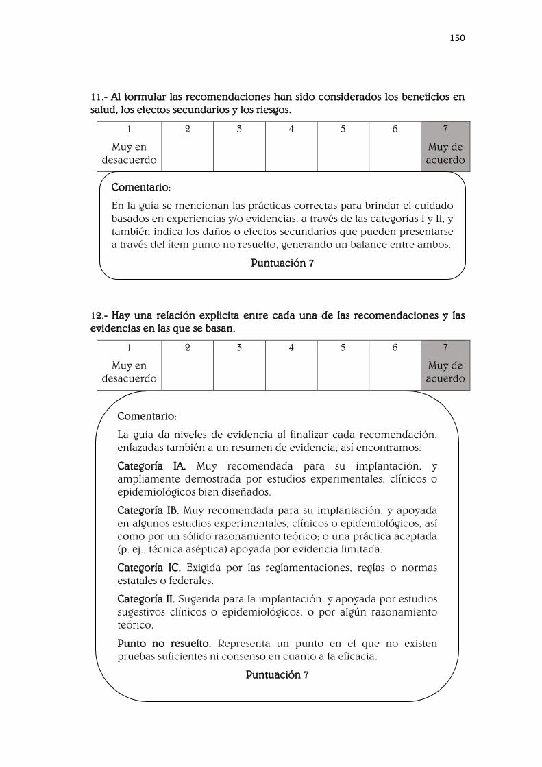
150
11.- Al formular las recomendaciones han sido considerados los beneficios en
salud, los efectos secundarios y los riesgos.
1
Muy en
desacuerdo
2 3 4 5 6 7
Muy de
acuerdo
12.- Hay una relación explicita entre cada una de las recomendaciones y las
evidencias en las que se basan.
1
Muy en
desacuerdo
2 3 4 5 6 7
Muy de
acuerdo
Comentario:
En la guía se mencionan las prácticas correctas para brindar el cuidado
basados en experiencias y/o evidencias, a través de las categorías I y II, y
también indica los daños o efectos secundarios que pueden presentarse
a través del ítem punto no resuelto, generando un balance entre ambos.
Puntuación 7
Comentario:
La guía da niveles de evidencia al finalizar cada recomendación,
enlazadas también a un resumen de evidencia; así encontramos:
Categoría IA. Muy recomendada para su implantación, y
ampliamente demostrada por estudios experimentales, clínicos o
epidemiológicos bien diseñados.
Categoría IB. Muy recomendada para su implantación, y apoyada
en algunos estudios experimentales, clínicos o epidemiológicos, así
como por un sólido razonamiento teórico; o una práctica aceptada
(p. ej., técnica aséptica) apoyada por evidencia limitada.
Categoría IC. Exigida por las reglamentaciones, reglas o normas
estatales o federales.
Categoría II. Sugerida para la implantación, y apoyada por estudios
sugestivos clínicos o epidemiológicos, o por algún razonamiento
teórico.
Punto no resuelto. Representa un punto en el que no existen
pruebas suficientes ni consenso en cuanto a la eficacia.
Puntuación 7

151
13.- La guía ha sido revisada por expertos externos antes de su publicación.
1
Muy en
desacuerdo
2 3 4 5 6 7
Muy de
acuerdo
14.- Se incluye un procedimiento para actualizar la guía.
1
Muy en
desacuerdo
2 3 4 5 6 7
Muy de
acuerdo
Comentario:
Con el fin de rediseñar su proceso de elaboración de la guía, en su
nuevo enfoque, cada directriz es desarrollada por un grupo de
trabajo en consulta con un panel de expertos en el tema, el cual se
compone de tres o más expertos tanto interna como externamente
para HICPAC. Estos expertos son elegidos a discreción, participando
y proporcionando retroalimentación en las actualizaciones regulares
del progreso, y proporcionando una revisión a fondo de las
cuestiones claves, de la bibliografía resultante de la búsqueda inicial
de la literatura, del proyecto de informe de la evidencia y de las
recomendaciones de las guías.
Puntuación 7
Comentario:
La presente actualización de la metodología de directriz HICPAC se basa
en las fortalezas del pasado y los avances actuales en la directriz de
desarrollo e implementación, permitiendo a HICPAC mejorar la validez
y la utilidad de sus directrices al tiempo que aborda desafíos en la
directriz de desarrollo emergente en el ámbito de la prevención y
control de infecciones. A pesar de la actualización, los desafíos
metodológicos y operativos persisten, y HICPAC está listo para integrar
los avances futuros en sus procesos como apropiado.
Puntuación 7

152
Dominio 4: Claridad de presentación:
15.- Las recomendaciones son específicas y no son ambiguas.
1
Muy en
desacuerdo
2 3 4 5 6 7
Muy de
acuerdo
16.- Las distintas opciones para el manejo de la enfermedad o condición de
salud se presentan claramente.
1
Muy en
desacuerdo
2 3 4 5 6 7
Muy de
acuerdo
Comentario:
En la guía se detalla paso a paso las recomendaciones brindadas ante la
evidencia, buscando prevenir o reducir la incidencia tanto como sea
posible de las infecciones del torrente sanguíneo relacionadas con el
catéter. Asimismo, estas se encuentran secuencialmente ordenadas,
abocadas a cada ítem, además se individualizan según el público
objetivo, modificándose levemente entre la población pediátrica y
adulta; lo que permite la comprensión completa de la persona que lo
lee.
Puntuación 7
Comentario:
Por ser esta una guía de amplio alcance, sus recomendaciones se
aplican a una serie de situaciones en las que se basan los catéteres
intravenosos, entre los cuales encontramos: educación, formación y
dotación del personal y selección de catéteres y lugares de inserción,
entre los cuales apreciamos: catéteres venosos centrales, higiene de las
manos y técnica aséptica, precauciones de máxima barrera estéril,
preparación de la piel, regímenes de apósitos en el sitio de inserción
del catéter, sustitución de catéteres venosos centrales, incluidos PICC y
catéteres de hemodiálisis, mejora de la actuación, entre otros.
Puntuación 7

153
17.- Las recomendaciones clave son fácilmente identificables.
1
Muy en
desacuerdo
2 3 4 5 6 7
Muy de
acuerdo
Dominio 5: Aplicabilidad:
18.- La guía describe factores facilitadores y barreras para su aplicación.
1
Muy en
desacuerdo
2 3 4 5 6 7
Muy de
acuerdo
Comentario:
No se encuentran en la guía recomendaciones clave que se encuentren en
un cuadro resumen con letra negrita y subrayad, ya que todas las
recomendaciones son muy importantes, sin embargo, las
recomendaciones están agrupadas en cada sección.
Puntuación 6
Comentario:
Una vez que las prácticas han sido determinadas como efectivas y
económicamente eficientes, el siguiente paso es implementarlas de
forma que se conviertan en parte de la rutina de los cuidados clínicos.
Lamentablemente, la implantación de prácticas preventivas basadas en
la evidencia no ha sido óptima, pues prácticas ampliamente
recomendadas en las directrices publicadas en el 2002 como el uso de
gluconato de clorhexidina como desinfectante local no se usa
habitualmente. Numerosos estudios de mejora de la calidad se han
publicado, y han usado diversos métodos, como la formación del
personal sanitario, la auditoría y la retroalimentación, el cambio
organizativo y los avisos clínicos.
Puntuación 7
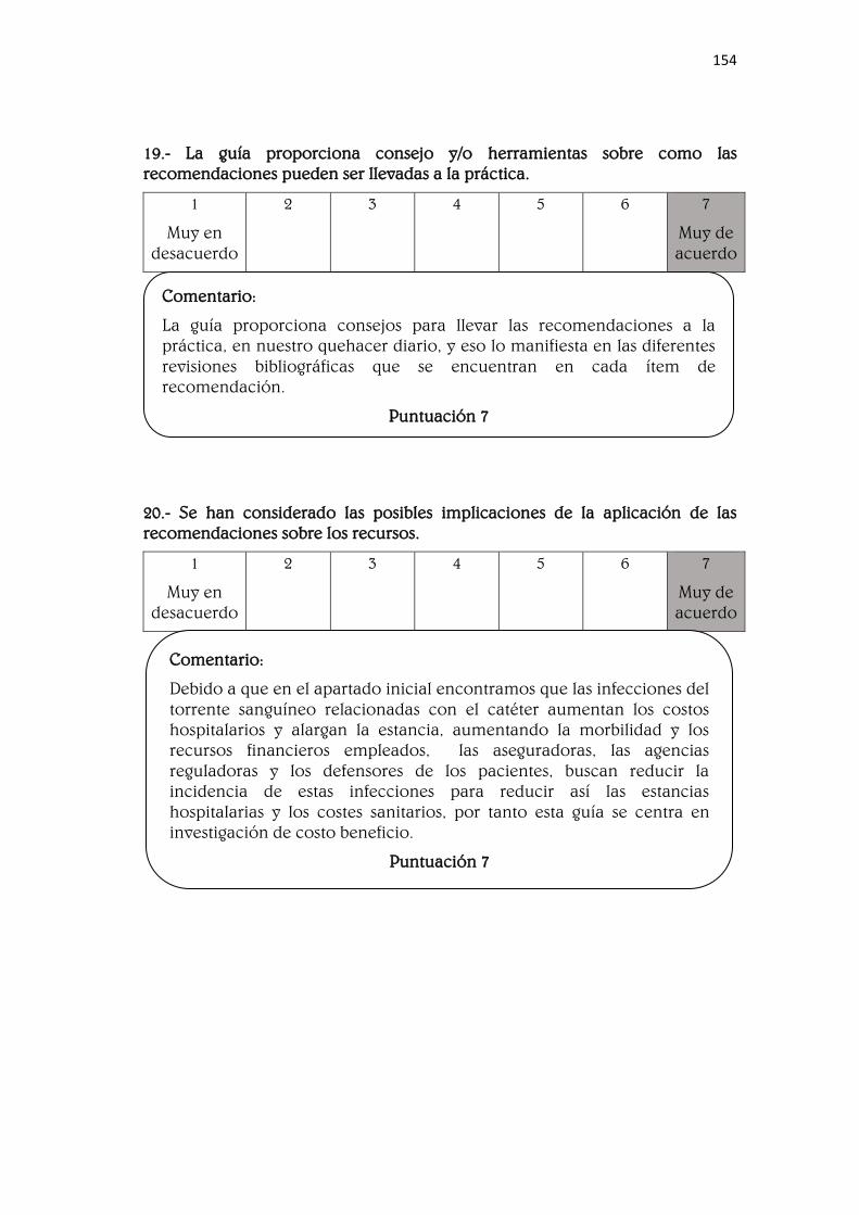
154
19.- La guía proporciona consejo y/o herramientas sobre como las
recomendaciones pueden ser llevadas a la práctica.
1
Muy en
desacuerdo
2 3 4 5 6 7
Muy de
acuerdo
20.- Se han considerado las posibles implicaciones de la aplicación de las
recomendaciones sobre los recursos.
1
Muy en
desacuerdo
2 3 4 5 6 7
Muy de
acuerdo
Comentario:
Debido a que en el apartado inicial encontramos que las infecciones del
torrente sanguíneo relacionadas con el catéter aumentan los costos
hospitalarios y alargan la estancia, aumentando la morbilidad y los
recursos financieros empleados, las aseguradoras, las agencias
reguladoras y los defensores de los pacientes, buscan reducir la
incidencia de estas infecciones para reducir así las estancias
hospitalarias y los costes sanitarios, por tanto esta guía se centra en
investigación de costo beneficio.
Puntuación 7
Comentario:
La guía proporciona consejos para llevar las recomendaciones a la
práctica, en nuestro quehacer diario, y eso lo manifiesta en las diferentes
revisiones bibliográficas que se encuentran en cada ítem de
recomendación.
Puntuación 7

155
21.- La guía ofrece criterios para monitorización y/o auditoría.
1
Muy en
desacuerdo
2 3 4 5 6 7
Muy de
acuerdo
Dominio 6: Independencia editorial:
22.- Los puntos de vista de la entidad financiadora no han influido en el
contenido de la guía.
1
Muy en
desacuerdo
2 3 4 5 6 7
Muy de
acuerdo
Comentario:
La guía establece una serie de recomendaciones para facilitar su
uso continuado, y es que describe acciones claras y definidas en
cuanto a los cuidados para prevenir las infecciones del torrente
sanguíneo relacionados con el catéter como:
➰ Garantizar unos niveles adecuados para el personal de
enfermería. Estudios de observación sugieren que una proporción
elevada de pacientes por enfermera se asocian a infecciones del
torrente sanguíneo relacionadas con el catéter donde las
enfermeras tratan pacientes con CVC. Categoría IB.
➰ Cambiar los apósitos utilizados en los sitios de inserción de CVC
de corta duración cada 2 días en caso de apósitos de gasa.
Categoría II.
➰ Cambiar los apósitos transparentes utilizados en los sitios de
inserción de CVC de corta duración al menos cada 7 días, excepto
en aquellos pacientes pediátricos en los que el riesgo de mover el
catéter sea mayor que las ventajas derivadas del cambio de apósito.
Categoría IB.
Puntuación 7
Comentario:
En la sección de la divulgación de los posibles conflictos de intereses
menciona a una serie de colaboradores los cuales son las fuentes de
financiación.
Puntuación 7

156
23.- Se han registrado y abordado los conflictos de intereses de los miembros
del grupo elaborador de la guía.
1
Muy en
desacuerdo
2 3 4 5 6 7
Muy de
acuerdo
Evaluación global de la guía:
1.- Puntúe la calidad global de la guía.
1
La
calidad
más baja
posible
2 3 4 5 6 7
La
calidad
más alta
posible
2.- ¿Recomendaría esta guía para su uso?
Sí
Sí, con modificaciones
No.
Comentario:
En la sección de la divulgación de los posibles conflictos de intereses
menciona que no hubo conflictos de intereses en los autores.
Puntuación 7
Notas:

157
PUNTUACIÓN DE LOS DOMINIOS:
Fórmula:
Dominio 1: Alcance y objetivo:
Ítem 1 Ítem 2 Ítem 3 Total
7 7 7 21
Puntuación máxima posible: 7 * 3 * 1 = 21
Puntuación mínima posible: 1 * 3 * 1 = 3
Puntuación estandarizada del dominio sería:
21 – 3
21 – 3
Dominio aprobado para su uso.
* 100 = 100 %
Puntuación máxima posible: muy de acuerdo * número de ítems *
número de evaluadores =
Puntuación mínima posible: muy en desacuerdo * número de ítems
* número de evaluadores =
Puntuación estandarizada del dominio sería:
Puntuación obtenida – puntuación mínima posible
Puntuación máxima posible – puntuación mínima posible
100 % =

158
Dominio 2: Participación de los implicados:
Ítem 4 Ítem 5 Ítem 6 Total
7 6 7 20
Dominio 3: Rigor en la elaboración:
Ítem
7
Ítem 8 Ítem 9 Ítem 10 Ítem 11 Ítem 12 Ítem 13 Ítem 14 Total
7 7 7 7 7 7 7 7 56
Puntuación máxima posible: 7 * 3 * 1 = 21
Puntuación mínima posible: 1 * 3 * 1 = 3
Puntuación estandarizada del dominio sería:
20 – 3
21 – 3
Dominio aprobado para su uso.
* 100 = 94 %
Puntuación máxima posible: 7 * 8 * 1 = 56
Puntuación mínima posible: 1 * 8 * 1 = 8
Puntuación estandarizada del dominio sería:
56 – 8
56 – 8
Dominio aprobado para su uso.
* 100 = 100 %

159
Dominio 4: Claridad de presentación:
Ítem 15 Ítem 16 Ítem 17 Total
7 7 6 20
Dominio 5: Aplicabilidad:
Ítem 18 Ítem 19 Ítem 20 Ítem 21 Total
7 7 7 7 28
Puntuación máxima posible: 7 * 3 * 1 = 21
Puntuación mínima posible: 1 * 3 * 1 = 3
Puntuación estandarizada del dominio sería:
20 – 3
21 – 3
Dominio aprobado para su uso.
* 100 = 94 %
Puntuación máxima posible: 7 * 4 * 1 = 28
Puntuación mínima posible: 1 * 4 * 1 = 4
Puntuación estandarizada del dominio sería:
28 – 4
28 – 4
Dominio aprobado para su uso.
* 100 = 100 %

160
Dominio 6: Independencia editorial:
Ítem 22 Ítem 23 Total
7 7 14
Puntuación máxima posible: 7 * 2 * 1 = 14
Puntuación mínima posible: 1 * 2 * 1 = 2
Puntuación estandarizada del dominio sería:
14 – 2
14 – 2
Dominio aprobado para su uso.
* 100 = 100 %