Embed Size (px)
Citation preview
8/29/11
1
(Unit II) Chapter 6: The Structure of DNA
Introduc;on to DNA Structure: The Importance of DNA Structure
• DNA, since it carries all the informa;on for a given organism, must be a molecule that contains an incredible amount of informa;on
• Contains informa;on for proper development of an organism – Allows the proper structures to form at the
appropriate ;me – Allows appropriate growth at the
appropriate ;me
• Contains the informa;on for proper cellular func;on – DNA encodes the informa;on to produce
proteins involved in respira;on – DNA encodes the informa;on to produce
proteins that are important in sending and receiving signals between cells
• All the appropriate informa;on is also passed on to subsequent genera;ons – Cellular reproduc;on (asexual) – Organismal reproduc;on (sexual or asexual)
8/29/11
2
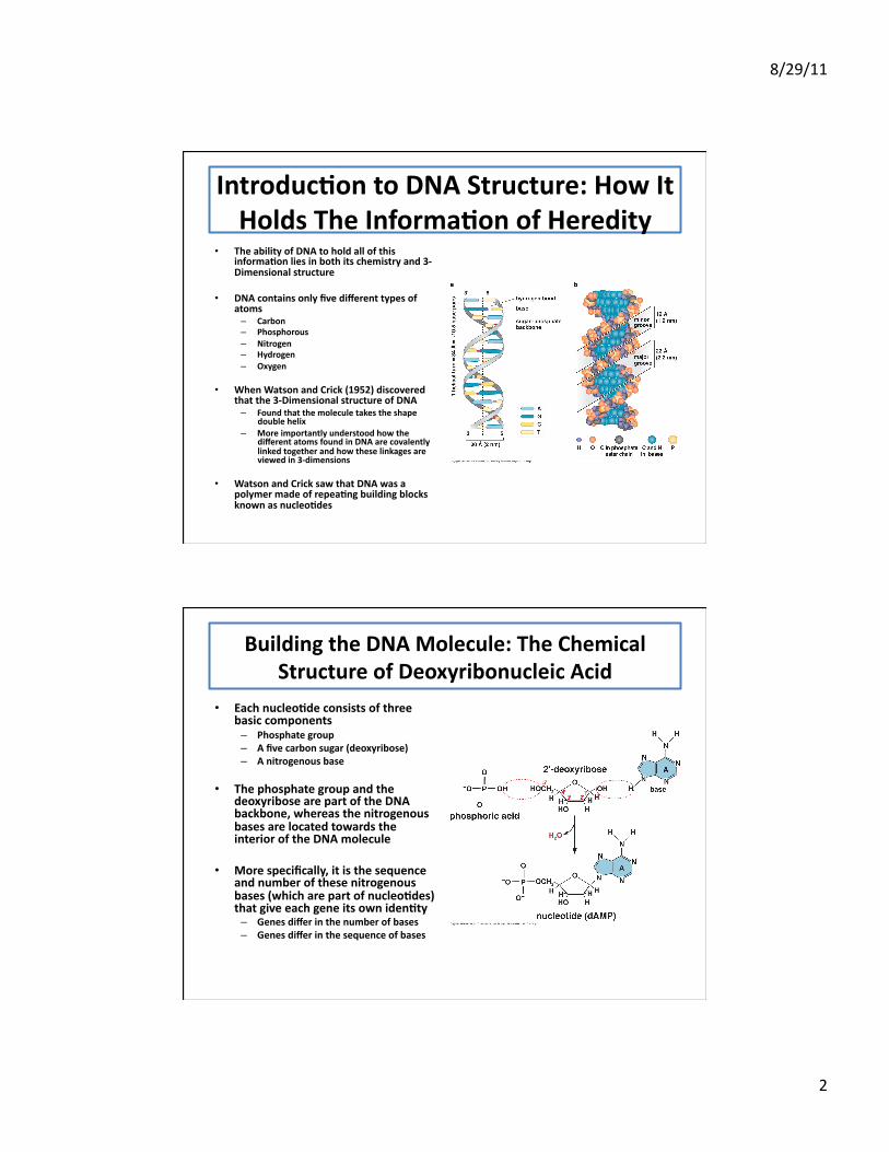
Introduc;on to DNA Structure: How It Holds The Informa;on of Heredity
• The ability of DNA to hold all of this informa;on lies in both its chemistry and 3-‐Dimensional structure
• DNA contains only five different types of atoms – Carbon – Phosphorous – Nitrogen – Hydrogen – Oxygen
• When Watson and Crick (1952) discovered that the 3-‐Dimensional structure of DNA – Found that the molecule takes the shape
double helix – More importantly understood how the
different atoms found in DNA are covalently linked together and how these linkages are viewed in 3-‐dimensions
• Watson and Crick saw that DNA was a polymer made of repea;ng building blocks known as nucleo;des
Building the DNA Molecule: The Chemical Structure of Deoxyribonucleic Acid
• Each nucleo;de consists of three basic components – Phosphate group – A five carbon sugar (deoxyribose) – A nitrogenous base
• The phosphate group and the deoxyribose are part of the DNA backbone, whereas the nitrogenous bases are located towards the interior of the DNA molecule
• More specifically, it is the sequence and number of these nitrogenous bases (which are part of nucleo;des) that give each gene its own iden;ty – Genes differ in the number of bases – Genes differ in the sequence of bases
8/29/11
3
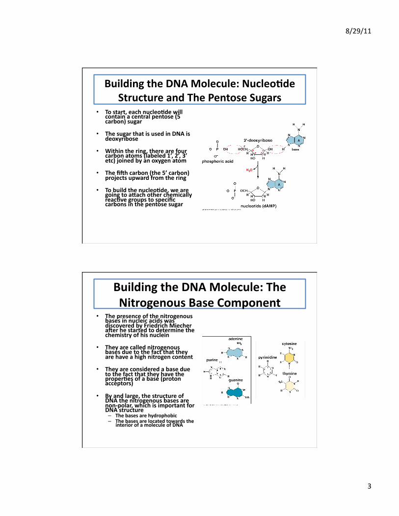
Building the DNA Molecule: Nucleo;de Structure and The Pentose Sugars
• To start, each nucleo;de will contain a central pentose (5 carbon) sugar
• The sugar that is used in DNA is deoxyribose
• Within the ring, there are four carbon atoms (labeled 1’, 2’, 3’ etc) joined by an oxygen atom
• The fi[h carbon (the 5’ carbon) projects upward from the ring
• To build the nucleo;de, we are going to a\ach other chemically reac;ve groups to specific carbons in the pentose sugar
Building the DNA Molecule: The Nitrogenous Base Component
• The presence of the nitrogenous bases in nucleic acids was discovered by Friedrich Miecher a[er he started to determine the chemistry of his nuclein
• They are called nitrogenous bases due to the fact that they are have a high nitrogen content
• They are considered a base due to the fact that they have the proper;es of a base (proton acceptors)
• By and large, the structure of DNA the nitrogenous bases are non-‐polar, which is important for DNA structure – The bases are hydrophobic – The bases are located towards the
interior of a molecule of DNA
8/29/11
4
Building the DNA Molecule: The Nitrogenous Base Component
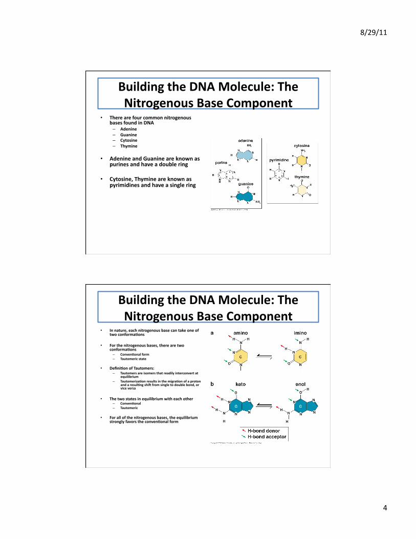
• There are four common nitrogenous bases found in DNA – Adenine – Guanine – Cytosine – Thymine
• Adenine and Guanine are known as purines and have a double ring
• Cytosine, Thymine are known as pyrimidines and have a single ring
Building the DNA Molecule: The Nitrogenous Base Component
• In nature, each nitrogenous base can take one of two conforma;ons
• For the nitrogenous bases, there are two conforma;ons – Conven;onal form – Tautomeric state
• Defini;on of Tautomers: – Tautomers are isomers that readily interconvert at
equilibrium – Tautomeriza;on results in the migra;on of a proton
and a resul;ng shi[ from single to double bond, or vice versa
• The two states in equilibrium with each other – Conven;onal – Tautomeric
• For all of the nitrogenous bases, the equilibrium strongly favors the conven;onal form
8/29/11
5
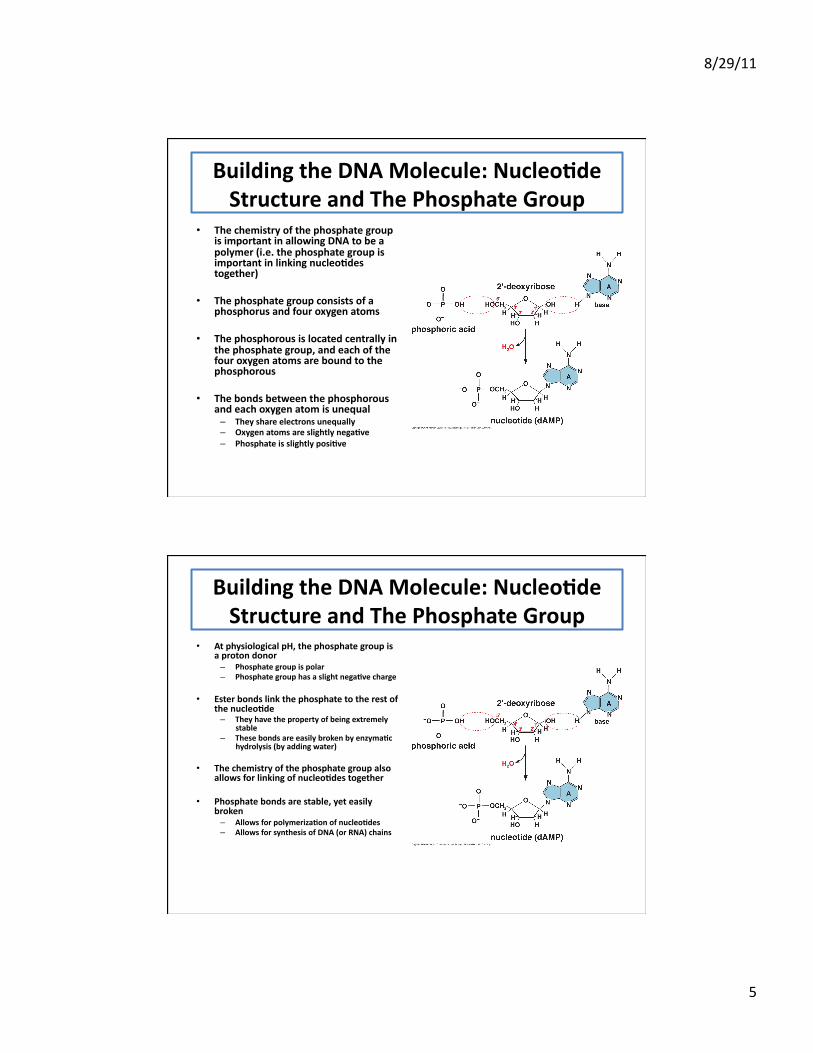
Building the DNA Molecule: Nucleo;de Structure and The Phosphate Group
• The chemistry of the phosphate group is important in allowing DNA to be a polymer (i.e. the phosphate group is important in linking nucleo;des together)
• The phosphate group consists of a phosphorus and four oxygen atoms
• The phosphorous is located centrally in the phosphate group, and each of the four oxygen atoms are bound to the phosphorous
• The bonds between the phosphorous and each oxygen atom is unequal – They share electrons unequally – Oxygen atoms are slightly nega;ve – Phosphate is slightly posi;ve
Building the DNA Molecule: Nucleo;de Structure and The Phosphate Group
• At physiological pH, the phosphate group is a proton donor – Phosphate group is polar – Phosphate group has a slight nega;ve charge
• Ester bonds link the phosphate to the rest of the nucleo;de – They have the property of being extremely
stable – These bonds are easily broken by enzyma;c
hydrolysis (by adding water)
• The chemistry of the phosphate group also allows for linking of nucleo;des together
• Phosphate bonds are stable, yet easily broken – Allows for polymeriza;on of nucleo;des – Allows for synthesis of DNA (or RNA) chains
8/29/11
6
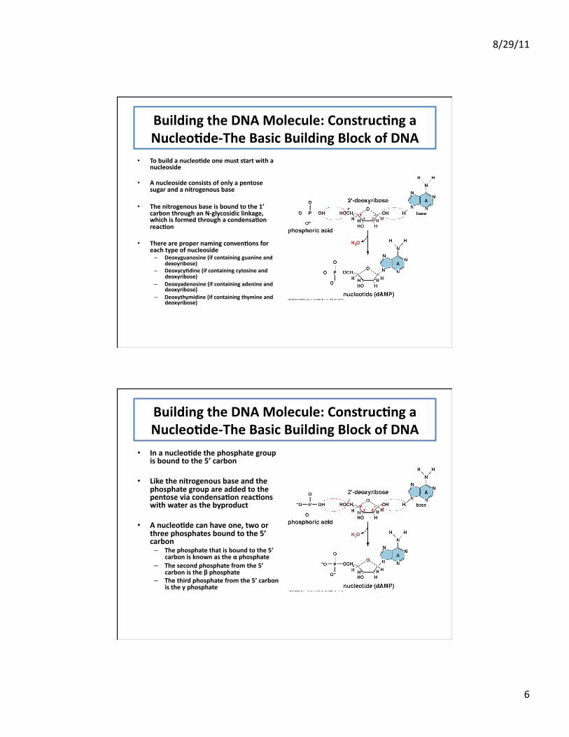
Building the DNA Molecule: Construc;ng a Nucleo;de-‐The Basic Building Block of DNA
• To build a nucleo;de one must start with a nucleoside
• A nucleoside consists of only a pentose sugar and a nitrogenous base
• The nitrogenous base is bound to the 1’ carbon through an N-‐glycosidic linkage, which is formed through a condensa;on reac;on
• There are proper naming conven;ons for each type of nucleoside – Deoxyguanosine (if containing guanine and
dexoyribose) – Deoxycy;dine (if containing cytosine and
deoxyribose) – Deoxyadenosine (if containing adenine and
deoxyribose) – Deoxythymidine (if containing thymine and
deoxyribose)
Building the DNA Molecule: Construc;ng a Nucleo;de-‐The Basic Building Block of DNA
• In a nucleo;de the phosphate group is bound to the 5’ carbon
• Like the nitrogenous base and the phosphate group are added to the pentose via condensa;on reac;ons with water as the byproduct
• A nucleo;de can have one, two or three phosphates bound to the 5’ carbon – The phosphate that is bound to the 5’
carbon is known as the α phosphate – The second phosphate from the 5’
carbon is the β phosphate – The third phosphate from the 5’ carbon
is the γ phosphate
8/29/11
7
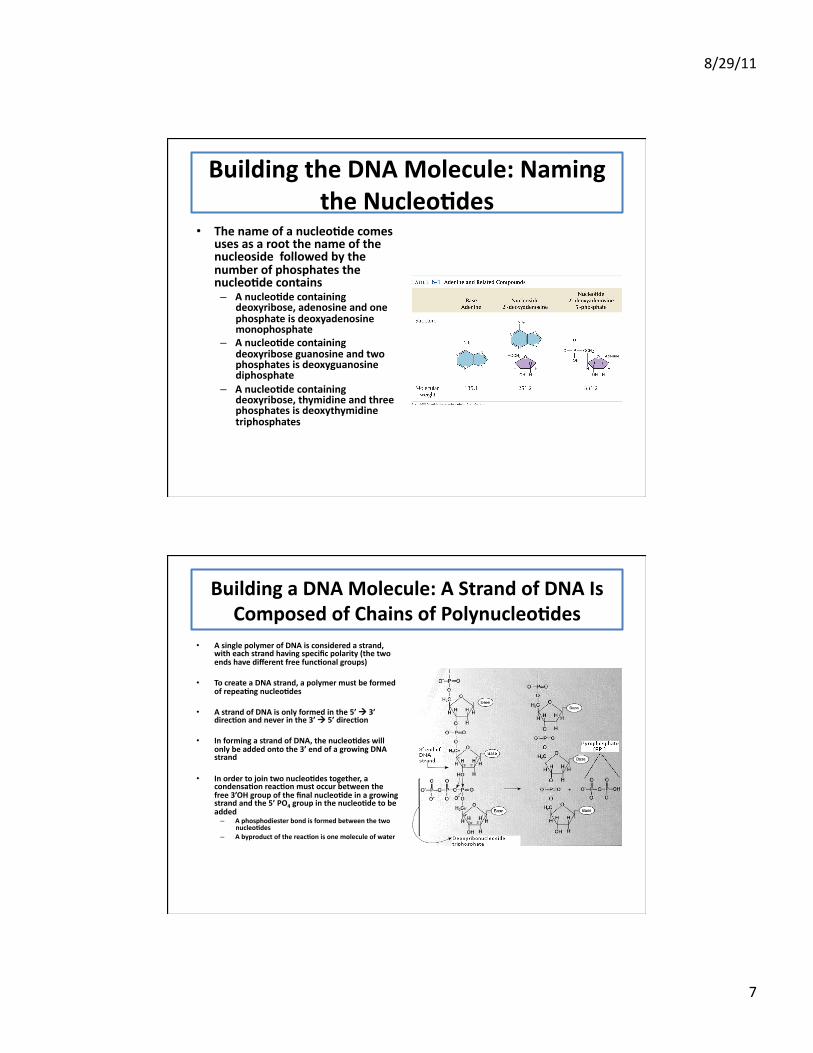
Building the DNA Molecule: Naming the Nucleo;des
• The name of a nucleo;de comes uses as a root the name of the nucleoside followed by the number of phosphates the nucleo;de contains – A nucleo;de containing
deoxyribose, adenosine and one phosphate is deoxyadenosine monophosphate
– A nucleo;de containing deoxyribose guanosine and two phosphates is deoxyguanosine diphosphate
– A nucleo;de containing deoxyribose, thymidine and three phosphates is deoxythymidine triphosphates
Building a DNA Molecule: A Strand of DNA Is Composed of Chains of Polynucleo;des
• A single polymer of DNA is considered a strand, with each strand having specific polarity (the two ends have different free func;onal groups)
• To create a DNA strand, a polymer must be formed of repea;ng nucleo;des
• A strand of DNA is only formed in the 5’ 3’ direc;on and never in the 3’ 5’ direc;on
• In forming a strand of DNA, the nucleo;des will only be added onto the 3’ end of a growing DNA strand
• In order to join two nucleo;des together, a condensa;on reac;on must occur between the free 3’OH group of the final nucleo;de in a growing strand and the 5’ PO4 group in the nucleo;de to be added – A phosphodiester bond is formed between the two
nucleo;des – A byproduct of the reac;on is one molecule of water
8/29/11
8
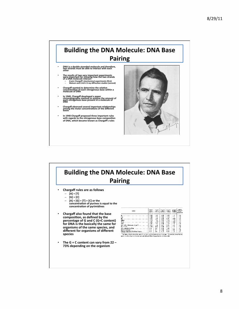
Building the DNA Molecule: DNA Base Pairing
• DNA is a double stranded molecule and therefore, two strands must be able to interact with each other
• The results of two very important experiments were important for showing how the two strands of a DNA molecule interact – Erwin Chargaff’s biochemical experiments (first) – Watson and Crick’s X-‐ray diffrac;on studies (second)
• Chargaff wanted to determine the rela;ve concentra;on of each nitrogenous base within a molecule of DNA
• In 1940, Chargaff developed a paper chromatography method to analyze the amount of each nitrogenous base present in a molecule of DNA
• Chargaff observed several important rela;onships among the molar concentra;ons of the different bases
• In 1940 Chargaff proposed three important rules with regards to the nitrogenous base composi;on of DNA, which became known as Chargaff’s rules
Building the DNA Molecule: DNA Base Pairing
• Chargaff rules are as follows – [A] = [T] – [G] = [C] – [A] + [G] = [T] + [C] or the
concentra;on of purines is equal to the concentra;on of pyrimidines
• Chargaff also found that the base composi;on, as defined by the percentage of G and C (G+C content) for DNA is the basically the same for organisms of the same species, and different for organisms of different species
• The G + C content can vary from 22 – 73% depending on the organism
8/29/11
9
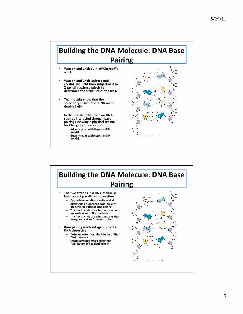
Building the DNA Molecule: DNA Base Pairing
• Watson and Crick built off Chargaff’s work
• Watson and Crick isolated and crystallized DNA then subjected it to X-‐ray diffrac;on analysis to determine the structure of the DNA
• Their results show that the secondary structure of DNA was a double helix
• In the double helix, the two DNA strands interacted through base pairing (showing a physical reason for Chargaff’s observa;ons – Adenine pairs with thymine (2 H
bonds) – Guanine pairs with cytosine (3 H
bonds)
Building the DNA Molecule: DNA Base Pairing
• The two strands in a DNA molecule lie in an an;parallel configura;on – Opposite orienta;on = an;-‐parallel – Allows the nitrogenous bases to align
properly for efficient base pairing – The free 5’ ends of each strand are on
opposite sides of the molecule – The free 3’ ends of each strand are also
on opposite sides from each other
• Base pairing is advantageous to the DNA chemistry – Excludes water from the interior of the
DNA molecule – Creates entropy which allows for
stabiliza;on of the double helix
8/29/11
10
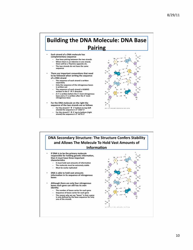
Building the DNA Molecule: DNA Base Pairing
• Each strand of a DNA molecule has complementary sequence – Due base pairing between the two strands – Where there is an Adenine in one strand,
there will be a Thymine opposite etc. – The two strands do not have the same
sequence
• There are important conven;ons that need to be followed when wri;ng the sequence of a DNA strand – The sequence of each strand is wri\en
separately – Only the sequence of the nitrogenous bases
is wri\en out – The sequence of each strand is ALWAYS
wri\en in the 5’ 3’ direc;on – A 5’ is wri\en before the 5’ most nitrogenous
base and a 3’ is wri\en a[er the 3’ most nitrogenous base
• For the DNA molecule on the right the sequence of the two strands are as follows – For the strand 5’ 3’ bo\om to top (le[
strand) the sequence is 5’ CAGT 3’ – For the strand 5’ 3’ top to bo\om (right
strand) the sequence is 5’ ACTG 3’
DNA Secondary Structure: The Structure Confers Stability and Allows The Molecule To Hold Vast Amounts of
Informa;on • If DNA is to be the primary molecule
responsible for holding gene;c informa;on, then it must have three important characteris;cs – It must hold vast amounts of informa;on – The molecule must be extremely stable – Must be easily replicated
• DNA is able to hold vast amounts informa;on in its sequence of nitrogenous bases
• Although there are only four nitrogenous bases each gene can s;ll has its own iden;ty – The number of bases varies for each gene – Sequence of bases varies for each gene – The reason why we say “bases” is that a gene
is only defined by the base sequence for only one of the strands

8/29/11
11
DNA Secondary Structure: The Structure Confers Stability and Allows The Molecule To Hold Vast Amounts of
Informa;on • The stability of the double stranded DNA
molecule comes from two important forces – Hydrogen bonding between the base pairs – Base stacking interac;ons
• In actuality, the base pairs lie flat upon one another and so instead of looking like “rungs on a ladder” they look like a stack of coins
• The bases in DNA stack together, which results in increased stability by elimina;ng water from the interior of the DNA molecule
• In order to have the base pairs lie flat on one another, each base pair must be slightly twisted with respect to previous base pair
DNA Secondary Structure: The Structure Confers Stability and Allows The Molecule To Hold Vast
Amounts of Informa;on
8/29/11
12
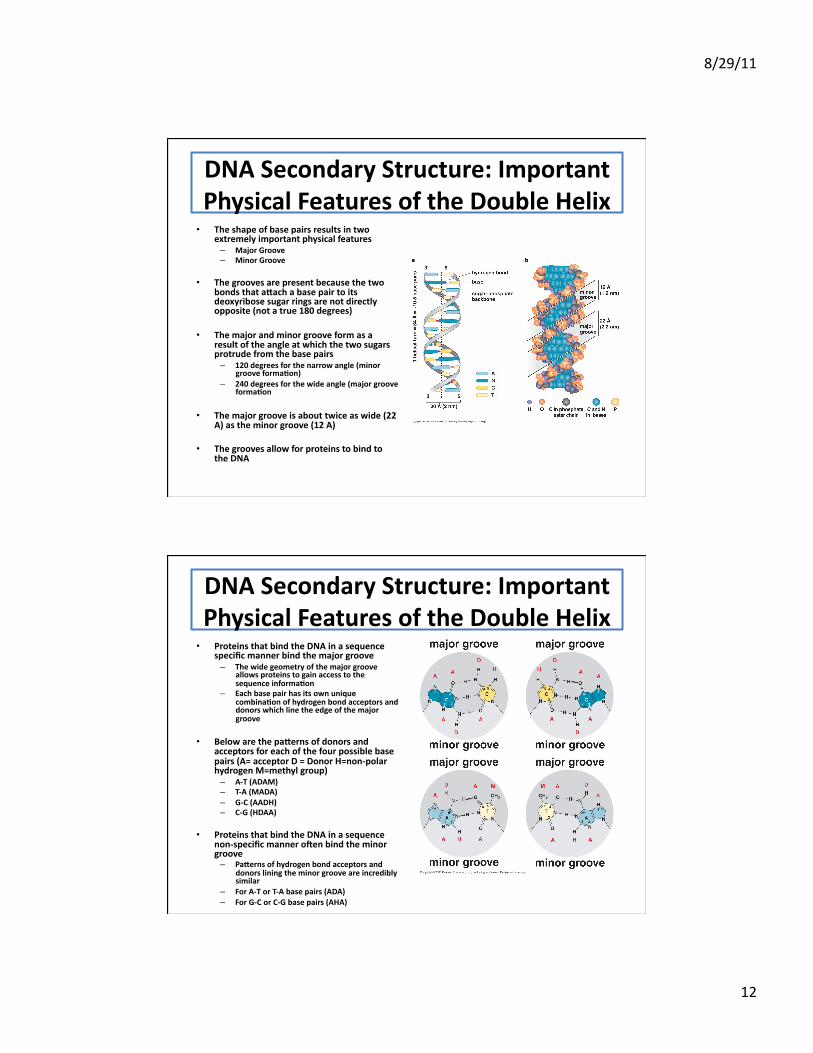
DNA Secondary Structure: Important Physical Features of the Double Helix • The shape of base pairs results in two
extremely important physical features – Major Groove – Minor Groove
• The grooves are present because the two bonds that a\ach a base pair to its deoxyribose sugar rings are not directly opposite (not a true 180 degrees)
• The major and minor groove form as a result of the angle at which the two sugars protrude from the base pairs – 120 degrees for the narrow angle (minor
groove forma;on) – 240 degrees for the wide angle (major groove
forma;on
• The major groove is about twice as wide (22 A) as the minor groove (12 A)
• The grooves allow for proteins to bind to the DNA
DNA Secondary Structure: Important Physical Features of the Double Helix • Proteins that bind the DNA in a sequence
specific manner bind the major groove – The wide geometry of the major groove
allows proteins to gain access to the sequence informa;on
– Each base pair has its own unique combina;on of hydrogen bond acceptors and donors which line the edge of the major groove
• Below are the pa\erns of donors and acceptors for each of the four possible base pairs (A= acceptor D = Donor H=non-‐polar hydrogen M=methyl group) – A-‐T (ADAM) – T-‐A (MADA) – G-‐C (AADH) – C-‐G (HDAA)
• Proteins that bind the DNA in a sequence non-‐specific manner o[en bind the minor groove – Pa\erns of hydrogen bond acceptors and
donors lining the minor groove are incredibly similar
– For A-‐T or T-‐A base pairs (ADA) – For G-‐C or C-‐G base pairs (AHA)
8/29/11
13
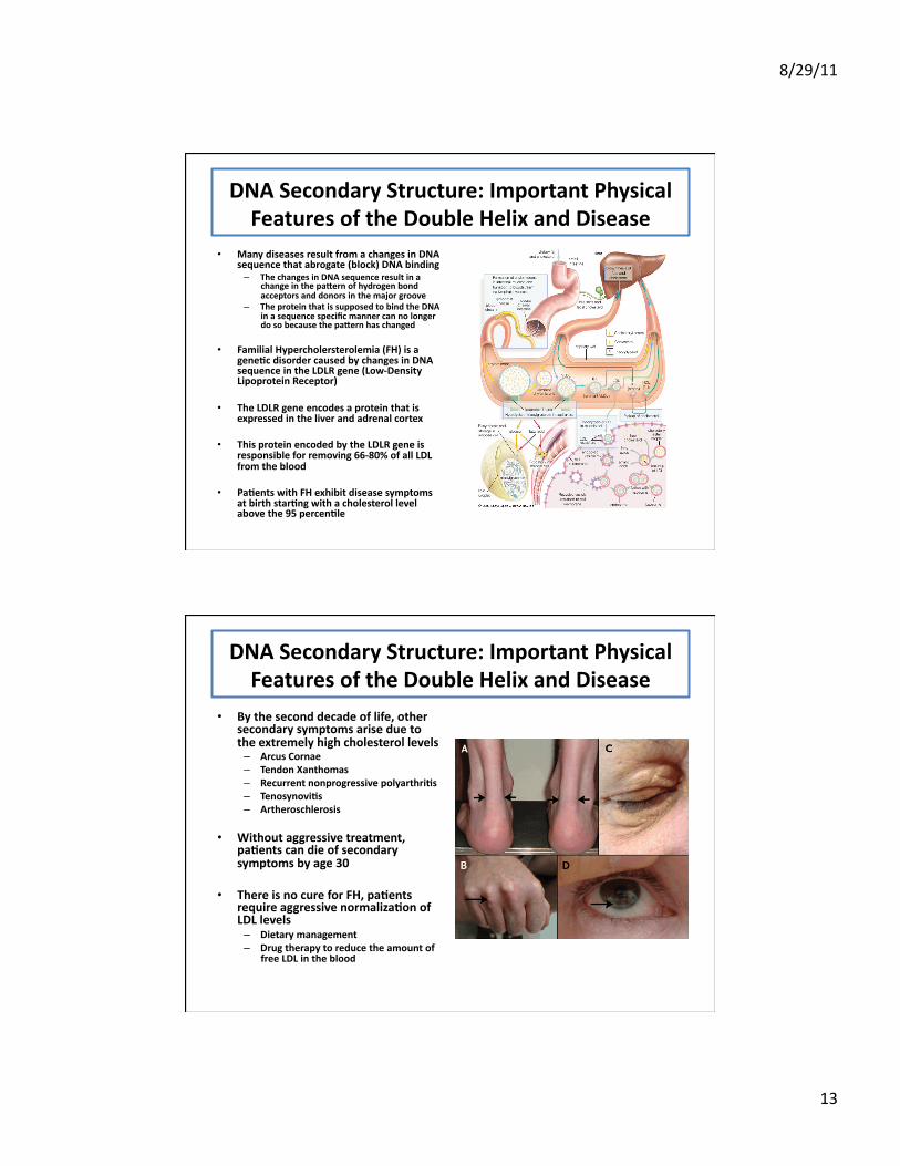
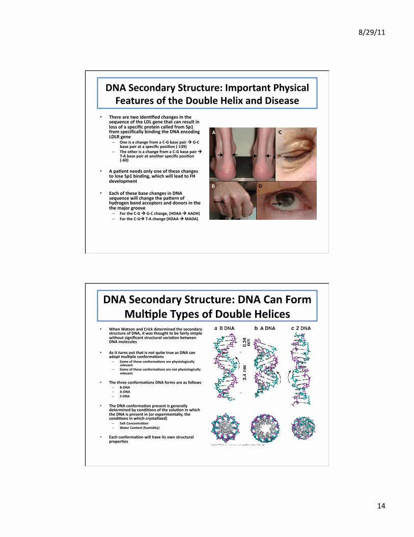
DNA Secondary Structure: Important Physical Features of the Double Helix and Disease
• Many diseases result from a changes in DNA sequence that abrogate (block) DNA binding – The changes in DNA sequence result in a
change in the pa\ern of hydrogen bond acceptors and donors in the major groove
– The protein that is supposed to bind the DNA in a sequence specific manner can no longer do so because the pa\ern has changed
• Familial Hypercholersterolemia (FH) is a gene;c disorder caused by changes in DNA sequence in the LDLR gene (Low-‐Density Lipoprotein Receptor)
• The LDLR gene encodes a protein that is expressed in the liver and adrenal cortex
• This protein encoded by the LDLR gene is responsible for removing 66-‐80% of all LDL from the blood
• Pa;ents with FH exhibit disease symptoms at birth star;ng with a cholesterol level above the 95 percen;le
DNA Secondary Structure: Important Physical Features of the Double Helix and Disease
• By the second decade of life, other secondary symptoms arise due to the extremely high cholesterol levels – Arcus Cornae – Tendon Xanthomas – Recurrent nonprogressive polyarthri;s – Tenosynovi;s – Artheroschlerosis
• Without aggressive treatment, pa;ents can die of secondary symptoms by age 30
• There is no cure for FH, pa;ents require aggressive normaliza;on of LDL levels – Dietary management – Drug therapy to reduce the amount of
free LDL in the blood
8/29/11
14
DNA Secondary Structure: Important Physical Features of the Double Helix and Disease
• There are two iden;fied changes in the sequence of the LDL gene that can result in loss of a specific protein called from Sp1 from specifically binding the DNA encoding LDLR gene – One is a change from a C-‐G base pair G-‐C
base pair at a specific posi;on (-‐139) – The other is a change from a C-‐G base pair
T-‐A base pair at another specific posi;on (-‐60)
• A pa;ent needs only one of these changes to lose Sp1 binding, which will lead to FH development
• Each of these base changes in DNA sequence will change the pa\ern of hydrogen bond acceptors and donors in the the major groove – For the C-‐G G-‐C change, (HDAA AADH) – For the C-‐G T-‐A change (HDAA MADA)
DNA Secondary Structure: DNA Can Form Mul;ple Types of Double Helices
• When Watson and Crick determined the secondary structure of DNA, it was thought to be fairly simple without significant structural varia;on between DNA molecules
• As it turns out that is not quite true as DNA can adopt mul;ple conforma;ons – Some of these conforma;ons are physiologically
relevant – Some of these conforma;ons are not physiologically
relevant
• The three conforma;ons DNA forms are as follows – B-‐DNA – A-‐DNA – Z-‐DNA
• The DNA conforma;on present is generally determined by condi;ons of the solu;on in which the DNA is present in (or experimentally, the condi;ons in which crystallized) – Salt Concentra;on – Water Content (humidity)
• Each conforma;on will have its own structural proper;es
8/29/11
15
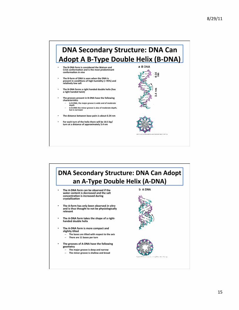
DNA Secondary Structure: DNA Can Adopt A B-‐Type Double Helix (B-‐DNA) • The B-‐DNA form is considered the Watson and
Crick conforma;on and is the most predominant conforma;on in vivo
• The B-‐form of DNA is seen when the DNA is present in condi;ons of high humidity (> 95%) and rela;vely low salt
• The B-‐DNA forms a right handed double helix (has a right handed twist)
• The grooves present in B-‐DNA have the following characteris;cs – In B-‐DNA, the major groove is wide and of moderate
depth – In B-‐DNA the minor groove is also of moderate depth,
but is narrower
• The distance between base pairs is about 0.34 nm
• For each turn of the helix there will be 10.5 bp/turn at a distance of approximately 3.4 nm
DNA Secondary Structure: DNA Can Adopt an A-‐Type Double Helix (A-‐DNA)
• The A-‐DNA form can be observed if the water content is decreased and the salt concentra;on is increased during crystalliza;on
• The A-‐form has only been observed in vitro and is thus thought to not be physiologically relevant
• The A-‐DNA form takes the shape of a right-‐handed double helix
• The A-‐DNA form is more compact and slightly ;lted – The bases are ;lted with respect to the axis – There are 11 bases per turn
• The grooves of A-‐DNA have the following geometry – The major groove is deep and narrow – The minor groove is shallow and broad
8/29/11
16
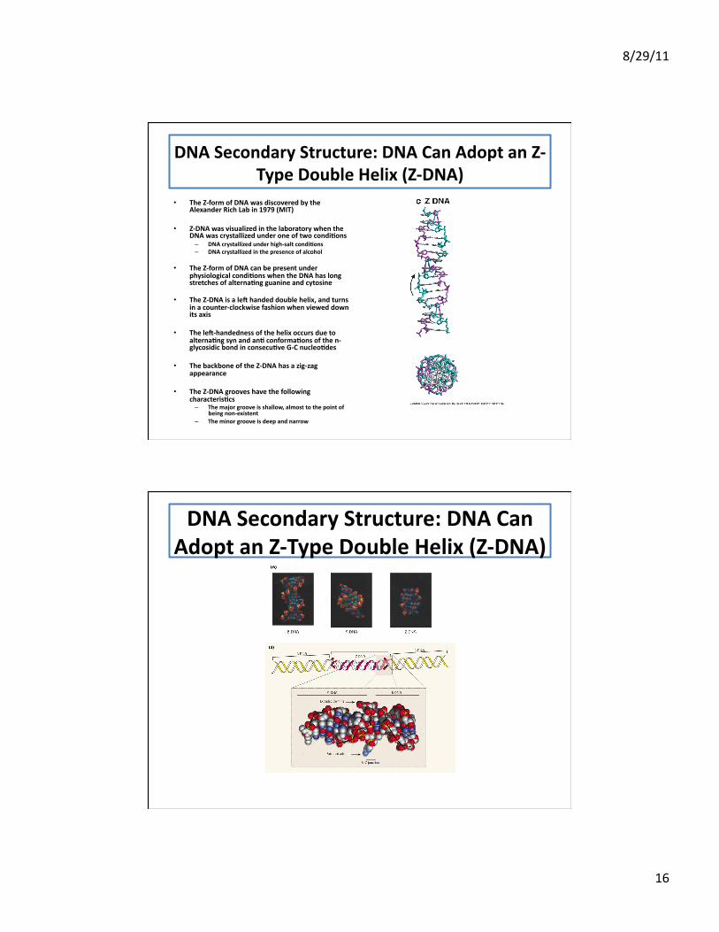
DNA Secondary Structure: DNA Can Adopt an Z-‐Type Double Helix (Z-‐DNA)
• The Z-‐form of DNA was discovered by the Alexander Rich Lab in 1979 (MIT)
• Z-‐DNA was visualized in the laboratory when the DNA was crystallized under one of two condi;ons – DNA crystallized under high-‐salt condi;ons – DNA crystallized in the presence of alcohol
• The Z-‐form of DNA can be present under physiological condi;ons when the DNA has long stretches of alterna;ng guanine and cytosine
• The Z-‐DNA is a le[ handed double helix, and turns in a counter-‐clockwise fashion when viewed down its axis
• The le[-‐handedness of the helix occurs due to alterna;ng syn and an; conforma;ons of the n-‐glycosidic bond in consecu;ve G-‐C nucleo;des
• The backbone of the Z-‐DNA has a zig-‐zag appearance
• The Z-‐DNA grooves have the following characteris;cs – The major groove is shallow, almost to the point of
being non-‐existent – The minor groove is deep and narrow
DNA Secondary Structure: DNA Can Adopt an Z-‐Type Double Helix (Z-‐DNA)
8/29/11
17
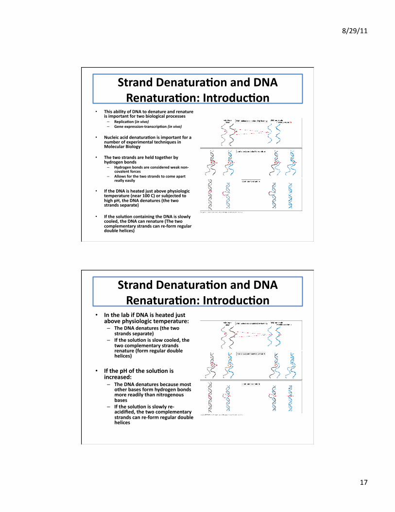
Strand Denatura;on and DNA Renatura;on: Introduc;on
• This ability of DNA to denature and renature is important for two biological processes – Replica;on (in vivo) – Gene expression-‐transcrip;on (in vivo)
• Nucleic acid denatura;on is important for a number of experimental techniques in Molecular Biology
• The two strands are held together by hydrogen bonds – Hydrogen bonds are considered weak non-‐
covalent forces – Allows for the two strands to come apart
really easily
• If the DNA is heated just above physiologic temperature (near 100 C) or subjected to high pH, the DNA denatures (the two strands separate)
• If the solu;on containing the DNA is slowly cooled, the DNA can renature (The two complementary strands can re-‐form regular double helices)
Strand Denatura;on and DNA Renatura;on: Introduc;on
• In the lab if DNA is heated just above physiologic temperature: – The DNA denatures (the two
strands separate) – If the solu;on is slow cooled, the
two complementary strands renature (form regular double helices)
• If the pH of the solu;on is increased: – The DNA denatures because most
other bases form hydrogen bonds more readily than nitrogenous bases
– If the solu;on is slowly re-‐acidified, the two complementary strands can re-‐form regular double helices
8/29/11
18
Strand Denatura;on and DNA Renatura;on: Introduc;on
• The process of adding heat to denature the DNA only affects the hydrogen bonds (weak bonds) that allow base pairing to occur
• The phosphodiester bonds are covalent linkages which are much stronger than hydrogen bonds and are unaffected by temperature
• Enzyma;c ac;vity is needed to break phosphodiester linkages – DNases – Restric;on Endonucleases
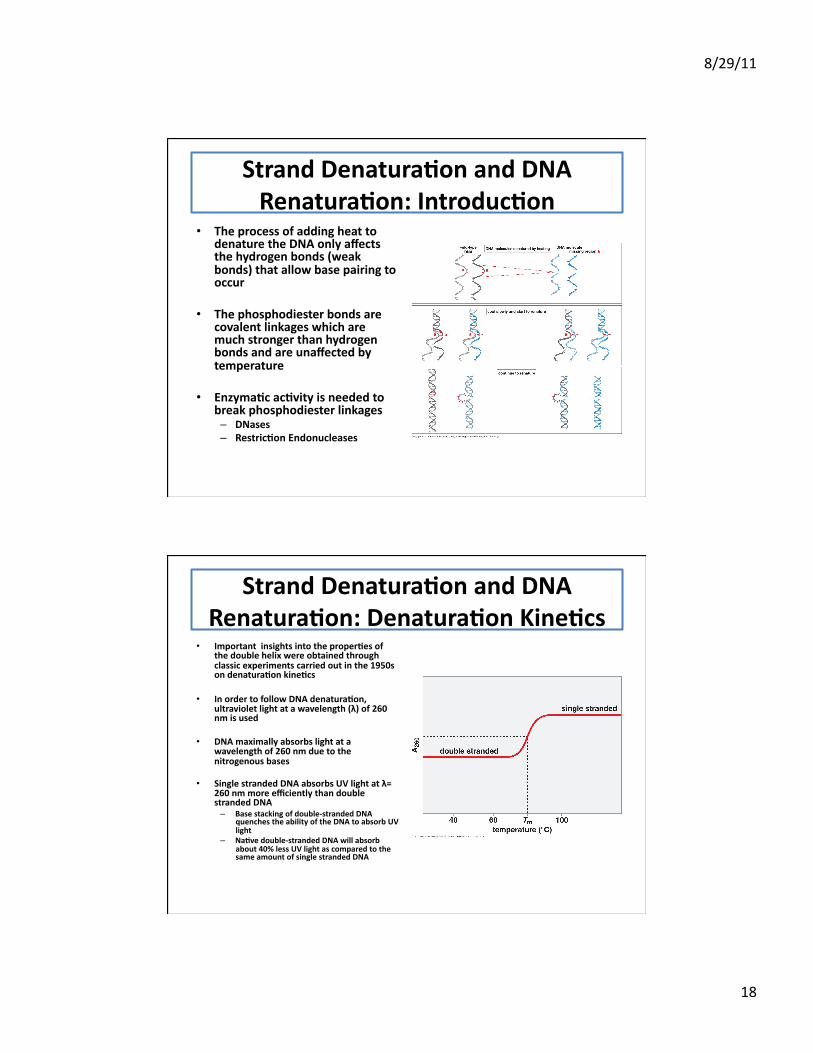
Strand Denatura;on and DNA Renatura;on: Denatura;on Kine;cs
• Important insights into the proper;es of the double helix were obtained through classic experiments carried out in the 1950s on denatura;on kine;cs
• In order to follow DNA denatura;on, ultraviolet light at a wavelength (λ) of 260 nm is used
• DNA maximally absorbs light at a wavelength of 260 nm due to the nitrogenous bases
• Single stranded DNA absorbs UV light at λ= 260 nm more efficiently than double stranded DNA – Base stacking of double-‐stranded DNA
quenches the ability of the DNA to absorb UV light
– Na;ve double-‐stranded DNA will absorb about 40% less UV light as compared to the same amount of single stranded DNA
8/29/11
19
Strand Denatura;on and DNA Renatura;on: Denatura;on Kine;cs
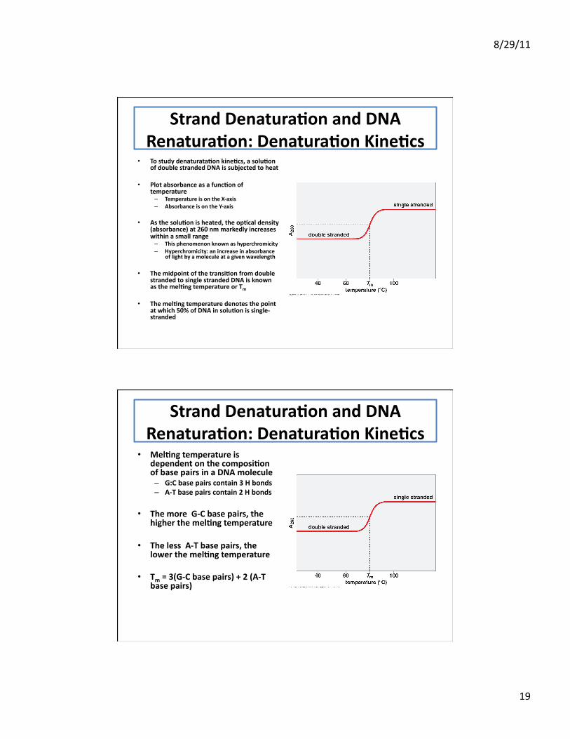
• To study denaturata;on kine;cs, a solu;on of double stranded DNA is subjected to heat
• Plot absorbance as a func;on of temperature – Temperature is on the X-‐axis – Absorbance is on the Y-‐axis
• As the solu;on is heated, the op;cal density (absorbance) at 260 nm markedly increases within a small range – This phenomenon known as hyperchromicity – Hyperchromicity: an increase in absorbance
of light by a molecule at a given wavelength
• The midpoint of the transi;on from double stranded to single stranded DNA is known as the mel;ng temperature or Tm
• The mel;ng temperature denotes the point at which 50% of DNA in solu;on is single-‐stranded
Strand Denatura;on and DNA Renatura;on: Denatura;on Kine;cs
• Mel;ng temperature is dependent on the composi;on of base pairs in a DNA molecule – G:C base pairs contain 3 H bonds – A-‐T base pairs contain 2 H bonds
• The more G-‐C base pairs, the higher the mel;ng temperature
• The less A-‐T base pairs, the lower the mel;ng temperature
• Tm = 3(G-‐C base pairs) + 2 (A-‐T base pairs)
8/29/11
20
RNA Structure: Introduc;on • RNA is chemically similar to DNA with a couple of
major differences – Single stranded – Can also base pair and form significant secondary
structure
• Instead of base pairing with a second strand, a single RNA strand can base pair with itself
• “The structure of RNA is breathtakingly intricate and graceful” -‐Harry Noller (2005)
• There are many types of RNA that can adopt significant number of structures that are important for biological func;on – tRNA (transla;on) – rRNA (ribosomal RNA) – snRNA (splicing) – snoRNA (rRNA processing) – Ribozymes (enzyma;c func;on) – mRNA (gene regula;on)
• The significant secondary structural mo;fs are stabilized by base pairing – Conven;onal base pairing (Watson-‐Crick) – Unconven;onal base pairing
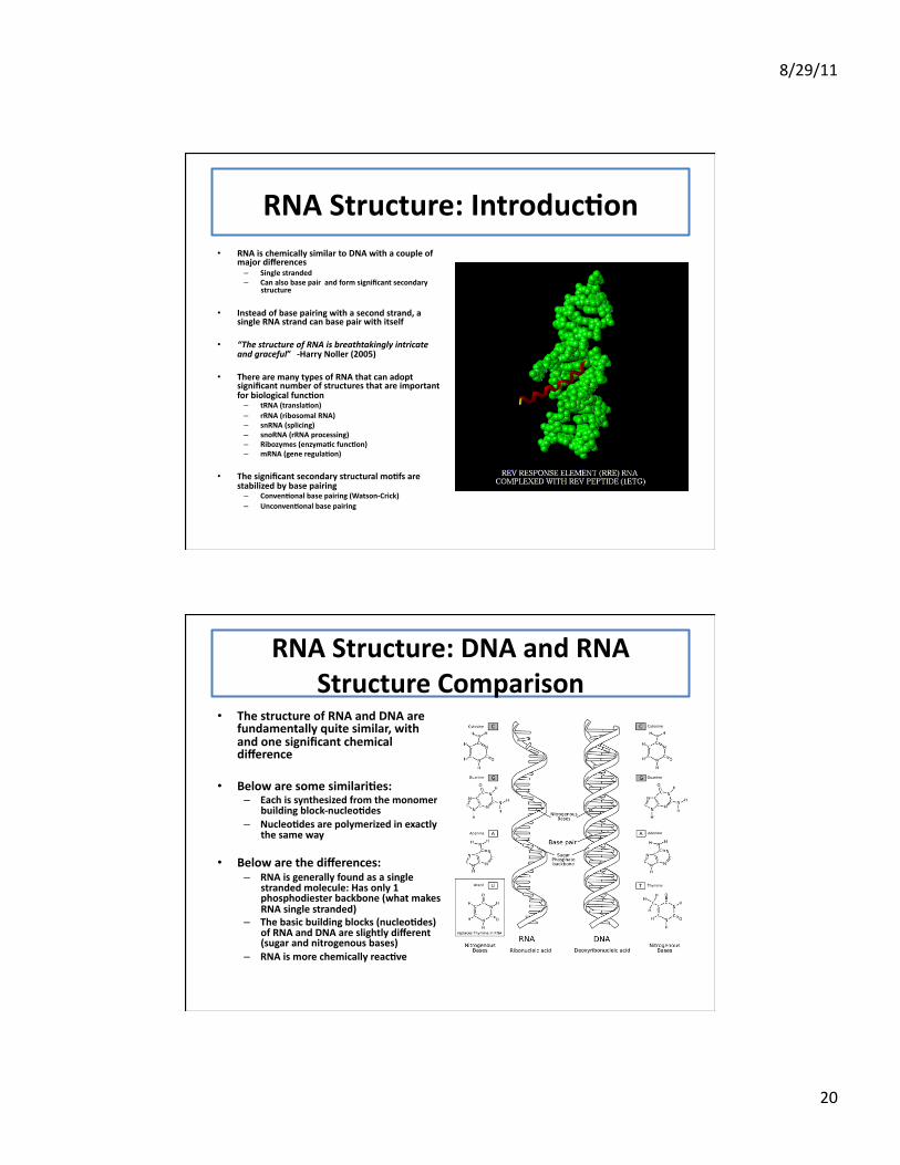
RNA Structure: DNA and RNA Structure Comparison
• The structure of RNA and DNA are fundamentally quite similar, with and one significant chemical difference
• Below are some similari;es: – Each is synthesized from the monomer
building block-‐nucleo;des – Nucleo;des are polymerized in exactly
the same way
• Below are the differences: – RNA is generally found as a single
stranded molecule: Has only 1 phosphodiester backbone (what makes RNA single stranded)
– The basic building blocks (nucleo;des) of RNA and DNA are slightly different (sugar and nitrogenous bases)
– RNA is more chemically reac;ve
8/29/11
21
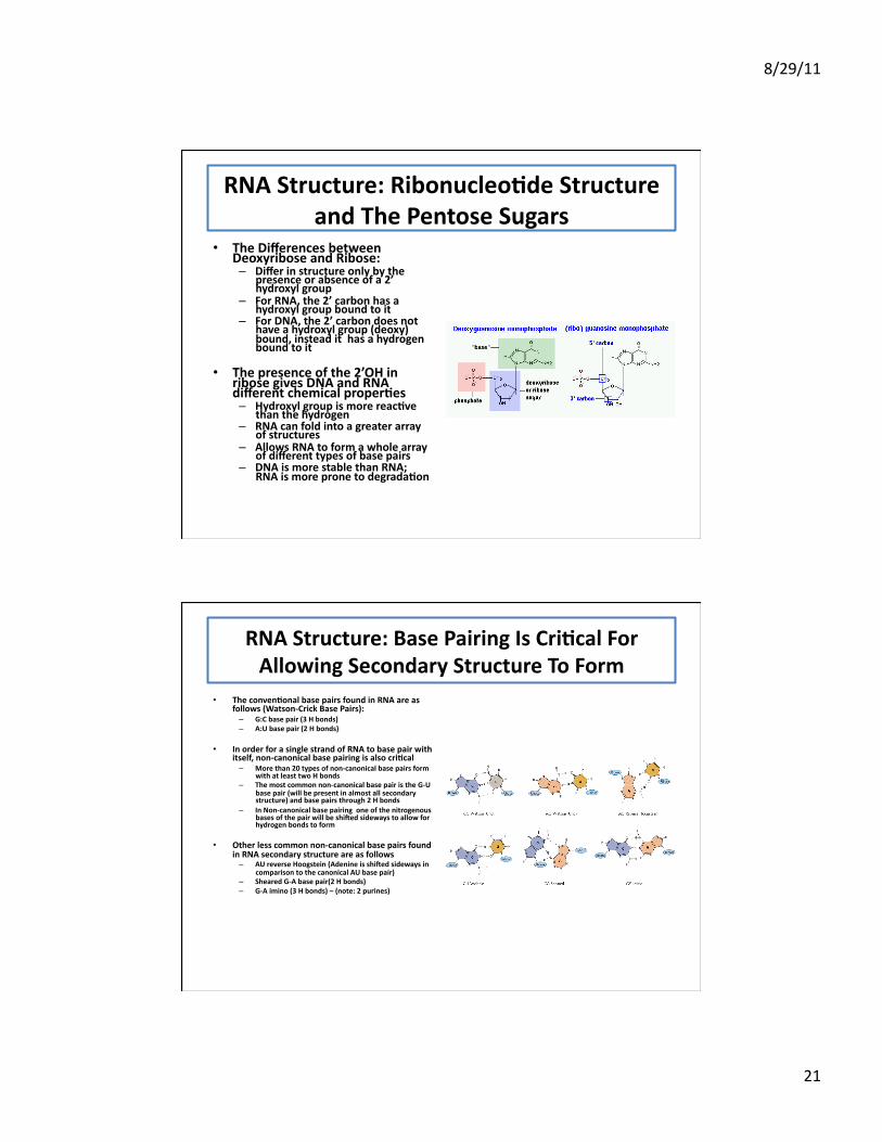
RNA Structure: Ribonucleo;de Structure and The Pentose Sugars
• The Differences between Deoxyribose and Ribose: – Differ in structure only by the
presence or absence of a 2’ hydroxyl group
– For RNA, the 2’ carbon has a hydroxyl group bound to it
– For DNA, the 2’ carbon does not have a hydroxyl group (deoxy) bound, instead it has a hydrogen bound to it
• The presence of the 2’OH in ribose gives DNA and RNA different chemical proper;es – Hydroxyl group is more reac;ve
than the hydrogen – RNA can fold into a greater array
of structures – Allows RNA to form a whole array
of different types of base pairs – DNA is more stable than RNA;
RNA is more prone to degrada;on
RNA Structure: Base Pairing Is Cri;cal For Allowing Secondary Structure To Form
• The conven;onal base pairs found in RNA are as follows (Watson-‐Crick Base Pairs): – G:C base pair (3 H bonds) – A:U base pair (2 H bonds)
• In order for a single strand of RNA to base pair with itself, non-‐canonical base pairing is also cri;cal – More than 20 types of non-‐canonical base pairs form
with at least two H bonds – The most common non-‐canonical base pair is the G-‐U
base pair (will be present in almost all secondary structure) and base pairs through 2 H bonds
– In Non-‐canonical base pairing one of the nitrogenous bases of the pair will be shi[ed sideways to allow for hydrogen bonds to form
• Other less common non-‐canonical base pairs found in RNA secondary structure are as follows – AU reverse Hoogstein (Adenine is shi[ed sideways in
comparison to the canonical AU base pair) – Sheared G-‐A base pair(2 H bonds) – G-‐A imino (3 H bonds) – (note: 2 purines)
8/29/11
22
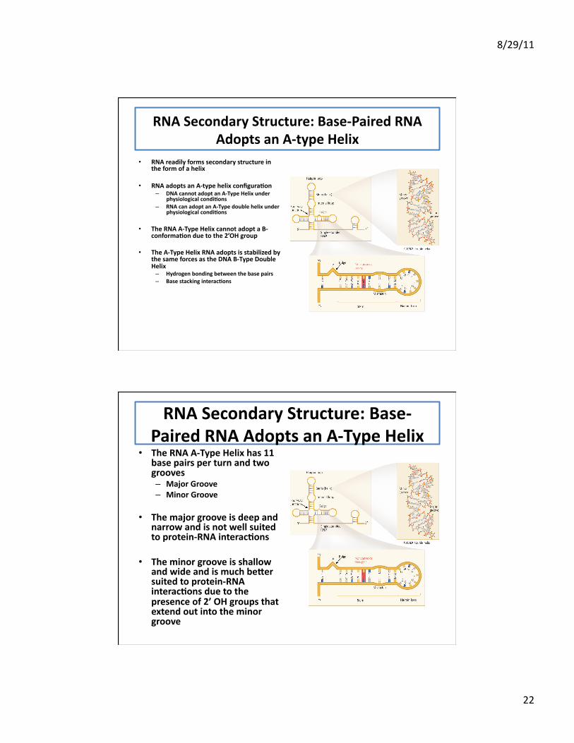
RNA Secondary Structure: Base-‐Paired RNA Adopts an A-‐type Helix
• RNA readily forms secondary structure in the form of a helix
• RNA adopts an A-‐type helix configura;on – DNA cannot adopt an A-‐Type Helix under
physiological condi;ons – RNA can adopt an A-‐Type double helix under
physiological condi;ons
• The RNA A-‐Type Helix cannot adopt a B-‐conforma;on due to the 2’OH group
• The A-‐Type Helix RNA adopts is stabilized by the same forces as the DNA B-‐Type Double Helix – Hydrogen bonding between the base pairs – Base stacking interac;ons
RNA Secondary Structure: Base-‐Paired RNA Adopts an A-‐Type Helix
• The RNA A-‐Type Helix has 11 base pairs per turn and two grooves – Major Groove – Minor Groove
• The major groove is deep and narrow and is not well suited to protein-‐RNA interac;ons
• The minor groove is shallow and wide and is much be\er suited to protein-‐RNA interac;ons due to the presence of 2’ OH groups that extend out into the minor groove
8/29/11
23
RNA Secondary Structure: Base-‐Paired RNA Adopts an A-‐Type Helix
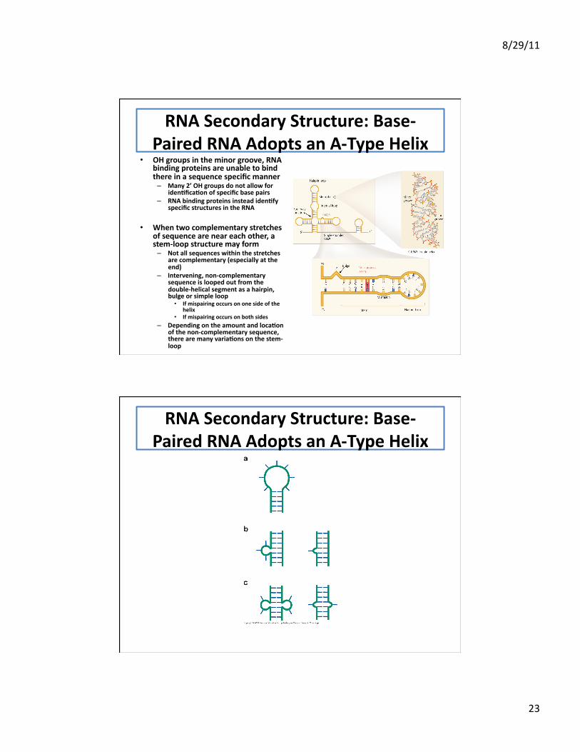
• OH groups in the minor groove, RNA binding proteins are unable to bind there in a sequence specific manner – Many 2’ OH groups do not allow for
iden;fica;on of specific base pairs – RNA binding proteins instead iden;fy
specific structures in the RNA
• When two complementary stretches of sequence are near each other, a stem-‐loop structure may form – Not all sequences within the stretches
are complementary (especially at the end)
– Intervening, non-‐complementary sequence is looped out from the double-‐helical segment as a hairpin, bulge or simple loop • If mispairing occurs on one side of the
helix • If mispairing occurs on both sides
– Depending on the amount and loca;on of the non-‐complementary sequence, there are many varia;ons on the stem-‐loop
RNA Secondary Structure: Base-‐Paired RNA Adopts an A-‐Type Helix
8/29/11
24
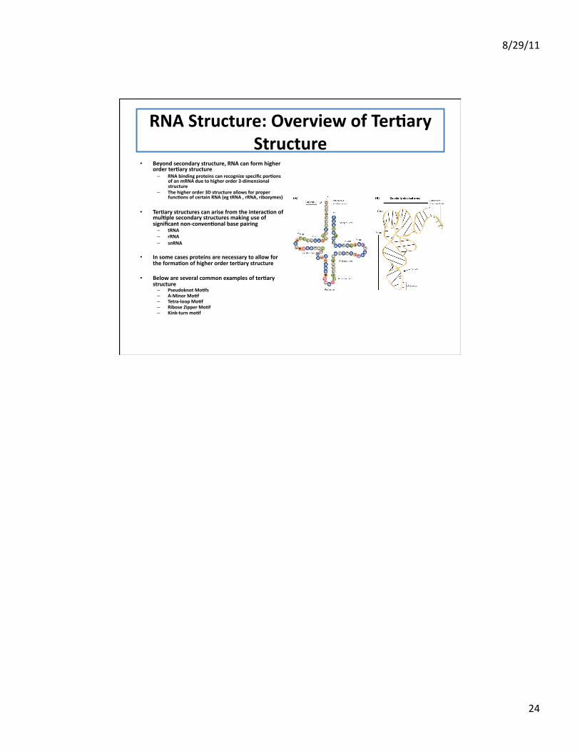
RNA Structure: Overview of Ter;ary Structure
• Beyond secondary structure, RNA can form higher order ter;ary structure – RNA binding proteins can recognize specific por;ons
of an mRNA due to higher order 3-‐dimensional structure
– The higher order 3D structure allows for proper func;ons of certain RNA (eg tRNA , rRNA, ribozymes)
• Ter;ary structures can arise from the interac;on of mul;ple secondary structures making use of significant non-‐conven;onal base pairing – tRNA – rRNA – snRNA
• In some cases proteins are necessary to allow for the forma;on of higher order ter;ary structure
• Below are several common examples of ter;ary structure – Pseudoknot Mo;fs – A-‐Minor Mo;f – Tetra-‐loop Mo;f – Ribose Zipper Mo;f – Kink-‐turn mo;f