Embed Size (px)
Citation preview
Unit 5

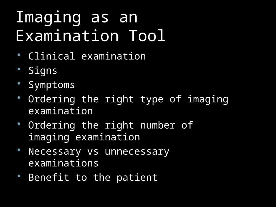
Imaging as an Examination Tool Clinical examination Signs Symptoms Ordering the right type of imaging
examination Ordering the right number of imaging
examination Necessary vs unnecessary examinations Benefit to the patient

Viewing condition Viewbox Monitor Prints Ambient light reduced Quiet room Intraoral films mounted on a opaque holder Equal intensity of light on the view box Monitors: calibration Magnification Software limitations

Systematic Approach Intraoral images
Teeth, periodontium, bone, adjacent structuresTooth #1 to #16, and then #17 to #32
Extraoral images;Panoramic, other extraoral plain radiographs
Cross sectional imagesCT, MRI

Localize the abnormality
How many lesions? Where is the lesion? Localized vs generalized Single arch or both the arches Inside the bone or outside Relation to the crown Relation to the root Superior to the mandibular canal

Periphery
Well defined or ill defined? Sharp margins Corticated margins Sclerotic margins Radiolucent band Blends into adjacent area Irregular margins

Shape
Circular Oval Scalloped Multilocular

Internal structures
Radiolucent Mixed Radiopaque Trabeculation Septa Calcifications Tooth or similar entities
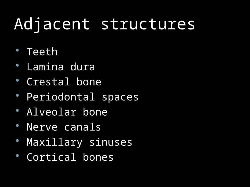
Adjacent structures
Teeth Lamina dura Crestal bone Periodontal spaces Alveolar bone Nerve canals Maxillary sinuses Cortical bones

Description, description, description Speak out loudly List it down Compare findings in different images Clinical information


Vindicate your D/D Vascular Infection Neoplasm Drugs Idiopathic/inflammatory Congenital Autoimmune Trauma Endocrine/metabolic

In the Land of 10,000 lakes,…

…we see the fish!


Liar, Liar!! Do Our Eyes Lie?

Dirty words!
Sun-ray appearance Ground glass Cotton wool Onion skin Driven snow Etc, etc

When not to order imaging You have not clinically / radiographically
evaluated the patient No benefit to the patient Additional images may not provide extra
information No ‘routine’ radiograph

What goes on the report
Patient, doctor, clinic identification Date, type and number of examination Reasons for the examination Clinical information Relevant observation Radiographic Impressions Any further tests, examinations