Embed Size (px)
Citation preview

Unit 3: Genetics and Information TransferBig Idea 3: Living systems store, retrieve, transmit and respond to information
essential to life processes.
3.A.1: DNA, and in some cases RNA, is the primary source of heritable information. 5.5, 27.1, 16.1, 16.2, 17.1, 17.2, 17.3, 17.4, 19.2, 20.1, 20.2
Concept 5.5 Nucleic acids store and transmit hereditary informationDNA and RNA will be the core topics of Chapter 17. For now, you should just review the general functions and know the components.1. The flow of genetic information is from DNA RNA protein. Use this figure to explain the process.
Label the nucleus, DNA, mRNA, ribosome, and amino acids.
2. The components of a nucleic acid are a sugar, a nitrogenous base, and a phosphate group. Label each on the figure below.
3. You may recall that early in this chapter we looked at the numbering system for the carbons of a sugar. Label the end of the strand on the left side of the figure below that has the number 5 sugar 5' and the other end of the chain 3'.
Holtzclaw, Fred & Theresa; Copyright © 2010 Pearson Education, Inc. Unit 3: Genetics and Information Transfer page 1 of 83

4. Notice that there are five nitrogen bases. Which four are found in DNA?
5. Which four are found in RNA?
6. How do ribose and deoxyribose sugars differ?
7. To summarize, what are the three components of a nucleotide?
8. Here is a model of DNA, which was proposed by James Watson and Francis Crick. What is this shape called?
9. Why are the strands said to be antiparallel?
10.What two molecules make up the “uprights”?
11.What molecules make up the rungs?
12.For the two nucleotides of DNA below, provide the complementary base. A— C—
Holtzclaw, Fred & Theresa; Copyright © 2010 Pearson Education, Inc. Unit 3: Genetics and Information Transfer page 2 of 83

13.In a DNA double helix, a region along one DNA strand has this sequence of nitrogenous bases: 5'-
T A G G C C T-3'
Write the complementary strand. Indicate the 5' and 3' ends of the new strand.
This summary table from the Chapter 5 Review is an excellent study tool. Use it to organize material from this chapter in your mind.
Holtzclaw, Fred & Theresa; Copyright © 2010 Pearson Education, Inc. Unit 3: Genetics and Information Transfer page 3 of 83

Concept 27.1 Structural and functional adaptations contribute to prokaryotic success1.The chapter opens with amazing tales of life at the extreme edge. What are the “masters of adaptation”? Describe the one case you thought most dramatic.
2.Which two domains include prokaryotes?
3.Let’s focus on some general details about prokaryotes.a.Are they multicellular or unicellular?
b.Compare their size relative to eukaryotic cells.
c.What three shapes are most common? Label them on the figure.
d.What is the composition of the typical bacterial cell wall?
4.A key feature of prokaryotic cells is the cell wall. What three functions does it provide for the cell?
5.Quick review! What material comprises the cell wall of plants? of fungi?
6.The cell walls of Archaeans are different. They lack _______________ but contain _____________ and _________________.
7.Explain the difference between Gram-positive and Gram-negative bacteria.
8.What is a bacterial capsule? What functions may it serve?
9.Many prokaryotes are capable of directional movement. What is this called?
Holtzclaw, Fred & Theresa; Copyright © 2010 Pearson Education, Inc. Unit 3: Genetics and Information Transfer page 4 of 83

10.What bacterial feature makes this possible?
11.Under ideal conditions, how quickly can E. coli divide? What conditions check prokaryotic reproduction?
12.What three key features allow prokaryotic populations to consist of trillions of individuals?
13.Compare prokaryotes to eukaryotes in terms of the following characteristics:
14.What are the small, circular, self-replicating pieces of DNA found in bacteria called?
15.Label the following structures of a typical prokaryote seen here: cell wall, sex pilus, circular chromosome, nucleoid region, ribosomes, flagella, capsule, and fimbriae. Sketch in a plasmid or two, and label them. For each structure, know the function. (Go to the end of the chapter, p. 573, for help with this figure.)
Holtzclaw, Fred & Theresa; Copyright © 2010 Pearson Education, Inc. Unit 3: Genetics and Information Transfer page 5 of 83

16.When conditions for survival are difficult, some species produce endospores. What are these? Can you name any species that form endospores? As a hint, consider what causes botulism or tetanus.
Concept 16.1 DNA is the genetic material1.What are the two chemical components of chromosomes?
2.Why did researchers originally think that protein was the genetic material?
3.Distinguish between the virulent and nonvirulent strains of Streptococcus pneumoniae studied by Frederick Griffith.
4.What was the purpose of Griffith’s studies?
5.Use this figure to summarize the experiment in which Griffith became aware that hereditary information could be transmitted between two organisms in an unusual manner.
6.Define transformation.
Holtzclaw, Fred & Theresa; Copyright © 2010 Pearson Education, Inc. Unit 3: Genetics and Information Transfer page 6 of 83
7.What did Oswald Avery determine to be the transforming factor? ___________ Explain his experimental approach.
8.Sketch a T2 bacteriophage and label its head, tail sheath, tail fiber, and DNA.
9.How does a bacteriophage destroy a bacterial cell? Look ahead to Chapter 19, Figure 19.5, to explain this.
10.How did Hershey and Chase “label” viral DNA and viral protein so that they could be distinguished? Explain why they chose each radioactive tag in light of the chemical composition of DNA and protein.
11.Describe the means by which Hershey and Chase established that only the DNA of a phage enters an E. coli cell. What conclusions did these scientists draw based on these observations?
12.What are Chargaff’s rules? How did he arrive at them?
13.List the three components of a nucleotide.
14.Who built the first model of DNA and shared the 1962 Nobel Prize for discovery of its structure?
15.What was the role of Rosalind Franklin in the discovery of the double helix?
16.Distinguish between the structure of pyrimidines and purines. Explain why adenine bonds only to thymine.
Holtzclaw, Fred & Theresa; Copyright © 2010 Pearson Education, Inc. Unit 3: Genetics and Information Transfer page 7 of 83

17.How did Watson and Crick’s model explain the basis for Chargaff’s rules?
18.Given that the DNA of a certain fly species consists of 27.3% adenine and 22.5% guanine, use Chargaff’s rules to deduce the percentages of thymine and cytosine.
19.Name the five nitrogenous bases, and put a checkmark in the correct column for each base. Also indicate if the base is found in DNA (D), RNA (R), or both (B).
20.Explain the base-pairing rule.
21.Describe the structure of DNA relative to each of the following:
a. distance across molecule _______________________
b. distance between nucleotides ____________________
c. distance between turns _________________________
d. components of the backbone ____________________
e. components of the “rungs”_____________________
Holtzclaw, Fred & Theresa; Copyright © 2010 Pearson Education, Inc. Unit 3: Genetics and Information Transfer page 8 of 83

22.Explain what is meant by 5' and 3' ends of the nucleotide.
23.What do we mean when we say the two strands of DNA are antiparallel?
Concept 16.2 Many proteins work together in DNA replication and repair1.What is the semiconservative model of replication?
2.Who performed the experiments that elucidated the correct mechanism of DNA replication?
3.How did Meselson and Stahl create “heavy” DNA for their experiments?
4.Use Figure 16.11 to explain how Meselson and Stahl confirmed the semiconservative mechanism of DNA replication.
5.Define the origins of replication.
6.Distinguish between the leading and the lagging strands during DNA replication.
7.What is the direction of synthesis of the new strand?
Holtzclaw, Fred & Theresa; Copyright © 2010 Pearson Education, Inc. Unit 3: Genetics and Information Transfer page 9 of 83

8.What are Okazaki fragments? How are they welded together?
9.Which enzyme . . .?
10.Label the following figures. Include 3' and 5' strands, RNA primer, primase, SSBP, topoisomerase, helicase, leading strand, lagging strand, DNA pol I, DNA pol III, DNA ligase, parental DNA, and new DNA.
Holtzclaw, Fred & Theresa; Copyright © 2010 Pearson Education, Inc. Unit 3: Genetics and Information Transfer page 10 of 83

11.Put it all together! Make a detailed list of the steps that occur in the synthesis of a new strand.
12.Explain the roles of each of the following enzymes in DNA proofreading and repair.
13.What is a thymine dimer? How might it occur? How is it repaired?
14.Make a sketch of a chromosome and label the telomeres.
15.Explain telomere erosion and the role of telomerase.
16.Why are cancer cells immortal, but most body cells have a limited life span?
Holtzclaw, Fred & Theresa; Copyright © 2010 Pearson Education, Inc. Unit 3: Genetics and Information Transfer page 11 of 83

Concept 17.1 Genes specify proteins via transcription and translation1.What situation did Archibald Garrod suggest caused inborn errors of metabolism?
2.Describe one example Garrod used to illustrate his hypothesis.
3.State the hypothesis formulated by George Beadle while studying eye color mutations in Drosophila.
4.What strategy did Beadle and Tatum adopt to test this hypothesis?
5.Which organism did Beadle and Tatum use in their research? How did this organism’s nutritional requirements facilitate this research?
6.How were Neurospora spores treated to increase the mutation rate?
7.Study Figure 17.2 carefully. On the figure below, outline the technique used to identify and isolate mutant fungi.
Holtzclaw, Fred & Theresa; Copyright © 2010 Pearson Education, Inc. Unit 3: Genetics and Information Transfer page 12 of 83

8.Cite two significant findings that resulted from the research of Beadle and Tatum.
9.What revision of detail (but not of basic principle) did this hypothesis undergo as more information was gained? Write this restatement and then box or highlight it. This is an important concept!
Basic Principles of Transcription and TranslationThis section will introduce you to the processes and associated terminology in the form of an overview. Once you have the big picture, you will take a closer look in the next few concepts.10.From the first paragraph in this section, find three ways in which RNA differs from DNA.
11.What are the monomers of DNA and RNA? Of proteins?
12.Define each of these processes that are essential to the formation of a protein:
transcription:
translation:
13.Complete the following table to summarize each process.
14.In eukaryotes, what is the pre-mRNA called?
15.What is the central dogma of molecular genetics, as proclaimed by Francis Crick.
Holtzclaw, Fred & Theresa; Copyright © 2010 Pearson Education, Inc. Unit 3: Genetics and Information Transfer page 13 of 83

16.How many nucleotide bases are there? ____________________ How many amino acids? ____________________ .
17.How many nucleotides are required to code for these 20 amino acids? ___________________
18.So, the language of DNA is a triplet code. How many unique triplets exist? ________________
19.DNA is double-stranded, but for each protein, only one of these two strands is used to produce an mRNA transcript. What is the coding strand called?
20.Here is a short DNA template. Below it, assemble the complementary mRNA strand.
3'A C G A C C A G T A A A 5'
21.How many codons are there above? ____________ Label one codon.
22.Describe Nirenberg’s experiment in which he identified the first codon.
23.What was the first codon–amino acid pair to be identified? ____________________________
24.Of the 64 possible codons, how many code for amino acids? __________________________
25.What event is coded fro by UAA, UAG and UGA? ___________________________________
26.What is the start codon? _______________________________________________________
27.Why is the genetic code said to be redundant but not ambiguous?
28.Explain the concept of reading frame.
29.Now here is an important idea: DNA is DNA is DNA. By this we mean that the code is nearly universal, and because of this, jellyfish genes can be inserted into pigs, or firefly genes can make a tobacco plant glow. Enjoy a look at Figure 17.6 in your text . . . and no question to answer here!
Holtzclaw, Fred & Theresa; Copyright © 2010 Pearson Education, Inc. Unit 3: Genetics and Information Transfer page 14 of 83

Concept 17.2 Transcription is the DNA-directed synthesis of RNA: A closer look1.Name the enzyme that uses the DNA template strand to transcribe a new mRNA strand.
2.You will recall from Chapter 16 that DNA polymerase III adds new nucleotides to the template DNA strand to assemble each new strand of DNA. Both enzymes can assemble a new polynucleotide only in the 5'direction. Which enzyme, DNA polymerase III or RNA polymerase, does not require a primer to begin synthesis?
3.What is a transcription unit?
4.Figure 17.7 in your text will require a bit of study. Use it to label the following elements on the figure below: promoter, RNA polymerase, transcription unit, DNA template, nontemplate DNA, and RNA transcript. Then, to the right of the figure, name the three stages of transcription and briefly describe each stage.
Holtzclaw, Fred & Theresa; Copyright © 2010 Pearson Education, Inc. Unit 3: Genetics and Information Transfer page 15 of 83

5.Let’s now take a closer look at initiation. Read the paragraph titled “RNA Polymerase Binding and Initiation of Transcription” carefully. List three important facts about the promoter here.
(1)
(2)
(3)
6.Use Figure 17.8 in your text to label the following elements of the figure below: TATA box, RNA polymerase II, transcription factors, template DNA strand, start point, 5' and 3', and mRNA transcript. To the right of the figure, explain the three stages of initiation that are shown.
7.What is the TATA box? How do you think it got this name?
8.What comprises a transcription initiation complex?
Holtzclaw, Fred & Theresa; Copyright © 2010 Pearson Education, Inc. Unit 3: Genetics and Information Transfer page 16 of 83

9.Now it is time to put all of the elements of transcription together. Write an essay below to describe the process by which mRNA is formed. Use these terms correctly in your essay, and underline each one: TATA box, gene, terminator, promoter, elongation, 5’ to 3', termination, initiation RNA, polymerase RNA nucleotides, template, start point, termination signal, and transcription factors. This essay is typical of what you might be asked to write on the AP Biology exam.
________________________________________________________________________________
________________________________________________________________________________
________________________________________________________________________________
________________________________________________________________________________
________________________________________________________________________________
________________________________________________________________________________
________________________________________________________________________________
________________________________________________________________________________
________________________________________________________________________________
________________________________________________________________________________
________________________________________________________________________________
________________________________________________________________________________
________________________________________________________________________________
________________________________________________________________________________
________________________________________________________________________________
________________________________________________________________________________
________________________________________________________________________________
________________________________________________________________________________
________________________________________________________________________________
________________________________________________________________________________
________________________________________________________________________________
________________________________________________________________________________Holtzclaw, Fred & Theresa; Copyright © 2010 Pearson Education, Inc. Unit 3: Genetics and Information Transfer page 17 of 83

Concept 17.3 Eukaryotic cells modify RNA after transcription1.RNA processing occurs only in eukaryotic cells. The primary transcript is altered at both ends, and sections in the middle are removed. a. What happens at the 5' end?
b. What happens at the 3' end?
2.What are three important functions of the 5' cap and poly-A tail?
3.Distinguish between introns and exons. Perhaps it will help to remember this: Exons are expressed.
4.On the figure below, label: pre-mRNA, 5' cap, poly-A tail, introns, and exons.
5.What are snRNPs? What two types of molecules make up a snurp?
6.You will be introduced to a number of small RNAs in this course. What type is the RNA in a snRNP?
7.Snurps band together in little snurp groups to form spliceosomes. How do spliceosomes work?
Holtzclaw, Fred & Theresa; Copyright © 2010 Pearson Education, Inc. Unit 3: Genetics and Information Transfer page 18 of 83

8.On the figure below, label the following: pre-mRNA, snRNPs, snRNA, protein, spliceosomes, intron, and other proteins.
9.Study the figure and text carefully to explain how the splice sites are recognized.
10.What is a ribozyme?
11.What commonly held idea was rendered obsolete by the discovery of ribozymes?
12.What are three properties of RNA that allow it to function as an enzyme?
(1)
(2)
(3)
Holtzclaw, Fred & Theresa; Copyright © 2010 Pearson Education, Inc. Unit 3: Genetics and Information Transfer page 19 of 83

13.What is the consequence of alternative splicing of identical mRNA transcripts?
Concept 17.4 Translation is the RNA-directed synthesis of a polypeptide: A closer look1.You may need to read on in this section in order to answer this question as well as think back to earlier information about mRNA. Come back to this question later if you wish. Three types of RNA are needed for protein synthesis. Complete the chart below.
2.What is an anticodon?
3.Transfer RNA has two attachment sites. What binds at each site? Sketch tRNA, indicate the 2 attachment sites, and note where complementary base pairing and hydrogen bonding occur to give tRNA its shape.
4.How many different aminoacyl-tRNA synthetases are there? ____________________________
5.Scientists expected to find one aminoacyl-tRNA synthetase per codon, but far fewer have been discovered. How does wobble explain this?
Holtzclaw, Fred & Theresa; Copyright © 2010 Pearson Education, Inc. Unit 3: Genetics and Information Transfer page 20 of 83

6.Use the figure below to explain the process of a specific amino acid being joined to a tRNA. Also add these labels: aminoacyl-tRNA synthetase, ATP, amino acid, and tRNA.
7.Describe the structure of a eukaryotic ribosome.
8.How does a prokaryotic ribosome differ from a eukaryotic ribosome? What is the medical significance of this difference?
9.On this figure, label the large subunit, small subunit, A, P, and E sites, mRNA binding site. To the right of the figure, explain the functions of the A, P, and E sites.
Holtzclaw, Fred & Theresa; Copyright © 2010 Pearson Education, Inc. Unit 3: Genetics and Information Transfer page 21 of 83

10.Much like transcription, we can divide translation into three stages. List them.
11.Summarize the events of initiation. Include these components: small ribosomal subunit, large ribosomal subunit, mRNA, initiator codon, tRNA, Met, initiation complex, P site, and GTP. The figure below may help you.
12.What is always the first amino acid in the new polypeptide?
13.Now, summarize the events of elongation. Include these components: mRNA, A site, tRNA, codon, anticodon, ribozyme, P site, and E site. Again, the figure may help you.
Holtzclaw, Fred & Theresa; Copyright © 2010 Pearson Education, Inc. Unit 3: Genetics and Information Transfer page 22 of 83

14.What is a release factor? By what mechanism is termination accomplished?
15.What is a polyribosome?
16.What are some of the things that will result in a final-form functional protein?
17.Describe at least three types of post-translational modifications.
18.Use the following figure to explain how proteins are targeted for the ER.
Holtzclaw, Fred & Theresa; Copyright © 2010 Pearson Education, Inc. Unit 3: Genetics and Information Transfer page 23 of 83

Concept 19.2 Viruses reproduce only in host cells1.What property of a virus determines its attachment to a host cell membrane?
2.Viruses are obligate intracellular parasites. What does this mean?
3.What is meant by host range? Distinguish between a virus with a broad host range and one with an extremely limited host range, and give an example of each.
4.Compare the host range for the rabies virus to that of the human cold virus.
5.What components of the host cell does a virus use to reproduce itself?
6.How does a DNA virus reproduce its genome?
7.How do most RNA viruses replicate their genome?
8.On this figure of a simplified viral reproductive cycle, label arrows to show these processes: transcription, translation, infection, replication, and self-assembly. Annotate your labels to explain the process of viral reproduction.
Holtzclaw, Fred & Theresa; Copyright © 2010 Pearson Education, Inc. Unit 3: Genetics and Information Transfer page 24 of 83

9.What are bacteriophages? Distinguish between virulent and temperate phages.
10.What portion of a phage enters the host cell? How does it do this?
11.What are restriction enzymes? What is their role in bacteria?
12.Why don’t restriction enzymes destroy the DNA of the bacterial cells that produce them?
13.What are three ways bacteria may win the battle against the phages?
14.What is a prophage?
15.Since cells that have incorporated phage DNA into their genome may continue to divide and propagate the viral genome, this might be considered somewhat like the Trojan horse. What might trigger the switchover from lysogenic to lytic mode?
16.Label the following elements of the figure below: lysogenic phage, lysogenic cycle, lytic cycle, prophage, phage DNA, bacterial chromosome, and self assembly.
Holtzclaw, Fred & Theresa; Copyright © 2010 Pearson Education, Inc. Unit 3: Genetics and Information Transfer page 25 of 83

17.Describe the lytic and lysogenic modes of bacteriophage reproduction.
18.There are some general differences between bacteriophages and animal viruses. What are two elements that nearly all animal viruses have?
19.What is a retrovirus? How do retroviruses, such as HIV, replicate their genome?
20.Here is a sketch of HIV. Label these parts: envelope, reverse transcriptase, RNA, and capsid.
21.Compare and contrast a prophage and a provirus. Which one are you likely to carry?
Holtzclaw, Fred & Theresa; Copyright © 2010 Pearson Education, Inc. Unit 3: Genetics and Information Transfer page 26 of 83

22.This sketch shows the infection of a cell by HIV. Extend label lines to give a complete explanation of the process. Refer to your text Figure 19.8 for details.
23.The final section in this concept is titled “Evolution of Viruses.” From this part, describe the two possible sources of viral genomes. You will see each of these important mobile genetic elements again.
Holtzclaw, Fred & Theresa; Copyright © 2010 Pearson Education, Inc. Unit 3: Genetics and Information Transfer page 27 of 83
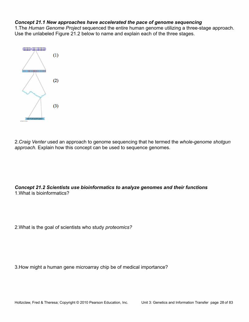
Concept 21.1 New approaches have accelerated the pace of genome sequencing1.The Human Genome Project sequenced the entire human genome utilizing a three-stage approach. Use the unlabeled Figure 21.2 below to name and explain each of the three stages.
2.Craig Venter used an approach to genome sequencing that he termed the whole-genome shotgun approach. Explain how this concept can be used to sequence genomes.
Concept 21.2 Scientists use bioinformatics to analyze genomes and their functions1.What is bioinformatics?
2.What is the goal of scientists who study proteomics?
3.How might a human gene microarray chip be of medical importance?
Holtzclaw, Fred & Theresa; Copyright © 2010 Pearson Education, Inc. Unit 3: Genetics and Information Transfer page 28 of 83

3.A.2 In eukaryotes, heritable information is passed to the next generation via processes that include the cell cycle and mitosis, or meiosis plus fertilization. 12.1, 12.2, 12.3 13.1, 13.2, 13.3
Concept 12.1 Cell division results in genetically identical daughter cells1.What are the three key roles of cell division? State each role, and give an example.
2.What is meant by the cell cycle?
3.What is the meaning of genome? Compare your genome to that of a prokaryotic cell.
4.How many chromosomes are in a human somatic cell?
5.Name two types of somatic cells in your body.
6.What is a gamete?
7.Name the two types of gametes.
8.How many chromosomes in a human gamete?
9.Define chromatin.
Holtzclaw, Fred & Theresa; Copyright © 2010 Pearson Education, Inc. Unit 3: Genetics and Information Transfer page 29 of 83

10.Think carefully, now. How many DNA molecules are in each of your somatic cells?
11.You are going to have to learn the difference between a number of similar-sounding terms. The sketch that looks like an X represents a replicated chromosome that has two sister chromatids. The narrow “waist” represents the location of the centromere. Students often get all these terms confused, so take time now to label the indicated areas of the figure and then define each of the terms below.
chromosome
chromatid
centromere
chromatin
12.Study Figure 12.4. Label the figure below, and summarize what occurs at the DNA level in each stage.
13.What is mitosis? How is it different from cytokinesis?
Holtzclaw, Fred & Theresa; Copyright © 2010 Pearson Education, Inc. Unit 3: Genetics and Information Transfer page 30 of 83

14.What occurs in meiosis? How is the chromosome number of daughter cells different?
15.Select either mitosis or meiosis to answer the following questions.
________________________ By what process are the damaged cells in a wound replaced?
________________________ By what process are eggs formed?
________________________ By what process does a zygote develop into a multicellular organism?
________________________ In which process are identical daughter cells produced?
________________________ Which process reduces chromosome number of daughter cells?
16.Don’t skip the Concept Check Questions! They are a good way to verify your understanding. Here is a variation of question 3. Answer it here: A hedgehog has 90 chromosomes in its somatic cells.
a.How many chromosomes did the hedgehog inherit from each parent?
b.How many chromosomes are in each of the hedgehog’s gametes?
c.How many chromosomes will be in each somatic cell of the hedgehog’s offspring?
Concept 12.2 The mitotic phase alternates with interphase in the cell cycle1.Label each of the parts of the cell cycle listed below, and give a brief explanation of what happens in each phase.
G1
S
G2
M
Holtzclaw, Fred & Theresa; Copyright © 2010 Pearson Education, Inc. Unit 3: Genetics and Information Transfer page 31 of 83
2.What are the components of the mitotic spindle? What is the source of these components?
3.In animal cells, the assembly of spindle microtubules starts at the centrosome. What is another name for the centrosome?
4.Sketch and label a centrosome with two centrioles.
5.Describe what happens to the centrosome during interphase and then prophase.
6.What is a kinetochore? Read your text carefully, and then make a labeled sketch that shows a replicated chromosome with two kinetechores and some attached spindle fibers. Figure 12.7 may help.
Holtzclaw, Fred & Theresa; Copyright © 2010 Pearson Education, Inc. Unit 3: Genetics and Information Transfer page 32 of 83

7.You will need to spend some serious time with Figure 12.6. Use it to help you label this figure. Label each phase by name; then label the smaller structures. Finally, make 2 or 3 summary statements that indicate important features to note about the phase.
Holtzclaw, Fred & Theresa; Copyright © 2010 Pearson Education, Inc. Unit 3: Genetics and Information Transfer page 33 of 83

8.Explain the difference between kinetochore and nonkinetechore microtubules. What is the function of each?
9.What are the components of the mitotic spindle?
10.At which end do kinetochore microtubules shorten during anaphase? Explain the data that supports where this shortening occurs.
11.Describe cytokinesis in an animal cell. Use a labeled sketch that shows the cleavage furrow.
12.Describe cytokinesis in a plant cell. Use a labeled sketch that shows the cell plate.
13.How is the cell plate formed? What is the source of the material for the cell plate?
14.Prokaryote reproduction does not involve mitosis, but instead occurs by binary fission. This process involves an origin of replication. Describe binary fission.
15.Notice that now you are learning a number of differences between prokaryotic and eukaryotic cells. Besides the fact that prokaryotes lack a membrane-bounded nucleus, describe the following differences:
Mode of reproduction?
Number of chromosomes?
Shape of the bacterial chromosome?
Holtzclaw, Fred & Theresa; Copyright © 2010 Pearson Education, Inc. Unit 3: Genetics and Information Transfer page 34 of 83

Concept 12.3 The eukaryotic cell cycle is regulated by a molecular control system1.What controls the cell cycle? Study the Inquiry Figure 12.13 to help you answer this question.
2.What is a cell cycle checkpoint?
3.Summarize what happens at each checkpoint. You may add to this chart as you study this section.
4.What is the Go phase? Describe this phase.
5.What is a protein kinase?
6.Kinases drive the cell cycle, but they must be activated by attachment of a _________________.
7.The activity of cyclin-dependent kinases (CDks) rises and falls. Why?
8.What does MPF trigger? What are some specific activities that it triggers?
9.What happens if all the chromosome kinetochores are not attached to spindle fibers? When this occurs, which checkpoint is not passed?
10.What are growth factors? How does PDGF stimulate fibroblast division?
Holtzclaw, Fred & Theresa; Copyright © 2010 Pearson Education, Inc. Unit 3: Genetics and Information Transfer page 35 of 83

11.Cancer cells exhibit different behaviors than normal cells. Here are two normal behaviors they no longer show. Explain each behavior.
density-dependent inhibition
anchorage dependence
12.Cancer cells also show loss of cell cycle controls and may divide without being checked. The story of HeLa cells is worth noting. What is their source? How old are they? Note that, unlike normal cells, HeLa cells are immortal!
13.What is transformation? metastasis?
14.Distinguish between a benign tumor and a malignant tumor.
15.List two specific cancer treatments, and tell how each treatment works.
16.Identify each phase of the cell cycle.
Holtzclaw, Fred & Theresa; Copyright © 2010 Pearson Education, Inc. Unit 3: Genetics and Information Transfer page 36 of 83

Concept 13.1 Offspring acquire genes from parents by inheriting chromosomes1.Let’s begin with a review of several terms that you may already know. Define: gene
locus
gamete
male gamete
female gamete
asexual reproduction
sexual reproduction
2.How many chromosomes are in human cells? What is a chromosome?
3.Which type of reproduction will result in genetically identical offspring?
Concept 13.2 Fertilization and meiosis alternate in sexual life cycles1.What is a somatic cell? Give examples of two human somatic cell types.
2.How does a somatic cell compare to a gamete in terms of chromosome number?
3.Distinguish between sex chromosomes and autosomes. How many of each are found in human cells?
Holtzclaw, Fred & Theresa; Copyright © 2010 Pearson Education, Inc. Unit 3: Genetics and Information Transfer page 37 of 83

4.What is a karyotype? How is it prepared? What are three things that can be determined from a karyotype?
5.Explain what is meant by homologous chromosomes.
6.Cells that have only one of each homologous pair are said to be haploid, a condition that is represented by n. Cells that have two of each homologous pair are said to be diploid or 2n. For each of the following, is the cell haploid or diploid?
liver cell____________________________ gamete_________________________________
egg_________________________________ zygote________________________________
skin cell_____________________________ sperm _________________________________
somatic cell__________________________ sex cell________________________________
7.The muscle cells of a dog have 78 chromosomes. Fill in the correct chromosome number in a:
bone cell_______ sperm_______ haploid cell_______ somatic cell_______ zygote_______
8.In the cell at right, the chromosomes are shaded in two colors to represent the parent of origin. On this sketch, label the following:
a.sister chromatids
b.homologous chromosomes
c.centromere
d.replicated chromosome
e.maternal chromosomes
9.How many chromosomes does the cell above have?
How many homologous pairs?
How many chromatids?
Is this cell haploid or diploid?
Holtzclaw, Fred & Theresa; Copyright © 2010 Pearson Education, Inc. Unit 3: Genetics and Information Transfer page 38 of 83

10.Where are the gametes of an animal produced? Be specific as to male and female gametes.
11.By what process are gametes produced?
12.What is another term for a fertilized egg? __________________What is the chromosome number of the fertilized egg? (Answer this in general terms, haploid, n, or diploid, 2n.)
13.What is the purpose of meiosis?
14.Study Figure 13.6. You will see that plants have a life cycle that involves spores, which form as a result of meiosis, so these spores are haploid. Notice also that both haploid and diploid cells can divide by mitosis. However, meiosis always begins with cells that are ______________________, and as a result of meiosis, daughter cells are formed that are always ______________________. These cells can be gametes (in animals) or spores (in plants).
15.Your study of plants this year will include knowing that they exhibit alternation of generations.
What does this mean?
What are the two generations?
Which is haploid, and which is diploid?
Use this information to label the moss life cycle here.
Holtzclaw, Fred & Theresa; Copyright © 2010 Pearson Education, Inc. Unit 3: Genetics and Information Transfer page 39 of 83

Concept 13.3 Meiosis reduces the number of chromosome sets from diploid to haploid1.What are alleles? Give an example.
2.In meiosis, the DNA is replicated during interphase, followed by two divisions. The first division is meiosis I. Study the events of prophase I as they are significant. Explain each of these events:
synapsis
crossing over
chiasmata
3.The figure at the right shows metaphase I. How is the arrangement of chromosomes different from metaphase of mitosis?
4.There will be two divisions in meiosis. What will separate in the first division in meiosis I?
5.Now study the chromosomes in anaphase I and telophase I carefully. How many chromosomes are in each cell at the end of the first meiotic division? Are the resultant daughter cells haploid, or diploid?
Holtzclaw, Fred & Theresa; Copyright © 2010 Pearson Education, Inc. Unit 3: Genetics and Information Transfer page 40 of 83

6.From this figure, you should see that chromosome number is reduced in meiosis I and that the daughter cells at the end of meiosis I are haploid. Remember this!
7.During meiosis I, homologous chromosomes separate. What separates during meiosis II?
8.To check that you have the big picture, here are some quick review questions.
a.What happens to chromosome number in meiosis?
b.During which division is the chromosome number reduced?
c.What is the purpose of meiosis?
d.How many times does the cell divide in meiosis?
e.How many times do the chromosomes duplicate?
f.How many daughter cells are formed?
g.What is the chromosome number?
h.What are homologs (homologous chromosomes)?
i.What occurs in synapsis?
j.What is crossing over?
Holtzclaw, Fred & Theresa; Copyright © 2010 Pearson Education, Inc. Unit 3: Genetics and Information Transfer page 41 of 83

9.Use Figure 13.9 to compare of mitosis and meiosis. Add these labels: Parent cell, Mitosis, Meiosis, Synapsis, Homologous chromosomes, Replicated chromosomes, Sister chromatids, Daughter cells, Meiosis I, Meiosis II, Crossing over
As you label the drawing, carefully think about each process and review its important features
10.Students often get confused about the differences between mitosis and meiosis. To help with this, work through the following chart:
Holtzclaw, Fred & Theresa; Copyright © 2010 Pearson Education, Inc. Unit 3: Genetics and Information Transfer page 42 of 83
11.Synapsis and crossing over are unique to meiosis. During what specific phase do these occur?
12.Explain the physical events of crossing over. You may wish to make a sketch of the event. Include these terms: synaptonemal complex, chiasmata, homologs, sister chromatids.
3.A.3 The chromosomal basis of inheritance provides an understanding of the pattern of passage (transmission) of genes from parent to offspring 14.1, 14.2, 14.3, 14.4
Concept 14.1 Mendel used the scientific approach to identify two laws of inheritance1.In the 1800s the most widely favored explanation of genetics was blending. Explain the concept of blending, and then describe how Mendel’s particulate (gene) theory was different.
2.One of the keys to success for Mendel was using pea plants. Explain how using pea plants allowed Mendel to control mating; that is, how did this approach let Mendel be positive about the exact characteristics of each parent?
3.Define the following terms. Then, consider your own family. Which generation would your Mom’s grandparents be? Your Mom? You?
P generation
F1 generation
F2 generation
4.Explain how Mendel’s simple cross of purple and white flowers did the following:
refuted blending
determined dominant and recessive characteristics
demonstrated the merit of experiments that covered multiple generations
Holtzclaw, Fred & Theresa; Copyright © 2010 Pearson Education, Inc. Unit 3: Genetics and Information Transfer page 43 of 83

5.Alternate versions of the same gene, like purple and white flower color, are termed _____________.
6.On the figure at below, label the allele for both purple and white flower color, a homologous pair, and the locus of the flower color gene.
7.In sexually reproducing organisms, why are there exactly two chromosomes in each homologue?
8.Mendel’s model consists of four concepts. Describe each concept in the appropriate space below. Indicate which of the concepts can be observed during meiosis by placing an asterisk by the concept.
9.Using Figure 14.5 as your guide, provide the missing notations for the figure below. (P, F1, F2).
a.What is the F2 phenotypic and genotypic ratio?
b.Which generation is completely heterozygous?
c.Which generation has both heterozygous and homozygous offspring?
Holtzclaw, Fred & Theresa; Copyright © 2010 Pearson Education, Inc. Unit 3: Genetics and Information Transfer page 44 of 83

4.In pea plants, T is the allele for tall plants, while t is the allele for dwarf plants. If you have a tall plant, demonstrate with a test cross how it could be determined if the plant is homozygous tall or heterozygous tall.
5.Explain the difference between a monohybrid cross and a dihybrid cross.
6.As you start to work word problems in genetics, two things are critical: the parent’s genotype must be correct, and the gametes must be formed correctly. Using Figure 14.8 as your guide, explain how the gametes are derived for the following cross. (You should have four different gametes).
YyRr × YyRr
7.Complete the cross given in questions 12 by placing the gametes in a Punnett square. Then provide the phenotypic ratio of the offspring.
Phenotype Ratio
8.Explain Mendel’s law of independent assortment.
Before leaving this concept, it would be helpful to complete the three problems in the 14.1 Concept Check on page 269 of your textbook. The problems are worked and explained in the Answer section on page A-10 at the back of the book.
Concept 14.2 The laws of probability govern Mendelian inheritance1. An event that is certain to occur has a probability of _______, while an event that is certain not to
occur has a probability of ________.
2. In probability, what is an independent event?
Holtzclaw, Fred & Theresa; Copyright © 2010 Pearson Education, Inc. Unit 3: Genetics and Information Transfer page 45 of 83
3. State the multiplication rule and give an original example.
4. State the addition rule and give an original example.
5. What is the probability that a couple will have a girl, a boy, a girl, and a boy in this specific order?
Concept 14.3 Inheritance patterns are often more complex than those predicted by simple Mendelian genetics1.Explain how incomplete dominance is different from complete dominance, and give an example of incomplete dominance.
2.Compare and contrast codominance with incomplete dominance.
3.Dominant alleles are not necessarily more common than recessive alleles in the gene pool. Explain why this is true.
4.Explain what is meant when a gene is said to have multiple alleles.
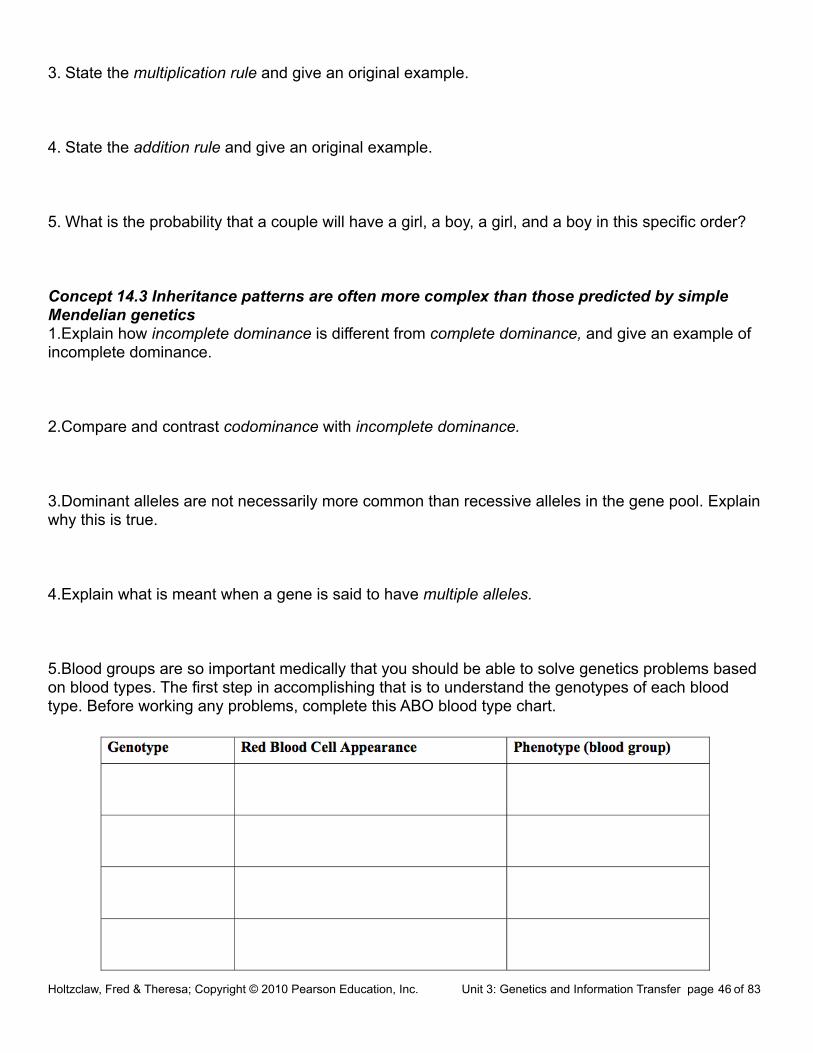
5.Blood groups are so important medically that you should be able to solve genetics problems based on blood types. The first step in accomplishing that is to understand the genotypes of each blood type. Before working any problems, complete this ABO blood type chart.
Holtzclaw, Fred & Theresa; Copyright © 2010 Pearson Education, Inc. Unit 3: Genetics and Information Transfer page 46 of 83

6.Question 2 in the 14.3 Concept Check is a blood type problem. Complete it here, and show your work.
7.What is pleiotropy? Explain why this is important in diseases like cystic fibrosis and sickle- cell disease.
8.Explain epistasis.
9.Explain why the dihybrid cross detailed in Figure 14.12 has 4 white mice instead of the 3 that would have been predicted by Mendel’s work.
10.Why is height a good example of polygenic inheritance?
11.Quantitative variation usually indicates _________________________________________.
12.Using the terms norm of reaction and multifactorial, explain the potential influence of the environment on phenotypic expression.
Concept 14.4 Many human traits follow Mendelian patterns of inheritance1.Pedigree analysis is often used to determine the mode of inheritance (dominant or recessive, for example). Be sure to read the “Tips for pedigree analysis” in Figure 14.15; then complete the unlabeled pedigree by indicating the genotypes for all involved. What is the mode of inheritance for this pedigree?
Holtzclaw, Fred & Theresa; Copyright © 2010 Pearson Education, Inc. Unit 3: Genetics and Information Transfer page 47 of 83

2.Explain why you know the genotype of one female in the third generation, but are unsure of the other.
3.Describe what you think is important to know medically about the behavior of recessive alleles.
4.Students are expected to have a general knowledge of the pattern of inheritance and the common symptoms of a number of genetic disorders. Provide this information for the disorders listed below.
a. cystic fibrosis
b. sickle-cell disease
c. achondroplasia
d. Huntington’s disease
5. Amniocentesis and chorionic villus sampling are the two most widely used methods for testing a fetus for genetic disorders. Use the unlabeled diagram below to explain the three main steps in amniocentesis and the two main steps of CVS.
Holtzclaw, Fred & Theresa; Copyright © 2010 Pearson Education, Inc. Unit 3: Genetics and Information Transfer page 48 of 83

6. What are the strengths and weaknesses of each fetal test?
7. Explain the symptoms of phenylketonuria, and describe how newborn screening is used to identify children with this disorder.
3.A.4 The inheritance pattern of many traits cannot be explained by simple Mendelian genetics. 15.1, 15.2, 15.3, 15.5Concept 15.1 Mendelian inheritance has its physical basis in the behavior of chromosomes1.What is the chromosome theory of inheritance?
2.Explain the law of segregation. Use two different colored pencils to illustrate the segregation of alleles. You may want to consult Figure 15.2 in your text, and model your sketches on this.
3.Explain the law of independent assortment. To demonstrate that you understand this concept, consider a cell with two pairs of chromosomes. Sketch the two different ways these chromosomes might be arranged during metaphase I.
Holtzclaw, Fred & Theresa; Copyright © 2010 Pearson Education, Inc. Unit 3: Genetics and Information Transfer page 49 of 83

4.Thomas Hunt Morgan selected Drosophila melanogaster as his experimental organism. List at least three reasons the fruit fly is an excellent subject for genetic studies.
5.The notation for wild type and mutant traits follows some accepted conventions. Notate the following genotypes for a female fruit fly:
a.a fly homozygous for red eyes
b.a fly heterozygous for red eyes
c.a fly homozygous for white eyes
4.When Thomas Hunt Morgan mated a white-eyed male fly with a red-eyed female, he came to the startling conclusion that the trait for eye color was located on the chromosome that determines sex. Show this cross. Begin with the parental generation, and go through the F2.
parental generation
F1 generation
F2 generation
5. What unusual result suggested that the eye-color trait is located on the X chromosome?
Concept 15.2 Sex-linked genes exhibit unique patterns of inheritance1.There are several variations on the way sex is determined in different species. Complete Figure 15.6 (on the next page) to explain four different methods of sex determination.
2.What is the SRY gene? Where is it found, and what does it do?
Holtzclaw, Fred & Theresa; Copyright © 2010 Pearson Education, Inc. Unit 3: Genetics and Information Transfer page 50 of 83

3.What is the definition of a sex-linked gene?
4.In humans, how has that term been historically modified?
5.Name and describe three human sex-linked disorders.
6.Try the following problem (Figure 15.7b in your text). A female who carries an allele for colorblindness, but who is not color-blind, mates with a male who has normal color vision. What is the probability that they will have a son who is color-blind? A Punnett square to use for this problem is shown below.
7.What is a Barr body? Why do human females show a Barr body in their cells?
8.X inactivation maintains the proper gene dosage How is the X chromosome inactivated?
9.Why can you say that all calico cats are females?
Concept 15.3 Linked genes tend to be inherited together because they are located near each other on the same chromosome1.What are linked genes? Do linked genes sort independently?
Holtzclaw, Fred & Theresa; Copyright © 2010 Pearson Education, Inc. Unit 3: Genetics and Information Transfer page 51 of 83

2.If two genes are linked on the same chromosome, we call this combination the parental combination. These genes will be transmitted as a unit and will not sort independently. However, during meiosis, crossing over occurs between homologous chromosomes, and the linked genes can become “unlinked.” In general, the farther two genes are from each other along the chromosome, the more often they will come “unlinked.” Genetic recombination is the process during which linked genes become unlinked. What do geneticists call the offspring that show these new combinations?
3.Review meiosis. When does crossing over occur?
4.Alfred H. Sturtevant, a student of Thomas Hunt Morgan, used assumptions from observations of crossovers to map genes. What is a linkage map?
5.What is a map unit?
6.Use the figure below, which is from Figure 15.10. It shows the results of a cross between a fruit fly that is heterozygous for gray body with normal wings, and a fruit fly that has a black body with vestigial wings. Because these genes are linked, the results are not what might have been predicted. Show the phenotypes and number of each type of offspring. Indicate which offspring are the recombinants and which are the parental type. Finally, calculate the map distance between the two genes. Show all your work here.
Holtzclaw, Fred & Theresa; Copyright © 2010 Pearson Education, Inc. Unit 3: Genetics and Information Transfer page 52 of 83

Concept 15.5 Some inheritance patterns are exceptions to the standard chromosome theory1.A number of genes will cause a variation in phenotype, depending on whether the gene came from the father or the mother. This variation occurs because of genomic imprinting. Explain genomic imprinting.
2.Although you inherited one chromosome of each pair from your mother and your father, you have inherited a group of genes from your mother only. What genes are these?
3.You should have identified mitochondrial DNA as the correct response to question 28 above. What other organelle has its own genes? These are extranuclear genes.
3.B.1 Gene regulation results in differential gene expression, leading to cell specialization. 18.1, 18.2, 18.3Concept 18.1 Bacteria often respond to environmental change by regulating transcription1.All genes are not “on” all the time. Using the metabolic needs of E. coli, explain why not.
2.What are the two main ways of controlling metabolism in bacterial cells?
3.Feedback inhibition is a recurring mechanism throughout biological systems. In the case of E. coli regulating tryptophan synthesis, is it positive or negative inhibition? Explain your choice.
4.What is a promoter? Holtzclaw, Fred & Theresa; Copyright © 2010 Pearson Education, Inc. Unit 3: Genetics and Information Transfer page 53 of 83

5.What is the operator? What does it do?
6.What is an operon?
7.List the three components of an operon, and explain the role of each one.
8.How does a repressor protein work?
9.What are regulatory genes?
10.Distinguish between inducible and repressible operons, and describe one example of each type.
11.Label this sketch of the lac operon with the terms at right. Know the function of each structure.
Operon genes
Operon
RNA polymerase
mRNA
Repressor protein
Operator
Repressor
Regulatory gene
InducerHoltzclaw, Fred & Theresa; Copyright © 2010 Pearson Education, Inc. Unit 3: Genetics and Information Transfer page 54 of 83

12.Compare and contrast the lac operon and the trp operon. (Remember that compare means “to tell how they are similar,” and contrast means “to tell how they are different.”)
13.What happens when a repressor is bound to the operator?
14.What is CAP? How does CAP work?
15.Explain why CAP binding and stimulation of gene expression is positive regulation.
16.Describe the relationship between glucose supply, cAMP, and CAP.
17.How can both repressible and inducible operons be negative regulators?
Concept 18.2 Eukaryotic gene expression can be regulated at any stage1.Even though all cells of an organism have the same genes, there is differential gene expression. What does this mean?
2.What percentage of the genes of a typical human cell is expressed at any given time?
3.What is the common control point of gene expression for all organisms?
4.Gene expression can be regulated by modifications of the chromatin. Distinguish between heterochromatin and euchromatin as to their structure and activity.
Holtzclaw, Fred & Theresa; Copyright © 2010 Pearson Education, Inc. Unit 3: Genetics and Information Transfer page 55 of 83

5.What occurs in histone acetylation? How does it affect gene expression?
6.What is DNA methylation? What role may it play in gene expression?
7.The inactive mammalian X chromosome is heavily methylated. What is the result of this methylation?
8.What is genomic imprinting, and how is it maintained? Give an example discussed earlier in human genetics.
9.Explain what is meant by epigenetic inheritance, and give an example of epigenetic changes discussed in the text or in class.
10.Use the sketch below to explain how enhancers and activators interact with transcription factors to affect gene expression. Label the following elements: TATA box, promoter, gene, enhancer, activators, transcription factors, transcription initiation complex, RNA polymerase II, and DNA. Then place your explanation to the right of the figure.
Holtzclaw, Fred & Theresa; Copyright © 2010 Pearson Education, Inc. Unit 3: Genetics and Information Transfer page 56 of 83
11.In prokaryotes, functionally related genes are usually clustered in a single operon. What has been found to be the case in eukaryotes?
12.Operons have not been found in eukaryotic cells, and the genes coding for the enzymes of a particular metabolic pathway are often scattered over different chromosomes. What is a plausible mechanism for the coordination of gene expression?
13.How can alternative RNA splicing result in different proteins derived from the same initial RNA transcript?
14.Posttranscriptional control includes regulation of mRNA degradation. Explain how this affects translation.
15.How can proteins be activated, processed, and degraded? Give an example or describe each process.
16.An article in Scientific American about proteasomes was entitled “Little Chamber of Horrors.” Explain how proteins are targeted for degradation, and give a specific example of when this might occur.
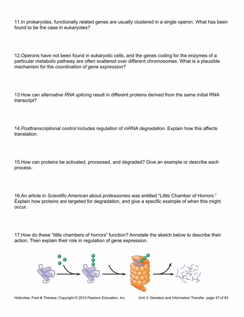
17.How do these “little chambers of horrors” function? Annotate the sketch below to describe their action. Then explain their role in regulation of gene expression.
Holtzclaw, Fred & Theresa; Copyright © 2010 Pearson Education, Inc. Unit 3: Genetics and Information Transfer page 57 of 83

Concept 18.3 Noncoding RNAs play multiple roles in controlling gene expression1.It is now known that much of the RNA that is transcribed is not translated into protein. these RNAs are called noncoding RNAs. Read carefully to discern a crucial role played by these RNAs. What is this role?
2.One of the noncoding RNAs that regulate gene expression is microRNA. On the sketch below, follow an RNA loop, called a “hairpin,” from its creation. Explain the two modes of action of microRNAs. Be sure to label the location of hydrogen bonds and Dicer.
3.B.2 A variety of intercellular and intracellular signal transmissions mediate gene expression. 11.1, 11.4, 18.1, 18.2, 18.3, 18.4Concept 11.1 External signals are converted into responses within the cell1.What is a signal transduction pathway?
2.How does yeast mating serve as an example of a signal transduction pathway?
Holtzclaw, Fred & Theresa; Copyright © 2010 Pearson Education, Inc. Unit 3: Genetics and Information Transfer page 58 of 83

3.Complete the chart of local chemical signaling in cell communication in animals.
4.How does a hormone qualify as a long-distance signaling example?
5.A signal transduction pathway has three stages. Use Figure 11.6 to label the missing parts of the preview figure below, and then explain each step.
Reception
Transduction
Response
Concept 11.4 Response: Cell signaling leads to regulation of transcription or cytoplasmic activities1.When cell signaling causes a response in the nucleus, what normally happens?
2.When cell signaling causes a response in the cytoplasm, what normally happens?
Holtzclaw, Fred & Theresa; Copyright © 2010 Pearson Education, Inc. Unit 3: Genetics and Information Transfer page 59 of 83

3.Figure 11.15 shows a single molecule of epinephrine resulting in the formation of __________ molecules of glucose-1-phosphate!
4.Figure 11.17 shows four different cellular results from a single signaling molecule. Briefly describe each response.
Cell A
Cell B
Cell C
Cell D
5.How do scaffolding proteins enhance a cellular response?
Concept 18.4 A program of differential gene expression leads to the different cell types in a multicellular organismThis concept deals with the regulation of gene expression in development. Animal development is also discussed in Chapter 47.1.What three processes lead to the transformation of a zygote into the organism?
2.Explain what occurs in cell differentiation and morphogenesis.
3.Differential gene expression results from different activators in different cells. How do different sets of activators come to be present in two cells? Explain how each of these occurs:
a.distribution of cytoplasmic determinants
b.different inductive signals
Holtzclaw, Fred & Theresa; Copyright © 2010 Pearson Education, Inc. Unit 3: Genetics and Information Transfer page 60 of 83

3.What is meant by determination? Explain what this means within an embryonic cell.
4.What process ensures that all the tissues and organs of an organism are in their characteristic places? Where do the molecular cues that control this process arise?
5.What is controlled by homeotic genes?
3.C.1 Changes in genotype can result in changes in phenotype. 15.4,16.2 17.5 23.4Concept 15.4 Alterations of chromosome number or structure cause some genetic disorders1.What occurs in nondisjunction?
2.Explain each of the following terms:
aneuploidy
monosomy
trisomy
polyploidy
3.Which of these events results in Down syndrome? What are four characteristics of Down syndrome?
Holtzclaw, Fred & Theresa; Copyright © 2010 Pearson Education, Inc. Unit 3: Genetics and Information Transfer page 61 of 83

4.For each of the following human aneuploidies, give the sex of the individual as well as any physical manifestation of the syndrome.
5.Chromosome structure can be altered in several ways. Label each type of alteration shown in this figure, and explain what occurs.
deletion
duplication
inversion
translocation
Concept 17.5 Point mutations can affect protein structure and function1.Define a mutation in terms of molecular genetics.
2.Define point mutations.
3.What are frameshift mutations?
Holtzclaw, Fred & Theresa; Copyright © 2010 Pearson Education, Inc. Unit 3: Genetics and Information Transfer page 62 of 83

4.Identify two mechanisms by which frameshifts may occur.
5.What is the difference between a nonsense and missense mutation?
6.How can a base-pair substitution result in a silent mutation?
7.What are the two categories of mutagens?
8.Describe the action of difference types of chemical mutagens.
Concept 23.4 Natural selection is the only mechanism that consistently causes adaptive evolution1.In evolutionary terms, fitness refers only to the ability to leave offspring and contribute to the gene pool of the next generation. It may have nothing to do with being big, or strong, or aggressive. Define relative fitness.
2.What is the relative fitness of a sterile mule? _______________________________________
3.Figure 23.13 is important because it helps explain the three modes of selection. Label each type of selection, and fill in the chart to explain what is occurring.
Holtzclaw, Fred & Theresa; Copyright © 2010 Pearson Education, Inc. Unit 3: Genetics and Information Transfer page 63 of 83

4.What is often the result of sexual selection?
5.What is the difference between intrasexual selection and intersexual selection? Give an example of each type of selection.
6.Explain two ways in which genetic variation is preserved in a population.
7.Discuss what is meant by heterozygote advantage, and use sickle-cell anemia as an example.
8.Finally, give four reasons why natural selection cannot produce perfect organisms.
Holtzclaw, Fred & Theresa; Copyright © 2010 Pearson Education, Inc. Unit 3: Genetics and Information Transfer page 64 of 83

3.C.2 Biological systems have multiple processes that increase genetic variation. 27.2 13.4Concept27.2 Rapid reproduction,mutation,and genetic recombination promote genetic diversity in prokaryotes1.You should now have some idea why there is so much potential for genetic diversity with bacterial populations. Although mutation is the major source of genetic variation in prokaryotes, listed below are the other three ways variation is introduced. Explain each one.
2.Define transformation. This idea was first described by Frederick Griffith. (You read about his work in Concept 16.1.)
3.What is transduction? What is the vector for this process?
4.Compare and contrast transduction and transformation.
5.What is a sex pilus? What is the F factor? And how are the two related?
Holtzclaw, Fred & Theresa; Copyright © 2010 Pearson Education, Inc. Unit 3: Genetics and Information Transfer page 65 of 83

6.The F factor is an episome. This is a piece of DNA that can be integrated within the main chromosome of the bacterium, or able to exist as an independent plasmid. What is the bacterial cell called:
when the F factor is in plasmid form?
when it lacks an F plasmid?
when it is integrated within the chromosome?
7.What occurs in bacterial conjugation?
8.When a mating bridge forms between an F+ cell and an F– cell and the F plasmid is replicated and transferred, what is the status of the F– cell afterward?
9.What is an Hfr cell?
10.How are Hfr cells created?
11.Summarize the transfer of genetic information from an Hfr cell to an F– cell.
12.An understanding of R plasmids and antibiotic resistance will be important when you do a bacterial transformation lab. What are R plasmids?
Holtzclaw, Fred & Theresa; Copyright © 2010 Pearson Education, Inc. Unit 3: Genetics and Information Transfer page 66 of 83
Concept 13.4 Genetic variation produced in sexual life cycles contributes to evolution1.An important idea for you to understand is that new alleles arise by changes in the DNA or mutation, but genetic diversity occurs when the deck that is dealt is simply reshuffled. So, there are three ways that sexually reproducing organisms “shuffle the deck.” They are listed below. Explain what occurs in each, and how this increases diversity.
independent assortment of chromosomes
crossing over
random fertilization
2.Here is a fun exercise to drive this point home. Pull out your calculator, and try your hand at this: When you were conceived, what were the odds that of the many possibilities, your parents would come up with you?
a.The number of different gametes that can be formed because of independent assortment is
2n, where n = the number of homologous pairs
Therefore, since humans have 46 chromosomes or 23 homologous pairs, what is the number of possible gametes that can be formed due to independent assortment of chromosomes?
b.Now, this is the number of unique gametes your mom could have made. Your father could have made the same number. To see the effect of random fertilization, multiply the number of gametes one parent could make by the number of unique gametes the other parent could make.
Your answer should be in the trillions, and all of this is without crossing over. See how special you are?
3.C.3 Viral replication results in genetic variation, and viral infection can introduce genetic variation into the hosts. 19.1, 19.2Concept 19.1 A virus consists of a nucleic acid surrounded by a protein coat1.What was some early evidence of the existence of viruses? Why were they difficult to study?
2.What was Wendell Stanley’s contribution to our knowledge of viruses?
Holtzclaw, Fred & Theresa; Copyright © 2010 Pearson Education, Inc. Unit 3: Genetics and Information Transfer page 67 of 83

3.What are the four forms of viral genomes?
4.What is a capsid? What are capsomeres? What different shapes may capsids have?
5.As you see, all viruses consist of a nucleic acid enclosed in a protein coat. Some viruses also have a membranous envelope. What are the components of a viral envelope? Which component is derived from the host cell, and which is of viral origin?
6.What is the role of an envelope in animal viruses?
7.For the virus shown below, label the protein capsid, tail fibers, head, tail sheath, and genome.
a.What type of virus is this?
b.What does its name mean?
c.What is its host?
d.Is the genome of this virus DNA, or RNA?
Holtzclaw, Fred & Theresa; Copyright © 2010 Pearson Education, Inc. Unit 3: Genetics and Information Transfer page 68 of 83

3.D.1 Cell communication processes share common features that reflect a shared evolutionary history. 11.1, 11.2
Concept 11.2 Reception: A signal molecule binds to a receptor protein, causing it to change shape1.Explain the term ligand. (This term is not restricted to cell signaling. You will see it in other situations during the year.)
2.The text will explain three major types of membrane receptors in Figure 11.7. This material is of fundamental importance, so we will work thorough the specific figures for each type of membrane receptor. The first example is a G protein-linked receptor. In the first figure, label the components and then describe the role of the three components.
3.Label and then describe what happens in step 2.
4.Label then describe what happens in step 3. (The yellow box at the bottom right is important!)
5.Equally important to starting a signal is stopping a signal. Step 4 stops the signal. (Failure to do so can lead to serious problems, like cancer.) Label and then describe how the signal is halted.
Holtzclaw, Fred & Theresa; Copyright © 2010 Pearson Education, Inc. Unit 3: Genetics and Information Transfer page 69 of 83

6.What activates a G protein?
7.A G protein is also a GTPase enzyme. Why is this important?
8.The second type of receptor described is receptor tyrosine kinase. Explain what a kinase enzyme does.
9.How does tyrosine kinase function in the membrane receptor?
10.What is a key difference between receptor tyrosine kinases and G protein-coupled receptors?
11.Provide all of the missing labels on the diagram; then explain what happens in step 1.
12.Label step 2 and then describe what happens to receptors tyrosine kinases when signaling molecules have attached.
Holtzclaw, Fred & Theresa; Copyright © 2010 Pearson Education, Inc. Unit 3: Genetics and Information Transfer page 70 of 83

13.Label and explain how the receptors are activated in step 3.
14.Use step 4 to explain how the activated receptor can stimulate multiple cellular response pathways.
15.Each activated protein in the figure above triggers a signal _________________ pathway leading to a ______________ response.
16.Moving to ion channel receptors, the example in Figure 11.7 shows the flow of ions into the cell. Ion channel receptors can also stop the flow of ions. These comparatively simple membrane receptors are explained in three steps. In the first step, label the diagram and then explain the role of the labeled molecules.
17.Label the diagram and then explain what has happened with the binding of the ligand to the receptor.
Holtzclaw, Fred & Theresa; Copyright © 2010 Pearson Education, Inc. Unit 3: Genetics and Information Transfer page 71 of 83

18.The ligand attachment to the receptor is brief. Label and explain what happens as the ligand dissociates.
19.In what body system are ligand-gated ion channels and voltage-gated ion channels of particular importance?
20.Intracellular receptors are found in the _______________ or _______________ of the cell, where they bond to chemical messengers that are ___________________ or very small, like nitric oxide.
21.This diagram uses testosterone, a hydrophobic hormone, to detail how intracellular receptors work. At each arrow, add an explanation of what is happening in the cell.
22.An important idea, transcription factors, is introduced in Figure11.8. Explain the function of transcription factors in the cell.
Holtzclaw, Fred & Theresa; Copyright © 2010 Pearson Education, Inc. Unit 3: Genetics and Information Transfer page 72 of 83

3.D.2 Cells communicate with each other through direct contact with other cells or from a distance via chemical signaling. 11.1, 11.2
3.D.3. Signal transduction pathways link signal reception with cellular response. 11.3Concept 11.3 Transduction: Cascades of molecular interactions relay signals from receptors to target molecules in the cell1.What are two benefits of multistep pathways like the one in Figure 11.9?
2.Explain the role of these two categories of enzymes in transduction.
Protein kinase
Protein phosphatases
3.Using Figure 11.9 as your guide, explain what is occurring in the cell at each arrow.
4.What is the difference between a first messenger and a second messenger?
Holtzclaw, Fred & Theresa; Copyright © 2010 Pearson Education, Inc. Unit 3: Genetics and Information Transfer page 73 of 83

5.Two common second messengers are cyclic AMP (cAMP) and calcium ions (Ca2+). Explain the role of the second messenger cAMP in Figure 11.11 from the text.
6.What is the important relationship between the second messenger and protein kinase A?
7.Figure 11.11 explains how to initiate a cellular response; how might that response be inhibited?
8.Using your new knowledge of cell signaling, explain the mechanism of disease in cholera.
9.List three types of pathways often induced by calcium ions.
10.What happens to the cytoplasmic concentration of calcium when it is used as a second messenger?
3.D.4. Changes in signal transduction pathways can alter cellular response. 11.4
3.E.1. Individuals can act on information and communicate it to others. 51.1Concept 51.1 Discrete sensory inputs can stimulate both simple and complex behaviors
1.How is behavior defined?
2.What is ethology?
Holtzclaw, Fred & Theresa; Copyright © 2010 Pearson Education, Inc. Unit 3: Genetics and Information Transfer page 74 of 83

3.What is the difference between proximate and ultimate causation?
4.Using red-crowned cranes, what is an example of a proximate causation question and an example of an ultimate causation question?
5.Who are the three ethologists who shared in a Nobel Prize for their work in 1973? We will look at work by each of them.
6.What is a fixed action pattern (FAP)? Give an example.
7.What is a sign stimulus? Give at least examples of sign stimuli.
8.Nicholas Tinbergen’s work with the stickleback fish is a classic study. Explain what he found. Use the terms fixed action pattern and sign stimulus in your response.
9.Define these behavior terms:
Holtzclaw, Fred & Theresa; Copyright © 2010 Pearson Education, Inc. Unit 3: Genetics and Information Transfer page 75 of 83

10.Explain what is meant by a circadian clock and circadian rhythms. Identify two behaviors, either plant or animal, that demonstrate a circadian rhythm. (You may need to refer to Chapter 49 or Chapter 36 for examples.)
11.Discuss two navigational strategies used by birds to migrate.
12.Animals communicate in various ways. Discuss at least three specific examples using different organisms.
13.Notice the pictures that show fruit fly courtship behavior (see AP Biology Lab 11B, “Reproductive Behavior in Fruit Flies”). What different modes of communication are used by the fruit fly?
14.Karl von Frisch studied European honeybees. What are the two types of dances that a returning worker bee does, and what information does each dance convey? Use a labeled sketch to describe each dance.
15.What are pheromones? Give three specific types of information that can be transmitted through pheromones.
Holtzclaw, Fred & Theresa; Copyright © 2010 Pearson Education, Inc. Unit 3: Genetics and Information Transfer page 76 of 83

3.E.2. Animals have nervous systems that detect external and internal signals, transmit and integrate information, and produce responses. 48.1, 48.2, 48.3, 48.4 49.2Concept 48.1 Neuron organization and structure reflect function in information transfer1.What is a neuron?
2.Neurons can be placed into three groups, based on their location and function.
3.Which division of the nervous system includes the brain and spinal cord?
4.This sketch shows two neurons. Label the following elements of this figure: cell body, dendrites, axon, synapse, presynaptic cell, postsynaptic cell, synaptic vesicles, synaptic terminal, and neurotransmitter.
Holtzclaw, Fred & Theresa; Copyright © 2010 Pearson Education, Inc. Unit 3: Genetics and Information Transfer page 77 of 83

5.What is shown in the box above? What do the red spheres represent?
6.What is indicated by the red arrows in the main figure?
7.What are glial cells?
Concept 48.2 Ion pumps and ion channels maintain the resting potential of a neuronIn this section you will need to recall information about the structure and function of the plasma membrane. Ions are not able to diffuse freely through the membrane, because they are charged and so must pass through protein channels specific for each ion.1.All cells have a membrane potential across their plasma membrane. What is the typical resting potential of a neuron?
2.On the sketch below, label the following: outside cell, inside cell. Show where the concentrations of Na+ and K+ are highest.
3.How are the concentration gradients of Na+ and K+ maintained?
Concept 48.3 Action potentials are the signals conducted by axons1.As you see in the figure above, in a resting neuron, the outside of the membrane is positively charged relative to the inside of the membrane. If positively charged ions flow out, the difference in charge between the two sides of the membrane becomes greater. What is the increase in the magnitude of the membrane potential called?
Holtzclaw, Fred & Theresa; Copyright © 2010 Pearson Education, Inc. Unit 3: Genetics and Information Transfer page 78 of 83

2.When a stimulus is applied, ion channels will open. If positively charged ions flow n,the membrane is said to depolarize. If depolarization causes the membrane potential to drop to a critical value, a wave of depolarization will follow. What is this critical value called?
3.What is the wave of depolarization called?
4.Just like toppling dominoes in a row, either the threshold of depolarization will be reached and an action potential will be generated, or the threshold will not be reached and no wave will occur. What is this response to stimulus called?
5.Figure 48.10 contains almost all you need to know about nerve impulse transmission, so it is worth some careful study time. Let’s approach it in steps.
a.Label Na+, K+, and their respective ion channels.b.Label the Resting state figure. Are the Na+ and K+ channels open, or closed? c.Label Depolarization. What triggers depolarization? What channels open? What occurs if the depolarization threshold is reached? d.Label Stage 4 in the figure Repolarization. How is the charge on the membrane reestablished? e.Label these regions of the graph: x- and y-axes, threshold, resting potential, depolarization, action potential, and repolarization.
Holtzclaw, Fred & Theresa; Copyright © 2010 Pearson Education, Inc. Unit 3: Genetics and Information Transfer page 79 of 83

f.Let’s see if you really understand this concept. Draw in another line on the graph to show what the change in membrane potential would look like if a stimulus were applied that did not reach the depolarization threshold.
6.Here is a closer look at what is happening along the membrane as a wave of depolarization (an action potential) travels along the length of the axon. Label the key elements of the figure; and to the right, explain how the action potential is conducted.
7.What are the two types of glial cells that produce myelin sheaths?
8.How does a myelin sheath speed impulse transmission? Use the figure below, and include a discussion of saltatory conduction and nodes of Ranvier in your response.
9.In the disease multiple sclerosis, the myelin sheaths harden and deteriorate. How would this affect nervous system function? Holtzclaw, Fred & Theresa; Copyright © 2010 Pearson Education, Inc. Unit 3: Genetics and Information Transfer page 80 of 83

Concept 48.4 Neurons communicate with other cells at synapses1.When the wave of depolarization arrives at the synaptic terminal, calcium ion channels open. What occurs to the synaptic vesicles as the Ca2+ level increases?
2.What is contained within the synaptic vesicles?
3.Label the figure below: synaptic vesicle, neurotransmitter, calcium ion channel, presynaptic membrane, postsynaptic membrane, and synapse.
4.Explain how an action potential is transmitted from one cell to another across a synapse by summarizing what is shown above in six steps.
(1)
(2)
(3)
(4)
(5)
(6)
Holtzclaw, Fred & Theresa; Copyright © 2010 Pearson Education, Inc. Unit 3: Genetics and Information Transfer page 81 of 83

5.There are many different types of neurotransmitters. Each neuron secretes only one type of neurotransmitter. Some neurotransmitters hyperpolarize the postsynaptic membrane. Are these excitatory or inhibitory neurotransmitters?
6.Define and explain summation.
7.A single postsynaptic neuron can be affected by neurotransmitter molecules released by many other neurons, some releasing excitatory and some releasing inhibitory neurotransmitters. What will determine whether an action potential is generated in the postsynaptic neuron?
8.Table 48.1 lists several of the major neurotransmitters. You are not expected to know their actions or secretion sites, but you should recognize that they are neurotransmitters! Go through the list that follows, and say each term aloud. Put a checkmark by any that you have heard mentioned before: acetylcholine, epinephrine, norepinephrine, dopamine, serotonin, GABA, glutamate, glycine, substance P, endorphins, and nitric oxide. That’s all for this question!
9.There is one neurotransmitter we want you to memorize. It is the most common neurotransmitter in both vertebrates and invertebrates, and it is released by the neurons that synapse with muscle cells at the neuromuscular junction. If you look ahead to Chapter 50, Figure 50.29, you will see a synapse between a neuron and a muscle cell, resulting in depolarization of the muscle cell and its contraction. What is this very important neurotransmitter?
Holtzclaw, Fred & Theresa; Copyright © 2010 Pearson Education, Inc. Unit 3: Genetics and Information Transfer page 82 of 83

Concept 49.2 The vertebrate brain is regionally specialized1.Label the following structures on the figure of the brain, and give a primary function of each labeled structure.
brainstem (includes pons, medulla)
cerebrum
cerebellum
thalamus
hypothalamus
2.Label the pituitary gland on this figure.
Holtzclaw, Fred & Theresa; Copyright © 2010 Pearson Education, Inc. Unit 3: Genetics and Information Transfer page 83 of 83