Embed Size (px)
Citation preview

Unit 3 Biology

succeeding in the vce, 2017
important notes Our policy at TSFX is to provide students with the most detailed and comprehensive set of notes that will maximise student performance and reduce study time. These materials, therefore, include a wide range of questions and applications, all of which cannot be addressed within the available lecture time i.e. Due to time constraints; it is possible that some of the materials included in this booklet will not be addressed during the course of these lectures.
Where applicable, fully worked solutions to the questions in this booklet will be handed to students on the last day of each subject lecture. Although great care is taken to ensure that these materials are mistake free, an error may appear from time to time. If you believe that there is an error in these notes or solutions, please let us know asap ([email protected]). Errors, as well as clarifications and important updates, will be posted at www.tsfx.com.au/vce-updates.
The views and opinions expressed in this booklet and corresponding lecture are those of the authors/lecturers and do not necessarily reflect the official policy or position of TSFX.
These materials are the copyright property of The School For Excellence and have been produced for the exclusive use of students attending this program. Reproduction of the whole or part of this document constitutes an infringement in copyright and can result in legal action. No part of this publication can be reproduced, copied, scanned, stored in a retrieval system, communicated, transmitted or disseminated, in any form or by any means, without the prior written consent of The School For Excellence (TSFX). The use of recording devices is STRICTLY PROHIBITED. Recording devices interfere with the microphones and send loud, high-pitched sounds throughout the theatre. Furthermore, recording without the lecturer’s permission is ILLEGAL. Students caught recording will be asked to leave the theatre, and will have all lecture materials confiscated.
it is illegal to use any kind of recording device during this lecture
extract from the master class teaching materials Our Master Classes form a component of a highly specialised weekly program, which is designed to ensure that students reach their full potential (including the elite A and A+ scores). These classes incorporate the content and teaching philosophies of many of the top schools in Victoria, ensuring students are prepared to a standard that is seldom achieved by only attending school. These classes are guaranteed to motivate students and greatly improve VCE scores! For additional information regarding the Master Classes, please do not hesitate to contact us on (03) 9663 3311 or visit our website at www.tsfx.com.au.
essential for all year 11 and 12 students!
copyright notice
TSFX - voted number one for excellence and quality in VCE programs.

EDITORIAL NOTE Due to the requests of many students, some concepts and topic areas have been retained from earlier Unit 3 Biology Study Designs. The detail of these areas has generally been moved to other units of Biology, i.e. Units 1, 2 or 4, and is no longer part of the core Unit 3 Biology curriculum. The knowledge and understanding of these areas is still recommended, and they are denoted with an asterisk (*) throughout the booklets.

© The School For Excellence 2017 Succeeding in the VCE – Unit 3 Biology Page 1
DEFENCE AGAINST DISEASE The body possesses a variety of defence mechanisms in order to protect itself against the wide diversity of invading pathogens and foreign matter. The defence mechanisms involve natural barriers (e.g. general mechanisms such as the skin, stomach acid, mucous membranes etc.), innate (non-specific) immune responses (e.g. the action of white blood cells – such as. dendritic cells – and proteins which non-specifically attack a variety of pathogens), or adaptive (specific) immune responses (e.g. T and B lymphocytes and antibodies) in which the response specifically targets one type of pathogen.
Natural Barriers Innate (Non-specific) Defence Mechanisms
Adaptive (Specific) Defence Mechanisms
First Line Second Line Third Line
Phagocytic white blood cells and natural killer (NK) cells
Complement proteins
Interferons
Inflammation response
Temperature response
dv.com

© The School For Excellence 2017 Succeeding in the VCE – Unit 3 Biology Page 2
NATURAL BARRIERS TO DISEASE As shown below, there are a variety of access points for pathogens to penetrate the internal environment of the human body:
A variety of physical and chemical barriers form the first line of defence. No memory of the pathogen is formed after any invasion. Chemical barriers
Acids: e.g. those found in stomach, urine and vagina.
Enzymes: Found in saliva (amylase) and tears (lysozyme which digests bacteria).
Mucus: Traps microorganisms.
Sebum: Secreted from sebaceous glands, contains antiseptics.
Sweat: An acidic, dehydrating repellent. Mechanical or physical barriers
Intact skin: Composed of tightly packed cells forming a tough, waterproof layer.
Mucous membranes: Line internal surfaces such as those found in reproductive and respiratory tracts that are exposed to air.
Cilia: Filter air passing through respiratory system. Beat in unison to guide pathogens trapped in mucus up towards the nose/mouth.
Nasal hairs: Filter inhaled air. Microbiological barriers
The gut flora (microorganism) community plays a direct role in defending against pathogens by fully colonising the space, making use of all available nutrients, and by secreting compounds that kill or inhibit pathogens that would compete for nutrients. Alteration of this flora with antibiotics can allow overgrowth of pathogenic microorganisms (e.g. Salmonella typhimurium) or superinfection with ordinarily commensal organisms (e.g. Candida albicans).
thefutureofthings.com
(Kinnear J & M Martin, 2000)
© The School For Excellence 2017 Succeeding in the VCE – Unit 3 Biology Page 3
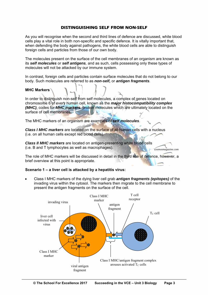
DISTINGUISHING SELF FROM NON-SELF As you will recognise when the second and third lines of defence are discussed, white blood cells play a vital role in both non-specific and specific defence. It is vitally important that, when defending the body against pathogens, the white blood cells are able to distinguish foreign cells and particles from those of our own body. The molecules present on the surface of the cell membranes of an organism are known as its self molecules or self antigens, and as such, cells possessing only these types of molecules will not be attacked by our immune system. In contrast, foreign cells and particles contain surface molecules that do not belong to our body. Such molecules are referred to as non-self, or antigen fragments. MHC Markers In order to distinguish non-self from self molecules, a complex of genes located on chromosome 6 of every human cell, known as the major histocompatibility complex (MHC), codes for MHC markers, protein molecules which are ultimately located on the surface of cell membranes. The MHC markers of an organism are examples of self molecules. Class I MHC markers are located on the surface of all human cells with a nucleus (i.e. on all human cells except red blood cells). Class II MHC markers are located on antigen-presenting white blood cells (i.e. B and T lymphocytes as well as macrophages). The role of MHC markers will be discussed in detail in the third line of defence, however, a brief overview at this point is appropriate. Scenario 1 – a liver cell is attacked by a hepatitis virus: Class I MHC markers of the dying liver cell grab antigen fragments (epitopes) of the
invading virus within the cytosol. The markers then migrate to the cell membrane to present the antigen fragments on the surface of the cell.
cosmosmagazine.com
invading virus
Class I MHC marker
liver cell infected with
virus
antigen fragment
T cell receptor
TC cell
Class I MHC/antigen fragment complex arouses activated TC cells
Class I MHC marker
viral antigen fragment

© The School For Excellence 2017 Succeeding in the VCE – Unit 3 Biology Page 4
Scenario 2 – a macrophage hunts down and engulfs a bacterium: Class II MHC markers grab antigen fragments of the engulfed bacterium. The markers
then migrate to the cell membrane to present the antigen fragments on the surface of the cell.
As will be discussed in full later, white blood cells of the third line treat a Class I MHC marker-antigen complex in a different manner to that of a Class II MHC marker-antigen complex. MHC markers are also responsible for tissue/organ rejection. It is in recognising the presence/absence of antigens that white blood cells will/will not initiate an immune response to the presence of a particular cell or substance.
A macrophage engulfs E coli bacteria on the outer surface of a blood vessel in the lung pleural cavity. It will present E coli
fragments on its surface using Class II markers
visualsunlimited.com
bacterium
Class II MHC marker
macrophage which has ingested bacterium
antigen fragment
T cell receptor
TH cell
Interleukin-1 activates TH cells

© The School For Excellence 2017 Succeeding in the VCE – Unit 3 Biology Page 5
SOURCE OF BLOOD CELLS All blood cells (red and white), as well as platelets, are produced in the bone marrow of the long bones of the body. Only one group of blood cells, the T cells, leave the bone marrow in an immature state. T cells migrate to the thymus gland, which is their site of maturation.
Macrophage consuming leukemic cells visualsunlimited.com
studyblue.com
*B lymphocytes *T lymphocytes *Natural killer cells
Macrophages & Dendritic C.
© The School For Excellence 2017 Succeeding in the VCE – Unit 3 Biology Page 6
THE SECOND LINE OF DEFENCE: INNATE (NON-SPECIFIC) If the first line of defence has failed, the second line of defence is employed. It involves innate responses to the presence of foreign particles in the internal environment of the organism. This line of defence cannot distinguish one type of micro-organism from another. No memory to pathogen is formed at the conclusion of the battle, i.e. this response is the same no matter how often a person is infected with the same pathogen.
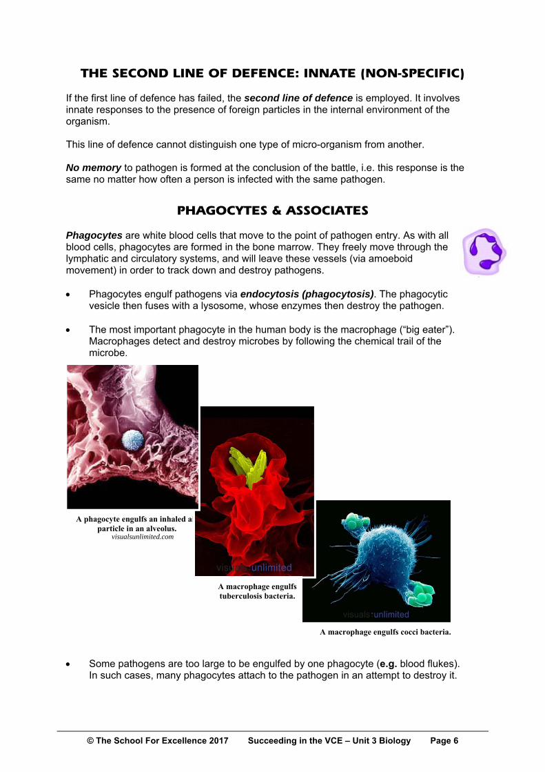
PHAGOCYTES & ASSOCIATES Phagocytes are white blood cells that move to the point of pathogen entry. As with all blood cells, phagocytes are formed in the bone marrow. They freely move through the lymphatic and circulatory systems, and will leave these vessels (via amoeboid movement) in order to track down and destroy pathogens. Phagocytes engulf pathogens via endocytosis (phagocytosis). The phagocytic
vesicle then fuses with a lysosome, whose enzymes then destroy the pathogen. The most important phagocyte in the human body is the macrophage (“big eater”).
Macrophages detect and destroy microbes by following the chemical trail of the microbe.
Some pathogens are too large to be engulfed by one phagocyte (e.g. blood flukes). In such cases, many phagocytes attach to the pathogen in an attempt to destroy it.
A phagocyte engulfs an inhaled air particle in an alveolus.
A macrophage engulfs tuberculosis bacteria.
A macrophage engulfs cocci bacteria.
visualsunlimited.com

© The School For Excellence 2017 Succeeding in the VCE – Unit 3 Biology Page 7
The presence or absence of an antigen on the surface of a foreign particle or organism (e.g. pollen grain, bacteria, roundworm, etc.) will determine whether or not a phagocyte will attack it.
After ingesting a pathogen, a macrophage will proceed to display the pathogen’s antigens on its own cell membrane in order to arouse recognition and further action on the part of the immune system.
Other white blood cells which ingest foreign particles include eosinophils and
basophils. As with basophils, eosinophils are involved in allergic responses. In addition to containing histamine, basophils contain the anti-clotting agent heparin.
Not only phagocytic, neutrophils, the most common white blood cell, also release
chemicals designed to kill bacteria. Natural killer (NK) cells operate in a different manner to the white blood cells that ingest foreign particles. Instead, they attack and kill viral-infected cells of the body in addition to directly killing cancer cells.
It is only in recent years that the
significant defensive role played by dendritic cells has become more apparent. They play a key role in processing antigen material and presenting it on their surface to other cells of the immune system, e.g. lymphocytes of the third line. Dendritic cells are present in small quantities in tissues that are in contact with the external environment, mainly the skin and the inner lining of the nose, lungs, stomach and intestines. Once activated, dendritic cells migrate to the lymphoid tissues where they interact with T cells and B cells of the third line of defence to initiate the appropriate immune response.
Two natural killer cells attack a cancer cell
thejustinwayneshow.com
Dendritic cells clearly revealing their dendrite-like extensions.

© The School For Excellence 2017 Succeeding in the VCE – Unit 3 Biology Page 8
PROTEINS THAT KILL OR IMPEDE INVADING MICROBES COMPLEMENT Complement proteins are a group of proteins that assist phagocytes in recognising the presence of pathogens. They continually circulate in blood plasma.
A particular group of proteins, called antibodies (discussed later in the booklet), circulate in the bloodstream, and they will attach to specific invading microorganisms. Complement proteins then attach to the antibodies on the invading micro-organisms (mainly bacteria) and act as flags, making the pathogen more readily identifiable to the immune system, and thus attracting phagocytes to the site of infection.
Complement proteins lyse the cell membranes of bacteria and fungi, and the leaking
cell contents further stimulate phagocytic action.
INTERFERON Interferon is a chemical that is secreted by viral infected cells. The release of interferon makes adjacent uninfected cells more resistant to the virus,
thus reducing their chances of infection. It triggers these cells to make particular enzymes which prevent the virus from synthesising more copies of itself inside the cell.
Interferon can reduce the success rates of viruses that do not have far to travel to a
host cell, e.g. common cold and influenza viruses.
(Campbell NA & JB Reece, 2002)

© The School For Excellence 2017 Succeeding in the VCE – Unit 3 Biology Page 9
INFLAMMATION Inflammation is a reaction to an infection whereby histamines, released from mast cells (a type of WBC lining the walls of blood vessels), cause local arterioles to expand, thus drawing more blood to the region. Phagocytes are carried with the blood to the area of infection. Under the influence of histamine, capillaries become highly permeable, so
macrophages and neutrophils pass easily from the bloodstream to the infected tissues.
Preliminary phagocytes that move to the area release histamine to attract more
phagocytes. Neutrophils (a type of phagocyte) also release chemicals which kill bacteria, and
macrophages clean up the mess.
inflammation.jpg
breakingmuscle.com

© The School For Excellence 2017 Succeeding in the VCE – Unit 3 Biology Page 10
TEMPERATURE RESPONSE If an invader has been detected in the body in considerable numbers, macrophages release a cytokine* called interleukin-1. Interleukin-1 travels to the brain, where it resets the hypothalamus, resulting in a fever. The invading microbes do not reproduce as well at the higher body temperature, giving the white blood cells, which perform well at the higher body temperature, an improved chance of overcoming them.
* Cytokines are a group of signalling molecules, typically protein, peptide or glycoprotein in structure that, as with hormones and neurotransmitters, are used extensively in communication between cells.)

© The School For Excellence 2017 Succeeding in the VCE – Unit 3 Biology Page 11
THE LYMPHATIC SYSTEM The lymphatic system plays a vital role in defending the human body against the myriad of pathogens it can be exposed to during a lifetime. It is a system of vessels and lymph nodes which, although separate from the circulatory system, does return fluid and protein to the blood. Lymph, blood plasma and interstitial fluid (fluid surrounding cells) are exchanged at all times. Fluid and plasma proteins enter the lymphatic system via tiny blind-ending lymph
capillaries. This fluid, once within the lymphatic system, is referred to as lymph. It is similar in composition to interstitial fluid.
Lymph capillaries merge to form increasingly larger vessels which ultimately drain
lymph back into the bloodstream at the vena cavae (the large veins returning blood to the heart from the body).
As with veins, lymph vessels contain
valves that prevent the backflow of fluid. This is because there is a one-way flow of lymph within the body. Hence, the squeezing of fluid within the lymphatic system towards the heart is largely dependent on movement of surrounding skeletal muscles of the body.
Along the length of lymph vessels are
sections known as lymph nodes. Inside each node are sections filled with white blood cells which filter the lymph passing through.
Lymph nodes swell during an infection, as the particular type of white blood cell responsible for fighting the infection multiplies rapidly in order to overcome and destroy the invader, typically a virus or bacterium. You may have noticed swollen nodes in your neck or tonsils during the course of an infection.
As well as lymph nodes located in various parts of the body, the spleen, tonsils and
appendix are also parts of the lymphatic system.
(Beckett, BS, 1979) sciencephoto.com

© The School For Excellence 2017 Succeeding in the VCE – Unit 3 Biology Page 12
PRIMARY LYMPHOID TISSUE Primary lymphoid organs generate lymphocytes from immature progenitor cells. The thymus (a gland in the chest), and the bone marrow comprise the primary lymphoid organs involved in the production and development of the cells of the immune system. Bone marrow is responsible for both the creation of T cells and the production and
maturation of B cells. From the bone marrow, B cells immediately join the circulatory system and travel to secondary lymphoid organs in search of pathogens.
T cells, on the other hand, travel from the bone marrow to the thymus where they develop further. Mature T cells join B cells in search of pathogens. The other 95% of T cells begin a process of apoptosis.
SECONDARY LYMPHOID TISSUE Secondary lymphoid organs, which include lymph nodes and the spleen, maintain mature naive lymphocytes and initiate an adaptive immune response. The secondary lymphoid organs are the sites of lymphocyte activation by antigens.
Secondary lymphoid tissue provides the environment for the foreign molecules (antigens) to be recognised by the lymphocytes. It is exemplified by the lymph nodes, and the lymphoid follicles in tonsils.
Lymph nodes are the key sites of lymph filtration, where antigens are screened. There are approximately 500 – 700 lymph nodes within a human body, and each node contains thousands of white blood cells. they are predominantly located in the neck, and chest, underarms, groin and intestines.
Activation leads to rapid mitosis (clonal expansion) of B and T cells. Mature lymphocytes recirculate between the blood and the secondary lymphoid organs until they encounter their specific antigen.
Blind-ending lymph capillary (above)
Lymph node (below)
(Campbell NA & JB Reece, 2000)

© The School For Excellence 2017 Succeeding in the VCE – Unit 3 Biology Page 13
THE THIRD LINE OF DEFENCE — ADAPTIVE (SPECIFIC) If the first and second lines of defence have not prevented an infection, specific immunity, involving the action of B cells (including their production of antibodies) and T cells, will come into play. The third line of defence involves: An adaptive (specific) response for each
type of infection. A memory of the pathogen resulting from
the infection. B cells mature in the bone marrow.
T cells mature in the thymus gland.
Other non-specific white blood cells (WBCs) assist the B cells and T cells in their operation. WBCs are continually circulating in the blood and lymphatic system acting as mobile defence forces for our body. Swollen lymph glands and the presence of white pus near sores are both clear signs of WBC activity. B Lymphocytes (B cells) and T lymphocytes (T cells) are highly specific in action:
Different varieties of T lymphocytes have the roles of mobilising the entire specific response to disease, destroying viral infected cells, foreign eukaryotic cells and cancer cells, and controlling the length of the immune response.
Each type of B lymphocyte secretes one specific type of antibody, which will specifically bind to one type of non-self antigen.
Approximately 80% of all lymphocytes are T cells and 20% are B cells.
Antigens & Antibodies Antibodies are proteins produced by B cells which specifically react with the antigens that caused their formation. We will discuss them shortly.
www.buzzle.com

© The School For Excellence 2017 Succeeding in the VCE – Unit 3 Biology Page 14
PRIMARY RESPONSE The primary response of the third line of defence occurs after first contact with a pathogen. It is slow to develop and the individual usually suffers some of the symptoms of the disease.
A macrophage captures the pathogen and advertises some of its antigens on its own cell membrane. The antigen fragment is displayed on a class II MHC marker of the macrophage. The macrophage also releases interleukin-1 in order to attract helper T cells. The helper T cells release interleukin-2 in order to activate B cells, cytotoxic T cells and more helper T cells.
A human macrophage on the move in search of foreign particles.
The first phase of the primary response: A phagocyte (e.g. a macrophage, dendritic cell etc.) presents an antigen fragment to a helper T cell.
bacterium
Class II MHC marker
macrophage which has ingested bacterium
antigen fragment
T cell receptor
TH cell
Interleukin-1 activates TH cells

© The School For Excellence 2017 Succeeding in the VCE – Unit 3 Biology Page 15
CELL MEDIATED IMMUNITY Cell mediated (cellular) immunity involves the resistance to disease resulting from the action of cells. It involves attacks on pathogens which are inside cells. Four types of T cells involved in cellular immunity: helper (TH) cells, cytotoxic (TC) cells, *regulatory (suppressor) cells and memory T cells. After engulfing an invader, a macrophage displays the
invader’s antigens on its own cell membrane. The activated macrophage is carried to the lymph node where the T cells are accumulated. This improves the chances of finding a matching T cell rapidly.
The macrophage also releases interleukin-1
(a cytokine) to attract the appropriate helper T cells. Using their T cell receptors, helper T cells also recognise the foreign antigens displayed by the class II markers of the macrophage and bind to the antigen. The CD4 receptors of the helper T cell directly bind to the class II MHC marker of the macrophage, and amplify the signal generated by the T cell receptor. Interleukin-1 also generates a fever in the body, which aids in disease resistance.
Once recognition has occurred, helper T cells stimulate other helper T cells, cytotoxic
T cells and B cells specific to that particular disease to become activated (clonal selection). They do this by releasing interleukin-2 (another cytokine). Without notification from helper T cells, cytotoxic T cells and most B cells will remain inactive against the particular pathogen or parasite they are programmed to attack.
Y
bacterium Class II MHC
marker
macrophage which has ingested bacterium
antigen fragment
T cell receptor
TH cell
TC cell
B cell
Interleukin-2 activates TH cells, B cells and TC
cells
Interleukin-1 activates TH cells
CD4 receptor
tcells.org

© The School For Excellence 2017 Succeeding in the VCE – Unit 3 Biology Page 16
Cytotoxic T cells kill body cells that have been infected with a pathogen (e.g. viruses). They use their T cell receptors to recognise the class I MHC marker-antigen complex on the infected cell surface, and their CD8 receptors also bind to the class I MHC marker of the infected cell. The cytotoxic T cell then secretes perforin proteins that punch holes in the membrane of the infected cell. This kills the infected cell, and prevents the further spread of the disease from that cell.
*Once the battle has been successfully fought, the regulatory T cells (formerly called suppressor T cells) turn off the specific immune system (B and T cells) for that disease.
*Regulatory T cells act by: Directly killing immune cells. Inhibiting the secretions of immune cells. Secreting proteins that alter immune cell functions.
viral antigen fragment Class I MHC
marker
antigen-presenting cell (a viral-infected cell)
antigen fragment
T cell receptor
cytotoxic T cell (TC cell)
CD8 receptor
library.thinkquest.org

© The School For Excellence 2017 Succeeding in the VCE – Unit 3 Biology Page 17
Cellular immunity is specific because a T cell surface receptor on a T cell will only attach to one type of antigen. An antigen has to bind to a T cell’s surface receptor in order for the T cell to be stimulated. Summary of the roles of the four classes of T cells: Helper T cells: The most important of all white blood cells, for without their presence,
no specific immune response, from either T cells or B cells, could take place effectively. Once activated by interleukin-1, helper T cells release interleukin-2 which mobilises other T cells and B cells specific to the particular infection. They also release interferon.
Cytotoxic T cells: Recognise and destroy viral infected cells, cancer cells and foreign
eukaryotic cells (e.g. protists and fungi). Cytotoxic T cells lyse infected cells with perforin and release toxins to destroy the cell and its contents. The release of cytokines further stimulates phagocytosis by macrophages. Cytotoxic T cells can also release interferon.
*Regulatory T cells: Regulate the immune response and stop its
activity when the invader has been defeated. Hence, resources are not wasted.
Memory T cells: Retain the ability to quickly recognise foreign
antigens so that a rapid response occurs with subsequent invasions.
T cells only seem to respond to antigens if they are presented by MHC markers on a eukaryotic cell. They are blind to isolated or free antigens. e.g. cytotoxic T cells will not attack free viruses – the viral antigen must be displayed with a class I MHC molecule (self-antigen) to activate a cytotoxic T cell attack. Another group of cytotoxic lymphocytes do exist. Its members, however, seem to exhibit nonspecific immune activity. They are neither T nor B cells. These natural killer cells are not activated by specific MHC-antigen interactions, and so are not specific to one type of pathogen-infected cell. Their mode of operation, however, is otherwise similar to that of cytotoxic T cells. They destroy a variety of viral-infected cells and cancer cells by secreting powerful cytotoxins.
A natural killer cell binds to two tumour cells
conkwest.com

© The School For Excellence 2017 Succeeding in the VCE – Unit 3 Biology Page 18
CELL MEDIATED IMMUNITY IN ACTION
Invasion of body by a pathogen (for example, virus or bacterium) which acts as an antigen.
Macrophages carry the antigen to lymph nodes. Interleukin-1 released.
Interleukin-1 and antigen "activate" helper T lymphocytes.
Helper T cells release interleukin-2. Helper T lymphocytes enlarge and divide repeatedly
to form a clone of identical cells. Most of these T lymphocytes Some form T memory cells. differentiate to form cytotoxic T cells, helper T lymphocytes and suppressor T lymphocytes. T memory cells remain in lymph nodes. Cytotoxic T cells leave lymph nodes More cytotoxic T lymphocytes and migrate to sites of infection. are rapidly produced on
re-exposure to the pathogen (secondary immune response). Cytotoxic T cells release lymphokines or interleukins. Destroy Cause more Increase Stimulate macrophages Pathogens. T cells to inflammation. to ingest pathogens. become "activated"
and so attract more macrophages.

© The School For Excellence 2017 Succeeding in the VCE – Unit 3 Biology Page 19
HUMORAL IMMUNITY Humoral immunity involves the resistance to disease by the production of antibodies (immunoglobulins) that bind to specific antigens. Antigens can be defined as a group of compounds, typically proteins, that:
_________________________________________________________________________
_________________________________________________________________________
An antibody is a protein molecule designed to attach to only one specific type of antigen. B cells produce antibodies (immunoglobulins), and each time a different type of pathogen enters a host, a different B cell must produce the specific antibody that is required for defence against the disease. An antibody has four chains; two heavy chains and two light (shorter) chains. Two
antigen-binding sites are present on each antibody. Consequently, each antibody can attach to two antigens of the one specific type.
B cells typically carry antibodies on their cell membranes. Millions of types of B cells
are produced by the body, each with a different type of antibody on its surface. Hence, many different antibodies exist in the body, e.g. Immunoglobulin G (IgG) etc.
www.accessexcellence.org
Fvasconcellos

© The School For Excellence 2017 Succeeding in the VCE – Unit 3 Biology Page 20
TYPES OF ANTIBODIES (IMMUNOGLOBULINS) IgMs (pentamers) are the first circulating antibodies to appear in response to an initial exposure to an antigen. Their concentration in the blood then declines rapidly. Their presence indicates a current infection. Very effective in agglutination and in reactions involving complement. Too large to cross the placenta. IgG is the most abundant of the circulating antibodies. It readily crosses walls of blood vessels and enters tissue fluids. It also crosses the placenta and confers passive immunity to the foetus. IgG protects against bacteria, viruses, toxins in the blood and lymph, and triggers the action of complement. IgA (a dimer) is produced by cells in mucous membranes, with the primary role of preventing the attachment of viruses and bacteria to epithelial linings. IgA is also found in many body secretions such as saliva, tears and perspiration. Its presence in milk helps to prevent gastrointestinal infections in babies. IgD antibodies are mostly found on the surfaces of B cells, probably functioning as antigen receptors that help to initiate the differentiation of B cells into plasma cells and memory cells. They do not activate complement nor cross the placenta. IgE antibodies are slightly larger than IgG and represent only a small fraction of blood antibodies. The tails attach to mast cells and basophils, and when triggered by an antigen, cause these cells to release histamine which causes an allergic reaction.
All forms of an antibody have a constant region, e.g. all IgG antibodies have the same
constant region; however, the variable region differs according to the target antigen.
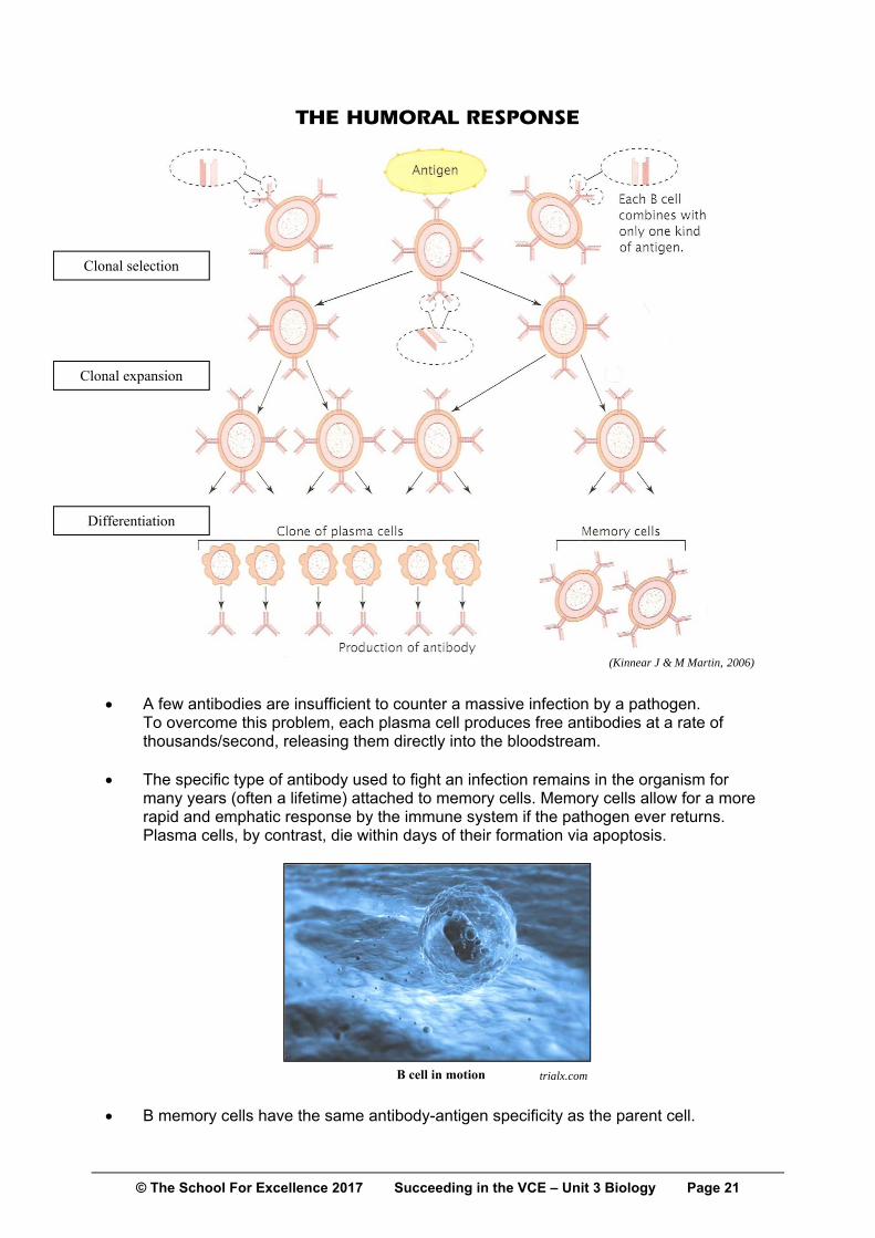
When an antigen enters the body it may be engulfed by a macrophage and delivered to the lymph nodes with the aid of a helper T cell. It will pass many B cells before it meets the B cell with the matching antibody.
Upon presentation of the particular antigen (clonal selection), the B cell is stimulated to
divide rapidly by mitosis, resulting in the production of many clones (clonal expansion). These clones become specialised (differentiation), forming many plasma cells and some memory cells.
B cell with matching antibodies
Helper T cell
Macrophage advertising antigen
© The School For Excellence 2017 Succeeding in the VCE – Unit 3 Biology Page 21
THE HUMORAL RESPONSE
A few antibodies are insufficient to counter a massive infection by a pathogen. To overcome this problem, each plasma cell produces free antibodies at a rate of thousands/second, releasing them directly into the bloodstream.
The specific type of antibody used to fight an infection remains in the organism for
many years (often a lifetime) attached to memory cells. Memory cells allow for a more rapid and emphatic response by the immune system if the pathogen ever returns. Plasma cells, by contrast, die within days of their formation via apoptosis.
B memory cells have the same antibody-antigen specificity as the parent cell.
B cell in motion trialx.com
(Kinnear J & M Martin, 2006)
Clonal selection
Clonal expansion
Differentiation

© The School For Excellence 2017 Succeeding in the VCE – Unit 3 Biology Page 22
Antibodies can act in a number of ways: 1. Neutralisation: By coating pathogens, antibodies neutralise viral particles and cellular
pathogens preventing them from attaching to host cells.
2. Agglutination of antigen-bearing particles exposes the pathogens to being engulfed by
phagocytes. 3. Precipitation of soluble antigens (e.g. snake
venom) by antibodies also stimulates an attack by phagocytes such as macrophages. The toxins are rendered useless in a precipitated form.
4. Antibodies activate complement proteins
which attach to the pathogen and act as flags to attract immune cells. They also directly puncture pathogen cell membranes.
(Campbell, NA et al, 2002)

© The School For Excellence 2017 Succeeding in the VCE – Unit 3 Biology Page 23
SECONDARY IMMUNE RESPONSE A secondary immune response occurs after a second exposure to same pathogen. B cells react more quickly and vigorously than they did in the initial response. Antibodies are produced much more rapidly, and in much greater quantity, due to the presence of memory cells.
Typically, an individual does not suffer any symptoms of the disease.
(Kinnear J & M Martin, 2000)
(Kinnear J & M Martin, 1993)

© The School For Excellence 2017 Succeeding in the VCE – Unit 3 Biology Page 24
HUMORAL IMMUNITY
Invasion of body by a pathogen (e.g. virus or bacterium), which acts as an antigen.
Macrophages carry the antigen to lymph nodes.
Antigen "activates" B lymphocytes.
B lymphocytes enlarge and divide repeatedly to form a clone (clonal expansion, clonal proliferation) of identical cells
(by mitosis). Most of these B lymphocytes Some B lymphocytes become differentiate to form plasma cells. B memory cells. Plasma cells secrete antibodies Long-term immunity. (up to 2,000 per sec). Cell does not live very long.
Pathogen or its toxin Stimulation of phagocytosis of Activation of the is inactivated. the pathogen by neutrophils Complement system. and macrophages.
B memory cells will rapidly divide to produce appropriate plasma cells which make more antibodies on re-exposure to the pathogen (secondary immune response). This will enable a much more rapid and emphatic response to a second attack by the antigen.
sciencephoto.com

© The School For Excellence 2017 Succeeding in the VCE – Unit 3 Biology Page 25
In summary:
Non-specific immunity (first and second lines of defence) involves the same response to any type of pathogen irrespective of how many times the patient has been exposed to the agent. No flexibility exists with this response.
Specific immunity (T and B cells) involves the production of specialised cells and substances that act against one particular infection. Specific immunity has a memory so that when the patient is faced with the same pathogen again, a more rapid and profound response occurs, reducing the chance of a recurrence of disease.
ABO ANTIGENS Antigens present on red blood cells include those of the ABO system (glycolipids) and the Rhesus factor (a protein). Ambulance officers ensure that the correct blood type is used when giving a transfusion to an accident victim.
Individuals with a Rhesus negative blood type cannot accept blood with the Rhesus positive antigen. The Type O antigen, however, is a short version of the Type A and Type B antigens, so it is deemed as a self antigen by individuals of all blood types.
TRANSPLANT REJECTION
Donor organs contain non-self antigens. Therefore, a patient’s body produces antibodies in the process of rejecting the donor tissue.
Drugs such as cyclosporine have been developed which reduce the intensity of a patient’s immune system response (it reduces levels of a chemical that stimulates certain cytotoxic T cells) so that it does not reject the donated antigens.
Without the use of immunosuppressive drugs, a transplanted tissue (organ) would be recognised as foreign by cytotoxic T cells. Along with phagocytes and natural killer cells, the cytotoxic T cells would eventually destroy all of the transplanted tissue.
The Rhesus monkey from which the Rhesus factor was investigated
ABO antigens

© The School For Excellence 2017 Succeeding in the VCE – Unit 3 Biology Page 26
RHESUS DISEASE After the birth of a first Rh+ baby, an Rh– mother has memory cells for Rhesus antigen. This is due to the fact that late in the term of the pregnancy, foetal red blood cell fragments often cross the placenta and enter the mother’s bloodstream. By the time the Rh+ antigens have been detected and responded to by the mother’s immune system, the baby has been born. The mother, however, now has memory cells for use in future pregnancies. Hence, in subsequent pregnancies involving Rh+ babies, the baby is at risk from the immune response of mother.

© The School For Excellence 2017 Succeeding in the VCE – Unit 3 Biology Page 27
SAMPLE QUESTIONS QUESTION 1 The first line of defence is used to keep pathogens outside of the internal environment of an organism. Possible chemical barriers of the first line include: A Interferons B Sweat C Macrophages D Water QUESTION 2 Natural killer cells are a type of lymphocyte. They can be distinguished from B and T lymphocytes in that they: A Are non-specific in their action. B Are produced in the bone marrow. C Spend most of their lives within the lymphatic system. D Are activated by Class I MHC-antigen fragment complexes. QUESTION 3 Histamines are chemicals synthesised by certain cells for the purpose of dilating blood vessels. White blood cells responsible for secreting histamines include: A Basophils and mast cells. B Neutrophils. C Basophils and macrophages. D Dendritic cells and mast cells. QUESTION 4 When a virus infects a eukaryotic cell, its DNA enters the nucleus, reprogramming the host cell to synthesise more copies of the virus. In such a situation, a virus-infected kidney cell would be expected to: A Release interferons to attract complement proteins to the site of infection. B Release histamine to attract white blood cells of the third line of defence. C Display antigen fragments using class I MHC markers. D Display antigen fragments using class II MHC markers. QUESTION 5 Pathogens which have penetrated the first line of defence would include: A Bacteria in the gut of the host. B Viruses adhered to the mucous lining of a bronchiole. C Bacteria within the host’s bladder. D Viruses within a lymph node.
© The School For Excellence 2017 Succeeding in the VCE – Unit 3 Biology Page 28
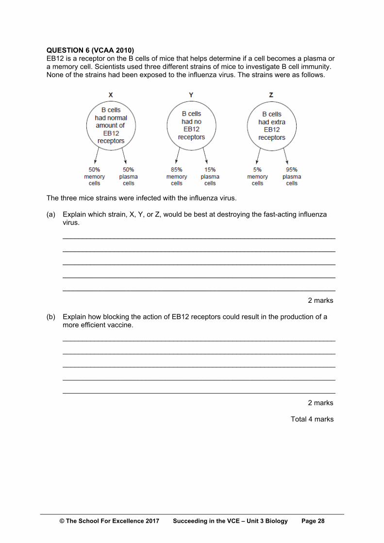
QUESTION 6 (VCAA 2010) EB12 is a receptor on the B cells of mice that helps determine if a cell becomes a plasma or a memory cell. Scientists used three different strains of mice to investigate B cell immunity. None of the strains had been exposed to the influenza virus. The strains were as follows.
The three mice strains were infected with the influenza virus. (a) Explain which strain, X, Y, or Z, would be best at destroying the fast-acting influenza virus.
_____________________________________________________________________
_____________________________________________________________________
_____________________________________________________________________
_____________________________________________________________________
_____________________________________________________________________
2 marks (b) Explain how blocking the action of EB12 receptors could result in the production of a more efficient vaccine.
_____________________________________________________________________
_____________________________________________________________________
_____________________________________________________________________
_____________________________________________________________________
_____________________________________________________________________
2 marks Total 4 marks
© The School For Excellence 2017 Succeeding in the VCE – Unit 3 Biology Page 29
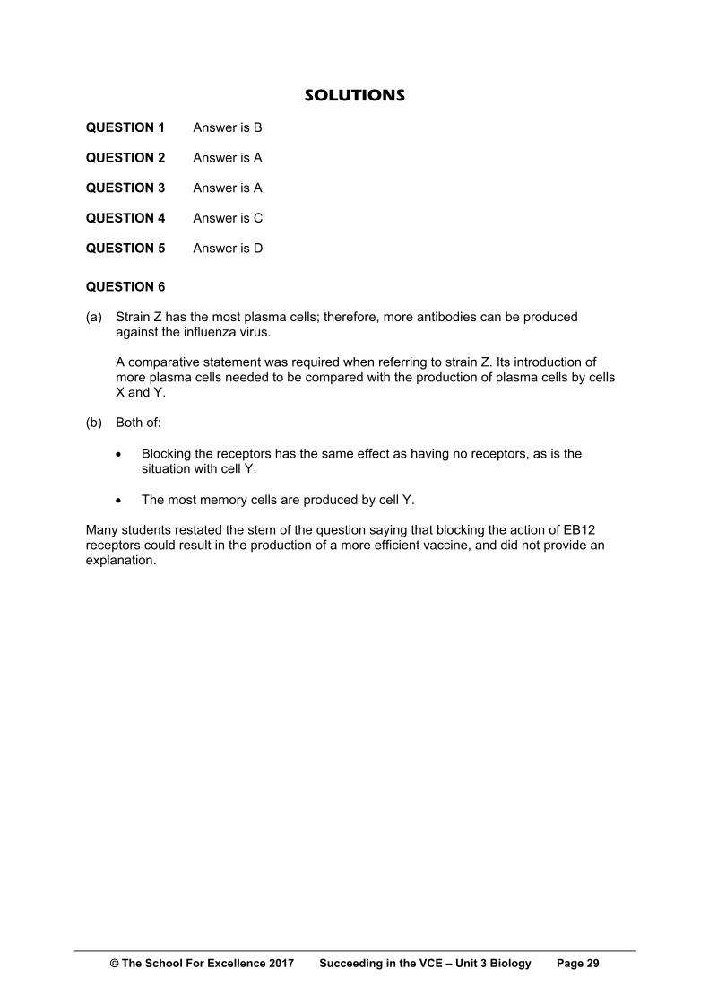
SOLUTIONS QUESTION 1 Answer is B QUESTION 2 Answer is A QUESTION 3 Answer is A QUESTION 4 Answer is C QUESTION 5 Answer is D
QUESTION 6 (a) Strain Z has the most plasma cells; therefore, more antibodies can be produced
against the influenza virus.
A comparative statement was required when referring to strain Z. Its introduction of more plasma cells needed to be compared with the production of plasma cells by cells X and Y.
(b) Both of:
Blocking the receptors has the same effect as having no receptors, as is the situation with cell Y.
The most memory cells are produced by cell Y. Many students restated the stem of the question saying that blocking the action of EB12 receptors could result in the production of a more efficient vaccine, and did not provide an explanation.