Embed Size (px)
Citation preview
Unit 2.4Communication with the Outside World
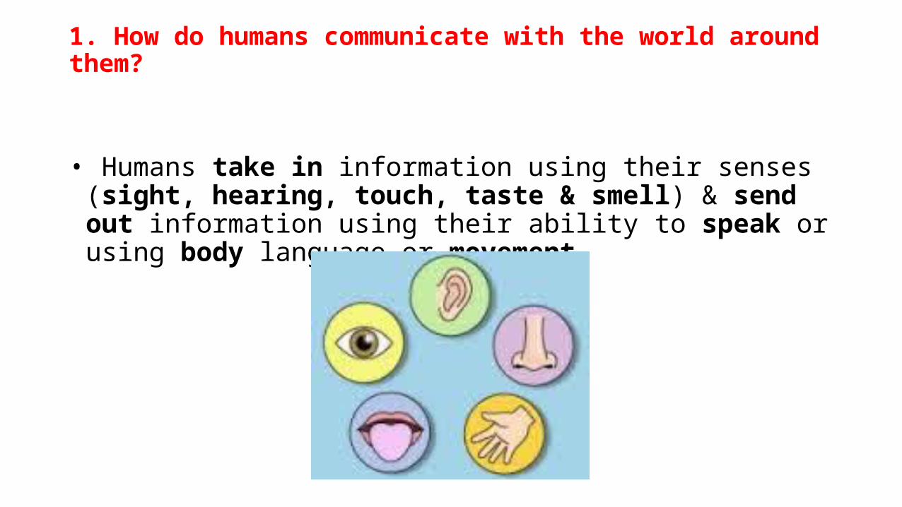
1. How do humans communicate with the world around them?
• Humans take in information using their senses (sight, hearing, touch, taste & smell) & send out information using their ability to speak or using body language or movement.
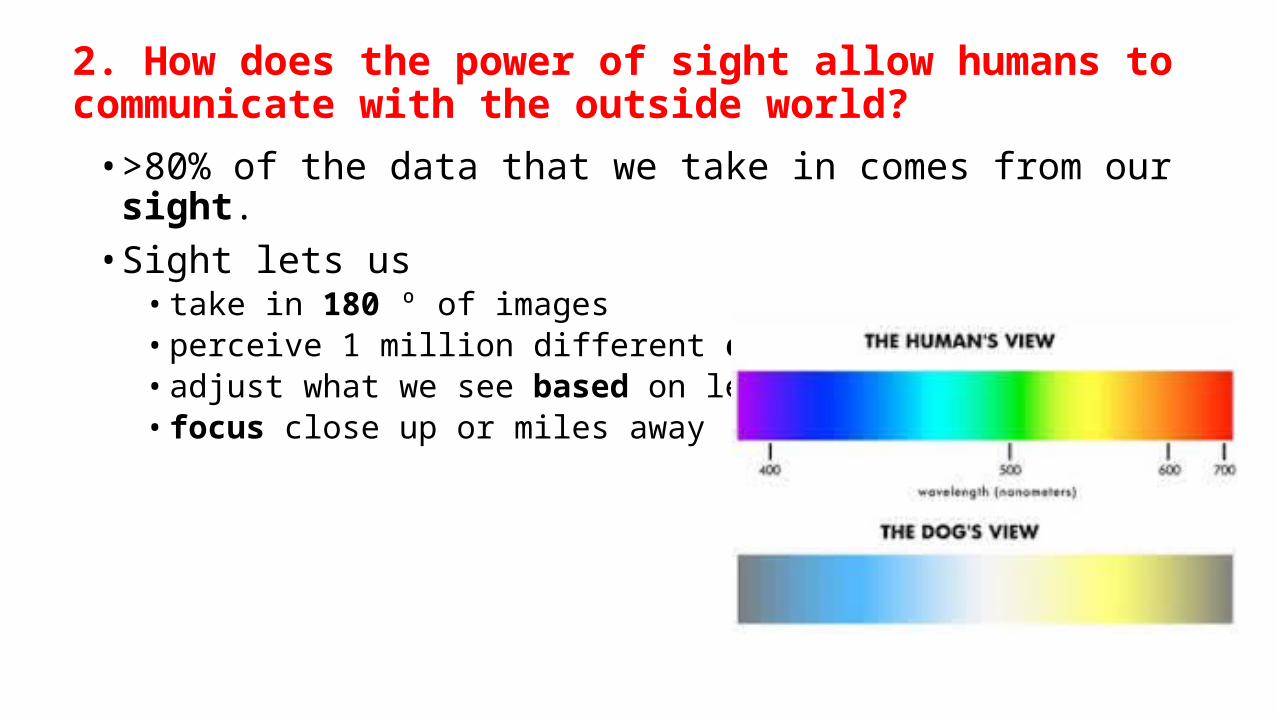
2. How does the power of sight allow humans to communicate with the outside world?
• >80% of the data that we take in comes from our sight. • Sight lets us
• take in 180 º of images • perceive 1 million different colors• adjust what we see based on level of light • focus close up or miles away
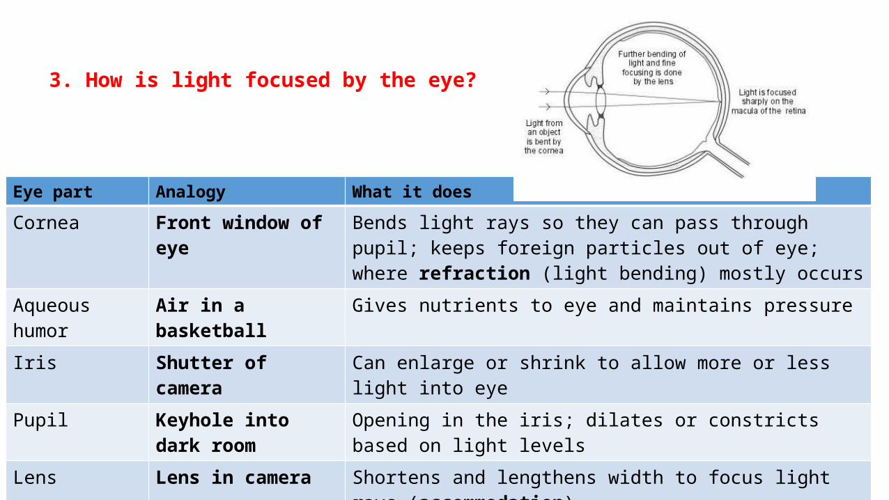
3. How is light focused by the eye?
Eye part Analogy What it does
Cornea Front window of eye Bends light rays so they can pass through pupil; keeps foreign particles out of eye; where refraction (light bending) mostly occurs
Aqueous humor Air in a basketball Gives nutrients to eye and maintains pressureIris Shutter of camera Can enlarge or shrink to allow more or less light into eye Pupil Keyhole into dark room Opening in the iris; dilates or constricts based on light levels Lens Lens in camera Shortens and lengthens width to focus light rays (accommodation)
Vitreous humor Air in a basketball Lets light pass through while helping eye keep its shape
Retina Film on camera Captures light rays & processes them w/ millions of nerve endings; sends light impulses through >1 million nerve fibers to optic nerve
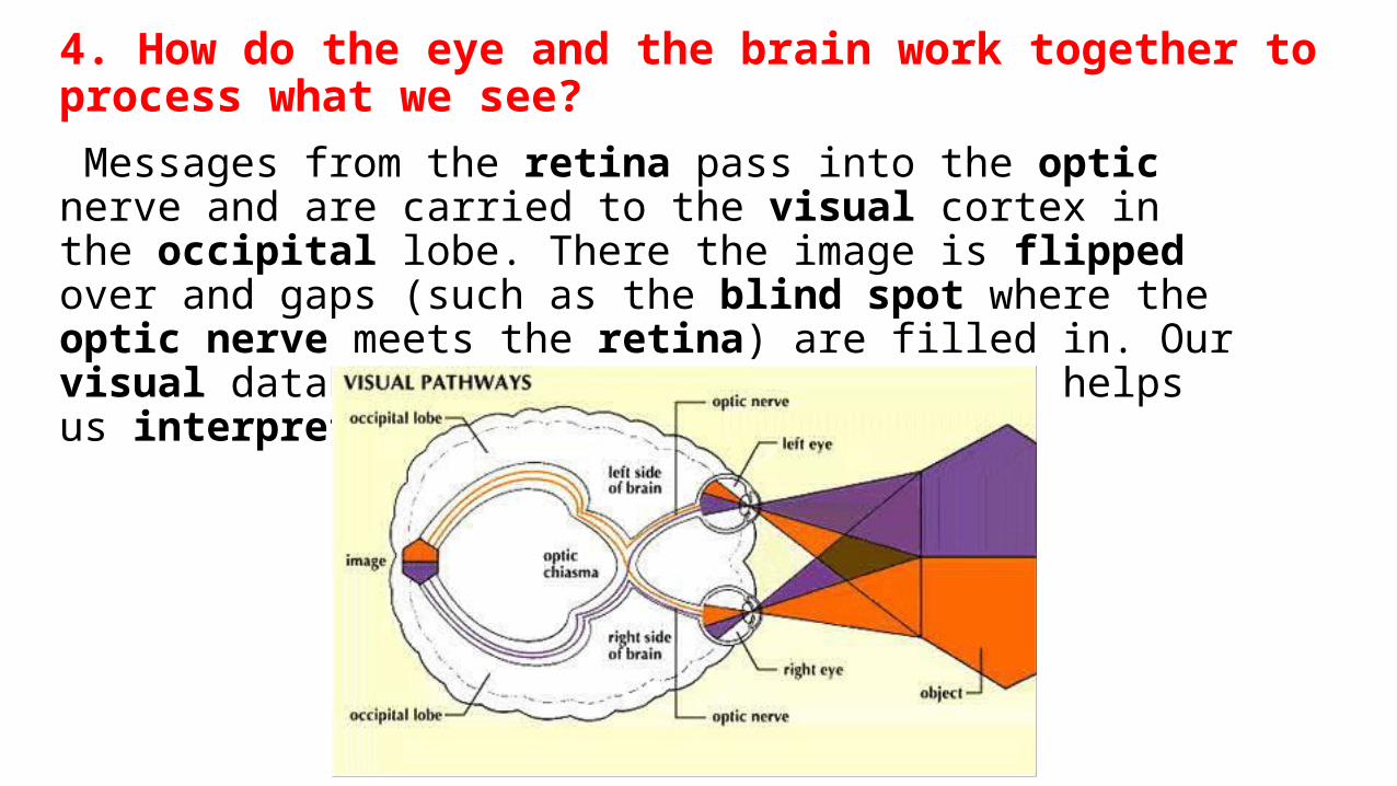
4. How do the eye and the brain work together to process what we see?
Messages from the retina pass into the optic nerve and are carried to the visual cortex in the occipital lobe. There the image is flipped over and gaps (such as the blind spot where the optic nerve meets the retina) are filled in. Our visual database (built during childhood) helps us interpret images.
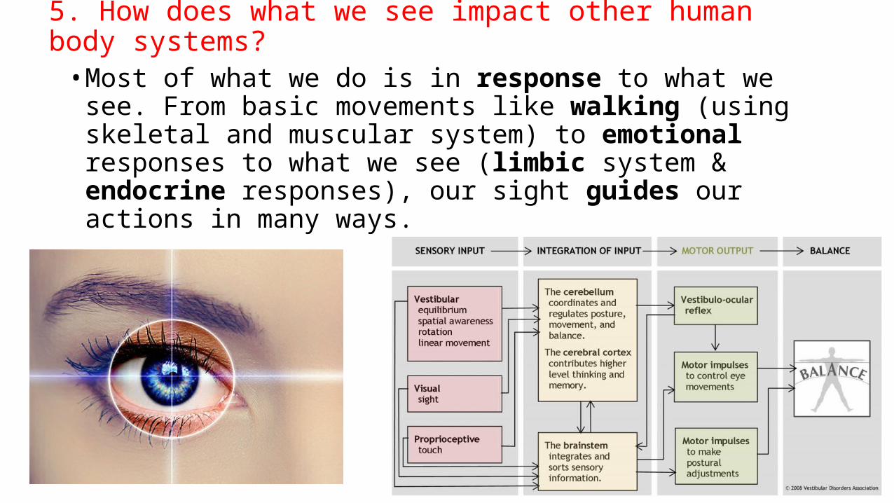
5. How does what we see impact other human body systems?
• Most of what we do is in response to what we see. From basic movements like walking (using skeletal and muscular system) to emotional responses to what we see (limbic system & endocrine responses), our sight guides our actions in many ways.
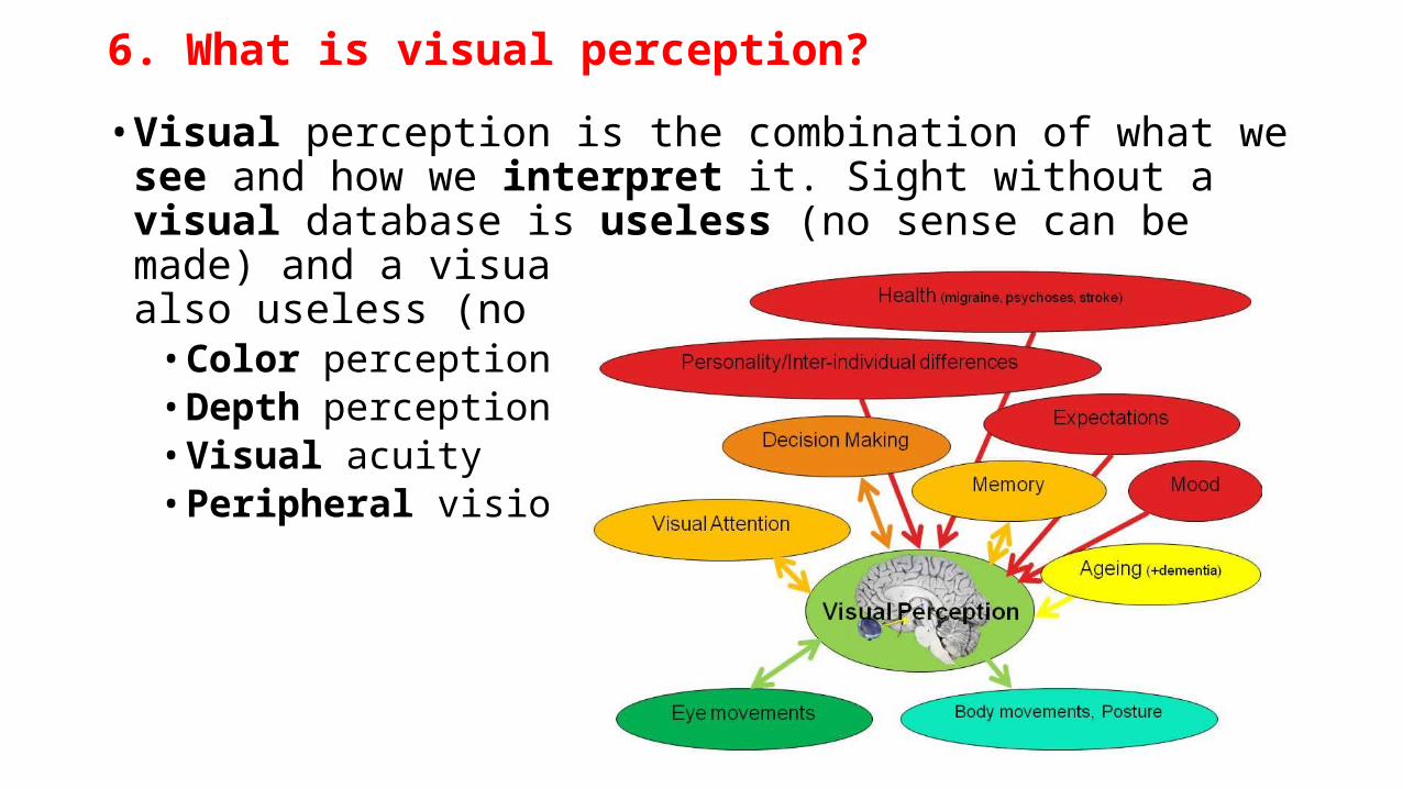
6. What is visual perception?
• Visual perception is the combination of what we see and how we interpret it. Sight without a visual database is useless (no sense can be made) and a visual database without sight is also useless (no input).
• Color perception • Depth perception • Visual acuity • Peripheral vision
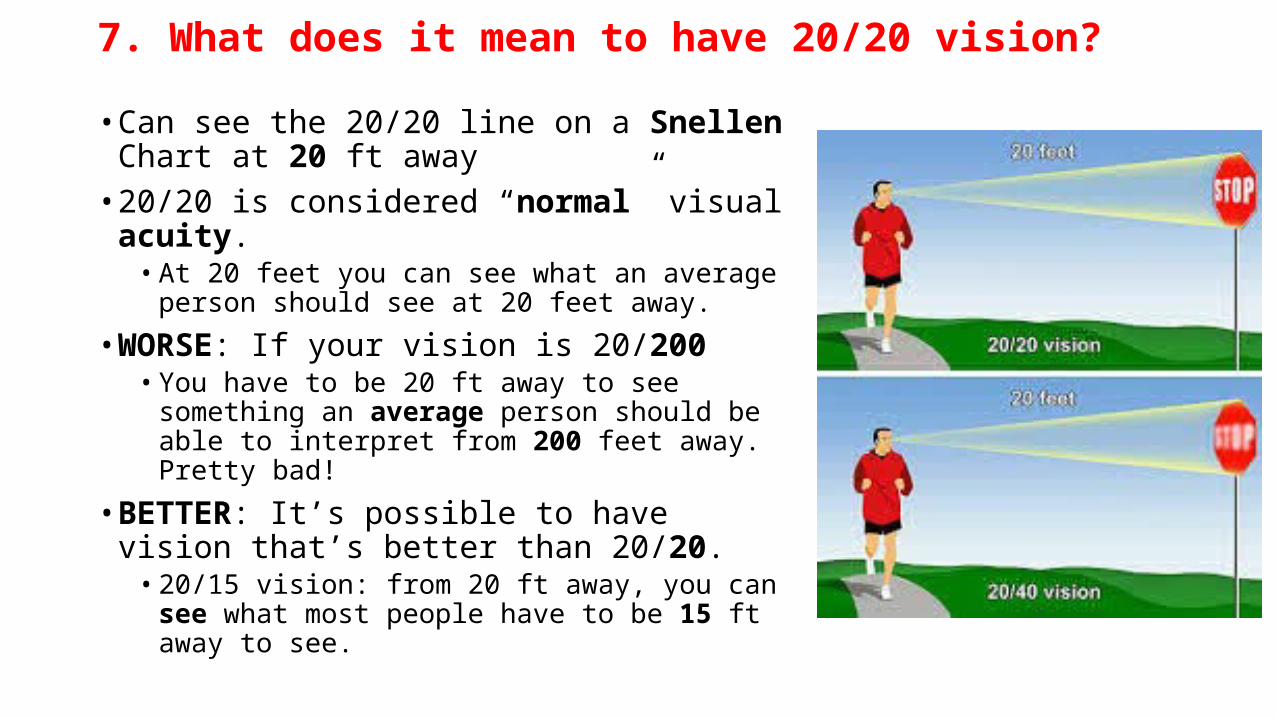
7. What does it mean to have 20/20 vision?
• Can see the 20/20 line on a Snellen Chart at 20 ft away
• 20/20 is considered “normal” visual acuity. • At 20 feet you can see what an average person
should see at 20 feet away.
• WORSE: If your vision is 20/200 • You have to be 20 ft away to see something an
average person should be able to interpret from 200 feet away. Pretty bad!
• BETTER: It’s possible to have vision that’s better than 20/20.
• 20/15 vision: from 20 ft away, you can see what most people have to be 15 ft away to see.
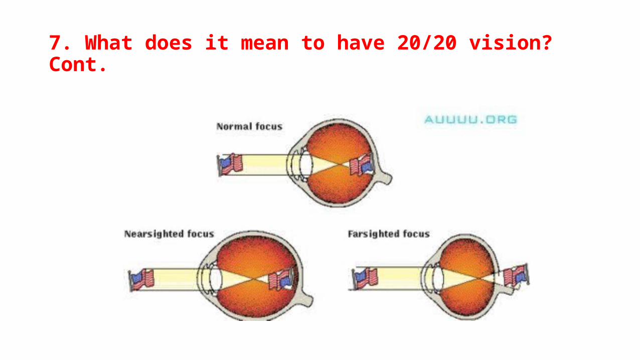
7. What does it mean to have 20/20 vision? Cont.
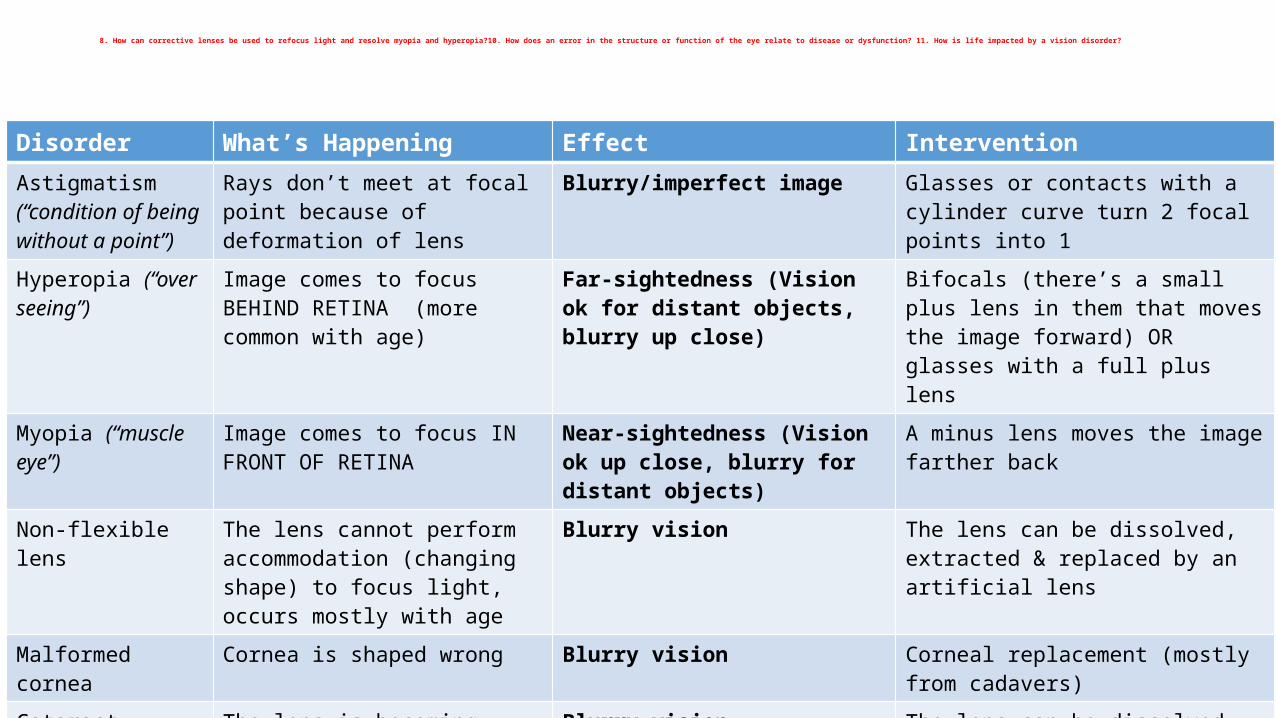
8. How can corrective lenses be used to refocus light and resolve myopia and hyperopia?10. How does an error in the structure or function of the eye relate to disease or dysfunction? 11. How is life impacted by a vision disorder?
Disorder What’s Happening Effect InterventionAstigmatism (“condition of being without a point”)
Rays don’t meet at focal point because of deformation of lens
Blurry/imperfect image Glasses or contacts with a cylinder curve turn 2 focal points into 1
Hyperopia (“over seeing”)
Image comes to focus BEHIND RETINA (more common with age)
Far-sightedness (Vision ok for distant objects, blurry up close)
Bifocals (there’s a small plus lens in them that moves the image forward) OR glasses with a full plus lens
Myopia (“muscle eye”)
Image comes to focus IN FRONT OF RETINA
Near-sightedness (Vision ok up close, blurry for distant objects)
A minus lens moves the image farther back
Non-flexible lens The lens cannot perform accommodation (changing shape) to focus light, occurs mostly with age
Blurry vision The lens can be dissolved, extracted & replaced by an artificial lens
Malformed cornea
Cornea is shaped wrong Blurry vision Corneal replacement (mostly from cadavers)
Cataract The lens is becoming cloudy Blurry vision The lens can be dissolved, extracted & replaced by an artificial lens
Crossed eyes The eyes do not focus together Double vision Lenses move the image to match the “wayward” eye, correcting double vision
9. How does the eye perceive depth, color and optical illusions?
• Color• The retina is composed of photoreceptors
• >95% rods (found all over retina & used for seeing black and white, peripheral vision & low light viewing) • <5% cones (for COLOR PERCEPTION, concentrated in fovea, tiny spot with detailed vision)
• Depth Perception• Depends on retina • The visual database contains information about size of objects from previous
experience & gauges size based on that.• Moving the head from side to side allows you to see how far objects move (less
movement means farther away). • Comparing the image from one eye to the combined images, tells retina how far
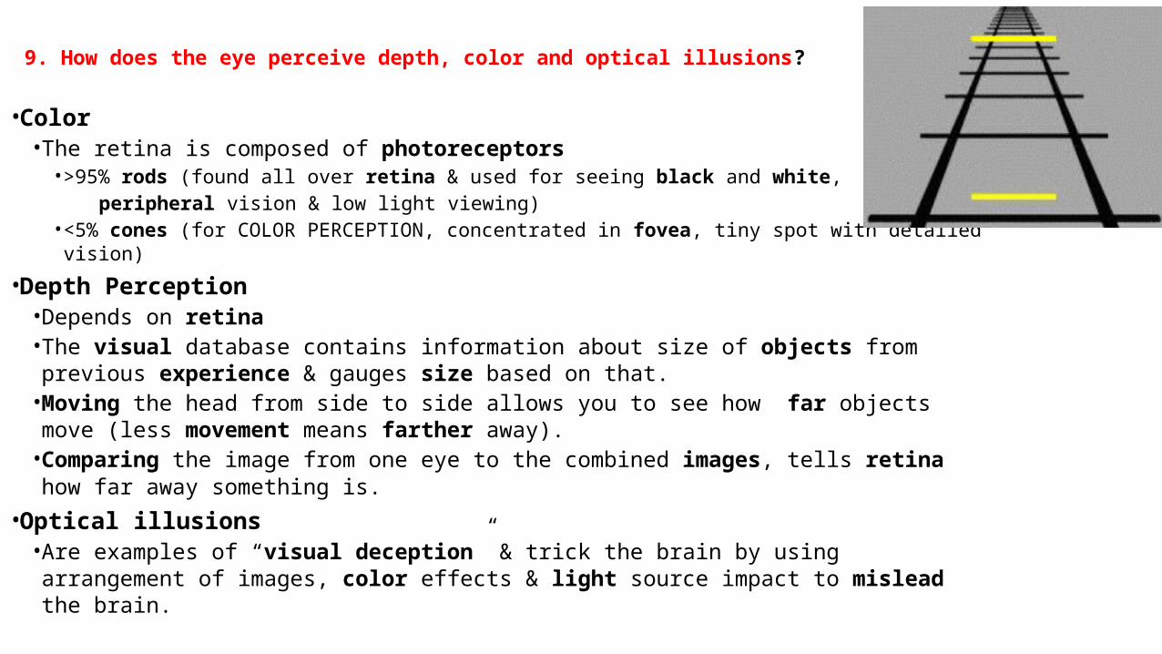
away something is. • Optical illusions
• Are examples of “visual deception” & trick the brain by using arrangement of images, color effects & light source impact to mislead the brain.
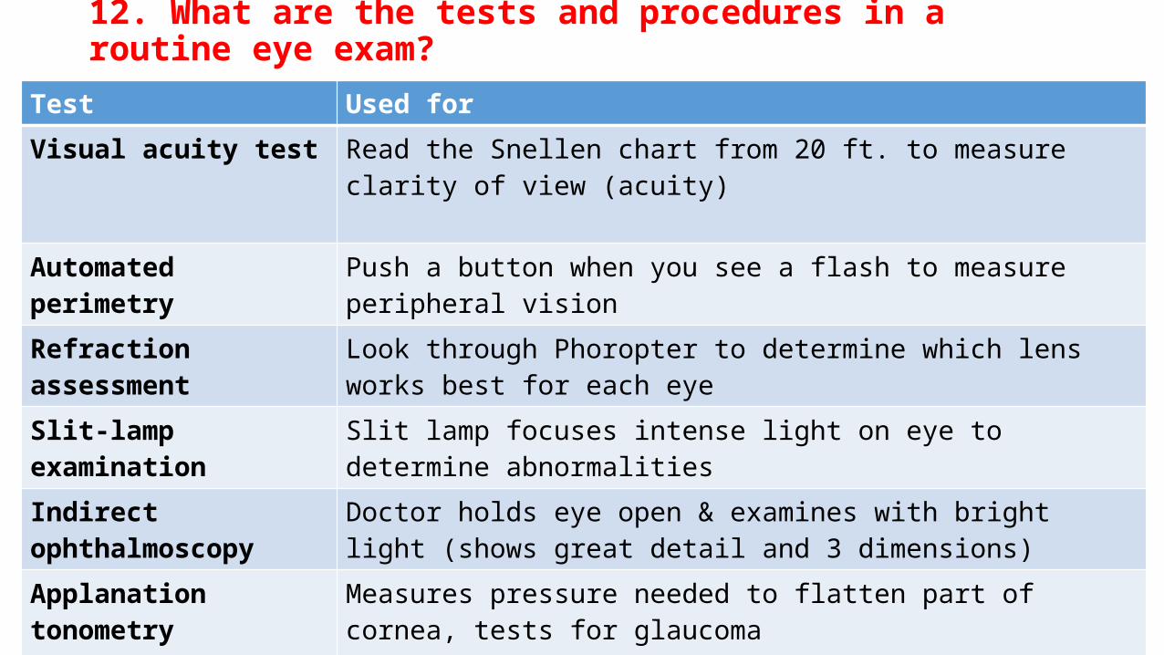
12. What are the tests and procedures in a routine eye exam?
Test Used for
Visual acuity test Read the Snellen chart from 20 ft. to measure clarity of view (acuity)
Automated perimetry Push a button when you see a flash to measure peripheral vision
Refraction assessment Look through Phoropter to determine which lens works best for each eye
Slit-lamp examination Slit lamp focuses intense light on eye to determine abnormalities
Indirect ophthalmoscopy
Doctor holds eye open & examines with bright light (shows great detail and 3 dimensions)
Applanation tonometry Measures pressure needed to flatten part of cornea, tests for glaucoma