Embed Size (px)
Citation preview

Unit 2: Vital signs Fundamental of Nursing
1
Unit 2: Vital signs Outlines
- Body temperature.
- Pulse / heart rate.
- Respiration.
- Blood Pressure.
Vital Signs
Vital signs are measures of various physiological status, in order to
assess the most basic body functions. When these values are not zero,
they indicate that a person is alive.
All of these vital signs can be observed, measured, and monitored.
This will enable the assessment of the level at which an individual
functioning. Normal ranges of measurements of vital signs change with
age and medical condition.
Vital signs are useful in detecting or monitoring medical problems.
Vital signs can be measured in a medical setting, at home, at the site of a
medical emergency, or elsewhere.
Vital Signs
Are measurements of the body's most basic functions:
1. Body temperature (Temp).
2. Pulse / heart rate.
3. Respiration.
4. Blood pressure (BP).
When to Assess Vital Signs
1. Upon admission to any healthcare agency.
2. Based on agency institutional policy and procedures.

Unit 2: Vital signs Fundamental of Nursing
2
3. Any time there is a change in the patient’s condition.
4. Before and after surgical or invasive diagnostic procedures.
5. Before and after activity that may increase risk.
6. Before and after administering medications that affect cardiovascular
or respiratory functioning.
Physiological Basis of Body Temperature
Body temperature is the balance between the heat production due
to chemical activities by the body and heat lost from the body through
radiation, conduction, convection, and vaporization( evaporation) .
Types of body temperature:
1. Core temperature:
Is the temperature of deep tissues of the body, e.g., cranium, thorax
and abdominal cavity. It remains relatively constant (37Cº or 98.6 Fº).
True core temperature readings can only be measured by invasive
means, such as placing a temperature probe into the esophagus,
pulmonary artery or urinary bladder.
Non-invasive sites such as the rectum, oral cavity, axilla, temporal
artery (forehead) and external auditory canal are accessible and are
believed to provide the best estimation of the core temperature.
2.Surface temperature:
Is the temperature of the skin, the subcutaneous tissue and fat. It,
by contrast rises and falls in response to the environmental changes.
When measured orally, the average body temperature of an adult is
between 36.7 Cº( 98 Fº) and 37 Cº( 98.6Fº).
Assessing Body Temperature
The normal range of the body temperature is between 36.2 to 37.2 Cº.

Unit 2: Vital signs Fundamental of Nursing
3
Factors Affecting Body's heat production
1.Basal metabolic rate ( BMR): The basal metabolic rate is the rate of
energy utilization in the body to maintain essential activities such as
breathing. BMRs vary with age and sex.
2.Muscle activity: It including shivering, can greatly increase metabolic
rate.
3.Thyroxin output: Increased thyroxin output increases the rate of
cellular metabolism throughout the body.
4.Epinephrine and sympathetic stimulation, these immediately increase
the rate of cellular metabolism in many body tissues.
5.Age: Very young and very old are more sensitive to change in
environmental temperature due to decreased thermoregulatory controls
6.Gender: women tend to have more function in body temperature than
men the increase in progesterone secretion at ovulation increase body
temperature .
7.Diurnal variation: body temperature normally change throughout the
day, varying as much as I Cº ( I.8 ºF) between the early morning and the
late afternoon.
8.Exercise: Hard work or strenuous exercise can increase body
temperature to as high as 38.3Cº to 40 Cº( 101 to 104 ºF) measured
orally.
Alterations in Body Temperature
Pyrexia: A body temperature above the usual range is called pyrexia,
hyperthermia, or ( in lay terms) fever. A very high temperature, e.g. 41Cº
(105 ºF) is called hyperpyrexia.

Unit 2: Vital signs Fundamental of Nursing
4
Common types of fevers
1.Intermittent Fever: during this type of fever, the body temperature
alternates at regular intervals between periods of fever and periods of
normal temperatures.
2.Remittent Fever: during this type of fever, a wide range of temperature
fluctuations occurs over the 2 hour period, all of which are above normal.
3.Relapsing Fever: In a relapsing fever, short febrile periods of a few
days are interspersed with periods of 1 or 2 days of normal temperature.
4.Constant Fever: during a constant fever, the body temperature
fluctuates minimally but always remains elevated.
Clinical Signs of Fever
A: Onset ( cold or chill stage)
1. increased heart rate and respiratory rate and depth.
2. Shivering due to increased skeletal muscle tension and contraction.
3. Cold skin due to vasoconstriction.
4. Cyanotic nail beds due to vasoconstriction.
5. Complain of feeling cold.
6. Gooseflesh appearance of the skin.
7. Rise in body temperature.
B: Course
1.Skin feels warm.
2. increased pulse and respiratory rate.
3. increased thirst.
4. mild to severe dehydration.
5. drowsiness, restlessness, or delirium and convulsions due to irritation
of the nerve cells/
6. loss of appetite with prolonged fever.

Unit 2: Vital signs Fundamental of Nursing
5
7. malaise, weakness, and aching muscles due to protein breakdown.
C: Abatement stage
1. Flushed and warm skin .
2. Sweating.
3. Decreased shivering.
4. Possible dehydration.
Treatment of Increasing Body Temperature
1. Antipyretics.
2. Cold sponge bath .
3. Cold compresses .
Nursing Interventions for patient with Fever
1. Monitor vital signs.
2.Assess skin color and temperature.
3.Monitor WBCs count and other pertinent laboratory records.
4. Remove excess clothes when the patient feels warm, but provide extra
warmth when the patient feels chilled.
5. Measure intake and output.
6.Reduced physical activity to limit heat production.
7. Provide oral hygiene to keep the mucous membranes moist. They can
become dry and cracked as a result of excessive fluid loss.
8. Applied moist cold applications such as cold compresses tepid sponge
and ice bag to increase loss through conduction.
9. Provide cool circulating air by using a fan to increase heat loss through
convection.
Hypothermia
Unit 2: Vital signs Fundamental of Nursing
6
It is a core body temperature below the lower limit of normal. The
ability of hypothalamus to regulate temperature is greatly impaired when
the body temperature falls below 34.5Cº ( 94 ºF), and death usually
occurs when the temperature falls below 34 Cº (93.2 ºF).
Physiological Process of hypothermia
1. Excessive cold environment.
2. Inadequate heat production to counteract the heat loss.
3. Impaired hypothalamus thermoregulation.
Clinical signs of hypothermia
1. Decreased body temperature.
2. Pale, cool, waxy skin.
3. Hypotension.
4. Decrease urine output.
5. Lack of muscle coordination.
6. Disorientation.
7. Drowsiness may progressing to coma.
Sites for Assessing Body Temperature
1.Orally (common way). 37 C° (3–5 min).
The oral cavity temperature is considered to be reliable when the
thermometer is placed posteriorly into the sublingual pocket. This
landmark is close to the sublingual artery, so this site tracks changes in
core body temperature.
2.Axillary (safe way). 36 C° + 0.5 C° (10 min).
Temperature is measured at the axilla by placing the thermometer
in the central position and adducting the arm close to the chest wall. is
considered to be an unreliable site for estimating core body temperature

Unit 2: Vital signs Fundamental of Nursing
7
because there are no main blood vessels around this area, therefore should
add 0.5C° to the actual reading.
3. Rectal (accurate reading).37 C° – 0.5 C° (2 – 3 min).
Rectal temperature is the most accurate method for measuring the
core temperature, and should reduce 0.5 C° to the actual reading.
4. Tympanic membrane.
The tympanic thermometer senses reflected infrared emissions
from the tympanic membrane through a probe placed in the external
auditory canal. This method is quick (<1 minute), minimally invasive and
easy to perform. It has been reported to estimate rapid fluctuations in core
temperature accurately because the tympanic membrane is close to the
hypothalamus.
Contraindications of oral thermometer
1. The child under 6 years .
2. Unconscious patients .
3. Psychiatric patients .
4. Patient who cannot breath from his nose
5. Mouth surgery or infection .
6. Patient on oxygen mask.
Contraindications of rectal thermometer
1. rectal surgery .
2. rectal disorders ( hemorrhoids. Rectal fissure..etc.).
3. diarrhea.
Types of Thermometers
1. Electronic thermometer.
2. Glass thermometer.

Unit 2: Vital signs Fundamental of Nursing
8
3. Paper thermometer.
4. Tympanic membrane thermometer.
Alterations in Thermoregulation
Alterations
Definition
Characteristics
Heat exhaustion
An increase in body temperature
(38_C– 40_C, 100.4_F–104.0_F) in
response to environmental
conditions that, in turn, causes
diaphoresis (profuse perspiration)
An increase in body temperature
(38_C–40_C, 100.4_F–104.0_F) in
response to environmental
conditions that, in turn, causes
diaphoresis (profuse perspiration).
Loss of excessive amounts
of water and sodium from
perspiring leads to thirst,
nausea, vomiting,
weakness, and
disorientation.
Heat stroke
A critical increase in body
temperature (41_C–44_C, 100.6_F–
112.0_F) resulting
from exposure to high
environmental temperature.
Dry, hot skin is the most
important sign.
confused or delirious and
experiences thirst,
abdominal distress, muscle
cramps, and visual
disturbances.
occurs if untreated.
Hypothermia
A body temperature of 35_C (95_F)
or lower resulting from cold
weather exposure or artificial
induction
Decrease in metabolism
leads to impairedmental
functioning and depressed
pulse, respiration, and
blood pressure; can result
in cardiac arrest if
untreated.
leads to impairedmental
functioning and depressed
pulse, respiration, and
blood pressure; can result
in cardiac arrest if
untreated.
Frostbite
Freezing of the body’s surface areas
(earlobes, fingers, and toes) in
extremely low temperatures.
Circulatory impairment
may be followed by
gangrene.
Conversion Formulas

Unit 2: Vital signs Fundamental of Nursing
9
Sometimes a health professional staff need to convert a Celsius
reading to Fahrenheit, or vice versa.
a. To convert from Fahrenheit to Celsius, deduct 32 from the Fahrenheit,
and then multiply by 5/9
C= (Fahrenheit temperature -32) × 5/9
For example, convert 98.6 Fahrenheit to Celsius reading
C= ( 98.6- 32) × 5/9
C=( 66.6) × 5/9
C = 37 Celsius degree.
b. To convert from Celsius to Fahrenheit, multiply the Celsius reading by
the fraction 9/5 and then add 32 .
For example, convert 37 Celsius degree toFahrenheitreading
F = ( 37 × 9/5) + 32
F = ( 66.6) + 32
F = 98.6 ºF
Nursing Diagnosis
Potential altered body temperature related to:
a. illness or trauma affecting temperature regulation.
b. medication or vigorous activity.
Altered body temperature (hyperthermia) related to exposure to
excessively hot environment, increase metabolic rate, or dehydration.
Altered body temperature (hypothermia) related exposure to
excessively cool environment, debilitating or trauma, or lack of adequate
clothing and shelter.
Ineffective thermoregulation related to decreased basal metabolism
secondary to aging, or trauma, or illness.
Risk for imbalanced body temperature, at risk for failure to maintain
body temperature within normal range.

Unit 2: Vital signs Fundamental of Nursing
11
Pulse
Pulse is a wave of blood created by contraction of the left ventricle
of the heart. The heart is a pulsate pump and the blood enters the arteries
with each heartbeat, causing pulse waves.
Pulse assessment is the measurement of a pressure pulsation
created when the heart contracts and ejects blood into the aorta.
Characteristics of Pulse
1. Quality.
2. Rate.
3. Rhythm.
4. Volume (strength or amplitude).
1. Pulse quality refers to the ‘‘feel’’ of the pulse, its rhythm and
forcefulness.

Unit 2: Vital signs Fundamental of Nursing
11
2. Pulse rate is an indirect measurement of cardiac output obtained by
counting the number of apical or peripheral pulse waves over a pulse
point.
- A normal pulse rate for adults is between 60 and 100 beats per minute.
- Bradycardia is a heart rate less than 60 beats per minute in an adult.
- Tachycardia is a heart rate in excess of 100 beats per minute in an
adult.
3. Pulse rhythm is the regularity of the heartbeat. It describes how
evenly the heart is beating:
- Regular (the beats are evenly spaced).
- Irregular (the beats are not evenly spaced).
- Dysrhythmia (arrhythmia) is an irregular rhythm caused by an early,
late, or missed heartbeat.
4. Pulse volume is a measurement of the strength or amplitude of force
exerted by the ejected blood against the arterial wall with each
contraction.
- It is described as normal (full, easily palpable).
- Weak (thready and usually rapid), or
- Strong (bounding).

Unit 2: Vital signs Fundamental of Nursing
12
Factors Contribute to Increase Pulse Rate
1. Pain.
2. Fever.
3. Stress, exercise .
4. Bleeding.
5. Decrease in blood pressure .
6. Some medications as (adrenalin, aminophylline).
Factors May Slow The Pulse
1. Rest .
2. Increasing age.
3. People with thin body size .
4. Some Medications.
5. Thyroid gland disturbances .

Unit 2: Vital signs Fundamental of Nursing
13
Pulse Point assessment
Pulse
point
Anatomical location Assessment Criteria
Temporal Over temporal bone, superior
and lateral to eye
Accessible; used routinely for
infants and when radial is
inaccessible.
Carotid Bilateral, under lower jaw in
neck along medial edge of
sternocleidomastoid muscle.
Accessible; used routinely for
infants and during shock or
cardiac arrest when other
peripheral pulses are too
weak to palpate; also used to
assess cranial circulation.
Apical Left midclavicular line at
fourth to fifth intercostal
space.
Used to auscultate heart
sounds and assess apical-
radial deficit.
Brachial Inner aspect between groove
of biceps and triceps muscle
at antecubital fossa.
Used in cardiac arrest for
infants, to assess lower arm
circulation, and to auscultate
blood pressure.
Radial Inner aspect of forearm on
thumb side of wrist.
Accessible; used routinely in
adults to assess character of
peripheral pulse.
Ulnar Outer aspect of forearm on
finger side of wrist.
Used to assess circulation to
ulnar side of hand and to
perform the Allens test.
Femoral In groin, below inguinal
ligament (midpoint between
symphysis pubis and
anterosuperior iliac spine).
Used to assess circulation to
legs and during cardiac arrest.
Popliteal Behind knee, at center in
popliteal fossa.
Used to assess circulation to
legs and to auscultate leg
blood pressure.
Posterior
tibial
Inner aspect of ankle between
Achilles tendon and tibia
(below medial malleolus).
Used to assess circulation to
feet.
Dorsalis Over instep, midpoint
between extension tendons of
great and second toe.
Used to assess circulation to
feet.

Unit 2: Vital signs Fundamental of Nursing
14
Unit 2: Vital signs Fundamental of Nursing
15
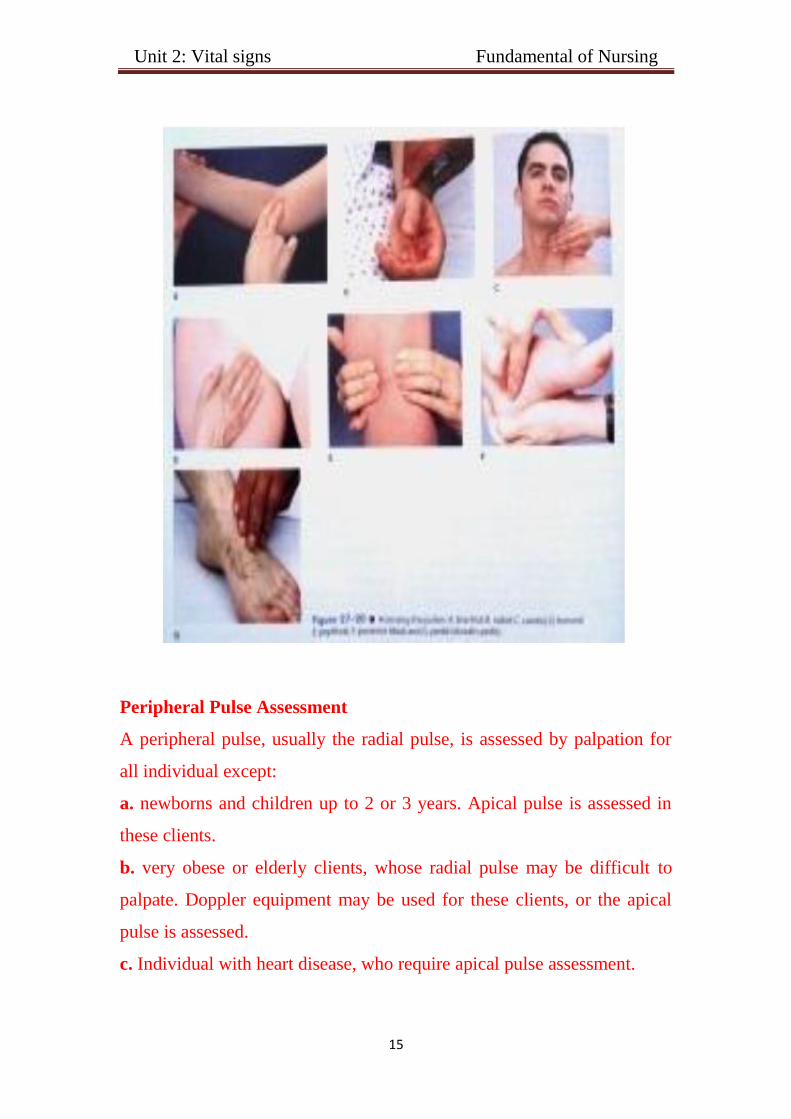
Peripheral Pulse Assessment
A peripheral pulse, usually the radial pulse, is assessed by palpation for
all individual except:
a. newborns and children up to 2 or 3 years. Apical pulse is assessed in
these clients.
b. very obese or elderly clients, whose radial pulse may be difficult to
palpate. Doppler equipment may be used for these clients, or the apical
pulse is assessed.
c. Individual with heart disease, who require apical pulse assessment.

Unit 2: Vital signs Fundamental of Nursing
16
d. Individuals in whom the circulation to a specific body part must be
assessed, e.g. following leg surgery the pedal ( dorsalis pedis) is assessed.
Apical Pulse Assessment
Assessment of the apical pulse is indicator for clients whose
peripheral pulse is irregular as well as for clients with known
cardiovascular, pulmonary, and renal diseases. It is commonly assessed
prior to administering medications that effect heart rate. The apical side is
also used to assess the pulse for newborns, infants, and children up to 2-3
years old.
Apical –Radial Pulse
An apical-radial pulse may need to be assessed for clients with
certain cardiovascular disorders. Normally the apical and radial rates are
identical.
An apical pulse rate greater than a redial pulse rate can indicate
that the thrust of the blood from the heart is too feeble for the wave to be
felt at the peripheral pulse site, (or) it can indicate the vascular disease is
preventing impulses from being transmitted. Any discrepancy between
the two pulse rates need to be reported promptly. In no instance is the
radial pulse greater than the apical pulse.
Pulse deficit
Pulse deficit is the difference in the apical pulse and the radial
pulse. These should be taken at the same time, which will require that 2
people take the pulse. One with a stethoscope and one at the wrist. Count
for 1 full minute. Then subtract the radial from the apical. This is the
Pulse Deficit.
Unit 2: Vital signs Fundamental of Nursing
17
Respiration
- Pulmonary ventilation (breathing ): movement of air in and out of
the lungs.
- Inspiration (inhalation) is the act of breathing in.
- Expiration (exhalation ) is the act of breathing out .
Factors Affecting Respiration
1. Pain, anxiety, exercise .
2. Medications .
3. Trauma .
4. Infection.
5. Respiratory and cardiovascular disease .
6. Alteration in fluids, electrolytes, acid- base balances.
Assessing Respirations
- Inspection.
- Listening with stethoscope.
- Monitoring arterial _ blood gas results.
- Using a pulse oximeter.
Control of Breathing
Respiration is controlled by:
1. Respiratory center in the medulla oblongata and the pons of the brain.
2. Chemoreceptors located centrally in the medulla in peripherally in the
carotid and aortic bodies. These centers and receptors respond to changes
in the concentration of oxygen ( O2), carbon dioxide ( Co 2), and
hydrogen ( H+) levels in the arterial blood.

Unit 2: Vital signs Fundamental of Nursing
18
Characteristics of Normal and Abnormal Breathing Sounds
Eupnea: refers to easy respirations with a normal rate of breaths per
minute that is age specific.
Bradypnea: is a respiratory rate of 10 or fewer breaths per minute.
Hypoventilation: is characterized by shallow respirations.
Tachypnea: is a respiratory rate greater than 24 breaths per minute.
Hyperventilation: is characterized by deep, rapid respirations.
The nurse can also observe alterations in the movement of the chest
wall:
Costal (thoracic) breathing: occurs when external intercostal muscles
and the other accessory muscles are used to move the chest upward and
outward.
Diaphragmatic(abdominal) breathing: occurs when the diaphragm
contracts and relaxes as observed by movement of the abdomen.
Dyspnea: refers to difficulty in breathing as observed by labored or
forced respirations through the use of accessory muscles in the chest and
neck to breathe.
Apnea : respirations cease for several seconds. Persistent cessation is
called respiratory arrest.
Cheyne–Stockes respiration: respiratory rhythm is irregular,
characterized by alternating periods of apnea and hyperventilation. The
respiratory cycle begins with slow, shallow breaths that gradually
increase to abnormal depth and rapidity. Gradually breathing slows and
becomes shallower, climaxing in a 10 to 20 seconds period of apnea
before respiration resumes.

Unit 2: Vital signs Fundamental of Nursing
19
Kussmaul respiration: respirations are abnormally deep but regular,
similar to hyperventilation. Characteristic of clients with diabetic
ketoacidosis.
Orthopnea: respiratory condition in which a person must sit or stand in
order to breathe deeply or comfortably.
Assessment of respiration includes;
1-Depth [by assessing the degree of excursion or movement in the chest
wall; shallow, deep or normal.
2-Rhythm.
3- Rate the nurse observes a full inspiration & expiration when counting.
Normal range: 12 – 20 breath / minute
Sites of breathing measurement
- Normal breathing is slightly observable, effortless, quiet,
automatic, and regular. It can be assessed by observing chest wall
expansion and bilateral symmetrical movement of the thorax.
- Another method the nurse can use to assess breathing is to place
the back of the hand next to the client’s nose and mouth to feel the
expired air.
IMPORTANT NOTE :
- (Nurse must not tell the patient that he or she will assess his
respiration because the patient can control his breathing so that will
give a wrong assessment).
- a complete cycle of an inspiration composes one respiration .

Unit 2: Vital signs Fundamental of Nursing
21
Patterns of Respiration
Respiration Desperation
Tachypnea 24b / min shallow
Bradypnea 10 b / min Regular
Hyperventilation Increased rate and depth
Hypoventilation Decreased rate and depth Irregular
Blood Pressure
Blood pressure: is the force required by the heart to pump blood from
the ventricles of the heart into the arteries. It is measured in systolic and
diastolic pressure.
- Systolic pressure : it is known as the force to pump blood out of
the
- Diastolic pressure: it is known as relaxation period of the heart
pump (ventricles ).
Sites for measurement of Blood Pressure
- The most common site for indirect blood pressure measurement is the
client’s arm over the brachial artery.
- When the client's condition prevents auscultation of the brachial artery,
the nurse should assess the blood pressure in the forearm or leg sites .
-When pressure measurements in the upper extremities are not accessible,
the popliteal artery, located behind the knee, becomes the site of choice.
- The nurse can also assess the blood pressure in other sites, such as the
radial artery in the forearm and the posterior tibial or dorsalis pedis artery
in the lower leg.
- Because it is difficult to auscultate sounds over the radial, tibial, and
dorsalis pedis arteries, these sites are usually palpated to obtain a systolic
reading.

Unit 2: Vital signs Fundamental of Nursing
21
The normal BP is 120/ 80 mmHg.
Hypertension: refers to a systolic blood pressure more than 120 mm Hg
or 20 to 30 mm Hg more the client’s normal systolic pressure.
Hypotension, refers to a systolic blood pressure less than 90 mm Hg or
20 to 30 mm Hg below the client’s normal systolic pressure.
Factors increasing blood pressure :
1. Age: in older adults, the diastolic pressure often increase as a result of
the reduced compliance of the arteries.
2. Exercise: physical activity increase both the cardiac output and hence
blood pressure, thus, a rest of 20 to 30 minutes following exercise is
indicated before the blood pressure can be reliably assessed.
3. Stress: stimulation of the sympathetic nervous system increases
cardiac output and vasoconstriction of the arterioles, thus increasing the
blood pressure reading, however, severe pain can decrease blood pressure
greatly and cause shock by inhibiting the vasomotor center and producing
vasodilatation.
4. Obesity.
5. Sex: after puberty , females usually have lower blood pressure than
males of the same age, this difference is thought to be due to hormonal
variations. After menopause , women generally have higher blood
pressure than before.
6. Medications: many medications may increase or decrease the blood
pressure.

Unit 2: Vital signs Fundamental of Nursing
22
7. Disease process: any condition affecting the cardiac output, blood
viscosity, and or compliance of the arteries has a direct effect on the
blood pressure.
Selected Conditions Affecting Blood Pressure
Condition Effect Possible cause
Fever Increase Increases metabolic
rate
Stress Increase Increases cardiac
output
Arteriosclerosis Increase Decrease artery
compliance
obesity Increase Increases peripheral
resistance
Hemorrhage Decrease Decreases blood
volume
Low hematocrit Decrease Decreases blood
viscosity
External heat Decrease Increases vasodilation
and thus decreases
peripheral vascular
resistance.
Exposure to cold Increase Causes
vasoconstriction and
thus increases
peripheral vascular
resistance.
Unit 2: Vital signs Fundamental of Nursing
23
Equipment for Assessing Blood Pressure
- Stethoscope and sphygmomanometer.
- Electronic or digital devices.
- Alcohol cotton swap.
Measurement of blood pressure
five phases in the series of sound called Korotkoff's sounds.
First, the nurse pumps the cuff up to about 30 mmHg above the point
where the last sound is heard, that is the point when the blood flow in the
artery is stopped.
the nurse observes the pressure readings on the manometer and relates
them to the sounds heard through the stethoscope.
Phases (Korotkoff’s Sounds Correlated to Pressure Dynamics)
Phase I: The period initiated by the first faint clear taping sound. These
sound gradually become more intense.
Phase II: The period during which the sounds have a swishing quality.
Phase III: The period during which the sounds are crisper and more
intense.
Phase IV: The period , during which the sounds become muffled and
have a soft, blowing quality.
Phase V: The period where the muffled, blowing sound disappear.

Unit 2: Vital signs Fundamental of Nursing
24
Pulse Pressure
Pulse pressure is the numeric difference between the systolic and diastolic
blood pressure . For example, if the resting blood pressure is 120/80
millimeters of mercury (mm Hg), the pulse pressure is 40 .
1. A pulse pressure within 40 is the normal and healthy pulse pressure .
2. A pulse pressure greater than 40 mm Hg is abnormal. A high pulse
pressure may be a strong predictor of heart problems (valve
regurgitation), especially for older adults.
3. A pulse pressure lower than 40 may mean a patient have poor heart
function.