Embed Size (px)
Citation preview
Unit 1. Revision notes in accordance with syllabus specifications.
Grade 12, CHSE 2004. - 1 - By Stafford Valentine Redden.
1 - Describe the properties of some important biological molecules, recall, recognize
and identify the general formulae and structure of these molecules, understand
their roles.
Water, carbohydrates, lipids, nucleic acids, proteins are some of the important biological
molecules.
Water - H2O;
Carbohydrates, (CH2O)n;
Lipids - made up of C, H and O, but ratio of oxygen is very less compared to C and H.
Proteins - contain C, H, O, N and sometimes sulphur.
Nucleic acids - contain C, H, O, N and P.
2 - Understand the importance of water as a solvent; Understand its dipolar nature,
understand formation of hydrogen bonds; understand other roles of water
related to its high latent heat of vaporization, specific heat capacity, density and
surface tension.
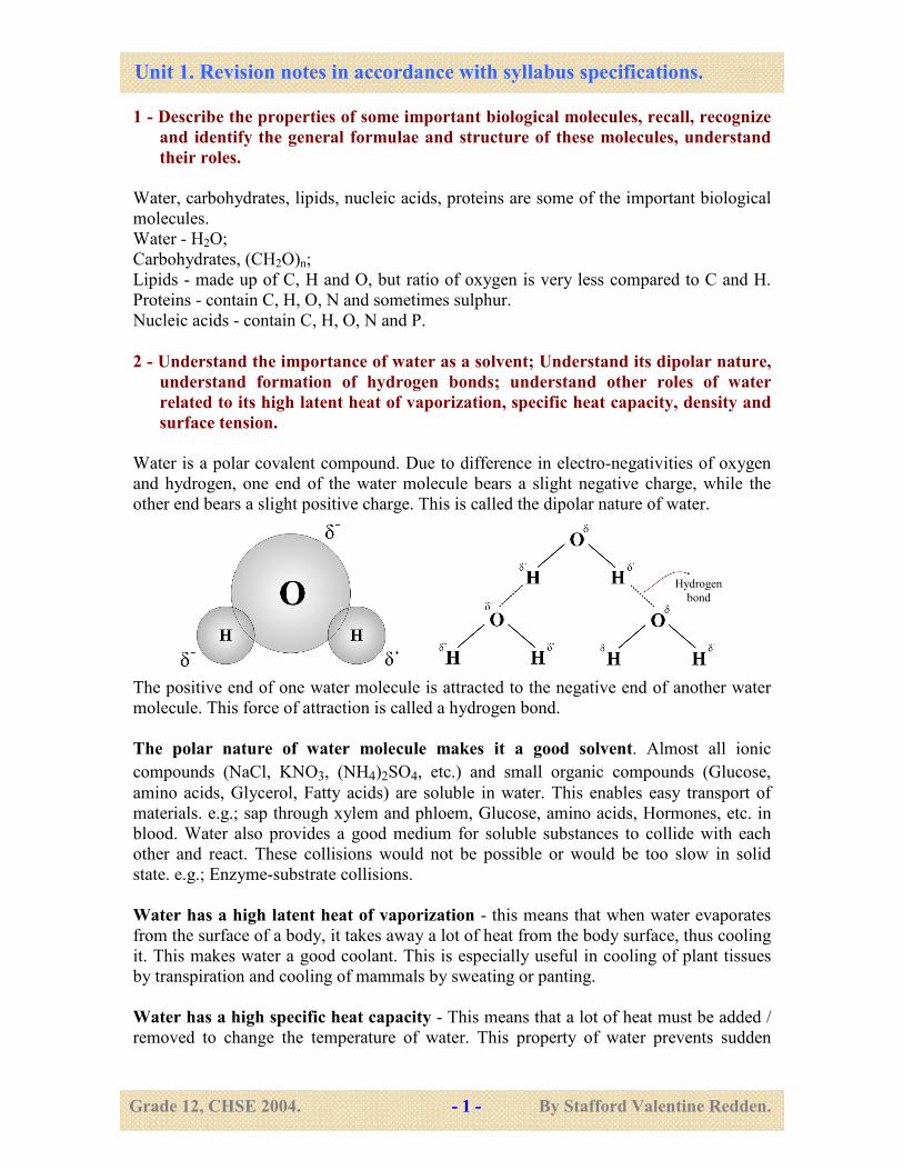
Water is a polar covalent compound. Due to difference in electro-negativities of oxygen
and hydrogen, one end of the water molecule bears a slight negative charge, while the
other end bears a slight positive charge. This is called the dipolar nature of water.
The positive end of one water molecule is attracted to the negative end of another water
molecule. This force of attraction is called a hydrogen bond.
The polar nature of water molecule makes it a good solvent. Almost all ionic
compounds (NaCl, KNO3, (NH4)2SO4, etc.) and small organic compounds (Glucose,
amino acids, Glycerol, Fatty acids) are soluble in water. This enables easy transport of
materials. e.g.; sap through xylem and phloem, Glucose, amino acids, Hormones, etc. in
blood. Water also provides a good medium for soluble substances to collide with each
other and react. These collisions would not be possible or would be too slow in solid
state. e.g.; Enzyme-substrate collisions.
Water has a high latent heat of vaporization - this means that when water evaporates
from the surface of a body, it takes away a lot of heat from the body surface, thus cooling
it. This makes water a good coolant. This is especially useful in cooling of plant tissues
by transpiration and cooling of mammals by sweating or panting.
Water has a high specific heat capacity - This means that a lot of heat must be added /
removed to change the temperature of water. This property of water prevents sudden
Hydrogen
bond
Unit 1. Revision notes in accordance with syllabus specifications.
Grade 12, CHSE 2004. - 2 - By Stafford Valentine Redden.
fluctuations in temperature of organisms or aquatic environment. The gradual change in
temperature gives organisms enough time to cope with the change. The high specific heat
capacity also helps to resist temperature changes (maintain constant temperature).
The density of pure water is 1g/cm3.The density of water changes with temperature. Thus
in aquatic habitats there will be layers of water with different densities. The differences in
density cause the circulation of water and nutrients within the habitat, thus affecting the
vertical distribution of organisms.
Water has maximum density at 40 C. This means that the densest water (at 4
0 C) will
remain at the bottom of an aquatic habitat. This prevents aquatic habitats from freezing
completely, so that aquatic organisms can survive at the bottom (unfrozen at 40 C).
Surface tension is the property of a liquid which makes its surface behave like a
stretched membrane, mainly caused due to hydrogen bonding between molecules (water).
This is especially useful to some aquatic invertebrates that can skate or lay eggs on the
water surface. Mosquito larvae also use the surface tension of water to cling to the
surface and breathe air, through siphons. Surface tension decreases the ease with which
gases dissolve into water.
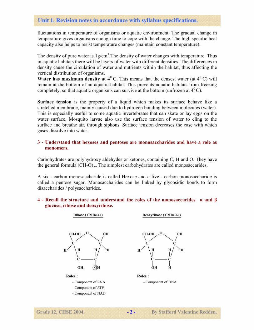
3 - Understand that hexoses and pentoses are monosaccharides and have a role as
monomers.
Carbohydrates are polyhydroxy aldehydes or ketones, containing C, H and O. They have
the general formula (CH2O) n. The simplest carbohydrates are called monosaccarides.
A six - carbon monosaccharide is called Hexose and a five - carbon monosaccharide is
called a pentose sugar. Monosaccharides can be linked by glycosidic bonds to form
disaccharides / polysaccharides.
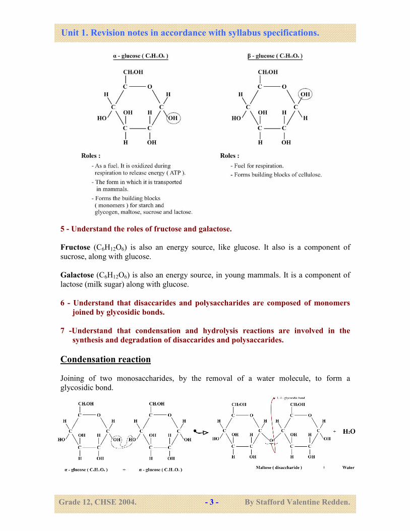
4 - Recall the structure and understand the roles of the monosaccarides α and β
glucose, ribose and deoxyribose.
Unit 1. Revision notes in accordance with syllabus specifications.
Grade 12, CHSE 2004. - 3 - By Stafford Valentine Redden.
5 - Understand the roles of fructose and galactose.
Fructose (C6H12O6) is also an energy source, like glucose. It also is a component of
sucrose, along with glucose.
Galactose (C6H12O6) is also an energy source, in young mammals. It is a component of
lactose (milk sugar) along with glucose.
6 - Understand that disaccarides and polysaccharides are composed of monomers
joined by glycosidic bonds.
7 -Understand that condensation and hydrolysis reactions are involved in the
synthesis and degradation of disaccarides and polysaccarides.
Condensation reaction
Joining of two monosaccharides, by the removal of a water molecule, to form a
glycosidic bond.
Unit 1. Revision notes in accordance with syllabus specifications.
Grade 12, CHSE 2004. - 4 - By Stafford Valentine Redden.
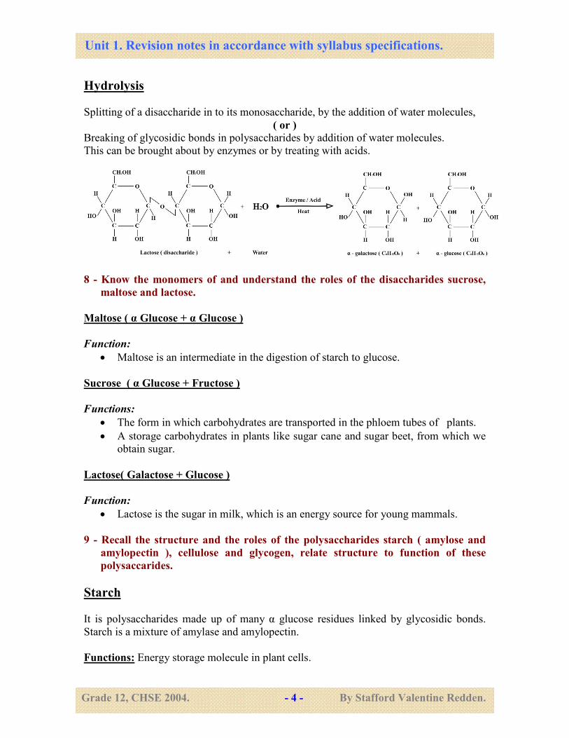
Hydrolysis
Splitting of a disaccharide in to its monosaccharide, by the addition of water molecules,
( or )
Breaking of glycosidic bonds in polysaccharides by addition of water molecules.
This can be brought about by enzymes or by treating with acids.
8 - Know the monomers of and understand the roles of the disaccharides sucrose,
maltose and lactose.
Maltose ( α Glucose + α Glucose )
Function:
• Maltose is an intermediate in the digestion of starch to glucose.
Sucrose ( α Glucose + Fructose )
Functions:
• The form in which carbohydrates are transported in the phloem tubes of plants.
• A storage carbohydrates in plants like sugar cane and sugar beet, from which we
obtain sugar.
Lactose( Galactose + Glucose )
Function:
• Lactose is the sugar in milk, which is an energy source for young mammals.
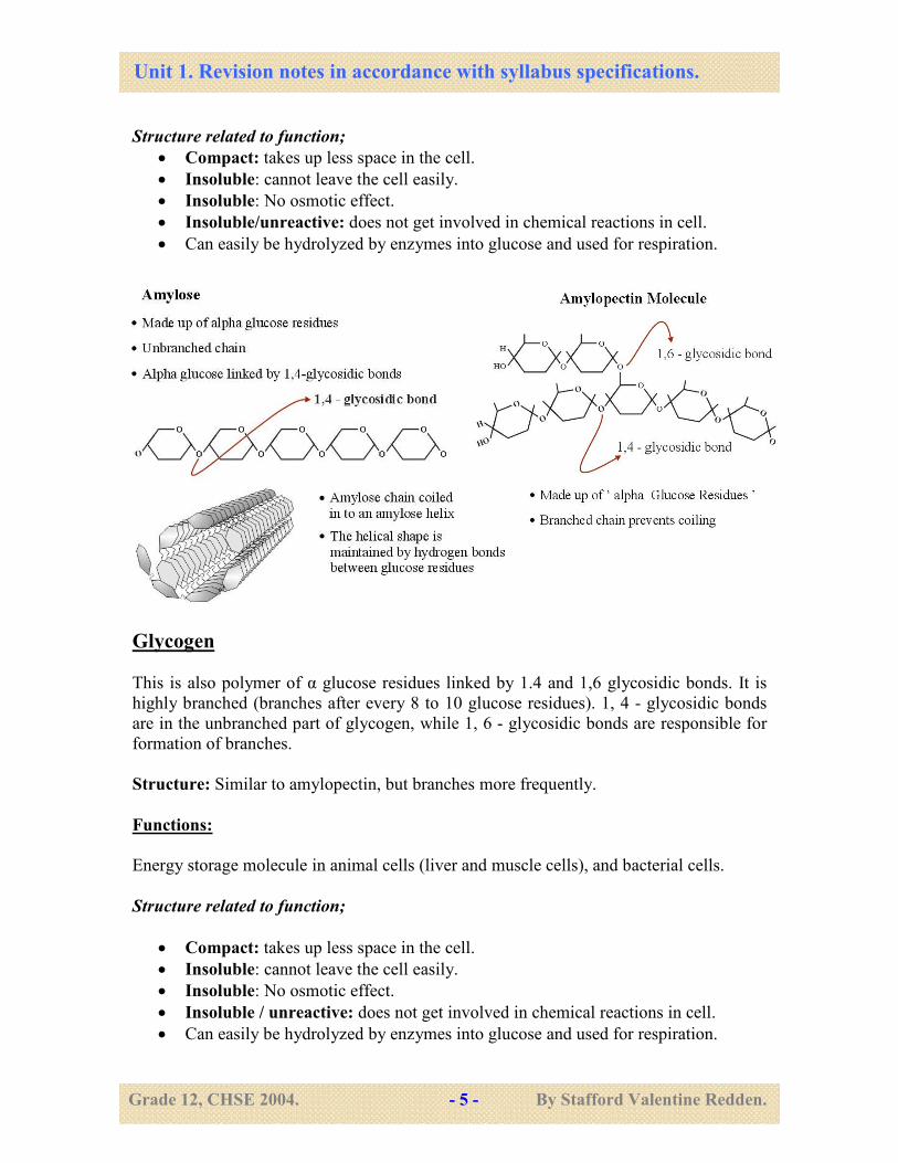
9 - Recall the structure and the roles of the polysaccharides starch ( amylose and
amylopectin ), cellulose and glycogen, relate structure to function of these
polysaccarides.
Starch
It is polysaccharides made up of many α glucose residues linked by glycosidic bonds.
Starch is a mixture of amylase and amylopectin.
Functions: Energy storage molecule in plant cells.
Unit 1. Revision notes in accordance with syllabus specifications.
Grade 12, CHSE 2004. - 5 - By Stafford Valentine Redden.
Structure related to function;
• Compact: takes up less space in the cell.
• Insoluble: cannot leave the cell easily.
• Insoluble: No osmotic effect.
• Insoluble/unreactive: does not get involved in chemical reactions in cell.
• Can easily be hydrolyzed by enzymes into glucose and used for respiration.
Glycogen
This is also polymer of α glucose residues linked by 1.4 and 1,6 glycosidic bonds. It is
highly branched (branches after every 8 to 10 glucose residues). 1, 4 - glycosidic bonds
are in the unbranched part of glycogen, while 1, 6 - glycosidic bonds are responsible for
formation of branches.
Structure: Similar to amylopectin, but branches more frequently.
Functions:
Energy storage molecule in animal cells (liver and muscle cells), and bacterial cells.
Structure related to function;
• Compact: takes up less space in the cell.
• Insoluble: cannot leave the cell easily.
• Insoluble: No osmotic effect.
• Insoluble / unreactive: does not get involved in chemical reactions in cell.
• Can easily be hydrolyzed by enzymes into glucose and used for respiration.
Unit 1. Revision notes in accordance with syllabus specifications.
Grade 12, CHSE 2004. - 6 - By Stafford Valentine Redden.
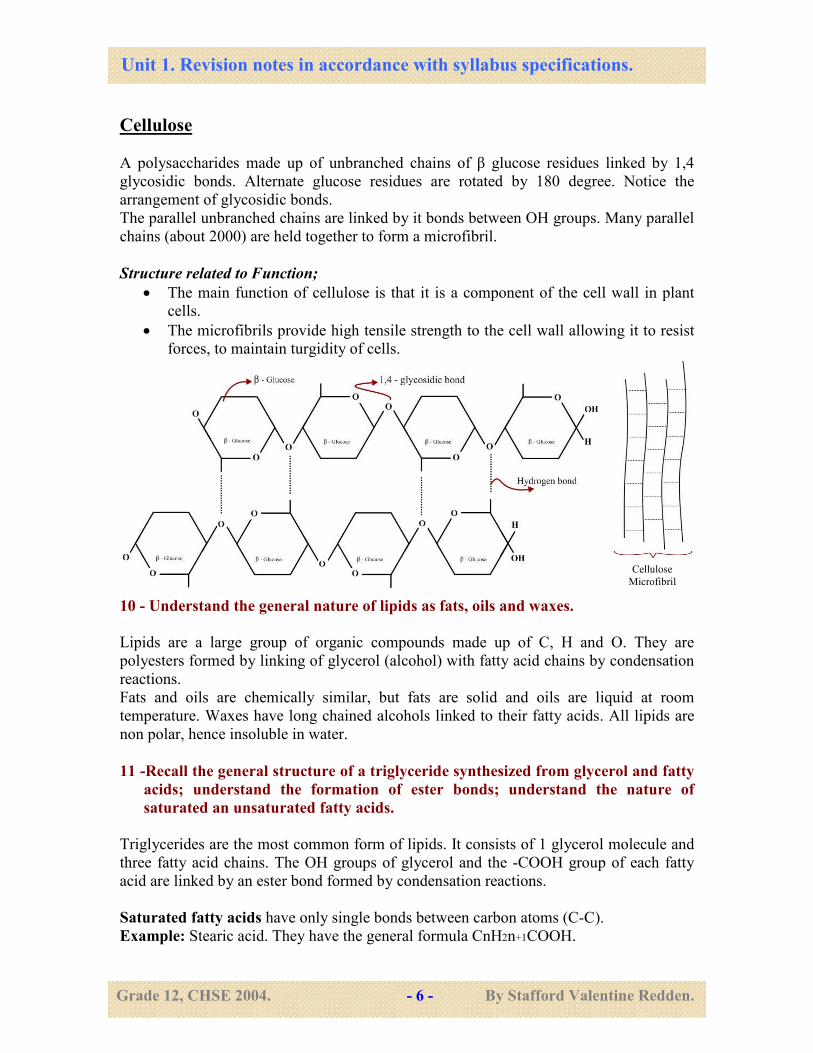
Cellulose
A polysaccharides made up of unbranched chains of β glucose residues linked by 1,4
glycosidic bonds. Alternate glucose residues are rotated by 180 degree. Notice the
arrangement of glycosidic bonds.
The parallel unbranched chains are linked by it bonds between OH groups. Many parallel
chains (about 2000) are held together to form a microfibril.
Structure related to Function;
• The main function of cellulose is that it is a component of the cell wall in plant
cells.
• The microfibrils provide high tensile strength to the cell wall allowing it to resist
forces, to maintain turgidity of cells.
10 - Understand the general nature of lipids as fats, oils and waxes.
Lipids are a large group of organic compounds made up of C, H and O. They are
polyesters formed by linking of glycerol (alcohol) with fatty acid chains by condensation
reactions.
Fats and oils are chemically similar, but fats are solid and oils are liquid at room
temperature. Waxes have long chained alcohols linked to their fatty acids. All lipids are
non polar, hence insoluble in water.
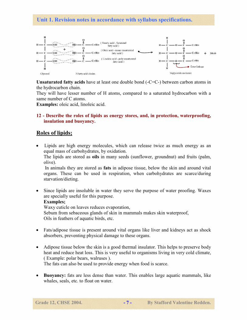
11 -Recall the general structure of a triglyceride synthesized from glycerol and fatty
acids; understand the formation of ester bonds; understand the nature of
saturated an unsaturated fatty acids.
Triglycerides are the most common form of lipids. It consists of 1 glycerol molecule and
three fatty acid chains. The OH groups of glycerol and the -COOH group of each fatty
acid are linked by an ester bond formed by condensation reactions.
Saturated fatty acids have only single bonds between carbon atoms (C-C).
Example: Stearic acid. They have the general formula CnH2n+1COOH.
Cellulose
Microfibril
Unit 1. Revision notes in accordance with syllabus specifications.
Grade 12, CHSE 2004. - 7 - By Stafford Valentine Redden.
Unsaturated fatty acids have at least one double bond (-C=C-) between carbon atoms in
the hydrocarbon chain.
They will have lesser number of H atoms, compared to a saturated hydrocarbon with a
same number of C atoms.
Examples: oleic acid, linoleic acid.
12 - Describe the roles of lipids as energy stores, and, in protection, waterproofing,
insulation and buoyancy.
Roles of lipids;
• Lipids are high energy molecules, which can release twice as much energy as an
equal mass of carbohydrates, by oxidation.
The lipids are stored as oils in many seeds (sunflower, groundnut) and fruits (palm,
olive).
In animals they are stored as fats in adipose tissue, below the skin and around vital
organs. These can be used in respiration, when carbohydrates are scarce/during
starvation/dieting.
• Since lipids are insoluble in water they serve the purpose of water proofing. Waxes
are specially useful for this purpose.
Examples; Waxy cuticle on leaves reduces evaporation,
Sebum from sebaceous glands of skin in mammals makes skin waterproof,
Oils in feathers of aquatic birds, etc.
• Fats/adipose tissue is present around vital organs like liver and kidneys act as shock
absorbers, preventing physical damage to these organs.
• Adipose tissue below the skin is a good thermal insulator. This helps to preserve body
heat and reduce heat loss. This is very useful to organisms living in very cold climate,
( Example: polar bears, walruses ).
The fats can also be used to provide energy when food is scarce.
• Buoyancy: fats are less dense than water. This enables large aquatic mammals, like
whales, seals, etc. to float on water.
Unit 1. Revision notes in accordance with syllabus specifications.
Grade 12, CHSE 2004. - 8 - By Stafford Valentine Redden.
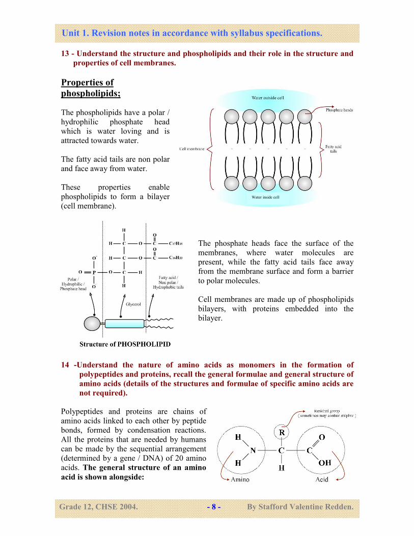
13 - Understand the structure and phospholipids and their role in the structure and
properties of cell membranes.
Properties of
phospholipids;
The phospholipids have a polar /
hydrophilic phosphate head
which is water loving and is
attracted towards water.
The fatty acid tails are non polar
and face away from water.
These properties enable
phospholipids to form a bilayer
(cell membrane).
The phosphate heads face the surface of the
membranes, where water molecules are
present, while the fatty acid tails face away
from the membrane surface and form a barrier
to polar molecules.
Cell membranes are made up of phospholipids
bilayers, with proteins embedded into the
bilayer.
14 -Understand the nature of amino acids as monomers in the formation of
polypeptides and proteins, recall the general formulae and general structure of
amino acids (details of the structures and formulae of specific amino acids are
not required).
Polypeptides and proteins are chains of
amino acids linked to each other by peptide
bonds, formed by condensation reactions.
All the proteins that are needed by humans
can be made by the sequential arrangement
(determined by a gene / DNA) of 20 amino
acids. The general structure of an amino
acid is shown alongside:
Water inside cell
Unit 1. Revision notes in accordance with syllabus specifications.
Grade 12, CHSE 2004. - 9 - By Stafford Valentine Redden.
-NH2 and -COOH groups are involved in the formation of peptide bonds.
R - groups are involved in hydrogen bond, ionic bond and covalent bond formation.
15 -Understand that amino acids are linked by peptide bonds to form polypeptides;
describe the formation of a peptide bond.
17 -Understand that condensation and hydrolysis reactions are involved in the
synthesis and degradation of polypeptides and proteins.
Amino acids are linked to each other by the formation of a peptide bond. The bond
forms by a condensation reaction between the -COOH group of one amino acid and the
-NH2 group of another amino acid.
Two amino acids linked by a peptide bond is called a peptide, while many amino acids
linked by peptide bonds is a polypeptide.
• Condensation is the joining of two amino acids by the removal of a water molecule
(forms a peptide bond).
• Hydrolysis is the splitting of a dipeptide / polypeptide by the addition of water
molecules (breaks peptide bonds).
16 -Understand the meaning of the terms primary, secondary, tertiary and
quaternary structure and their importance in the structure of enzymes.
Primary structure of a protein
The primary structure is the sequence of amino acids in a polypeptide chain. This
sequence is determined by the genetic code on DNA.
The primary structure determines the secondary, tertiary or quaternary structure of a
protein.
Examples:
The eventual shape and function of both polypeptide chains is going to be different.
Unit 1. Revision notes in accordance with syllabus specifications.
Grade 12, CHSE 2004. - 10 - By Stafford Valentine Redden.
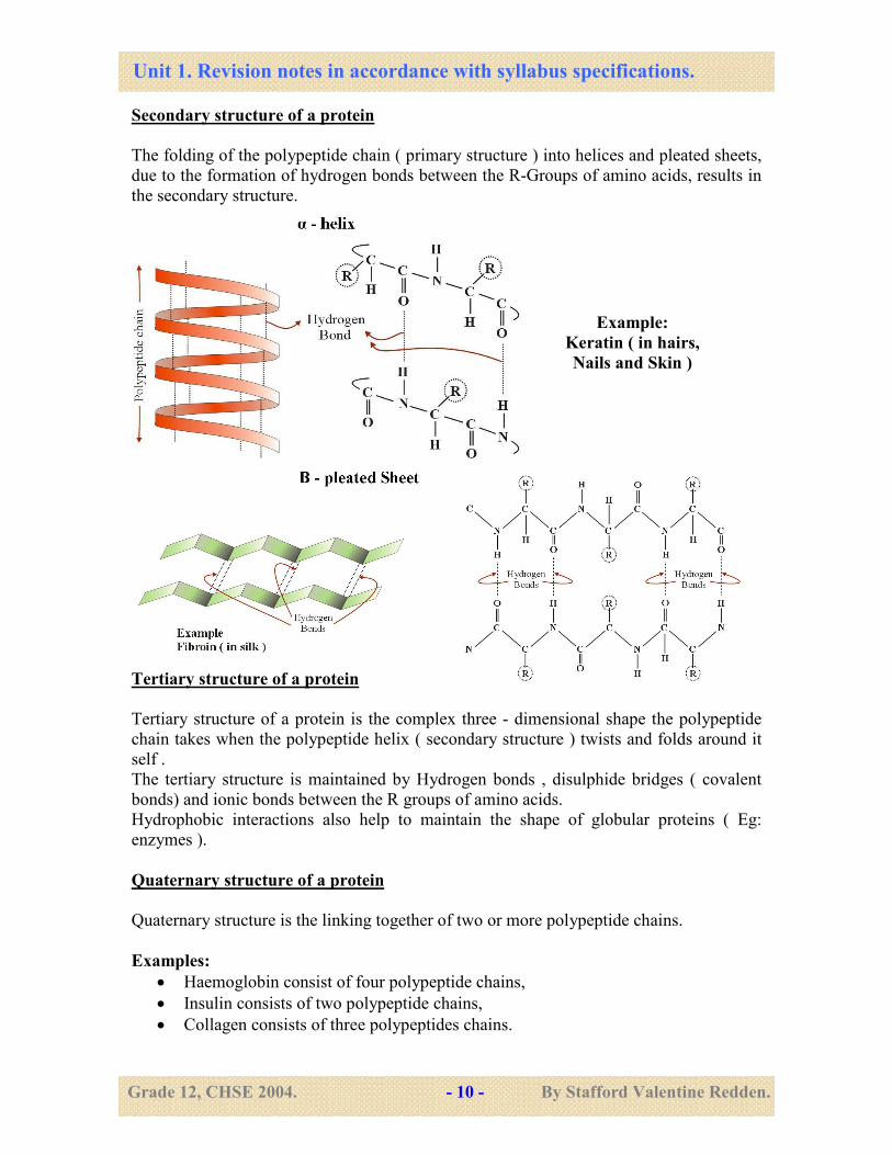
Secondary structure of a protein
The folding of the polypeptide chain ( primary structure ) into helices and pleated sheets,
due to the formation of hydrogen bonds between the R-Groups of amino acids, results in
the secondary structure.
Tertiary structure of a protein
Tertiary structure of a protein is the complex three - dimensional shape the polypeptide
chain takes when the polypeptide helix ( secondary structure ) twists and folds around it
self .
The tertiary structure is maintained by Hydrogen bonds , disulphide bridges ( covalent
bonds) and ionic bonds between the R groups of amino acids.
Hydrophobic interactions also help to maintain the shape of globular proteins ( Eg:
enzymes ).
Quaternary structure of a protein
Quaternary structure is the linking together of two or more polypeptide chains.
Examples:
• Haemoglobin consist of four polypeptide chains,
• Insulin consists of two polypeptide chains,
• Collagen consists of three polypeptides chains.
Example:
Keratin ( in hairs,
Nails and Skin )
Unit 1. Revision notes in accordance with syllabus specifications.
Grade 12, CHSE 2004. - 11 - By Stafford Valentine Redden.
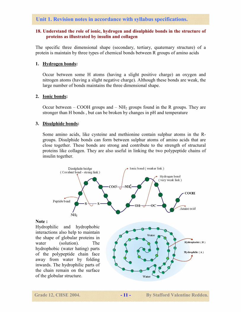
18. Understand the role of ionic, hydrogen and disulphide bonds in the structure of
proteins as illustrated by insulin and collagen
The specific three dimensional shape (secondary, tertiary, quaternary structure) of a
protein is maintain by three types of chemical bonds between R groups of amino acids
1. Hydrogen bonds:
Occur between some H atoms (having a slight positive charge) an oxygen and
nitrogen atoms (having a slight negative charge). Although these bonds are weak, the
large number of bonds maintains the three dimensional shape.
2. Ionic bonds:
Occur between – COOH groups and – NH2 groups found in the R groups. They are
stronger than H bonds , but can be broken by changes in pH and temperature
3. Disulphide bonds:
Some amino acids, like cysteine and methionine contain sulphur atoms in the R-
groups. Disulphide bonds can form between sulphur atoms of amino acids that are
close together. These bonds are strong and contribute to the strength of structural
proteins like collagen. They are also useful in linking the two polypeptide chains of
insulin together.
Note :
Hydrophilic and hydrophobic
interactions also help to maintain
the shape of globular proteins in
water (solution). The
hydrophobic (water hating) parts
of the polypeptide chain face
away from water by folding
inwards. The hydrophilic parts of
the chain remain on the surface
of the globular structure.
Unit 1. Revision notes in accordance with syllabus specifications.
Grade 12, CHSE 2004. - 12 - By Stafford Valentine Redden.
19. Understand the nature and roles of fibrous and globular proteins as illustrated
by collagen and insulin
Insulin is a globular protein. It is made up of two polypeptide chains which are linked to
each other by two disulphide bridges (bonds) - Quaternary structure.
The polypeptide chains are highly twisted (tertiary structure) and rolled up in to a globule
when dissolved in water (hydrophobic interactions).
Collagen is a fibrous protein. It is made up of three polypeptide chains (quaternary
structure) each polypeptide chain is twisted to form a helix. The three polypeptide helices
wind around each other like a rope with three strands. Hydrogen bonds hold the three
strands in place. This makes collagen very stable, insoluble, flexible, but inelastic.
Collagen is found mainly in tendons and bones.
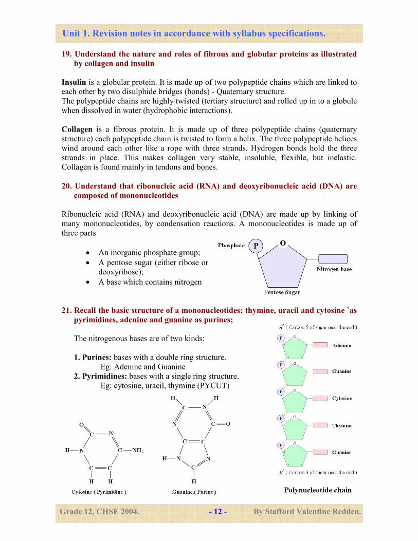
20. Understand that ribonucleic acid (RNA) and deoxyribonucleic acid (DNA) are
composed of mononucleotides
Ribonucleic acid (RNA) and deoxyribonucleic acid (DNA) are made up by linking of
many mononucleotides, by condensation reactions. A mononucleotides is made up of
three parts
• An inorganic phosphate group;
• A pentose sugar (either ribose or
deoxyribose);
• A base which contains nitrogen
21. Recall the basic structure of a mononucleotides; thymine, uracil and cytosine `as
pyrimidines, adcnine and guanine as purines;
The nitrogenous bases are of two kinds:
1. Purines: bases with a double ring structure.
Eg: Adenine and Guanine
2. Pyrimidines: bases with a single ring structure.
Eg: cytosine, uracil, thymine (PYCUT)
Unit 1. Revision notes in accordance with syllabus specifications.
Grade 12, CHSE 2004. - 13 - By Stafford Valentine Redden.
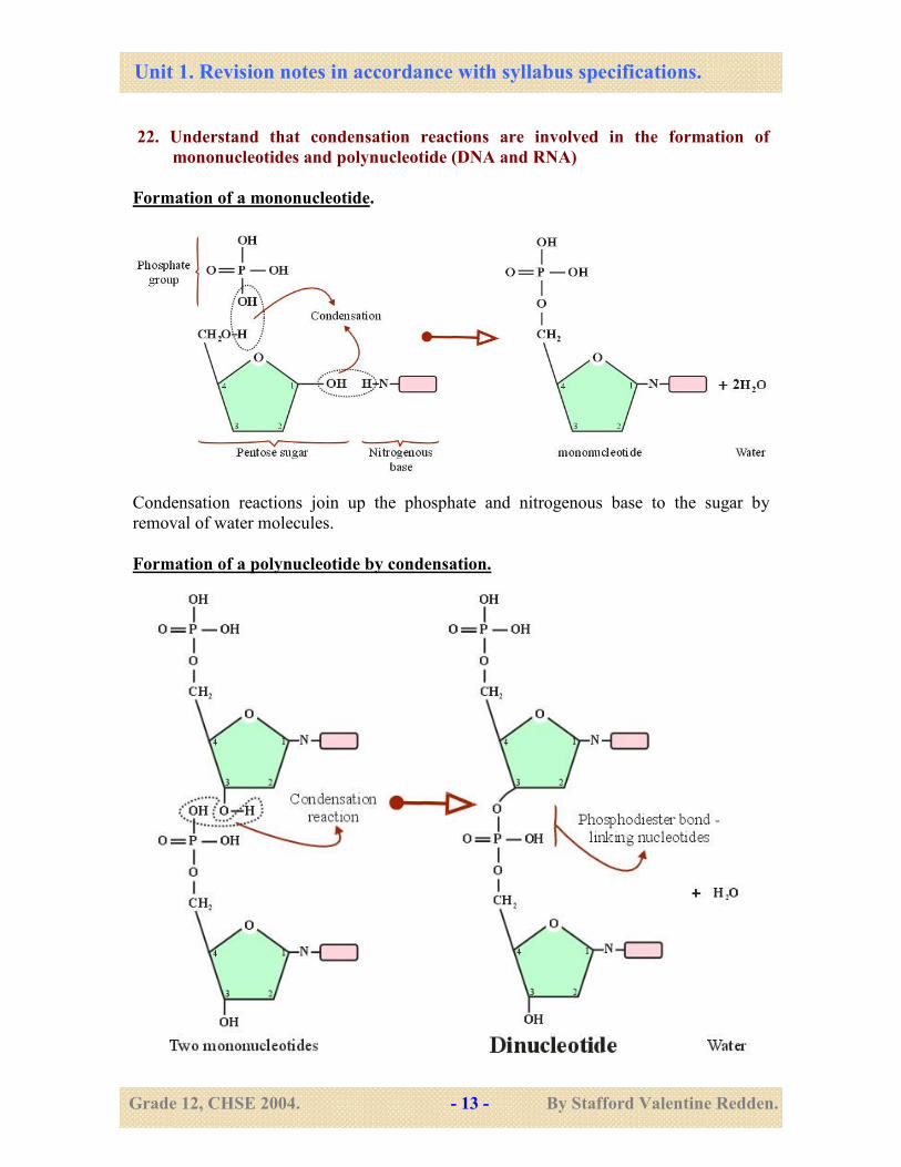
22. Understand that condensation reactions are involved in the formation of
mononucleotides and polynucleotide (DNA and RNA)
Formation of a mononucleotide.
Condensation reactions join up the phosphate and nitrogenous base to the sugar by
removal of water molecules.
Formation of a polynucleotide by condensation.
Unit 1. Revision notes in accordance with syllabus specifications.
Grade 12, CHSE 2004. - 14 - By Stafford Valentine Redden.
• Many nucleotides can be linked by phosphodiester bonds to form a polynucleotide.
• Condensation reactions are involved in the formation of phosphodiester bonds.
• Carbon 5’ of pentose binds with carbon 3’of pentose on another nucleotide by a
phosphate group.
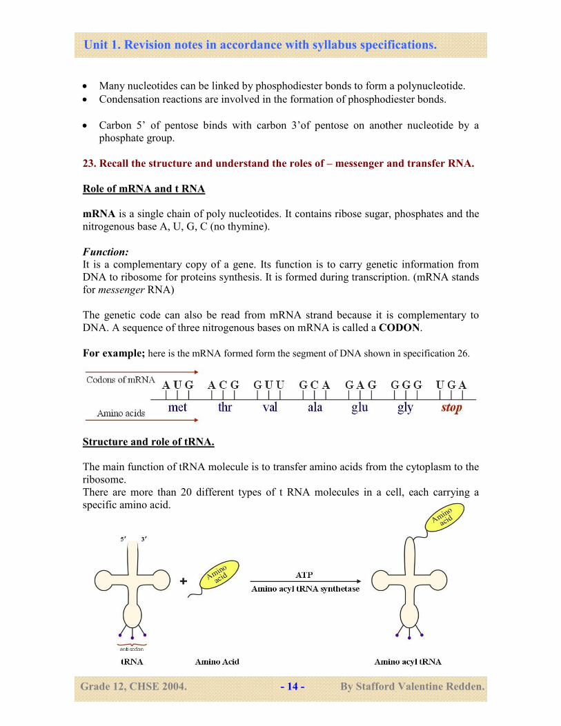
23. Recall the structure and understand the roles of – messenger and transfer RNA.
Role of mRNA and t RNA
mRNA is a single chain of poly nucleotides. It contains ribose sugar, phosphates and the
nitrogenous base A, U, G, C (no thymine).
Function:
It is a complementary copy of a gene. Its function is to carry genetic information from
DNA to ribosome for proteins synthesis. It is formed during transcription. (mRNA stands
for messenger RNA)
The genetic code can also be read from mRNA strand because it is complementary to
DNA. A sequence of three nitrogenous bases on mRNA is called a CODON.
For example; here is the mRNA formed form the segment of DNA shown in specification 26.
Structure and role of tRNA.
The main function of tRNA molecule is to transfer amino acids from the cytoplasm to the
ribosome.
There are more than 20 different types of t RNA molecules in a cell, each carrying a
specific amino acid.
Unit 1. Revision notes in accordance with syllabus specifications.
Grade 12, CHSE 2004. - 15 - By Stafford Valentine Redden.
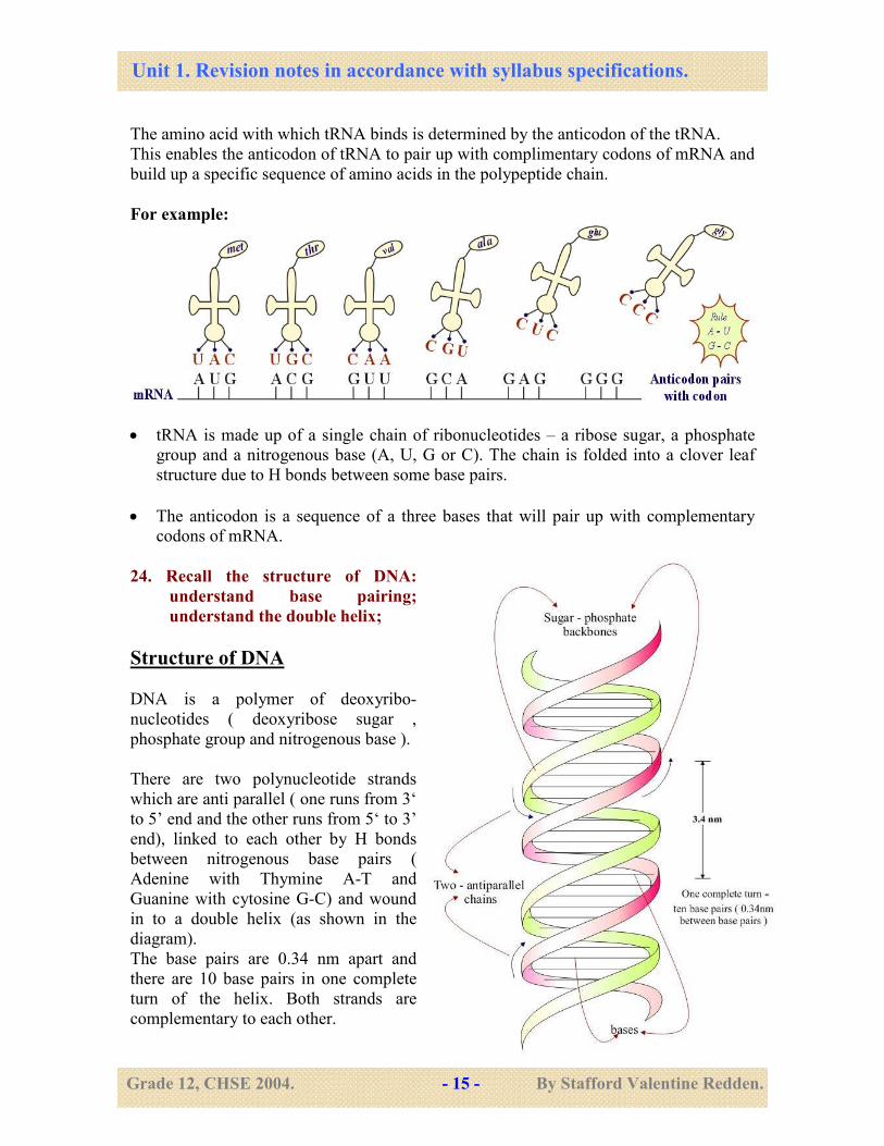
The amino acid with which tRNA binds is determined by the anticodon of the tRNA.
This enables the anticodon of tRNA to pair up with complimentary codons of mRNA and
build up a specific sequence of amino acids in the polypeptide chain.
For example:
• tRNA is made up of a single chain of ribonucleotides – a ribose sugar, a phosphate
group and a nitrogenous base (A, U, G or C). The chain is folded into a clover leaf
structure due to H bonds between some base pairs.
• The anticodon is a sequence of a three bases that will pair up with complementary
codons of mRNA.
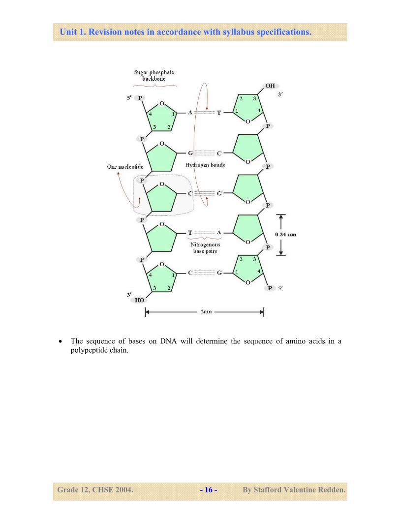
24. Recall the structure of DNA:
understand base pairing;
understand the double helix;
Structure of DNA
DNA is a polymer of deoxyribo-
nucleotides ( deoxyribose sugar ,
phosphate group and nitrogenous base ).
There are two polynucleotide strands
which are anti parallel ( one runs from 3‘
to 5’ end and the other runs from 5‘ to 3’
end), linked to each other by H bonds
between nitrogenous base pairs (
Adenine with Thymine A-T and
Guanine with cytosine G-C) and wound
in to a double helix (as shown in the
diagram).
The base pairs are 0.34 nm apart and
there are 10 base pairs in one complete
turn of the helix. Both strands are
complementary to each other.
Unit 1. Revision notes in accordance with syllabus specifications.
Grade 12, CHSE 2004. - 16 - By Stafford Valentine Redden.
• The sequence of bases on DNA will determine the sequence of amino acids in a
polypeptide chain.
Unit 1. Revision notes in accordance with syllabus specifications.
Grade 12, CHSE 2004. - 17 - By Stafford Valentine Redden.
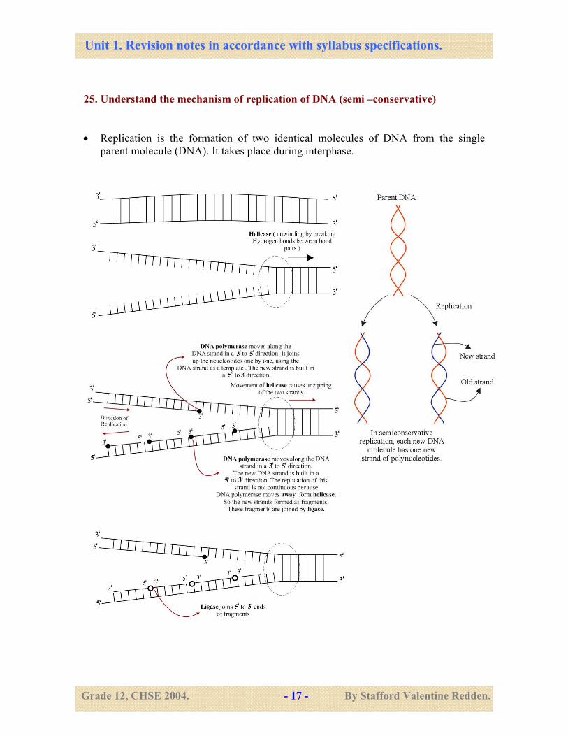
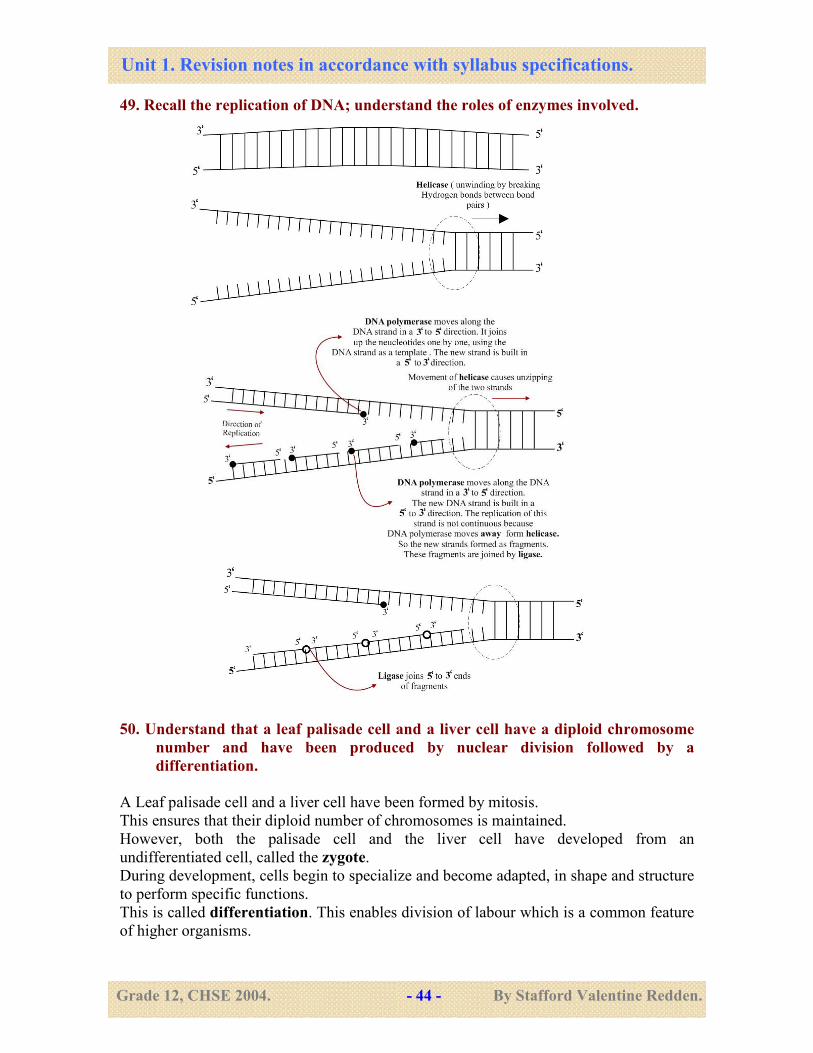
25. Understand the mechanism of replication of DNA (semi –conservative)
• Replication is the formation of two identical molecules of DNA from the single
parent molecule (DNA). It takes place during interphase.
Unit 1. Revision notes in accordance with syllabus specifications.
Grade 12, CHSE 2004. - 18 - By Stafford Valentine Redden.
26. Understand the nature of the genetic code; understand that gene is a sequence of
bases on the DNA molecule which codes for a sequence of amino acids in a
polypeptide chain;
The genetic code is the sequence of bases on DNA that determines the sequence of amino
acids in a polypeptide chain (primary protein structure).
• The genetic code is a triplet code - this means that a sequence of three nitrogenous
base ( triplet ) on DNA , codes for a single amino acid. eg;
• The genetic code is degenerate - this means that a given amino acid can be coded for
by more than one triplet code.
Eg: GUC, GCC, GCA, GCG all code for the amino acid alanine.
• The genetic code is universal - The same triplets code for the same amino acids in
all organisms (except for a few triplets in mitochondrial DNA and ancient bacteria).
What is a Gene?
A gene is a sequence of bases on DNA that codes for a sequence of amino acids in a
polypeptide chain (primary protein structure).
27. Understand the processes of transcription and translation in the synthesis of
proteins; understand that amino acid sequences are specified by DNA and know
the function of the ribosomes; understand the codons and anticodons in relation
to messenger and transfer RNA.
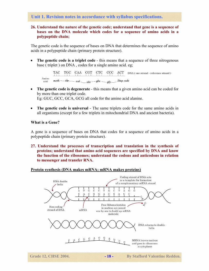
Protein synthesis (DNA makes mRNA; mRNA makes proteins)
Unit 1. Revision notes in accordance with syllabus specifications.
Grade 12, CHSE 2004. - 19 - By Stafford Valentine Redden.
Transcription
This is the making of mRNA from DNA.
A length of DNA (a gene) is copied into an mRNA molecule.
o The unwinding of the double helix (by breaking H bonds) and the linking up of
ribonucleotides to form mRNA is catalyzed by the enzyme RNA polymerase.
o Before leaving the nucleus, some parts of mRNA are cut of. These parts remain in the
nucleus and are called introns. The remaining nucleotides rejoin and are called exons.
The exons (mRNA) leaves the nucleus.
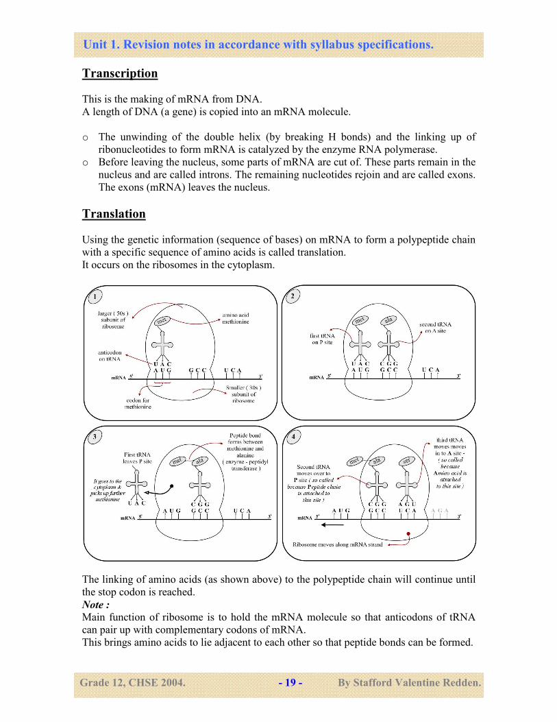
Translation
Using the genetic information (sequence of bases) on mRNA to form a polypeptide chain
with a specific sequence of amino acids is called translation.
It occurs on the ribosomes in the cytoplasm.
The linking of amino acids (as shown above) to the polypeptide chain will continue until
the stop codon is reached.
Note :
Main function of ribosome is to hold the mRNA molecule so that anticodons of tRNA
can pair up with complementary codons of mRNA.
This brings amino acids to lie adjacent to each other so that peptide bonds can be formed.
Unit 1. Revision notes in accordance with syllabus specifications.
Grade 12, CHSE 2004. - 20 - By Stafford Valentine Redden.
28 - Appreciate the Human Genome Project in the light of the structure and roles of
nucleic acids; consider the spiritual, moral, ethical, social and cultural issues of
this project.
Human Genome Project (HGP) - Goal: to identify the chromosomal location of every
human gene and to determine the precise chemical structure (sequence of bases) and its
application in health and diseases. It has revealed that the human genome has about
30,000 to 40,000 genes. The main role of these genes is to produce proteins.
Social, ethical, legal issues to be considered: the HGP has brought up many social,
legal and ethical complications that have to be considered. Issues like who should be
given access to an individuals’ genome? Will there be discrimination on the basis of the
type of genes that an individual possesses? Will parents abort children if their genome is
undesirable? Will insurance companies refuse to insure people who have Huntington’s
genes? Will employment opportunities be based on the genes of intelligence?
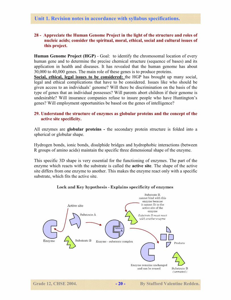
29. Understand the structure of enzymes as globular proteins and the concept of the
active site specificity.
All enzymes are globular proteins - the secondary protein structure is folded into a
spherical or globular shape.
Hydrogen bonds, ionic bonds, disulphide bridges and hydrophobic interactions (between
R groups of amino acids) maintain the specific three dimensional shape of the enzyme.
This specific 3D shape is very essential for the functioning of enzymes. The part of the
enzyme which reacts with the substrate is called the active site. The shape of the active
site differs from one enzyme to another. This makes the enzyme react only with a specific
substrate, which fits the active site.
Unit 1. Revision notes in accordance with syllabus specifications.
Grade 12, CHSE 2004. - 21 - By Stafford Valentine Redden.
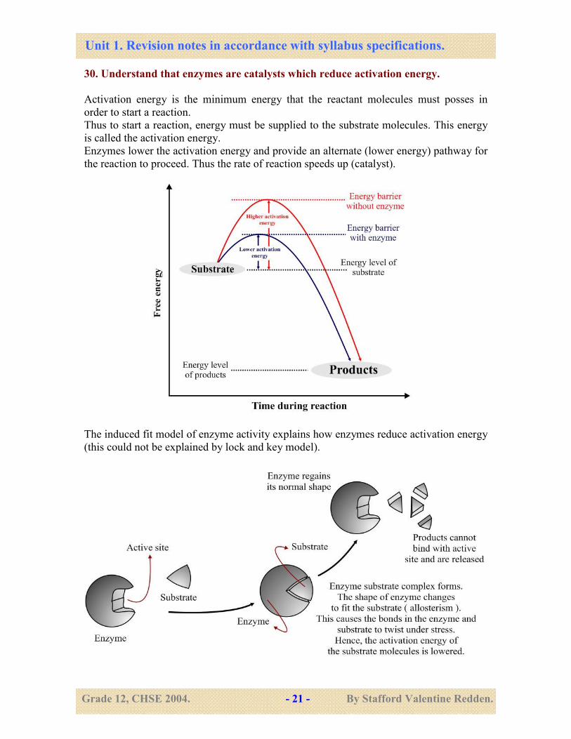
30. Understand that enzymes are catalysts which reduce activation energy.
Activation energy is the minimum energy that the reactant molecules must posses in
order to start a reaction.
Thus to start a reaction, energy must be supplied to the substrate molecules. This energy
is called the activation energy.
Enzymes lower the activation energy and provide an alternate (lower energy) pathway for
the reaction to proceed. Thus the rate of reaction speeds up (catalyst).
The induced fit model of enzyme activity explains how enzymes reduce activation energy
(this could not be explained by lock and key model).
Unit 1. Revision notes in accordance with syllabus specifications.
Grade 12, CHSE 2004. - 22 - By Stafford Valentine Redden.
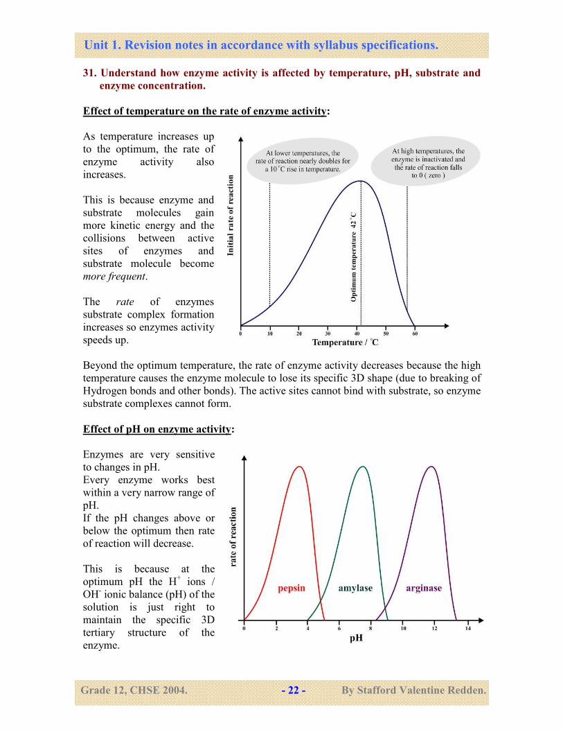
31. Understand how enzyme activity is affected by temperature, pH, substrate and
enzyme concentration.
Effect of temperature on the rate of enzyme activity:
As temperature increases up
to the optimum, the rate of
enzyme activity also
increases.
This is because enzyme and
substrate molecules gain
more kinetic energy and the
collisions between active
sites of enzymes and
substrate molecule become
more frequent.
The rate of enzymes
substrate complex formation
increases so enzymes activity
speeds up.
Beyond the optimum temperature, the rate of enzyme activity decreases because the high
temperature causes the enzyme molecule to lose its specific 3D shape (due to breaking of
Hydrogen bonds and other bonds). The active sites cannot bind with substrate, so enzyme
substrate complexes cannot form.
Effect of pH on enzyme activity:
Enzymes are very sensitive
to changes in pH.
Every enzyme works best
within a very narrow range of
pH.
If the pH changes above or
below the optimum then rate
of reaction will decrease.
This is because at the
optimum pH the H+ ions /
OH- ionic balance (pH) of the
solution is just right to
maintain the specific 3D
tertiary structure of the
enzyme.
Unit 1. Revision notes in accordance with syllabus specifications.
Grade 12, CHSE 2004. - 23 - By Stafford Valentine Redden.
If pH changes then the change in H+ ion concentration will disrupt the H bonds and the
ionic bonds which maintain the tertiary structure of the enzyme.
This will cause the enzyme to change its shape so that the active site cannot bind with
substrate molecules.
Enzyme substrate complexes will form at a slower rate, so rate of enzyme activity
decreases / stops.
Enzyme concentration:
As the enzyme concentration
increases (at constant substrate
concentration) the rate of
reaction increases until it reaches
a maximum rate (V max).
This is because there will be
more number of free active sites,
at any given time.
So the rate of enzyme substrate
complex formation increases.
Thus rate of reaction increases.
The rate doesn’t increase beyond
the V max because the substrate
concentration becomes a limiting
factor.
Even though there will be many free active sites, there will not be enough substrate
molecules to bind with them. So, rate of enzyme substrate complex formation remains
constant at V max.
But, increasing substrate concentration would further increase the rate of reaction.
Substrate concentration:
Rate will increase further if
enzyme concentration is
increased.
Unit 1. Revision notes in accordance with syllabus specifications.
Grade 12, CHSE 2004. - 24 - By Stafford Valentine Redden.
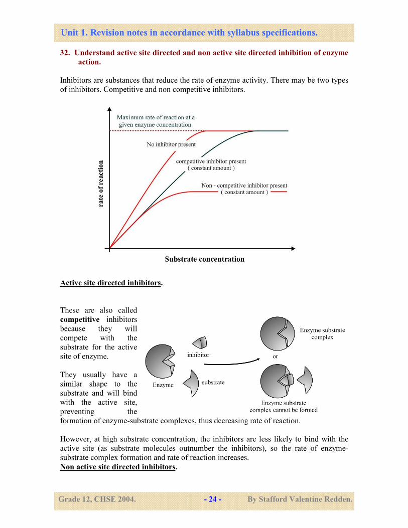
32. Understand active site directed and non active site directed inhibition of enzyme
action.
Inhibitors are substances that reduce the rate of enzyme activity. There may be two types
of inhibitors. Competitive and non competitive inhibitors.
Active site directed inhibitors.
These are also called
competitive inhibitors
because they will
compete with the
substrate for the active
site of enzyme.
They usually have a
similar shape to the
substrate and will bind
with the active site,
preventing the
formation of enzyme-substrate complexes, thus decreasing rate of reaction.
However, at high substrate concentration, the inhibitors are less likely to bind with the
active site (as substrate molecules outnumber the inhibitors), so the rate of enzyme-
substrate complex formation and rate of reaction increases.
Non active site directed inhibitors.
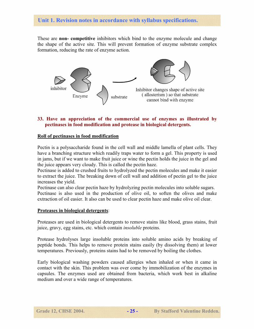
Unit 1. Revision notes in accordance with syllabus specifications.
Grade 12, CHSE 2004. - 25 - By Stafford Valentine Redden.
These are non- competitive inhibitors which bind to the enzyme molecule and change
the shape of the active site. This will prevent formation of enzyme substrate complex
formation, reducing the rate of enzyme action.
33. Have an appreciation of the commercial use of enzymes as illustrated by
pectinases in food modification and protease in biological detergents.
Roll of pectinases in food modification
Pectin is a polysaccharide found in the cell wall and middle lamella of plant cells. They
have a branching structure which readily traps water to form a gel. This property is used
in jams, but if we want to make fruit juice or wine the pectin holds the juice in the gel and
the juice appears very cloudy. This is called the pectin haze.
Pectinase is added to crushed fruits to hydrolyzed the pectin molecules and make it easier
to extract the juice. The breaking down of cell wall and addition of pectin gel to the juice
increases the yield.
Pectinase can also clear pectin haze by hydrolyzing pectin molecules into soluble sugars.
Pectinase is also used in the production of olive oil, to soften the olives and make
extraction of oil easier. It also can be used to clear pectin haze and make olive oil clear.
Proteases in biological detergents:
Proteases are used in biological detergents to remove stains like blood, grass stains, fruit
juice, gravy, egg stains, etc. which contain insoluble proteins.
Protease hydrolyses large insoluble proteins into soluble amino acids by breaking of
peptide bonds. This helps to remove protein stains easily (by dissolving them) at lower
temperatures. Previously, proteins stains had to be removed by boiling the clothes.
Early biological washing powders caused allergies when inhaled or when it came in
contact with the skin. This problem was over come by immobilization of the enzymes in
capsules. The enzymes used are obtained from bacteria, which work best in alkaline
medium and over a wide range of temperatures.
Unit 1. Revision notes in accordance with syllabus specifications.
Grade 12, CHSE 2004. - 26 - By Stafford Valentine Redden.
34. Discuss the advantages of the immobilization of commercial enzymes, as
illustrated by lactase.
Immobilization of enzymes (holding enzymes in place):
Immobilization is a process by which enzymes are trapped in insoluble material such as
beads of alginate or in cellulose fibers. The main advantages of using immobilized
enzymes are:
The enzymes can be re used: this reduces the overall cost of production. Less money
but must be spent to buy expensive enzymes.
Enzymes do not have to be separated from the products: this reduces the cost of
purification of products and also saves valuable time.
Enzymes are more stable: less likely to be affected / denatured by changes in
temperature or pH. They can be used at higher temperatures which will decrease reaction
time. This makes the reactions faster. Temperature and pH regulation becomes easier.
More than one enzyme can be fixed in order: this gives us greater control in industrial
process. It allows industrial processes to use several enzymes, one after another,
continuously, allowing the use of automated machines. This reduces cost and save time,
making production more efficient and less labour intensive.
Immobilized lactase is used in modification of lactose in milk. It hydrolyses lactose
into glucose and galactose.
This has many applications:
• Lactose in milk is hydrolysed so that it can be consumed by lactose intolerant
people who cannot digest lactose (as they do not produce lactase).
• It sweetens the milk without adding additional sugars, because glucose and
galactose are sweeter than lactose. This is useful in ice cream production.
• Lactose crystallizes at low temperatures. This would give ice-cream a
sandy texture.
Use of lactase in ice cream production will remove lactose, so that no crystals
form and the ice cream is creamy and smooth.
• Whey is the liquid obtained from milk during cheese production. This is usually
discarded as a waste product.
However; treatment of whey with lactase can yield sweet syrup, containing
glucose and galactose.
Unit 1. Revision notes in accordance with syllabus specifications.
Grade 12, CHSE 2004. - 27 - By Stafford Valentine Redden.
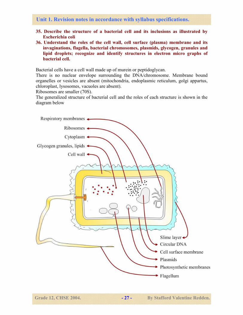
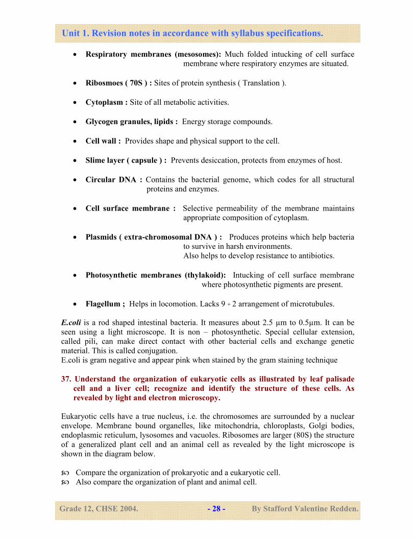
35. Describe the structure of a bacterial cell and its inclusions as illustrated by
Escherichia coli
36. Understand the roles of the cell wall, cell surface (plasma) membrane and its
invaginations, flagella, bacterial chromosomes, plasmids, glycogen, granules and
lipid droplets; recognize and identify structures in electron micro graphs of
bacterial cell.
Bacterial cells have a cell wall made up of murein or peptidoglycan.
There is no nuclear envelope surrounding the DNA/chromosome. Membrane bound
organelles or vesicles are absent (mitochondria, endoplasmic reticulum, golgi appartus,
chloroplast, lysosomes, vacuoles are absent).
Ribosomes are smaller (70S).
The generalized structure of bacterial cell and the roles of each structure is shown in the
diagram below
Unit 1. Revision notes in accordance with syllabus specifications.
Grade 12, CHSE 2004. - 28 - By Stafford Valentine Redden.
• Respiratory membranes (mesosomes): Much folded intucking of cell surface
membrane where respiratory enzymes are situated.
• Ribosmoes ( 70S ) : Sites of protein synthesis ( Translation ).
• Cytoplasm : Site of all metabolic activities.
• Glycogen granules, lipids : Energy storage compounds.
• Cell wall : Provides shape and physical support to the cell.
• Slime layer ( capsule ) : Prevents desiccation, protects from enzymes of host.
• Circular DNA : Contains the bacterial genome, which codes for all structural
proteins and enzymes.
• Cell surface membrane : Selective permeability of the membrane maintains
appropriate composition of cytoplasm.
• Plasmids ( extra-chromosomal DNA ) : Produces proteins which help bacteria
to survive in harsh environments.
Also helps to develop resistance to antibiotics.
• Photosynthetic membranes (thylakoid): Intucking of cell surface membrane
where photosynthetic pigments are present.
• Flagellum ; Helps in locomotion. Lacks 9 + 2 arrangement of microtubules.
E.coli is a rod shaped intestinal bacteria. It measures about 2.5 µm to 0.5µm. It can be
seen using a light microscope. It is non – photosynthetic. Special cellular extension,
called pili, can make direct contact with other bacterial cells and exchange genetic
material. This is called conjugation.
E.coli is gram negative and appear pink when stained by the gram staining technique
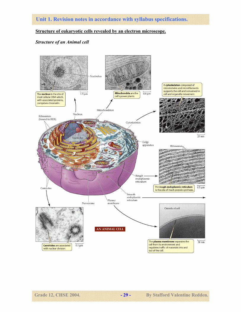
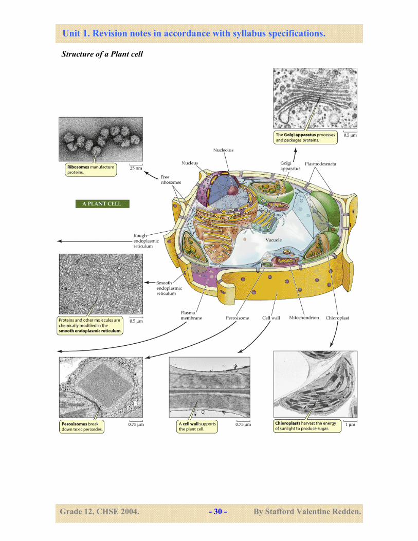
37. Understand the organization of eukaryotic cells as illustrated by leaf palisade
cell and a liver cell; recognize and identify the structure of these cells. As
revealed by light and electron microscopy.
Eukaryotic cells have a true nucleus, i.e. the chromosomes are surrounded by a nuclear
envelope. Membrane bound organelles, like mitochondria, chloroplasts, Golgi bodies,
endoplasmic reticulum, lysosomes and vacuoles. Ribosomes are larger (80S) the structure
of a generalized plant cell and an animal cell as revealed by the light microscope is
shown in the diagram below.
� Compare the organization of prokaryotic and a eukaryotic cell.
� Also compare the organization of plant and animal cell.
Unit 1. Revision notes in accordance with syllabus specifications.
Grade 12, CHSE 2004. - 29 - By Stafford Valentine Redden.
Structure of eukaryotic cells revealed by an electron microscope.
Structure of an Animal cell
Unit 1. Revision notes in accordance with syllabus specifications.
Grade 12, CHSE 2004. - 30 - By Stafford Valentine Redden.
Structure of a Plant cell
Unit 1. Revision notes in accordance with syllabus specifications.
Grade 12, CHSE 2004. - 31 - By Stafford Valentine Redden.
38. Understand the magnification and resolution that can be achieved using light
and electron microscopy. Interpret electronmicrographs and identify the
organelles.
Magnification and resolution:
Magnification is how many times larger an image is when compared to the object.
Magnification = size of image / size of object.
The magnification produced by a light microscope depends on the strength of the
objective lens and the eye piece lens.
For example if you are using a 40x objective lens and a 10x eye piece lens then the
specimen is being magnified 400 times.
There is no limit to the magnification of a light microscope. However, at higher
magnification the image becomes blurred and you would not be able to see any more
details than before. To see more details a microscope of higher resolution must be used (a
electron microscope).
Resolution is the degree of detail which can be seen.
The limit of resolution is the minimum distance between two points which can be seen
clearly.
The limit of resolution of a light microscope is 200 nm. This means that object smaller
than 200nm will be invisible, or, two points which are less than 200nm apart (150 nm)
will be seen as a single point. This is because the distance between the points is too small
to be seen. The limit of resolution of an electron microscope is 0.5 nm.
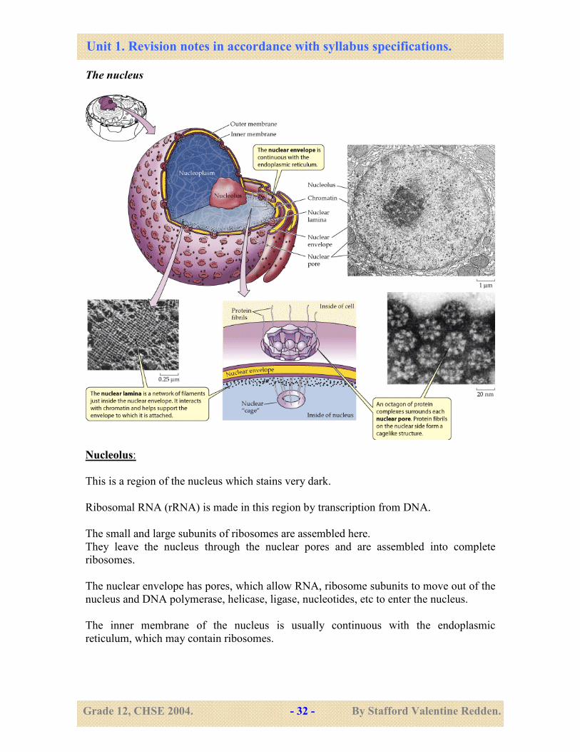
39- Describe the structure and understand the roles of the nucleus, nucleolus, rough
and smooth endoplasmic reticulum, golgi apparatus, lysosomes, chloroplast,
mitochondria, ribosome centrioles and microtubules, the cellulose cell wall;
Nucleus:
The nucleus is the part of the cell that contains the DNA. It is surrounded by a nuclear
envelope (double membrane with a space between them).
In an interphase cell, the DNA is not visible as chromosomes but appear as disorganized
material called chromatin.
In some parts the chromatin appears to be densely packed and is called heterochromatin.
In other parts it looks lighter in color. This is the euchromatin.
DNA in heterochromatin is not active (as it is coiled up around histones), but DNA in
euchromatin is involved in transcription or replication.
Unit 1. Revision notes in accordance with syllabus specifications.
Grade 12, CHSE 2004. - 32 - By Stafford Valentine Redden.
The nucleus
Nucleolus:
This is a region of the nucleus which stains very dark.
Ribosomal RNA (rRNA) is made in this region by transcription from DNA.
The small and large subunits of ribosomes are assembled here.
They leave the nucleus through the nuclear pores and are assembled into complete
ribosomes.
The nuclear envelope has pores, which allow RNA, ribosome subunits to move out of the
nucleus and DNA polymerase, helicase, ligase, nucleotides, etc to enter the nucleus.
The inner membrane of the nucleus is usually continuous with the endoplasmic
reticulum, which may contain ribosomes.
Unit 1. Revision notes in accordance with syllabus specifications.
Grade 12, CHSE 2004. - 33 - By Stafford Valentine Redden.
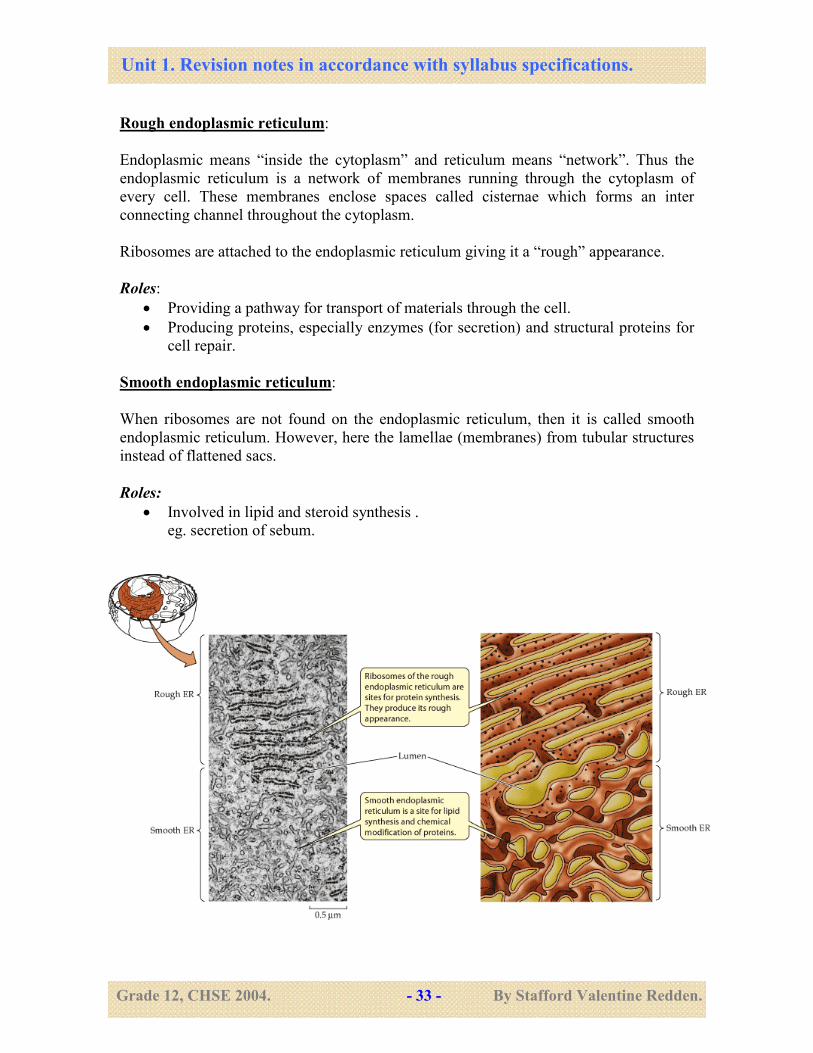
Rough endoplasmic reticulum:
Endoplasmic means “inside the cytoplasm” and reticulum means “network”. Thus the
endoplasmic reticulum is a network of membranes running through the cytoplasm of
every cell. These membranes enclose spaces called cisternae which forms an inter
connecting channel throughout the cytoplasm.
Ribosomes are attached to the endoplasmic reticulum giving it a “rough” appearance.
Roles:
• Providing a pathway for transport of materials through the cell.
• Producing proteins, especially enzymes (for secretion) and structural proteins for
cell repair.
Smooth endoplasmic reticulum:
When ribosomes are not found on the endoplasmic reticulum, then it is called smooth
endoplasmic reticulum. However, here the lamellae (membranes) from tubular structures
instead of flattened sacs.
Roles:
• Involved in lipid and steroid synthesis .
eg. secretion of sebum.
Unit 1. Revision notes in accordance with syllabus specifications.
Grade 12, CHSE 2004. - 34 - By Stafford Valentine Redden.
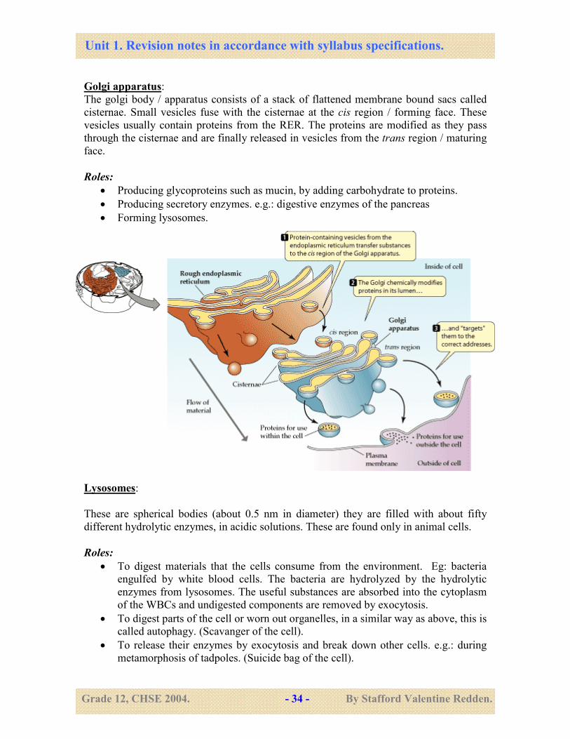
Golgi apparatus:
The golgi body / apparatus consists of a stack of flattened membrane bound sacs called
cisternae. Small vesicles fuse with the cisternae at the cis region / forming face. These
vesicles usually contain proteins from the RER. The proteins are modified as they pass
through the cisternae and are finally released in vesicles from the trans region / maturing
face.
Roles:
• Producing glycoproteins such as mucin, by adding carbohydrate to proteins.
• Producing secretory enzymes. e.g.: digestive enzymes of the pancreas
• Forming lysosomes.
Lysosomes:
These are spherical bodies (about 0.5 nm in diameter) they are filled with about fifty
different hydrolytic enzymes, in acidic solutions. These are found only in animal cells.
Roles:
• To digest materials that the cells consume from the environment. Eg: bacteria
engulfed by white blood cells. The bacteria are hydrolyzed by the hydrolytic
enzymes from lysosomes. The useful substances are absorbed into the cytoplasm
of the WBCs and undigested components are removed by exocytosis.
• To digest parts of the cell or worn out organelles, in a similar way as above, this is
called autophagy. (Scavanger of the cell).
• To release their enzymes by exocytosis and break down other cells. e.g.: during
metamorphosis of tadpoles. (Suicide bag of the cell).
Unit 1. Revision notes in accordance with syllabus specifications.
Grade 12, CHSE 2004. - 35 - By Stafford Valentine Redden.
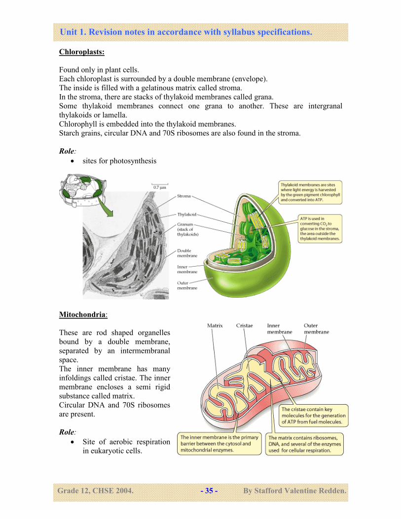
Chloroplasts:
Found only in plant cells.
Each chloroplast is surrounded by a double membrane (envelope).
The inside is filled with a gelatinous matrix called stroma.
In the stroma, there are stacks of thylakoid membranes called grana.
Some thylakoid membranes connect one grana to another. These are intergranal
thylakoids or lamella.
Chlorophyll is embedded into the thylakoid membranes.
Starch grains, circular DNA and 70S ribosomes are also found in the stroma.
Role:
• sites for photosynthesis
Mitochondria:
These are rod shaped organelles
bound by a double membrane,
separated by an intermembranal
space.
The inner membrane has many
infoldings called cristae. The inner
membrane encloses a semi rigid
substance called matrix.
Circular DNA and 70S ribosomes
are present.
Role:
• Site of aerobic respiration
in eukaryotic cells.
Unit 1. Revision notes in accordance with syllabus specifications.
Grade 12, CHSE 2004. - 36 - By Stafford Valentine Redden.
Ribosomes:
Ribosomes appear as small black dots in electron micrographs. Some are found free in
the cytoplasm, while others are attached to the outer surface of membranes of the rough
endoplasmic reticulum (RER). Each ribosome is made up of a small sub unit (30S) and a
larger subunit (50S). The larger sub unit is made up of two molecules of rRNA and
proteins, the smaller subunit is made up of one rRNA molecule and proteins.
Role:
• Sites of protein synthesis (translation).
Centrioles
Centrioles are hollow cylinders of microtubules. Each centriole is made up of 9 triplets of
microtubules. They are found in the cytoplasm. There are two centrioles arranged at right
angles to each other, to form the centrosome.
Role:
• During cell division they replicate and move towards the poles of the cells and
help in organizing the spindle fibers during cell division in animal cells.
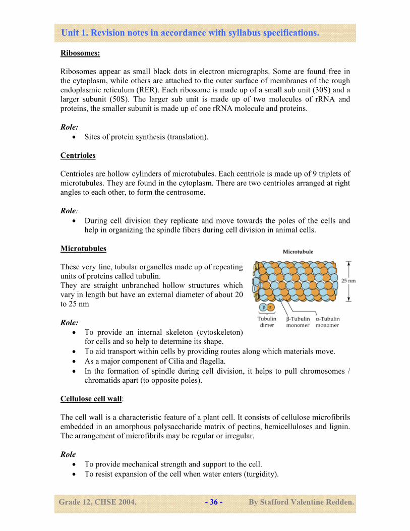
Microtubules
These very fine, tubular organelles made up of repeating
units of proteins called tubulin.
They are straight unbranched hollow structures which
vary in length but have an external diameter of about 20
to 25 nm
Role:
• To provide an internal skeleton (cytoskeleton)
for cells and so help to determine its shape.
• To aid transport within cells by providing routes along which materials move.
• As a major component of Cilia and flagella.
• In the formation of spindle during cell division, it helps to pull chromosomes /
chromatids apart (to opposite poles).
Cellulose cell wall:
The cell wall is a characteristic feature of a plant cell. It consists of cellulose microfibrils
embedded in an amorphous polysaccharide matrix of pectins, hemicelluloses and lignin.
The arrangement of microfibrils may be regular or irregular.
Role
• To provide mechanical strength and support to the cell.
• To resist expansion of the cell when water enters (turgidity).
Unit 1. Revision notes in accordance with syllabus specifications.
Grade 12, CHSE 2004. - 37 - By Stafford Valentine Redden.
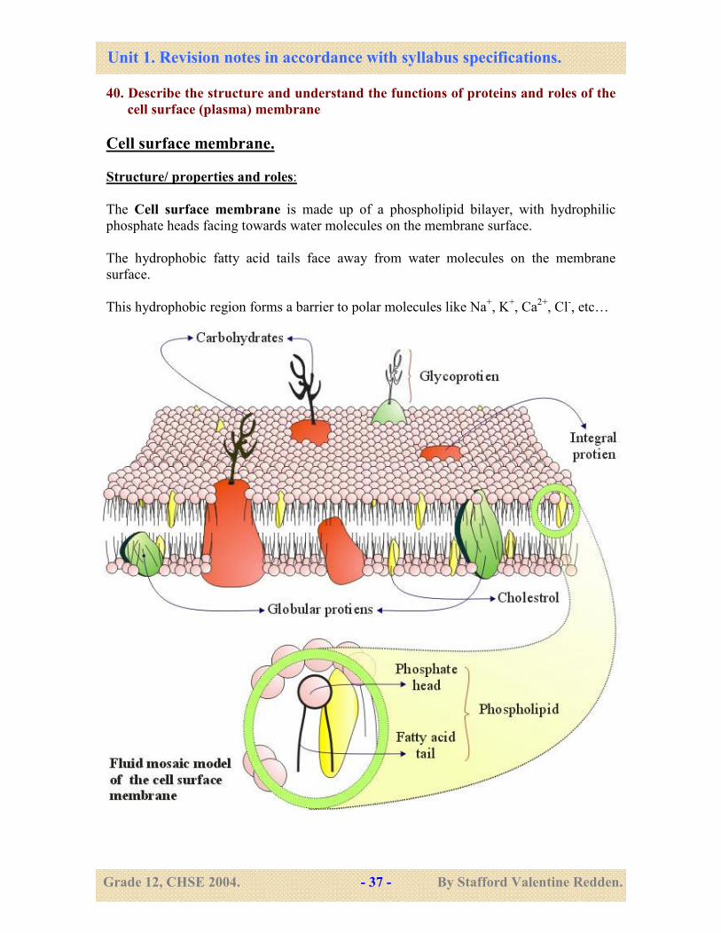
40. Describe the structure and understand the functions of proteins and roles of the
cell surface (plasma) membrane
Cell surface membrane.
Structure/ properties and roles:
The Cell surface membrane is made up of a phospholipid bilayer, with hydrophilic
phosphate heads facing towards water molecules on the membrane surface.
The hydrophobic fatty acid tails face away from water molecules on the membrane
surface.
This hydrophobic region forms a barrier to polar molecules like Na+, K
+, Ca
2+, Cl
-, etc…
Unit 1. Revision notes in accordance with syllabus specifications.
Grade 12, CHSE 2004. - 38 - By Stafford Valentine Redden.
However , lipid soluble, non polar molecules like CO2, O2, cholesterol, fatty acids etc…
can pass across the phospholipid bilayer. Water can pass through the phospholipid
bilayer because of its small size and relatively low polarity.
Proteins are embedded into the phospholipid bilayer. These proteins allow certain polar
molecules and ions to pass across the membrane. Hence they are often referred to as
channel proteins, carrier proteins or transporter proteins. They are specific and will allow
only certain substances to pass across.
Some of the proteins (extrinsic proteins) act as enzymes, recognition sites and electron
carriers. The proteins also provide structural support for the membrane. Branched chains
of carbohydrates maybe attached to some phospholipid molecules (glycolipids) or to
proteins (glycoproteins).
The carbohydrates act as recognition sites for neurotransmitters, hormones or for cell to
cell recognition.
Cholesterol makes the membranes less fluid and more stable. This model of cell surface
membrane is called the ‘fluid mosaic’ model.
Fluid means that molecules can change places within the membrane.
Mosaic means that proteins are embedded randomly in to the phospholipid bilayer.
Role:
• Selectively permeable membrane helps to prevent passage of some substances and
allow passage of other substances. This helps to maintain the appropriate
composition of the cytoplasm.
41. Understand how molecules and ions move into and out of cell
Molecules move in and out of cell across the selectively permeable cell membrane. There
are four basic processes, namely, diffusion, osmosis, active transport and bulk transport
(exocytosis and endocytosis). Diffusion and osmosis are passive processes which use
kinetic energy of molecules (not ATP), but active transport and bulk transport are active
processes, which use metabolic energy (ATP) from the cell.
42. Understand the principles involved in passive transport by diffusion and
facilitated diffusion
Diffusion is the net movement of particles from a region of their high concentration to a
region of their lower concentration down a concentration gradient.
It is a passive process, which means that it does not require ATP. It occurs due to the
random movement of particles across the membrane. The particles move in both
directions across the membrane, but the rate of movement of particles from higher to
lower concentration is greater than the movement in the opposite direction.
Unit 1. Revision notes in accordance with syllabus specifications.
Grade 12, CHSE 2004. - 39 - By Stafford Valentine Redden.
Each type of molecule / ion moves down its own diffusion gradient, independent of other
molecules.
For Example: O2 and CO2 diffuse in different directions in the lungs.
Factors affecting diffusion: The rate of diffusion across membranes depends on the
following factors.
a) - Surface area of membrane: rate of diffusion is directly proportional to surface area.
b) - Difference in concentration across the membrane: rate of diffusion is directly
proportional to the concentration gradient.
c) – Thickness of membrane: rate of diffusion is inversely proportional to the thickness
of the membrane or the diffusion distance.
d) – Temperature: rate of diffusion is directly proportional to the temperature as the
kinetic energy of particles increase with temperature.
e) - Size of particles: Smaller / lighter particles diffuse faster.
Substances that can be exchanged by diffusion
• O2 and CO2 are non polar, small molecules which can diffuse rapidly across the
phospholipid bilayer.
• Ions and large polar molecules, like glucose , amino acids ,Na+, Cl
- are repelled by
the hydrophobic region (fatty acid tails) of the phospholipids and diffuse across
extremely slowly, if at all.
• Steroid hormones are lipid soluble and can diffuse across the membrane easily.
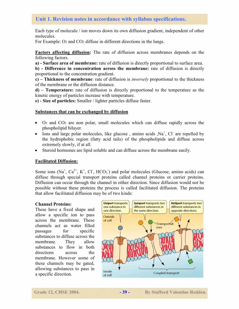
Facilitated Diffusion:
Some ions (Na+, Ca
2+, K
+, Cl
-, HCO3
-) and polar molecules (Glucose, amino acids) can
diffuse through special transport proteins called channel proteins or carrier proteins.
Diffusion can occur through the channel in either direction. Since diffusion would not be
possible without these proteins the process is called facilitated diffusion. The proteins
that allow facilitated diffusion may be of two kinds:
Channel Proteins: These have a fixed shape and
allow a specific ion to pass
across the membrane. These
channels act as water filled
passages for specific
substances to diffuse across the
membrane. They allow
substances to flow in both
directions across the
membrane. However some of
these channels may be gated,
allowing substances to pass in
a specific direction.
Unit 1. Revision notes in accordance with syllabus specifications.
Grade 12, CHSE 2004. - 40 - By Stafford Valentine Redden.
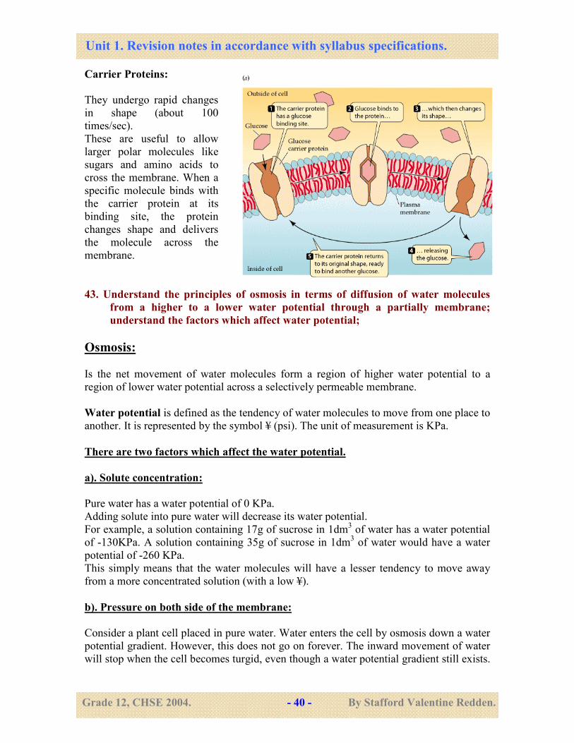
Carrier Proteins:
They undergo rapid changes
in shape (about 100
times/sec).
These are useful to allow
larger polar molecules like
sugars and amino acids to
cross the membrane. When a
specific molecule binds with
the carrier protein at its
binding site, the protein
changes shape and delivers
the molecule across the
membrane.
43. Understand the principles of osmosis in terms of diffusion of water molecules
from a higher to a lower water potential through a partially membrane;
understand the factors which affect water potential;
Osmosis:
Is the net movement of water molecules form a region of higher water potential to a
region of lower water potential across a selectively permeable membrane.
Water potential is defined as the tendency of water molecules to move from one place to
another. It is represented by the symbol ¥ (psi). The unit of measurement is KPa.
There are two factors which affect the water potential.
a). Solute concentration:
Pure water has a water potential of 0 KPa.
Adding solute into pure water will decrease its water potential.
For example, a solution containing 17g of sucrose in 1dm3 of water has a water potential
of -130KPa. A solution containing 35g of sucrose in 1dm3 of water would have a water
potential of -260 KPa.
This simply means that the water molecules will have a lesser tendency to move away
from a more concentrated solution (with a low ¥).
b). Pressure on both side of the membrane:
Consider a plant cell placed in pure water. Water enters the cell by osmosis down a water
potential gradient. However, this does not go on forever. The inward movement of water
will stop when the cell becomes turgid, even though a water potential gradient still exists.
Unit 1. Revision notes in accordance with syllabus specifications.
Grade 12, CHSE 2004. - 41 - By Stafford Valentine Redden.
This is because the cell wall exerts a pressure on the water molecules and decreases its
tendency (water potential) to enter the cells.
44. Understand the principles involved in active transport; Endocytosis and
Exocytosis.
Active Transport:
It is the uptake of molecules or ions against a concentration gradient using energy from
respiration (ATP).
How does it work?
• The molecule or ion combines with a specific carrier protein in the cell surface
membrane.
• ATP transfers a phosphate group to the carrier protein on the inside of the membrane.
This causes the carrier protein to undergo a change of shape which causes the
molecule or ion to move across the membrane.
• The molecule or ion is then released and the protein changes back to its original
shape.
Due to energy needed for this process, the cells involved tend to contain more
mitochondria and a high rate of respiration.
Their ability to carry out active uptake is affected by temperature, oxygen concentration
and the presence of respiratory poisons like Cyanide. (All factors which affect
respiration).
Some processes involving active transport are:
Nerve impulse transmission, muscle contraction, absorption of amino acids in ileum,
absorption of ions by root hair cells of plants, protein synthesis, selective re-absorption in
kidney.
Exocytosis and Endocytosis are processes by which bulk transport of materials take
place (irrespective of the concentration gradient).
Endocytosis: ( Taking substances into the cell ).
The cell surface membrane wraps around the substance (forming an invagination). This
invagination deepens and finally pinches off to enclose the substance in a vesicle inside
the cytoplasm.
Eg: Phagocytes (white blood cells engulf bacteria), amoeba engulfing prey.
Taking in of solid substances is Phagocytosis. Taking in of liquids is Pinocytosis.
Unit 1. Revision notes in accordance with syllabus specifications.
Grade 12, CHSE 2004. - 42 - By Stafford Valentine Redden.
Exocytosis:
This is the reverse of Endocytosis. It is the passage of materials out of the cell. This
method is often used for secretion of enzymes, hormones or mucus.
The secretory vesicles fuse with the cell surface membrane and release their contents to
the outside.
45. Understand that tissues are aggregations of cells of common origin, structure
and function, as illustrated by the tissues of a mesophytic leaf.
Tissues are a group of cells of common origin, structure and function. The different
tissues found in a mesophytic leaf are:
a). Parenchyma:
These cells are roughly spherical or elongated. They have a thin primary cell wall of
cellulose and have an active cytoplasm. They are living tissues and are found in modified
forms in the epidermis, palisade and spongy mesophyll layers and between xylem and
phloem of leaves.
Functions: protection, storage, photosynthesis.
b). Collenchyma:
These are elongated cells, usually polygonal in shape with tapering ends. They have less
cytoplasm. They contain a nucleus and are living. They are found in the midrib of leaves.
Function: Provide mechanical support.
c). Sclerenchyma:
These are elongated cells. They are Polygonal in shape with tapering and interlocking
ends. They have highly lignified cell walls. They are dead cells with no cytoplasm.
Function: Provide mechanical support. Found in xylem and phloem (referred to as
fibres).
d). Xylem and Phloem:
These are tissues which are composed of more than one type of cells. Found in
veins/midrib of leaves. They are referred to as vascular tissues. (Refer to structure of
xylem and phloem in unit 2 notes).
Unit 1. Revision notes in accordance with syllabus specifications.
Grade 12, CHSE 2004. - 43 - By Stafford Valentine Redden.
46. Understand that the leaf and liver are organs and composed of aggregations of
tissues.
Organs
These are groups of tissues performing a similar function.
Eg: leaf is an organ. All the tissues of the leaf work to perform photosynthesis.
The liver is an organ. It is composed of cells called Hepatocytes. These cells perform all
the functions of the liver. However, blood cells, nervous tissues and connective tissues
are all necessary to enable the hepatocytes to function normally. Thus, all these tissues
function together as an organ.
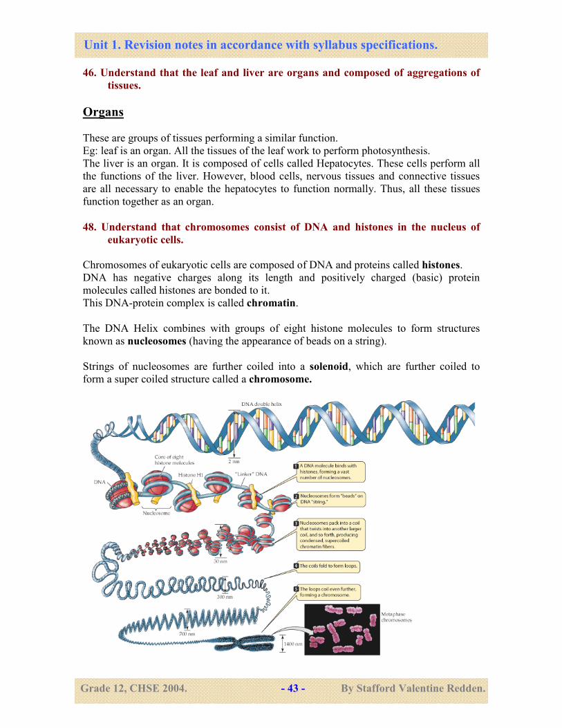
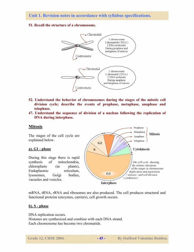
48. Understand that chromosomes consist of DNA and histones in the nucleus of
eukaryotic cells.
Chromosomes of eukaryotic cells are composed of DNA and proteins called histones.
DNA has negative charges along its length and positively charged (basic) protein
molecules called histones are bonded to it.
This DNA-protein complex is called chromatin.
The DNA Helix combines with groups of eight histone molecules to form structures
known as nucleosomes (having the appearance of beads on a string).
Strings of nucleosomes are further coiled into a solenoid, which are further coiled to
form a super coiled structure called a chromosome.
Unit 1. Revision notes in accordance with syllabus specifications.
Grade 12, CHSE 2004. - 44 - By Stafford Valentine Redden.
49. Recall the replication of DNA; understand the roles of enzymes involved.
50. Understand that a leaf palisade cell and a liver cell have a diploid chromosome
number and have been produced by nuclear division followed by a
differentiation.
A Leaf palisade cell and a liver cell have been formed by mitosis.
This ensures that their diploid number of chromosomes is maintained.
However, both the palisade cell and the liver cell have developed from an
undifferentiated cell, called the zygote.
During development, cells begin to specialize and become adapted, in shape and structure
to perform specific functions.
This is called differentiation. This enables division of labour which is a common feature
of higher organisms.
Unit 1. Revision notes in accordance with syllabus specifications.
Grade 12, CHSE 2004. - 45 - By Stafford Valentine Redden.
51. Recall the structure of a chromosome.
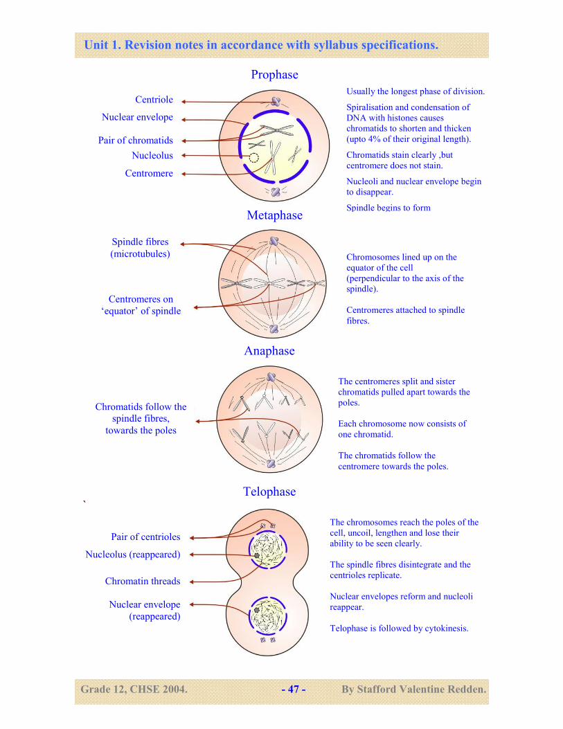
52. Understand the behavior of chromosomes during the stages of the mitotic cell
division cycle; describe the events of prophase, metaphase, anaphase and
telophase.
47. Understand the sequence of division of a nucleus following the replication of
DNA during interphase.
Mitosis
The stages of the cell cycle are
explained below.
a). G1 - phase
During this stage there is rapid
synthesis of mitochondria,
chloroplasts (in plants),
Endoplasmic reticulum,
lysosomes, Golgi bodies,
vacuoles and vesicles.
mRNA, tRNA, rRNA and ribosomes are also produced. The cell produces structural and
functional proteins (enzymes, carriers), cell growth occurs.
b). S - phase
DNA replication occurs.
Histones are synthesized and combine with each DNA strand.
Each chromosome has become two chromatids.
Unit 1. Revision notes in accordance with syllabus specifications.
Grade 12, CHSE 2004. - 46 - By Stafford Valentine Redden.
c). G2 - phase
Mitochondria and chloroplasts divide.
Energy stores increase and mitotic spindle begins to form.
d). M - phase
Nuclear division occurs in four stages (prophase, metaphase, anaphase and telophase)
e). C - phase
Cytokinesis: Equal distribution of organelles and cytoplasm into each daughter cell.
Interphase
Interphase consists of G1, S and
G2 phases.
DNA replication occurs during
this stage.
The DNA is not highly coiled
and not visible as chromosomes.
The DNA appears as a network
of fine threads called chromatin.
Unit 1. Revision notes in accordance with syllabus specifications.
Grade 12, CHSE 2004. - 47 - By Stafford Valentine Redden.
`
Usually the longest phase of division. Spiralisation and condensation of
DNA with histones causes
chromatids to shorten and thicken
(upto 4% of their original length). Chromatids stain clearly ,but
centromere does not stain. Nucleoli and nuclear envelope begin
to disappear. Spindle begins to form
Centriole
Nuclear envelope
Pair of chromatids
Nucleolus
Centromere
Spindle fibres
(microtubules)
Centromeres on
‘equator’ of spindle
Chromatids follow the
spindle fibres,
towards the poles
Pair of centrioles
Nucleolus (reappeared)
Chromatin threads
Nuclear envelope
(reappeared)
Chromosomes lined up on the
equator of the cell
(perpendicular to the axis of the
spindle).
Centromeres attached to spindle
fibres.
The centromeres split and sister
chromatids pulled apart towards the
poles.
Each chromosome now consists of
one chromatid.
The chromatids follow the
centromere towards the poles.
The chromosomes reach the poles of the
cell, uncoil, lengthen and lose their
ability to be seen clearly.
The spindle fibres disintegrate and the
centrioles replicate.
Nuclear envelopes reform and nucleoli
reappear.
Telophase is followed by cytokinesis.
Telophase
Anaphase
Metaphase
Prophase
Unit 1. Revision notes in accordance with syllabus specifications.
Grade 12, CHSE 2004. - 48 - By Stafford Valentine Redden.
53. Understand the significance of mitosis in growth and replacement; understand
the significance of daughter nuclei with chromosomes identical in number and
type.
Significance of mitosis
• Genetic stability - Mitosis produces two nuclei which have the same (identical)
number of chromosomes as the parent cell. Moreover, each chromosome is
genetically identical to parent DNA as it is formed by replication. These genetically
identical cells are called clones.
• Growth - Growth is achieved by an increase in the number of cells in an organism.
Mitosis helps to increase cell numbers, thus causing growth.
• Asexual reproduction, regeneration and cell replacement - Binary fission in
amoeba, budding in hydra, growth of plants from stem cuttings, bulbs and tubers form
new individuals from parents, by mitosis ( Meiosis is not involved ). Regeneration of
missing parts (such as legs in crustaceans, arms of starfish ) and healing of wounds
also involves mitosis.
54. Understand that the production of new individuals involves the transfer of
genetic information from parent to offspring;
55. Understand that inherited information in the offspring is identical to that of the
parent; understand the significance off mitosis in achieving this;
As state above, mitosis maintains genetic stability during growth and development.
Reproduction is the ability to produce a new generation of individuals of the same
species.
This involves the transfer of genetic information from parental generation to the next,
there by ensuring that the characteristics, not only of the species, but also of the parental
organisms, are perpetuated.
A new individual has to go through a period of growth and development before it reaches
a stage where it can reproduce itself.
Mitosis maintains genetic stability during this period. It also helps to preserve genetic
characters from generation to generation during asexual reproduction.
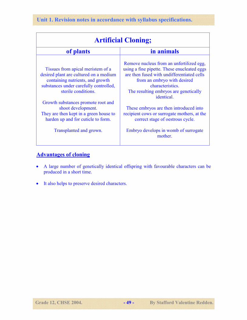
56. Understand the nature of natural and artificial cloning in plants and animals.
Cloning is the production of a genetically identical offspring ( genetically identical to the
parent ) by asexual reproduction.
Asexual reproduction in plants, yeasts, bacteria are examples of natural cloning.
However, artificial cloning is possible in both plants and animals.
An outline of the process is given below;
Unit 1. Revision notes in accordance with syllabus specifications.
Grade 12, CHSE 2004. - 49 - By Stafford Valentine Redden.
Advantages of cloning
• A large number of genetically identical offspring with favourable characters can be
produced in a short time.
• It also helps to preserve desired characters.
Artificial Cloning;
of plants in animals
Tissues from apical meristem of a
desired plant are cultured on a medium
containing nutrients, and growth
substances under carefully controlled,
sterile conditions.
Growth substances promote root and
shoot development.
They are then kept in a green house to
harden up and for cuticle to form.
Transplanted and grown.
Remove nucleus from an unfertilized egg,
using a fine pipette. These enucleated eggs
are then fused with undifferentiated cells
from an embryo with desired
characteristics.
The resulting embryos are genetically
identical.
These embryos are then introduced into
recipient cows or surrogate mothers, at the
correct stage of oestrous cycle.
Embryo develops in womb of surrogate
mother.