Embed Size (px)
Citation preview
Unit 1Cell and Molecular
Biology
Section 2
Cell cycle, cell growth and differentiation
The cell cycle
The cell cycle, or mitosis, is the process by whichnew cells are produced for growth. All cellsresulting from this process are identical to eachother and the parent cell
The cycle has 4 main stages as follows :- G1 – Cell growth and synthesis of organelles S – DNA replication G2 - Cell growth and synthesis of organelles M – Cell division
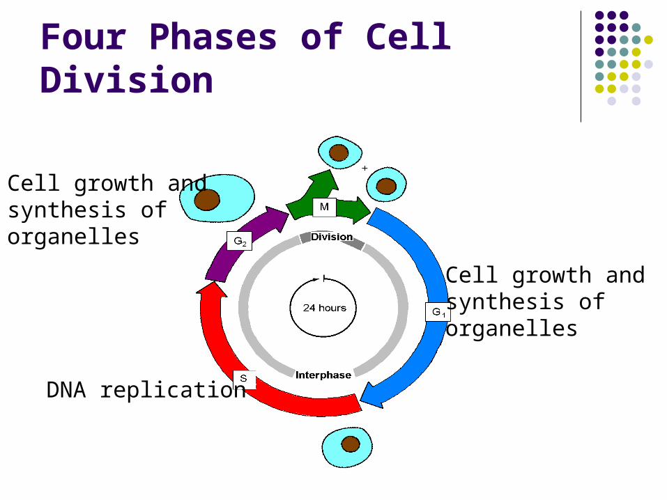
Four Phases of Cell Division
Cell growth andsynthesis of organelles
Cell growth andsynthesis of organelles
DNA replication
The stages G1, S and G2 are collectivelyknown as interphase.
The M phase or cell division phase is known asthe mitotic phase.
Mitosis has a number of stages as shown
below:
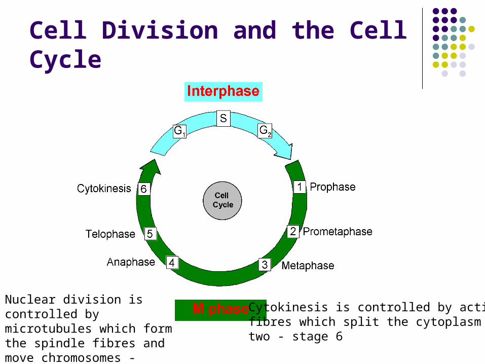
Cell Division and the Cell Cycle
Nuclear division is controlled by microtubules which form the spindle fibres and move chromosomes - stages 1-5
Cytokinesis is controlled by actinfibres which split the cytoplasm intwo - stage 6
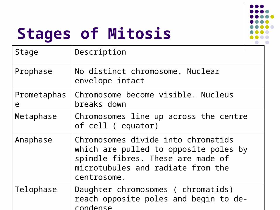
Stages of MitosisStage Description
Prophase No distinct chromosome. Nuclear envelope intact
Prometaphase Chromosome become visible. Nucleus breaks down
Metaphase Chromosomes line up across the centre of cell ( equator)
Anaphase Chromosomes divide into chromatids which are pulled to opposite poles by spindle fibres. These are made of microtubules and radiate from the centrosome.
Telophase Daughter chromosomes ( chromatids) reach opposite poles and begin to de-condense
Cytokinesis Cell divides into two by contraction of actin fibres
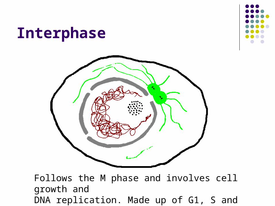
Interphase
Follows the M phase and involves cell growth andDNA replication. Made up of G1, S and G2
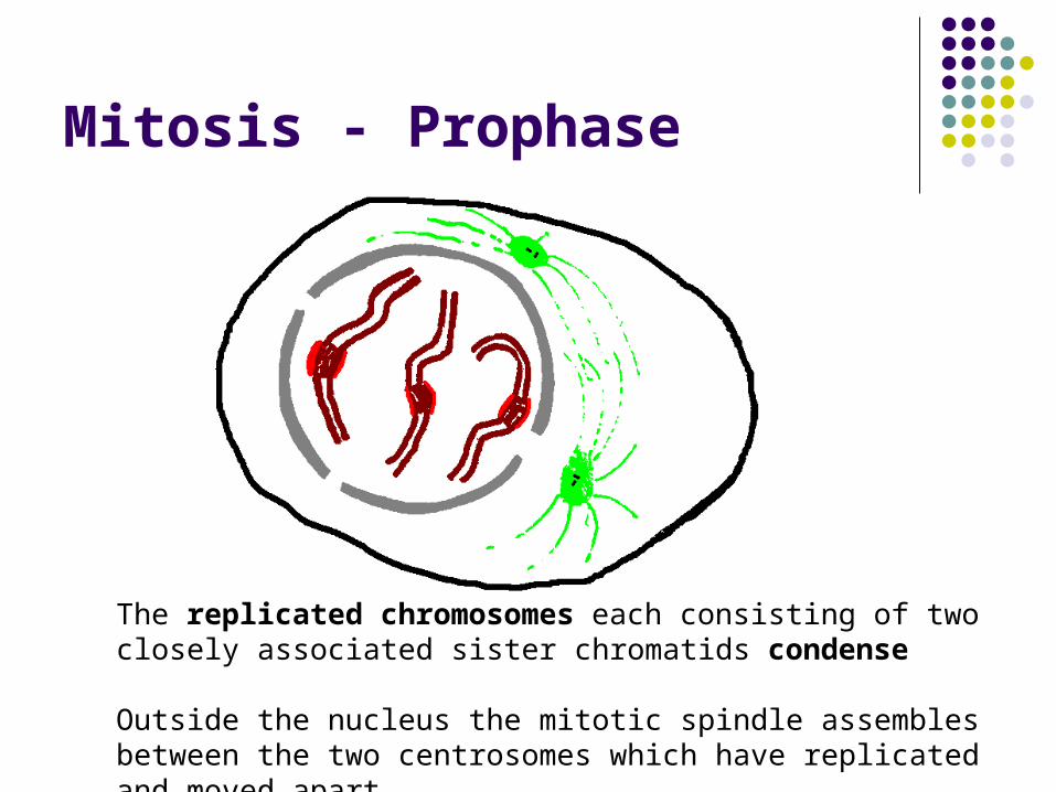
Mitosis - Prophase
The replicated chromosomes each consisting of two closely associated sister chromatids condense
Outside the nucleus the mitotic spindle assembles between the two centrosomes which have replicated and moved apart.
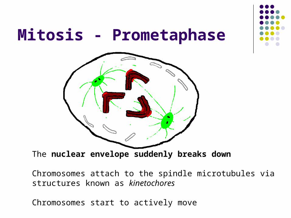
Mitosis - Prometaphase
The nuclear envelope suddenly breaks down
Chromosomes attach to the spindle microtubules via structures known as kinetochores
Chromosomes start to actively move
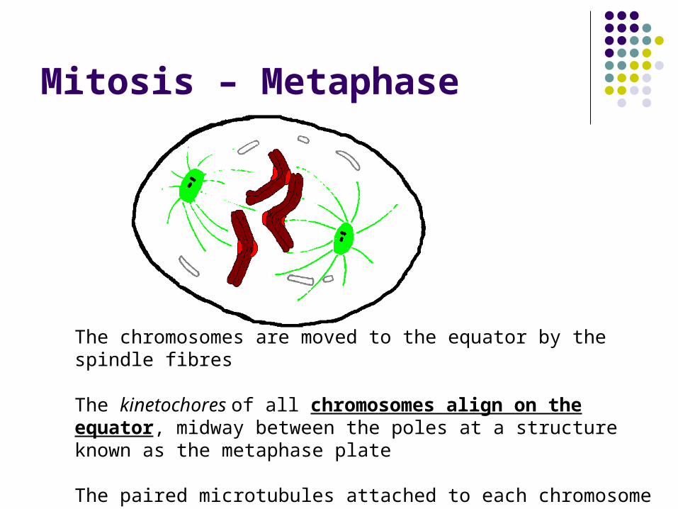
Mitosis – Metaphase
The chromosomes are moved to the equator by the spindle fibres
The kinetochores of all chromosomes align on the equator, midway between the poles at a structure known as the metaphase plate
The paired microtubules attached to each chromosome attach to opposite poles of the spindle
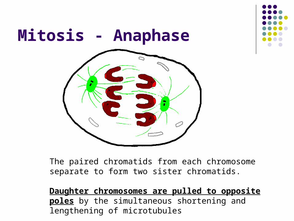
Mitosis - Anaphase
The paired chromatids from each chromosome separate to form two sister chromatids.
Daughter chromosomes are pulled to opposite poles by the simultaneous shortening and lengthening of microtubules
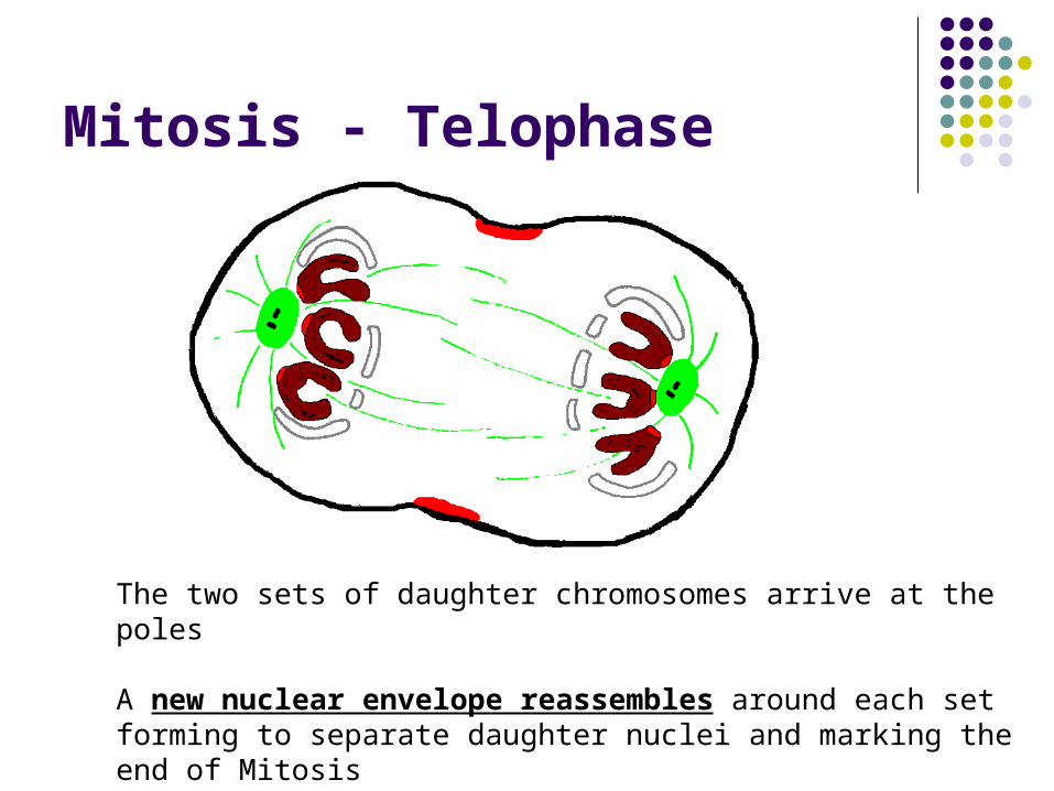
Mitosis - Telophase
The two sets of daughter chromosomes arrive at the poles
A new nuclear envelope reassembles around each set forming to separate daughter nuclei and marking the end of Mitosis
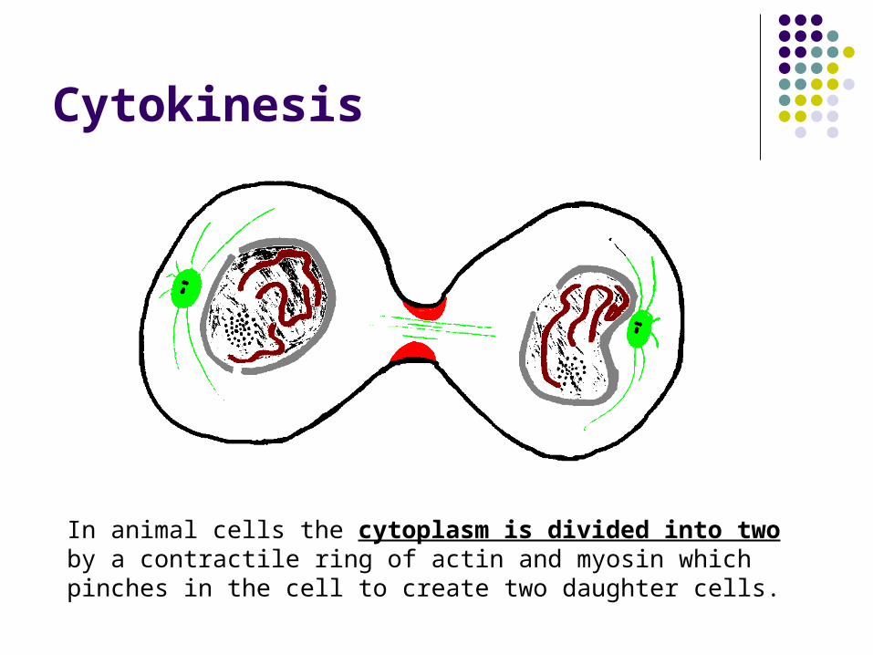
Cytokinesis
In animal cells the cytoplasm is divided into two by a contractile ring of actin and myosin which pinches in the cell to create two daughter cells.
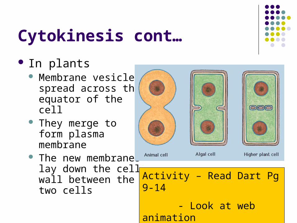
Cytokinesis cont…
In plants Membrane vesicles
spread across the equator of the cell
They merge to form plasma membrane
The new membranes lay down the cell wall between the two cells
Activity – Read Dart Pg 9-14
- Look at web animation (www.biozone.co.uk/links.html)
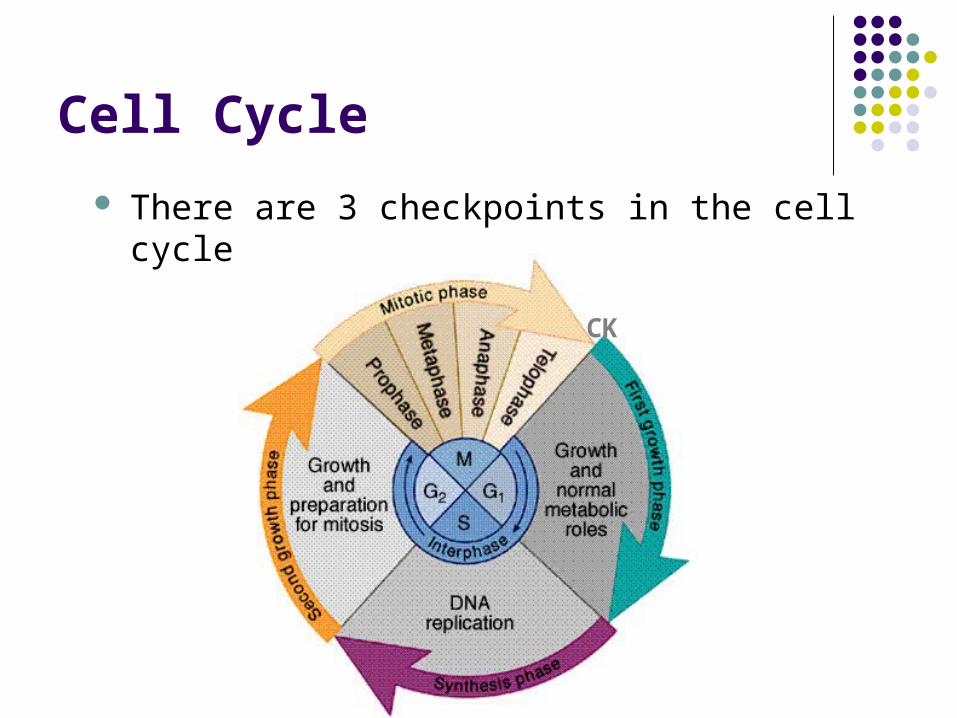
Cell Cycle
There are 3 checkpoints in the cell cycle
CK
Control of cell cycle
G1 Checkpoint End of the G1 phase – the cell size is assessed.
If large enough the cell enters S-phase. The cell is usually pushed past this point by
signals (growth factors) from outside the cell.
If conditions are met DNA replication enzymes called polymerase are transcribed to allow S-phase to begin
If conditions are not met Cells don’t divide and remin in G0 Many mature cells e.g. nerve cells, skeletal muscle
cells, RBC’s don’t divide.
G2 Checkpoint DNA replication success is monitored If replication is successful
DNA polymerase enzymes are deactivated Metaphase enzymes are activated
If replication is unsuccessful Any cell with unreplicated or damaged DNA that cant
be repaired is destroyed (apoptosis = cell suicide)
Control of cell cycle - MPF
Mitosis (maturation) Promoting Factor (MPF) Promotes transition of G2 to M phase Acts as a catalyst for the conversion of
metaphase enzymes from an inactive to an active state (by phosphorylation)
M Checkpoint Occurs during metaphase
Checks the spindle has assembled properly
All chromosomes are attached properly (by the kinetochores
If conditions are met Metaphase enzymes are deactivated Anaphase enzymes are activated
Abnormal cell division: Cancer
Cancer cells by-pass normal cell control mechanisms. As a result they divide uncontrollably to form lumps of tissue (tumours) that no longer carry out their function.
Mutation to Proliferation Genes Normal proliferation genes are called Proto-oncogenes
During normal cell division proto-oncogenes code for proteins (e.g. growth factors) that promote cell division
Mutated Proliferation genes are called oncogenes Oncogenes act to produce cells that are not required.
E.g. Produce a protein which triggers a response in the
cell as if growth factors are present Over production of growth factors
Oncogenes are dominantOnly 1 gene in the pair of alleles needs to
mutate for it to have an effect.Mutations in several different genes are
usually required for cancer to develop.
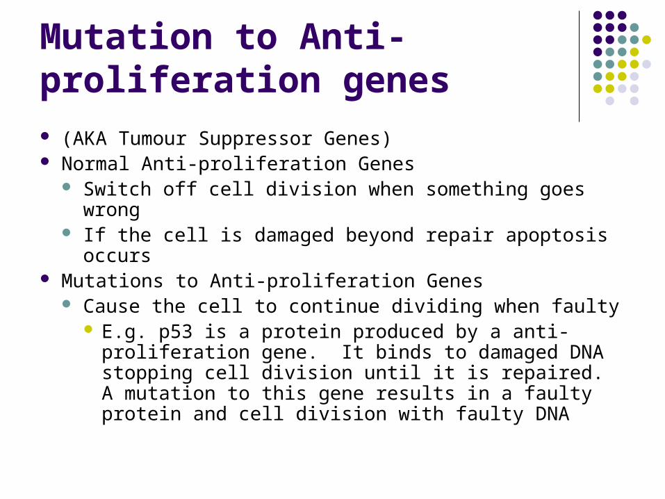
Mutation to Anti-proliferation genes (AKA Tumour Suppressor Genes) Normal Anti-proliferation Genes
Switch off cell division when something goes wrong If the cell is damaged beyond repair apoptosis occurs
Mutations to Anti-proliferation Genes Cause the cell to continue dividing when faulty
E.g. p53 is a protein produced by a anti-proliferation gene. It binds to damaged DNA stopping cell division until it is repaired. A mutation to this gene results in a faulty protein and cell division with faulty DNA
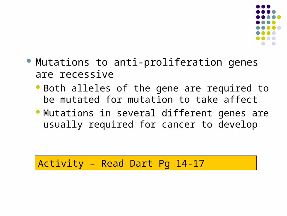
Mutations to anti-proliferation genes are recessiveBoth alleles of the gene are required to be
mutated for mutation to take affectMutations in several different genes are
usually required for cancer to develop
Activity – Read Dart Pg 14-17
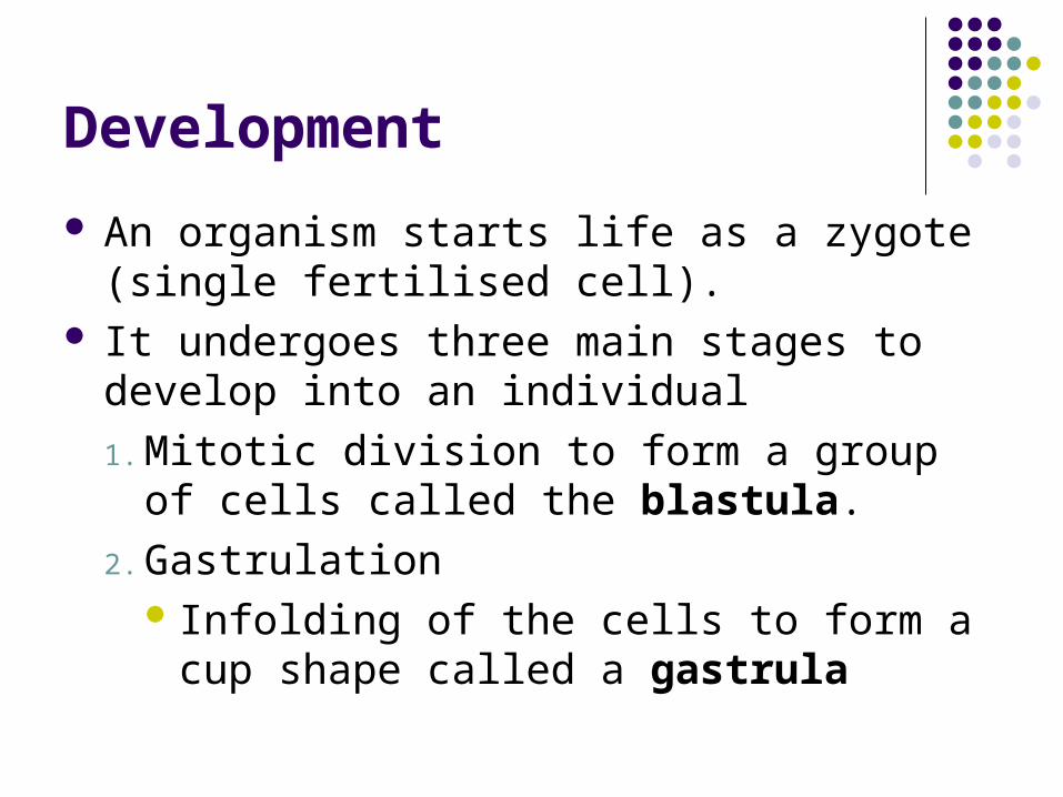
Development
An organism starts life as a zygote (single fertilised cell).
It undergoes three main stages to develop into an individual
1. Mitotic division to form a group of cells called the blastula.
2. Gastrulation Infolding of the cells to form a cup shape
called a gastrula
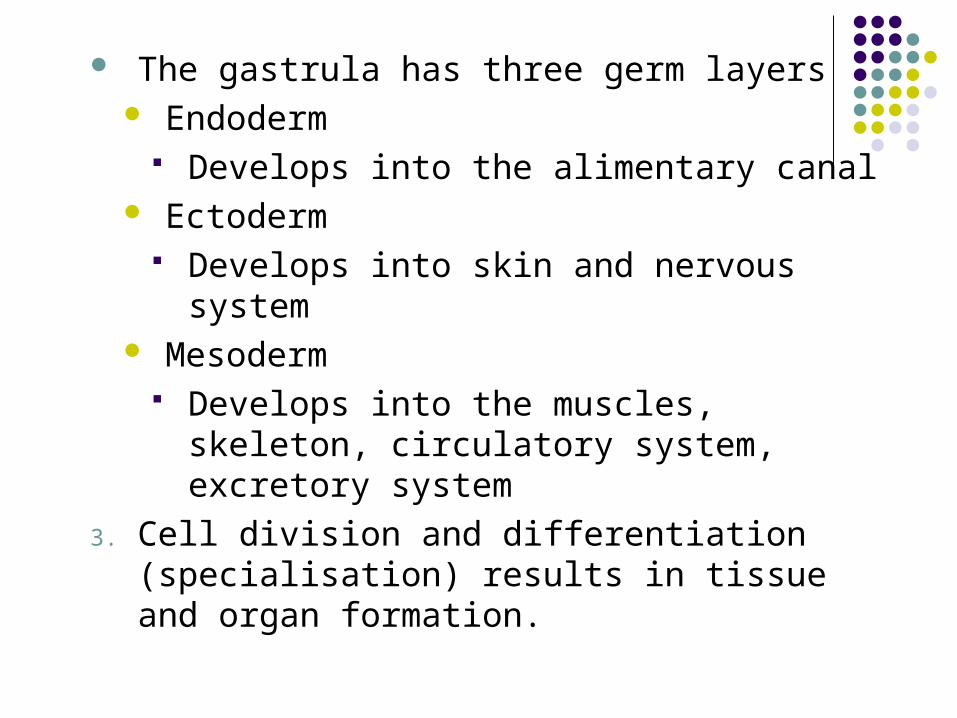
The gastrula has three germ layers Endoderm
Develops into the alimentary canal Ectoderm
Develops into skin and nervous system Mesoderm
Develops into the muscles, skeleton, circulatory system, excretory system
3. Cell division and differentiation (specialisation) results in tissue and organ formation.
Differentiation
Nearly all cells in an organism have the same DNA
Differentiation depends on gene expression (the transcription of a gene into mRNA)
i.e. which genes are ‘switched on’ and which genes are ‘switched off’.
During development the control of gene expression may be:Temporal (different genes expressed at
different times in development)Spatial (cells in different places in the
embryo expressing different genes)
Example of differentiation to form an organism:
Drosophila melanogaster
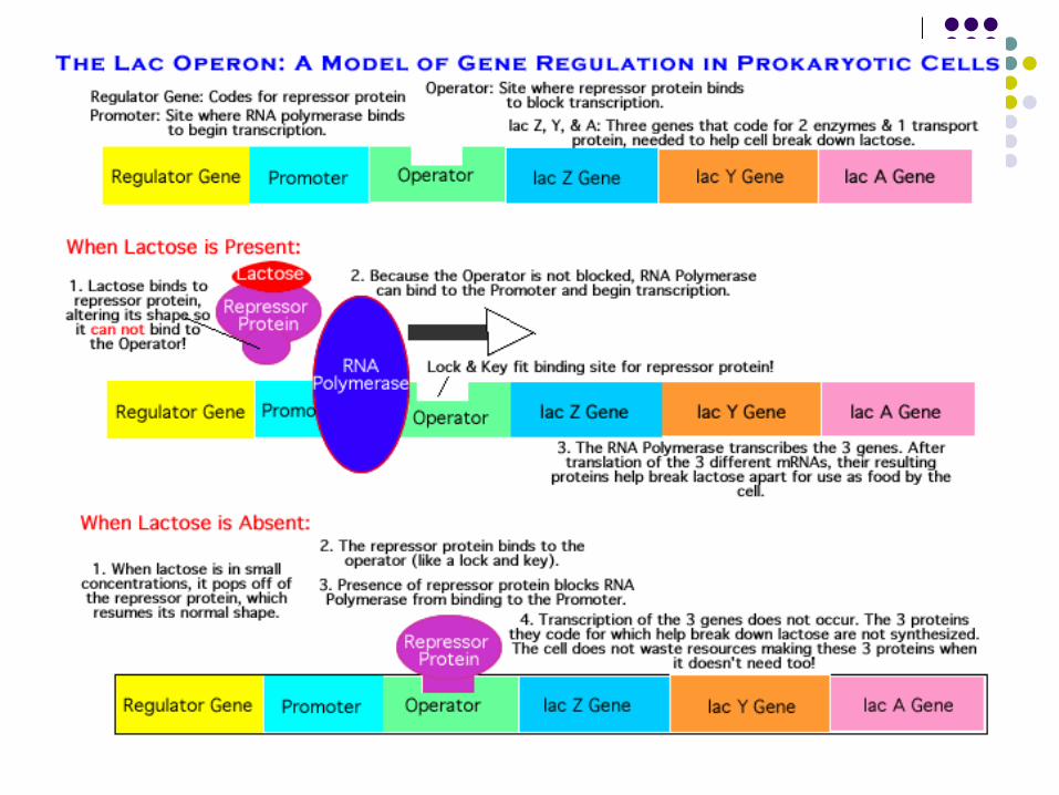
Control of gene expression in bacteria
Lac operon (aka Jacob-Monod hypothesis)
Stem Cells
A stem cell is an undifferentiated cell which can undergo unlimited division to form other cells
Source of stem cells Adult e.g. bone marrow Embryonic (from blastula stage ~ 150 cell stage) Cancer cells Umbilical Cord Blood
Stem cells have the ability to differentiate, unlike specialised cells
Activity –
•Read Dart Pg 18-20
•Web animations from Biozone website
•http://www.sumanasinc.com/webcontent/anisamples/nonmajorsbiology/stemcells.html
•http://www.sumanasinc.com/webcontent/anisamples/majorsbiology/lacoperon.html
•http://science.howstuffworks.com/stem-cell.htm