Embed Size (px)
Citation preview

Unilateral Cholesteatoma in the First Millennium BC
*Nuria Armentano, *Assumpcio Malgosa, †Brıgida Martınez, ‡Pedro Abello,§Manuel de Juan Delago, *Gemma Prats-Munoz, and *†Albert Isidro
*Unitat d’Antropologia biologica, Dept. Biologia Animal, Biologia Vegetal i Ecologia, Universitat Autonomade Barcelona (UAB); ÞHospital Universitari Sagrat Cor de Barcelona; þUniversitat Autonoma de Barcelona
(UAB); and §Neuroradiologia. Hospital de Sant Pau, UAB, Barcelona, Spain
Objective: To analyze the bone lesions of the ear region from a lateBronze Age individual to establish the most probable diagnosis.Background: There has been evidence of diseases of the ear re-gion since way back in history, but few human remains have beenrecognized. The case presented here corresponds to an ear lesionfrom a prehistoric skeleton found in the archeological site of LaCova des Pas (900Y800 cal yr BC), located on Minorca island, inthe western Mediterranean.Methods: Macroscopic and radiologic (iCT) analysis had beenperformed.
Results: The remains belong to an elderly female subject who hada large cavity on the tympanic cavity as a result of the completeerosion of the outer wall of the attic and a large increase in thediameter of the outer ear canal. The cavity extends posterior tothe mastoid.Conclusion: The diagnosis suggests a probable cholesteatoma,being one of the oldest cases in Europe.KeyWords: Bronze AgeVComputed tomographic scanVEar region diseaseVMinorcaVPaleopathology.Otol Neurotol 35:561Y564, 2014.
Diseases of the ear region have been known since theEgyptians and the Assyrians (1). The Ebers Papyrus (764/770) and the Berlin Papyrus (70/71 and 200/203) datingfrom the New Kingdom, XIX dynasty, both described earaffections and their treatment (2). There is no high preva-lence of aural region damage in archeological remains, andthere are many difficulties with interpreting past ear dis-eases. Taphonomic changes to old bones due to externalfactors such as the chemical composition of the soil,changes in temperature, humidity, direct sunlight, or plantand animal modifications lead to the misdiagnosis of someof these lesions.
Otitis media (OM) and its complications, such as mas-toiditis, chronic otitis, and cholesteatoma, may have had amajor impact on ancient populations. The morphologicstudy of this region is often difficult, but there have beenseveral previous case studies of ear diseases that havemainlyfocused on the radiologic examination of the mastoid. The
temporal bone is a complex 3-dimensional bone that makesit difficult to identify fine structures, whereas the absenceof the ossicular chain is frequent given its fragility.
The oldest case of disease affecting the temporal bone isthat of the Broken Hill skull dated between 300,000 and125,000 years old discovered by A.S. Woodward in 1921 inthe former Southern Rhodesia. Yearsley (3) was the first todescribe the temporal pathology, and he thought that thepresence of serious dental caries with alveolar abscesses plusa pneumatic anatomic variation of the mastoid and the loss ofposterior wall of the attic and posterior tympanic spine couldbe diagnosed as Bezold’s mastoiditis. Later, this wasquestioned because of a lack of antral involvement in thisspecimen (4). More recent and accurate diagnoses havebeen made of this case using medical endoscopes and ra-diographic images. For this lesion, the authors proposed,speculatively, a differential diagnosis such as an intradiploicdermoid or eosinophilic granuloma (5).
The case we present was found in the archeological siteof La Cova des Pas, on Minorca (Balearic Islands) exca-vated from 2005 to 2006 (6). It can be considered an ex-ceptional archeological discovery in the prehistoric Balearicand western Mediterranean region as it presents superbpreservation and conservation of archeological and anthro-pologic records. The cave was of natural origin and wasused as a necropolis by a pre-Talayotic culture community,in the late Bronze Age (900Y800 cal yr BC) (7). Located inan inaccessible location of the wall of a ravine, had been
Address correspondence and reprint requests to Albert Isidro, M.D.,Capio,Hospital Universitari Sagrat Cor deBarcelona iUnitat d’Antropologiabiologica, Dept. Biologia Animal, Biologia Vegetal i Ecologia, UniversitatAutonoma de Barcelona, 08193 Bellaterra-Cerdanyola del Valles. Spain;E-mail: [email protected], [email protected] work was partially funded by the Spanish MEC: CGL2008-
00800 and Generalitat de Catalunya SGR-2009-566.The authors disclose no conflicts of interest.
Otology & Neurotology35:561Y564 � 2014, Otology & Neurotology, Inc.
561
Copyright © 2014 Otology & Neurotology, Inc. Unauthorized reproduction of this article is prohibited.
used to bury a minimum of 66 persons, representing bothsexes and all age groups. During the funeral rite, the bodywas left on the cave floor and kept in a forced flexed po-sition by the use of plant fiber ropes and shrouds. Thespecific environmental conditions of the cave also allowedthe preservation of mummified soft tissue and hair, as wellas remnants of wood and ropes (8,9).
MATERIALS AND METHODS
Individual DescriptionThe skeleton of the individual CP-15 (6) was partially pre-
served. Sex and age were determined according to skeletal mor-phologic traits that suggest an older woman of 60 to 70 years ofage (10Y13).She presents several diseases and bone abnormalities. A small
trauma was observed on the frontal bone, but its remodeled aspectindicates that the injury occurred long before dying (14). Theparietal bones show a significant bilateral thinning that is com-mon in older women (15) and almost complete resorption of allobservable alveoli of the mandible. Degenerative signs associatedwith osteoarthritis of the Atlas vertebra have also been observed.However, the most important injury affected the right temporalbone. The analysis had been performed in Phillips Brilliance iCT,cutting every 0.2 mm.
RESULTS
Macroscopic DescriptionThe left temporal bone (Fig. 1) of the individual shows a
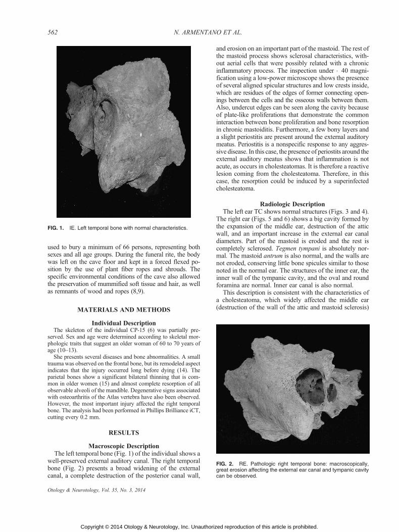
well-preserved external auditory canal. The right temporalbone (Fig. 2) presents a broad widening of the externalcanal, a complete destruction of the posterior canal wall,
and erosion on an important part of the mastoid. The rest ofthe mastoid process shows sclerosal characteristics, with-out aerial cells that were possibly related with a chronicinflammatory process. The inspection under �40 magni-fication using a low-power microscope shows the presenceof several aligned spicular structures and low crests inside,which are residues of the edges of former connecting open-ings between the cells and the osseous walls between them.Also, undercut edges can be seen along the cavity becauseof plate-like proliferations that demonstrate the commoninteraction between bone proliferation and bone resorptionin chronic mastoiditis. Furthermore, a few bony layers anda slight periostitis are present around the external auditorymeatus. Periostitis is a nonspecific response to any aggres-sive disease. In this case, the presence of periostits around theexternal auditory meatus shows that inflammation is notacute, as occurs in cholesteatomas. It is therefore a reactivelesion coming from the cholesteatoma. Therefore, in thiscase, the resorption could be induced by a superinfectedcholesteatoma.
Radiologic DescriptionThe left ear TC shows normal structures (Figs. 3 and 4).
The right ear (Figs. 5 and 6) shows a big cavity formed bythe expansion of the middle ear, destruction of the atticwall, and an important increase in the external ear canaldiameters. Part of the mastoid is eroded and the rest iscompletely sclerosed. Tegmen tympani is absolutely nor-mal. The mastoid antrum is also normal, and the walls arenot eroded, conserving little bone spicules similar to thosenoted in the normal ear. The structures of the inner ear, theinner wall of the tympanic cavity, and the oval and roundforamina are normal. Inner ear canal is also normal.
This description is consistent with the characteristics ofa cholesteatoma, which widely affected the middle ear(destruction of the wall of the attic and mastoid sclerosis)
FIG. 1. IE. Left temporal bone with normal characteristics.
FIG. 2. RE. Pathologic right temporal bone: macroscopically,great erosion affecting the external ear canal and tympanic cavitycan be observed.
562 N. ARMENTANO ET AL.
Otology & Neurotology, Vol. 35, No. 3, 2014
Copyright © 2014 Otology & Neurotology, Inc. Unauthorized reproduction of this article is prohibited.

and was laterally externalized, destroying the superior andposterior walls of the ear canal. However, no signs of com-plications were found in the inner ear or in the endocranealcavity. Therefore, after the macroscopic and radiologicanalysis, the most accurate diagnosis is cholesteatoma.
DISCUSSION
Taking into account the age of the individual and thelesion patterns, the most probable diagnosis is an acquiredunilateral cholesteatoma. There are very few published
cases of this kind of lesion originating in prehistoric times.However, attribution of these ancient lesions to choleste-atoma is not always confident as are based exclusively inmacroscopic exam.
The oldest case of probable cholesteatoma belongs to aLate Stone Age skull from Boskof (Transvaal / RSA) dis-covered in 1913 and described by Singer (16). In AncientEgypt the presence of otologic diseases are not uncommon.Both in Predynastic and Protodynastic times, some ex-amples of lesions diagnosed as possible cholesteatomas are
FIG. 3. IE. Axial CT of left temporal bone. Normal pneuma-tization of mastoid process and correct sizes of ear canal andtympanic cavity can be observed.
FIG. 4. IE. Coronal CT of the normal left ear where the wall of theattic is well differentiated (1).
FIG. 5. RE.Axial CT scan of the right ear: (1) great erosion causinga large cavity formed by the tympanic cavity and part of the mastoidprocess, (2) disappearance ofmastoidpneumatization, and (3) goodpreservation of the structures of the inner ear.
FIG. 6. RE. Coronal cut of the right ear. The image shows thefollowing: (1) destruction of the attic external wall and erosion ofthe tympanic cavity roof, (2) tegmen tympani preserved, (3) normalinner ear structures, and (4) major extension of the tympaniccavity and the CAE.
563ANCIENT CHOLESTEATOMA
Otology & Neurotology, Vol. 35, No. 3, 2014
Copyright © 2014 Otology & Neurotology, Inc. Unauthorized reproduction of this article is prohibited.

found in literature (17), some of them involving tempo-romandibular joints. From this period, a feasible case ofcholesteatoma was found in a Predynastic skull from Nubia,now stored at the British Museum, the right temporal boneof which shows a considerable destruction of the mastoid(18). The cranium shows no sign of healing, and the in-dividual probably died from the extensive inflammation inthe ear region. Otherwise, it resembles the early dynasticskull from the Tarkhan discovered by Fitzsimons in Nubiathat shows a well-defined breach in the meatal wall, possiblybecause of a mastoid abscess (19). Thus, the most impressivefind of this piece is the presence of a fairly well-healed tre-phine hole at some distance on the inferior parietal bone thatcould be an ancient curative procedure. In America and inAsia, there have been few studies of the prevalence ofmastoid infection in prehistoric times (20,21).
In Europe, few cases have been described of ear/mastoiddisease in Prehistoric and Classical times. The oldest couldbe a Neolithic left mastoidal fistula belonging to a 20-year-old female temporal bone fromNerja (Malaga, Spain) (22).From the beginnings of the Bronze Age (Catalan Mega-lithic Culture), a 12-year-old child from the Dolmen at theCementiri dels Moros (Torrent, Girona / Spain) shows ahole in the postero-superior wall of the left temporal bonethat affects the sigmoid sinus in addition to a pneumaticmastoid process (15). However, both cases of cholesteatomaare doubtful. Also dating from the Bronze Age is the Irishskull from Knockast (23) with a lesion involving the tem-poromandibular joint.
More recent specimens described to experience cholestea-toma come from the PunicYRoman Necropolis of Cadiz (24).A possible case of cholesteatoma was found in a computer-assisted tomographic study of 33 temporal bones; the caseis that of a female subject dating from the 4th century AD.Two other possible cases of cholesteatoma were describedfrom Sedgeford and Red CastleYThetford/Norfolk burials,both dating from the Late Saxon times (25) and anotherfrom Quarrington II (Lincolnshire) dating back to the 5thto 6th centuries AD (26). Finally, cholesteatomas werementioned in four Merovingian skulls dating from the 5thto 7th century (27) in Germany.
After this revision of ancient cases of diagnosed cho-lesteatomas from prehistoric and historic times, the case ofLa Cova des Pas could be one of the most ancient cases ofcholesteatoma in Europe ever to be published, if not themost ancient case. Also, it could be one of the most true asdemonstrated by the morphologic and radiologic charac-teristics of these very well conserved ancient bones.
Acknowledgments: The authors thank the team and sponsors(Consell Insular de Menorca and Caixa de Catalunya) of theexcavation of La Cova des Pas.
REFERENCES
1. Thompson RC. Assyrian prescriptions for diseases of the ear. J RAsiat Soc GB Irel 1931;1:1Y25.
2. Nunn JF.Medicine in Ancient Egypt. London, UK: British MuseumPress, 1966.
3. YearsleyM. The pathology of the left temporal bone of the Rhodesianskull. In: Pycraft, WP, eds. Rhodesian man and Associated Remains.London, UK: British Museum (Natural History), 1928;59Y63.
4. McKenzie W, Brothwell D. Disease in the ear region. In: BrothwellD, Sandison AT, eds.Diseases in Antiquity. London, UK: Charles CThomas Publisher, 1967:464Y73.
5. Montgomery PQ, Williams HOL, Reading N, Stringer CB. An as-sessment of the temporal bone lesions of the Broken Hill cranium. JArchaeol Sci 1994;21:331Y37.
6. Armentano N, Jordana X, Fadrique T, Galtes I, Malgosa A. La Covades Pas (Ferreries, Menorca). In: Nieto JL, Obon JA, Baena S, eds.Genes, ambiente y enfermedades en poblaciones humanas. Zaragoza,Spain: Prensas Universitarias de Zaragoza, 2008:61Y72.
7. van Strydonck M, Boudin M, Guerrero Ayuso VM, Calvo M,Fullola JM, Petit MA. The necessity of sample quality assessment in14C AMS dating: The case of Cova des Pas (Menorca Y Spain).Nucl Instrum Methods Phys Res B 2010;268:990Y4.
8. Cabanes D, Albert MR. Microarchaeology of a collective burial:Cova des Pas (Minorca). J Archaeol Sci 2011;38:1119Y26.
9. Simon M, Gonzalez-Ruiz M, Prats-Munoz F, Malgosa A. Com-parison of two DNA extraction methods in a Spanish Bronze Ageburial cave. Quat Int 2012;247:358Y62.
10. Ferembach D, Schwidetzky I, Stloukal M. Recommendations forage and sex diagnoses of skeletons. J Hum Evol 1980;9:517Y49.
11. Krogman WM, Iscan YM. The Human Skeleton in Forensic Med-icine. Springfield, New Zealand: ChC Thomas Ed., 1986.
12. Brothwell DR. Digging Up Bones. London, UK: British Museum,1963.
13. Aleman I, Botella MC, Ruiz L. Determinacion del sexo en elesqueleto postcraneal. Estudio de una poblacion mediterranea ac-tual. Arch Esp Morfol 1997;2:2Y17.
14. Etxeberrıa F. Patologıa traumatica. In: Isidro A, Malgosa A, eds.Paleopatologıa, la enfermedad no escrita. Barcelona, Spain: Ed.Masson, 2003;195Y207.
15. Campillo D. Paleopatologia. Els primers vestigis de la malaltia.Barcelona, Spain: Fundacio Uriach 1838, 1994. vol. 2.
16. Singer R. Pathology in the temporal bone of the Boskop skull. S AArch Bull 1961;16:103.
17. Derry DE. Anatomical report. Archaeol Survey Nubia Bull 1909;3:29Y52.
18. Elliot-Smith G, Dawson WR. Egyptian Mummies. London, UK:George Allen and Unwin Ltd, 1924.
19. Oakley KP, Brooke W, Akester AR, Brothwell D. Contribution ontrepanning or trephination in ancient and modern times. Man 1959;60:122.
20. Titche LL, Coulthard SW, Wachter RD, Thies AC, Harries LL.Prevalence of mastoid infection in prehistoric Arizona Indians. Am JPhys Anthrop 1981;56:269Y73.
21. Rathbun TA, Mallin R. Middle ear disease in a prehistoric Iranianpopulation. Bull NY Acad Med 1977;53:901Y5.
22. Garcıa-Sanchez M. El enterramiento epipaleolıtico de la Cueva deNerja (Malaga). Estudio preliminar. Antropologıa y PaleoecologıaHumana 1986;4:3Y13.
23. Hencken HO,Movius H. The cemetery-cairn of Knockast. Proc RoyIrish Acad. Section C 1932;3441:232Y84.
24. Macıas M, Villanueva A, Mateo A, Ruzaperez-Barquero M.Enfermedades otologicas halladas en una muestra de poblacionPunica y Romana de Cadiz. In: Sanchez Sanchez JA, ed.Sistematizacion metodologica en Paleopatologıa. Actas del VCongreso Nacional de Paleopatologıa. 2001:103Y12. Available at:http://pendientedemigracion.ucm.es/info/aep/boletin/actas/14.pdf.Accessed March 11, 2013.
25. Wells C. Three cases of aural pathology of Anglo-Saxon date. J LaryngOtol 1962;76:931Y3.
26. MayS, Holst M. Paleo-otology of cholesteatoma. Int J Osteoarchaeol2006;16:1Y15.
27. Schultz M. Disease of the ear region in early and prehistoricpopulations. J Hum Evol 1979;8(6):575Y80.
564 N. ARMENTANO ET AL.
Otology & Neurotology, Vol. 35, No. 3, 2014
Copyright © 2014 Otology & Neurotology, Inc. Unauthorized reproduction of this article is prohibited.