Embed Size (px)
Citation preview

Understanding the Etiology of Inflammatory Complications Following Ileal Pouch-Anal
Anastomosis
A Study of the Genetic, Serological and Microbial Factors Associated with Ileal
Inflammation
by
Andrea Dayna Tyler
A thesis submitted in conformity with the requirements
for the degree of Doctor of Philosophy
Graduate Department of Institute of Medical Science
University of Toronto
© Copyright by Andrea D. Tyler, 2013
ii
Understanding the Etiology of Inflammatory Complications Following Ileal Pouch-Anal
Anastomosis
A Study of the Genetic, Serological and Microbial Factors Associated with Ileal
Inflammation
Andrea Dayna Tyler
Doctor of Philosophy
Institute of Medical Science
University of Toronto
2013
Abstract
Introduction: Inflammatory pouch complications, including pouchitis, chronic pouchitis
(CP) and a Crohn’s disease-like phenotype (CDL) of the pouch following ileal pouch-
anal anastomosis (IPAA), are relatively common, and arise via unknown mechanisms.
The phenotypic similarities between pouch inflammation and inflammatory bowel
disease (IBD) suggest there may be common pathways involved in both disorders. The
aim of this thesis is to investigate the serological, genetic and microbial factors
contributing to the development of pouch inflammation in a large, well characterized
patient cohort.
Methods: Subjects with IPAA were recruited, and clinical and demographic information
was obtained through medical chart review and patient questionnaire, allowing patients to
be grouped based on post-surgical phenotype. Blood and tissue was collected for genetic,
serological and microbial analyses. Anti-microbial antibodies were detected using
enzyme-linked immunosorbent assay (ELISA), genotyping was carried out using the
iii
Illumina Goldengate custom SNP assay and Sequenome iPLEX platform, and tissue-
associated microbial communities were assessed using 454 pyrosequencing.
Results: Among our cohort, smoking was associated with CDL (P=0.003) and Ashkenazi
Jewish heritage with CP (P<0.008). NOD2insC (rs2066847) (P=7.4x10⁻⁵), anti-CBir1
(P<0.0001) and ASCA (IgG) (P=0.03) were significantly associated with inflammatory
pouch outcomes. Additional SNPs in NOX3, DAGLB, and NCF4 were also marginally
associated with pouch outcome. A multi-variable risk model combining clinical,
serologic and genetic markers was constructed and could differentiate between chronic
pouch inflammatory phenotypes and no pouchitis. Genus level microbial analysis
demonstrated that several organisms (Bacteroides, Parabacteroides, Blautia and
Moryella) were detected less frequently among the inflammatory outcome groups
(P<0.05). These associations remained significant even following adjustment for
antibiotic use, smoking, country of birth and gender.
Conclusions: CD-associated anti-microbial antibodies and genetic markers are associated
with chronic inflammatory pouch phenotypes. Additionally, changes in the composition
of the pouch associated microbiome are associated with inflammation. These
observations suggest that similar mechanisms may be involved in non-surgical IBD and
pouchitis.

iv
Acknowledgements
I would like to thank my Supervisor, Dr. Mark Silverberg and my thesis advisory committee (Dr. Zane Cohen, Dr. Nicola Jones, Dr. Ken Croitoru, Dr. David Guttman) for their support and encouragement throughout my graduate studies. Their guidance and advice was instrumental in the completion of this work. I was so lucky to have had the opportunity to have learned from the expertise of each of these individuals.
I would also like to thank the members of the Silverberg lab, most especially Boyko Kabakchiev and Joanne Stempak for their assistance with these studies, their willingness to teach, and their friendship over the years.
All of the work done as part of this thesis could not have been performed without the enthusiastic support of the patients included in these studies. These individuals happily shared their blood, tissue and time, in hopes of contributing to this work. I thank them for their unwavering support and for being a constant reminder of why I chose to pursue a degree in inflammatory bowel disease research.
I am also grateful for the financial support provided to me to complete this work by the Crohn’s and Colitis Foundation of Canada, and both The Department of Medicine and the Samuel Lunenfeld Research Institute at Mount Sinai Hospital.
Finally, I would like to thank my parents (Maureen and Kip Tyler), my Grandmothers (Dr. Marvis Tutiah, and Wilma Mitchell) and my Toronto ‘family’ for their support, guidance, availability for counselling, home cooking, laughter, and for inspiring me to reach higher than I might otherwise have imagined possible.

v
Table of Contents
Chapter 1: Introduction and Background ............................................................................ 1
1.1 Inflammatory bowel disease ..................................................................................... 1
1.1.2 Risk factors for IBD ......................................................................................... 2
1.1.3 Ileal pouch-anal anastomosis and pouch inflammatory complications ............ 3
1.2 Serology .................................................................................................................... 6
1.2.1 A description of the anti-microbial antibodies identified in IBD .......................... 7
1.2.2 Association of serological markers with CD and UC .................................... 10
1.2.3 IBD serology in pouch inflammatory complications ..................................... 12
1.2.4 The role of anti-microbial antibodies in inflammatory disease pathogenesis ...... 12
1.3 Genetics ................................................................................................................... 15
1.3.1 Analyzing the human genome ........................................................................ 15
1.3.2 SNP genotyping: platforms and analysis ............................................................. 17
1.3.3 - Genetic Associations with IBD .................................................................... 23
1.3.4 IBD genetic association studies in non-Caucasian populations ..................... 27
1.4 Bacteria ................................................................................................................... 29
1.4.1 Phylogenetic organization and taxonomic classification of bacteria ............. 30
1.4.2 Human microbiome analysis techniques ........................................................ 35
1.4.3 Culture-independent microbial community analysis ...................................... 36
1.4.3.1 Sample procurement ............................................................................... 36
1.4.3.2 Microbial DNA extraction from samples ............................................... 37
1.4.3.3 Overview of Sequence-based microbial analysis tools ........................... 38
1.4.3.4 A more detailed examination of next-generation sequencing and how it is applied to the analysis of the microbiome. ......................................................... 41
1.4.3.5 Sequence quality trimming, alignment and taxonomic assignment ....... 44
1.4.3.6 Statistical analysis of microbial data ...................................................... 50
1.4.4 The Human Microbiome – lessons from community level analysis .............. 53
1.4.5 Location-specific microbiome heterogeneity ................................................. 56
1.4.7 Genes, serology and microbiome ................................................................... 59
1.4.8 Microbiome in IBD ........................................................................................ 60
1.4.9 Microbiome and pouchitis .............................................................................. 64
1.5 Overarching theme - bringing it all together ........................................................... 65

vi
1.6 Hypothesis and Aims .............................................................................................. 65
Chapter 2: Antimicrobial Antibodies Are Associated with a Crohn's Disease-Like Phenotype following Ileal Pouch-Anal Anastomosis ....................................................... 67
2.1 Abstract: .................................................................................................................. 70
2.2 Introduction ............................................................................................................. 71
2.3 Materials and Methods ............................................................................................ 71
2.4 Results ..................................................................................................................... 74
2.5 Discussion ............................................................................................................... 81
Chapter 3: The NOD2insC polymorphism is associated with worse outcome following ileal pouch-anal anastomosis for ulcerative colitis ........................................................... 85
3.1 Abstract: .................................................................................................................. 87
3.2 Introduction: ............................................................................................................ 89
3.3 Methods: ................................................................................................................. 89
3.4 Results: .................................................................................................................... 93
3.5 Discussion ............................................................................................................. 105
Chapter 4: Characterization of the gut-associated microbiome in inflammatory pouch complications following ileal pouch-anal anastomosis. ................................................. 109
4.1 Abstract: ................................................................................................................ 111
4.2 Introduction ........................................................................................................... 112
4.3 Materials and Methods .......................................................................................... 113
4.4 Results ................................................................................................................... 115
4.5 Discussion ............................................................................................................. 128
4.6 Chapter 4, Supplementary Methods ...................................................................... 134
5.1 Conclusions and general discussion .......................................................................... 136
5.2 Future directions ....................................................................................................... 147
Appendix ......................................................................................................................... 150
References ....................................................................................................................... 185
vii
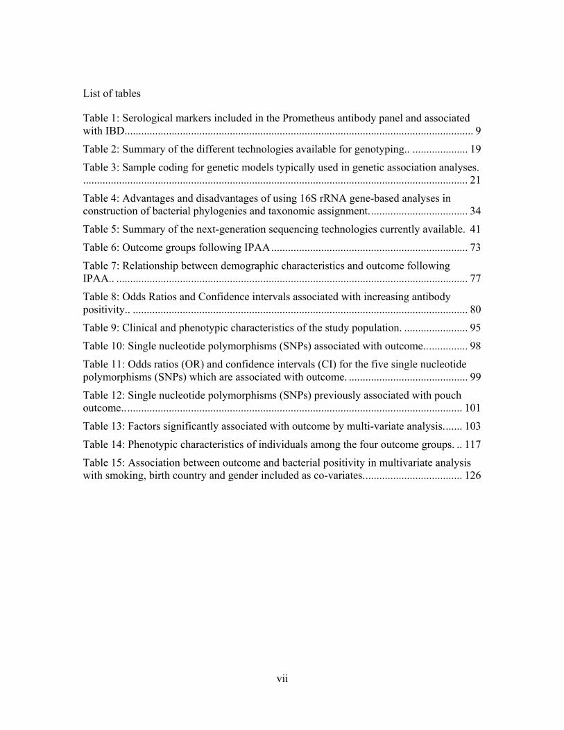
List of tables
Table 1: Serological markers included in the Prometheus antibody panel and associated with IBD. ............................................................................................................................. 9
Table 2: Summary of the different technologies available for genotyping.. .................... 19
Table 3: Sample coding for genetic models typically used in genetic association analyses. ........................................................................................................................................... 21
Table 4: Advantages and disadvantages of using 16S rRNA gene-based analyses in construction of bacterial phylogenies and taxonomic assignment. ................................... 34
Table 5: Summary of the next-generation sequencing technologies currently available. 41
Table 6: Outcome groups following IPAA ....................................................................... 73
Table 7: Relationship between demographic characteristics and outcome following IPAA.. ............................................................................................................................... 77
Table 8: Odds Ratios and Confidence intervals associated with increasing antibody positivity.. ......................................................................................................................... 80
Table 9: Clinical and phenotypic characteristics of the study population. ....................... 95
Table 10: Single nucleotide polymorphisms (SNPs) associated with outcome.. .............. 98
Table 11: Odds ratios (OR) and confidence intervals (CI) for the five single nucleotide polymorphisms (SNPs) which are associated with outcome. ........................................... 99
Table 12: Single nucleotide polymorphisms (SNPs) previously associated with pouch outcome.. ......................................................................................................................... 101
Table 13: Factors significantly associated with outcome by multi-variate analysis. ...... 103
Table 14: Phenotypic characteristics of individuals among the four outcome groups. .. 117
Table 15: Association between outcome and bacterial positivity in multivariate analysis with smoking, birth country and gender included as co-variates. ................................... 126
viii
List of figures
Figure 1: Hierarchical organization of taxonomic levels used for classifying organisms 32
Figure 2: Regions of 16S rRNA molecule typically used for evaluating the human microbiome. ...................................................................................................................... 42
Figure 3: Proportions of patients (n=399) in each of the three outcome groups .............. 76
Figure 4: Overall levels of antibody positivity among three outcome groups .................. 79
Figure 5: Proportion of individuals in each specific outcome group. ............................... 96
Figure 6: ROC curves generated for the risk score analysis. .......................................... 104
Figure 7: Phylum level comparisons between four outcome groups. ............................. 119
Figure 8: Proportion of patients positive for genera which were significantly associated with outcome ................................................................................................................... 121
Figure 9: Proportion of patients positive for genera in individuals with inflamed vs not-inflamed pouches. ........................................................................................................... 123
Figure 10: Hypothesized model of the pathogenesis of ileal inflammation ................... 143
ix
Abbreviations
IBD – Inflammatory bowel disease
CD – Crohn’s disease
UC – Ulcerative colitis
IBDU – IBD type unclassified
IC – Indeterminate colitis
IPAA – Ileal pouch anal anastomosis
PSC - Primary sclerosing cholangitis
FAP – Familial adenomatous polyposis
TNF – Tumor necrosis factor
ELISA - Enzyme linked immunosorbent assay
ASCA – Anti-Saccharomyces cerevisiae antibodies
pANCA - Perinuclear antineutrophil cytoplasmic antibodies
OmpC – Outer membrane porin C
SNP - Single nucleotide polymorphism
TDT – Transmission disequilibrium test
HWE - Hardy-Weinberg equilibrium
GWA – Genome-wide association
MAF - Minor allele frequency
FDR – False discovery rate
ROS – Reactive oxygen species
eQTL – Expression quantitative trait loci
HGT – Horizontal gene transfer
RDP – Ribosomal database project
T-RFLP – Terminal restriction fragment length polymorphism
SSCP - Single-strand conformation polymorphism
qPCR – Quantitative polymerase chain reaction
FISH – Fluorescence in situ hybridization
OTU – Operational taxonomic unit
BLAST – Basic local alignment search tool
NCBI – National centre for biotechnology information
MSA – Multiple sequence alignment
x
5-ASA – 5-aminosalicylic acid
MAMP – Microbial-associated molecular pattern
PSA – Polysaccharide A
TLR – Toll-like receptor
IL – Interleukin
NOD - Nucleotide-binding oligomerization domain-containing protein
IFN – Interferon
MHC – Major histocompatibility complex
LPS - Lipopolysaccharide
CRP – C-reactive protein
ESR – Erythrocyte sedimentation rate
SLiME - Synthetic learning in microbial ecology
AIEC – Adherent-invasive E. coli
FT – Fecal transplant
NP/NCP – No pouchitis/no chronic pouchitis
CP – Chronic pouchitis
P – Pouchitis
CDL – Crohn’s disease-like phenotype
MSH - Mount Sinai Hospital
HMC - Penn State Milton S. Hershey Medical Center
IBD-U – IBD unclassified
OR – Odds ratio
CI – Confidence interval
ROC - Receiver operating characteristic
AUC - Area under the curve
PDAI – Pouchitis disease activity index
PAS - Pouchitis activity score

1
Chapter 1: Introduction and Background
1.1 Inflammatory bowel disease
The Inflammatory Bowel Diseases (IBD), comprising Crohn’s disease (CD) and
ulcerative colitis (UC) are chronic inflammatory conditions of the digestive tract of
unknown etiology. In Canada, where rates of IBD are among the highest in the world,
between 0.67 and 0.9% of the population is affected, with an approximately equal
proportion of patients with a diagnosis of UC and CD 1-3. It is conservatively estimated,
in a report provided by the Crohn’s and Colitis Foundation of Canada, that 28 Canadians
are diagnosed with IBD daily. Symptoms are varied, depending on the location of
disease, however commonly include abdominal cramping, nausea or vomiting, increased
stool frequency, bloody stool, fatigue, anemia and weight loss2. Children diagnosed with
IBD may experience delays in physical and psychological development. Medical
management of both CD and UC are similar with anti-inflammatories (5-ASA),
immunosuppressants (methotrexate, azathioprine), steroids and biological agents (anti-
TNFα) the current mainstays of treatment. However, IBD has no cure, and represents a
significant healthcare burden2.
Historically, CD and UC have been considered separate entities with different
phenotypic characteristics and outcomes. CD can discontinuously affect any part of the
digestive tract from mouth to anus and is often characterized by transmural inflammation.
Additionally, patients may go on to develop stricturing or fistulizing phenotypes which
frequently require surgical management. As a result of such complications, up to 80% of
CD patients will have to undergo some form of surgical resection during the course of
their disease4. While CD surgery is typically a temporary solution, with disease often
recurring in nearby tissue, benefits include the obviation of obstructive and penetrating
symptoms, decreased need for immunosuppressive medication and an increase in patient
quality of life. UC, on the other hand, affects mainly the superficial layers of mucosa of
the large bowel, and extends in a continuous fashion proximally from the rectum. Patients
with UC do not develop structuring or fistulizing phenotypes and seldom have
inflammation extending into the small bowel. In UC cases where surgical intervention is
2
necessary, subtotal colectomy or procto-colectomy is performed, and is considered
curative in intent as all UC-susceptible tissue (colon) is removed. An additional subset of
patients display traits characteristic of both disorders and are referred to as IBD type
unclassified (IBDU). The Montreal classification further identifies patients with
diagnostic features of both CD and UC specifically at the time of surgical resection as
indeterminate colitis (IC)5. However, evidence suggests that the IBDs exist more as a
spectrum, with overlapping phenotypic characteristics, disease mechanisms and
responses to therapeutic and surgical management.
1.1.2 Risk factors for IBD
A great deal of study has been devoted to identifying risk factors for the
development of IBD. Evidence of heritable risk factors include the increased incidence of
IBD among individuals of Ashkenazi Jewish heritage6, and among individuals with a
family history of IBD 7. This relationship is slightly stronger in CD, but important in both
diseases. Smoking and exposure to second hand smoke is among the best studied
environmental risk factors, having been associated with an increased risk of CD, and a
decreased risk of UC8, 9. Additional environmental factors which have been suggested to
play a role in the recent dramatic increase in IBD prevalence through much of the
developed world include reduced breastfeeding, increased hygiene and sanitation,
reduction in physical activity, exposure to pollution, consumption of a Western diet 10,
and use of antibiotics among children11. However, studies examining these factors have
been associative in nature and are complicated by the difficulties in obtaining information
on patients prior to the onset of symptoms or disease.
There is also evidence that multiple auto-inflammatory processes may occur via
similar mechanisms. Individuals with IBD are at increased risk of comorbidities
including primary sclerosing cholangitis (PSC), pyoderma gangrenosum, arthritis, and
uveitis among others12. Furthermore, the presence of such co-morbidities may also
signify a more severe disease course.
IBD is predominantly a Western disease with significantly higher incidence and
prevalence in North America and northern Europe compared to other more equatorial

3
locations. This has led to speculation that environmental factors specific to Western
nations, in conjunction with increased genetic susceptibility, leads to disease. Evidence
supporting this hypothesis includes changes in the prevalence of IBD among first
generation Canadians. While newly arrived immigrants, particularly those coming from
equatorial locations, tend to have lower rates of IBD than do their Canadian counterparts,
the risk of IBD among their children is equal to or exceeds the Canadian average13, 14.
Further, a recent study examining the risk of IBD among South Asian immigrant
populations within Canada observed that rates of IBD among second generation
Canadians (children) was significantly higher than non-South Asians4, 15. Disease
phenotypes tended to be more severe among this group as well, with extensive colonic
disease more common than among non-south Asian individuals13. Similar results were
obtained in a study conducted in the United Kingdom15.
Prevailing hypotheses regarding IBD pathogenesis take into account both genetic
and environmental factors. It is believed that IBD susceptibility is partially genetically
mediated with specific genetic loci modulating disease risk. However, in the absence of a
specific environmental trigger, which is commonly believed to be microbial in origin,
IBD may not develop. Thus, IBD is thought to result from a complex interaction between
host genetics and microbial environment, with ultimate phenotype determined by the
specific gene-microbe interactions.
1.1.3 Ileal pouch-anal anastomosis and pouch inflammatory complications
Individuals with severe or medication refractory UC, or who develop dysplasia,
are candidates for total procto-colectomy and ileostomy or ileal pouch-anal anastomosis
(IPAA). These patients are considered surgically ‘cured’ of their disease, due to the
localized disease pattern characteristic of this form of IBD. Rather than receiving a
conventional ileostomy, individuals undergoing colectomy for UC may elect to undergo
IPAA, which allows transanal passage of stool and eliminates the requirement of an
ileostomy. This procedure involves the surgical construction of a pouch or reservoir from
the terminal ileum which is then connected to the rectal cuff or anus. IPAA is only rarely
performed on individuals with CD, due to an increased risk of post-surgical
complications, such as recurrence of chronic inflammation in the pouch or afferent limb,

4
or development of CD-related fistulas16. Individuals with familial adenomatous polyposis
(FAP), a condition characterized by proliferation of polyps throughout the large bowel,
also often undergo IPAA, in order to prevent the development of colorectal cancer17.
When performed by experienced physicians, colectomy with IPAA has a relatively low
rate of surgical complication and has been shown to enhance patient experience and
quality of life when compared to an ileostomy18.
Despite the surgical removal of UC susceptible tissue, many patients experience
pouchitis, defined as inflammation of the ileal reservoir. Pouchitis is the most common
complication following IPAA, affecting up to 50% of UC patients at some time following
surgery, while occurring only rarely among individuals with FAP. Patients with pouchitis
experience increased stool frequency, rectal bleeding, fecal urgency, cramping, malaise
and occasionally fever. The endoscopic and histological picture is similar to that of IBD,
with edema, erythema, loss of vascular pattern friability and ulceration seen on
pouchoscopy and polymorphonuclear/mononuclear leukocyte infiltration, microscopic
ulceration, and villus atrophy observed on histological examination19, 20. Unlike IBD,
most cases of pouchitis respond rapidly to antibiotic therapy and are transient in nature21.
However, a subset of patients with pouchitis will require long term antibiotics in order to
maintain healthy pouches, or will require the use of additional therapeutic agents in order
to remain healthy. Medications which may be successfully used in treating antibiotic
refractory pouchitis include those which are typically used to treat IBD, such as
corticosteroids, immunomodulators and anti-tumor necrosis factor (TNFα) agents. An
additional subset of patients with IPAA for UC, will develop what has been described as
a CD-like phenotype, with inflammation proximal to the pouch in either the afferent limb
or upper digestive tract, or the development of a stricture, fistula or abscess which is not
related to surgical complications21. Such chronic inflammatory phenotypes have been
suggested to have been misdiagnosed pre-colectomy CD, yet thorough chart review often
demonstrates no evidence for this diagnosis prior to surgery or following pathological
review of the surgical specimen22.
Physicians often successfully treat patients presenting with symptoms of pouchitis
empirically with antibiotics. However, it is often unclear whether pouch inflammation is
indeed the cause of these symptoms. Several clinical tools for diagnosing pouchitis, and

5
differentiating this outcome from others have been developed. The pouchitis disease
activity index (PDAI) and pouchitis activity score (PAS) make use of a combination of
clinical, endoscopic and histological data in order to diagnose patients with pouchitis20. In
a recent analysis by Ben Bassat et al23, poor correlation was shown between the clinical
and both endoscopic and histological components of each of these scores, as well as
between the endoscopic and histological subscores. This suggests that clinical symptoms
are non-specific, and highlights the importance of using more objective measures of
pouch inflammation, especially in the context of studies specifically analyzing etiological
factors contributing to inflammation. Objective measures for accurately assessing
inflammation which rely on biomarkers rather than invasive procedures such as
endoscopy, would clearly be beneficial.
Several factors have been associated with an increased risk of pouchitis
development following IPAA. The most important risk factor is a pre-colectomy
diagnosis of IBD. Individuals with FAP rarely develop pouch inflammation, whereas
those with UC are much more susceptible. Individuals with CD are only rarely offered
this procedure due to the likelihood of disease recurrence. IC has been associated with an
increased risk of complications compared to UC in some studies, although not all,
suggesting that the risk of pouch inflammation for these individuals may be similar to
that seen for those with severe UC24. Some evidence also suggests that more extensive
UC prior to surgery or the presence of backwash ileitis increases the risk of inflammatory
complications, although findings regarding these traits have shown mixed results25-27.
Among UC patients undergoing IPAA, a diagnosis of PSC, presence of other extra-
intestinal manifestations of IBD, a family history of CD among first degree relatives and
smoking history have all been associated with an increased risk of pouch inflammation25,
28-30.
As with IBD in general, the etiology of pelvic pouch inflammatory complications
is poorly understood. The phenotypic similarities between pouch inflammation and both
CD and UC suggest that common disease pathways are involved in these inflammatory
processes. As such, the pelvic pouch represents a useful human model for evaluating de
novo ileal inflammation and may be a useful proxy for gaining insight into general
mechanisms of IBD. Furthermore, the importance of describing predictive factors which

6
accurately stratify patients’ risk of inflammatory complications, will have broad
applications in determining which individuals are good candidates for IPAA surgery.
1.2 Serology
The current gold-standard for the diagnosis and classification of patients with IBD
is ileocolonoscopy with biopsy. While highly accurate and relatively safe, this procedure
is invasive and costly. Further, in complicated cases, colonoscopy may not allow
differentiation between IBD subtypes. As such, the development of non-invasive,
inexpensive tests which are capable of differentiating between different forms of IBD and
assessing disease activity, would be useful for physicians trying to diagnose patients.
Currently available and widely used clinical blood tests which are helpful in managing
IBD include measurements of C-reactive protein (CRP), and erythrocyte sedimentation
rate (ESR)31 for example, each of which provide a broad measure of inflammatory
activity. CRP is perhaps the most well studied blood-based biomarker, and has been
shown to provide an effective estimation of disease severity in CD32. There is also some
evidence that this marker may help to predict relapse or need for colectomy in UC33.
However, these blood markers can also be elevated in other inflammatory conditions, and
thus are not specific enough on their own to provide a definitive diagnosis of IBD34.
Several other studies have also shown poorer correlations between CRP and bowel
disease activity, and have suggested that it does not provide sufficient resolution to
distinguish between CD and UC, highlighting the need for additional markers35.
A newer marker, fecal calprotectin, has been shown to correlate well with various
measures of IBD disease activity especially in colonic disease35. Several studies have
demonstrated that this marker is at least as useful as CRP in measuring disease activity,
yet it too is unable to differentiate between CD and UC36. Furthermore, the necessity of
obtaining stool samples to carry out this test has prevented its more widespread utility.
The value of biomarkers clinically, and in IBD research is apparent. However,
none of the above tests, on their own, are capable of accurately distinguishing between
phenotypes, or predicting which patients are at risk of a more severe outcome. Several
7
newer biomarkers with interesting applications for IBD research and clinical
management, both as diagnostic tools, and to assist in the sub-classification of patients
with a diagnosis of CD or UC, are anti-microbial antibodies which can be detected in the
serum of patients with IBD. Such markers have been well studied and demonstrate high
specificity (90-100%) but rather low sensitivity (50-70%) for bowel disease37. Thus,
while these markers do not obviate the need for colonoscopy, they have shown potential
in aiding in disease management. Furthermore, the ease of obtaining serological samples
(draw peripheral blood from patient, allow sample to clot in the tube, remove serum for
testing or storage), combined with the relative cost-effectiveness of their measurement,
would make tests based on these markers widely appealing for both clinical and research
applications.
1.2.1 A description of the anti-microbial antibodies identified in IBD
Many of the common and well-studied IBD associated serological markers,
including anti-Saccharomyces cerevisiae antibodies (ASCA), anti-CBir1, anti-outer
membrane porin C (OmpC), and anti-I2 are antibodies which target specific microbial
motifs. ASCAs are human IgA or IgG antibodies which specifically target the mannan
motifs on the cell wall of S. cerevisiae38 yet which are capable of also recognizing
homologous sequences from other organisms more commonly found in the digestive
tract. For example, ASCAs can also be generated against Candida albicans39. Anti-CBir1
targets a flagellin moiety and anti-OmpC a membrane porin of Escherichia coli, while
anti-I2 targets a membrane component of Pseudomonas fluorescens40. These microbes
are part of the commensal microbiota, yet among healthy individuals rarely result in the
production of antibodies.
Another commonly used serological marker with diagnostic use in studying IBD
is perinuclear anti-neutrophil cytoplasmic antibody (pANCA). Rather than specifically
recognizing microbial motifs, this marker is an autoantibody with specificity for an as yet
unknown component of neutrophil granules41. While ANCAs have been associated with
several inflammatory diseases, detection of pANCA is a much more IBD-specific

8
phenomenon. Unlike ANCA, pANCA has nuclear localization of the staining pattern
which can be detected by indirect immunofluorescence, and which disappears following
treatment with DNase41.
For each of these markers, antibody titres are assessed using enzyme linked
immunosorbent assay (ELISA). Established positivity cutoff values, based on
experimental results, are also commonly applied to enhance the diagnostic value and
interpretability of data. Prometheus Therapeutics and Diagnostics (San Diego, CA) offers
diagnostic testing for anti-microbial makers important in IBD, to provide more
information to physicians attempting to make an accurate diagnosis of IBD or to
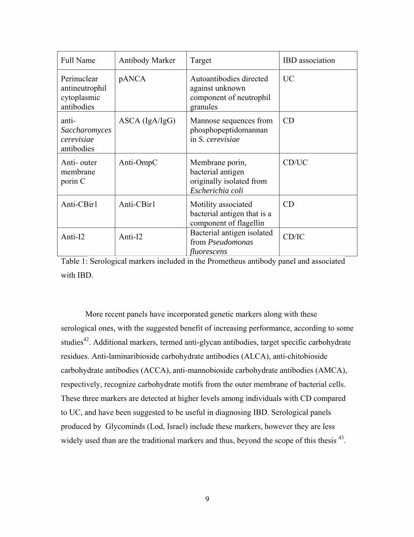
distinguish between UC and CD. Table 1 describes the markers which are typically
assessed by Prometheus and their described associations with IBD.
9
Full Name Antibody Marker Target IBD association
Perinuclear antineutrophil cytoplasmic antibodies
pANCA Autoantibodies directed against unknown component of neutrophil granules
UC
anti-Saccharomyces cerevisiae antibodies
ASCA (IgA/IgG) Mannose sequences from phosphopeptidomannan in S. cerevisiae
CD
Anti- outer membrane porin C
Anti-OmpC Membrane porin, bacterial antigen originally isolated from Escherichia coli
CD/UC
Anti-CBir1 Anti-CBir1 Motility associated bacterial antigen that is a component of flagellin
CD
Anti-I2 Anti-I2 Bacterial antigen isolated from Pseudomonas fluorescens
CD/IC
Table 1: Serological markers included in the Prometheus antibody panel and associated
with IBD.
More recent panels have incorporated genetic markers along with these
serological ones, with the suggested benefit of increasing performance, according to some
studies42. Additional markers, termed anti-glycan antibodies, target specific carbohydrate
residues. Anti-laminaribioside carbohydrate antibodies (ALCA), anti-chitobioside
carbohydrate antibodies (ACCA), anti-mannobioside carbohydrate antibodies (AMCA),
respectively, recognize carbohydrate motifs from the outer membrane of bacterial cells.
These three markers are detected at higher levels among individuals with CD compared
to UC, and have been suggested to be useful in diagnosing IBD. Serological panels
produced by Glycominds (Lod, Israel) include these markers, however they are less
widely used than are the traditional markers and thus, beyond the scope of this thesis 43.
10
1.2.2 Association of serological markers with CD and UC
The utility of serum associated markers in IBD diagnostics have been well
documented. In each case, these markers are detected significantly more frequently
among individuals with disease compared to healthy controls. These serological markers
also have the potential to distinguish between CD and UC. Several studies have
demonstrated a strong association between ASCA (IgA/IgG) and CD, and between
pANCA and UC44-46. ASCA IgA and IgG correlate well with one another47, and overall
ASCA positivity tends to correlate highly with small bowel CD and poorly with UC.
Patients with IC have been shown to have ASCA levels which are intermediate between
those found in individuals with CD and UC, an interesting observation given the
intermediate nature of the IC phenotype37. pANCA, on the other hand, is associated with
UC and colonic disease location in CD48-50. Both of these markers are also associated
with more severe outcomes which commonly require surgical intervention51 . Moreover,
individuals with CD and higher numbers of CD-associated markers are at increased risk
of complicated disease behaviour and need for surgery52, 53. When measured alone, these
markers are useful, however, the sensitivity and specificity of using them together is
substantially increased54.
Despite the diagnostic potential of ASCA and pANCA, approximately 30% of
patients with IBD are not positive for either of these markers50. Further, although fairly
specific, there is some overlap in marker detection among phenotypic outcome groups,
with 5-15% of UC patients positive for ASCA, and approximately 6-20% of CD patients
positive for pANCA50, 54, 55. This relationship becomes more complicated when different
disease phenotypes are evaluated, as colonic CD often demonstrates a serological profile
more similar to that of UC. In order to optimize diagnostic sensitivity and specificity,
additional markers including anti-CBir1 and anti-OmpC, were identified and have
demonstrated diagnostic potential.
Anti-OmpC is detected more frequently among individuals with CD and their
unaffected relatives compared to those with UC and healthy controls 56. This marker has
also been associated with internal penetrating and stricturing CD phenotypes57. Yet the
specificity of this marker in appropriately diagnosing IBD in the absence of the other
markers has been called into question. In a small study examining its utility in a pediatric

11
cohort, seven patients were falsely found to have IBD based on anti-OmpC seroreactivity
alone58. Anti-CBir1 is also strongly associated with CD. This marker is detected more
commonly among individuals with small bowel disease and requirement for resection, as
well as among individuals with fibrostenotic and internal penetrating disease phenotypes.
It is negatively associated with a more UC-like disease course40. Furthermore, among CD
patients who are positive for pANCA this marker was more commonly detected,
suggesting that it may add specificity to the diagnostic capabilities of anti-microbial
antibody panels40, 59. Interestingly, anti-CBir1 is detected more frequently among younger
children (<8 years old) with IBD, and may therefore represent a better early indicator of
IBD among this population60. Anti-I2 is associated with CD, and a need for surgery61.
Interestingly, this marker was also associated with response following fecal diversion for
medically resistant proctocolitis, with individuals positive for this marker more likely to
experience clinical remission following this procedure62. Seropositivity for all of these
markers is also highly dependent on the length of time that an individual has had disease,
as well as disease severity and tendency towards progression61.
The majority of studies examining anti-microbial antibody markers have been
conducted retrospectively. As such, the observation that serum levels of ASCA, pANCA
and anti-OmpC have been shown to be fairly static over time, not varying greatly with
disease activity or surgery, is important63. Of the results from studies which do
prospectively evaluate these markers, most findings are similar to those found in
retrospective studies. One study demonstrated a substantial increase in the risk of
developing IBD in first-degree relatives from multiply affected families, among
individuals who were positive for at least one serological marker (measured prior to
diagnosis)64. On the other hand, a small study evaluating pANCA positivity following
colectomy showed a slight reduction in pANCA prevalence between individuals who had
undergone surgery compared to those who had not65. The few studies which have
prospectively evaluated these markers have supported their predictive potential, and
demonstrate that the described anti-microbial antibodies are detectable in many patients
prior to symptom onset and IBD diagnosis, although often at lower titres66.

12
1.2.3 IBD serology in pouch inflammatory complications
The beneficial potential of serological biomarkers in easily diagnosing and
monitoring inflammatory pouch complications following IPAA is also apparent. As such,
serological markers have also been examined in this context. Such studies have been
limited by small sample sizes and unclear post-colectomy patient phenotypic
classifications. Results to date have been mixed, with some demonstrating an association
between pANCA, ASCA and anti-CBir1 and pouch outcome, with these markers detected
at higher frequency among individuals with inflammatory complications compared to
those with healthy pouches67 68-70. Other studies have shown no relationship71. An
additional study demonstrated that ASCA was associated with the development of fistulas
following surgery72. Whether these markers were present prior to IPAA, and thus could
be used as predictors of post-surgical outcome is unknown. However the previously
described stability of markers over time suggests that they may have a use in stratifying
patient risk prior to colectomy. As well, a prospective study demonstrated that pre-
colectomy levels of pANCA, and anti-CBir1 were associated with an increased risk of
acute and chronic pouchitis development following IPAA67. The relationship between
pouch outcome and serological markers is described in greater detail in Chapter 2.
1.2.4 The role of anti-microbial antibodies in inflammatory disease pathogenesis
Increased prevalence of several of the anti-microbial antibody markers (mainly
ASCA and pANCA) have been detected in numerous inflammatory conditions, including
IBD, ankylosing spondylitis73, chronic granulomatous disease74, and celiac disease75. In
each case, the physiological processes which contribute to the specificity of antibodies to
normally commensal organisms are unknown. While there is no evidence that these
antibodies themselves have a pathogenic role, or are even detectable in the bowel
mucosa76, their high prevalence among individuals with inflammatory diseases suggests
that they may be byproducts of disease processes. Further, given that these markers are
detected in numerous diseases, they may provide additional evidence that these diseases
proceed via similar mechanisms.

13
The pathophysiological relevance of anti-microbial antibodies in IBD, and more
generally in autoinflammatory diseases, is unclear. On the one hand it is possible that the
antibodies detected in CD and UC, which target specific microbial surface antigens, are
indicative of immunological recognition of a pathogenic organism which is of importance
in diseases pathogenesis. In this case, marker specificity for flagellin (CBir1) and E. coli
surface porin (OmpC) or the unknown protein detected from Pseudomonas fluorescens
(I2) would suggest that these organisms may have a specific role in IBD. As will be
discussed in section 1.4.8, specific strains of E. coli have indeed been detected more
commonly in tissue samples from ileal CD77, although have not been detected among all
patients with disease. However, no specific correlations between anti-CBir1 and any
organism, or anti-OmpC and E. coli have been documented. P. fluorescens has not been
shown to associate with CD, although some evidence suggests that the Pseudomonas
genus may be detected more frequently among children with ileal disease78. Additionally,
certain strains of P. fluorescens have been shown to increase epithelial permeability in
cell culture79. However, little evidence can be found supporting the hypothesis that a
single organism is responsible for IBD, even among subsets of the population.
An alternative hypothesis is that the presence of anti-microbial antibodies in the
serum of patients with IBD may be indicative of a more general breakdown of intestinal
barrier function or of the innate immune response, resulting in an increase in the detection
of microbes by the adaptive immune system80. This in turn has been speculated to lead to
increased production of antibodies targeting microbial motifs from normally commensal
organisms. Observations that individuals with chronic granulomatous disease, a condition
where failure to produce reactive oxygen species (an important innate immune process)
leads to reduced ability to clear infections, also express these markers, would seem to
suggest that innate immune function is important74. Further, there is evidence that
individuals with IBD have additional anti-microbial antibodies in their blood, with
specificities for numerous different microbial antigens47. In fact, enhanced diagnostic
utility of antibodies against a mixture of bacterial antigens obtained from a preparation of
the surface antigens of a single species (Bacteroides vulgatus), or complex community,
compared to the individual markers currently tested, has been demonstrated47. This
suggests that the current evaluation of antibodies specific for certain epitopes may be
missing important information. These observations are in keeping with a more

14
generalized breakdown in intestinal barrier function leading to increased antibody
detection. One study demonstrated a marginal correlation between increasing ASCA IgG
and intestinal permeability81. However, few studies have definitively demonstrated that
increased gut permeability is associated with antibody titres or positivity.
It is also possible that, rather than a general breakdown in overall barrier function,
there is a loss of specific innate immune functions which typically prevent certain
antigens from eliciting adaptive immune processes. ASCA positive patients, for example,
have decreased expression of mannan-binding lectin (MBL)82. This molecule acts as a
pattern-recognition receptor directed against oligomannan, (the epitope detected by
ASCA, which is also found on the cell surface of numerous other microorganisms). Upon
the binding of MBL to a microorganism, the complement system is activated, leading to
clearance of the targeted organism82. This would suggest that specific host alterations are
responsible for the production of anti-microbial antibodies, and that these alterations may
also play a role in disease pathogenesis. A further discussion of the relationship between
host factors (genetics) and anti-microbial antibody positivity is included in section 1.3.5.
As with many aspects of IBD, determining whether a breakdown in barrier
function precedes inflammation, or is merely a consequence of macroscopic or
microscopic disease, is difficult. Inflammatory processes themselves result in a less
cohesive intestinal barrier and could, therefore, result in increased access of commensal
organisms to adaptive immune processes. Yet studies which have supported the
prognostic capabilities of antibody markers suggest that anti-microbial markers can be
detected in the serum of patients preceding the onset of inflammation66. This suggests
that alterations in barrier function (should they occur), which lead to anti-microbial
antibody generation, precede the onset of IBD.
Despite the strong evidence regarding the diagnostic utility of these markers in
IBD, on their own they leave many unanswered questions regarding mechanisms of
disease pathogenesis. To gain a further understanding of disease mechanisms, and how
these serologic markers fit into a model of disease etiology, it is necessary to further
explore additional pathways, including host genetic and microbial factors.
15
1.3 Genetics
With the sequencing of the human genome ‘completed’ in 200183, 84, it became
clear that a great deal more work was required before a genetic roadmap of human
disease was available. Early drafts of the human genome provided evidence of previously
unrecognized variability in the genetic landscape, with some regions characterized by
high gene density, and others more sparsely populated. Further, the number of coding
genes was found to be lower than expected, with around 30-40K protein coding genes.
On the other hand, these fewer genes are more complex than those described in other
organisms, with greater potential for alternate splicing83. While not providing a cure for
all human genetic disease, the complete sequencing of the human genome provided many
useful tools for better understanding the genetic variability between individuals and
groups, and for evaluating the genetic contribution to many disorders. Subsequent
projects such as the HapMap and 1000 Genomes Project, which both seek to discover,
genotype and provide accurate haplotype information on human DNA polymorphisms in
multiple populations, shed new light on human genetic diversity both within and between
ethnic groups85, 86. These tools can be used to evaluate the complex architecture of
genetically mediated diseases, and have identified millions of single nucleotide
polymorphisms (SNP)s, allowing quantification of genetic variability across populations
and phenotypes87.
1.3.1 Analyzing the human genome
The role of genetic factors in health and disease is complex. Several diseases
including sickle cell anemia and cystic fibrosis for example, result from disease-causing
mutations in a single gene (monogenic)88, 89. Yet other diseases, including diabetes,
obesity and IBD demonstrate complex heritability, with numerous loci modulating
disease risk. Indeed, while genetic variability likely impacts susceptibility to most human
illness, with effects ranging from those seen in monogenic disorders, to the modulation of
individual susceptibility towards infectious agents (ie. decreased susceptibility to HIV
infection among CCL3L1high copy number carriers)90, few of these interactions have
been fully elucidated. In the case of complex diseases, incomplete penetrance (differential
expression of a genetic trait despite genotype), polygenicity (multiple genes contributing
16
to a phenotype) and differential epigenetic regulation (genomic modification not resulting
from direct nucleotide changes) complicate genetic analysis as each may be responsible
for alterations in phenotype. Genetic heterogeneity, where multiple genes may cause
similar phenotypes, and copy number variation, where variation in the numbers of genes
or gene segments may lead to alterations in phenotype, also complicate genetic
analyses91, 92. Additionally, phenotype may be modified by a host of environmental
factors which can be difficult to accurately measure.
Despite these challenges, several approaches have been used to evaluate the
genetic contribution to specific diseases. Earlier genetic studies used existing knowledge
of either a disease or basic cellular process, to identify candidate genes and then
attempted to determine whether alterations to that gene were associated with disease
pathogenesis. Such studies provided useful information on some disorders, but were
small in scale and less effective for evaluating genetics in complex, multifactorial
diseases where little is known regarding etiology. To address this limitation, positional
cloning and linkage analysis were developed and have been used to identify
chromosomal regions of interest which may contain loci involved in disease. Such studies
allowed associations to be detected independently of functional knowledge.
Early linkage studies used microsatellite markers, composed of tandemly repeated
nucleotides with different alleles containing altered numbers of repeats, regularly
interspersed throughout the genome, to detect regions of interest91. Recent studies more
commonly make use of SNP markers, with substitutions (nucleotide switch ie. from A-G)
the most common form of polymorphism measured in genotyping studies, and insertions
(addition of an extra nucleotide into the genetic sequence) and deletions (removal of a
nucleotide from the wildtype sequence) less common. These studies assume that there
will be linkage between markers which are located near disease polymorphisms. Linkage
between genetic loci results when the physical distance between markers is insufficient to
allow regular crossing over to occur during meiosis. This leads to physically close loci
being more commonly inherited together than are unlinked loci91. In order to identify
regions of interest, initial studies used transmission disequilibrium testing (TDT) to
assess how often a specific marker allele is passed from a heterozygote parent to an
affected offspring. Early studies made use of this technique in well characterized families

17
with both affected and unaffected members. This method is very powerful, and unlike the
case control studies described below, is reasonably robust to population stratification93.
Once a region of interest is found to associate with disease, increased resolution of that
region can be obtained using more closely spaced polymorphisms91. These approaches
were useful in early genetic studies, but were both cost and labor intensive, and required a
large number of family groups to provide adequate statistical power. Furthermore,
changes with smaller effect sizes which are typical of complex diseases often went
undetected. In order to ameliorate these issues, larger numbers of markers are required, as
are increased sample sizes to improve power.
Current approaches to identifying polymorphisms or loci of interest in complex
disease involve the use of case-control study designs with high-throughput technologies
to specifically assess regions of interest, and genome-wide association (GWA) studies
(some more rare diseases may also still use family based studies with transmission
disequilibrium testing). Recent advances in genotyping technology has allowed the large
scale testing of genetic variation among increased numbers of individuals with complex
diseases. For such studies, allele frequencies of SNPs are measured in large cohorts
(1000s) of both affected individuals and unrelated, healthy controls. When a marker is
shown to have an altered allele frequency among cases compared to controls, that marker
and the surrounding region are candidates for disease association94. In order to achieve
the most informative results, accurate phenotyping of populations is essential, and both
disease and healthy control groups must be carefully selected to ensure that they are
ethnically matched and to minimize population stratification. Healthy controls must be
well characterized as well to ensure that they themselves are not at an increased risk of
possessing risk factors for the trait in question (ie. having affected family members). With
an appropriately large sample size, this approach allows identification of markers which
confer smaller amounts of risk, and is thus useful for evaluating many complex traits.
1.3.2 SNP genotyping: platforms and analysis
Several genotyping platforms have been developed which allow the high
throughput identification of loci of interest for genetic association studies. These can be

18
used for both candidate-SNP based analyses and larger-scale GWA studies. The
application of large-scale genetic analyses to the study of complex diseases, and the
subsequent requirement for increased accuracy has contributed to enhancing data quality,
with the accuracy of SNP calls estimated to be greater than 98%87 95. Several different
companies provide technologies for SNP-based genotyping (Table 2).

19
Company Technology Availability of custom arrays
GWAS arrays available
Features
Illumina BeadArray YES, including Immunochip
YES - up to 4.5 million SNP coverage (OMNI quad beadchip)
-can add additional 500K custom markers
Affymetrix GeneChip YES YES - up to 1.8 million markers 906 K SNPs, 946K CNVs (Genome wide human array 6.0)
Sequenom iPLEX YES NO - Custom SNP analysis of up to 40 SNPs per reaction
Applied Biosystems
TaqMan YES NO - Custom SNP analysis with up to 4.5 million pre-designed SNP assays
Table 2: Summary of the different technologies available for genotyping. This list is not
intended to be exhaustive, but to provide a broad outline of the technologies available for
SNP-based genotyping.
While scientists are able to select any number of candidate SNPs for analysis
using custom platforms, GWA chips include set numbers of markers interspersed
throughout the genome in an unbiased fashion. Some areas are poorly represented (ie.
centromeres), however outside of these regions, markers are spaced regularly with
varying levels of coverage depending on the technology. SNP markers are selected from
either HapMap96 or the 1000 Genomes Project97 datasets and must be detectable in a
sufficient proportion of the population (detection in ≥1% of the population is considered
the standard definition of a polymorphism) to be measured in experiments. Current GWA

20
chips can provide information on up to 4.5 million SNP markers (Illumina), thus
providing a huge amount of information on samples, relatively rapidly. There are,
however, advantages to using a more targeted approach compared to GWA studies, as
custom assays may reduce costs in the event where the association of candidate SNPs
with disease is being measured or specific hypotheses tested. Custom assays may also
allow scientists to explore regions of interest with increased resolution beyond what can
typically be achieved through standard GWA chips.
Following genotyping, quality control filters are applied to ensure that only high
quality results are included in analyses. Samples in which fewer than 95% of the SNPs
analyzed produce results are typically excluded as inclusion of samples falling below this
threshold results in higher error rates. SNPs with an experimental minor allele frequency
(MAF) below 0.05 are also typically excluded. However, use of this threshold prevents
rare variants from being analyzed98. Exclusion of such variants could have important
ramifications for experimental results, as rare variants may, in some cases, play a role in
pathogenesis. SNPs are also tested for agreement with the Hardy-Weinberg principle.
SNPs showing a significant (p<0.001) deviation from equilibrium are excluded as this is
indicative of population heterogeneity, non-random mating (unlikely in human studies) or
genotyping problems93. Additional quality assurance measures can include performing a
sex check to ensure that genotype information matches that obtained through patient
phenotyping, and genotyping several replicate samples to determine sequencing error
rates99.
Following quality trimming procedures, SNPs must be tested for associations with
phenotypic outcome groups. The most basic association test involves comparing MAFs
between phenotypes (ie. cases versus controls). Using a basic chi-square (parametric) or
Fisher's exact (exact) test, association can be assessed and probability quantified to
provide evidence as to whether a particular allele is associated with an increased or
decreased risk of a phenotype of interest. The role of genetic factors in influencing
quantitative traits (ie. height) can also be determined using a Wald test. However, these
methods evaluate only differences in MAFs between outcome groups, which have little
biological or functional relevance. More complex models which take into account the
heritance pattern of alleles, and have the potential to adjust for additional covariates in the
analysis, include logistic and linear regression with dominant, recessive, co-dominant or
21
additive inheritance models. A dominant model assumes that an allele will have an effect
on phenotype regardless of whether an individual is homozygous or heterozygous for it.
Recessive models imply just the opposite, with an alleles' effect only observed when an
individual is homozygous for that allele93. Both of these models are especially useful for
detecting associations when the MAF is very low. An additive model, on the other hand,
assumes that the heterozygous state has an increased risk of a phenotype compared to
wildtype, and that the homozygous minor allele has two times this risk93. This closely
approximates a co-dominant model, which assumes that the heterozygous state will be
intermediate between the two homozygous forms, although without specifying that the
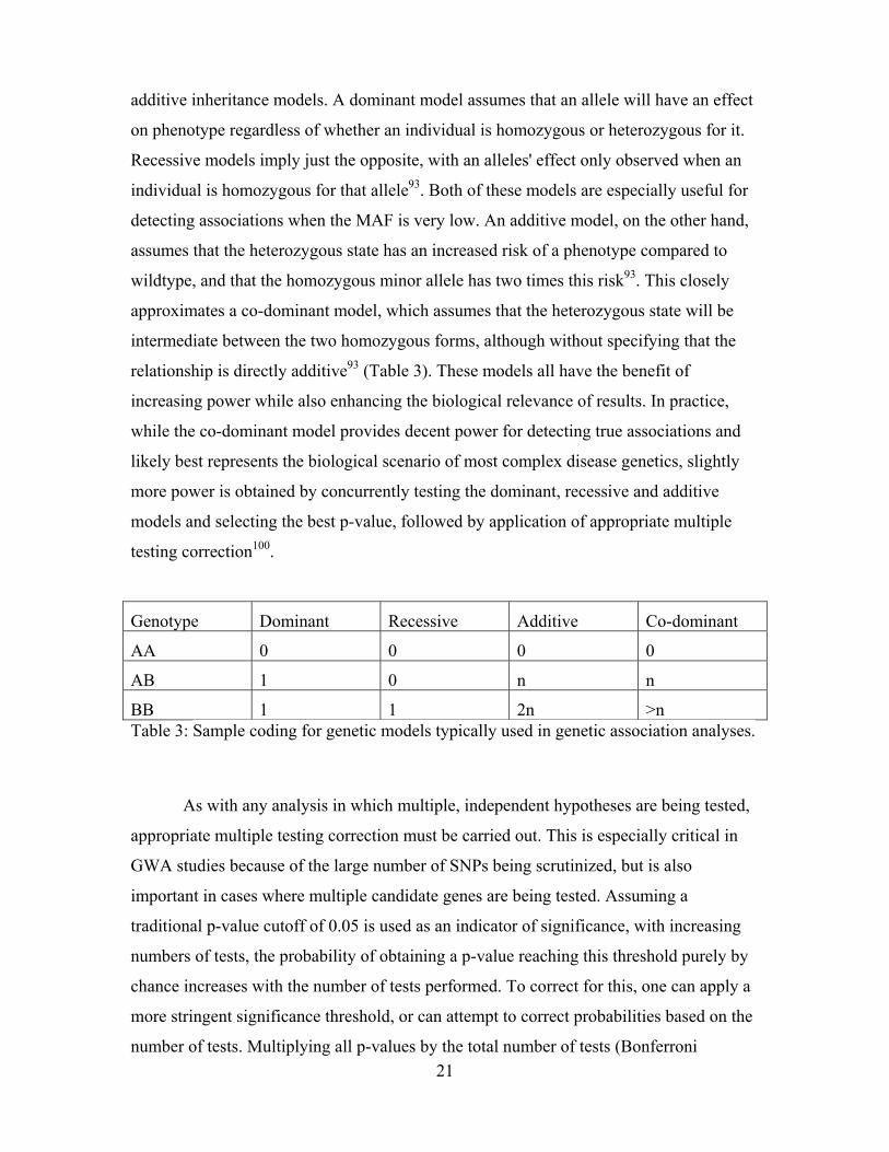
relationship is directly additive93 (Table 3). These models all have the benefit of
increasing power while also enhancing the biological relevance of results. In practice,
while the co-dominant model provides decent power for detecting true associations and
likely best represents the biological scenario of most complex disease genetics, slightly
more power is obtained by concurrently testing the dominant, recessive and additive
models and selecting the best p-value, followed by application of appropriate multiple
testing correction100.
Genotype Dominant Recessive Additive Co-dominant
AA 0 0 0 0
AB 1 0 n n
BB 1 1 2n >n Table 3: Sample coding for genetic models typically used in genetic association analyses.
As with any analysis in which multiple, independent hypotheses are being tested,
appropriate multiple testing correction must be carried out. This is especially critical in
GWA studies because of the large number of SNPs being scrutinized, but is also
important in cases where multiple candidate genes are being tested. Assuming a
traditional p-value cutoff of 0.05 is used as an indicator of significance, with increasing
numbers of tests, the probability of obtaining a p-value reaching this threshold purely by
chance increases with the number of tests performed. To correct for this, one can apply a
more stringent significance threshold, or can attempt to correct probabilities based on the
number of tests. Multiplying all p-values by the total number of tests (Bonferroni
22
correction) provides a correction which removes most false positive results but likely at
the expense of inflating type II errors. Additionally, given that some SNPs may be in
linkage disequilibrium and therefore not truly independent, this correction may lead to the
needless rejection of true associations93. Despite this limitation, Bonferroni corrections
have become standard of practice in many GWA studies. Application of this technique in
conjunction with test reduction strategies such as selecting or prioritizing specific SNPs,
conducting analyses on haplotype blocks rather than individual SNPs, or performing a
two-stage analysis, is most effective.
Alternatives to Bonferroni include Sidak and Holmes corrections which follow
many similar assumptions as Bonferroni, but are less stringent101. These methods are
rarely used in genetics research. The false-discovery rate (FDR) correction also assumes
that tests are independent, and additionally requires all loci to conform to a similar
distribution. FDR is less stringent than the methods previously described and attempts to
identify important effects from the many 'significant' results detected in multiple
experiments, by taking into account the number of hypotheses tested and the level of
significance of each102. Yet another approach is to use permutation testing to establish a
p-value cutoff level which is appropriately stringent to achieve optimal levels of type I
and II error101.
Despite best efforts to reduce the likelihood of false positive results, mathematical
correction is not always a sufficient guarantee of high quality results as these corrections
do not take into account subtle population effects. Replication of findings in
independently accrued populations is, therefore, considered the gold standard for genetic
association analysis and has the ability to substantially reduce Type I error. Results which
are replicated in multiple populations are considered true associations, while those which
do not replicate are called into question, and may demand further analyses to determine
whether they are true associations101.
While these methods have been used to confirm that genetics do indeed play a
role in many human diseases, the lack of functional information available which would
provide valuable insight into disease mechanisms, especially in regards to disease
pathogenesis, is lacking in many cases. For some SNPs, the marker itself may confer
disease risk. In many others, genetic associations with specific loci have been found in
chromosomal regions with genes of known or hypothesized function which could
23
reasonably be involved in disease. In others, however, marker SNPs are located in either
gene deserts, or regions with high gene density, producing none or numerous candidate
genes for disease association. This ambiguity highlights the need for further study beyond
genetic association studies.
1.3.3 - Genetic Associations with IBD
Several lines of evidence suggest that IBD is at least partially genetically
mediated: monozygotic twin concordance rates for IBD are significantly higher than are
rates among dizygotic twins103; individuals with a family history of IBD are at a
significantly higher risk of developing the disease themselves7; and the preponderance of
disease among certain ethnic groups, most notably individuals of Ashkenazi Jewish
heritage6 are all indirect evidence of a genetic role in disease pathogenesis. As such, a
great deal of study has gone into evaluating different genetic markers in the context of
IBD, first with small scale and candidate gene studies, and more recently with larger
cohorts and GWA studies.
To date more than 163 SNPs have been associated with IBD, some specific to CD
(18%) or UC (14%), with those remaining conferring risk or protection from both
disorders104. Of the loci associated exclusively with CD or UC, many show similar
directionality between both disorders. Only SNPs in two genes (NOD2 and PTPN22)
have been associated with an increased risk of CD and protection against UC (although in
the case of NOD2, not reaching genome-wide significance levels in UC). Many of the
SNPs associated with IBD are located in or around genes involved in innate or adaptive
immune processes, suggesting the importance of host immune factors in disease
pathogenesis. Indeed, among the most common IBD-associated SNPs are those near
genes involved in biological processes including innate immune function, lymphocyte
activation, maintenance of barrier function, and production of reactive oxygen species
(ROS)105. Furthermore, many of the SNPs associated with IBD are involved in other
complex diseases or have been demonstrated to have a modulating effect on the
pathogenesis of bacterially mediated diseases such as leprosy, or susceptibility to other
mycobacterial diseases104.

24
The most striking genetic association with IBD is that of the NOD2insC variant
(rs2066847) with an increased risk of ileal CD106, 107. Despite its low MAF, this finding
has been replicated in many experiments and in different patient populations.
Furthermore, several additional SNPs in this gene have also been associated with CD108 .
This variant is highly associated with risk of fibrostenotic and fistulizing phenotypes,
ileal disease location, and requirement for surgery among both adult and pediatric
cohorts, yet is protective against large bowel disease109, 110. NOD2 signals downstream
effectors after recognition of bacterial muramyl dipeptide (MDP), and specific viral
motifs including single stranded RNA111, 112. The role of variant forms of NOD2 in IBD
pathogenesis has been well studied and suggests several possible mechanisms through
which this loci may contribute to disease: variants of this protein may have a reduced
ability to detect pathogens or to mount an appropriate innate immune response through
NFkB induced production of pro-inflammatory molecules, resulting in adaptive immune
processes becoming activated against normally non-pathogenic organisms113. NOD2 has
also been implicated in immune regulatory pathways, with an important role in
establishing immune tolerance105. Additionally, NOD2 variants may have decreased
ability to activate autophagy pathways in response to infection. Along with NOD2,
ATG16L1 and IRGM are components of the autophagy pathway which is important in
bacterial recognition and processing, regulation of cell trafficking, and activation of both
innate and adaptive immune pathways114. Variants in these genes are also highly
associated with IBD, ATG16L1 (rs12994997) exclusively with CD, and IRGM
(rs11741861) with both CD and UC, further supporting this pathway’s role in disease
pathogenesis104.
Several polymorphisms in IL23R, have been associated with IBD both CD and
UC, with different SNPs associated with an increased, and decreased risk of CD104, 115.
The protein encoded by this gene, as well as those from several other IBD-associated
genes including TNFSF15, STAT3, IL12B, CCR6 and JAK2 are important components of
pathways involved in activation and differentiation of T-helper 17 (TH17) cells. This cell
group is important in inflammatory processes, and inhibits development of regulatory T-
cells (Treg)116. The minor allele at rs11209026 in IL23R, which is protective against IBD
development and results in a missense mutation in the protein, has been shown to
decrease populations of IL23 responsive cells, and pro-inflammatory cytokine

25
production117. Of further interest is the observation made in one study showing a
moderate (though not significant after multiple testing correction) decrease in risk for the
development of colorectal cancer among individuals who were carriers of IL23R
polymorphisms118.
IL10 cytokine and receptor mutants are rare, however can lead to disease
phenotypes in the offspring of consanguineously mating individuals. Children who are
homozygous for these mutations experience a severe form of infantile onset IBD without
seemingly requiring any environmental stimulus. Such mutations which result in a loss of
function of IL10 or IL10 receptor, lead to a subsequent inability to produce inhibitors of
proinflammatory cytokines. Successful treatment of individuals with this genotype has
relied on bone marrow transplant113, 119, 120.
Several polymorphisms have also been exclusively associated with UC. Among
these are rs10797432 in TNFRSF14 and a variant at 7p22 (rs798502) believed to
implicate GNA12 121 104. Less is known about the pathological roles of the genes
associated with these variants. However, TNFRSF14 encodes a member of the TNF
superfamily with a role in innate immunity, and which has been shown to be an important
inhibitor of inflammation in mouse models121. GNA12 is involved in the assembly of tight
junctions for intestinal epithelial cells, suggesting the importance of barrier function in
UC pathogenesis122. Additional SNPs which have been evaluated in IBD are included in
Appendix Chapter 2, Supplementary Table 1.
Many of the variant SNPs associated with IBD are in non-coding regions, or have
not been associated with a candidate gene. These SNPs are typically thought to be in
linkage disequilibrium with other genetic variants which are the true contributors to
pathogenesis. However, it is also possible that such apparently 'non-coding' SNPs may be
in regions of transcriptional regulation. rs10065172, another variant near IRGM for
example, leads to altered binding of several regulatory miRNAs, with the risk allele
leading to increased levels of IRGM protein detected in cells123. Alternate splicing may
also account for the function of some of these polymorphisms. A rare protective variant
in an intron of CARD9, for example, has been shown to result in a truncated mRNA
transcript caused by a skipped exon. This variant protein is less functional than the
wildtype108. Furthermore, many of the IBD-associated SNPs have been associated with

26
altered expression levels of either nearby (cis) or genomically distant (trans) genes. These
expression quantitative trait loci (eQTL)s, are described in a recent paper by Kabakchiev
et al124.
The majority of loci which have been associated with IBD have also been
associated with additional comorbidities104. A large number of IBD-associated loci are
also involved in primary immunodeficiencies, ankylosing spondylitis and psoriasis
among others125 104. rs11209026 in IL23R, for example, is protective against the
development of both psoriasis and IBD126. Several UC susceptibility loci including those
near genes L2, REL and CARD9 are also associated with PSC, a liver condition which is
much more common among individuals with UC127. While most of the associations
between polymorphisms and multiple disorders are similar in direction, a notable
exception to this pattern is observed in rs2476601 in PTPN22. While the minor allele of
this variant is a risk factor for development of Type I diabetes, rhumatoid arthritis and
vitiligo, the same allele is protective against the development of CD108. As studies
evaluating IBD genetics have been some of the largest, it is likely with the generation of
more data through larger studies in other complex diseases that the number of disease
overlapping loci will increase. Such data suggests shared pathways which are likely
important in the etiology of both disorders.
Despite the work done in investigating the role of genetics in IBD, the 163
disease-associated loci explain only approximately 14% of disease variance in CD, and
8% in UC104. Additional alterations in gene expression, epigenetic regulation, rare genetic
variants or environmental factors may help to account for this ‘missing variability’. The
SNPs described so far have been 'common' variants, all with minor allele frequencies
above 1%. However, a small, exploratory analysis examining patients with severe CD
and UC, found that individuals possessing no known IBD polymorphisms, were often
found to have rare, and in some cases previously undescribed variants in exomes of IBD-
associated genes128. Additionally, eQTL analysis has demonstrated that the common
IBD-associated SNPs result in transcriptomic changes, with cis or trans gene products
either up or down-regulated, suggesting that alterations in gene expression play a role in
pathogenesis124. Host environment also likely plays a strong role in pathogenesis, but
challenges associated with investigating such risk factors have prevented significant
advancement in this field. However, it has been suggested that the reason that genetic
27
studies to date have failed to explain greater amounts of heritability, is that IBD is not in
fact a genetic disease. Instead, in a manner similar to peptic ulcer disease, which was also
once thought to be genetically mediated129, IBD is actually bacterial in nature, with a
single organism (analogous to H. pylori in the case of peptic ulcers130) responsible for
disease processes. This hypothesis remains controversial, and has not been supported by
experimental evidence or clinical trials.
1.3.4 IBD genetic association studies in non-Caucasian populations
The majority of IBD-associated genetic polymorphisms have been studied
primarily in Caucasian populations. Studies examining the role of these genetic factors in
individuals of Ashkenazi Jewish heritage demonstrate that several of the strongest IBD-
associated polymorphisms in Caucasians are also associated with IBD among this
group131. However, evidence for a role of these specific polymorphisms among
individuals of other ethnic groups is sparse. Healthy populations of individuals classified
as North American First Nations, for example, have many of the common IBD-associated
polymorphisms detected at differential rates compared to those of Caucasian ancestry.
For example, ATG16L1 and NOD2 polymorphisms are detected less frequently than they
are among Caucasians, and risk variants in IL23R, IL12B, and TNFSF15 are detected
more commonly132. These differences may explain the reduced prevalence of IBD among
individuals of this ethnicity, although raise puzzling questions when considered alongside
evidence that other autoimmune conditions including diabetes and arthritis are more
common among this group133, 134.
Although IBD prevalence is lower in Asian countries than is typically observed in
the West, a recent rise in the number of cases detected in these regions has been
observed135. Unlike Caucasian populations, the dominant IBD-associated polymorphisms
in NOD2 are rare among those of Asian descent, and have not been associated with
IBD136-138. Furthermore, neither of the strongly IBD-associated SNPs in ATG16L1 or
IRGM, have shown disease associations among Asians, although several novel
polymorphisms in these genes are associated with IBD among individuals of this
ethnicity139, 140. Alternative NOD2 variants can also be detected among these
populations, and like the variants associated with disease in Caucasians, are typically
28
associated with severe, early onset CD. Furthermore, variants in this gene are also
associated with an increased risk of infectious diseases including tuberculosis and
leprosy138, 141. The IL23R locus has shown mixed results, but the lack of replication of
associations among Asian cohorts, compared to the strong associations seen among
Caucasians suggests that this loci does not have the same role in IBD pathogenesis
among these groups140, 142, 143. The HLA region also shows similar associations with UC
among Caucasian and Asian populations. TNFSF15, on the other hand is much more
strongly associated with IBD among Asian groups than their Caucasian counterparts138.
The cohorts used for many of the studies investigating IBD genetics among non-
Caucasians are typically much smaller than those used for studies among Caucasians.
However, findings in different ethnic groups suggest that many of the polymorphisms
that have been well associated with disease pathogenesis among Caucasians, are rare
among other ethnic groups in which IBD prevalence is on the rise. Given that too little
time has passed for genetic drift to explain the recent spike in IBD among non-Western
populations, one may speculate that recent environmental or dietary changes may be
responsible for increased disease risk. Additional study of such factors among individuals
of varied ethnicities will be of benefit in answering some of the fundamental questions
regarding IBD pathogenesis.
1.3.5 Genetics in IPAA
As observed with IBD, individuals with inflammatory complications following
IPAA (both pouchitis and CDL) have demonstrated increased prevalence of genetic risk
factors. Unlike the large-scale IBD genetic association studies, those investigating genetic
susceptibility to pouch inflammatory complications are typically characterized by low
sample numbers and unclear phenotyping. However, several studies implicate genetic
markers in pouch inflammatory pathologies, with specific markers discussed in greater
detail in Chapter 3.
1.3.6 Evidence for genetic associations with anti-microbial antibody positivity
29
Observations demonstrating differences in the carriage rates of ASCA and
pANCA between individuals of different ethnicities suggest that positivity for these
markers is a complex phenomenon which may be partially genetically mediated144 145.
First-degree relatives of patients with IBD are also more likely to be positive for anti-
microbial markers than are unrelated healthy controls, further supporting this hypothesis.
Several IBD-associated genetic polymorphisms, including those in NOD2, ATG16L1, and
TLR5, have been associated with antibody positivity146. The NOD2 and ATG16L1
variants are associated with an increased number of antibody markers detected in serum,
and have also both been associated with increased titres and positivity for ASCA, and in
some studies, anti-OmpC147-150. Additionally, anti-CBir1 is more prevalent in individuals
with IRGM polymorphisms, although this marker is detected at decreased levels among
those with variant TLR5146. This marker is also detected at increased titres among
individuals with at least one NOD2 polymorphism59. The IBD-associated TNFSF15
polymorphism is associated with anti-OmpC positivity among individuals of Ashkenazi
Jewish heritage 151. However, whether there is a causal relationship between these factors
remains to be determined.
The clinical utility of combining such markers in managing and diagnosing
patients with IBD has been a topic of much study. Recent work evaluating the diagnostic
potential of these factors in IBD, or differentiating between CD and UC, suggest that
combination panels, which include described serological markers and well-characterized
gene polymorphisms, more accurately predict phenotype than do those measuring
serological markers alone42, 152. Panels which incorporate eight serological, four genetic
and five inflammatory markers (ie. C-reactive protein) have been developed by
Prometheus and are capable of diagnosing IBD with sensitivity of 63% and specificity of
around 90%153. The sub-classification (of patients with CD or UC) power of this panel
has not yet been independently reviewed.
1.4 Bacteria
The reason for the dramatic recent rise in the prevalence of IBD and other
autoimmune diseases through much of the developed world is unclear. For many years,
30
scientists postulated that an important factor is the increasingly 'clean' environments
typical of these areas154. This so called 'hygiene hypothesis' suggests that exposure to
fewer bacteria, viruses or eukaryotic parasites, prevents the immune system from
developing properly during childhood and adolescence, leading to dysregulation and
disease. Thus, improvements in sanitation, increased use of antibiotics and vaccines, and
reduced exposure to pathogens, although exceedingly successful in improving public
health, may have had unpredictable consequences on the proper functioning of the
immune system. Evidence regarding the role of any single pathogenic organism in
preventing or causing autoimmune disease is lacking. It is possible that 'cleaner'
environments alter human microbial community composition as a whole, which in turn
prevents the development of host immune tolerance and homeostasis. However,
evaluating complex microbial communities in the context of the human host was, until
recently, too time consuming and expensive to be broadly undertaken. The application of
next-generation sequencing technologies, reduced sequencing cost, as well as an
increased availability of funding have greatly enhanced our knowledge of the structure of
human-associated microbial communities, and led to the development of better tools for
increasing the accuracy of information obtained from such experiments.
1.4.1 Phylogenetic organization and taxonomic classification of bacteria
Phylogenetics refers to the evaluation of the evolutionary relationship among
organisms, and is usually based on a combination of genetic and phenotypic information.
Taxonomy is informed by the phylogenetic relationships between groups, and refers to
the classification, identification and naming of organisms. Traditional measures of
organism relatedness and subsequent taxonomic assignments were based strictly on
phenotypic traits, however, the incorporation of genetic information into classification
schemes has resulted in a more accurate approximation of the relationships between
organisms155. In some cases the increased availability of genetic data has led to confusion
surrounding specific organisms’ evolutionary relationships, with those once thought to be
closely related based on phenotype, shown to be less closely related based on DNA
sequence similarity – a problem specifically apparent in the analysis of microbes where
horizontal gene transfer (HGT) is quite common. Sequence-based approaches to

31
microbial analysis have become more widely used, leading to challenges in interpreting
the results of sequencing experiments. Thus, where possible, both genetics and phenotype
should be used to inform phylogenetic relationships and taxonomic assignment.
All living organisms are currently hierarchically classified into eight major levels,
with domain being the most general, and species the most specific (Figure 1). Sub-
groupings are also often included, to provide additional information. There are 3 major
domains (Archaea, Bacteria, Eubacteria), each of which have numerous sublevels,
characterized by distinguishing features allowing organisms to be classified with their
most similar cousins. To better illustrate the nature of these hierarchical systems one
might consider our own species, Homo sapiens. A member of the genus Homo, the
family Hominidae, the order Primate, class Mammalia, phylum Chordata, kingdom
Anamalia, and domain Eukarya (Figure 1). Here it may be seen that analyses on any one
of these levels could be conducted, each providing different amounts of information.
While the most precise information is gained from the species-level assignment, there is
also value in higher level analyses, especially in cases where, unlike our example of
Homo sapiens above, species identification is not possible.

32
KINGDOM
PHYLUM
CLASS
FAMILY
GENUS
SPECIES
ORDER
Anamalia
Chordata
Mammalia
Hominidae
Homo
Homo sapiens
Primates
DOMAIN Eukarya
Figure 1: Hierarchical organization of taxonomic levels used for classifying organisms
Accurate phylogenies and taxonomic assignments are critical to microbiome
analysis and interpretation of results, especially for experiments which are conducted
strictly based on DNA sequences. Our example of humans is a simplistic one, as the
phylogenetic relationship between Homo sapiens and related species is extremely well
defined. Bacteria, on the other hand, display more complex evolutionary relationships
between one another, and new species and even phyla are still being discovered.
Determining where novel species fit among known organisms and assigning them
accurate taxonomic designations can be challenging. Further complicating bacterial
phylogenetic analysis, is the fact that bacterial genomes are much more subject to
variability than are those of eukaryotes, and the rapid doubling time characteristic of most
prokaryotic cells allows populations to evolve quickly in response to stress. An example
of this can be seen in the rapid development and spread of antibiotic resistance among
bacteria exposed to a specific antibiotic156.
33
Our inability to culture many recently identified organisms has led to increased
emphasis being placed on genetic information. However, HGT between different species,
and even more distantly related groups may confound many metagenomic analyses and
present problems for accurately determining phylogenetic relationships. Transformation,
transduction and conjugation all facilitate the transfer of DNA between different bacterial
cells and can be used to share genes between distantly related organisms. Transferred
genes conferring anti-microbial resistance or other functions which may confer benefit to
the recipient, are more likely to be selected for and spread among a population. The major
limiting factor to successful, stable and useful incorporation of a novel gene into the
recipient cell is the ability of the recipient to utilize the donated gene, with more closely
related organisms more likely to do so successfully157. Locations with high bacterial
population densities, such as that found in the human gut, are characterized by higher
potential for HGT. Therefore, the plasticity in bacterial genomes caused by gene transfer
must be taken into account when performing sequence based analyses of microbial
community structure.
Although much of the bacterial genome is relatively plastic, certain genetic
markers are more stable, and can therefore, be considered candidates for phylogenetic
analysis. Most commonly used among these is the 16S rRNA gene, which is present in all
Bacteria and Archaea although with different copy numbers in different species, ranging
from one to as many as 15 per organism158. The 16S rRNA gene encodes an essential
component of the bacterial ribosome that is necessary for all protein synthesis, and as
such, is comparatively well conserved155. Further adding to this genes’ utility in
phylogenetic analysis is the sequence itself, which is characterized by conserved regions
suitable for use as annealing targets for PCR amplification of sequences from the
majority of organisms present in a sample, flanked by regions of substantially higher
variability which may be used in conjunction with curated 16S databases to identify
organisms. While transfer of these units between organisms is believed to be relatively
rare, it is possible for highly divergent species to utilize 16S genes transferred from
remotely related species with only limited reduction in function observed among recipient
cells159. A summary of the advantages and disadvantages of 16S-based approaches are
described in Table 4.
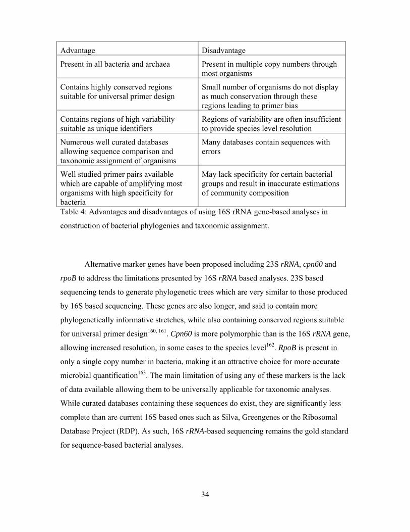
34
Advantage Disadvantage
Present in all bacteria and archaea Present in multiple copy numbers through most organisms
Contains highly conserved regions suitable for universal primer design
Small number of organisms do not display as much conservation through these regions leading to primer bias
Contains regions of high variability suitable as unique identifiers
Regions of variability are often insufficient to provide species level resolution
Numerous well curated databases allowing sequence comparison and taxonomic assignment of organisms
Many databases contain sequences with errors
Well studied primer pairs available which are capable of amplifying most organisms with high specificity for bacteria
May lack specificity for certain bacterial groups and result in inaccurate estimations of community composition
Table 4: Advantages and disadvantages of using 16S rRNA gene-based analyses in
construction of bacterial phylogenies and taxonomic assignment.
Alternative marker genes have been proposed including 23S rRNA, cpn60 and
rpoB to address the limitations presented by 16S rRNA based analyses. 23S based
sequencing tends to generate phylogenetic trees which are very similar to those produced
by 16S based sequencing. These genes are also longer, and said to contain more
phylogenetically informative stretches, while also containing conserved regions suitable
for universal primer design160, 161. Cpn60 is more polymorphic than is the 16S rRNA gene,
allowing increased resolution, in some cases to the species level162. RpoB is present in
only a single copy number in bacteria, making it an attractive choice for more accurate
microbial quantification163. The main limitation of using any of these markers is the lack
of data available allowing them to be universally applicable for taxonomic analyses.
While curated databases containing these sequences do exist, they are significantly less
complete than are current 16S based ones such as Silva, Greengenes or the Ribosomal
Database Project (RDP). As such, 16S rRNA-based sequencing remains the gold standard
for sequence-based bacterial analyses.
35
1.4.2 Human microbiome analysis techniques
Perhaps surprisingly, bacteria exist only rarely in isolation, and are instead most
commonly found in complex community assemblages or biofilms, in which numerous
different organisms share a similar ecological niche164. In many cases, these organisms
are co-dependent on one another, requiring metabolic support from additional members
of the community for survival165. Organisms residing in or upon higher order taxa display
even more complex ecology, as the interaction between host and microbe requires a
delicate balance to ensure that both symbionts are capable of optimal growth. The human
microbiome is defined as the collection of organisms and their genomes inhabiting
different anatomical locations both in and on the human host166. The human gut alone is
home to hundreds of trillions of microorganisms and contains more genetic information
than that which exists in the human genome167. In most cases, these organisms exist with
the host in a symbiotic or mutualistic relationship: microbes help with the digestion of
nutrients, prevent colonization of the host by pathogenic organisms, and aid in the proper
development of both the intestinal epithelium and immune system168, while the host
provides nutrients and a suitable habitat for bacterial growth. However, recent studies
have implicated a reduced diversity of microorganisms in the etiology of several chronic
conditions including inflammatory bowel disease (IBD), diabetes and obesity169-173.
Prior to the development and application of sequence-based molecular tools to
microbiome analysis, culture-based methods were the norm. Traditional, culture based
microbiology relies on the ability to cultivate viable organisms outside of their natural
habitat, which can be difficult as many organisms which are well adapted to life in the
human gut are not viable in different locations. Providing the necessary nutritional and
atmospheric conditions to promote growth for all organisms is difficult, and in the past
has resulted in underestimation of the complexity of the human gut microbial ecosystem.
However, advantages of being able to directly culture organisms include the greater
amount of information obtained regarding bacterial metabolism and growth requirements,
and the potential future use of cultured strains in experimentally evaluating the
interaction between microbes and host.
Early studies relying on traditional approaches to culture organisms significantly
underestimated the complexity of the gut ecosystem in particular, in large part because of

36
the difficulties inherent in culturing many of the organisms occupying this niche174. To
solve this problem, culture-independent techniques have been applied to the analysis of
complex microbial communities. Yet a surprising majority of human microbiome studies
to date have focused on high level taxonomic assignments for analysis, providing
information on whether specific phyla or families are associated with a particular habitat,
phenotypic outcome, or environmental factor in the human host. For the most part, this is
due to the limitations present in current technologies which have provided insufficient
data to allow a more detailed understanding. However, applying a phylum level analysis
to our human example above, humans would be classified together with 43,000 other
species including tunicates, ascidiacea (aka sea squirts), and amphibians to name just a
few. Thus, while some important information is gathered from this knowledge, it is clear
that much ambiguity remains as well. As such, the advantage of increasing the resolution
of microbiome studies is apparent, and the quest to obtain greater information from
current technologies remains essential.
1.4.3 Culture-independent microbial community analysis
Recent sequence-based approaches to evaluating the microbiome have been
developed to circumvent the limitations of culture and use bacterial DNA sequence as a
proxy for estimation of organism identity, relative abundance and functional potential.
However, the challenges and sources of error of sequence-based analyses are important to
understand in order to accurately interpret results.
1.4.3.1 Sample procurement
The first step in analyzing the microbiome is to obtain samples from the site of
interest. The type and number of samples to be obtained is dependent on the specific
experimental question to be answered. In evaluating the gut microbiome, the most
common samples used are stool, and biopsies obtained during patient endoscopy. While
stool is commonly used, easy to get and provides a great deal of sample material, its
microbial profile is substantially different from that obtained from tissue175. Biopsies, on
the other hand, are difficult to obtain and the microbiome may be altered by the

37
requirement that patients take laxatives prior to endoscopy176. Furthermore, differences in
micronutrient bioavailability or the presence of microscopic inflammation at the site of
biopsy, may make it difficult to get an accurate picture of overall community structure.
However, tissue samples have the benefit of providing a picture of the organisms present
at sites immediately adjacent to the host and, therefore, most likely to interact with the
immune system.
Due to the ubiquity of bacteria and bacterial DNA, contamination of all samples
by environmental sequences is likely to occur regardless of measures taken to reduce
these factors. Attempts should be made to minimize contamination, and equivalent
sampling collection and handling procedures must be applied to all samples. Small
changes in sample handling procedures profoundly influence observed community
composition, with, for example, freezing stool samples altering the
Bacteroidetes/Firmicutes ratio compared to samples which are never frozen177.
1.4.3.2 Microbial DNA extraction from samples
Once samples have been obtained, total DNA is extracted directly, typically using
a combination of mechanical and enzymatic disruption. Extraction method is highly
dependent on the samples from which DNA is being extracted, with tissue samples
requiring the use of slightly different protocols compared to samples from soil or stool.
Furthermore, the relative efficiency of disrupting organisms is highly dependent on the
structure of the bacterial cell178. This means that obtaining an appropriate balance
between an extraction techniques' harshness, which could result in shearing the DNA of
easily lysed organisms (gram negatives for example), and mildness, which would prevent
difficult to lyse organisms (gram positives, spores, mycobacteria) from having their DNA
extracted, can have important implications for downstream community analysis, and may
lead to alterations in apparent community composition179-181.
While the advantages of optimizing extraction protocols to provide increasingly
accurate views of bacterial community structure are obvious, no methods are currently
capable of providing a truly unbiased DNA sample182, 183. Extraction methods using a
combination of bead beating for physical disruption and enzymatic digestion using
38
lysozyme, mutanolysin, lysostaphin, or combinations of these, appear to provide
sequence information which most closely approximates community structure183. Other
studies have suggested that mechanical lysis steps, such as bead beating, actually
decrease the overall amount of DNA obtained from samples184. While no extraction
protocol is yet capable of rendering perfect extraction efficiency across all cell types,
ensuring methodological consistency between samples included in an experiment is
critical for obtaining unbiased results. All extraction methods have demonstrated
reproducible results when used to extract DNA from multiple identical samples (ie.
samples obtained from the same stool sample)183, suggesting that once an extraction
method has been selected, it is important to extract all samples using the same protocol in
order to ensure that different biases are not applied to samples included in the same
experiment.
To provide even greater consistency between extraction batches, numerous
commercial kits are available which can be modified through the addition of the above
mentioned bead-beating steps and enzymatic lysis to generate reasonably accurate
accounts of community structure. Such kits have the additional benefit of being subjected
to stringent quality control procedures demanded of commercial manufacturers. Among
these are kits provided by QIAGEN, MoBio, MP Biomedicals, and Sigma-Aldrich to
name just a few. Most commonly used for human microbiome analyses are the QIAGEN
DNA Mini, DNeasy and stool kits, and the MoBio PowerSoil isolation kit185. While the
former can be used to extract total DNA from tissues, the latter is primarily used in stool
analyses. In each case, the presence of human and archaeal DNA can reduce the number
of bacterial reads obtained during sequencing. However these can be easily detected and
removed during downstream processing. Operator contamination is more difficult to
manage and at present, can only be minimized by performing extractions in a clean
environment, and by recording technician as well as kit lot numbers used for different
extraction batches.
1.4.3.3 Overview of Sequence-based microbial analysis tools
Initial sequence-based microbial community analysis approaches involved
construction of libraries, consisting of bacterial clones transformed with sequences of
39
interest from the community being studied186, 187. Extracted DNA from each colony could
then be sequenced using Sanger sequencing, and relative quantification of sequences
could be inferred. While a significant advancement over culture-based approaches, this
technique remains time consuming and laborious, and often does not provide the
necessary coverage to accurately estimate the variability in the human microbiome.
DNA fingerprinting techniques, including terminal restriction fragment length
polymorphism (T-RFLP), and single-strand conformation polymorphism (SSCP), have
also been applied to microbial analysis in conjunction with normal or capillary
electrophoresis, and while significantly faster than library construction, are incapable of
providing adequate levels of resolution for characterization of complex communities174,
188, 189. The utility of these techniques can be most easily observed in performing basic
comparisons between organisms with well-characterized restriction patterns or 16S rRNA
secondary structure respectively190. However, these methods dramatically under-represent
community diversity and they do not provide quantitative data allowing estimation of the
relative abundance of different organisms. In an interesting application of these
techniques, Hiibel et al concurrently measured levels of the 16S rRNA gene (DNA) and
16S rRNA product to deduce a specific organisms’ cellular activity. In so doing, they
were able to accurately predict the metabolic state of the organisms in question (ie.
determine whether the organisms were alive and replicating, or inactive/dead)191.
Sanger sequencing and quantitative (q) PCR, are accurate, able to provide long
read lengths to achieve high degrees of sequence resolution, and relatively simple to use.
In fact, qPCR is considered the gold standard for quantifying bacterial abundance192.
However, these methods are slow, and labour intensive and, in the case of qPCR, require
the use of specifically designed and validated primers in order to provide taxonomically
informative data. Thus, these methods have only limited potential for use in complex
community analysis, especially in cases where little is known a priori. Their widest
application is in determining whether a specific organism is detected in a sample, and for
confirming the results obtained through other, more high-throughput methodologies.
Additional indirect analysis techniques include fluorescence in situ hybridization
(FISH) and the use of dot-blot or microarrays to assess whether particular sequences are
present in a sample. Both of these techniques can be used for the detection, and relative

40
quantification of known sequences174. In both cases, oligonucleotide probes targeting
specific organisms are used to capture complementary sequences in a sample. The most
commonly used microarray, PhyloChip (2nd generation), is capable of providing data on
more than 8700 strains of bacteria and archaea193. However, these methods are limited in
that they require use of a specific probe in order to detect an organism, preventing novel
microbes from being detected. Furthermore, cross-hybridization between sequences from
non-target organisms may conflate results and, in the case of the PhyloChip, have been
shown to lead to distortions in perceived community structure193.
The use of next-generation sequencing circumvents many of these limitations.
The two main technologies currently used for next-generation microbiome analysis are
454 pyrosequencing (Roche) and Illumina platforms. Several additional technologies
from other companies are also available (Table 5), but are less widely used and as such
will not be described in depth. Both 454 pyrosequencing and Illumina provide several
platforms offering different levels of coverage (number of sequences in a sequencing run)
and sequence lengths (base pair length of the sequence). While Illumina is able to provide
more coverage at lower cost, 454 pyrosequencing is capable of generating longer
sequences, often corresponding to an increase in taxonomic resolution (Table 5). Both
Illumina and pyrosequencing-based technologies can be applied to either 16S rRNA gene
or metagenomic analyses. While the former focuses on determining which organisms are
present in a sample, the latter can be used to provide functional information as well.
However the added cost and (currently) limited resources available for downstream
processing (ie genome assembly and functional interpretation) make metagenomic
analyses significantly more resource intensive. As such, the focus of work discussed in
this thesis relates to 16S-based approaches.

41
Technology Maximum Sequence Length
Sequences per run
Error rate
Roche 454 pyrosequencing (GS FLX Titanium XL+)
<1000bp 1,000,000 1.07-1.6% 194,
195
Illumina (MiSeq) <150bp paired end reads
1x10⁹ 0.4-5%194, 196
Applied Biosystems SOLiD
85 1.5x10⁹ 0.01-0.5% 197,
198
Pacific BioSciences
>2500 bp 3,000,000 13-15%196, 199
Ion Torrent <250 11,000,000 0.5-1.8%196, 200 Table 5: Summary of the next-generation sequencing technologies currently available195. Listed in brackets are the newest technologies which are available, in the case of 454, the Titanium XL+ had not yet been adopted for use in 16S microbiome analyses at the time of writing this thesis201.
1.4.3.4 A more detailed examination of next-generation sequencing and how it is applied
to the analysis of the microbiome.
Current next-generation sequencing technologies are limited by the length of
sequences they are able to provide, with ~400 bp being the upper limit of sequence
lengths achievable through currently applied technologies. As such, it is not possible to
utilize the entire length of the 16S rRNA gene (~1550 bp), and specific region(s) of the
gene must be selected to target for analysis. The 16S rRNA gene is composed of nine
regions of high variability, termed hypervariable regions, flanked by relatively conserved
regions which can be used as binding sites for universal primers. Different regions have
different biases, and levels of resolution, meaning that few comparisons can be made
between experiments conducted by evaluating different sites (Figure 2). Despite the well-
characterized universal nature of primers typically used in microbiome experiments,
amplification biases remain, and are not consistent between conserved regions202.
Furthermore, specific groups such as the Bifidobacteria are consistently underrepresented
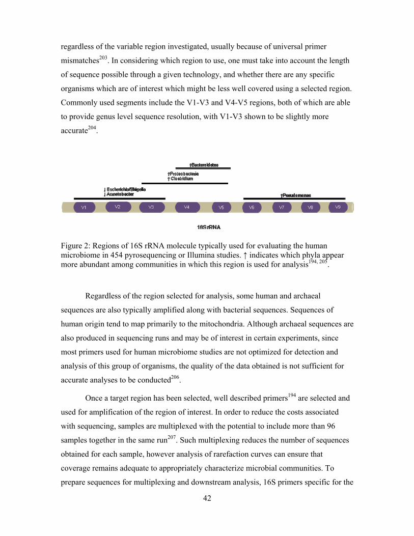
42
regardless of the variable region investigated, usually because of universal primer
mismatches203. In considering which region to use, one must take into account the length
of sequence possible through a given technology, and whether there are any specific
organisms which are of interest which might be less well covered using a selected region.
Commonly used segments include the V1-V3 and V4-V5 regions, both of which are able
to provide genus level sequence resolution, with V1-V3 shown to be slightly more
accurate204.
Figure 2: Regions of 16S rRNA molecule typically used for evaluating the human microbiome in 454 pyrosequencing or Illumina studies. ↑ indicates which phyla appear more abundant among communities in which this region is used for analysis194, 205.
Regardless of the region selected for analysis, some human and archaeal
sequences are also typically amplified along with bacterial sequences. Sequences of
human origin tend to map primarily to the mitochondria. Although archaeal sequences are
also produced in sequencing runs and may be of interest in certain experiments, since
most primers used for human microbiome studies are not optimized for detection and
analysis of this group of organisms, the quality of the data obtained is not sufficient for
accurate analyses to be conducted206.
Once a target region has been selected, well described primers194 are selected and
used for amplification of the region of interest. In order to reduce the costs associated
with sequencing, samples are multiplexed with the potential to include more than 96
samples together in the same run207. Such multiplexing reduces the number of sequences
obtained for each sample, however analysis of rarefaction curves can ensure that
coverage remains adequate to appropriately characterize microbial communities. To
prepare sequences for multiplexing and downstream analysis, 16S primers specific for the
43
region of interest and containing customized barcodes and adapter sequences for ligation
to either 454 pyrosequencing beads or the Illumina flowcell are used. Barcode length is
dependent on the degree of multiplexing desired, but are commonly approximately 10-
base sequences, which are sufficiently unique compared to other barcodes to prevent
sequencing errors leading to assignment of a sequence to an incorrect sample208. These
are sequenced along with the selected 16S region, and are subsequently used to
accurately parse data.
Next-generation sequencing technologies each make use of different chemistries
to provide sequence information. 454 pyrosequencing makes use of pyrophosphate
chemistry for sequence resolution. A single query sequence is ligated to a bead and
undergoes emulsion PCR to coat the bead with thousands of copies of that sequence.
Beads are placed into a single well of a picotitre plate, and nucleotides are added
sequentially (across all wells). Unincorporated nucleotides are removed prior to the
addition of the next base. The pyrophosphate which is released when nucleotides are
incorporated into the growing DNA strand, is measured using an ATP
sulfurylase/luciferase reaction which generates light. The light is subsequently detected
by a charge coupled device (ccd) camera and converted to a peak in a pyrogram. The
height of the pyrogram peak is a function of the amount of light produced by the
reaction209. The sequential addition of bases allows determination of base calls, and the
number of nucleotides incorporated is proportional to the intensity of the light output (up
to 8bp).
Illumina uses a different method which involves deposition of sequences on a
flowcell followed by isothermal bridge amplification to increase the sequence density at
that specific location. Reverse strands are cleaved and washed away, leaving millions of
copies of the original sequence for analysis. Sequencing is conducted base-by-base using
fluorescently labelled nucleotides. Following incorporation and fluorescence reading,
tags are removed and the next base may be added to the growing chain. Unlike
pyrosequencing, paired end reads can be generated, in which sequencing is conducted
from both ends of the DNA fragment201. This can be used to create longer read lengths
and to enhance the accuracy of measurements.
44
Sequencing error rates and common types are different between Illumina and 454
pyrosequencing, with Illumina sequencing more prone to mismatching, while 454
typically has higher rates of insertions and deletions, particularly around homopolymeric
regions 195, 210. Longer sequences are, on average, of higher quality than are shorter
sequences, although may still contain erroneous calls. However, for both of these
technologies, extended sequencing results in a dramatic rise in sequencing error rate with
a higher number of errors occurring after 100 bases in Illumina and 400 bases in 454202 194. Error rates for 454 tend to be spread across many sequences, making it difficult to
remove all that are suspect without a corresponding reduction in sequence coverage195.
Given the run- and sequence-specific nature of error rates, in order to minimize
propagation of errors through workflows, it is important to include standardized control
sequences and error-corrected base callers in each run to allow estimation of the true
error rate. Furthermore, downstream sequence processing which specifically targets and
culls sequences with a higher probability of containing erroneous bases, is useful.
1.4.3.5 Sequence quality trimming, alignment and taxonomic assignment
In order to ensure the best quality sequencing information is included in analyses,
and to prevent inaccurate sequence information from falsely inflating levels of diversity
among samples, sequences are quality trimmed prior to taxonomic assignment and
subsequent analysis. Typical methods of quality control are aimed at addressing the
specific limitations of the sequencing technology being used. For example, given that
shorter 454 sequences tend to have higher error rates, and extended homopolymers are a
constant source of error, sequences which do not meet minimum length requirements
(with the threshold used typically dependent on the technology used and choice of
reagents), and which include extended homopolymeric stretches (typically 8 nucleotides)
are removed from downstream processing195. Sequences with homopolymers extending
beyond 8 nucleotides have error rates above 0.01, as do sequences shorter than 200
nucleotides (error rates are defined as the total errors detected per base call). The
presence of ambiguous bases, or primer mismatches are also indicative of lower quality
sequences. More than two primer mismatches correspond to an error rate of 0.0056, while
the presence of any ambiguous bases substantially increases error (above 0.01)202. The

45
expected alignment location of a sequence of interest can further be used to identify
incorrect sequences. If an experiment is relying on primers specific to a given
hypervariable region of 16S, yet preliminary database alignment suggests that a different
region from that expected has a stronger alignment score, the quality of that sequence is
called into question.
Rather than simply removing sequences with the features described above,
alternative solutions include removing portions of sequences with overall low quality,
thus reducing the amount of information lost at this stage211. Algorithms such as
PyroNoise and DeNoiser have been developed which allow base calls to be corrected
using the raw flowgram data212, 213. However, the increased computational intensity
demanded by both of these approaches has prevented their widespread utility in large
datasets.
An additional method to remove spurious sequences, proposed by Huse et al
makes use of a pre-clustering step which identifies highly similar sequences and separates
out those which are unique214. The logic behind this approach is that more abundant
sequences, which cluster together based on a predetermined distance measure, are more
likely to be real than those which are less abundant. Sequences which are rare and which
fail to cluster into groups can then be removed. The simplest implementation of this
algorithm is to perform pairwise comparisons between all sequences and determine the
number of mismatches between them. Those with more than the defined cutoff (one
mismatch is commonly used) will not be grouped together. This approach has been
observed to decrease error rates to 0.0004, when applied in conjunction with the
previously described steps202.
Providing more of a challenge for ensuring sequence quality, is the ubiquity of
chimeric sequences throughout many sequencing projects. Chimeric sequences are an
artefact of PCR amplification in which different parts of a sequence arise from different
parent strands. Such sequences typically result from an incomplete sequence dissociating
from its parent strand and acting as a primer for another, different sequence. This hybrid
is then amplified leading to spurious groups. Because these sequences are not a reflection
of poor sequencing quality, they are much more difficult to detect than are errors
resulting from low quality reads. Chimeras are so ubiquitous in sequence-based analyses
46
that they can be found in many 16S databases, especially among organisms which have
not yet been cultured. Chimeras are estimated to make up anywhere from 5-45% of
sequences in a run and thus represent a huge source of error for next generation
sequencing approaches215. Furthermore, 16S rRNA databases have been shown to contain
a great many anomalies, with approximately 5% showing errors (RDP database release
9.22) prior to the widespread use of chimera detection algorithms216. However, through
improvements in computer based algorithms for detecting chimeric sequences, and the
application of standardized sequence processing pipelines which are capable of reducing
the number of errors in data, improved accuracy in taxonomic assignment is possible.
Several algorithms have been developed which attempt to detect and remove
chimeric sequences from datasets. Each can be used in conjunction with well-curated
databases in which it is known that no chimeric sequences are found, or run
independently of reference databases, with sequences in a run compared to one another.
While database approaches have some advantages, especially in terms of decreased
computer power and resources, they are forced to rely on often incomplete and inaccurate
datasets. Because the highest quality sequences are available from previously cultured
organisms, these databases would not be useful for samples in which many of the
organisms have not previously been cultured. Further, in applying a more broad
collection of database sequences, one may inadvertently incorporate lower quality
reference reads which would not add to the accuracy of the analysis. As such, where
possible it is advantageous to make use of a database free approach.
ChimeraSlayer detects a large proportion (87%) of chimeric sequences with
greater than 4% pair divergence, meaning it is capable of detecting a large proportion of
intra-genus chimeras (chimeras where both parent sequences are members of the same
genus), and can be run with or without reference database dependence215. This algorithm
represented a substantial improvement over those which had been previously developed,
and was able to detect more erroneous sequences then earlier approaches such as
Bellerephon or Pintail, especially for similar sequences217. Another approach, Perseus,
identifies chimeras independently of a reference database. It assumes that parent strands
of a chimera must have experienced at least one more round of replication than the
chimera, therefore allowing restriction of searches to those sequences present in greater

47
abundance than the query. Unfortunately, this method assumes that chimeras result from
two parents, not taking into account the possibility of increased numbers of parent
strands218. The most recent algorithm, UCHIME, shows significant improvements over
ChimeraSlayer, especially in cases where the chimeric sequence are small or arose from
more than two parents. A further advantage of UCHIME, is that it performs significantly
faster than both Perseus and ChimeraSlayer218.
Once raw sequences have been quality trimmed, they can be assigned to
taxonomic outcome groups to generate more meaningful information for downstream
analyses. The two main approaches include direct assignment of sequences to phylotypes
and operational taxonomic unit (OTU)-based processing219. These methods each have
their own benefits and limitations. When sequences are assigned directly to a taxonomy
based on comparisons of sequences with known databases, assignment is highly
contingent on the accuracy of the sequencing platform and the reference database being
used. The alternate approach, which is used quite commonly in microbiome research,
seeks to group sequences based on sequence similarity, and independent of a reference
database. These groups, termed OTUs, are composed of sequences clustered at a given
similarity threshold (typically 97%). OTUs are artificial constructs which do not
correspond well with biological units (ie. species)219. Following clustering, representative
sequences are selected for assignment of each OTU to a taxonomic group. OTU based
methods often inflate estimates of sample diversity with direct phylotype assignment less
subject to this bias202. However the ability to group sequences independently of 16S
databases for initial steps, especially in the context of early databases which were
incomplete and plagued by erroneous reads, has made OTU-based analyses popular220.
Clustering organisms together based on a given distance threshold can be time
consuming and computationally intensive. Calculating the distance between two
sequences involves either direct sequence comparisons, or may take into account the
three dimensional structure of the rRNA molecule. Numerous algorithms have been
developed to perform sequence alignments, all balancing a need for accuracy with limited
computing power. Dynamic programming, including the Needleman-Wunsch, and Smith-
Waterman algorithms were designed to align any types of sequences, and to guarantee
that the optimal alignment is found. These methods involve construction of global and
48
local distance matrices respectively, which are then used to calculate optimal alignments
between paired sequences. These processes are incredibly computationally intensive
however, and therefore useful only on a small number of sequences221. Secondary
structure based methods such as Infernal are particularly useful for more distantly related
organisms as, in these cases, sequences may be highly divergent, although protein folding
remains conserved. However, Infernal often overestimates distance calculations except in
cases where the distance between organisms is already large222.
Word-based methods and heuristic algorithms have been designed to reduce the
amount of computer power required to generate high quality alignments. Such methods
use early results to influence and reduce the number of future calculations, and decrease
the number of overall calculations required and amount of computer resources. While not
guaranteeing optimal alignment, the speed of such approaches has made them popular for
aligning large numbers of query sequences. Included in this group are several of the most
commonly used database search algorithms. Both the Basic Local Alignment Search Tool
(BLAST)223 and FASTA224, used by the National Centre for Biotechnology Information
(NCBI), make use of small 'words' extracted from query sequences and searched against
target sequences in a database. When word hits are found, dynamic programming is used
to extend those matches outward as long as a good alignment is possible. While FASTA
will provide only the best alignment, BLAST will display many 'top' alignments, with bit
scores allowing scientists to select options which make the most biological sense. Various
iterations of both of these algorithms can be used to align DNA, RNA, and proteins.
Calculating the distances or relative similarities between multiple sequences from
a run requires even more computer resources due to the sheer number of comparisons to
be made. Large amounts of data, such as that produced by next-generation sequencing,
makes the use of dynamic programming infeasible214. To address this, multiple sequence
alignment (MSA) algorithms have been developed and applied to sequence processing
pipelines, and aim to reduce the number of comparisons conducted on a set of sequences.
Propagative or progressive MSA methods involve grouping together sequences which are
most alike first, and adding less similar sequences later. This dramatically reduces the
number of comparisons required, but has the disadvantage of propagating early errors
forward without correction. Iterative MSA methods attempt to address this problem by

49
continually realigning subgroups in order to classify sequences into an accurate
phylogenetic tree. Studies have demonstrated that MSA-based algorithms including
MUSCLE (progressive) and MAFFT (progressive and iterative functionality), falsely
inflate the amount of genetic distance between 16S sequences and artificially increase the
number of OTUs generated225.
Additional tools have been developed which provide accurate pairwise-distances
and are widely used for OTU generation in microbiome research. To minimize resource
use, the main algorithms used include the DNADIST program from the PHYLIP package
and k-mer based distance calculations with various mer sizes. Additional procedures are
offered through different analysis pipelines226. Using calculated distances, different
heuristic algorithms are then used to cluster sequences at a given threshold, including but
not limited to nearest, average and furthest neighbour. Additional algorithms include CD-
HIT, UClust, ESPRIT, and BlastClust. The choice of method has been shown to have a
strong impact on observed community richness, so selection of appropriate, well
validated parameters is essential. One simulation and mock community study
demonstrated that the average-neighbour algorithm is capable of producing the most
robust OTU groupings219.
To further simplify sequence processing and analysis, several suites of tools have
been developed and are available online, offering iterations of common or proprietary
algorithms for sequence alignment and OTU generation, as well as providing methods for
performing quality control checks and generating graphical representations of data. These
include dotur, mothur, and Qiime among others. These utilities provide a fair amount of
flexibility in sequence quality trimming and data clustering, allowing the user to specify
which alignment and clustering algorithms are used. Further, within those algorithms
users are given the flexibility of modifying a number of the default parameters to
optimize their data. Qiime, for example, offers seven different clustering methods
(including each of those listed above) for assignment of sequences to OTUs, allows the
user to specify which 16S rRNA database to use for taxonomic assignment, and allows
the user to view data either graphically or through alternate data formats for use in other
programs (Qiime 1.6.0).
50
1.4.3.6 Statistical analysis of microbial data
As with any new technology, consensus on the most robust way of analyzing
microbiome data has yet to be reached. Many studies make use of traditional ecological
indicators of community composition including estimates of coverage, and alpha and beta
diversity. Such tools are specifically useful as they are not impacted by the distribution of
the data. Diversity is a concept which is used often in microbiome research, but can be
difficult to interpret and quantify. In general, diversity estimates incorporate information
regarding species richness (different organisms detected in a sample) and abundance
(number of a specific type of organism present in a sample). Alpha diversity is a measure
of the mean diversity within a specific sample or ecosystem, whereas beta diversity is
typically considered to involve a comparison of diversities between ecosystems or
samples227. Numerous methods have been developed to estimate alpha diversity, with the
Shannon, Simpson and Chao1 indices most commonly used. These metrics were initially
created for macro-ecology, and have subsequently been applied to microbiome
analyses228, 229. However, of these measures, none control well for rare organisms, and
their applicability for comparisons between samples must be carefully considered. As
such, measures of diversity may be a useful tool which should only be used in
conjunction with other, more detailed information.
Beta diversity, in the context of microbiome research has been used to perform
comparisons between samples based strictly on their microbial composition (Qiime
website). As such, each sample community is explicitly compared to others through
calculation of the distance or variance between samples. Evaluation of these distances is
complex and may make use of different calculation matrices such as Jaccard, Euclidian,
weighted or unweighted Unifrac. Unifrac is the most popular technique used to calculate
beta-diversity in microbiome analyses, as it takes into account the phylogenetic structure
of a community in calculating distance matrices and probabilities. Furthermore, both
frequency (weighted Unifrac) and dichotomous (unweighted Unifrac) data can be
analyzed, which is useful for ensuring the accuracy of statistical inferences230.
Analyses which aim to determine whether specific organisms or OTUs are
associated with outcome may also be performed, and can be used to provide more
detailed and specific information regarding associations between environments or
51
phenotypes and microbes. Microbiome data can be analyzed as a dichotomous variable,
based on whether an organism is detected in an experiment, or as a semi-continuous
variable based on relative abundance. For dichotomous results, presence of an organism
may be a reflection of the coverage achieved in an experiment, making it difficult to
make comparisons between sequencing runs or platforms. Abundance data can also be
problematic as interpretation of count data obtained from next-generation sequencing,
given the lack of standardized internal controls, makes data normalization between
batches and experiments next to impossible. As such, most samples rely on frequency
data, whereby a degree of normalization is achieved by converting raw count data to
frequencies in samples. Neither of these methods address the issue of primer bias towards
specific sequences or the differential sequencing efficiency of different samples.
However, dichotomous and abundance data of individual organisms can be used together
to assess whether there are common differences between phenotypic groups.
Differences in dichotomous or abundance data between groups must then be
assessed using statistics. Major challenges in analyzing microbiome data include the lack
of conformation of data to a normal distribution, discrete abundance data (organism or
OTU counts) which is later converted to semi-continuous frequency data (continuous, but
bound by 0 and 1), and the observation that many organisms in microbial communities
are detected in only a few samples, resulting in zero-inflation of data. Common statistical
tests rely on the assumption of normality to accurately estimate probability, and break
down when this condition is not met. To address this, different data transformation
methods have been applied to microbiome data, including various log-based methods
(which require additional manipulation with zero-inflated data), arcsine square root,
Dirichlet multinomial231, and negative binomial models232. The advantages of these
approaches, is that once data has been transformed, traditional statistical tests including
analysis of variance (ANOVA), linear regression and t-tests, may be applied to detect
associations. These methods are typically more powerful than exact or non-parametric
approaches and are therefore especially useful in cases in which smaller sample sizes
negatively influence experimental power. On the other hand, exact and non-parametric
statistics can be used on many different types of data, and do not make assumptions about
data structure or rely on complex transformations for analysis. However, zero-inflated
data remains problematic for all analyses as the exact meaning of a zero in next-

52
generation microbiome sequencing is unclear. This makes it difficult to obtain an
accurate estimate of the variance among subjects for sparsely detected organisms233.
Obtaining similar results when multiple types of analyses are applied may be considered
indicative of robust results.
Regardless of the chosen approach, proper correction for multiple testing must be
performed in order to account for the number of tests being analyzed and avoid type I
errors. Stringent correction approaches such as a Bonferroni, may inflate the type II error,
resulting in the rejection of true results. As such, a balance must be struck between
maintaining statistical stringency and power. Unlike genetics, the FDR method is most
commonly used in microbiome analyses. In addition, some groups apply a higher
significance threshold (Corrected P-value< 0.3) combined with evidence of biological
plausibility as a check for suspected associations, which can be effective when
information is available regarding groups of interest, but is less so in analyzing novel
organisms. However, biological plausibility in itself must not be used as the sole
verification of results, as it does not necessarily imply that egregious analytical errors
have not occurred. As such, this approach must be used with caution to prevent reporting
inaccurate results
Because of the comparative newness of microbiome research, gold-standard
protocols for analysis have not yet been developed and widely accepted by the research
community. Different groups make use of different methodologies from sampling to
sequence processing and analysis, which have profound impacts on data quality and can
prevent the accurate comparison of results between studies. Some of the methodological
issues which can have an impact on perceived microbiome composition include:
extraction method (whether physical, chemical or combination protocols are used),
extraction kit (commercial variety or homemade), primer selection (differential primer
bias)234, sequencing method (Sanger vs 454 vs Illumina vs microarray), clustering and
alignment algorithm, database used for taxonomic assignment, transformation method,
and statistical approach. These issues may be minimized within a study by ensuring that
standard protocols are used for all samples, however are difficult to account for in
comparisons between studies, and the inherent biases of chosen methodologies must be
considered in interpreting data. Despite these challenges, a great deal has been learned

53
recently about the populations of microbes inhabiting different anatomical locations on
the human host.
1.4.4 The Human Microbiome – lessons from community level analysis
Before understanding which changes are necessary for the development of
disease, it is necessary to first attempt to identify a 'core' microbiome, or set of organisms
which are present at a given sampling location among many individuals. Depending on
the parameters used in identifying 'core' features, different results can be obtained. Using
the most universal definition of inclusion, that to be included as part of a 'core'
microbiome an organism must be detected in a site of interest among every individual
studied, several phyla are identified which fall into this category in the intestinal tract.
While most gut or stool microbiome studies typically identify up to 20 phyla in samples,
only Actinobacteria, Firmicutes, Bacteroidetes, Proteobacteria and Fusobacteria may be
considered to be universal in that they are detected across all samples. A study by Huse et
al attempted to identify a core set of organisms at the OTU level by compiling results
obtained from several studies of healthy subjects, and across many different biological
habitats235. In this study, stool was found to have fewer 'core' OTUs than were oral cavity
sites, but more than were detected in skin and vaginal sites. When a lower threshold was
used for inclusion as a core organism such as detection of an OTU in 50% of individuals,
the number of OTUs reaching this threshold was approximately 15%235. Notably, both
definitions of 'core' do not take into account organism abundance, so some of the
organisms were present in high numbers among certain samples, and were rare in others.
To date, no intra-individual ‘core’ organisms have been found which can be detected
across all body habitats. Additional work is now focusing on identifying factors which
are responsible for shaping an individuals’ microbiome, in hopes of explaining some of
this variability.
Microbial colonization of humans begins at birth, with mode of delivery being the
first factor known to influence the microbiome236. Slightly later, breast feeding versus
formula feeding impact the development and composition of the gut associated
microbiome in early childhood, as does weaning, and helps to explain the large amount of
temporal variability undergone by microbial populations early in life237. The speed of

54
change begins to level off as children approach adolescence, and large scale changes in
microbiome structure are not typically observed again until much later in life. Typifying
this change is the correlation between increasing age and decreasing Bifidobacterium
levels238. However, despite the relative stability of the microbiome through adulthood, in
time-series experiments, few organisms can be detected across all sampling times
(approximately 10% in stool, over 100 daily samples)239. Additionally, use of antibiotics
leads to more drastic perturbations of the host microbiota. These agents are capable of
significantly altering the microbiome, and while communities often revert to a state
similar to that which existed prior to medication use, they frequently do not fully
recover240, 241.
Evidence that the microbiome is at least partly shaped by dietary and
environmental factors is compelling. Host organisms with diverse dietary patterns
(herbivores, carnivores, omnivores) have differences in the structure of their microbiome
which transcend phylogenetic relationships between host species242. Furthermore, similar
observations may be seen among human populations who consume broadly different
diets. In a comparison between children from Africa and Italy where there are substantial
differences in the amount of fibre, calories and protein consumed, broad differences in
the gut microbiome are detected and include a significantly higher overall ratio of
Firmicutes/Bacteroidetes among individuals from Europe compared to Africans.
Furthermore, several genera are present exclusively among African children, including
Prevotella, Xylanibacter (Bacteroidetes) and Treponema (Spirochaetes) which may all
contribute to enhancing nutritional uptake from plant polysaccharides243. Yet breast fed
children in both locations displayed similarities which caused them to group
independently of either larger group. This suggests that diet is an important driver of
microbial composition among healthy, well matched individuals consuming different
diets243. Dietary patterns among Western populations have also shown evidence of
association with microbiome profiles, with individuals consuming a protein/animal fat
rich diet having increased Bacteroides, and those consuming more carbohydrates
enriched for Prevotella. Consumption of large amounts of fruits and vegetables further
alters the composition of the human microbiome244, 245.
55
Other factors which alter gut bacterial communities include smoking, medication
use and ethnicity. Indeed, smoking has been associated with changes in the microbiome,
as have various medications which were not previously recognized to have bactericidal
effects, such as 5-aminosalicylic acid medications (5-ASA)s246. Such differences could
account for the differential risk of CD and UC among smokers and non-smokers, and the
efficacy of different drugs in reducing inflammatory activity. However, these results need
to be replicated in additional cohorts to determine their validity, as associations to date
have been marginal. Additional studies have suggested that ethnicity may contribute to
microbiome heterogeneity247, 248, which may make it difficult to evaluate the confounding
effect of other environmental differences and the potential impact of genetic
heterogeneity, on microbial composition.
Additional microbiome heterogeneity between subjects can be attributed to
cooperation and competitive exclusion between the microbes themselves. Certain species
or genera often co-associate with other distinct organisms, often independently of
phylogenetic relationships. These co-associations likely result from synergistic
relationships between groups. Marvinbryantia formatexigens, for example, grows more
rapidly in the presence of formate, and has been observed to positively co-occur with
Butyrivibrio fibrisolvens, a formate producer165. Samples taken at birth, and following
antibiotic treatment are more likely to consist of 'opportunistic' organisms, capable of fast
growth, and without a stringent requirement for the presence of additional organisms to
provide metabolic co-factors. As time following birth or removal of antibiotic application
lengthens, fewer such opportunistic organisms are detected, as they are replaced by more
symbiotic groups165. This successional colonization likely has important implications
following IBD-related surgery and may explain why the use of probiotics and antibiotics
later in life often have transient effects on the microbiome.
The microbiome also plays a role in regulating its human niche, most notably
through influencing and modulating the function of cells and the immune system.
Helicobacter pylori, the causative agent in stomach and duodenal ulcers, for example, is
known to modulate the macroenvironment of the stomach lumen, increasing or
decreasing acid secretion depending on the location of infection249. Other organisms
directly impact the host immune system, such as segmented filamentous bacteria (SFB)
which induces production of Th17 cells in rodents, and Faecalibacterium prausnitzii,
56
which excretes an as yet unidentified compound with anti-inflammatory effects250, 251.
Additionally, temporal variation in microbial populations, including changes in
population structures during a lifetime, suggest that the functional benefits conferred by
certain organisms on immune development may have a finite opportunity to exert their
effects. For example, while few studies have described detection of the
immunomodulating organism SFB in humans, those which have suggest a near absence
of SFB among adults compared to children252. Organisms such as these with known
function, represent only a tiny fraction of the intestinal microbiome, as such it is likely
that with more study, a great many additional species with similar host modulating
properties will be found.
In an attempt to better understand these relationships, the Human Microbiome
Project (HMP) was conceived. In many ways analogous to the human genome project
which first sought to obtain a roadmap of the human genome, the aim of the HMP is to
catalogue the typical organisms present among healthy individuals. The HMP has done a
great deal of work cataloguing the organisms present in numerous human sites, including
the digestive tract, skin, oral and nasal cavities, and urogenital tract with the ultimate goal
of determining the impact of microbial communities on human health. To answer this
question, it is necessary to discover which organisms are present in specific sites, and
what functions they are capable of performing. While both of these questions are
important, to date, the majority of work has focused on the former, with little yet known
about the functional implication of such findings. This work has lead to the generation of
reference genome sequences for numerous previously unidentified or unstudied
organisms, and has provided a basic catalogue of the bacteria present in different body
habitats. Further work aimed at investigating specific disease phenotypes and evaluating
how the human microbiota contributes to disease, either through altering immunity or
other as yet unknown mechanistic pathways, has also been done.
1.4.5 Location-specific microbiome heterogeneity
The microbial composition of different environments is reflective of macro-
ecological factors, including nutrient bioavailability, pH, salinity, and in the case of the
human microbiome, immunological factors including components of the innate and

57
adaptive immune systems. These environmental differences shape the microbiome and
lead to significant variation in microbial profiles between sampling sites. Along the
digestive tract alone, it has been demonstrated that microbial profiles are significantly
different between the mouth, stomach, small and large intestines and stool187, 253, 254. Even
physically close sites such as the dorsum of the tongue and hard palate can be
substantially different, suggesting the importance of local selective pressure on
community composition255.
Numerous studies cataloguing site-specific bacterial composition have been
conducted. The most diverse habitats in the human host are found in the oral cavity and
lower large bowel248. Interindividual variability is high as is temporal variability at a
given site, with the oral cavity seemingly less susceptible to change over time than other
sites. Few organisms are detected in all body habitats (including stool, skin sites, oral and
nasal cavity)248, 256. Interestingly, when finer mapping has been performed examining
different skin micro-locations it appears that variation in environmental characteristics
shape microbial populations, with sebum producing areas such as the forehead and face
dominated by Propionibacterineae, and more dry locations such as the trunk and legs
dominated by Staphylococcus species256. Other sites also appear to be dominated by
different organisms or groups of organisms. Stool, for example, has a higher amount of
Bacteroides, and several genera from within the Firmicutes, vaginal sites tend to be
dominated by Lactobacillus, and the mouth by Streptococcus, Haemophilus, Prevotella
and Veillonella248. Differences between sampling locations within the digestive system
are also apparent. As a general rule, diversity increases from the stomach to lower large
bowel. Differences in the composition of the intestinal microbiome are striking between
the ileum and the colon; however, subtle differences can also be observed between
biopsies taken from left and right colonic tissue, including altered frequencies of
Roseburia, Altistipes and Fusobacterium. Yet some of the largest differences in
community structure are observed between stool and any intestinal biopsy location,
suggesting caution must be used in interpreting the biological significance of stool-based
studies246.
Despite the recognized taxonomic diversity of the intestinal ecosystem, the
functional capabilities of organisms in this site are more conserved than observed in
58
other, less diverse habitats. A recent study by Cantarel et al demonstrated that specific
carbohydrate bioavailability in different locations is a primary driver of microbial
population structure, and that although community membership composition may be
different, the metabolic potential of regions with specific carbohydrate sources may be
less variable257. Furthermore, while stool and oral samples are taxonomically distinct,
organisms from both sites demonstrate distinct functional potential from non-digestive
sites as they more commonly possess genes involved in the breakdown of plant and
fungal carbohydrates257. It has been recognized, therefore, that despite phenotypic
relatedness (at the family level), there is a high degree of variability in the number and
type of metabolic processes encoded in bacterial genomes, suggesting that species-level
resolution is necessary in order to accurately deduce functional potential257. The
importance of the relationship between taxonomy and sequence-deduced metabolic
potential has not been determined. However, the incomplete nature of current
metagenomic datasets, highlights the importance of adding to current reference sequence
collections to allow more accurate functional inference.
1.4.6 Microbiome and Immunity
Germ free mice have altered immune structures compared to those raised with
normal commensal bacteria. Alterations include smaller splenic lymphoid follicles, fewer
splenic CD4+ T cells, smaller Peyer’s patches, fewer intestinal lymphocytes, and an
immune profile skewed towards a more Th2 type response. These changes can be
reversed following colonization with specific members of the commensal microbiota168.
Interestingly, whole microbes may not be required to shape this relationship, as specific
microbial associated molecular patterns (MAMP)s may also be capable of inducing more
‘typical’ immune processes. B. fragilis polysaccharide A (PSA), for example, has the
ability to increase the number of CD4+ T-cells to normal levels in germ free mice,
independent of B. fragilis colonization. PSA alone enhances production of IL10,
produced systemically by regulatory T-cells and by several other cell types in response to
prolonged inflammation168, 258. Furthermore, B. fragilis-derived PSA appears to signal
directly through TLR2 to promote immunogenic tolerance, a feature which is necessary
for colonization of the host intestinal tract by commensals259. Of note, is the observation

59
that several organisms falling into the B. fragilis group (which includes 10 species in
total) are decreased among individuals with IBD compared to healthy controls260. Other
organisms have also shown immunomodulatory potential, include SFB which induce
production of Th17 cells in rodents through an as yet unidentified epitope261, and
Clostridia groups IV and XIVa which induce the accumulation of Treg cells in the lamina
propria of the colon262.
Variation in the overall composition of the intestinal microbiota brought on by
differences in hygiene, have also demonstrated influence on tissue gene expression
patterns. Animals housed in a clean environment with microbial profiles dominated by
Bacteroidetes and reduced Firmicutes, have increased expression of pathways involving
immune response-IFN alpha/beta signaling, and antigen presentation by MHC I
compared to animals housed in less sterile environments263. Of further interest is the
observation that different microbes have the ability to alter host response in a location-
specific manner; for example, interacting with the immune system in different ways
depending on whether colonizing the intestinal lumen or blood168, 264. Additionally,
stress-induced alterations in bacterial community structure265, and increased detection of
bacterial lipopolysaccharide (LPS) in blood brought on by increased intestinal
permeability, is at least partially responsible for the cytokine response (increased IL-1β
and IL-18) typically observed in rats exposed to stressors266.
1.4.7 Genes, serology and microbiome
To date, no associations between the anti-microbial antibodies and different
components of the microbiome have been observed. However, in a small number of
pouch patients, several inflammatory markers including CRP, TNF-α and erythrocyte
sedimentation rate (ESR), showed evidence of correlation with specific microbes. In
these patients, serum CRP was inversely correlated with Streptococcaceae, TNF-α was
correlated or inversely correlated with several different bacterial families, and ESR
correlated with Staphilococcaceae and Bacillaceae but inversely with total
Moraxellaceae267. This study was small, however, and the results have not been replicated
in either another pouch cohort, or among individuals with colonic disease.
60
Gene-microbe associations are also beginning to be evaluated, in hopes of
determining which endogenous host factors are responsible for a specific microbial
'phenotype'. Genetically distinct mouse strains harbour divergent microbial communities,
regardless of housing conditions and diet, with different OTUs dominating the bacterial
community in different strains. In total, 42 OTUs from the uncultured Firmicutes and
Bacteroidetes were highly variable between ten different mouse strains tested268.
Furthermore, among humans, NOD2 genotype may be associated with microbial
community composition, and differential frequencies of the groups Actinobacteria,
Bacteroidetes, Clostridium GroupIV (Firmicutes) and Bacillus (Firmicutes), although this
result requires validation in an additional cohort.
1.4.8 Microbiome in IBD
In the case of complex diseases, differences in bacterial growth patterns may have
a profound impact on disease development. In IBD, the idea that bacteria occupying
certain niches either alter gene expression patterns in those areas or interact with specific
host features to alter immune response, leading to the development of location specific
inflammation, has gained much traction. Tissue gene expression patterns have been
observed to be affected by the presence of bacterial DNA, with several genes involved in
inflammatory pathways either up or down regulated in response to this stimulus. In
individuals with CD and UC, the number of gene transcripts displaying this phenomenon
is more than doubled269.
IBD and several other diseases including Type II diabetes and obesity, are
characterized by a decreased level of microbial diversity, reduced numbers of typically
ubiquitous organisms, and community structures which are more susceptible to drastic
perturbation than are those of healthy individuals171, 172, 270, 271. It has been suggested that
decreased diversity may lead to communities which are less resistant to perturbation272.
However, it is also possible that even in the context of exposure to a wide variety of
microbes, healthy guts will be dominated by a limited number of beneficial organisms263.
Numerous studies have implicated specific organisms and changes in community
structure in the pathogenesis of IBD. Faecalibacterium prausnitzii and adherent-invasive
E. coli (AIEC) are perhaps the best studied organisms demonstrating association with

61
IBD. F. prausnitzii is decreased in the stool of many individuals with IBD, and AIEC
shows evidence of being detected more frequently among individuals with ileal CD246, 273.
Numerous additional organisms have also been associated with inflammation,
detected more or less frequently in stool or tissue biopsies from those with inflammation.
Examples include Roseburia, the genus Escherichia and Odoribacter which have
demonstrated associations with IBD, the Roseburia and Escherichia exclusively with
ileal CD, and Odoribacter with ileal CD and pan-colitis246. Bacteroides decrease in
inflammation (both CD and UC), as do Lactobacillus, Ruminococcus, and
Bifidobacterium. Alistipes are decreased in both ileal and colonic CD331. Like F.
prausnitzii, many of these organisms are known producers of immune-modulating
compounds including butyrate246, suggesting a potential important role in regulating the
inflammatory response among individuals without IBD. Some of the organisms suggested
to be increased in inflammation include members of the phylum Proteobacteria in ileal
inflammation331, as well as the genera Methanobrevibacter, Campylobacter, Collinsella,
Bifidobacterium and sulfate reducing bacteria in both CD and UC compared to controls331 344. Further microbial associations with IBD are discussed in Chapter 4.
Several murine studies have also demonstrated the importance of microbes in the
etiology of IBD. Specific strains of Proteus mirabilis and Klebsiella pneumoniae derived
from T-bet−/− × Rag2−/− mice are capable of transferring colitis to wild-type mice274.
NLRP6 or ASC deficiencies also lead to changes in the gut microbiota of mice, which are
transferrable to wild type animals upon cohabitation. As components of multiprotein
inflammasomes, these proteins are critical in innate immune response to MAMPs through
triggering cleavage of pro-inflammatory cytokines. The changes to the microbiome
engendered by knock-outs of these proteins include increases in Prevotella, an additional
unknown member of the family Prevotellaceae and several members of the TM7
phylum275.
Changes in host immunity resulting from the inflammatory process, also likely
influence the function and composition of the gut microbiome. The lumen of the healthy
colon is normally relatively anaerobic, and therefore supports the growth and expansion
of obligately anaerobic organisms. During inflammation increased prevalence of
facultative anaerobes (ie. Enterobacteriaceae) may be caused by increased production of
62
ROSs by the innate immune system as part of the inflammatory process276. These
organisms gain a competitive advantage in this scenario, as they are capable of using
several ROS as terminal electron acceptors for anaerobic respiration276. Some organisms
(ie. Salmonella spp.), actually illicit inflammation in order to gain a competitive
advantage over other commensal bacteria277. Furthermore, chronic intestinal
inflammation in mice has been demonstrated to contribute to the upregulation of bacterial
stress response proteins, providing some measure of protection from oxidative stress278.
Such findings demonstrate the important role of the innate immune system in modulating
the structure of the colonic microbiome. These changes may have an important role in
propagating an inflammatory state.
A potential role for microbes in diagnosing or differentiating IBD subtypes has
also been explored. The diagnostic potential of synthetic learning in microbial ecology
(SLiME) analysis of 16S sequence data among stool samples from a pediatric cohort,
observed that they were able to detect IBD with 80.3% sensitivity and 69.7% specificity.
Differentiating between CD and UC was less accurate (specificity 49%, sensitivity 95%).
Interestingly, this algorithm performed better at distinguishing CD from UC among
individuals in remission than among those with active disease271. This suggests that
inflammation-specific microbial changes are likely similar between CD and UC, and that
these may overwhelm signals which are characteristic of and specific to these
phenotypes. While not as accurate as current serology and genetics based approaches, this
method demonstrates a great deal of future potential, given the amount of study which is
currently directed at this field.
Several studies have also begun to investigate the human virome and fungome
(collection of viruses and micro-eukaryotes respectively, co-existing with humans) in the
context of IBD. The human virome, for example, is highly individual-specific and
appears to be dominated by bacteriophages279. These particles are capable of shaping the
composition of the observed microbiome through the transmission of genetic material to
bacterial hosts, with high specificity for certain species280, 281. While evidence for a viral
role in IBD development is scant, mouse studies have indicated that in the context of
specific IBD-associated genetic polymorphisms and a normal microbiota, viral infection
with a murine novovirus may lead to the development of colitis282. The diversity of
63
micro-eukaryotes, on the other hand, tends to be low and fungal communities tend to be
more stable temporally than either the microbiome or virome283. Contrary to the
microbiome, fungal diversity in individuals with CD has been suggested to be higher than
that observed in healthy controls284. Although it is likely that these organisms play a role
in influencing disease outcomes, additional studies are required to confirm their
importance.
Given the apparent profound effect of microbes on health and disease, it seems
logical that altering the microbiome through ingestion of specific microorganisms with
known beneficial effects might result in positive outcomes for patients with IBD. The
development of probiotic formulations, designed to treat a variety of conditions, have this
aim in mind. In actual fact, the effect of probiotics on altering disease processes has met
with limited success in the case of IBD285-289, and has been only slightly more successful
in the treatment of pouchitis287, 290. Several reasons may exist for this apparent
contradiction: the organisms which are used in common probiotic formulas may not be
those capable of modulating the host immune system, and rather their described benefits
were determined based on studies which were associative in nature rather than causative.
Alternatively, in the context of already-established inflammation, the use of probiotics
may be insufficient to improve disease processes291. It is also possible that the already-
established microbiota prevents colonization of the host with non-native organisms. This
final observation has been supported by studies demonstrating that, while transient
changes to the host microbiota may be observed in response to probiotic organisms, these
are most commonly rapidly reversed after probiotic treatment is stopped292.
Fecal transplantation (FT) also has the potential to modulate the structure of the
gut microbiome, and has been demonstrated to be an effective treatment for Clostridium
difficile infection in cases where antibiotics are unsuccessful. The use of this treatment
for IBD is still being investigated, however, small reports and case studies have suggested
that FT may result in decreased disease symptoms and medication use293. However, one
case study describes an individual with quiescent UC treated with fecal transplant for C.
difficile, developing a transient UC flare following transplantation294. Such data
highlights the need for additional study prior to the widespread use of this procedure in
patients.

64
1.4.9 Microbiome and pouchitis
As with pouch genetic studies, those investigating the microbiome in pelvic
pouches and pouch inflammation have been small, have not consistently made use of next
generation sequencing approaches for analysis, and have had flaws in study design or
sample collection and produced inconclusive results. A prospective study of ten patients
found that pouches had increased levels of Enterobacteria and coagulase negative
Staphylococci compared to pre-surgical ileum samples, and increased Bacteroides,
Lactobacillus and Veillonella compared to pre-surgical rectal samples295. The majority of
organisms detected in pouch samples belong to one of the usual four major phyla,
although their prevalence is quite different from that typically seen in colonic tissue296.
Little work has investigated whether differences in genus or species level groups are
detectable between pouch and non-pouch samples, representing a large gap in current
knowledge.
Some studies have shown that pouchitis episodes are characterized by decreased
numbers of anaerobic bacteria, and increased numbers of aerobic organisms297 and a
generalized alteration of the tissue associated microbiota, characterized by reduction in
organism diversity296, 298, 299. Yet other studies have found no difference in bacterial
diversity or ratio of aerobic to anaerobic organisms between individuals with pouchitis
and those without, and that only rare organisms were differentially abundant between
inflamed and uninflamed pouches267, 300. Recent work presented evidence that, at the
family level, Enterococcaceae, Streptococcaceae, Lachnospiraceae and Alcaligenaceae
were reduced in patients with pouchitis; however, these changes were not consistently
statistically significant, and not replicated across all studies267, 296, 299. At the genus level,
increased Roseburia, Clostridium, Lachnospira, Veillonella, Prevotella and Akkermansia
were detected in pooled samples from individuals with pouchitis compared to FAP, while
Bacteroides, Parabacteroides, Escherichia and Faecalibacterium were decreased301.
Furthermore, treatment with ciprofloxacin and metronidazole, reduces the number of
aerobic and anaerobic organisms, respectively, which are detected in inflamed pouches,
with reductions closely mirroring improvements in symptoms among those treated with
ciprofloxacin297.
65
1.5 Overarching theme - bringing it all together
One of the greatest difficulties in studying IBD is the difficulty in determining
which factors are associated with disease development and which are the result of
disease. To get an accurate picture of the environmental and bacterial triggers leading to
disease development among a genetically susceptible host, it is necessary to either
evaluate individuals prior to the onset of disease, or study the recurrence of inflammation
in a well-characterized model, preferably human. The evidence which has been presented
suggests that genetic and microbial factors both play a role in pouch inflammatory
processes. The detection of anti-microbial antibodies in the serum of individuals with
pouch inflammation, and the phenotypic similarities characteristic of both IBD and
chronic pouch inflammation, suggest that these processes may proceed via common
mechanisms. Thus, inflammation of the pouch following IPAA is an appropriate human
model for studying de novo intestinal inflammation.
The precise changes which small bowel tissue undergoes in order to become
susceptible to inflammation, among individuals in whom disease had been previously
confined to the large bowel (UC), has important implications for pathology. A better
understanding of the role of microbes in the context of a well defined genetic
predisposition to bowel inflammation, will be useful for better understanding pouch
inflammation, as well as IBD in general. Furthermore, additional insight relating to the
subtle differences in phenotype between pouchitis and IBD, such as the antibiotic
responsive nature of the majority of pouchitis cases, may be gained from a wider study
examining these factors.
1.6 Hypothesis and Aims
It is hypothesized that pouch inflammatory complications arise from similar mechanisms
to those involved in IBD pathogenesis (both CD and UC) and that susceptibility for both
IBD and pouch inflammation result from a similar genetic predisposition and microbial
profile, with anti-microbial antibody markers demonstrating patterns which are

66
characteristic of chronic inflammation. Furthermore, individuals with UC and pouches
who demonstrate CD-like or chronic inflammatory phenotypes following IPAA will have
a genetic, serological and microbial profile similar to individuals with CD. As such, the
aim of this thesis was to evaluate the genetic, serological and microbial factors which are
associated with pouch inflammatory complications in a large, well-characterized cohort
of patients with IPAA.
The specific aims of this study were to evaluate:
1) Whether specific, well characterized IBD-associated serological markers are also
associated with pouch inflammation.
2) Whether genetic factors which are known to be associated with IBD, or which are
suggested to be involved in inflammatory or immune pathways, are associated with
chronic pouch inflammation.
3) Whether specific alterations in the microbiota of the pelvic pouch were associated with
pouch inflammation.

67
Chapter 2: Antimicrobial Antibodies Are Associated with a Crohn's Disease-Like
Phenotype following Ileal Pouch-Anal Anastomosis
Reprinted from Clinical Gastroenterology and Hepatology, 10(5), Andrea D. Tyler,
Raquel Milgrom, Wei Xu, Joanne M. Stempak, A. Hillary Steinhart, Robin S. McLeod,
Gordon R. Greenberg, Zane Cohen, Mark S. Silverberg. Antimicrobial Antibodies Are
Associated with a Crohn's Disease-Like Phenotype following Ileal Pouch-Anal
Anastomosis, Pages 507-12, Copyright (2012), with permission from Elsevier

68
Title: Anti-Microbial Antibodies Are Associated with a Crohn's Disease-Like Phenotype
following Ileal Pouch-Anal Anastomosis
Authors: Andrea D. Tyler1,2, Raquel Milgrom2, Wei Xu3, Joanne M. Stempak2, A. Hillary
Steinhart2, Robin S. McLeod2, Gordon R. Greenberg2, Zane Cohen2, Mark S. Silverberg2
Specific Author Contributions:
Andrea D. Tyler: Conception and design of the study; generation, collection, assembly,
analysis and interpretation of data; drafting and revision of the manuscript; approval of
the final version of the manuscript
Raquel Milgrom:Collection and assembly of data; drafting of the manuscript
Wei Xu: Analysis and interpretation of data; drafting and version of manuscript; approval
of the final version of the manuscript
Joanne M. Stempak: Design of the study; generation, collection, assembly and
interpretation of data; drafting and revision of the manuscript; approval of the final
version of the manuscript
A. Hillary Steinhart: Collection of data; Drafting and revision of the manuscript; approval
of the final version of the manuscript
Robin S. McLeod: Collection of data; Drafting and revision of the manuscript; approval
of the final version of the manuscript
Gordon R. Greenberg: Conception of the study; Drafting/review of manuscript; approval
of the final version of the manucript
Zane Cohen: Conception of the study; collection and interpretation of the data;
Drafting/review of manuscript; approval of the final version of the manucript
Mark S. Silverberg: Conception and design of the study; generation, collection, assembly,
analysis and interpretation of data; drafting and revision of the manuscript; approval of
the final version of the manuscript

69
Acknowledgements:
The authors would like to thank Diane Verbeeten and Lucy T. Zhang for their assistance
with the study, and Harden Huang and Brenda O’Connor for maintaining and providing
access to the Mount Sinai Hospital Pelvic Pouch Database. This study was supported by a
grant from the Crohn’s and Colitis Foundation of Canada.

70
2.1 Abstract:
Background & Aims: Pouchitis and Crohn’s disease-like (CDL) complications of the
pouch occur at rates near 50% and 20%, respectively, following colectomy with ileal-
pouch anal anastomosis (IPAA) for ulcerative colitis (UC). We investigated whether anti-
microbial antibodies are associated with pouch outcome following IPAA.
Methods: We studied clinical and endoscopic data from 399 individuals with UC who
underwent colectomy with IPAA at Mount Sinai Hospital in Toronto, Canada; patients
were classified as no pouchitis, chronic pouchitis (CP), or CDL. Serum samples were
analyzed from 341 patients for antibodies against Saccharomyces cerevisiae (ASCA),
OmpC, CBir1, and perinuclear neutrophil cytoplasm (pANCA).
Results: Of the subjects, 70.7% had no pouchitis, 16.8% developed CP, and 12.5%
developed CDL. Smoking was associated with CDL (P=.003). Ashkenazi Jewish
individuals more commonly had CP (P<.008). Of patients with CDL, 53.5% and 14.0%
had positive test results for anti-CBir1 and ASCA (immunoglobulin [Ig] G), respectively,
compared with 21.4% and 3.8% of those with no pouchitis and 28.3% and 5.0% of those
with CP (P<.0001 and P=.03). Anti-CBir1 was associated with CDL, compared with no
pouchitis (P=2.8x10⁻⁵; odds ratio [OR]=4.2; 95% confidence interval [CI], 2.2–8.3) or CP
(P=.011; OR=2.9; 95% CI, 1.3–6.6). ASCA IgG was associated with CDL, compared to
patients with no pouchitis (P=.01; OR=4.1; 95% CI, 1.4–12.3). In a model, levels of
pANCA and the anti-microbial antibodies were associated with CP (P=.029) and CDL
(P=4.7 x 10⁻⁴).
Conclusions: Anti-microbial antibodies and pANCA are associated with inflammatory
complications of the pouch. The CDL phenotype is associated with factors that
characterize Crohn’s disease, including smoking, anti-CBir1, and ASCA.
KEY WORDS: ileal pouch-anal anastomosis; serologic markers; inflammation;; surgical
complications, ulcerative colitis; inflammatory bowel disease; pouchitis
71
2.2 Introduction
Ulcerative colitis (UC) is a chronic inflammatory disease of unknown etiology
affecting primarily the colon. Several studies have shown that greater than 20% of
affected individuals will require surgical management at some point in their disease
course 302, 303. The surgical approach in the setting of fulminant or chronic, treatment
refractory UC and colonic dysplasia is colectomy with ileal-pouch anal anastomosis
(IPAA) 304, 305. Despite the surgical removal of the colon, de novo inflammation of the
ileal reservoir, termed pouchitis, occurs in 12% to 50% of patients 24, 304, 306. Treatment of
pouchitis is often sub-optimal. Symptoms may resolve after a defined treatment course
with broad spectrum antibiotics, however, some cases require continuous therapy,
immunomodulators or biologics in order to maintain symptom control 307. Up to 20% of
patients with a pouch go on to develop a Crohn’s disease (CD)-like phenotype,
characterized by inflammation in the afferent limb, the presence of proximal small bowel
strictures unrelated to surgery, or perianal/abdominal fistulas or abscesses that occur at a
remote time from the IPAA procedure 22, 307, 308.
Prior studies have identified factors which may be associated with inflammatory
complications of the ileal pouch in IBD such as smoking, colonic disease extent, primary
sclerosing cholangitis (PSC), and extra-intestinal manifestations (EIM) 25, 309-311. Family
history of CD has been shown to increase the risk of a CD-like phenotype of the pouch 69.
In addition, the utility of serological markers such as pANCA and ASCA, in the setting of
pouchitis and CD-like complications, have been evaluated with variable results. The aim
of this study was to determine which clinical and serological factors are associated with
outcomes after IPAA in a large cohort of UC patients.
2.3 Materials and Methods
Study Population
More than 1600 patients having undergone IPAA prior to 2007 at Mount Sinai
Hospital (MSH) in Toronto, Canada, and who were registered in a Pelvic Pouch database,
72
were contacted to participate in the study which was approved by the Research Ethics
Board of MSH. All patients provided written informed consent.
Patients with a confirmed pre-colectomy diagnosis of UC who had a minimum of
two years follow up after ileostomy closure were eligible for the study. Patients with
confirmed or suspected pre-colectomy CD, inflammatory bowel disease unclassified
(IBDU)/indeterminate colitis (IC), or familial adenomatous polyposis (FAP) were
excluded. A pre-colectomy diagnosis of UC was based on clinical, endoscopic,
histologic, and when available, radiologic evidence of disease as documented in patient
clinical files. Colectomy pathology was also reviewed to verify the diagnosis. Patients
with evidence of backwash ileitis (n = 11) pre-colectomy were included.
Clinical Data Collection
Study data was retrospectively collected by thorough clinical chart review and
patient interview. Pre-colectomy disease extent was characterized using the Montreal
Classification (E1, E2 and E3)5. Clinical data collected included gender, date and age of
IBD diagnosis, time from diagnosis to colectomy, family history of IBD, ethnicity, EIMs
including arthralgias, osteoporosis/osteopenia, erythema nodosum, pyoderma
gangrenosum, PSC, ocular inflammation, previous appendectomy and post-surgical
outcomes. Only 7 patients in the cohort had PSC so no analyses were performed for this
group specifically. Smoking history was also documented: individuals smoking at least
one cigarette per day were considered smokers and those having quit prior to recruitment
ex-smokers.
Post-Surgical Classification
Review of clinical records and patient interview to document disease course
following ileostomy closure was performed. Patients were grouped into one of three
categories based on post-surgical outcomes (Table 6). The “no pouchitis” (NP) group
included individuals who had no episodes of pouchitis or who had not experienced more
than three acute episodes of pouchitis per year. An acute episode was defined as one
where there was a complete clinical response to less than or equal to a two week course

73
of antibiotics (ciprofloxacin, metronidazole, or combination) which is the usual treatment
protocol at MSH. The “chronic pouchitis" (CP) group included antibiotic-dependent and
antibiotic- refractory patients who required either prolonged (>1 month) antibiotic
therapy or required the use of second- or third-line medications (5-ASA, steroids,
immunomodulators, biologics). The final group included individuals with a Crohn’s
disease-like phenotype (CDL). These were defined by one or more of the following
criteria: a) development of a perianal fistula greater than one year after ileostomy closure;
b) a stricture proximal to the pouch which was not related to a surgical complication; c)
evidence of endoscopic inflammation (ulceration, erythema, friability) in the afferent
limb/pre-pouch ileum or more proximal small intestine. Additional adverse post-surgical
outcomes such as outlet obstruction, anal strictures, or rectal cuff inflammation were
documented, but did not lead to exclusion. Time to development of pouch outcome was
defined as the time from ileostomy closure to the onset of symptoms and diagnosis.
Table 6: Outcome groups following IPAA
No Pouchitis (NP) Chronic Pouchitis (CP) CD-like phenotype (CDL)
- No episodes of pouchitis
-OR-
- <4 antibiotic responsive episodes of pouchitis per year
- Patients requiring long-term (>1month) use of antibiotics to maintain remission
-OR-
- Patients require second- or third-line medications (i.e. 5-ASA, steroids, immunomodulators, biologics) to maintain symptom control
- Abdominal or perianal fistula not related to surgical complication (>1year post-ileostomy closure)
-AND/OR-
- Inflammation in the afferent limb (pre-pouch ileum) or more proximal small bowel
-AND/OR-
- Proximal bowel stricture not related to surgical complication
Sample collection and serology testing
Blood samples were obtained by venipuncture and collected using standard serum
or serum separation tubes at the time of study enrollment. Samples were spun and serum
was aliquoted into sterile freezer vials and stored at -80⁰C for future testing. Serologic
assays for a panel of anti-microbial antibodies were carried out at Prometheus

74
Laboratories (San Diego, CA) using enzyme linked immunosorbent assay (ELISA) and
immunofluorescence, as previously described 63. An antibody test was considered
positive if the titres were greater than the cut off limits as provided by Prometheus:
ASCA IgA >20 EU/mL; ASCA IgG >40 EU/mL; pANCA >12.1 EU/mL and pANCA
IFA DNAse sensitive; anti-OmpC >16.4 EU/mL; anti-CBir1 >21 EU/mL.
Statistical Analysis
Descriptive statistics were provided with mean, standard deviation and range for
continuous variables, and frequency and percentage for categorical variables. Chi-square
test and ANOVA were used to detect the differences in clinical and serological variables
between the NP, CP, and CDL subgroups. Logistic regression models were applied to
estimate the odds ratio (OR) and corresponding 95% confidence interval (CI). We
applied multivariate analysis on CP vs. NP and CDL vs. NP to identify important risk
factors. The quartile sums of serum markers were explored in conjunction with other
clinical factors including age, smoking, race, and pre-colectomy UC disease extent. For
all analyses, two-sided tests were used. Results were considered significant if p-value was
≤0.05. All statistical analyses were applied using SAS version 9.2 (SAS institute, Cary,
NC).
2.4 Results
1427 patients from the MSH Pelvic Pouch Database were contacted and 420
patients with a pre-colectomy diagnosis of UC were included and provided consent to
participate in the study. Twenty-one patients were excluded after medical records review:
ten patients with FAP, ten with a pre-colectomy diagnosis of IC or IBDU and one due to
a pre-colectomy diagnosis of microscopic colitis. The remaining 399 subjects were
included in the study and had sufficient medical documentation for classification. Serum
samples were provided by 341 of these individuals.
70.7% of the study population met criteria for the NP group, 16.8% for the CP
group and 12.5% in the CDL group (Figure 3). Among individuals in the NP group,
60.3% had never experienced an episode of pouchitis, while 39.7% experienced at least
75
one but less than four episodes per year. The comparison of the clinical characteristics of
the study population by pouch outcome are shown in Table 7. The mean duration of
pouch follow up at the time of study entry was 9.2 ± 6.8 years with no significant
difference between the three groups. More than 90% of the population was Caucasian.
Ashkenazi Jewish ethnicity was significantly associated with pouch outcome (P=0.02)
with those affected by CP more likely to be of Ashkenazi Jewish heritage compared to
those with NP (P<0.008, OR 2.7). First degree family history of IBD was not associated
with outcome. Over 90% of the study population had extensive UC prior to surgery
(Montreal Classification E3). Only 1 of 399 subjects had distal UC (Montreal
Classification E1). There was no significant difference among pouch outcome in terms
of gender, duration of UC prior to colectomy, age at UC diagnosis or age at IPAA. Nine
patients had pre-colectomy evidence of skin tags or anal fissures but were still considered
to have a diagnosis of UC (6 NP, 2 CP, 1 CDL, P=0.91). Ten patients had evidence of
backwash ileitis prior to colectomy (6 NP, 2 CP, 2 CDL; P=0.71).
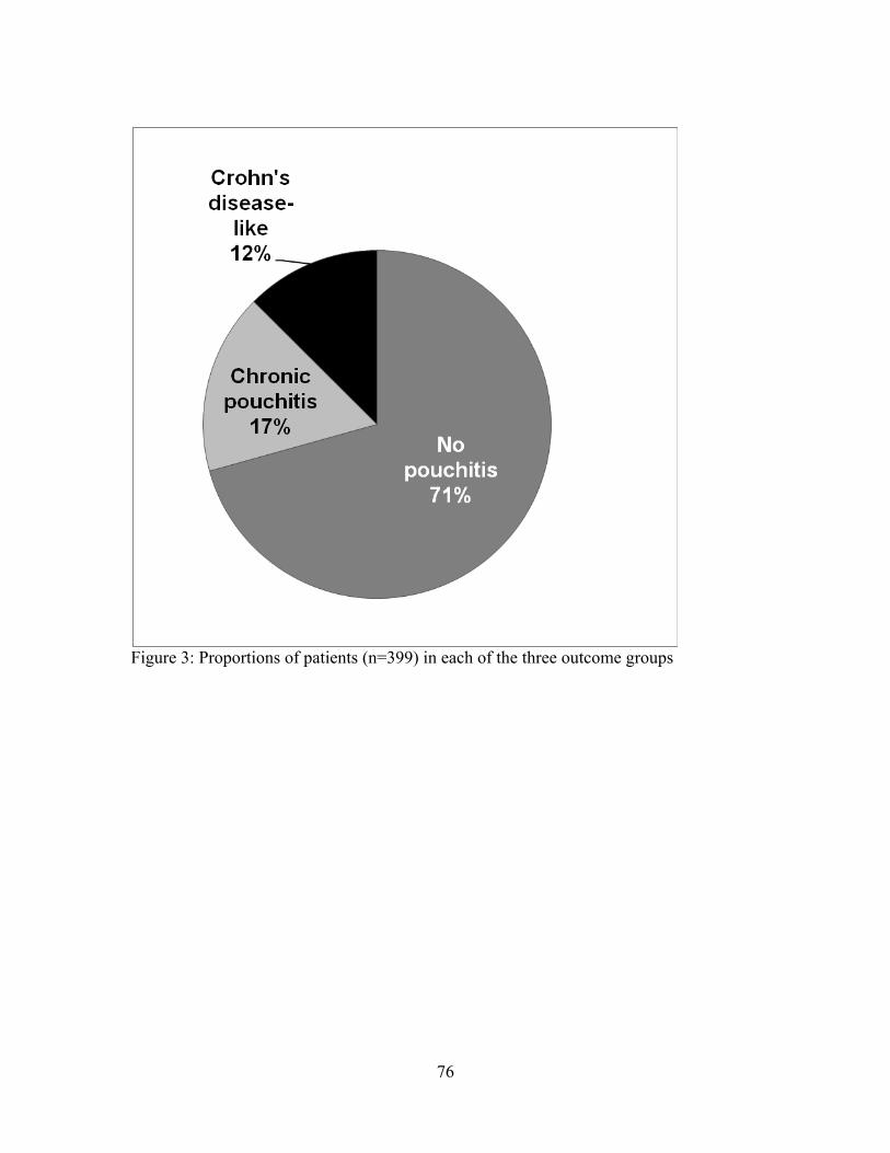
76
Figure 3: Proportions of patients (n=399) in each of the three outcome groups
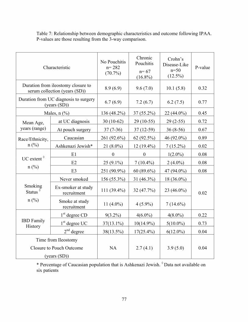
77
Table 7: Relationship between demographic characteristics and outcome following IPAA. P-values are those resulting from the 3-way comparison.
Characteristic No Pouchitis
n= 282 (70.7%)
Chronic Pouchitis
n= 67 (16.8%)
Crohn’s Disease-Like
n=50 (12.5%)
P-value
Duration from ileostomy closure to serum collection (years (SD)) 8.9 (6.9) 9.6 (7.0) 10.1 (5.8) 0.32
Duration from UC diagnosis to surgery (years (SD)) 6.7 (6.9) 7.2 (6.7) 6.2 (7.5) 0.77
Males, n (%) 136 (48.2%) 37 (55.2%) 22 (44.0%) 0.45
Mean Age, years (range)
at UC diagnosis 30 (10-62) 29 (10-55) 29 (2-55) 0.72
At pouch surgery 37 (7-36) 37 (12-59) 36 (8-56) 0.67
Race/Ethnicity, n (%)
Caucasian 261 (92.6%) 62 (92.5%) 46 (92.0%) 0.89
Ashkenazi Jewish* 21 (8.0%) 12 (19.4%) 7 (15.2%) 0.02
UC extent ◊
n (%)
E1 0 0 1(2.0%) 0.08
E2 25 (9.1%) 7 (10.4%) 2 (4.0%) 0.08
E3 251 (90.9%) 60 (89.6%) 47 (94.0%) 0.08
Smoking Status ◊
n (%)
Never smoked 156 (55.3%) 31 (46.3%) 18 (36.0%)
0.02 Ex-smoker at study
recruitment 111 (39.4%) 32 (47.7%) 23 (46.0%)
Smoke at study recruitment 11 (4.0%) 4 (5.9%) 7 (14.6%)
IBD Family History
1st degree CD 9(3.2%) 4(6.0%) 4(8.0%) 0.22
1st degree UC 37(13.1%) 10(14.9%) 5(10.0%) 0.73
2nd degree 38(13.5%) 17(25.4%) 6(12.0%) 0.04
Time from Ileostomy
Closure to Pouch Outcome
(years (SD))
NA 2.7 (4.1) 3.9 (5.0) 0.04
* Percentage of Caucasian population that is Ashkenazi Jewish. ◊ Data not available on six patients
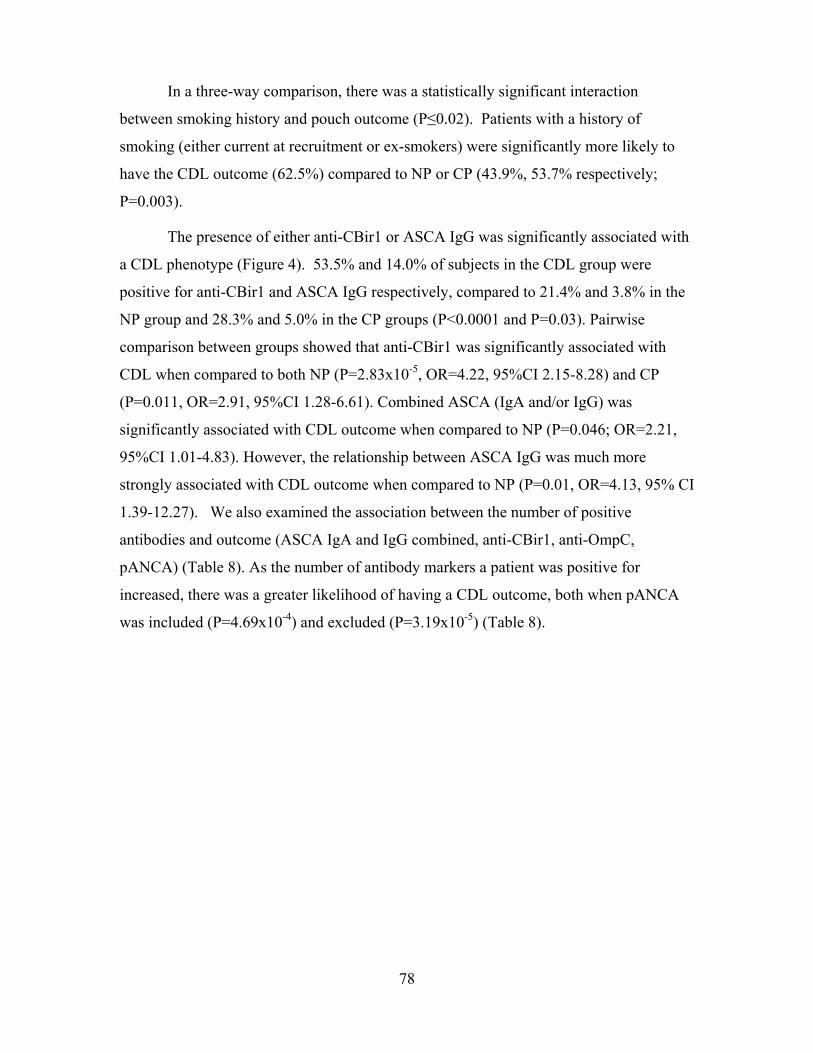
78
In a three-way comparison, there was a statistically significant interaction
between smoking history and pouch outcome (P≤0.02). Patients with a history of
smoking (either current at recruitment or ex-smokers) were significantly more likely to
have the CDL outcome (62.5%) compared to NP or CP (43.9%, 53.7% respectively;
P=0.003).
The presence of either anti-CBir1 or ASCA IgG was significantly associated with
a CDL phenotype (Figure 4). 53.5% and 14.0% of subjects in the CDL group were
positive for anti-CBir1 and ASCA IgG respectively, compared to 21.4% and 3.8% in the
NP group and 28.3% and 5.0% in the CP groups (P<0.0001 and P=0.03). Pairwise
comparison between groups showed that anti-CBir1 was significantly associated with
CDL when compared to both NP (P=2.83x10⁻⁵, OR=4.22, 95%CI 2.15-8.28) and CP
(P=0.011, OR=2.91, 95%CI 1.28-6.61). Combined ASCA (IgA and/or IgG) was
significantly associated with CDL outcome when compared to NP (P=0.046; OR=2.21,
95%CI 1.01-4.83). However, the relationship between ASCA IgG was much more
strongly associated with CDL outcome when compared to NP (P=0.01, OR=4.13, 95% CI
1.39-12.27). We also examined the association between the number of positive
antibodies and outcome (ASCA IgA and IgG combined, anti-CBir1, anti-OmpC,
pANCA) (Table 8). As the number of antibody markers a patient was positive for
increased, there was a greater likelihood of having a CDL outcome, both when pANCA
was included (P=4.69x10⁻⁴) and excluded (P=3.19x10⁻⁵) (Table 8).
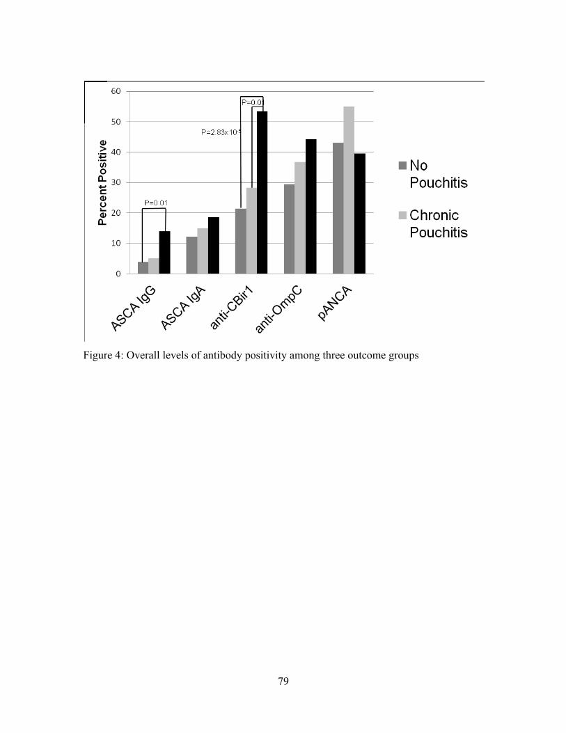
79
Figure 4: Overall levels of antibody positivity among three outcome groups
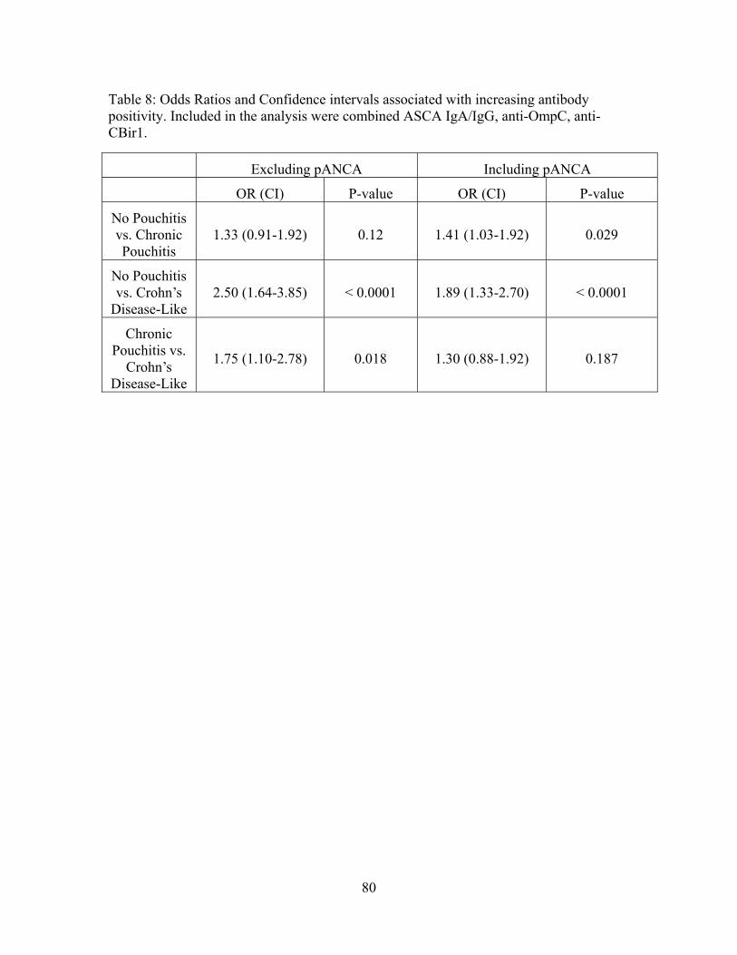
80
Table 8: Odds Ratios and Confidence intervals associated with increasing antibody positivity. Included in the analysis were combined ASCA IgA/IgG, anti-OmpC, anti-CBir1.
Excluding pANCA Including pANCA
OR (CI) P-value OR (CI) P-value
No Pouchitis vs. Chronic Pouchitis
1.33 (0.91-1.92) 0.12 1.41 (1.03-1.92) 0.029
No Pouchitis vs. Crohn’s
Disease-Like 2.50 (1.64-3.85) < 0.0001 1.89 (1.33-2.70) < 0.0001
Chronic Pouchitis vs.
Crohn’s Disease-Like
1.75 (1.10-2.78) 0.018 1.30 (0.88-1.92) 0.187

81
Mean antibody titres for each of the markers in the three outcome groups are
described as a supplementary figure (Appendix Chapter 2, Supplementary figure 1). In
quartile sum analysis, only mean antibody titres for anti-CBir1 were significantly
different between outcome groups (p=0.001). In a multivariate model, which included
quartile sum of combined serum markers as well as age, smoking, race, and UC extent, it
was found that for CP vs. NP, only the quartile sum of serum markers were marginally
associated with disease (p=0.05). However, when comparing CDL with NP, both
smoking (p=0.0023) and the quartile sum of serum markers (p= 0.0006) were associated
with outcome.
The overall seroprevalence rate of pANCA was 44.6% in the entire cohort (n =
341) of patients with serum available. The rate was highest in CP with 55.0% of patients
positive for this marker. However there were no significant differences in rates of
pANCA positivity among the three outcome groups. Additionally, there were no
differences in seroprevalence among any of the markers when comparing individuals
with no pouchitis ever with those who had had episodes of acute antibiotic responsive
pouchitis (data not shown). Finally, we compared both CP and CDL with the subset of
NP individuals who had never experienced pouchitis (n=146) and found that these results
were identical to those seen in the comparisons made using the entire NP cohort.
2.5 Discussion
The development of chronic pouchitis and a CD-like phenotype of the pouch
following IPAA for UC are among the greatest risk factors for pouch failure following
surgery 307. A model to help identify UC patients at risk for complications following
IPAA would be very useful when counseling patients where colectomy is a consideration
for management. A number of studies have examined clinical risk factors as predictors of
outcome demonstrating that smoking, family history of CD and Ashkenazi Jewish
ethnicity are associated with a greater risk of complications following IPAA 69, 311.
Several studies have also shown that the presence of PSC or other immune disorders are
associated with poor pouch outcome 312, 313. In this report, we confirm that Ashkenazi
Jewish individuals and those who have a history of smoking (either current or ex-
82
smokers) were significantly more likely to develop inflammatory pouch complications.
We did not observe any association between outcome and first-degree family history of
IBD.
Anti-microbial antibodies have also been previously described as important
markers associated with pouch outcomes including pouchitis and a CD-like phenotype 67,
71, 72, 314. Typically, pANCA is associated with acute and chronic pouchitis, and ASCA
with a CD-like phenotype after IPAA 69, 72. Previously published work described a
modest association between anti-CBir1 and either acute or chronic pouchitis 67 however,
this marker has not previously been associated with a CDL outcome. These observations
are in keeping with our findings demonstrating the independent association of anti-CBir1
and ASCA IgG with the CDL outcome. Interestingly, the prevalence of anti-CBir1
among UC-IPAA patients with a CDL phenotype, is similar to rates previously reported
in CD patients 59, 148. Additionally, previous studies have shown that presence of both
anti-CBir1 and pANCA together increased the risk of developing pouchitis 67. Extending
these data, our results show that an increasing number of positive antibodies typically
found in CD (ASCA, anti-CBir1 and anti-OmpC) are strongly associated with CDL
outcome and also with chronic pouchitis when pANCA is included.
Several previous studies have suggested that pANCA is associated with chronic
pouchitis 67, 70 and this marker has been associated with UC generally in numerous studies 50, 315, 316. In our study population 44.6% of subjects were pANCA positive and there were
no significant differences in prevalence among the three outcome groups. This rate,
resembles that previously reported in other UC populations 50. The pANCA rate found in
our general UC population (55.6%, data not published) is slightly higher than that
described here, suggesting perhaps some diminution in antibody titre over time following
colectomy. Conversely, the rates of anti-OmpC and anti-CBir1 reported in this population
appear higher than the usual rates found in a UC population. The prevalence of ASCA in
IPAA patients without CDL phenotype is similar to other UC cohorts. The divergent rates
of anti-microbial antibody prevalence in this cohort compared to a general UC or CD
cohort may reflect a change in response to alterations of the microbial flora in the ileal
pouch compared to the flora found in the ileum of a UC patient prior to colectomy. It is
83
also conceivable that CD-related anti-microbial antibodies are found more frequently in
severe UC requiring colectomy as was reported by Fleshner et al 67, 311. Further studies
sampling serum both before and after colectomy will help to clarify this observation.
Genetic studies also suggest that considerable overlap may exist in early etiologic
events leading to UC and CD as illustrated by reports of overlapping CD and UC risk
variants 317-319. Many such polymorphisms are thought to result in increased intestinal
permeability to microbial antigens triggering an adaptive immune response which may be
characterized by the production of anti-microbial antibodies. NOD2 polymorphisms have
previously been associated with increases in serum concentration of ASCA, anti-OmpC
and anti-CBir1 148. The findings that anti-CBir1, ASCA, and a history of smoking,
features typically associated with CD 40, 59, were associated with the CDL phenotype
following IPAA in a cohort of patients with a confirmed UC diagnosis prior to surgery,
suggests that these individuals may be genetically susceptible to IBD via pathways which
are important in both CD and UC phenotypes. Following surgery, such individuals may
be more susceptible to a CD-like phenotype in response to changes in the microbiota of
the pouch, thereby contributing to changes in anti-microbial antibody titres. We
hypothesize that patients with CD-like outcomes after pelvic pouch surgery for UC are
not a “missed” diagnosis of CD but rather an evolution of the IBD-phenotype to one that
more closely resembles CD rather than UC. To date, few genetic factors have been
implicated in the etiology of pouchitis. Polymorphisms in NOD2, IL-1RA and TNF have
shown only weak association with pouchitis, and no genetic variants have as yet been
associated with the CDL outcome 71, 320, 321. However, serologic evidence such as that
described in this study, continues to suggest a role for a breakdown in mucosal and innate
immunity in the pathogenesis of both pouchitis and CD of the pouch.
The onset of ileal inflammation in the pelvic pouch model represents a unique
opportunity to identify the mechanisms that contribute to intestinal inflammation more
generally in IBD. To further characterize this process additional study is required to
evaluate the genetic risk variants associated with pouch outcome and the composition of
the pouch microbiome, ideally in a prospective fashion. In the interim, these data add to
the accumulating literature suggesting that anti-microbial antibodies may be useful in

84
stratifying patients into groups that are at varying risk for inflammatory pouch
complications.
85
Chapter 3: The NOD2insC polymorphism is associated with worse outcome following
ileal pouch-anal anastomosis for ulcerative colitis
This article has been accepted for publication in Gut.
The definitive copyedited, typeset version: Tyler AD, Milgrom R, Stempak JM, Xu W,
Brumell JH, Muise AM, Sehgal R, Cohen Z, Koltun W, Shen B, Silverberg MS. The
NOD2insC polymorphism is associated with worse outcome following ileal pouch-anal
anastomosis for ulcerative colitis. Gut. 2012 Aug 9. [Epub ahead of print]. is available
online at: http://gut.bmj.com/content/early/2012/08/08/gutjnl-2011-301957.abstract

86
Title: The NOD2insC polymorphism is associated with worse outcome following ileal pouch-anal anastomosis for ulcerative colitis
Authors: Andrea D Tyler1,2, Raquel Milgrom2, Joanne M Stempak2, Wei Xu3, John H
Brumell1,5,6, Aleixo M Muise1,4,5, Rishabh Sehgal7, Zane Cohen1,2, Walter Koltun7, Bo
Shen8, Mark S Silverberg1,2
Contributorship statement:
ADT implemented the trial in Toronto, data acquisition, analysis and interpretation, and
drafting and revising the manuscript. RM data analysis and critical revision of the
manuscript. JMS implemented the trial in Toronto and critical revision of the manuscript.
WX statistical analysis and revision of the manuscript. JHB critical revision of the
manuscript. AM critical revision of the manuscript. ZC critical revision of the manuscript
and assisted with patient recruitment. RS implemented the trial for the Hershey site and
monitored data collection for that site. WK implemented the trial in Hershey and critical
revision of the manuscript. BS implemented the trial in Cleveland and critical revision of
the manuscript. MSS initiated the collaborative project, designed data collection tools,
implemented the trial in Toronto, and critical revision of the manuscript. ADT and MSS
are guarantors.
Acknowledgements:
The authors would like to thank Diane Verbeeten and Lucy T. Zhang for their assistance
with the study, and Harden Huang and Brenda O’Connor for maintaining and providing
access to the Mount Sinai Hospital Pelvic Pouch Database. This study was supported by a
grant from the Crohn’s and Colitis Foundation of Canada. Dr. Silverberg is partially
supported by the Gale and Graham Wright Research Chair in Digestive Disease. Partial
funding for genotyping was supported by a CIHR operating grant (MOP97756) to AMM
and JHB.
87
3.1 Abstract: Objective: Inflammatory complications after ileal pouch-anal anastomosis (IPAA) for
ulcerative colitis (UC) are common. The aim of this study was to investigate whether
genetic factors are associated with adverse pouch outcomes such as chronic pouchitis
(CP) and a Crohn’s disease-like (CDL) phenotype. Design: 866 patients were recruited
from three centres in North America: Mount Sinai Hospital (Toronto, ON), the
Cleveland Clinic (Cleveland, OH) and Penn State Milton S. Hershey Medical Center
(Hershey, PA). DNA as well as clinical and demographic information was collected.
Subjects were classified into post-surgical outcome groups: no chronic pouchitis (NCP),
CP, and CDL phenotype. Results: Clinical and genetic data was available on 714
individuals. 487 (68.2%) were classified as NCP, 118 (16.5%) CP, and 109 (15.3%)
CDL. The presence of arthritis or arthropathy (p=0.02), primary sclerosing cholongitis
(p=0.009) and duration of time from ileostomy closure to recruitment (p=0.001) were
significantly associated with outcome. The NOD2insC (rs2066847) risk variant was the
SNP most significantly associated with pouch outcome (p=7.4x10⁻⁵). Specifically, it was
associated with both CP and CDL when compared to NCP (odds ratio [OR]=3.2 and 4.3
respectively). Additionally, SNPs in NOX3 (rs6557421, rs12661812), DAGLB
(rs836518), and NCF4 (rs8137602) were shown to be associated with pouch outcome
with slightly weaker effects. A multi-variable risk model combining previously
identified clinical (smoking status, family history of IBD), serologic (ASCA IgG,
pANCA, and anti-CBir1) and genetic markers was constructed and resulted in an OR of
2.72 (p=8.89x10⁻⁷) for NCP vs CP/CDL and 3.22 (p=4.11x10⁻⁸) for NCP vs CDL
respectively. Conclusion: Genetic polymorphisms, in particular, the NOD2insC risk
allele, are associated with chronic inflammatory pouch outcomes among patients with UC
and IPAA.
88
What is already known:
- Several genetic factors have previously been associated with inflammatory pouch
outcomes in small cohorts.
- Patients with familial adenomatous polyposis and a pelvic pouch do not typically
develop inflammatory pouch complications, suggesting genetic factors are
important in pouchitis and a Crohn’s disease-like phenotype among individuals
with ulcerative colitis.
What is new in this study:
- The NOD2 insC polymorphism, which is important in innate immune recognition
of microbial muramyl dipeptide, is associated with inflammatory pouch outcomes.
- Additional genes which are mediators in the generation of reactive oxygen species
may also have a role in pouch inflammation
Possible clinical implications:
- These polymorphisms may be useful for predicting which patients are at an
increased risk of developing chronic pouch inflammatory complications following
surgery for ulcerative colitis
- A combination of clinical, serologic and genetic factors may be a useful clinical
tool for assisting in decision making for patients considering a colectomy and
pelvic pouch for UC

89
3.2 Introduction:
Ulcerative colitis (UC) is a chronic inflammatory disorder of unknown etiology.
Approximately 20% of affected individuals will require colectomy at some point in their
disease course303, 322-324. Restorative proctocolectomy with ileal-pouch anal anastomosis
(IPAA) is the surgical procedure of choice in the setting of fulminant or chronic,
treatment-refractory UC and UC-associated colonic neoplasia304, 305. However, de novo
inflammation of the ileal reservoir (pouchitis) is a common post-surgical complication
with prevalence rates ranging from 12% to greater than 50% 24, 29, 304, 306. An additional
subset of patients (up to 17%) will develop a Crohn’s Disease (CD)-like phenotype
characterized by abdominal or perianal fistulae or inflammation of the small bowel
proximal to the pouch (afferent limb) 307.
The causes of ileal inflammation in the pouch in patients with a preoperative
diagnosis of UC are unknown. Clearly the evolution of the microbiome of the pouch
after closure of a diverting ileostomy plays a role, as patients rarely develop pouchitis
prior to restoration of continuity of the fecal stream. The decreased risk of inflammation
among those with familial adenomatous polyposis (FAP), and reports by some groups
that a family history of CD may increase the risk for the CDL phenotype in the pouch,
suggests that heritable susceptibility may be a critical factor in disease pathogenesis28, 69.
However, previous genetic studies have been small in scale and have not yielded
consistently reproducible results 325, 326. Among others, variants in IL-1RN 71, 327, and
TLR1328 have been associated with pouch inflammation. Additional studies have
tentatively linked NOD2 variants with chronic pouchitis 106, 107, 321. The aim of this study
was to evaluate whether IBD susceptibility polymorphisms which have been previously
implicated in CD, UC or pouchitis are associated with chronic pouch inflammatory
outcomes following surgery for UC in a large IPAA cohort.
3.3 Methods:
Study Population

90
All study protocols were carried out in accordance with the research ethics boards at each
center and informed consent was obtained from all patients prior to their enrolment.
Patients having undergone colectomy with IPAA at Mount Sinai Hospital (MSH),
Cleveland Clinic and the Penn State Milton S. Hershey Medical Center (HMC) were
contacted by research staff between 2007 and 2010. The demographic and clinical
features of subjects from MSH have been previously reported and described in detail329,
and the NOD2 genotype status has been previously reported on the HMC cohort321.
Included in the study were any patients with a confirmed pre-colectomy diagnosis of UC
and who had had their ileostomy closed a minimum of one year prior to study enrolment.
UC diagnosis was confirmed based on clinical, endoscopic and pathologic evidence –
particularly from the colectomy specimen. Those having undergone their procedure
outside of any of these institutions were included only in the event that medical
documentation was available. Patients with CD, inflammatory bowel disease of the colon
– type unclassified (IBD-U) or indeterminate colitis (IC) based on pre-colectomy medical
chart review or colectomy surgical pathology, were excluded.
Clinical Data Collection
Study data were obtained through detailed retrospective chart review and patient
questionnaire with investigators blinded to patients’ genetic results. To ensure uniformity
between sites, rigorous definitions were agreed upon and applied to classify patients into
outcome groups. Data collected directly by patient interview included gender, age, family
history of IBD, smoking status, and clinical symptoms of pouch function following
surgery (daily number of bowel movements, incontinence, presence or absence of blood
in stool). UC diagnosis date, surgical history and dates, presence of any extraintestinal
manifestations of IBD such as arthritis or arthropathy, osteoporosis/osteopenia, erythema
nodosum, pyoderma gangrenosum, primary sclerosing cholangitis (PSC) or ocular
inflammation, as well as the post-surgical outcome were confirmed through medical chart
review. Pre-colectomy disease extent was classified based on the Montreal classification5.
The presence of backwash ileitis (inflammation of the terminal ileum in individuals with
pan-colitis) pre-colectomy was documented and patients were included only in the event
that all other findings were consistent with UC.

91
Post-Surgical Classification
Patients were classified into one of three outcome groups based on clinical, endoscopic
and histologic factors. The “no chronic pouchitis” (NCP) group included individuals
without an episode of pouchitis, as well as those who experienced fewer than four clinical
acute pouchitis episodes per year, each responding to two weeks or less of antibiotics
(ciprofloxacin, metronidazole, or combination). This clinical definition of acute pouchitis
has been previously described22. Typically, these subjects may not have had a full clinical
assessment but clearly represent a distinct category as demonstrated by their immediate
response to antibiotics. The “chronic pouchitis” (CP) group included antibiotic-
dependent and antibiotic- refractory patients who required either prolonged (>1 month)
antibiotic therapy, required medical intervention for pouchitis more than three times per
year, or required the use of second- or third-line medications (5-ASA, steroids,
immunomodulators, biologics). All such subjects had endoscopic assessment at some
point during their disease course 22. The final group was the “Crohn’s disease-like”
phenotype (CDL). Individuals classified into this group met at least one of the following
criteria: a) development of a perianal fistula more than one year after ileostomy closure
documented through physical examination, examination under anesthesia or imaging; b)
development of a stricture proximal to the pouch which was not related to a surgical
complication and was confirmed by endoscopy or small bowel imaging; c) evidence of
inflammation (ulceration, erythema, friability) extending above the pouch inlet and into
the afferent limb/pre-pouch ileum or more proximal small intestine detected on
pouchoscopy or upper endoscopy. Anastomotic ulceration or ulceration around the pouch
inlet alone was not sufficient to classify patients into the CDL outcome group. Additional
post-surgical outcomes including mechanical or surgical complications or cuffitis were
also documented22. Time to diagnosis of pouchitis was defined as time from ileostomy
closure to the time of onset of symptoms and diagnosis.
DNA Collection and Genotyping
3-6mL of whole blood was obtained by venipuncture using standard EDTA collection
tubes at both MSH and HMC. DNA was extracted using the QIAGEN Gentra Puregene

92
Blood Kit. DNA samples from the Cleveland Clinic were extracted from clotted blood
using the Maxwell 16 Tissue DNA purification kit according to manufacturer’s protocol.
DNA from all study sites was then stored in sealed Matrix screw top tubes at 4°C prior to
spectrophotometric quality and quantity check using the NanoDrop 1000 (Thermo
Scientific). The majority of DNA samples yielded concentrations ranging from 100-300
μg with all concentrations above 20ng/μL. Those falling below 18 ng/uL were whole
genome amplified using the Illustra Genomiphi HY DNA Amplification Kit (GE
Healthcare). Single nucleotide polymorphism (SNP) genotyping was performed using
the Illumina Goldengate custom SNP assay on the Illumina BeadStation500G (San
Diego, CA) at The Center for Applied Genomics (TCAG, Toronto, Canada) and the
Sequenome iPLEX platform (Génome Québec, Montreal, Canada). NOD2insC
genotyping was performed using the TaqMan SNP Genotyping platform (TCAG).
Statistical Analysis and Quality Control
Descriptive statistics were reported as mean and range for continuous variables and
frequencies and proportions for categorical variables. For phenotypic results, Fisher’s
exact test and Pearson’s chi-square test were used to compare proportions; non-
parametric tests were used to compare the continuous variables. PLINK version 1.06330
was used to obtain descriptive statistics of the SNPs such as the allele frequency,
genotype distribution and to test for Hardy-Weinberg equilibrium (HWE) for each marker
based on Pearson’s chi-square test. All SNPs which were successfully genotyped in at
least 95% of the study cohort, satisfied HWE criteria (p HWE>0.001) and had a minor
allele frequency >0.01, were included in the analysis. To ensure that results were not due
to population stratification, only Caucasian individuals were included in the genetic
analysis. Logistic regression models were applied for the association analysis. Although
an additive genetic model was used for the primary analysis 331 we also explored
dominant and recessive genetic models. Throughout this report the p-values are those
obtained from the additive genetic model unless otherwise stated. Odds ratios (OR) and
95% confidence intervals (CI) were calculated. Two-sided statistical tests were applied
and all analyses were performed with SAS 9.2 (SAS Institute, Cary, NC, USA).
93
A list of 646 SNPs was generated and chosen for the analysis in the initial cohort
based on their previous known associations with CD, UC or pouchitis. To accommodate
the Sequenom platform, the top 46 hits from the initial analysis as well as 20 previously
described important CD/UC/pouchitis associated SNPs were selected for genotyping in
the replication cohort (Appendix: Chapter 3, Supplementary table 1). Due to the large
number of tests performed, nominal significance was defined in both the preliminary
analysis and the combined cohort as p < 0.01. Stringent Bonferroni correction was
applied to the final p-value to adjust for multiple comparisons with p < 7.5x10⁻⁴ required
to declare significance.
Finally, multivariate analysis was performed with factors previously shown by our
group and others to be associated with pouch outcome. These included the serologic
markers anti-saccharomyces cerevisiae antibody (ASCA) IgG, perinuclear anti-neutrophil
cytoplasmic antibody (pANCA) and anti-CBir1329; clinical factors included smoking and
family history of IBD; and the SNPs which were significantly associated with outcome
in our Caucasian cohort. This analysis was performed in a subset of 341 patients in whom
all of the preceding data was available. A step-wise procedure was performed to generate
a risk score based on the significant risk factors from the multivariate analysis. The risk
score was based on the weighted combination of the risk factors with the standardized
logistic regression coefficient as the weight332. Receiver operating characteristic (ROC)
curves were generated to calculate the area under the curve (AUC) and the sensitivity and
specificity of the risk score332.
3.4 Results:
Study Cohort
The initial cohort consisted of 399 patients recruited from MSH and the second included
467 patients recruited from MSH, Cleveland Clinic and HMC, for a total cohort of 866.
Within both the first and second cohorts, 339 and 401 respectively had both genotype and
phenotype information available for a total genetic cohort of 740. Among these
individuals 714 (96.5%) were Caucasian and only these subjects were subsequently
analyzed.
94
Clinical Variables Associated with Pouch Outcome
Clinical and phenotypic characteristics of the study population are shown in Table 9. A
description of the pouch outcome groups among the combined Caucasian cohort is shown
in Figure 5. There were 487 (68.2%) NCP, 118 (16.5%) CP, and 109 (15.3%) CDL. The
mean age at UC diagnosis (29, range 2-59 years) and at IPAA (37, range 7-61 years) was
the same between groups. Within the CDL group, 56.6 % were diagnosed based on the
presence of a fistula or abscess developing more than one year following surgery, 36.4%
based on inflammation extending into the afferent limb, and 7.0% met both criteria.
Factors previously related to pouch outcome such as smoking and family history of
IBD69, 307 were not found to be significantly associated in this cohort. However, the
presence of large-joint arthritis (p=0.02) and PSC (p=0.009) were significantly associated
with both the CP and CDL outcomes. A longer duration of time from ileostomy closure
to study enrolment was associated with a worse pouch outcome (p=0.001). The mean
time to diagnosis of CP and CDL was 2.4 and 4.4 years respectively, well below the
mean post-surgical follow-up time for any of the outcome groups.
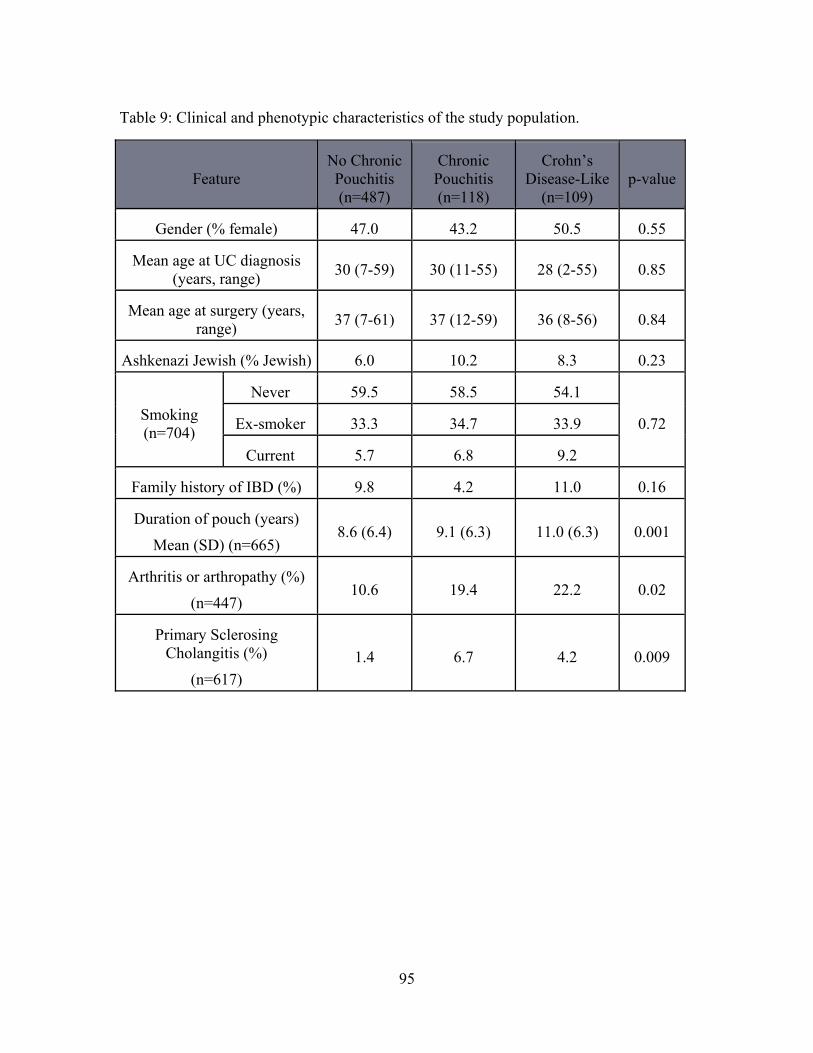
95
Table 9: Clinical and phenotypic characteristics of the study population.
Feature No Chronic Pouchitis (n=487)
Chronic Pouchitis (n=118)
Crohn’s Disease-Like
(n=109) p-value
Gender (% female) 47.0 43.2 50.5 0.55
Mean age at UC diagnosis (years, range) 30 (7-59) 30 (11-55) 28 (2-55) 0.85
Mean age at surgery (years, range) 37 (7-61) 37 (12-59) 36 (8-56) 0.84
Ashkenazi Jewish (% Jewish) 6.0 10.2 8.3 0.23
Smoking (n=704)
Never 59.5 58.5 54.1
0.72 Ex-smoker 33.3 34.7 33.9
Current 5.7 6.8 9.2
Family history of IBD (%) 9.8 4.2 11.0 0.16
Duration of pouch (years)
Mean (SD) (n=665) 8.6 (6.4) 9.1 (6.3) 11.0 (6.3) 0.001
Arthritis or arthropathy (%)
(n=447) 10.6 19.4 22.2 0.02
Primary Sclerosing Cholangitis (%)
(n=617) 1.4 6.7 4.2 0.009

96
Figure 5: Proportion of individuals in each specific outcome group.
97
Association of SNPs with Pouch Outcome
Of the 646 SNPs selected for analysis in the initial cohort of subjects (n = 369), 66 SNPs
which met initial criteria for significance or were SNPs of importance in prior IBD
genetic studies were gentoyped in the additional cohort (n = 345). Of these, 12 had a low
call rate and were not included in the analysis. Of the remaining 54 SNPs, five were
significantly associated with outcome at a p-value threshold of p<0.01 in a three way
comparison. However, only the NOD2 insertion variant (NOD2 insC, rs2066847)
remained significantly associated with pouch outcome after stringent Bonferroni
correction (p=7.4x10⁻⁵; pcorr=4.9x10⁻³) (Table 10). Specifically, the NOD2insC variant
was detected significantly more frequently among individuals with CP and CDL
compared to those with NCP, with OR of 3.21 (CI=1.38-7.47) and 4.30 (CI=1.90-9.77),
respectively (Table 11). This association remained significant even when the previously
reported samples obtained from HMC were excluded.
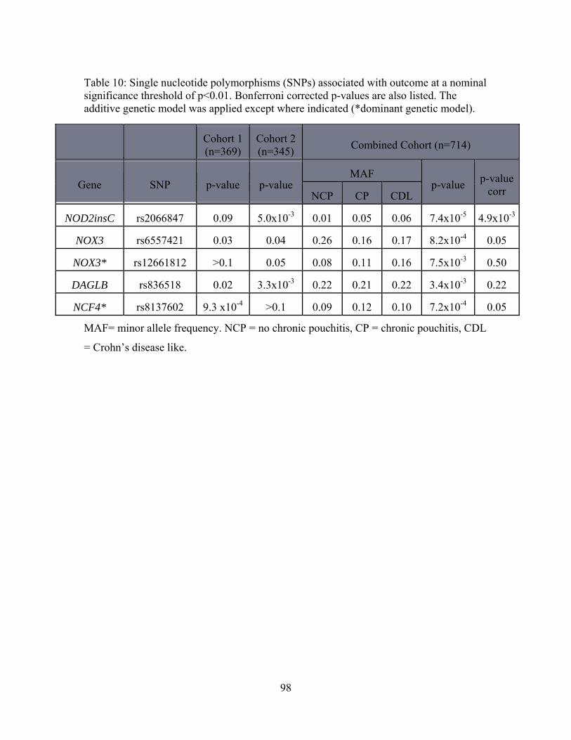
98
Table 10: Single nucleotide polymorphisms (SNPs) associated with outcome at a nominal significance threshold of p<0.01. Bonferroni corrected p-values are also listed. The additive genetic model was applied except where indicated (*dominant genetic model).
Cohort 1 (n=369)
Cohort 2 (n=345) Combined Cohort (n=714)
Gene SNP p-value p-value MAF
p-value p-value corr NCP CP CDL
NOD2insC rs2066847 0.09 5.0x10⁻³ 0.01 0.05 0.06 7.4x10⁻⁵ 4.9x10-3
NOX3 rs6557421 0.03 0.04 0.26 0.16 0.17 8.2x10⁻⁴ 0.05
NOX3* rs12661812 >0.1 0.05 0.08 0.11 0.16 7.5x10⁻³ 0.50
DAGLB rs836518 0.02 3.3x10⁻³ 0.22 0.21 0.22 3.4x10⁻³ 0.22
NCF4* rs8137602 9.3 x10⁻⁴ >0.1 0.09 0.12 0.10 7.2x10⁻⁴ 0.05
MAF= minor allele frequency. NCP = no chronic pouchitis, CP = chronic pouchitis, CDL
= Crohn’s disease like.

99
Table 11: Odds ratios (OR) and confidence intervals (CI) for the five single nucleotide polymorphisms (SNPs) which are associated with outcome. (*dominant genetic model).
Gene SNP CP vs NCP CDL vs NCP CDL vs CP CP/CDL vs
NCP
OR CI OR CI OR CI OR CI
NOD2insC rs2066847 3.21 1.38-7.47 4.30 1.90-9.77 1.28 0.57-2.88 3.67 1.81-7.45
NOX3 rs6557421 0.55 0.36-0.82 0.56 0.37-0.87 1.05 0.62-1.79 0.56 0.40-0.76
NOX3* rs12661812 1.25 0.71-2.19 1.09 0.59-2.00 0.87 0.41-1.83 1.17 0.74-1.84
DAGLB rs836518 0.93 0.63-1.38 0.45 0.27-0.74 0.49 0.27-0.88 0.69 0.50-0.96
NCF4* rs8137602 1.01 0.58-1.74 1.10 0.63-1.93 1.09 0.54-2.21 1.05 0.69-1.61
NCP = no chronic pouchitis, CP = chronic pouchitis, CDL = Crohn’s disease like.

100
Given the low minor allele frequency of the NOD2insC allele in our cohort, to
fully assess the risk associated with variants in NOD2 and pouch related inflammatory
complications, we examined the risk associated with compound heterozygosity or
homozygosity for combinations of markers (rs2066847, rs2066845 and rs2066844). We
found that increased numbers of variants at this loci were associated with increased risk
of CDL when compared to NCP, (p=.002; OR=2.08; CI=1.31-3.50), however, no
increased risk was observed when NCP was compared to CP.
Additional SNPs associated with outcome in the three-way analysis (p<0.01) were
variants in NOX3 (rs6557421) and DAGLB (rs836518), however, these did not remain
significant after correction for multiple testing (Table 10). In the pairwise analysis,
rs6557421 trended towards being protective for both of the inflammatory outcomes and
rs836518 was protective against only the CDL phenotype (Table 11). Additional variants
in NOX3 (rs12661812) and NCF4 (rs8137602) were found to be associated with outcome
(p<0.01) using the dominant genetic model, with variants at both loci more common in
individuals with inflammatory pouch phenotypes. However, these polymorphisms did not
remain significantly associated with outcome after correction for multiple testing.
We next conducted analyses in which we compared the NCP group with the
combined CP/CDL cohort (ncomb=227). We found that both rs2066847 (NOD2insC) and
rs6557421 (NOX3) were associated with outcome with the minor allele of rs2066847
(p=3.3x10⁻⁴) increasing disease risk and that of rs6557421 (p=2.8x10⁻⁴) protective against
inflammatory phenotypes (Table 11).
Variants which had been previously associated with pouch complications,
including those in TLR1 328, CD14, TLR9 326 as well as other NOD2 variants, were not
found to be associated with outcome among our cohort (Table 12). IL1RN327 trended
towards significance in our initial analysis, however did not remain significant after
correction for multiple testing. Additionally, many of the other well known IBD-
associated genetic polymorphisms including those specific to CD (i.e. ATG16L1) and
those associated with both CD and UC (i.e. IL23R), were not associated with any of the
outcome groups.
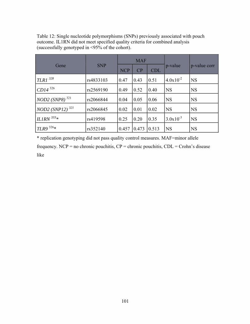
101
Table 12: Single nucleotide polymorphisms (SNPs) previously associated with pouch outcome. IL1RN did not meet specified quality criteria for combined analysis (successfully genotyped in <95% of the cohort).
Gene SNP MAF
p-value p-value corr NCP CP CDL
TLR1 328 rs4833103 0.47 0.43 0.51 4.0x10⁻² NS
CD14 326 rs2569190 0.49 0.52 0.40 NS NS
NOD2 (SNP8) 321 rs2066844 0.04 0.05 0.06 NS NS
NOD2 (SNP12) 321 rs2066845 0.02 0.01 0.02 NS NS
IL1RN 333* rs419598 0.25 0.20 0.35 3.0x10⁻³ NS
TLR9 326* rs352140 0.457 0.473 0.513 NS NS
* replication genotyping did not pass quality control measures. MAF=minor allele
frequency. NCP = no chronic pouchitis, CP = chronic pouchitis, CDL = Crohn’s disease
like

102
Multivariate analysis and risk score
In the preliminary multivariate analysis, five factors were found to independently
associate with outcome and were included in the subsequent multivariate analysis,
including rs2066847 (NOD2insC), rs6557421 (NOX3), rs836518 (DAGLB), anti-CBir1
and smoking (Table 13). These factors were used to generate a weighted risk score, with
the comparison of both NCP with CDL (p=4.11x10⁻⁸; OR=3.22, CI=2.12-4.89) and NCP
with combined CP and CDL (p=8.89x10⁻⁷; OR=2.72; CI=1.89-3.91) resulting in highly
significant associations. The sensitivity and specificity were assessed by generating ROC
curves. The best model was that comparing CDL with NCP and containing all three
genetic markers, as well as the serologic and clinical factors (sensitivity = 80.0%;
specificity = 70.3%). This model performed significantly better than did those generated
using smoking alone, or smoking and anti-CBir1 (Figure 6).

103
Table 13: Factors significantly associated with outcome by multi-variate analysis.
Factor CP vs. NCP CDL vs. NCP CDL vs. CP
OR (CI) p-value OR (CI) p-value OR (CI) p-
value
NOD2insC*
rs2066847
2.62
(0.39-17.64)
0.32 13.51
(2.00-90.9) 0.008
2.99
(0.45-20.0)
0.26
NOX3*
rs6557421
0.54
(0.31-0.94)
0.03 0.35
(0.15-0.78) 0.01
0.62
(0.24-1.59)
0.32
DAGLB*
rs836518
1.10
(0.69-1.74)
0.70 0.29
(0.12-0.67) 0.004
0.25
(0.10-0.66)
0.005
anti-CBir1
(+/-)
2.30
(0.84-3.21)
0.15 6.38
(2.81-14.43)
<0.0001
3.48
(1.36-8.90)
0.009
Smoking
Current smoking vs.
Never smoked
2.30
(0.55-9.71)
0.38 11.96
(3.07-46.53)
0.0002 5.68
(1.10-29.25)
0.04
Ex-smoker
vs.
Never smoked
1.52
(0.83-2.77)
0.99 2.20
(0.95-5.10) 0.30
1.16
(0.44-3.05)
0.18
*: For the genetic markers an additive genetic model to compare homozygote recessive
vs. heterozygote vs. homozygote dominant genotypes was applied
NCP = no chronic pouchitis, CP = chronic pouchitis, CDL = Crohn’s disease like.
OR=odds ratio; CI=confidence interval.
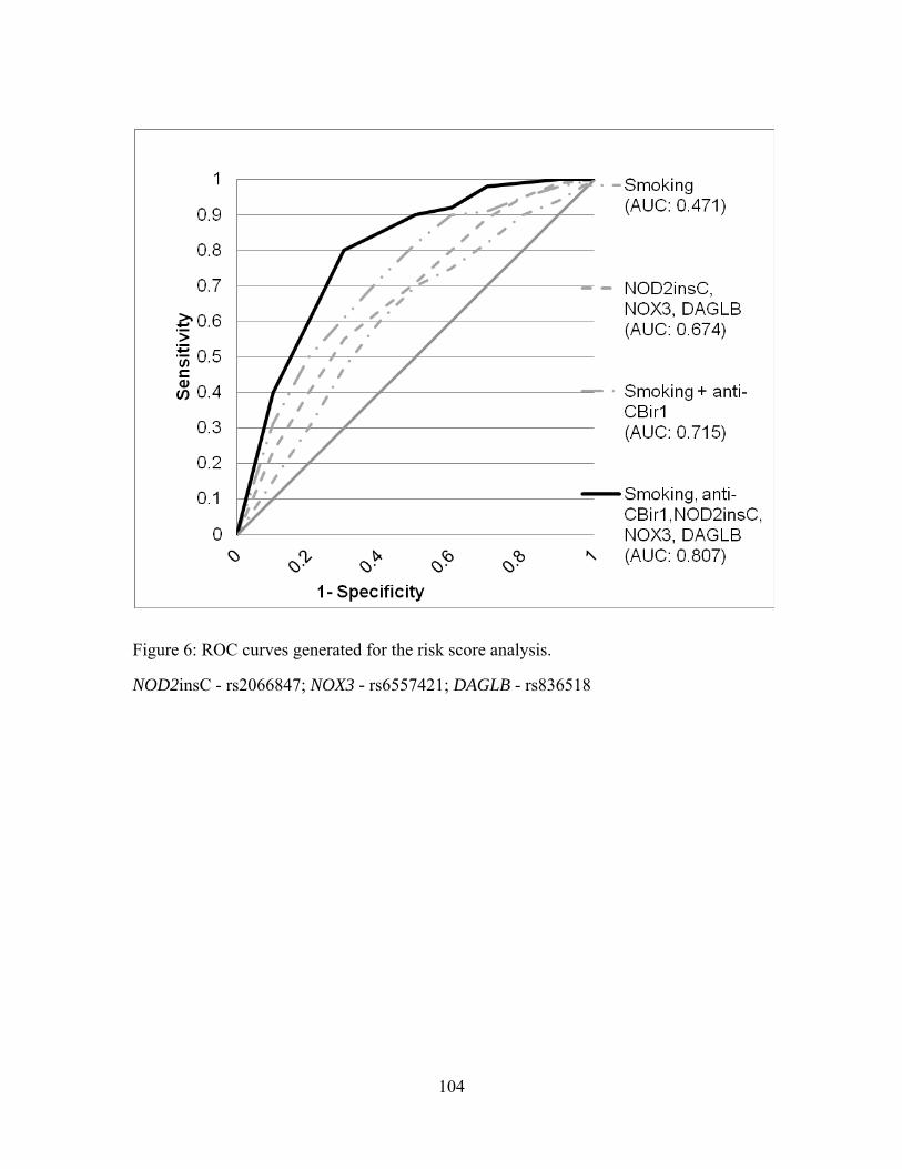
104
Figure 6: ROC curves generated for the risk score analysis.
NOD2insC - rs2066847; NOX3 - rs6557421; DAGLB - rs836518
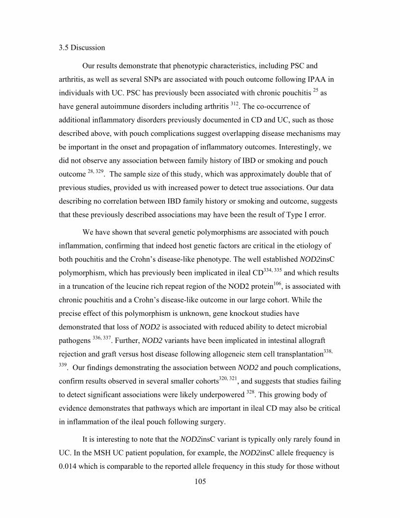
105
3.5 Discussion
Our results demonstrate that phenotypic characteristics, including PSC and
arthritis, as well as several SNPs are associated with pouch outcome following IPAA in
individuals with UC. PSC has previously been associated with chronic pouchitis 25 as
have general autoimmune disorders including arthritis 312. The co-occurrence of
additional inflammatory disorders previously documented in CD and UC, such as those
described above, with pouch complications suggest overlapping disease mechanisms may
be important in the onset and propagation of inflammatory outcomes. Interestingly, we
did not observe any association between family history of IBD or smoking and pouch
outcome 28, 329. The sample size of this study, which was approximately double that of
previous studies, provided us with increased power to detect true associations. Our data
describing no correlation between IBD family history or smoking and outcome, suggests
that these previously described associations may have been the result of Type I error.
We have shown that several genetic polymorphisms are associated with pouch
inflammation, confirming that indeed host genetic factors are critical in the etiology of
both pouchitis and the Crohn’s disease-like phenotype. The well established NOD2insC
polymorphism, which has previously been implicated in ileal CD334, 335 and which results
in a truncation of the leucine rich repeat region of the NOD2 protein106, is associated with
chronic pouchitis and a Crohn’s disease-like outcome in our large cohort. While the
precise effect of this polymorphism is unknown, gene knockout studies have
demonstrated that loss of NOD2 is associated with reduced ability to detect microbial
pathogens 336, 337. Further, NOD2 variants have been implicated in intestinal allograft
rejection and graft versus host disease following allogeneic stem cell transplantation338,
339. Our findings demonstrating the association between NOD2 and pouch complications,
confirm results observed in several smaller cohorts320, 321, and suggests that studies failing
to detect significant associations were likely underpowered 328. This growing body of
evidence demonstrates that pathways which are important in ileal CD may also be critical
in inflammation of the ileal pouch following surgery.
It is interesting to note that the NOD2insC variant is typically only rarely found in
UC. In the MSH UC patient population, for example, the NOD2insC allele frequency is
0.014 which is comparable to the reported allele frequency in this study for those without
106
inflammatory pouch complications340. However in the 227 subjects with CP or CDL, the
NOD2insC allele frequency is over 0.05. These patients were all carefully chart reviewed
to exclude any clinical or pathological evidence of CD or IBDU prior to colectomy.
Additionally, the colectomy pathology was all carefully reviewed and there were no
features of CD seen in these subjects. These data therefore suggest that the utility of
traditional clinical and histologic diagnostic classification and phenotyping is not
sufficient to categorize subjects in terms of prognosticating outcome after pelvic pouch
surgery. That is, a diagnosis of clinical UC may not imply that pouch complications are
less likely to arise but rather, the subject’s genotype may be a more important
determinant of outcome.
Other variants which demonstrated modest association with outcome include
those which are located in the non-coding regions of NOX3 and DAGLB or adjacent to
NCF4. NCF4, encoding p40phox which is a component of the NADPH oxidase complex,
was recently identified in a GWAS as an ileal CD susceptibility gene341. Mechanistic
studies have demonstrated that CD patients who are carriers of risk alleles at this locus
have significantly decreased amounts of reactive oxygen species - important for host
innate immunity and defense from microbial pathogens - generated from granulocyte-
macrophage colony-stimulating factor-primed neutrophils compared to patients not
carrying these mutations342. NOX3, another component of this complex in various tissue
types, is also important in the production of reactive oxygen species 343, 344. Together,
these findings suggest an important role for this pathway in the pathogenesis of not only
pouch inflammatory outcomes but IBD in general. The function of DAGLB, and a
potential role for it in IBD pathogenesis remains unclear, however, the SNP in this gene
is in a region of high linkage disequilibrium with RAC1; a gene involved in host immune
defence and which has been previously associated with UC345.
Other variants which have been previously associated with pouch outcome among
smaller cohorts were not associated with outcome in our group. A possible explanation
for the difference between our results and others is that this study has a much larger
sample size, which would help to reduce Type I error. Additionally, population
stratification may account for different results as some previous studies did not control
for ethnicity 328, 333. To limit these effects, only Caucasian individuals were analyzed in

107
our study. We also applied very stringent statistical correction to our data in order to
reduce the likelihood of reporting false positives, which may have been too stringent to
allow detection of weaker associations. The four variants discussed in this paper which
did not remain significant after multiple testing correction, may therefore warrant further
investigation in an additional cohort to definitively assess their importance in pouchitis
pathogenesis. This would ideally be attempted in the setting of the large International
IBD Genetics Consortium Cohort but would require detailed phenotyping of pre-
colectomy data as well as of pouch outcomes, both of which may not be readily available
in existing databases.
We hypothesize that patients with NOD2insC may be more likely to develop
inflammatory pouch complications due to alteration in the microbial composition of the
pouch mucosa. This hypothesis is supported by our data showing that UC patients with a
CDL phenotype after pelvic pouch surgery are more likely to be positive for ASCA and
anti-CBir1329, serologic markers more typically associated with CD, and by other groups’
data demonstrating a strong association between the common UC associated serum
marker, pANCA, and pouch outcome67, 68. Additional evidence that NOD2 variants affect
the composition of the ileal flora suggests that there may be a relationship between IBD
associated genetic polymorphisms and the presence of serum antibodies337 148. Our
findings of the presence of both serologic markers of CD and genetic polymorphisms
which have been previously associated with ileal CD showing association with pouch
outcome, suggests that the mechanism of ileal inflammation which leads to a CP or CDL
outcome in the pouch may be similar to those which lead to the development of
inflammation within the ileum. Additionally, the inclusion of individuals with non-
chronic pouchitis in the NCP group, and the lack of evidence for any associations
between genotype and this outcome, suggests that the chronic inflammatory phenotypes
proceed via diverging mechanistic pathways from those which lead to antibiotic
responsive pouchitis. Furthermore, the overlap in genetic susceptibility between CP and
CDL suggests that distinction between these outcomes may be of limited consequence
compared to the more clinically relevant phenotype of chronic, medication refractory
inflammation which is characteristic of both disorders and results from a common
etiology.
108
While it is conceivable that these CDL patients were misclassified prior to
colectomy, the stringent phenotypic classification which was used to determine which
patients were included in this analysis would suggest that this is unlikely to be the case.
Rather, our results combined with recent data published by Waterman et al. showing few
differences in the prevalence of several IBD-associated SNPs between those with UC and
those with CD317, supports the concept of IBD as a mosaic of inflammatory disorders.
The varying phenotypes associated with overlapping genetic susceptibility loci suggests
that while genetic predisposition may be a key inflammatory mediator, other factors such
as bacteria or as yet unknown environmental stimuli are necessary in order for immune
dysregulation and inflammation to occur.
The etiology of pouch complications remains unknown, however, the data
presented in this paper emphasizes the similarities between pouch inflammation and IBD
in general, and suggests that the pelvic pouch model may be useful for evaluating factors
which contribute to de novo inflammation. It will be important to evaluate the impact of
the pouch microbiome and the impact of host genotype on the composition of the
microbiome to fully understand the mechanisms of pouch inflammation. Our data also
demonstrate that a model for assessing risk of pouch complications which encompasses
genetic and serologic factors may be a more useful tool than current clinical assessment.
109
Chapter 4: Characterization of the gut-associated microbiome in inflammatory pouch
complications following ileal pouch-anal anastomosis.
Accepted May 13, 2013: PLOS One.
Authors: Andrea D. Tyler1,2, Natalie Knox3, Boyko Kabakchiev1,2, Raquel Milgrom2,
Richard Kirsch5, Zane Cohen1,2, Robin S. McLeod2, David S. Guttman6, Denis O.
Krause3,4, Mark S. Silverberg1,2
Author contributions:
ADT: Generation, collection, assembly, analysis and interpretation of data; statistical plan
and analysis; drafting and revision of the manuscript. NK: assembly, analysis and
interpretation of data; revision of the manuscript. BK: analysis and interpretation of data;
statistical analysis; revision of the manuscript. RM: assembly of data and manuscript
revision. RK: collection, assembly, analysis and interpretation of data. ZC: conception of
the study; revision of the manuscript. RSM: conception of the study; revision of the
manuscript. DSG: interpretation of data; revision of the manuscript. DOK: conception
and design of the study; generation, assembly of data. MSS: conception and design of the
study; drafting or revision of the manuscript; approval of the final version of the
manuscript. ADT and MSS are guarantors.
Acknowledgements: The authors would like to thank Joanne Stempak, Lucy T. Zhang,
Tsedey Legesse, Rachel Caplan, Fuqin Zhu and other members of the Silverberg and
Krause labs for their assistance with the study. Additional thanks to Hillary A. Steinhart,
Gordon R. Greenberg, Kenneth Croitoru, Geoff C. Nguyen, Alvin Newman, Helen M.
MacRae, Robert Gryfe, Robert H. Riddell at the MSH Department of Gastroenterology
and Pathology and research fellows Smita Halder and Matti Waterman at MSH for aiding
in patient recruitment, and to Harden Huang and Brenda O’Connor for maintaining and

110
providing access to the Mount Sinai Hospital Pelvic Pouch Database. This study was
supported by a grant from the Crohn’s and Colitis Foundation of Canada. Dr. Silverberg
is partially supported by the Gale and Graham Wright Research Chair in Digestive
Disease.
111
4.1 Abstract:
Introduction: Inflammatory complications following ileal pouch-anal anastomosis (IPAA)
for ulcerative colitis (UC) are common and thought to arise through mechanisms similar
to de novo onset inflammatory bowel disease. The aim of this study was to determine
whether specific organisms in the tissue-associated microbiota are associated with
inflammatory pouch complications.
Methods: Patients having previously undergone IPAA were recruited from Mount Sinai
Hospital. Clinical and demographic information were collected and a pouchoscopy with
biopsy of both the pouch and afferent limb was performed. Patients were classified based
on post-surgical phenotype into four outcome groups: familial adenomatous polyposis
(FAP) controls, no pouchitis, pouchitis, and Crohn’s disease-like (CDL). Pyrosequencing
of the 16S rRNA V1-V3 hypervariable region, and quantitative PCR for bacteria of
interest, were used to identify organisms present in the afferent limb and pouch.
Associations with outcomes were evaluated using exact and non-parametric tests of
significance.
Results: Analysis at the phylum level indicated that Bacteroidetes were detected
significantly less frequently (P<0.0001) in the inflammatory outcome groups (pouchitis
and CDL) compared to both FAP and no pouchitis. Conversely, Proteobacteria were
detected more frequently in the inflammatory groups, although without reaching the same
levels of significance. At the genus level, organisms associated with outcome were
detected less frequently among the inflammatory groups compared to those without
inflammation. Several of these organisms, including Bacteroides (Pboth sites<0.0001),
Parabacteroides (Ppouch=1.2x10⁻⁴, Pafferent limb=2.2x10⁻³), Blautia (Ppouch=3.0x10⁻³, Pafferent
limb<0.0001) and Sutterella (Ppouch=2.5x10⁻³, Pafferent limb=1.7x10⁻³), were associated with
outcome in both the pouch and afferent limb. These associations remained significant
even following adjustment for antibiotic use, smoking, country of birth and gender.
Individuals with quiescent disease receiving antibiotic therapy displayed similar
reductions in these organisms as those with active pouch inflammation.
Conclusions: Specific genera are associated with inflammation of the ileal pouch, with a
reduction of typically ubiquitous organisms characterizing the inflammatory phenotypes.
112
4.2 Introduction
Recent studies have implicated microorganisms in the etiology of several chronic
conditions including inflammatory bowel disease (IBD)169, diabetes171 and obesity173. The
evidence for a role of microorganisms in IBD, both Crohn’s disease (CD) and ulcerative
colitis (UC), is compelling: numerous polymorphisms in genes associated with innate and
adaptive immunity, as well as those associated with barrier function, have been
associated with IBD, as have anti-microbial antibodies52, 121, 346. Additionally, some
studies have implicated adherent invasive Escherichia coli (AIEC) and Mycobacterium
avium subspecies paratuberculosis as potential contributors to pathogenesis347, 348. On
the other hand, decreased frequency of Faecalibacterium prausnitzii among those with
inflammation compared to healthy controls, suggests that this organism may have a
protective effect250, 349. Recent studies have shown that intestinal dysbiosis is associated
with disease, and advances in culture-independent sequencing approaches have
demonstrated a vast amount of heterogeneity within the microbiota of the gastrointestinal
tract337, 350. This highlights the need for further investigation in large and phenotypically
diverse cohorts.
Post-surgical models of IBD are useful for studying the role of microbes, as
recurrence can be viewed as a surrogate for de novo onset of disease. Among UC
patients, greater than 20% will require surgical management324, for which the treatment
of choice is a colectomy with ileal-pouch anal anastomosis (IPAA). Colectomy is often
considered a definitive treatment for UC, however, de novo inflammation of the ileal
reservoir (pouchitis) is a common post-surgical complication with prevalence rates
ranging from 12% to greater than 50%29 , 306. Additionally, 10-17% of patients go on to
develop a CD-like phenotype which is described as the development of abdominal or
perianal fistulas or abscesses, or inflammation of the small bowel proximal to the pouch
(afferent limb)307, 329. IPAA is also the treatment of choice among individuals with
familial adenomatous polyposis (FAP), however, inflammatory complications of the
pouch among this group are very rare. Recent genetic studies have shown that among
individuals with IPAA, those with polymorphisms in innate immune and bacterial
sensing and recognition genes are at an increased risk for inflammatory complications351.
However, inflammation rarely develops in the absence of fecal flow, suggesting that this
113
genetic predisposition alone does not itself cause inflammation, and that microbial factors
may have a critical role. The aim of this study was to characterize and evaluate the
mucosal microbiome of individuals having undergone IPAA for treatment of UC or FAP.
4.3 Materials and Methods
Ethics Statement
This study was approved by and carried out in accordance with the Research
Ethics Board of Mount Sinai Hospital (Toronto, Canada).
Subject Recruitment
Patients were recruited during regular pouch follow-up at Mount Sinai Hospital
(MSH) in Toronto, Canada. Any patients with confirmed UC or FAP and who had
undergone IPAA at least one year prior to recruitment were included in the study.
Biopsies were taken from within the pouch itself (1 biopsy) and 5-10 cm into the afferent
limb (1 biopsy), and were immediately placed into sterile, empty freezer vials and snap
frozen in liquid nitrogen. Two additional biopsies from the same locations were sent to
the MSH pathology lab for histological scoring. During the pouchoscopy, physicians
documented the appearance of the pouch and afferent limb using previously described
criteria for pouch inflammation. Peripheral blood was also collected for clinical
evaluation of C-reactive protein (CRP) levels.
All subjects were classified into outcome groups based on a combination of long-
term complications in conjunction with inflammatory activity at the time of the
procedure. Those with FAP were classified as such, while the remaining groups were
composed of individuals with UC prior to colectomy. To assess inflammation of both the
pouch and afferent limb, endoscopic appearance (erythema, friability, ulceration) and
histological (polymorphonuclear leukocyte infiltration, ulceration/erosions) scores at the
time of the study endoscopy were considered (Chapter 4, Supplementary Table 1)20, 352.
These traits were utilized as they appear to be objective categories within the PDAI and
PAS. Using these categories, a score greater than 3 was applied as the cutoff indicative of
114
inflammation. Individuals who had never been diagnosed with pouchitis and who had no
evidence of inflammation of the pouch or afferent limb at the time of sample collection
were classified into the “no pouchitis” group; individuals with evidence of inflammation
of only the pouch at the time of endoscopy, based on the criteria described above, were
classified as “pouchitis”; individuals with documented evidence of inflammation of the
afferent limb or proximal small bowel, historically, or at the time of their procedure, or
who had developed a stricture or fistula no sooner than one year following the closure of
their ileostomy were classified into the “CD-like” group (CDL). This group was made up
of individuals who had both active inflammation as well as those without. Individuals
with a previous diagnosis of CDL or chronic pouchitis, based on documented evidence of
complications and a need for long-term medical therapy, but who had an inflammatory
score below our cutoff for both the pouch and afferent limb, and no fistulae, were
considered a separate group (“quiescent”) and were not included in the primary analysis.
Microbial DNA Extraction and Analysis
Biopsies were thawed and processed in two batches at the Department of Animal
Science, University of Manitoba (Winnipeg, Canada). The QIAGEN DNeasy Blood and
Tissue Kit (QIAGEN, California, USA) with an additional bead beating step was used to
extract microbial DNA. Barcoded primers specific for the V1-V3 hypervariable region of
the 16S rRNA molecule were used for 454 pyrosequencing using the GS FLX Titanium
assay (Roche, USA)353. Sequences were processed and assigned to a taxonomy using
mothur (Chapter 4, Supplementary Methods).
Statistical analysis
Taxonomic groups which were detected in less than 5% of the samples were
excluded from further analysis. Analyses were performed separately on pouch and
afferent limb samples in order to avoid falsely inflating significance. Genus level results
were analyzed using presence-absence and frequency data with exact and non-parametric
methodologies (Chapter 4, Supplementary Methods). Confirmation of results and
adjustment for confounding variables was performed using exact logistic regression. FDR
corrected p-values below 0.05 were considered significant. All statistical analyses were
115
conducted using STATA version 11.1 (StataCorp Texas, USA) and R version 2.13.1 (R
Foundation for Statistical Computing, Vienna, Austria). Additional analyses were carried
out using the Linear Discriminant Analysis (LDA) Effect Size (LEfSe)354 tool (version
1.1.0).
Quantitative PCR (qPCR)
Organisms previously implicated in bowel inflammation including F. prausnitzii,
Clostridial cluster IV, AIEC, and Roseburia were evaluated using qPCR with previously
described primer pairs (Chapter 4, Supplementary Table 2). Relative abundance was
estimated as has been previously described, using the 16S rRNA Eubacteria gene as a
reference (Chapter 4, Supplementary Methods)355. Results from individual samples were
log2 transformed and analyzed using the Kruskal-Wallis test. Additionally, primers
specific for the Bacteroides genus were used to confirm pyrosequencing results by
evaluating the correlation between dichotomized (Matthew’s) and frequency (Spearman)
pyrosequencing and qPCR data.
4.4 Results
Patient phenotypic and clinical characteristics
274 patients were recruited to this study. To ensure a balanced study design, 78
were selected for analysis based on phenotype, with 71 classified into the four primary
outcome groups. Seven patients were excluded from the initial analysis having been
diagnosed with chronic pouchitis or CDL which was effectively treated with antibiotics
or other medications (quiescent). In total, there were 18 patients in the FAP group, 19 no
pouchitis, 15 pouchitis, and 19 CDL. Among those classified as CDL, two had only
perianal fistulae and an inflammation score of zero in both the pouch and afferent limb
(no evidence of inflammation at the time of endoscopy). One additional patient included
in this group had evidence of only mild inflammation in both the pouch and afferent limb
(inflammatory score less than or equal to 3), with additional features of the CDL
phenotype. Inflammatory activity, as measured by our objective inflammatory score, was

116
low among individuals in the FAP and no pouchitis groups, and higher in both the
pouchitis and CDL groups in the pouch (P<0.01). In the afferent limb, only individuals in
the CDL group displayed evidence of inflammation (P<0.01)(Chapter 4, Supplementary
figure 1).
Interestingly, C-reactive protein (CRP) levels were similarly elevated among each
of the UC groups, compared to individuals with FAP (P=0.002)(Table 14). There was an
approximately equal split across genders through each of the outcome groups and the
mean age at UC or FAP diagnosis and time from ileostomy closure to sampling were
approximately the same (Table 14). Smoking was significantly more common (P=0.007)
among individuals who were in the FAP group. The majority of patients (54%) had taken
antibiotics, (ciprofloxacin, metronidazole, or combination) at some point following their
pouch procedure (either for pouch complications, or for reasons not related to bowel
inflammation). Antibiotic use at the time of pouchoscopy, or during the month
immediately prior is documented in Table 14. Individuals in the FAP and no pouchitis
groups who were on antibiotics (amoxicillin, cefixime) reported sinus or urinary tract
infections as the reason for medication use. One individual in the FAP group took
medications because of possible pouchitis-like symptoms. However, endoscopic and
histological examinations at several time-points suggest that an inflammatory outcome
was not the appropriate diagnosis. Another individual in this group was characterized as
having pouch inflammation based on mild pouch erythema and histology showing
discrete patchy polymorphonuclear leukocyte infiltration in both the pouch and afferent
limb. This individual had no symptoms consistent with pouchitis and was taking Sulindac
for periampullary polyps.

117
Table 14: Phenotypic characteristics of individuals among the four outcome groups.
FAP (n=18) No Pouchitis (n=19)
Pouchitis (n=15)
Crohn’s disease-like (n=19)
p-value
Gender (% female)
55.6 26.3 26.7 52.6 0.14
Mean age at recruitment (years(SE))
38.1 (2.1) 52.8 (3.3) 41.3 (3.8) 46.3 (3.1) 0.009
Mean age at UC/FAP diagnosis (years (SE))
23.1 (3.0) 31.9 (3.3) 27.8 (3.0) 30.5(2.9) 0.20
Time from surgery to sample collection (years (SE))
11.1 (1.5) 10.6 (1.7) 9.3 (1.6) 9.9 (1.3) 0.88
C-reactive protein
(mean (SE))
2.0 (0.6) 7.1 (3.1) 9.8 (3.4) 7.7 (2.3) 0.002
Antibiotic previous month (% using, SE)
11.1 (7.4) 10.5 (7.0) 13.3 (8.7) 42.1 (11.3) 0.07
Antibiotics ever (% used, SE)
11.1 (7.4) 42.1 (11.3) 73.3 (44.2) 89.5 (30.6) 0.03
Biologics (% using, SE)
0 0 6.7 (6.4) 10.5 (7.0) 0.44
Smoking at recruitment (% using, SE)
41.2 (11.6) 5.3 (16.2) 6.7 (6.4) 5.3(5.1) 0.007
FAP = familial adenomatous polyposis; SE=standard error. Individuals who were classified as smoking at recruitment reported at least one cigarette per day within the last month.

118
Bacterial community diversity and phylum composition of the ileal pouch
The pyrosequencing experiment provided 557,215 (mean of 7143 sequences per
sample) raw sequences with a median read length of 453 basepairs (range 250-531).
However, following quality trimming and chimera checking, 345,466 (mean of 4429
sequences per sample) high quality reads remained. To ensure that our stringent inclusion
criteria did not adversely affect our ability to detect organisms in our samples, Good’s
coverage was calculated for each sample, and was on average greater than 92% for all
outcome groups. A single sample from the pouchitis group had coverage which was
below 80%. Diversity, measured using the Shannon228 and the inverse Simpson diversity
indices229, was lower among samples obtained from the pouchitis group (Chapter 4,
Supplementary table 3) in the pouch. Results in the afferent limb were similar, although
no significant differences between outcome groups were observed at this site.
The Firmicutes, Proteobacteria, Bacteroidetes, and Actinobacteria were the most
diverse phyla present with the majority of genera detected belonging to one of these
groups (Figure 7A; Chapter 4, Supplementary Figure 2). Firmicutes, Proteobacteria,
Bacteroidetes and Fusobacteria were numerically the most abundant organisms (Figure
7B; Chapter 4, Supplementary Figure 2). Additionally, Actinobacteria were detected
among 72% of the samples, although at very low abundance. There was no significant
difference in the number of genera detected per phylum between the four outcome
groups. In both the pouch and afferent limb, a significantly greater proportion of
sequences in the FAP and no pouchitis groups were Bacteroidetes with, on average,
37.4% and 26.7% of the community composition made up of organisms from these
groups compared to the 1.9% and 6.6% in the pouchitis and CDL groups respectively in
the pouch (P=0.0001) (Figure 7B, Chapter 4, Supplementary Table 4). Results in the
afferent limb were similar. While there was no significant association observed in the
four-way analysis, Proteobacteria were detected more commonly among individuals with
the CDL phenotype compared to FAP (P=0.004). Similar trends were observed between
the pouchitis and FAP group, although not reaching statistical significance. .
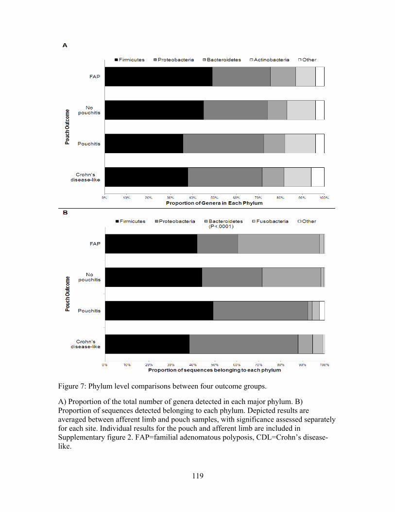
119
Figure 7: Phylum level comparisons between four outcome groups.
A) Proportion of the total number of genera detected in each major phylum. B) Proportion of sequences detected belonging to each phylum. Depicted results are averaged between afferent limb and pouch samples, with significance assessed separately for each site. Individual results for the pouch and afferent limb are included in Supplementary figure 2. FAP=familial adenomatous polyposis, CDL=Crohn’s disease-like.
120
Microbial composition of the ileal pouch at the genus level
In total, 287 genera were detected. Of these 83 were present in more than 5% of
samples. There was no statistical difference in genera positivity or abundance between
sampling locations (pouch vs. afferent limb). However, to avoid bias from including
multiple samples from the same individual, analysis was conducted separately on the
different biopsy sites.
Fisher’s exact test was used to conduct a four-way comparison evaluating whether
different proportions of individuals in each outcome group had a specific genus detected
at pouchoscopy. Within the pouch, 15 genera were associated with outcome at a nominal
significance threshold of P≤0.05, with five (Bacteroides, Parabacteroides, Moryella,
Sutterella and Blautia) remaining significant after FDR correction for multiple testing
(Pcorr≤0.05) (Figure 8A, Chapter 4, Supplementary Table 5). In the afferent limb, 16
genera were significantly associated with outcome, with seven (Blautia, Bacteroides,
Dorea, Anaerococcus, Sutterella, Parabacteroides, and Incertae Sedis
Erysipelotrichaceae) remaining significant after correction (Figure 8B; Chapter 4,
Supplementary Table 5). These genera were typically detected less frequently among
individuals with pouchitis and CDL compared to those with the non-inflammatory
outcomes.
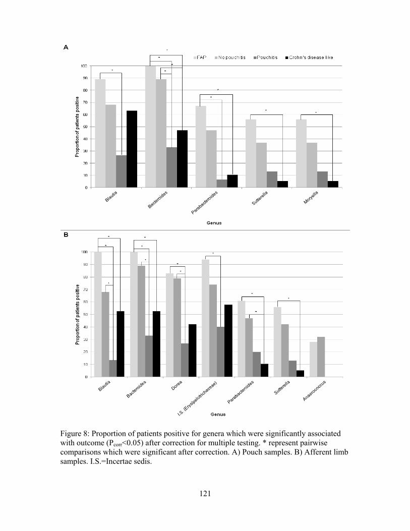
121
Figure 8: Proportion of patients positive for genera which were significantly associated with outcome (Pcorr<0.05) after correction for multiple testing. * represent pairwise comparisons which were significant after correction. A) Pouch samples. B) Afferent limb samples. I.S.=Incertae sedis.
122
As the CDL group was somewhat heterogeneous, containing individuals both with
and without active inflammation at the time of their pouchoscopy, we performed an
additional analysis, including only CDL individuals with inflammation at the time of
sampling (n=16). The results were similar to those in the previous analysis, although
Moryella in the pouch and Anaerococcus in the afferent limb did not remain significant
after multiple testing correction (Chapter 4, Supplementary Figure 3). When we
examined the effect of inflammation on bacterial composition we found that genera
associated with inflammation were similar to those described in our four group
comparison, with Bacteroides (P=2.5x10⁻⁵) and Parabacteroides (P=2.5x10⁻⁴) less
commonly detected among inflamed samples (Figure 9).

123
igure 9: Proportion of patients positive for genera in individuals with inflamed vs not-
corr
F
inflamed pouches. Depicted results are those associated with inflammation at a nominal
p-value threshold of P<0.01. * demonstrates associations which remained significant after
correction for multiple testing (P <0.05).

124
We next examined the relationship between quantitative genera abundance and
outcome. Results in both locations were similar to those observed for the dichotomized
results. In both sites, Bacteroides, Blautia, and Sutterella were positively associated with
outcome, as were, Parabacteroides and Moryella in only the pouch samples (Pcorr<0.05)
(Chapter 4, Supplementary Figure 4). In each case, genera were detected at lower levels
in the pouchitis and CDL groups compared to those with non-inflamed tissue.
Across all groups, use of one or more antibiotics at the time of the pouchoscopy
or during the preceding month was positively associated with the presence of
Lactobacillus in both the pouch and afferent limb (pouch P=4.6x10⁻⁴; afferent limb
P=8.9x10⁻⁴). Also associated with antibiotic use in the afferent limb were Dorea
(P=4.0x10⁻⁴) and Ralstonia (P=1.4x10⁻⁴), with Ralstonia detected more commonly
among individuals who were on antibiotics, and Dorea less common among individuals
on these medications. Several additional genera, including those which were associated
with outcome were shown to be marginally effected by antibiotic use at a nominal
significance threshold (P<0.05), however did not remain significant after correction for
multiple testing. Genera abundance data showed similar results, with Lactobacillus and
Haemophilus comprising an increased proportion of the microbiome among individuals
on antibiotics in both the pouch and afferent limb, and Ralstonia, detected at higher
frequency in the afferent limb. Dorea was detected at a lower frequency in the afferent
limb among individuals on antibiotics. No other medication use appeared to significantly
influence microbial composition, although few patients were available for this analysis.
53.5% of our population had taken antibiotics at some point in the past (pre- or
post-colectomy) (Table 14). To verify that our observed associations were between
inflammatory phenotype and specific genera, and to ensure that our observations were not
due to antibiotic use, we performed our analysis again evaluating only individuals who
were not on antibiotics during the month (n=57), or 6 months (n=55) preceding their
pouchoscopy. Results in the dichotomous analysis for both locations and time points
demonstrated that genera previously found to be associated with outcome after correction
were all associated in this smaller cohort (P<0.05) with the exception of Moryella in the

125
pouch and Parabacteroides in the afferent limb (Chapter4, Supplementary Figure 5). All
previously described organisms remained significant in the frequency analysis.
Exact logistic regression was performed with smoking, gender and Canadian vs.
other country of birth included as co-variates. In both anatomical locations, Bacteroides,
Sutterella and Blautia, remained significantly associated with outcome (P<0.01).
Moryella, and Parabacteroides were associated with outcome only in the pouch and
Dorea and Anaerococcus in the afferent limb (Table 15). Adjusted odds ratios
demonstrated a significant decrease in likelihood of pouch inflammatory complications
associated with the above genera. No significant results in this pair-wise analysis were
detected for the FAP vs. no pouchitis or pouchitis vs. CDL comparisons.

126
Table 15: Association between outcome and bacterial positivity in multivariate analysis with smoking, birth country and gender included as co-variates.
FAP vs. Pouchitis
FAP vs. CDL No Pouchitis vs. Pouchitis
No Pouchitis vs. CDL
P-value OR P-value OR P-value OR P-value OR
Pouch
Bacteroides .002* .06 .002* .04 .0005* .04 .03 .14
Parabacteroides .006* .05 .004* .05 .006* .04 .03 .13
Moryella .04 .08 .009* .05 .30 .29 .02 .07
Sutterella .01 .05 .001* .02 .21 .26 .06 .11
Blautia .009* .07 .31 .25 .02 .11 1.0 .89
Afferent Limb
Blautia .0001* .03 .02 .09 .005* .09 .50 .50
Bacteroides .0004* .03 .003* .05 .003* .08 .07 .18
Dorea .004* .06 .003* .05 .01 .12 .11 .26
I.S. (Erysipelotrichaceae)
.04 .10 .04 .08 .09 .19 .54 .50
Sutterella .06 .11 .006* .04 .14 .20 .02 .07
Anaerococcus .34 .28 .21 .22 .06 .13 .008* .06
Parabacteroides .13 .21 .02 .10 .14 .23 .03 .13
Described nominal p-values marked with a * are those which remain significant after correction for multiple testing (Pcorr<0.05). Genera tested are those which were significant in the preliminary analysis. No significant associations were seen for the FAP vs No Pouchitis or the Pouchitis vs CDL comparisons. FAP = familial adenomatous polyposis, CDL = Crohn’s disease like. OR = odds ratio I.S.= Incertae sedis.

127
Finally, analysis conducted using LEfSe confirmed many of our previous results,
and highlighted additional organisms with potential roles in the inflammatory phenotypes
(Chapter 4, Supplementary Figures 6). These included the class Bacilli (phylum
Firmicutes) which was detected at higher frequency among inflammatory groups, and
Proteobacteria detected more often among individuals with CDL (Chapter 4,
Supplementary Figures 6).
qPCR validation and targeted organism analysis
qPCR was performed on 86 samples from 43 individuals (FAP=12; No
Pouchitis=10; Pouchitis=11; CDL=8; quiescent=2). Bacteroides qPCR results correlated
with those obtained from pyrosequencing (Matthew’s correlation coefficient = 0.78;
Spearman correlation coefficient = 0.74). Furthermore, detection rates among the
outcome groups were similar with Bacteroides detected less frequently in the pouchitis
and CDL groups than in either FAP or no pouchitis groups (P<0.01). Additional
organisms associated with outcome included the Roseburia spp. / E. rectale group
(pouch) and Clostridium cluster IV (afferent limb). These organisms were present in
increased numbers in the FAP group compared to both pouchitis and CDL groups
(Chapter 4, Supplementary Figure 7).
Pyrosequencing results for treated quiescent, medication dependent samples
Seven patients not included in our analysis were classified as quiescent. These
individuals were on long term therapy to maintain remission, and displayed no features of
inflammation at the time of their study visit. Nominally significant differences (P<0.01)
were observed between the quiescent and FAP groups in several genera in both the pouch
(Bacteroides and Blautia) and the afferent limb (Blautia, Bacteroides, and Incertae Sedis
Erysipelotrichaceae) in both the dichotomized and frequency analyses (Chapter 4,
Supplementary Figure 8). In both the pouch and afferent limb, there were no significant
differences between the pouchitis or CDL groups and the quiescent individuals. Despite
the lack of evidence of inflammation, samples taken from individuals on long-term

128
therapy because of a diagnosis of chronic, medication dependent disease, seemed to have
a similar microbial profile among pouchitis-associated genera to the inflammatory
outcome groups.
4.5 Discussion
These data demonstrate decreased microbial diversity among individuals with
pouchitis compared to those with non-inflamed pouches, with broad changes at the
phylum and genus level observed. Similar reductions in diversity have also been
demonstrated in non-surgical bowel inflammation169, 350. Interestingly, both the non-
parametric Shannon and inverse Simpson indices demonstrated that all three pre-
colectomy UC groups had decreased diversity compared to the FAP group. Previous
histological evidence suggesting that subclinical inflammation may be present in
individuals with pre-colectomy UC, although not in those with FAP may offer an
explanation for this finding356. Thus, the observation that individuals classified into the
no pouchitis group had microbial characteristics intermediate between those seen in
individuals with pouchitis and FAP, may be related to subclinical inflammation present in
this group which was not captured by our reporting, but which was reflected in increased
CRP levels detected in this group. Also of note is the observation that the CDL group did
not demonstrate decreased diversity compared to individuals without pouch
inflammation. This may be related to the fact that several individuals with CDL were not
experiencing active pouch inflammation at sampling, and that the inflammatory activity
detected in this group at the time of pouch endoscopy was more heterogeneous than was
that observed in the pouchitis group. Indeed, this group had, on average, slightly lower
inflammatory scores than did the pouchitis group.
At the phylum level, significantly decreased levels of Bacteroidetes and marginal
increases in the Proteobacteria were associated with pouch outcome, among individuals
with inflamed pouches compared to those without inflammation. This confirms results
from other pouch studies, although results have been mixed in examining these organisms
in the context of non-surgical IBD296, 301, 350, 357, 358. However, our study demonstrates that
while Actinobacteria is detected in a majority of samples (72%), it is present at low

129
abundance (mean 0.8%). Fusobacteria, on the other hand, were detected in only 28% of
samples, yet among those individuals in whom these organisms were detected they were
dominant members of the bacterial community. This contrasts reports from others296, and
can most likely be attributed to the increased sequence coverage, and unique phenotypes
explored in our experiment.
Phylum level comparisons, while interesting, are of limited use in predicting
biological function. We subsequently demonstrated that specific genera are associated
with pouch outcome among our patient cohort. Among the Clostridia group XIVa,
Blautia, Moryella and Dorea were convincingly associated with outcome, detected more
frequently and in greater abundance among individuals without inflammatory
complications. Interestingly, a recent study of healthy subjects showed that Blautia was
one of the only genera detected in samples from all individuals, suggesting that it is a
common component of a healthy gut microbiota359. Furthermore, previous studies have
suggested that individuals with CD, including those in remission, have decreased levels
of Clostridia-related organisms detected360, 361. Both Bacteroides and Parabacteroides
were also strongly associated with outcome. These closely related genera have both been
previously implicated in pouch inflammation296, 301. Decreased levels of Bacteroides
have also been previously observed in some studies in inflamed UC and CD tissue
compared to healthy controls, while other studies have shown increases in this organism
to be associated with disease362-365. This further suggests the importance of this organism
in bowel inflammation, regardless of host or specific anatomic site. Furthermore,
Bacteroides species, specifically B. fragilis, have been shown to modulate immune
function, stimulating immune tolerance through Toll-like receptor (TLR)2259. Sutterella
was the only genus from the Proteobacteria phylum associated with outcome, and in
contrast with previous studies suggesting a potential pathogenic role for this organism in
IBD or no association at all366, was detected less frequently among inflammatory groups.
We noted an increase in the prevalence of Proteobacteria and the class Bacilli among the
inflamed groups, however this association was less dramatic. Further study investigating
the relevance of these groups of organisms is necessary to determine the significance of
this finding.

130
We specifically evaluated whether organisms previously associated with bowel
inflammation, including CD, were also involved in pouch inflammation to determine
whether similar biological processes might be of importance in both disorders. Contrary
to previous studies, we did not observe an association between F. prausnitzii or AIEC
and inflammatory outcome in our cohort. However, our results do support previous
observations that additional members of the Clostridium group358, and Roseburia spp./E.
rectale may be associated with pouch inflammation.
The use of subjects who had an IPAA for FAP, allowed us to evaluate changes
occurring in the pouch microbiome which were relevant specifically to individuals with
an inherited predisposition to intestinal inflammation. Individuals with FAP rarely
develop pouch inflammation, despite the fact that their digestive tract anatomy is
identical to those with UC. Additionally, subclinical inflammation may be present in
individuals with pre-colectomy UC, without a clinical diagnosis of pouchitis, as our
finding of elevated levels of CRP among this group may suggest. This subclinical
inflammation has important clinical and experimental implications, and is reflected in our
observations illustrating substantial changes between the microbiome of individuals with
inflammatory pouch complications compared to those with FAP. More moderate, yet
similar alterations occur in the microbiome of individuals with UC when comparing those
with and without inflammation. This likely reflects underlying differences in host genetic
predisposition in UC patients resulting in specific alterations in the pouch microbiome
that characterize the inflammatory phenotypes.
While there were no statistical differences in medication use between outcome
groups at the time of or immediately prior to pouchoscopy, it is conceivable that
medications, particularly antibiotics, may have impacted the tissue associated microbiota.
Yet the described genera were detected significantly less frequently among individuals in
the inflammatory outcome groups even when individuals who had taken antibiotics in the
preceding month or six months were excluded from the analysis. This would suggest that
the effect of inflammation on the microbiome supersedes the impact of antibiotic use.
Alternatively, if shifts in the microbiome of the pouch precede and possibly induce pouch
inflammation, then the use of antibiotics may not result in long term beneficial effects on
bacterial composition or lead to a reversion to a “healthy” pouch microbiome as would be
131
suspected by the typically antibiotic responsive nature of pouchitis. This may in turn be
responsible for the often recurrent nature of pouchitis following treatment. It is also
possible that past antibiotic use (beyond one month prior to procedure) leads to
permanent changes to the microbiome which are detectable long after cessation of
medications. Studies evaluating the long-term effect of antibiotics on intestinal microbial
composition have shown mixed results with some studies showing a relatively quick
reversion to a profile similar to the pre-medication state367, while others suggest long-
term changes, especially with antibiotic use in childhood 241, 368. Interestingly, antibiotic
use in childhood has been associated with an increased risk of CD, suggesting that
changes to the microbiota which occur early in life, whether they persist long-term or not,
may have a drastic impact on subsequent disease susceptibility11. However, as the levels
of these genera had recovered to detectable levels in FAP and individuals without
pouchitis, despite historic antibiotic use (data not shown), yet remained undetectable in
the most pouchitis and CDL individuals, we conclude that this decrease was associated
with the phenotype rather than medication use.
Interestingly, we did not observe differences in the microbial composition of the
pouch compared to the afferent limb among patients regardless of the inflammatory status
of either site. The anatomical alterations resulting from the IPAA procedure, namely the
surgically induced continuity between the pouch and afferent limb, likely promotes
similar bacterial community structure between these two locations. Previous studies have
documented the occurrence of variability in the microbiome between inflamed and non-
inflamed neighbouring sites, although none were consistent across many individuals357.
This suggests that individual-specific differences in the microbiome may be important in
pathology. Further, the absence of evidence for differences between inflamed and
uninflamed locations in the same individual suggests that variable host genetics or
location-specific gene expression patterns may be of importance in establishing disease
location and phenotype. It is also possible that microbial variation results from the
inflammatory process itself. In this case, loss of specific organisms may contribute
towards propagating disease but not initiate disease processes. Prospective studies are
required to evaluate this hypothesis.
132
Of particular interest, is the observation that the seven individuals who were
taking medication for chronically inflamed pouch phenotypes, and who had successfully
achieved resolution of their symptoms, did not demonstrate a change in their microbial
composition to resemble that of individuals with healthy pouches. While it appears that
the samples from individuals with quiescent disease were most similar to those from
individuals with pouchitis, it is conceivable that with a larger sample size differences
between these groups may have been observed. Additionally, changes at lower taxonomic
levels (ie. species), which were undetectable in this experiment could also have been
associated with different phenotypes. It is interesting to speculate, however, that perhaps
the reason for patients’ continual requirement for medical therapy is their inability to be
colonized by the ‘beneficial organisms’ described by our group.
The majority of associations described in this report suggest that a loss of
organism diversity is associated with inflammation. It is possible that individuals with
pouch inflammation may undergo a loss of specific anti-inflammatory organisms that
have important roles in modulating the immune response. Many of the organisms
detected less frequently in the inflammatory groups have been previously shown to have
anti-inflammatory potential: members of the Clostridia XIVa group (Blautia, Dorea and
Moryella) produce butyrate, a known anti-inflammatory molecule369, 370, and Bacteroides
produce vitamin K, which is associated with decreased levels of serum inflammatory
markers (ie TNFa, CD40 etc)371. Alternatively, the absence of these typically ubiquitous
organisms may allow increased colonization of tissue by atypical species. While we did
not observe increased prevalence of specific genera in our cohort, it is conceivable that
small changes among several groups of organisms may not have been detected. In any
event, endogenous gut organisms have the capacity to exert profound effects on the
function of the immune system, and play a pivotal role in maintaining gut immune
homeostasis259, 264, 372. Such changes in immune function may also be reflected by the
presence of anti-microbial antibodies in the serum which are also known to be associated
with pouch outcomes67, 329. In the future, functional metabolites from these organisms
may represent novel diagnostic targets or have therapeutic potential for patients with
active disease.
133
Our experiments have demonstrated that a reduction in specific organisms is associated
with inflammatory outcomes. These changes are characteristic of both pouchitis and the
Crohn’s disease-like phenotype, and include several organisms which have been
previously associated with non-surgical IBD. These observations demonstrate that the
role of these microbes in bowel inflammation may be common and independent of
surgical history or anatomic location of disease. Our previous findings showing that IBD-
associated genetic polymorphisms are also important in pouch pathology351, in
conjunction with this work suggests that like non-surgical IBD, pouch inflammation
arises in individuals with inherited genetic predisposition and changes in the tissue
associated microbiome. However, the event triggering disease onset remains elusive, and
will require large, prospective studies to evaluate.

134
4.6 Chapter 4, Supplementary Methods
Microbial DNA Extraction and Analysis
Sequences were parsed based on barcodes using a customized Perl script. Reads with an
average quality score below 35 were removed prior to sequence processing. Data was
then quality trimmed and aligned to a SILVA reference database (version SSUref_102)
using mothur (version 1.12.3)373. This resulted in removal of sequences which were less
than 250 base pairs long (excluding primers), and/or which contained ambiguous bases or
homopolymers extending eight or more nucleotides. The ChimeraSlayer algorithm215 was
used to remove potentially chimeric sequences from the dataset. Additionally, a pre-
cluster step was performed, as has been previously recommended by Huse et al., allowing
detection and removal of rare spurious sequences214. Individual sequences were then
assigned to taxonomic outcome groups, at the genus level where possible, using the Silva
taxonomy with mothur374. A Bayesian classification approach with 100 iterations, and an
applied bootstrap cutoff of 60 was used to assign sequences to a taxonomic outcome
groups.
Statistical analysis
Taxonomic groups which were detected in less than 5% of the samples were excluded
from further analysis. Analyses were performed separately on pouch and afferent limb
samples in order to avoid falsely inflating significance. Results were first dichotomized
based on whether or not a given genera was detected within a sample. Fisher’s exact test
was applied to determine whether there were statistically significant differences between
groups. Results at the genus level were confirmed using exact logistic regression with
smoking, country of birth and gender included as covariets to further validate results.
Continuous abundance was calculated by normalizing the sequences for each taxon
against the total number of sequences detected in a sample375. Genera abundance was
assessed for significance using the non-parametric Kruskal-Wallis and Wilcoxon signed
rank test. FDR corrected p-values below .05 were considered significant in both the
dichotomized and continuous abundance analyses. All statistical analyses were conducted

135
using STATA version 11.1 (StataCorp Texas, USA) and R version 2.13.1 (R Foundation
for Statistical Computing, Vienna, Austria). Additional analyses were carried out using
the Linear Discriminant Analysis (LDA) Effect Size (LEfSe)354 tool (version 1.1.0) to get
a more robust understanding of differences in community structure across phenotypic
outcome groups (Significance threshold P<.05).
Quantitative PCR (qPCR)
Organisms previously implicated in inflammation including F. prausnitzii, Clostridial
cluster IV, AIEC, and Roseburia were evaluated using qPCR with previously described
primer pairs (Supplementary Table 2). Relative abundance was estimated as has been
previously described355, with Eubacterial primers used to construct a standard curve.
Reactions were carried out using the Power SYBR green PCR master mix (Applied
Biosystems, Foster City, CA) with the AB 7300 system and sequence detection software
(version 1.3; Applied Biosystems, Foster City, CA). Results from individual samples
were log2 transformed and analyzed using Kruskal-Wallis test. Additionally, primers
specific for the Bacteroides genus were used to confirm pyrosequencing results by
evaluating the correlation between dichotomized (Matthew’s) and frequency (Spearman)
pyrosequencing and qPCR data. Dichotomous results were generated based on a
pyrosequencing detection limit of 10-4 sequences per sample, estimated from the
theoretical number of sequences possible per sample in the multiplexed pyrosequencing
reaction.

136
5.1 Conclusions and general discussion
This project involved accrual of the largest IBD-IPAA patient population yet
assembled, and studied a variety of important factors with the potential to contribute
towards, or arise in response to, ileal inflammation. As such, well powered experiments
allowed these analyses to be conducted with a high degree of statistical stringency. In
each aspect of this study, steps were taken to ensure that the effects of potentially
confounding factors were minimized, including restricting the sample size to include only
Caucasians in the genetic analysis, and controlling for antibiotic use, country of birth,
gender and smoking status when studying the microbiome. Furthermore, the strict patient
phenotyping and classification guidelines, allowed us to ensure that observed associations
were with the target outcomes of interest. As such, the findings presented in this thesis, of
alterations in the SNP frequency of the NOD2insC polymorphism, the frequency and
abundance of organisms in the pouch microbiome and seropositivity of antimicrobial
antibodies, suggests a model of how inflammatory conditions of the ileum may arise.
The size of the patient cohort, as well as the strict phenotyping criteria used in
these studies, ensured that patients with acute, antibiotic responsive pouchitis as well as
chronic pouchitis and a Crohn’s disease-like phenotype, were included in analyses. The
subclassification of patients into either the antibiotic responsive or refractory outcome
groups allowed determination of factors which were associated with disease chronicity,
and which with a more transient disease course. Observations of genetic and serological
profiles among individuals with chronic inflammatory phenotypes which differed from
those seen in individuals with acute pouchitis (which were included in the no pouchitis
group, although subgroup analyses were performed), suggest that these inflammatory
outcomes proceed via different mechanisms. However, findings of similar alterations to
the microbiota among individuals with active pouchitis (chronic and acute) and inactive,
but medication dependent chronic pouchitis and CDL, suggest that similar bacterial
alterations are involved in both pathologies. Additional exploration of this phenomenon
in another large cohort will be useful in confirming these findings.
Decreased microbial diversity among individuals with pouchitis compared to
those with non-inflamed pouches, with broad changes at the phylum and genus level were
137
observed. One of the major findings was that Bacteroides were detected significantly less
frequently in inflamed tissue compared to that which is uninflamed. Previous reports have
demonstrated that certain species of Bacteroides, specifically B. fragilis, modulate
immune function, and stimulate immune tolerance through TLR2259. This, combined with
observations by other groups that different members of this genus are able to decrease
circulating cytokine levels259, strongly suggests that Bacteroides spp. play an important
role in downregulating the immune response. Interestingly, B. fragilis, which has been
shown to have important immuno-regulatory effects via the production of PSA376, binds
to mucins in the mucus layer, rather than exclusively within the intestinal lumen where
organisms typically reside377. This intimate association between host and microbe,
demonstrates the ability of this organism to adapt to a specific host niche which is
resistant to colonization by other organisms. Other species within this genus, and the
closely related Parabacteroides, also produce PSA and related compounds, although they
have not been studied to the same degree as has this organism378.
The other genera which were associated with inflammatory outcomes have not
been as well studied. Blautia and Moryella, newly described members of the Clostridia
Group XIVa379, 380, were detected less frequently among individuals with inflammation.
Blautia is detected often in microbiome studies of the lower digestive tract of healthy
individuals359, but its precise function has not been elucidated. Both Blautia and Moryella
are members of the larger Clostridia group, however, and are therefore hypothetical
producers of butyrate, a molecule with anti-inflammatory potential369. The role of
Sutterella in modulating or propagating inflammation is more difficult to determine, as
some studies suggest that members of this group promote inflammation, while others
have shown just the opposite effect381 366. Additional work will be required in order to
determine the exact role of this group in IBD.
qPCR was also used to more closely evaluate whether particular Clostridial
groups and strains of organisms, which have been previously associated with bowel
inflammation, were also involved in pouch inflammation. Members of the Clostridium
cluster IV and XIV groups may be protective against the development of IBD, and play a
role in maintaining mucosal immune homeostasis187. Both F. prausnitzii (Clostridium
cluster IV) and the Eubacterium rectale/Roseburia (Clostridium cluster XIV) groups

138
produce butyrate, via varied pathways382. This short-chain fatty acid is thought to
contribute towards decreasing levels of IL-12 and IFN-gamma production and leads to
higher secretion of IL10250. Additionally, organisms from the Clostridium cluster IV
group have the capability of increasing the abundance of Treg cells in the colon, but not in
small bowel tissue, via activation of intestinal epithelial cells262. Mice colonized by these
organisms, and displaying a resultant increase in Treg cells throughout the colon, are
resistant to the development of experimental colitis262. Contrary to previous studies245, 343,
no statistically significant association between F. prausnitzii and inflammatory outcome
was observed in this cohort. Yet there did appear to be a moderate reduction in the
frequency of this organism among inflamed samples. qPCR experiments did, however,
demonstrate a reduced prevalence of the Eubacterium rectale/Roseburia group among the
inflammatory groups compared to uninflamed samples. This further emphasizes the
critical role of bacterially-produced immune-modulating compounds in pouch
inflammation.
AIEC has also been previously associated with IBD, detected more frequently in
ileal tissue of individuals with CD347. AIEC strains have the ability to adhere to and to
invade intestinal epithelial cells via FimH, are capable of surviving and replicating in
macrophages without inducing cell death, and induce increased production of the pro-
inflammatory cytokine TNF-α383. While there was no statistically significant association
between this organism and outcome in our cohort, increased abundance of AIEC among
individuals with the CDL phenotype was observed (not statistically significant),
mirroring results seen in previous IBD studies347. However, the high relative frequency of
this organism in individuals with FAP, compared to UC patients without pouchitis, was a
novel and surprising finding. Previous studies have observed higher levels of certain
mucosa-adherent strains of E. coli among individuals with colorectal cancer384,
suggesting that this organism may have a pathogenic role in bowel cancer development.
This might also explain the higher prevalence of this organism in individuals with FAP,
despite the lack of inflammation in this group. Additional evaluation in a larger cohort is
required to confirm this finding.
Together, these results suggest that a reduction in bacterially produced immune-
modulating compounds is important in IBD pathogenesis. These compounds likely play a

139
beneficial role in helping to maintain immune homeostasis among individuals without
bowel disease, and may have therapeutic potential among individuals with already
established inflammation. The observed associations between decreased organism
diversity, and reductions in the prevalence of specific organisms and disease status, may
be the result of two possible mechanisms: disease development may be initiated in
response to a reduction in beneficial organisms, which is caused by an unknown factor.
Alternatively, it is possible that microbial variation results from the inflammatory process
itself. In this case, the increased prevalence of anti-microbial antibodies among
individuals with inflammation in conjunction with observations of alterations in the
microbiome may suggest that an aberrant host response to intestinal microbes contributes
to a reduction in organism diversity, including loss of species with beneficial effects on
the host. In this case, genetic or immune processes may drive the development of
inflammation, and also contribute towards altering the composition of the gut microbiota.
The loss of specific organisms may then contribute towards propagating disease but
would not, themselves, initiate disease processes. In either scenario, the observation of
specific reductions in microorganisms with immune-modulating properties, and the lack
of strong evidence for an association with organisms which are more likely to be
pathogenic, highlights the importance of 'good' bacteria in regulating gut immunity and
homeostasis. However, as many of the changes to the microbiome which have been
observed involve reduced detection of organisms which are capable of producing anti-
inflammatory compounds, it is difficult to determine which comes first: decreased
immune tolerance towards commensals, or reduction in organisms with anti-
inflammatory properties. These observations do, however, emphasize the importance of
cross-talk between commensal organisms occupying the pelvic pouch and the immune
system in maintaining immune homeostasis.
The observed differences in the prevalence of several organisms (Blautia most
notably, but also observed in Bacteroides and Dorea (afferent limb)) between the active
pouchitis and the CD-like group may be a result of altered inflammatory activity, or may
suggest that divergent mechanisms of disease onset and propagation occur in each of
these phenotypic outcome groups. The CDL group was more heterogeneous than was the
pouchitis group, made up of individuals with inflammatory scores ranging from zero to
greater than ten. Further, there was a slight, but not significant, reduction in the mean

140
inflammatory score in the CD-like group compared to the pouchitis group. When only the
individuals with inflammation at the time of their pouchoscopy were included in the CD-
like group, the prevalence of all organisms but Blautia was similar in the active pouchitis
and CDL groups. Furthermore, no significant difference in organism prevalence between
these groups was observed in the pairwise analysis. It is possible that removal of these
individuals resulted in reduced power, and an inability to detect significant associations
with outcome. In this case, the increased prevalence of these organisms in individuals
with CD-like phenotypes (intermediate levels between individuals with no pouchitis and
pouchitis), may be indicative of important changes in the microbiome contributing to, or
resulting from different disease mechanisms.
These results also highlight the need for more robust mechanisms for measuring
inflammatory activity at intestinal sites. While several organisms were associated with
our four phenotypic outcome groups, fewer associations were seen when we compared
individuals with inflammation to those without. CRP levels among the three pre-surgery
UC groups were also similarly elevated compared to individuals without UC, suggesting
ongoing inflammatory activity was present despite the absence of endoscopic or
histological evidence. Further, this activity appeared to be capable of influencing the
composition of the pouch microbiome. Such observations suggest that a clinical diagnosis
of inflammation may be non-specific, when compared to molecular indicators of disease
activity. The relevance of such low-level inflammation in the context of patient outcomes
is unclear.
Additionally complicating the study of the human microbiome is the observation
that bacterial populations vary over time, with changes in bacterial communities resulting
from alterations in diet, smoking, country of residence, or medication use230, 231, 234, 235, 246.
Yet alterations in the microbiome alone appear insufficient to cause chronic
inflammation, as demonstrated by the observation that individuals with antibiotic
responsive pouchitis have microbial profiles which resemble those seen in individuals
with chronic inflammation. The pouch inflammation in patients with antibiotic responsive
pouchitis is uniquely responsive to these medications, and can be rapidly resolved.
However, among individuals with a genetic predisposition for IBD, it is possible that
normal or abnormal variation in the microbiome tips the balance of immunity, and lead to
141
disease. When this happens, increased immune recognition of commensal organisms
leads to the generation of anti-microbial antibodies, and contributes towards propagating
inflammation. Enhanced immune response likely causes further reductions in the
numbers of beneficial organisms present in the mucosa. As such, the specific
combination of altered host genotype and intestinal bacterial dysbiosis may be sufficient
to result in chronic inflammation.
Several genes are associated with increased susceptibility towards CD, IBD (both
CD and UC) and UC alone104. As a predominantly UC patient cohort, therefore, IPAA
patients are more likely to have higher rates of UC and IBD-associated polymorphisms
than are individuals without a previous diagnosis of IBD (ie. individuals with FAP prior
to colectomy). This observation likely explains the high prevalence of pouch
inflammatory complications among those who undergo IPAA for UC compared to FAP.
Several of the UC-associated polymorphisms tag genes which are involved in intestinal
barrier function such as HNF4A and E-cadherin (CDH1)385. Additionally, several CD-
associated genetic polymorphisms are also associated with chronic pouch inflammation,
confirming that additional host genetic factors are important determinants of phenotypic
outcome following surgery, and may suggest mechanistic differences between acute
(antibiotic responsive) and chronic pouch phenotypes. The well established NOD2insC
polymorphism results in a truncation of the leucine rich repeat region of the NOD2
protein which has demonstrated evidence of an inhibited ability to detect microbes336, 337.
Other genetic variants which demonstrated modest association with chronic inflammation
of the pouch include those which are located in the non-coding regions of NOX3 and
DAGLB or adjacent to NCF4. Individuals who are carriers of additional risk alleles at
these loci have significantly decreased production of reactive oxygen species (which are
important for defense from microbial pathogens) compared to patients not carrying these
mutations342 343, 344. NOD2 and NCF4 polymorphisms have also been demonstrated to
associate strongly with ileal CD106, 107, 341, 386. These findings, in conjunction with results
showing that microbes are also important in pathogenesis, suggest an important role for
innate immune activity in response to commensal microbes in the development of chronic
pouch inflammatory outcomes. It is likely that additional polymorphisms are also
associated with pouch complications; however, these may only be detectable in larger
sample sizes. Many of the SNPs associated with IBD have extremely low effect sizes,
142
requiring the use of extremely large cohorts (sample size greater than 10,000) to
measure104. However, these results definitively establish that host genotype, specifically
loci previously associated with ileal inflammation, is critical to the pathogenesis of
chronic pouch inflammatory complications.
The changes in immune function which are brought on by a reduced number of
anti-inflammatory organisms, and increased host susceptibility due to decreased innate
immune activity, is also reflected by the presence of anti-microbial antibodies in the
serum of individuals with chronic pouch inflammatory phenotypes. This hypothesis is
supported by data showing that UC patients with a CD-like phenotype after pelvic pouch
surgery are more likely to be positive for ASCA and anti-CBir1, and by that of other
groups’ demonstrating a strong association between the common UC associated serum
marker, pANCA, and pouch outcome67, 68. The presence of these anti-microbial
antibodies may be representative of a broader breakdown in mucosal function. While
individuals with chronic pouch inflammatory phenotypes were commonly positive for
markers typically associated with CD (ASCA and anti-CBir1), no association was seen
among individuals with acute, antibiotic responsive pouchitis. The genotype of these
individuals (acute pouchitis) was also similar to that seen in individuals who had never
experienced inflammatory pouch complications. This suggests that microbial alterations
brought on by the pouch surgery alone are not sufficient to promote the development of
anti-microbial antibodies. Instead, as with IBD, anti-microbial antibody positivity results
from a complex interaction between genetically-mediated alterations to the gut barrier or
innate immune functions of the mucosa, and the tissue-associated microbiome.
These results suggest that patients with NOD2insC (and possibly with variants in
the additional genes which show modest associations with outcome) are more likely to
develop chronic inflammatory pouch complications in response to alteration in the
microbial composition of the pouch mucosa. Decreased prevalence of organisms with
immune-modulating functions such as Clostridium group IV, XIV, and Bacteroides-like
organisms, resulting from either random variation or via host-mediated alterations in diet
or gene expression (or numerous other types of environmental instability), among
patients who are also carriers of these genetic susceptibility loci, results in an inability to
downregulate the inflammatory response directed towards commensal organisms at the

143
intestinal epithelium. The increased adaptive immune activity resulting from this
perturbation and imbalance leads to the production of anti-microbial antibodies, which
can be detected in the serum of patients with IBD (Figure 10). Since NOD2
polymorphisms are associated with increases in serum concentration of ASCA, anti-
OmpC and anti-CBir1, it is likely that alterations in this gene are a critical mediator in gut
barrier permeability148. Furthermore, these findings of the presence of both serologic
markers of CD and genetic polymorphisms, which have been previously associated with
ileal CD, showing association with pouch outcome, suggests that the mechanism of ileal
inflammation leading to chronic pouch inflammation is similar to that which leads to the
development of inflammation in the ileum.
Anti-microbial antibody
production
Changes in gut-associated microbiota
↓Bacteroides↓ClostridiaGroup XIVa↓anti-inflammatorycompounds
Host genetic predisposition
NOD2insCNOX3NCF4RAC1?
IlealInflammation
Figure 10: Hypothetical model of the pathogenesis of ileal inflammation

144
Additional proposed mechanisms of pouchitis development including ischemia,
mechanical failure, NSAID use and deposition of collagen on the pouch mucosa22 may
effect a subset of patients with pouch complications. However, this may be ruled out in
most cases based on the length of time between pouch surgery and inflammation
development, and the absence of medication use prior to symptom onset.
The majority of individuals in whom pouchitis develops, respond rapidly to the
administration of broad spectrum antibiotics. Yet the diagnosis of these individuals with
UC prior to surgery suggests that they are genetically predisposed towards the
development of chronic, colonic inflammation. Furthermore, the bacterial composition of
the pouch brought on by inflammation, is the same as that which is observed in
individuals with chronic pouch inflammation. Despite these similarities, they do not
develop a chronic inflammatory phenotype following surgery. It is possible that given
additional time, acute, medication-responsive pouchitis may evolve into a chronic
inflammatory phenotype. However, the long follow-up time between pouch surgery and
phenotyping in this cohort suggests that this is unlikely to be the case. Alternatively,
differences in the gene expression of key mediators of inflammation, between colonic and
ileal tissue could be responsible for the variable phenotypes. Such differences would
contribute to chronic disease in large bowel tissue, and acute disease in ileal pouches. It
may also be possible that specific differences in the mucosa-associated microbiome
between colonic inflammation and ileal pouch inflammation lead to differences in
phenotype and outcome. However, the most likely explanation for this phenotypic
difference is that the absence of key polymorphisms among the acute pouchitis group,
which are important mediators of ileal inflammation (ie. NOD2insC, NCF4) prevents the
development of chronic inflammation in the ileal pouch among individuals with antibiotic
responsive pouchitis. These individuals may, therefore, experience inflammation of ileal
tissue, but do not experience long-term inflammatory complications when exposed to the
same microbial stimuli.
Observations of recurrence of chronic inflammation among individuals who are
clinically diagnosed with UC prior to colectomy, suggests to some that among these
patients a missed diagnosis of CD is the reason for inflammation recurrence.
Alternatively, patients with chronic inflammation and CD-like outcomes after pelvic
145
pouch surgery for UC may not be a “missed” diagnosis of CD, but rather evolution of the
IBD-phenotype to one that more closely resembles CD rather than UC. These results in
conjunction with data published by Waterman et al. which showed that there are few
differences in the prevalence of several IBD-associated SNPs between those with UC and
those with CD317, support the concept of IBD as a mosaic of inflammatory disorders. The
varying phenotypes associated with IBD arise via a specific combination of genetic
(including alterations in gene expression, and epigenetic regulation) and microbial
predisposing factors. Antibody profiles in serum reflect the underlying pathogenic forces
contributing to disease development, rather than specificity for the clinical subtype into
which patients are classified. Furthermore, the genetic susceptibility overlap between
individuals with chronic pouchitis and those with the CD-like phenotype suggests that
distinguishing between these outcomes is of limited importance compared to the more
clinically relevant phenotype of chronic, medication refractory inflammation
characteristic of both disorders and potentially arising from a common etiology. This
concept of IBD has interesting implications for disease management, both prior to and
following surgical intervention. Therefore, future disease prognostication and medical
treatments must take into account clinical phenotype as only one aspect of disease
management, with additional pathway-specific factors also taken into account in
determining which patients are candidates for a specific procedure or medication.
The findings of this study also shed light on different avenues for investigation in
the development of novel molecular tests and more targeted therapies for managing IBD.
Molecules which stimulate the production of innate immune compounds or which
enhance the function of the epithelial barrier may prevent commensal organisms from
stimulating a severe and damaging adaptive immune response. Development and
application of more effective probiotics, such as members of the genera identified by our
group, might also have a beneficial effect on modulating disease course. Among patients
in whom surgery is a consideration, knowledge of the specific genotype and antimicrobial
antibody profile will help stratify patients based on their risk of post-surgical chronic
inflammatory outcomes. This approach will ensure that patients who are more likely to
develop complications are discouraged from undergoing IPAA, or are closely monitored
following surgery to ensure that they are rapidly diagnosed and treated in the event that
they develop post-surgical complications. Individuals in whom pouchitis does develop,
146
who have a profile suggestive of chronic inflammatory phenotypes, may be candidates
for more rapid escalation of therapy. Furthermore, identification of individuals who are
more susceptible to the development of either pouchitis or IBD, based on the diagnosis
with IBD of a family member, and the detection of risk polymorphisms or anti-microbial
serological markers, may be candidates for preventive therapy before disease onset. Such
individuals may be good candidates for probiotic or prebiotic therapies to enhance the
numbers of beneficial bacteria present in healthy pouch tissue. By ensuring that these
organisms are present in individuals at increased risk of disease, the effect of their
immunomodulating compounds may prevent other, more pro-inflammatory organisms
from becoming dominant members of the microbiome, and therefore enhance immune
homeostasis.
This hypothesis has broader applications as well, given that numerous other
diseases including PSC, arthritis, and diabetes have shown similar genetic and
antimicrobial antibody profiles to those observed in IBD. These conditions are also
characterized by intestinal bacterial dysbiosis. Furthermore, additional autoimmune
conditions have shown evidence of subclinical inflammation in the intestinal tract prior to
diagnosis, as well as increased intestinal permeability. In a rat model of type I diabetes,
for example, jejunal enteropathy and increased numbers of intraepithelial lymphocytes
were detected among animals predisposed to the development of diabetes prior to
symptom onset387. Further, even prior to diagnosis, specific pathogen treated mice may
have the natural history of their disease course altered via the introduction of specific
microorganisms, with several bacterial groups providing protection against the
development of diabetes388. Such findings demonstrate the importance of gaining a better
understanding of the complex relationship between gut immunity and the tissue
associated microbiome of the digestive tract in order to better understand processes
contributing to the development of immune-mediated conditions.
These results further demonstrate the utility of the pelvic pouch model for
studying de novo IBD and suggest that it may have important implications for
understanding disease processes in both IBD and other immune-mediated conditions.
Findings of similarities in genetic susceptibility towards chronic pouch inflammatory
outcomes and CD and altered populations of microbes in the pouches of individuals with

147
inflammation, which suggest that chronic inflammation arises from similar mechanisms,
provide a model for evaluating the instigating processes in de novo disease development.
Thus, the application of well-designed prospective studies which capture different time
points following ileostomy closure, will likely shed further light on initiating factors in de
novo ileal disease onset.
5.2 Future directions
The work described in this thesis highlights important factors in the pathogenesis
of inflammatory pouch complications. The similar genetic predisposition observed in
pouch complications and IBD (notably ileal CD) highlighted by these studies, and
specifically the role of innate and adaptive immune processes in modulating disease risk,
have been suggested. Additional host genes and pathways of importance which may be
specific to pouch inflammation or previously undiscovered IBD-associated
polymorphisms, may be identified using GWA studies. These studies will be conducted
in this cohort combined with cohorts from other centres in the future.
The cross-sectional nature of these studies makes it difficult to determine
causality, and the utility of the identified serologicial and microbial markers for
prognostication remains unknown. While the predictive value of SNP markers is high
considering that host genetics are stable and do not change over time in response to
external stimuli, the same cannot be said regarding either anti-microbial antibody
positivity or intestinal microbiome composition. Some studies have shown serological
markers to be stable longitudinally, yet to date no well-powered study has shown that pre-
surgical anti-microbial antibody profiles are similar following surgery. Additionally,
while the microbial profiles identified are definitively associated with pouch
inflammation, their role in disease initiation is unclear. As many studies have pointed out,
it is possible that the described changes are the result of inflammatory processes, and not
contributory towards disease onset or pathogenesis. However, although observed changes
may be caused by the inflammatory status of the tissue, the alterations to the microbiome
which are characteristic of inflammation may have a role in propagating inflammatory
processes and immune dysregulation. Future analyses evaluating both serology and the

148
microbiome prospectively and involving serial sampling (from multiple sample sites in
the case of the intestinal microbiota) will be useful for determining which factors are
important in the development of inflammation and which may be suitable for disease
diagnostics. Prospective microbiome studies will therefore be useful in determining
which organisms may be responsible for disease onset, and how those organisms
contribute towards propagating inflammation.
While this study has documented alterations in the microbiome of pouches with
inflammation, the functional effect of the loss of specific organisms is unknown. In
future, metabolomic analysis should be conducted to evaluate whether reductions in the
organisms we have described results in a cumulative reduction in the quantity of butyrate,
or other short chain fatty acids available to the host. Further, to determine which
additional microbial functional pathways are important in pouch health, it will be
important to perform a metagenomic analysis in pouch samples. This will allow us to
confirm our hypothesis that dysregulation of the inflammatory response is propagated via
a reduction in the production of immune modulating compounds. This approach may also
help to highlight additional microbial pathways and metabolites with pathogenic or
therapeutic potential.
It would also be useful for patients to have a measure for objectively evaluating
risk of disease recurrence following surgery. This score would likely encompass genetic,
demographic (smoking history) and phenotypic (presence of extra-intestinal
manifestations of disease, medication use) characteristics, pre-surgery serological results,
and incorporate early changes to the pouch microbiota prior to, and immediately
following ileostomy closure. Since it is unclear whether the ileal or colonic microbiome,
or both, contribute to the colonization of the pouch, the utility in measuring the
microbiota of these sites prior to surgery for inclusion as part of a risk score is unknown.
However, should evidence of predictive capabilities arise, they could be of great benefit.
The field of microbiome analysis is so new, that many different analyses and
studies are necessary to get a more comprehensive understanding of the changes which
are responsible for disease initiation, propagation and recurrence. Immediate evaluation
of the differences between anatomical sites, and between pouches and non-surgical IBD
may be useful for determining why pouchitis is responsive to antibiotics in more than

149
50% of cases, whereas non-surgical IBD is not. Another challenge going forward will be
to determine which genetic and microbial factors together contribute to establishing a
particular serological profile, and which host factors, be they genetic, dietary, or lifestyle,
are important in shaping the composition of the host microbiome. A greater evaluation of
these relationships will be critical in obtaining a more comprehensive understanding of
the relationship between colonizer and host and how they may impact immune function.
The relationship between the organisms inhabiting the digestive tract, and the host
are complex, and are only beginning to be understood. This work shows clear evidence
for an etiological role of genetic and microbial factors in pouch inflammatory
complications. While the mechanisms which are responsible for disease initiation and
propagation remain a mystery, this work has identified similarities and differences
between pouch inflammation and non-surgical IBD, with potential implications for a
more broad understanding of IBD in general. Future evaluation of the pathways and
organisms identified by this work may lead to the generation of novel therapeutics or
diagnostic strategies which are capable of enhancing patient care.

150
Appendix
Chapter 3, Supplementary Table 1: List of SNPs included in this analysis
CHR SNP Included in Replication CHR SNP
Included in Replication
1 rs1044879 1 rs3845461 1 rs10489628 Yes 1 rs3845466 1 rs1061622 1 rs396991 1 rs1061624 1 rs4266924 1 rs10789230 1 rs4353135 1 rs10797887 1 rs447536 1 rs10889676 1 rs448740 1 rs10889677 1 rs4645983 1 rs10911364 1 rs526201 1 rs10911365 1 rs6426833 1 rs11209003 1 rs6669582 1 rs11209008 1 rs6672995 1 rs11265519 1 rs7517847 1 rs11465802 1 rs763110 1 rs11465804 1 rs789180 1 rs11578964 Yes 1 rs789181 1 rs11584383 1 rs789185 1 rs11588654 1 rs789191 1 rs11811630 1 rs9286879 Yes 1 rs12035082 1 rs9988642 1 rs12094228 2 rs10210302 1 rs12567232 2 rs1048610 1 rs12753665 2 rs10495565 1 rs13294 Yes 2 rs1056204 1 rs1336480 2 rs10929587 1 rs17129659 2 rs10929590 1 rs17419032 2 rs1143634 1 rs1800896 2 rs11684747 1 rs1801274 Yes 2 rs12469362 1 rs2064689 2 rs12471449 1 rs2274910 2 rs13003464 1 rs2297909 2 rs13412102 1 rs2343331 2 rs1441090 1 rs2476601 2 rs16944 1 rs2490271 Yes 2 rs1839669 1 rs2819130 2 rs1880439 1 rs2902440 2 rs2001658 2 rs2241879 4 rs4833103 Yes 2 rs2241880 4 rs9999226 2 rs2276338 5 rs1002922 2 rs2289472 5 rs10045431 Yes
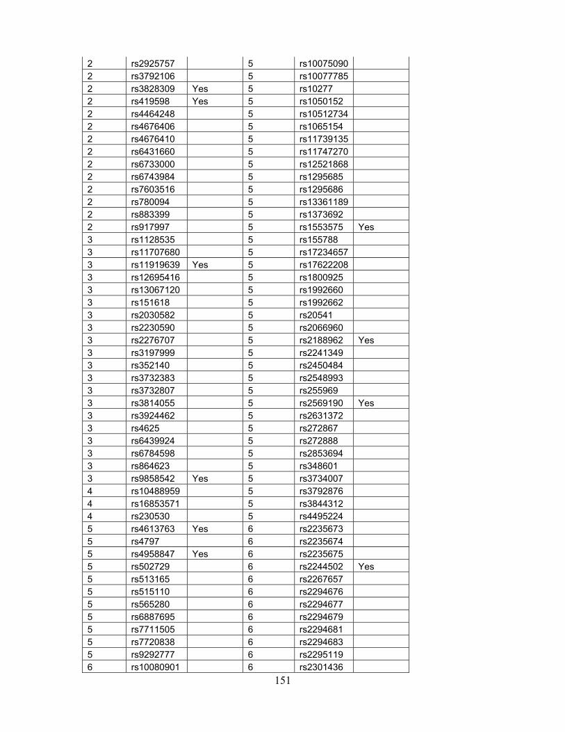
151
2 rs2925757 5 rs10075090 2 rs3792106 5 rs10077785 2 rs3828309 Yes 5 rs10277 2 rs419598 Yes 5 rs1050152 2 rs4464248 5 rs10512734 2 rs4676406 5 rs1065154 2 rs4676410 5 rs11739135 2 rs6431660 5 rs11747270 2 rs6733000 5 rs12521868 2 rs6743984 5 rs1295685 2 rs7603516 5 rs1295686 2 rs780094 5 rs13361189 2 rs883399 5 rs1373692 2 rs917997 5 rs1553575 Yes 3 rs1128535 5 rs155788 3 rs11707680 5 rs17234657 3 rs11919639 Yes 5 rs17622208 3 rs12695416 5 rs1800925 3 rs13067120 5 rs1992660 3 rs151618 5 rs1992662 3 rs2030582 5 rs20541 3 rs2230590 5 rs2066960 3 rs2276707 5 rs2188962 Yes 3 rs3197999 5 rs2241349 3 rs352140 5 rs2450484 3 rs3732383 5 rs2548993 3 rs3732807 5 rs255969 3 rs3814055 5 rs2569190 Yes 3 rs3924462 5 rs2631372 3 rs4625 5 rs272867 3 rs6439924 5 rs272888 3 rs6784598 5 rs2853694 3 rs864623 5 rs348601 3 rs9858542 Yes 5 rs3734007 4 rs10488959 5 rs3792876 4 rs16853571 5 rs3844312 4 rs230530 5 rs4495224 5 rs4613763 Yes 6 rs2235673 5 rs4797 6 rs2235674 5 rs4958847 Yes 6 rs2235675 5 rs502729 6 rs2244502 Yes 5 rs513165 6 rs2267657 5 rs515110 6 rs2294676 5 rs565280 6 rs2294677 5 rs6887695 6 rs2294679 5 rs7711505 6 rs2294681 5 rs7720838 6 rs2294683 5 rs9292777 6 rs2295119 6 rs10080901 6 rs2301436

152
6 rs1016259 6 rs231943 6 rs10223752 6 rs231944 6 rs1041981 6 rs231946 6 rs10484545 6 rs231947 6 rs10484603 6 rs231948 6 rs10484604 6 rs231952 6 rs11758155 6 rs231954 6 rs11758386 6 rs231956 6 rs11961314 6 rs231959 6 rs12195525 6 rs231961 6 rs12196156 6 rs2395182 6 rs12202693 6 rs2395185 6 rs12529198 6 rs2395269 6 rs12661812 Yes 6 rs3093661 6 rs13201732 6 rs3093662 6 rs14362 6 rs3093664 Yes 6 rs1546894 6 rs3093665 6 rs162990 6 rs3094204 6 rs163007 6 rs3117223 6 rs17309827 6 rs3129882 6 rs1799724 6 rs3130453 Yes 6 rs1799964 6 rs3130501 Yes 6 rs1800629 Yes 6 rs3132946 6 rs1800750 6 rs3134603 6 rs2229094 6 rs3134954 6 rs2235668 6 rs361525 6 rs2235670 6 rs3749930 6 rs2235671 6 rs3749946 6 rs2235672 6 rs3763313 Yes 6 rs4248160 6 rs9480140 6 rs4248161 6 rs995482 6 rs434841 7 rs1016189 6 rs438239 7 rs10238136 6 rs443198 7 rs10247855 6 rs4516954 7 rs10278217 6 rs4645839 7 rs1045642 6 rs6239 7 rs10499343 6 rs6557420 7 rs10951982 6 rs6557421 Yes 7 rs1406161 6 rs660895 Yes 7 rs1456893 6 rs6900372 7 rs17224513 6 rs6908425 Yes 7 rs1860695 6 rs6919626 7 rs2075820 6 rs6932514 7 rs2240478 6 rs6935359 7 rs2302004 6 rs746868 7 rs2302006 6 rs7739889 7 rs2303364 6 rs7746082 7 rs3789243 6 rs7754577 7 rs3800752

153
6 rs7758080 Yes 7 rs3813517 6 rs7769639 7 rs4720672 6 rs7774274 7 rs6947579 6 rs793697 7 rs6951997 6 rs849884 7 rs6960867 6 rs849886 7 rs6962966 Yes 6 rs909253 7 rs6964587 6 rs9268480 7 rs6976629 6 rs9271568 7 rs702484 6 rs9275184 7 rs7783685 6 rs9277554 7 rs836488 6 rs9322523 7 rs836518 Yes 6 rs9322524 7 rs836547 Yes 6 rs9371890 7 rs836554 6 rs9384323 7 rs836559 6 rs9384326 8 rs10505007 6 rs9397819 8 rs11362 6 rs9469964 8 rs11775611 6 rs9478652 8 rs2044999 6 rs9480136 8 rs2386922 6 rs9480137 8 rs2456449 8 rs2979040 10 rs2241833 8 rs4871857 10 rs2289310 8 rs6557634 10 rs3218621 8 rs830772 10 rs3936503 8 rs916552 Yes 10 rs4379776 8 rs921720 11 rs10501701 9 rs10821091 11 rs10765205 9 rs11534415 11 rs10830273 9 rs1322054 11 rs10830277 9 rs1885383 11 rs11018622 9 rs2295800 11 rs1176713 9 rs34155071 Yes 11 rs11821838 Yes 9 rs3789879 11 rs11826748 9 rs3789882 11 rs12574346 9 rs4263839 11 rs12576495 9 rs4378065 11 rs12799930 9 rs4574921 11 rs17193643 9 rs4743484 11 rs17195409 9 rs4878061 11 rs1793004 9 rs4986790 Yes 11 rs1827428 9 rs4986791 11 rs2036418 9 rs7849191 11 rs2164521 9 rs7868736 11 rs2164522 10 rs10509093 11 rs2712800 10 rs1058198 11 rs317147 10 rs10761659 Yes 11 rs317155 10 rs10763976 Yes 11 rs317185 10 rs10826240 11 rs317194
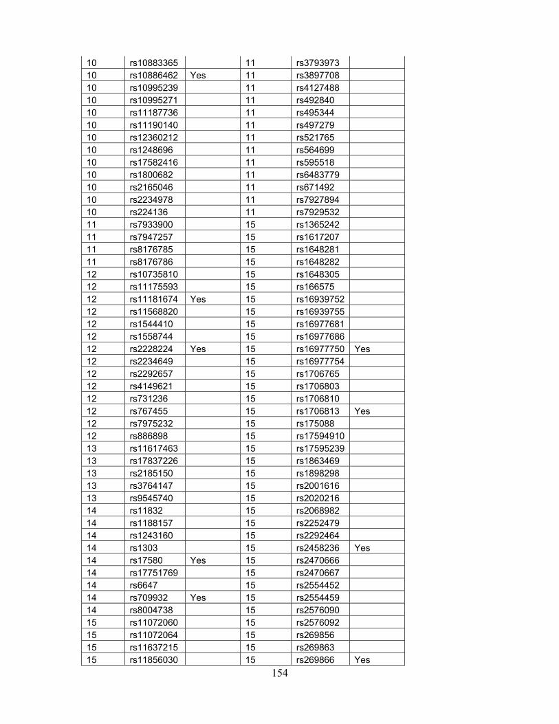
154
10 rs10883365 11 rs3793973 10 rs10886462 Yes 11 rs3897708 10 rs10995239 11 rs4127488 10 rs10995271 11 rs492840 10 rs11187736 11 rs495344 10 rs11190140 11 rs497279 10 rs12360212 11 rs521765 10 rs1248696 11 rs564699 10 rs17582416 11 rs595518 10 rs1800682 11 rs6483779 10 rs2165046 11 rs671492 10 rs2234978 11 rs7927894 10 rs224136 11 rs7929532 11 rs7933900 15 rs1365242 11 rs7947257 15 rs1617207 11 rs8176785 15 rs1648281 11 rs8176786 15 rs1648282 12 rs10735810 15 rs1648305 12 rs11175593 15 rs166575 12 rs11181674 Yes 15 rs16939752 12 rs11568820 15 rs16939755 12 rs1544410 15 rs16977681 12 rs1558744 15 rs16977686 12 rs2228224 Yes 15 rs16977750 Yes 12 rs2234649 15 rs16977754 12 rs2292657 15 rs1706765 12 rs4149621 15 rs1706803 12 rs731236 15 rs1706810 12 rs767455 15 rs1706813 Yes 12 rs7975232 15 rs175088 12 rs886898 15 rs17594910 13 rs11617463 15 rs17595239 13 rs17837226 15 rs1863469 13 rs2185150 15 rs1898298 13 rs3764147 15 rs2001616 13 rs9545740 15 rs2020216 14 rs11832 15 rs2068982 14 rs1188157 15 rs2252479 14 rs1243160 15 rs2292464 14 rs1303 15 rs2458236 Yes 14 rs17580 Yes 15 rs2470666 14 rs17751769 15 rs2470667 14 rs6647 15 rs2554452 14 rs709932 Yes 15 rs2554459 14 rs8004738 15 rs2576090 15 rs11072060 15 rs2576092 15 rs11072064 15 rs269856 15 rs11637215 15 rs269863 15 rs11856030 15 rs269866 Yes
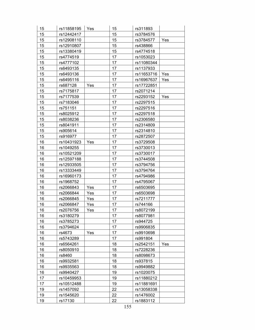
155
15 rs11858195 Yes 15 rs311893 15 rs12442417 15 rs3784576 15 rs12908110 15 rs3784577 Yes 15 rs12910807 15 rs438866 15 rs13380419 15 rs4774518 15 rs4774519 17 rs1053023 15 rs4777102 17 rs11080344 15 rs6493135 17 rs1137933 15 rs6493136 17 rs11653716 Yes 15 rs6495116 17 rs16967637 Yes 15 rs687128 Yes 17 rs17722851 15 rs7175817 17 rs2071214 15 rs7177539 17 rs2293152 Yes 15 rs7183046 17 rs2297515 15 rs751151 17 rs2297516 15 rs8025912 17 rs2297518 15 rs8038236 17 rs2306580 15 rs8041911 17 rs2314809 15 rs905614 17 rs2314810 15 rs916977 17 rs2872507 16 rs10431923 Yes 17 rs3729508 16 rs1049255 17 rs3730013 16 rs10521209 17 rs3730017 16 rs12597188 17 rs3744508 16 rs12933505 17 rs3794756 16 rs13333449 17 rs3794764 16 rs16960173 17 rs4794986 16 rs1968752 17 rs4795067 16 rs2066843 Yes 17 rs6503695 16 rs2066844 Yes 17 rs6503698 16 rs2066845 Yes 17 rs7211777 16 rs2066847 Yes 17 rs744166 16 rs2076756 Yes 17 rs8072199 16 rs3180279 17 rs8077981 16 rs3785273 17 rs944725 16 rs3794624 17 rs9906835 16 rs4673 Yes 17 rs9910698 16 rs5743289 17 rs991804 16 rs6564261 18 rs2542151 Yes 16 rs8050910 18 rs7228236 16 rs8460 18 rs8098673 16 rs9932581 18 rs937815 16 rs9935563 18 rs9949882 16 rs9940427 19 rs1020075 17 rs10459953 19 rs11880212 17 rs10512488 19 rs11881691 19 rs1457092 22 rs13058338 19 rs1545620 22 rs1476002 19 rs17130 22 rs1883112
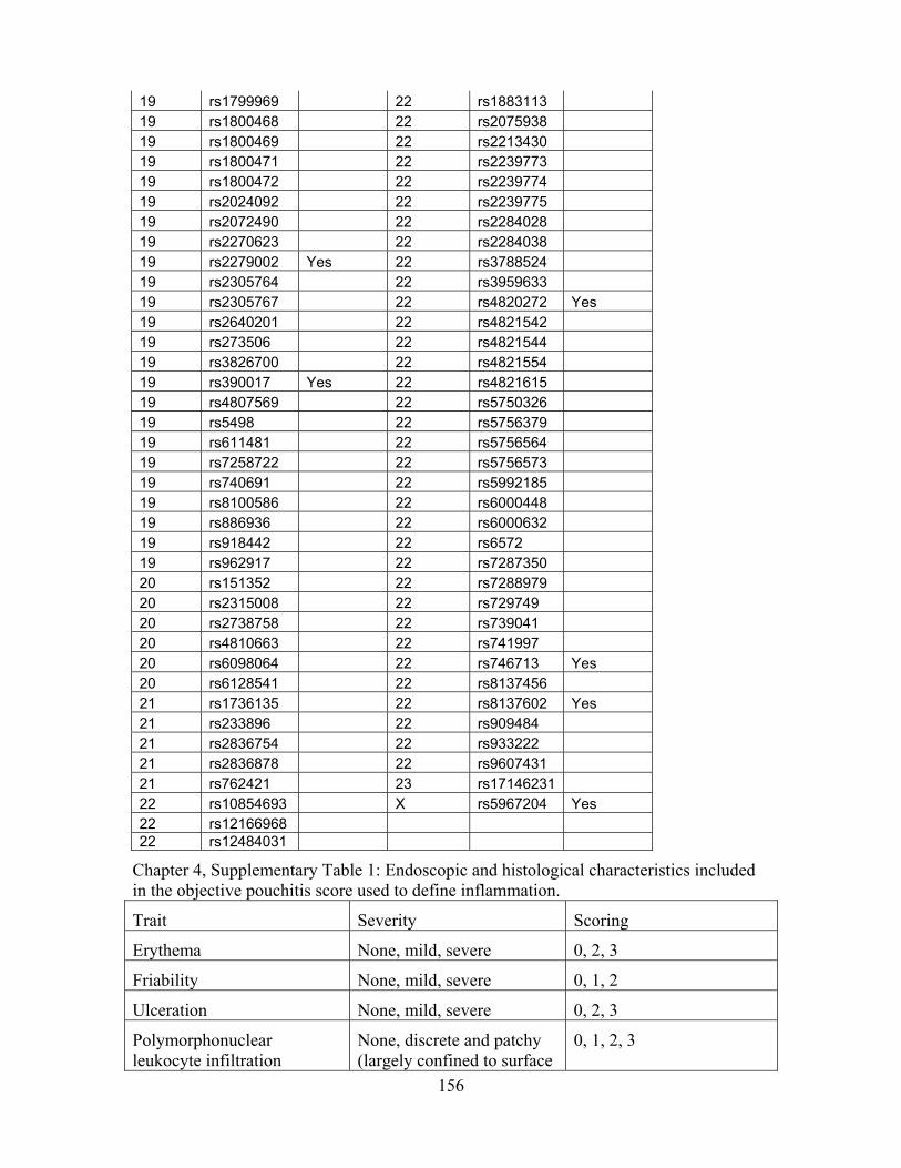
156
19 rs1799969 22 rs1883113 19 rs1800468 22 rs2075938 19 rs1800469 22 rs2213430 19 rs1800471 22 rs2239773 19 rs1800472 22 rs2239774 19 rs2024092 22 rs2239775 19 rs2072490 22 rs2284028 19 rs2270623 22 rs2284038 19 rs2279002 Yes 22 rs3788524 19 rs2305764 22 rs3959633 19 rs2305767 22 rs4820272 Yes 19 rs2640201 22 rs4821542 19 rs273506 22 rs4821544 19 rs3826700 22 rs4821554 19 rs390017 Yes 22 rs4821615 19 rs4807569 22 rs5750326 19 rs5498 22 rs5756379 19 rs611481 22 rs5756564 19 rs7258722 22 rs5756573 19 rs740691 22 rs5992185 19 rs8100586 22 rs6000448 19 rs886936 22 rs6000632 19 rs918442 22 rs6572 19 rs962917 22 rs7287350 20 rs151352 22 rs7288979 20 rs2315008 22 rs729749 20 rs2738758 22 rs739041 20 rs4810663 22 rs741997 20 rs6098064 22 rs746713 Yes 20 rs6128541 22 rs8137456 21 rs1736135 22 rs8137602 Yes 21 rs233896 22 rs909484 21 rs2836754 22 rs933222 21 rs2836878 22 rs9607431 21 rs762421 23 rs17146231 22 rs10854693 X rs5967204 Yes 22 rs12166968 22 rs12484031
Chapter 4, Supplementary Table 1: Endoscopic and histological characteristics included in the objective pouchitis score used to define inflammation.
Trait Severity Scoring
Erythema None, mild, severe 0, 2, 3
Friability None, mild, severe 0, 1, 2
Ulceration None, mild, severe 0, 2, 3
Polymorphonuclear leukocyte infiltration
None, discrete and patchy (largely confined to surface
0, 1, 2, 3
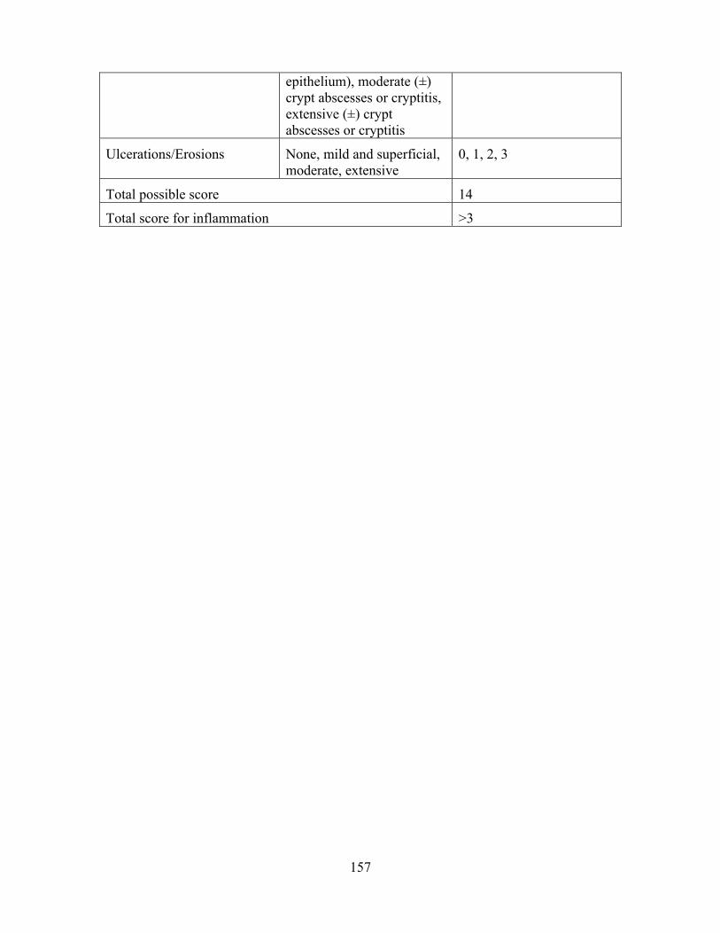
157
epithelium), moderate (±) crypt abscesses or cryptitis, extensive (±) crypt abscesses or cryptitis
Ulcerations/Erosions None, mild and superficial, moderate, extensive
0, 1, 2, 3
Total possible score 14
Total score for inflammation >3

158
Chapter 4, Supplementary Table 2: Primers used for pyrosequencing and qPCR experiments. Primers listed 5’-3’.
Primer Target Primer Names
Forward Primer Reverse Primer
VI-V3 specific universal primer set
Gray 28F / Gray 519R353
GAGTTTGATCNTGGCTCAG
GTNTTACNGCGGCKGCTG
Eubacteria 341-357F /
518-534R355
CCTACGGGAGGCAGCAG
ATTACCGCGGCTGCTGG
Bacteroides spp. Bac303F/ Bfr-Fmrev389
GAAGGTCCCCCACATTG
CGCKACTTGGCTGGTTCAG
Roseburia spp. and E. rectale
RrecF/ Rrec630mR38
9
GCGGTRCGGCAAGTCTGA
CCTCCGACGCTCTAGTMCGAC
Clostridial cluster IV
Clep866mF / Clept1240mR389
TTAACACAATAAGTWATCCACCTGG
ACCTTCCTCCGTTTTGTCAAC
Faecalibacterium prausnitzii
FPR-2F / Fprau645R389
GGAGGAAGAAGGTCTTCGG
AATTCCGCCTACCTCTGCACT
Adherent invasive E. coli (AIEC)
EcoliFimH2F / EcoliFimH2 355
GCCGGTGGCGCTTTATTTG
TCATCGCTGTTATAGTTGTTGGTCT
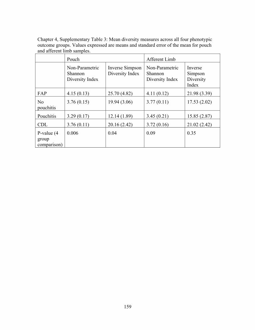
159
Chapter 4, Supplementary Table 3: Mean diversity measures across all four phenotypic outcome groups. Values expressed are means and standard error of the mean for pouch and afferent limb samples.
Pouch Afferent Limb
Non-Parametric Shannon Diversity Index
Inverse Simpson Diversity Index
Non-Parametric Shannon Diversity Index
Inverse Simpson Diversity Index
FAP 4.15 (0.13) 25.70 (4.82) 4.11 (0.12) 21.98 (3.39)
No pouchitis
3.76 (0.15) 19.94 (3.06) 3.77 (0.11) 17.53 (2.02)
Pouchitis 3.29 (0.17) 12.14 (1.89) 3.45 (0.21) 15.85 (2.87)
CDL 3.76 (0.11) 20.16 (2.42) 3.72 (0.16) 21.02 (2.42)
P-value (4 group comparison)
0.006 0.04 0.09 0.35
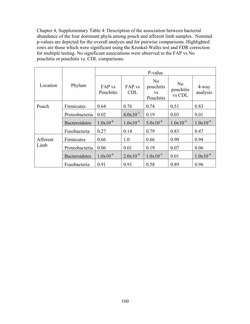
160
Chapter 4, Supplementary Table 4: Description of the association between bacterial abundance of the four dominant phyla among pouch and afferent limb samples. Nominal p-values are depicted for the overall analysis and for pairwise comparisons. Highlighted rows are those which were significant using the Kruskal-Wallis test and FDR correction for multiple testing. No significant associations were observed in the FAP vs No pouchitis or pouchitis vs. CDL comparisons.
Location Phylum
P-value
FAP vs Pouchitis
FAP vs CDL
No pouchitis
vs Pouchitis
No pouchitis vs CDL
4-way analysis
Pouch Firmicutes 0.64 0.76 0.74 0.51 0.83
Proteobacteria 0.02 4.0x10⁻³ 0.19 0.03 0.01
Bacteroidetes 1.0x10⁻⁴ 1.0x10⁻⁴ 5.0x10⁻⁴ 1.0x10⁻³ 1.0x10⁻⁴
Fusobacteria 0.27 0.14 0.79 0.83 0.47
Afferent Limb
Firmicutes 0.66 1.0 0.66 0.90 0.94
Proteobacteria 0.06 0.01 0.19 0.07 0.06
Bacteroidetes 1.0x10⁻⁴ 2.0x10⁻⁴ 1.0x10⁻³ 0.01 1.0x10⁻⁴
Fusobacteria 0.91 0.91 0.58 0.89 0.96
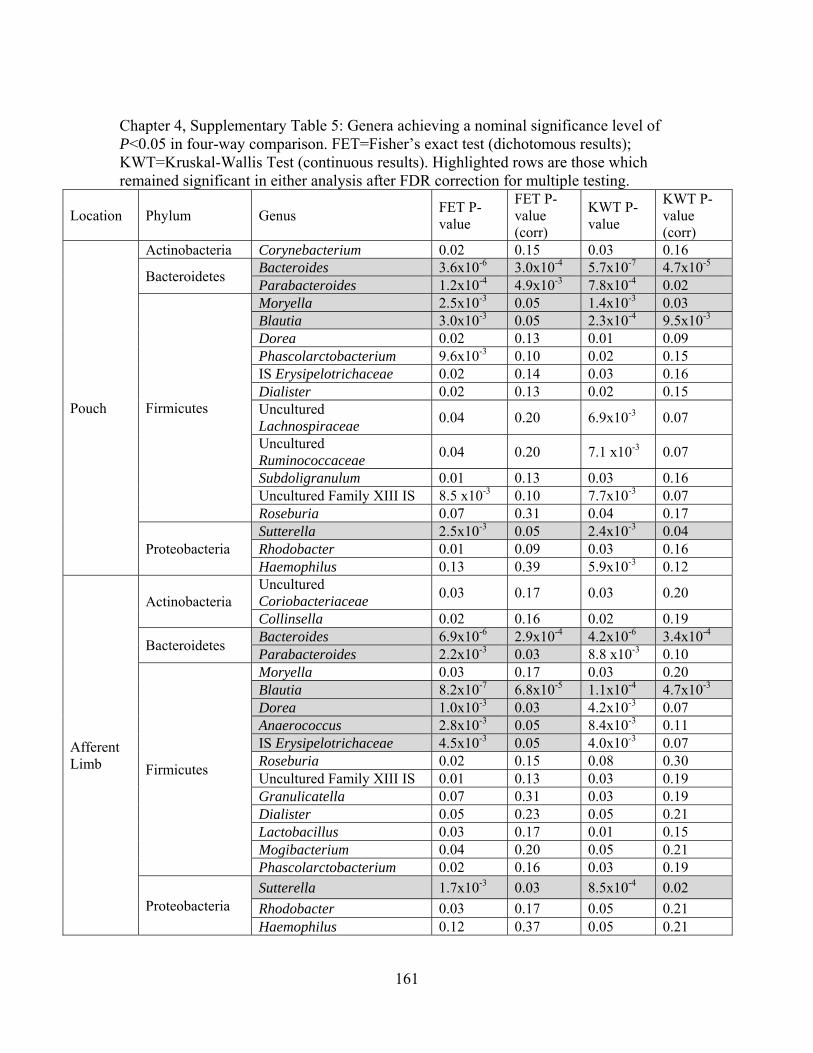
161
Chapter 4, Supplementary Table 5: Genera achieving a nominal significance level of P<0.05 in four-way comparison. FET=Fisher’s exact test (dichotomous results); KWT=Kruskal-Wallis Test (continuous results). Highlighted rows are those which remained significant in either analysis after FDR correction for multiple testing.
Location Phylum Genus FET P-value
FET P-value (corr)
KWT P-value
KWT P-value (corr)
Pouch
Actinobacteria Corynebacterium 0.02 0.15 0.03 0.16
Bacteroidetes Bacteroides 3.6x10⁻⁶ 3.0x10⁻⁴ 5.7x10⁻⁷ 4.7x10⁻⁵
Parabacteroides 1.2x10⁻⁴ 4.9x10⁻³ 7.8x10⁻⁴ 0.02
Firmicutes
Moryella 2.5x10⁻³ 0.05 1.4x10⁻³ 0.03Blautia 3.0x10⁻³ 0.05 2.3x10⁻⁴ 9.5x10⁻³
Dorea 0.02 0.13 0.01 0.09Phascolarctobacterium 9.6x10⁻³ 0.10 0.02 0.15IS Erysipelotrichaceae 0.02 0.14 0.03 0.16Dialister 0.02 0.13 0.02 0.15Uncultured Lachnospiraceae 0.04 0.20 6.9x10⁻³ 0.07 Uncultured Ruminococcaceae 0.04 0.20 7.1 x10⁻³ 0.07 Subdoligranulum 0.01 0.13 0.03 0.16Uncultured Family XIII IS 8.5 x10⁻³ 0.10 7.7x10⁻³ 0.07Roseburia 0.07 0.31 0.04 0.17
Proteobacteria Sutterella 2.5x10⁻³ 0.05 2.4x10⁻³ 0.04Rhodobacter 0.01 0.09 0.03 0.16Haemophilus 0.13 0.39 5.9x10⁻³ 0.12
Afferent Limb
Actinobacteria Uncultured Coriobacteriaceae 0.03 0.17 0.03 0.20 Collinsella 0.02 0.16 0.02 0.19
Bacteroidetes Bacteroides 6.9x10⁻⁶ 2.9x10⁻⁴ 4.2x10⁻⁶ 3.4x10⁻⁴
Parabacteroides 2.2x10⁻³ 0.03 8.8 x10⁻³ 0.10
Firmicutes
Moryella 0.03 0.17 0.03 0.20Blautia 8.2x10⁻⁷ 6.8x10⁻⁵ 1.1x10⁻⁴ 4.7x10⁻³
Dorea 1.0x10⁻³ 0.03 4.2x10⁻³ 0.07Anaerococcus 2.8x10⁻³ 0.05 8.4x10⁻³ 0.11IS Erysipelotrichaceae 4.5x10⁻³ 0.05 4.0x10⁻³ 0.07Roseburia 0.02 0.15 0.08 0.30Uncultured Family XIII IS 0.01 0.13 0.03 0.19Granulicatella 0.07 0.31 0.03 0.19Dialister 0.05 0.23 0.05 0.21Lactobacillus 0.03 0.17 0.01 0.15Mogibacterium 0.04 0.20 0.05 0.21Phascolarctobacterium 0.02 0.16 0.03 0.19
Proteobacteria Sutterella 1.7x10⁻³ 0.03 8.5x10⁻⁴ 0.02 Rhodobacter 0.03 0.17 0.05 0.21Haemophilus 0.12 0.37 0.05 0.21

162

163
Chapter 2, Supplementary figure 1: Mean antibody titres expressed in EU/mL. Error bars represent standard errors. Titres of anti-CBir1 were significantly associated with outcome (P=0.001)
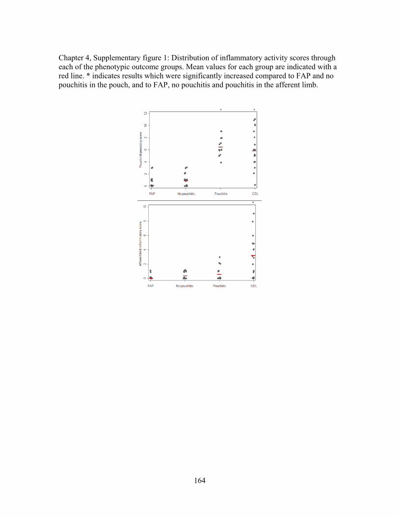
164
Chapter 4, Supplementary figure 1: Distribution of inflammatory activity scores through each of the phenotypic outcome groups. Mean values for each group are indicated with a red line. * indicates results which were significantly increased compared to FAP and no pouchitis in the pouch, and to FAP, no pouchitis and pouchitis in the afferent limb.

165
Chapter 4, Supplementary figure 2: Phylum level comparisons between four outcome groups for the pouch and afferent limb. FAP=familial adenomatous polyposis, CDL=Crohn’s disease-like.

166

167
Chapter 4, Supplementary figure 3: Proportion of patients positive for genera which were significantly associated with outcome (Pcorr<0.05) following removal of individuals without evidence of inflammation at study pouchoscopy from the CDL group. * represent pairwise comparisons which were significant (Pcorr <0.05) after correction. A) Pouch samples. B) Afferent limb samples. I.S. = Incertae sedis.
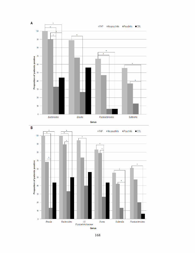
168
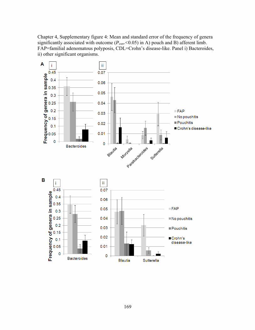
169
Chapter 4, Supplementary figure 4: Mean and standard error of the frequency of genera significantly associated with outcome (Pcorr<0.05) in A) pouch and B) afferent limb. FAP=familial adenomatous polyposis, CDL=Crohn’s disease-like. Panel i) Bacteroides, ii) other significant organisms.

170
Chapter 4, Supplementary figure 5: Proportion of patients positive for genera which were significantly associated with outcome (Pcorr<0.05) in the preliminary analysis, among the cohort of patients on no antibiotic therapy (n=57). * represent pairwise comparisons which were significant (Pcorr <0.05) after correction. A) Pouch samples. B) Afferent limb samples. I.S. = Incertae sedis.

171
Chapter 4, Supplementary Figure 6: A) Cladograms demonstrating the results obtained through LDA Effect Size (LEfSe) analysis. Highlighted results are those which were increased in the corresponding group. Significant associations are highlighted with outcome group(s) with increased proportions indicated. B) Differential abundance of organisms detected at significantly different frequencies via LEfSe.

172
Cladogram – Pouch samples, four group comparison
Significant associations as highlighted, with outcome group(s) where increased proportion was detectable indicated

Cladogram – Pouch samples, FAP vs Pouchitis
Significant associations as highlighted, with outcome group(s) where increased proportion was detectable indicated
173
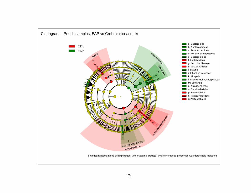
Cladogram – Pouch samples, FAP vs Crohn’s disease-like
Significant associations as highlighted, with outcome group(s) where increased proportion was detectable indicated
174
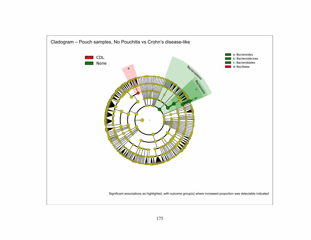
Cladogram – Pouch samples, No Pouchitis vs Crohn’s disease-like
Significant associations as highlighted, with outcome group(s) where increased proportion was detectable indicated
175
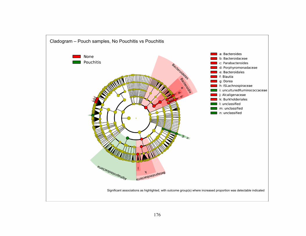
176
Cladogram – Pouch samples, No Pouchitis vs Pouchitis
Significant associations as highlighted, with outcome group(s) where increased proportion was detectable indicated
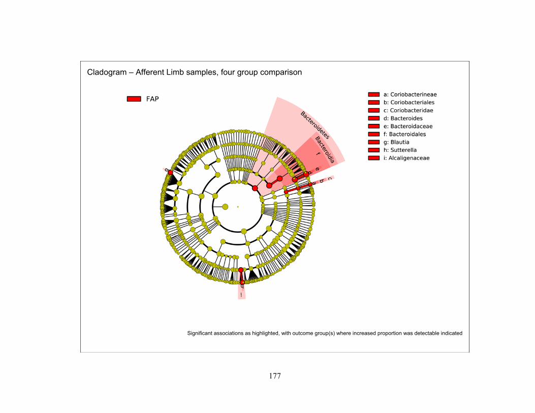
Cladogram – Afferent Limb samples, four group comparison
Significant associations as highlighted, with outcome group(s) where increased proportion was detectable indicated
177
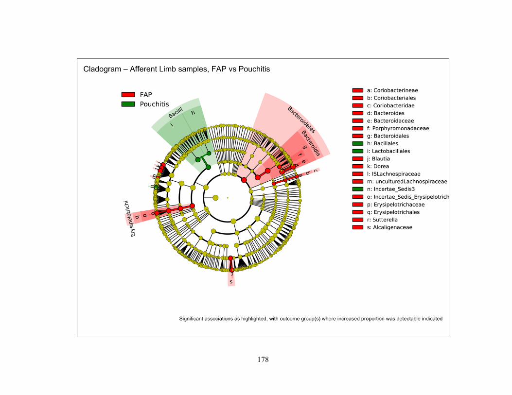
Cladogram – Afferent Limb samples, FAP vs Pouchitis
Significant associations as highlighted, with outcome group(s) where increased proportion was detectable indicated
178

Cladogram – Afferent Limb samples, FAP vs Crohn’s disease-like
Significant associations as highlighted, with outcome group(s) where increased proportion was detectable indicated
179
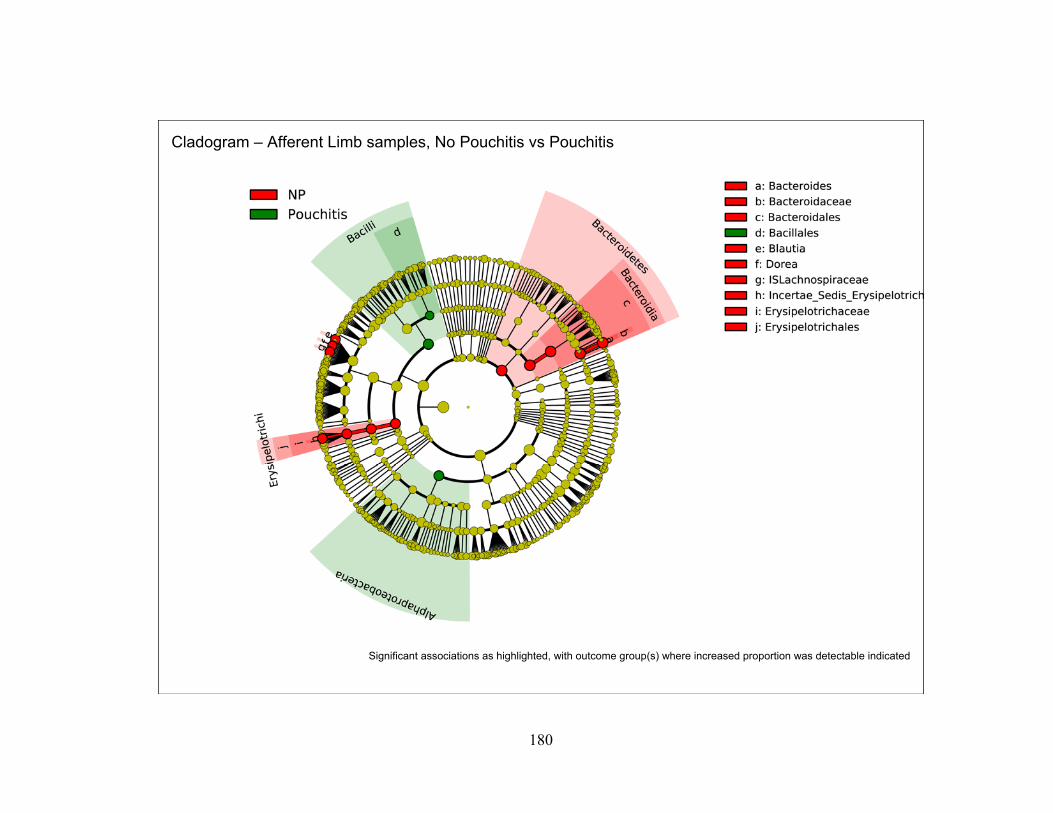
Cladogram – Afferent Limb samples, No Pouchitis vs Pouchitis
Significant associations as highlighted, with outcome group(s) where increased proportion was detectable indicated
180
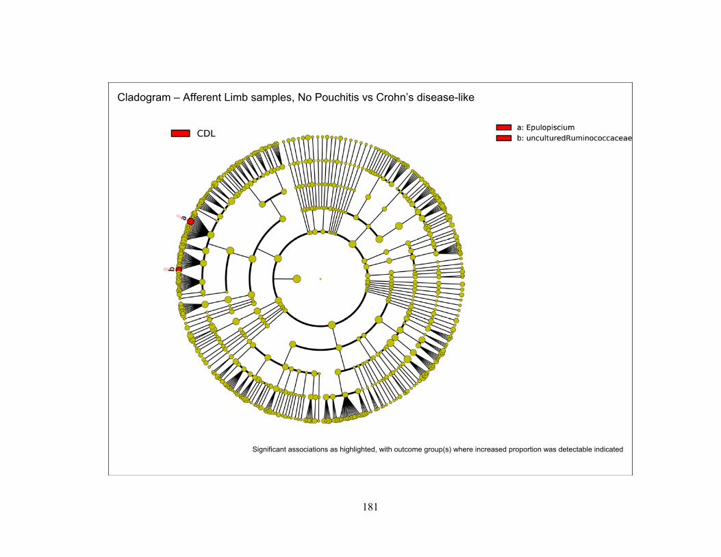
181
Significant associations as highlighted, with outcome group(s) where increased proportion was detectable indicated
Cladogram – Afferent Limb samples, No Pouchitis vs Crohn’s disease-like

182
Chapter 4, Supplementary Figure 7: log2 transformed relative abundance of organisms of interest from real-time quantitative PCR in the A) pouch and B) afferent limb. Significant results are marked with an astrix (Pcorr<0.05). FAP=familial adenomatous polyposis, CDL=Crohn’s disease-like.

183
Chapter 4, Supplementary figure 8: Proportion of individuals in each outcome group positive for genera which were previously associated with pouch inflammatory outcomes. FAP=familial adenomatous polyposis, Quiescent=seven individuals requiring long-term medical therapy to maintain remission. Lines represent associations between the quiescent group and others which reached nominal significance (P<0.05). A) Pouch samples. B) Afferent limb samples.
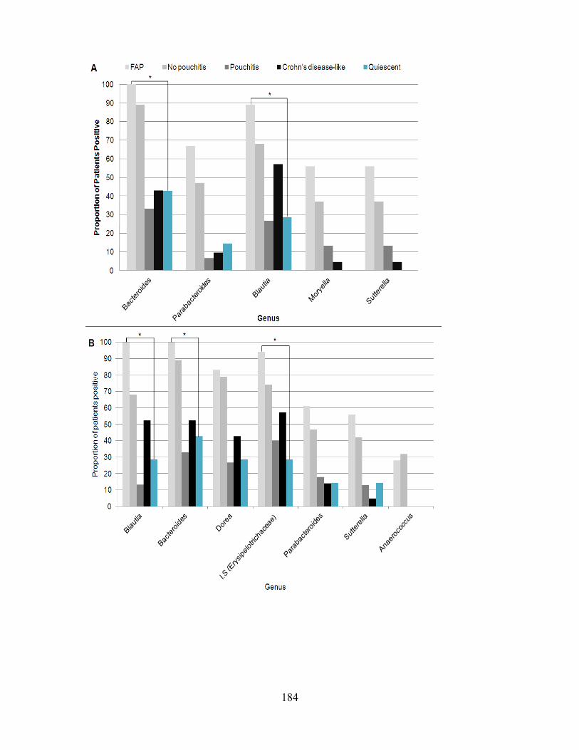
184

185
References
1. Bernstein CN, Wajda A, Svenson LW, et al. The epidemiology of inflammatory bowel disease in Canada: a population-based study. Am J Gastroenterol 2006;101:1559-68.
2. Canada CsaCFo. THE IMPACT OF INFLAMMATORY BOWEL DISEASE IN CANADA: 2012 Final Report and Recommendations. In: CCFC, ed. Toronto: CCFC, 2012.
3. Molodecky NA, Soon IS, Rabi DM, et al. Increasing incidence and prevalence of the Inflammatory Bowel Diseases with time, based on systematic review. Gastroenterology 2012;142:46-54 e42; quiz e30.
4. Loftus EV, Jr., Schoenfeld P, Sandborn WJ. The epidemiology and natural history of Crohn's disease in population-based patient cohorts from North America: a systematic review. Aliment Pharmacol Ther 2002;16:51-60.
5. Silverberg MS, Satsangi J, Ahmad T, et al. Toward an integrated clinical, molecular and serological classification of inflammatory bowel disease: Report of a Working Party of the 2005 Montreal World Congress of Gastroenterology. Can J Gastroenterol 2005;19 Suppl A:5-36.
6. Roth MP, Petersen GM, McElree C, et al. Geographic origins of Jewish patients with inflammatory bowel disease. Gastroenterology 1989;97:900-4.
7. Orholm M, Munkholm P, Langholz E, et al. Familial occurrence of inflammatory bowel disease. N Engl J Med 1991;324:84-8.
8. Jick H, Walker AM. Cigarette smoking and ulcerative colitis. N Engl J Med 1983;308:261-3.
9. Somerville KW, Logan RF, Edmond M, et al. Smoking and Crohn's disease. British medical journal 1984;289:954-6.
10. Baumgart DC, Sandborn WJ. Crohn's disease. Lancet 2012.
11. Hviid A, Svanstrom H, Frisch M. Antibiotic use and Inflammatory Bowel Diseases in childhood. Gut 2010;60:49-54.
12. Bernstein CN, Blanchard JF, Rawsthorne P, et al. The prevalence of extraintestinal diseases in inflammatory bowel disease: a population-based study. Am J Gastroenterol 2001;96:1116-22.
13. Pinsk V, Lemberg DA, Grewal K, et al. Inflammatory bowel disease in the South Asian pediatric population of British Columbia. Am J Gastroenterol 2007;102:1077-83.
14. Bernstein CN, Rawsthorne P, Cheang M, et al. A population-based case control study of potential risk factors for IBD. Am J Gastroenterol 2006;101:993-1002.
15. Carr I, Mayberry JF. The effects of migration on ulcerative colitis: a three-year prospective study among Europeans and first- and second- generation South Asians in Leicester (1991-1994). Am J Gastroenterol 1999;94:2918-22.
186
16. Le Q, Melmed G, Dubinsky M, et al. Surgical outcome of ileal pouch-anal anastomosis when used intentionally for well-defined Crohn's disease. Inflamm Bowel Dis 2012.
17. Campbell WJ, Spence RA, Parks TG. Familial adenomatous polyposis. Br J Surg 1994;81:1722-33.
18. McLeod RS. Surgery for Inflammatory Bowel Diseases. Dig Dis 2003;21:168-79.
19. Evgenikos N, Bartolo DC, Hamer-Hodges DW, et al. Comparison of the Moskowitz criteria and the pouchitis disease activity index (PDAI) for diagnosis of ileoanal pouch inflammation. Colorectal Dis 2001;3:161-4.
20. Heuschen UA, Allemeyer EH, Hinz U, et al. Diagnosing pouchitis: comparative validation of two scoring systems in routine follow-up. Dis Colon Rectum 2002;45:776-86; discussion 786-8.
21. Shen B, Lashner BA. Pouchitis: a spectrum of diseases. Curr Gastroenterol Rep 2005;7:404-11.
22. Shen B, Remzi FH, Lavery IC, et al. A proposed classification of ileal pouch disorders and associated complications after restorative proctocolectomy. Clin Gastroenterol Hepatol 2008;6:145-58; quiz 124.
23. Ben-Bassat O, Tyler, AD. , Xu, W. , Kirsch, R. , Schaeffer, DF. , Walsh, J. , Steinhart, HA. , Greenberg, GR. , Cohen, Z. , Silverberg, MS. , McLeod, RS. . Symptoms of Pouchitis and Inflammation-Are They Correlated? AGA Digestive Diseases Week 2012;San Diego.
24. Dayton MT, Larsen KR, Christiansen DD. Similar functional results and complications after ileal pouch-anal anastomosis in patients with indeterminate vs ulcerative colitis. Arch Surg 2002;137:690-4; discussion 694-5.
25. Abdelrazeq AS, Kandiyil N, Botterill ID, et al. Predictors for acute and chronic pouchitis following restorative proctocolectomy for ulcerative colitis. Colorectal Dis 2008;10:805-13.
26. Achkar JP, Al-Haddad M, Lashner B, et al. Differentiating risk factors for acute and chronic pouchitis. Clin Gastroenterol Hepatol 2005;3:60-6.
27. Gustavsson S, Weiland LH, Kelly KA. Relationship of backwash ileitis to ileal pouchitis after ileal pouch-anal anastomosis. Dis Colon Rectum 1987;30:25-8.
28. Shen B, Remzi FH, Hammel JP, et al. Family history of Crohn's disease is associated with an increased risk for Crohn's disease of the pouch. Inflamm Bowel Dis 2009;15:163-70.
29. Kuisma J, Jarvinen H, Kahri A, et al. Factors associated with disease activity of pouchitis after surgery for ulcerative colitis. Scand J Gastroenterol 2004;39:544-8.
30. Merrett MN, Mortensen N, Kettlewell M, et al. Smoking may prevent pouchitis in patients with restorative proctocolectomy for ulcerative colitis. Gut 1996;38:362-4.

187
31. Cakal B, Akoz AG, Ustundag Y, et al. Red cell distribution width for assessment of activity of inflammatory bowel disease. Digestive diseases and sciences 2009;54:842-7.
32. Shine B, Berghouse L, Jones JE, et al. C-reactive protein as an aid in the differentiation of functional and inflammatory bowel disorders. Clin Chim Acta 1985;148:105-9.
33. Moscandrew ME, Loftus EV, Jr. Diagnostic advances in inflammatory bowel disease (imaging and laboratory). Current gastroenterology reports 2009;11:488-95.
34. Young B, Gleeson M, Cripps AW. C-reactive protein: a critical review. Pathology 1991;23:118-24.
35. Schoepfer AM, Beglinger C, Straumann A, et al. Fecal calprotectin correlates more closely with the Simple Endoscopic Score for Crohn's disease (SES-CD) than CRP, blood leukocytes, and the CDAI. Am J Gastroenterol 2010;105:162-9.
36. Henderson P, Casey A, Lawrence SJ, et al. The diagnostic accuracy of fecal calprotectin during the investigation of suspected pediatric inflammatory bowel disease. Am J Gastroenterol 2012;107:941-9.
37. Walker LJ, Aldhous MC, Drummond HE, et al. Anti-Saccharomyces cerevisiae antibodies (ASCA) in Crohn's disease are associated with disease severity but not NOD2/CARD15 mutations. Clinical and experimental immunology 2004;135:490-6.
38. Sendid B, Colombel JF, Jacquinot PM, et al. Specific antibody response to oligomannosidic epitopes in Crohn's disease. Clinical and diagnostic laboratory immunology 1996;3:219-26.
39. Standaert-Vitse A, Jouault T, Vandewalle P, et al. Candida albicans is an immunogen for anti-Saccharomyces cerevisiae antibody markers of Crohn's disease. Gastroenterology 2006;130:1764-75.
40. Targan SR, Landers CJ, Yang H, et al. Antibodies to CBir1 flagellin define a unique response that is associated independently with complicated Crohn's disease. Gastroenterology 2005;128:2020-8.
41. Vidrich A, Lee J, James E, et al. Segregation of pANCA antigenic recognition by DNase treatment of neutrophils: ulcerative colitis, type 1 autoimmune hepatitis, and primary sclerosing cholangitis. Journal of clinical immunology 1995;15:293-9.
42. Lichtenstein GR, Targan SR, Dubinsky MC, et al. Combination of genetic and quantitative serological immune markers are associated with complicated Crohn's disease behavior. Inflamm Bowel Dis 2011;17:2488-96.
43. Dotan I. New serologic markers for inflammatory bowel disease diagnosis. Digestive diseases 2010;28:418-23.
44. Billing P, Tahir S, Calfin B, et al. Nuclear localization of the antigen detected by ulcerative colitis-associated perinuclear antineutrophil cytoplasmic antibodies. Am J Pathol 1995;147:979-87.
188
45. Buckland MS, Mylonaki M, Rampton D, et al. Serological markers (anti-Saccharomyces cerevisiae mannan antibodies and antineutrophil cytoplasmic antibodies) in inflammatory bowel disease: diagnostic utility and phenotypic correlation. Clin Diagn Lab Immunol 2005;12:1328-30.
46. Lombardi G, Annese V, Piepoli A, et al. Antineutrophil cytoplasmic antibodies in inflammatory bowel disease: clinical role and review of the literature. Dis Colon Rectum 2000;43:999-1007.
47. Adams RJ, Heazlewood SP, Gilshenan KS, et al. IgG antibodies against common gut bacteria are more diagnostic for Crohn's disease than IgG against mannan or flagellin. Am J Gastroenterol 2008;103:386-96.
48. Vasiliauskas EA, Kam LY, Karp LC, et al. Marker antibody expression stratifies Crohn's disease into immunologically homogeneous subgroups with distinct clinical characteristics. Gut 2000;47:487-96.
49. Vasiliauskas EA, Plevy SE, Landers CJ, et al. Perinuclear antineutrophil cytoplasmic antibodies in patients with Crohn's disease define a clinical subgroup. Gastroenterology 1996;110:1810-9.
50. Zholudev A, Zurakowski D, Young W, et al. Serologic testing with ANCA, ASCA, and anti-OmpC in children and young adults with Crohn's disease and ulcerative colitis: diagnostic value and correlation with disease phenotype. Am J Gastroenterol 2004;99:2235-41.
51. Amre DK, Lu SE, Costea F, et al. Utility of serological markers in predicting the early occurrence of complications and surgery in pediatric Crohn's disease patients. Am J Gastroenterol 2006;101:645-52.
52. Ferrante M, Henckaerts L, Joossens M, et al. New serological markers in inflammatory bowel disease are associated with complicated disease behaviour. Gut 2007;56:1394-403.
53. Papp M, Altorjay I, Dotan N, et al. New serological markers for inflammatory bowel disease are associated with earlier age at onset, complicated disease behavior, risk for surgery, and NOD2/CARD15 genotype in a Hungarian IBD cohort. Am J Gastroenterol 2008;103:665-81.
54. Quinton JF, Sendid B, Reumaux D, et al. Anti-Saccharomyces cerevisiae mannan antibodies combined with antineutrophil cytoplasmic autoantibodies in inflammatory bowel disease: prevalence and diagnostic role. Gut 1998;42:788-91.
55. Peyrin-Biroulet L, Standaert-Vitse A, Branche J, et al. IBD serological panels: facts and perspectives. Inflamm Bowel Dis 2007;13:1561-6.
56. Mei L, Targan SR, Landers CJ, et al. Familial expression of anti-Escherichia coli outer membrane porin C in relatives of patients with Crohn's disease. Gastroenterology 2006;130:1078-85.
57. Dubinsky MC, Lin YC, Dutridge D, et al. Serum immune responses predict rapid disease progression among children with Crohn's disease: immune responses predict disease progression. Am J Gastroenterol 2006;101:360-7.
189
58. Davis MK, Andres JM, Jolley CD, et al. Antibodies to Escherichia coli outer membrane porin C in the absence of anti-Saccharomyces cerevisiae antibodies and anti-neutrophil cytoplasmic antibodies are an unreliable marker of Crohn disease and ulcerative colitis. J Pediatr Gastroenterol Nutr 2007;45:409-13.
59. Papadakis KA, Yang H, Ippoliti A, et al. Anti-flagellin (CBir1) phenotypic and genetic Crohn's disease associations. Inflamm Bowel Dis 2007;13:524-30.
60. Markowitz J, Kugathasan S, Dubinsky M, et al. Age of diagnosis influences serologic responses in children with Crohn's disease: a possible clue to etiology? Inflamm Bowel Dis 2009;15:714-9.
61. Arnott ID, Landers CJ, Nimmo EJ, et al. Sero-reactivity to microbial components in Crohn's disease is associated with disease severity and progression, but not NOD2/CARD15 genotype. Am J Gastroenterol 2004;99:2376-84.
62. Spivak J, Landers CJ, Vasiliauskas EA, et al. Antibodies to I2 predict clinical response to fecal diversion in Crohn's disease. Inflamm Bowel Dis 2006;12:1122-30.
63. Landers CJ, Cohavy O, Misra R, et al. Selected loss of tolerance evidenced by Crohn's disease-associated immune responses to auto- and microbial antigens. Gastroenterology 2002;123:689-99.
64. Joossens M, Van Steen K, Branche J, et al. Familial aggregation and antimicrobial response dose-dependently affect the risk for Crohn's disease. Inflamm Bowel Dis 2010;16:58-67.
65. Esteve M, Mallolas J, Klaassen J, et al. Antineutrophil cytoplasmic antibodies in sera from colectomised ulcerative colitis patients and its relation to the presence of pouchitis. Gut 1996;38:894-8.
66. Israeli E, Grotto I, Gilburd B, et al. Anti-Saccharomyces cerevisiae and antineutrophil cytoplasmic antibodies as predictors of inflammatory bowel disease. Gut 2005;54:1232-6.
67. Fleshner P, Ippoliti A, Dubinsky M, et al. Both preoperative perinuclear antineutrophil cytoplasmic antibody and anti-CBir1 expression in ulcerative colitis patients influence pouchitis development after ileal pouch-anal anastomosis. Clin Gastroenterol Hepatol 2008;6:561-8.
68. Fleshner PR, Vasiliauskas EA, Kam LY, et al. High level perinuclear antineutrophil cytoplasmic antibody (pANCA) in ulcerative colitis patients before colectomy predicts the development of chronic pouchitis after ileal pouch-anal anastomosis. Gut 2001;49:671-7.
69. Melmed GY, Fleshner PR, Bardakcioglu O, et al. Family history and serology predict Crohn's disease after ileal pouch-anal anastomosis for ulcerative colitis. Dis Colon Rectum 2008;51:100-8.
70. Sandborn WJ, Landers CJ, Tremaine WJ, et al. Antineutrophil cytoplasmic antibody correlates with chronic pouchitis after ileal pouch-anal anastomosis. Am J Gastroenterol 1995;90:740-7.
190
71. Aisenberg J, Legnani PE, Nilubol N, et al. Are pANCA, ASCA, or cytokine gene polymorphisms associated with pouchitis? Long-term follow-up in 102 ulcerative colitis patients. Am J Gastroenterol 2004;99:432-41.
72. Dendrinos KG, Becker JM, Stucchi AF, et al. Anti-Saccharomyces cerevisiae antibodies are associated with the development of postoperative fistulas following ileal pouch-anal anastomosis. J Gastrointest Surg 2006;10:1060-4.
73. Mundwiler ML, Mei L, Landers CJ, et al. Inflammatory bowel disease serologies in ankylosing spondylitis patients: a pilot study. Arthritis research & therapy 2009;11:R177.
74. Yu JE, De Ravin SS, Uzel G, et al. High levels of Crohn's disease-associated anti-microbial antibodies are present and independent of colitis in chronic granulomatous disease. Clinical immunology 2011;138:14-22.
75. Ashorn S, Raukola H, Valineva T, et al. Elevated serum anti-Saccharomyces cerevisiae, anti-I2 and anti-OmpW antibody levels in patients with suspicion of celiac disease. Journal of clinical immunology 2008;28:486-94.
76. Mallant-Hent RC, Mooij M, von Blomberg BM, et al. Correlation between Saccharomyces cerevisiae DNA in intestinal mucosal samples and anti-Saccharomyces cerevisiae antibodies in serum of patients with IBD. World journal of gastroenterology : WJG 2006;12:292-7.
77. Carvalho FA, Barnich N, Sivignon A, et al. Crohn's disease adherent-invasive Escherichia coli colonize and induce strong gut inflammation in transgenic mice expressing human CEACAM. J Exp Med 2009;206:2179-89.
78. Wagner J, Short K, Catto-Smith AG, et al. Identification and characterisation of Pseudomonas 16S ribosomal DNA from ileal biopsies of children with Crohn's disease. PLoS ONE 2008;3:e3578.
79. Madi A, Svinareff P, Orange N, et al. Pseudomonas fluorescens alters epithelial permeability and translocates across Caco-2/TC7 intestinal cells. Gut pathogens 2010;2:16.
80. Dubinsky M. What is the role of serological markers in IBD? Pediatric and adult data. Digestive diseases 2009;27:259-68.
81. Harrer M, Reinisch W, Dejaco C, et al. Do high serum levels of anti-Saccharomyces cerevisiae antibodies result from a leakiness of the gut barrier in Crohn's disease? European journal of gastroenterology & hepatology 2003;15:1281-5.
82. Schoepfer AM, Flogerzi B, Seibold-Schmid B, et al. Low Mannan-binding lectin serum levels are associated with complicated Crohn's disease and reactivity to oligomannan (ASCA). Am J Gastroenterol 2009;104:2508-16.
83. Lander ES, Linton LM, Birren B, et al. Initial sequencing and analysis of the human genome. Nature 2001;409:860-921.
84. Venter JC, Adams MD, Myers EW, et al. The sequence of the human genome. Science 2001;291:1304-51.
191
85. Consortium IH. A haplotype map of the human genome. Nature 2005;437:1299-320.
86. Consortium TGP. A map of human genome variation from population-scale sequencing. Nature 2010;467:1061-73.
87. Li G, Gelernter J, Kranzler HR, et al. M(3): an improved SNP calling algorithm for Illumina BeadArray data. Bioinformatics 2012;28:358-65.
88. Cheng SH, Gregory RJ, Marshall J, et al. Defective intracellular transport and processing of CFTR is the molecular basis of most cystic fibrosis. Cell 1990;63:827-34.
89. Lehmann H. Sickle-cell anaemia and sickle-cell trait as home- and heterozygous gene-combinations. Nature 1951;167:931-3.
90. Liu S, Yao L, Ding D, et al. CCL3L1 copy number variation and susceptibility to HIV-1 infection: a meta-analysis. PLoS ONE 2010;5:e15778.
91. Hartwell LH, Hood L, Goldberg ML, et al. Genetics: From genes to genomes 2nd edition. McGraw-Hill Companies, 2004.
92. Girirajan S, Campbell CD, Eichler EE. Human copy number variation and complex genetic disease. Annual review of genetics 2011;45:203-26.
93. Lewis CM. Genetic association studies: design, analysis and interpretation. Briefings in bioinformatics 2002;3:146-53.
94. Stranger BE, Stahl EA, Raj T. Progress and promise of genome-wide association studies for human complex trait genetics. Genetics 2011;187:367-83.
95. Hong H, Xu L, Liu J, et al. Technical reproducibility of genotyping SNP arrays used in genome-wide association studies. PLoS ONE 2012;7:e44483.
96. Consortium TIH. A haplotype map of the human genome. Nature 2005;437:1299-320.
97. Abecasis GR, Auton A, Brooks LD, et al. An integrated map of genetic variation from 1,092 human genomes. Nature 2012;491:56-65.
98. Miyagawa T, Nishida N, Ohashi J, et al. Appropriate data cleaning methods for genome-wide association study. Journal of human genetics 2008;53:886-93.
99. Fan YH, Song YQ. IPGWAS: an integrated pipeline for rational quality control and association analysis of genome-wide genetic studies. Biochemical and biophysical research communications 2012;422:363-8.
100. Lettre G, Lange C, Hirschhorn JN. Genetic model testing and statistical power in population-based association studies of quantitative traits. Genetic epidemiology 2007;31:358-62.
101. Martin LJ, Woo JG, Avery CL, et al. Multiple testing in the genomics era: findings from Genetic Analysis Workshop 15, Group 15. Genetic epidemiology 2007;31 Suppl 1:S124-31.
102. Benjamini Y, Drai D, Elmer G, et al. Controlling the false discovery rate in behavior genetics research. Behavioural brain research 2001;125:279-84.
192
103. Tysk C, Lindberg E, Jarnerot G, et al. Ulcerative colitis and Crohn's disease in an unselected population of monozygotic and dizygotic twins. A study of heritability and the influence of smoking. Gut 1988;29:990-6.
104. Jostins L, Ripke S, Weersma RK, et al. Host-microbe interactions have shaped the genetic architecture of inflammatory bowel disease. Nature 2012;491:119-24.
105. Khor B, Gardet A, Xavier RJ. Genetics and pathogenesis of inflammatory bowel disease. Nature 2011;474:307-17.
106. Ogura Y, Bonen DK, Inohara N, et al. A frameshift mutation in NOD2 associated with susceptibility to Crohn's disease. Nature 2001;411:603-6.
107. Hugot JP, Chamaillard M, Zouali H, et al. Association of NOD2 leucine-rich repeat variants with susceptibility to Crohn's disease. Nature 2001;411:599-603.
108. Rivas MA, Beaudoin M, Gardet A, et al. Deep resequencing of GWAS loci identifies independent rare variants associated with inflammatory bowel disease. Nat Genet 2011;43:1066-73.
109. Russell RK, Drummond HE, Nimmo EE, et al. Genotype-phenotype analysis in childhood-onset Crohn's disease: NOD2/CARD15 variants consistently predict phenotypic characteristics of severe disease. Inflamm Bowel Dis 2005;11:955-64.
110. Cleynen I, Gonzalez JR, Figueroa C, et al. Genetic factors conferring an increased susceptibility to develop Crohn's disease also influence disease phenotype: results from the IBDchip European Project. Gut 2012.
111. Girardin SE, Boneca IG, Viala J, et al. Nod2 is a general sensor of peptidoglycan through muramyl dipeptide (MDP) detection. The Journal of biological chemistry 2003;278:8869-72.
112. Sabbah A, Chang TH, Harnack R, et al. Activation of innate immune antiviral responses by Nod2. Nature immunology 2009;10:1073-80.
113. Cho JH, Brant SR. Recent insights into the genetics of inflammatory bowel disease. Gastroenterology 2011;140:1704-1712 e2.
114. Zhou XJ, Zhang H. Autophagy in immunity: implications in etiology of autoimmune/autoinflammatory diseases. Autophagy 2012;8:1286-99.
115. Duerr RH, Taylor KD, Brant SR, et al. A genome-wide association study identifies IL23R as an inflammatory bowel disease gene. Science 2006;314:1461-3.
116. Raza A, Yousaf W, Giannella R, et al. Th17 cells: interactions with predisposing factors in the immunopathogenesis of inflammatory bowel disease. Expert review of clinical immunology 2012;8:161-8.
117. Pidasheva S, Trifari S, Phillips A, et al. Functional studies on the IBD susceptibility gene IL23R implicate reduced receptor function in the protective genetic variant R381Q. PLoS ONE 2011;6:e25038.
118. Poole EM, Curtin K, Hsu L, et al. Genetic variability in IL23R and risk of colorectal adenoma and colorectal cancer. Cancer epidemiology 2012;36:e104-10.

193
119. Glocker E. Inflammatory Bowel Disease and Mutations Affecting the Interleukin-10 Receptor. N Engl J Med 2009.
120. Glocker EO, Frede N, Perro M, et al. Infant colitis--it's in the genes. Lancet 2010;376:1272.
121. Anderson CA, Boucher G, Lees CW, et al. Meta-analysis identifies 29 additional ulcerative colitis risk loci, increasing the number of confirmed associations to 47. Nat Genet 2011;43:246-52.
122. Sabath E, Negoro H, Beaudry S, et al. Galpha12 regulates protein interactions within the MDCK cell tight junction and inhibits tight-junction assembly. Journal of cell science 2008;121:814-24.
123. Brest P, Lapaquette P, Souidi M, et al. A synonymous variant in IRGM alters a binding site for miR-196 and causes deregulation of IRGM-dependent xenophagy in Crohn's disease. Nat Genet 2011;43:242-5.
124. Kabakchiev B, Silverberg MS. Expression Quantitative Trait Loci Analysis Identifies Associations Between Genotype and Gene Expression in Human Intestine. Gastroenterology 2013.
125. Ellinghaus D, Ellinghaus E, Nair RP, et al. Combined analysis of genome-wide association studies for Crohn disease and psoriasis identifies seven shared susceptibility loci. American journal of human genetics 2012;90:636-47.
126. Cargill M, Schrodi SJ, Chang M, et al. A large-scale genetic association study confirms IL12B and leads to the identification of IL23R as psoriasis-risk genes. American journal of human genetics 2007;80:273-90.
127. Janse M, Lamberts LE, Franke L, et al. Three ulcerative colitis susceptibility loci are associated with primary sclerosing cholangitis and indicate a role for IL2, REL, and CARD9. Hepatology 2011;53:1977-85.
128. Christodoulou K, Wiskin AE, Gibson J, et al. Next generation exome sequencing of paediatric inflammatory bowel disease patients identifies rare and novel variants in candidate genes. Gut 2012.
129. Hanley WB. Hereditary Aspects of Duodenal Ulceration: Serum-Pepsinogen Level in Relation to Abo Blood Groups and Salivary Abh Secretor Status. British medical journal 1964;1:936-40.
130. Marshall BJ, Warren JR. Unidentified curved bacilli in the stomach of patients with gastritis and peptic ulceration. Lancet 1984;1:1311-5.
131. Peter I, Mitchell AA, Ozelius L, et al. Evaluation of 22 genetic variants with Crohn's disease risk in the Ashkenazi Jewish population: a case-control study. BMC medical genetics 2011;12:63.
132. Murdoch TB, Bernstein CN, El-Gabalawy H, et al. Prevalence of genetic variants associated with inflammatory bowel disease in a healthy First Nations cohort. CMAJ : Canadian Medical Association journal 2012;184:E435-41.
133. Johnson S, Martin D, Sarin C. Diabetes mellitus in the First Nations population of British Columbia, Canada. Part 3. Prevalence of diagnosed cases. International journal of circumpolar health 2002;61:260-4.

194
134. Peschken CA, Esdaile JM. Rheumatic diseases in North America's indigenous peoples. Seminars in arthritis and rheumatism 1999;28:368-91.
135. Ouyang Q, Tandon R, Goh KL, et al. The emergence of inflammatory bowel disease in the Asian Pacific region. Current opinion in gastroenterology 2005;21:408-13.
136. Inoue N, Tamura K, Kinouchi Y, et al. Lack of common NOD2 variants in Japanese patients with Crohn's disease. Gastroenterology 2002;123:86-91.
137. Croucher PJ, Mascheretti S, Hampe J, et al. Haplotype structure and association to Crohn's disease of CARD15 mutations in two ethnically divergent populations. European journal of human genetics : EJHG 2003;11:6-16.
138. Ng SC, Tsoi KK, Kamm MA, et al. Genetics of inflammatory bowel disease in Asia: systematic review and meta-analysis. Inflamm Bowel Dis 2012;18:1164-76.
139. Prescott NJ, Dominy KM, Kubo M, et al. Independent and population-specific association of risk variants at the IRGM locus with Crohn's disease. Human molecular genetics 2010;19:1828-39.
140. Yang SK, Park M, Lim J, et al. Contribution of IL23R but not ATG16L1 to Crohn's disease susceptibility in Koreans. Inflamm Bowel Dis 2009;15:1385-90.
141. Cheon JH. Genetics of Inflammatory Bowel Diseases: A Comparison between Western and Eastern Perspectives. Journal of gastroenterology and hepatology 2012.
142. Mahurkar S, Banerjee R, Rani VS, et al. Common variants in NOD2 and IL23R are not associated with inflammatory bowel disease in Indians. Journal of gastroenterology and hepatology 2011;26:694-9.
143. Yamazaki K, Onouchi Y, Takazoe M, et al. Association analysis of genetic variants in IL23R, ATG16L1 and 5p13.1 loci with Crohn's disease in Japanese patients. Journal of human genetics 2007;52:575-83.
144. Bernstein CN, El-Gabalawy H, Sargent M, et al. Assessing inflammatory bowel disease-associated antibodies in Caucasian and First Nations cohorts. Canadian journal of gastroenterology 2011;25:269-73.
145. Lawrance IC, Murray K, Hall A, et al. A prospective comparative study of ASCA and pANCA in Chinese and Caucasian IBD patients. Am J Gastroenterol 2004;99:2186-94.
146. Murdoch TB, Xu W, Stempak JM, et al. Pattern recognition receptor and autophagy gene variants are associated with development of antimicrobial antibodies in Crohn's disease. Inflamm Bowel Dis 2012;18:1743-8.
147. Ippoliti A, Devlin S, Mei L, et al. Combination of innate and adaptive immune alterations increased the likelihood of fibrostenosis in Crohn's disease. Inflamm Bowel Dis 2010;16:1279-85.
148. Devlin SM, Yang H, Ippoliti A, et al. NOD2 variants and antibody response to microbial antigens in Crohn's disease patients and their unaffected relatives. Gastroenterology 2007;132:576-86.
195
149. Papp M, Altorjay I, Norman GL, et al. Seroreactivity to microbial components in Crohn's disease is associated with ileal involvement, noninflammatory disease behavior and NOD2/CARD15 genotype, but not with risk for surgery in a Hungarian cohort of IBD patients. Inflamm Bowel Dis 2007;13:984-92.
150. Henckaerts L, Pierik M, Joossens M, et al. Mutations in pattern recognition receptor genes modulate seroreactivity to microbial antigens in patients with inflammatory bowel disease. Gut 2007;56:1536-42.
151. Michelsen KS, Thomas LS, Taylor KD, et al. IBD-associated TL1A gene (TNFSF15) haplotypes determine increased expression of TL1A protein. PLoS ONE 2009;4:e4719.
152. Wolf DC, Abraham BP, Afzali A, et al. Community Perspectives: Combining Serology, Genetics, and Inflammation Markers for the Diagnosis of IBD and Differentiation Between CD and UC. Gastroenterology & hepatology 2012;8:1-16.
153. Shirts B, von Roon AC, Tebo AE. The entire predictive value of the prometheus IBD sgi diagnostic product may be due to the three least expensive and most available components. Am J Gastroenterol 2012;107:1760-1.
154. Kukreja A, Maclaren NK. NKT cells and type-1 diabetes and the "hygiene hypothesis" to explain the rising incidence rates. Diabetes technology & therapeutics 2002;4:323-33.
155. Olsen GJ, Lane DJ, Giovannoni SJ, et al. Microbial ecology and evolution: a ribosomal RNA approach. Annual review of microbiology 1986;40:337-65.
156. Vaidya VK. Horizontal Transfer of Antimicrobial Resistance by Extended-Spectrum beta Lactamase-Producing Enterobacteriaceae. Journal of laboratory physicians 2011;3:37-42.
157. Lang AS, Zhaxybayeva O, Beatty JT. Gene transfer agents: phage-like elements of genetic exchange. Nature reviews. Microbiology 2012;10:472-82.
158. Kembel SW, Wu M, Eisen JA, et al. Incorporating 16S Gene Copy Number Information Improves Estimates of Microbial Diversity and Abundance. PLoS computational biology 2012;8:e1002743.
159. Kitahara K, Yasutake Y, Miyazaki K. Mutational robustness of 16S ribosomal RNA, shown by experimental horizontal gene transfer in Escherichia coli. Proceedings of the National Academy of Sciences of the United States of America 2012.
160. Hunt DE, Klepac-Ceraj V, Acinas SG, et al. Evaluation of 23S rRNA PCR primers for use in phylogenetic studies of bacterial diversity. Appl Environ Microbiol 2006;72:2221-5.
161. Ludwig W, Schleifer KH. Bacterial phylogeny based on 16S and 23S rRNA sequence analysis. FEMS microbiology reviews 1994;15:155-73.
162. Schellenberg J, Links MG, Hill JE, et al. Pyrosequencing of the chaperonin-60 (cpn60) universal target as a tool for determining the composition of microbial communities. Appl Environ Microbiol 2009.
196
163. Mollet C, Drancourt M, Raoult D. rpoB sequence analysis as a novel basis for bacterial identification. Molecular microbiology 1997;26:1005-11.
164. Saffo MB. Themes from variation: probing the commonalities of symbiotic associations. Integrative and comparative biology 2002;42:291-4.
165. Lozupone C, Faust K, Raes J, et al. Identifying genomic and metabolic features that can underlie early successional and opportunistic lifestyles of human gut symbionts. Genome research 2012;22:1974-84.
166. Yang J. The Human Microbiome Project: Extending the definition of what constitutes a human. In: Institute NHGR, ed. Volume 2013: National Human Genome Research Institute, 2012.
167. Qin J, Li R, Raes J, et al. A human gut microbial gene catalogue established by metagenomic sequencing. Nature 2010;464:59-65.
168. Surana NK, Kasper DL. The yin yang of bacterial polysaccharides: lessons learned from B. fragilis PSA. Immunological reviews 2012;245:13-26.
169. Manichanh C, Rigottier-Gois L, Bonnaud E, et al. Reduced diversity of faecal microbiota in Crohn's disease revealed by a metagenomic approach. Gut 2006;55:205-11.
170. Wu X, Ma C, Han L, et al. Molecular characterisation of the faecal microbiota in patients with type II diabetes. Curr Microbiol 2010;61:69-78.
171. Larsen N, Vogensen FK, van den Berg FW, et al. Gut microbiota in human adults with type 2 diabetes differs from non-diabetic adults. PLoS One 2010;5:e9085.
172. Turnbaugh PJ, Gordon JI. The core gut microbiome, energy balance and obesity. J Physiol 2009;587:4153-8.
173. Turnbaugh PJ, Hamady M, Yatsunenko T, et al. A core gut microbiome in obese and lean twins. Nature 2009;457:480-4.
174. Zoetendal EG, Collier CT, Koike S, et al. Molecular ecological analysis of the gastrointestinal microbiota: a review. The Journal of nutrition 2004;134:465-72.
175. Stearns JC, Lynch MD, Senadheera DB, et al. Bacterial biogeography of the human digestive tract. Scientific reports 2011;1:170.
176. Harrell L, Wang Y, Antonopoulos D, et al. Standard colonic lavage alters the natural state of mucosal-associated microbiota in the human colon. PLoS ONE 2012;7:e32545.
177. Bahl MI, Bergstrom A, Licht TR. Freezing fecal samples prior to DNA extraction affects the Firmicutes to Bacteroidetes ratio determined by downstream quantitative PCR analysis. FEMS microbiology letters 2012;329:193-7.
178. Carrigg C, Rice O, Kavanagh S, et al. DNA extraction method affects microbial community profiles from soils and sediment. Applied microbiology and biotechnology 2007;77:955-64.
179. Salonen A, Nikkila J, Jalanka-Tuovinen J, et al. Comparative analysis of fecal DNA extraction methods with phylogenetic microarray: effective recovery of

197
bacterial and archaeal DNA using mechanical cell lysis. Journal of microbiological methods 2010;81:127-34.
180. Ariefdjohan MW, Savaiano DA, Nakatsu CH. Comparison of DNA extraction kits for PCR-DGGE analysis of human intestinal microbial communities from fecal specimens. Nutrition journal 2010;9:23.
181. Kaser M, Ruf MT, Hauser J, et al. Optimized method for preparation of DNA from pathogenic and environmental mycobacteria. Appl Environ Microbiol 2009;75:414-8.
182. OCuiv P, Aguirre de Carcer D, Jones M, et al. The effects from DNA extraction methods on the evaluation of microbial diversity associated with human colonic tissue. Microbial ecology 2011;61:353-62.
183. Yuan S, Cohen DB, Ravel J, et al. Evaluation of methods for the extraction and purification of DNA from the human microbiome. PLoS ONE 2012;7:e33865.
184. Carbonero F, Nava GM, Benefiel AC, et al. Microbial DNA extraction from intestinal biopsies is improved by avoiding mechanical cell disruption. Journal of microbiological methods 2011;87:125-7.
185. McOrist AL, Jackson M, Bird AR. A comparison of five methods for extraction of bacterial DNA from human faecal samples. Journal of microbiological methods 2002;50:131-9.
186. Weinstock GM. Genomic approaches to studying the human microbiota. Nature 2012;489:250-6.
187. Frank DN, St Amand AL, Feldman RA, et al. Molecular-phylogenetic characterization of microbial community imbalances in human Inflammatory Bowel Diseases. Proceedings of the National Academy of Sciences of the United States of America 2007;104:13780-5.
188. Zinger L, Gury J, Giraud F, et al. Improvements of polymerase chain reaction and capillary electrophoresis single-strand conformation polymorphism methods in microbial ecology: toward a high-throughput method for microbial diversity studies in soil. Microbial ecology 2007;54:203-16.
189. Danovaro R, Luna GM, Dell'anno A, et al. Comparison of two fingerprinting techniques, terminal restriction fragment length polymorphism and automated ribosomal intergenic spacer analysis, for determination of bacterial diversity in aquatic environments. Appl Environ Microbiol 2006;72:5982-9.
190. Bent SJ, Pierson JD, Forney LJ, et al. Measuring species richness based on microbial community fingerprints: the emperor has no clothes. Appl Environ Microbiol 2007;73:2399-401; author reply 2399-401.
191. Hiibel SR, Pruden A, Crimi B, et al. Active community profiling via capillary electrophoresis single-strand conformation polymorphism analysis of amplified 16S rRNA and 16S rRNA genes. Journal of microbiological methods 2010;83:286-90.
192. Ludwig W, Schleifer KH. How quantitative is quantitative PCR with respect to cell counts? Systematic and applied microbiology 2000;23:556-62.
198
193. Midgley DJ, Greenfield P, Shaw JM, et al. Reanalysis and simulation suggest a phylogenetic microarray does not accurately profile microbial communities. PLoS ONE 2012;7:e33875.
194. Claesson MJ, Wang Q, O'Sullivan O, et al. Comparison of two next-generation sequencing technologies for resolving highly complex microbiota composition using tandem variable 16S rRNA gene regions. Nucleic Acids Res 2010.
195. Gilles A, Meglecz E, Pech N, et al. Accuracy and quality assessment of 454 GS-FLX Titanium pyrosequencing. BMC Genomics 2011;12:245.
196. Quail MA, Smith M, Coupland P, et al. A tale of three next generation sequencing platforms: comparison of Ion Torrent, Pacific Biosciences and Illumina MiSeq sequencers. BMC Genomics 2012;13:341.
197. Suzuki S, Ono N, Furusawa C, et al. Comparison of sequence reads obtained from three next-generation sequencing platforms. PLoS ONE 2011;6:e19534.
198. Liu L, Li Y, Li S, et al. Comparison of next-generation sequencing systems. Journal of biomedicine & biotechnology 2012;2012:251364.
199. Carneiro MO, Russ C, Ross MG, et al. Pacific biosciences sequencing technology for genotyping and variation discovery in human data. BMC Genomics 2012;13:375.
200. Merriman B, IT RDT, Rothberg JM. Progress in Ion Torrent semiconductor chip based sequencing. Electrophoresis 2012;33:3397-417.
201. Glenn TC. Field guide to next-generation DNA sequencers. Molecular ecology resources 2011;11:759-69.
202. Schloss PD, Gevers D, Westcott SL. Reducing the effects of PCR amplification and sequencing artifacts on 16S rRNA-based studies. PLoS ONE 2011;6:e27310.
203. Sim K, Cox MJ, Wopereis H, et al. Improved detection of bifidobacteria with optimised 16S rRNA-gene based pyrosequencing. PLoS ONE 2012;7:e32543.
204. Kim M, Morrison M, Yu Z. Evaluation of different partial 16S rRNA gene sequence regions for phylogenetic analysis of microbiomes. J Microbiol Methods 2011;84(1):81-7.
205. Consortium J. Evaluation of 16S rDNA-based community profiling for human microbiome research. PLoS ONE 2012;7:e39315.
206. Klindworth A, Pruesse E, Schweer T, et al. Evaluation of general 16S ribosomal RNA gene PCR primers for classical and next-generation sequencing-based diversity studies. Nucleic acids research 2013;41:e1.
207. Wong KH, Jin Y, Moqtaderi Z. Multiplex illumina sequencing using DNA barcoding. Current protocols in molecular biology / edited by Frederick M. Ausubel ... [et al.] 2013;Chapter 7:Unit7 11.
208. Hamady M, Walker JJ, Harris JK, et al. Error-correcting barcoded primers for pyrosequencing hundreds of samples in multiplex. Nature Methods 2008;5:235-7.
209. Siqueira JF, Jr., Fouad AF, Rocas IN. Pyrosequencing as a tool for better understanding of human microbiomes. Journal of oral microbiology 2012;4.

199
210. Luo C, Tsementzi D, Kyrpides N, et al. Direct comparisons of Illumina vs. Roche 454 sequencing technologies on the same microbial community DNA sample. PLoS One 2012;7:e30087.
211. Chou HH, Holmes MH. DNA sequence quality trimming and vector removal. Bioinformatics 2001;17:1093-104.
212. Quince C, Lanzen A, Davenport RJ, et al. Removing noise from pyrosequenced amplicons. BMC Bioinformatics 2011;12:38.
213. Reeder J, Knight R. Rapidly denoising pyrosequencing amplicon reads by exploiting rank-abundance distributions. Nature Methods 2010;7:668-9.
214. Huse SM, Welch DM, Morrison HG, et al. Ironing out the wrinkles in the rare biosphere through improved OTU clustering. Environ Microbiol 2011;12:1889-98.
215. Haas BJ, Gevers D, Earl AM, et al. Chimeric 16S rRNA sequence formation and detection in Sanger and 454-pyrosequenced PCR amplicons. Genome Res 2011;21:494-504.
216. Ashelford KE, Chuzhanova NA, Fry JC, et al. At least 1 in 20 16S rRNA sequence records currently held in public repositories is estimated to contain substantial anomalies. Appl Environ Microbiol 2005;71:7724-36.
217. Haas BJ, Gevers D, Earl A, et al. Chimeric 16S rRNA sequence formation and detection in Sanger and 454-pyrosequenced PCR amplicons. Genome Res 2011.
218. Edgar RC, Haas BJ, Clemente JC, et al. UCHIME improves sensitivity and speed of chimera detection. Bioinformatics 2011;27:2194-200.
219. Schloss PD, Westcott SL. Assessing and improving methods used in operational taxonomic unit-based approaches for 16S rRNA gene sequence analysis. Appl Environ Microbiol 2011;77:3219-26.
220. Li K, Bihan M, Yooseph S, et al. Analyses of the microbial diversity across the human microbiome. PLoS ONE 2012;7:e32118.
221. Huang X. On global sequence alignment. Computer applications in the biosciences : CABIOS 1994;10:227-35.
222. Wang X, Cai Y, Sun Y, et al. Secondary structure information does not improve OTU assignment for partial 16s rRNA sequences. The ISME journal 2012;6:1277-80.
223. Altschul SF, Gish W, Miller W, et al. Basic local alignment search tool. J Mol Biol 1990;215:403-10.
224. Pearson WR, Lipman DJ. Improved tools for biological sequence comparison. Proceedings of the National Academy of Sciences of the United States of America 1988;85:2444-8.
225. Sun Y, Cai Y, Liu L, et al. ESPRIT: estimating species richness using large collections of 16S rRNA pyrosequences. Nucleic Acids Res 2009;37:e76.

200
226. Schloss PD. The effects of alignment quality, distance calculation method, sequence filtering, and region on the analysis of 16S rRNA gene-based studies. PLoS computational biology 2010;6:e1000844.
227. Tuomisto H. A diversity of beta diversities: straightening up a concept gone awry. Part 1. Defining beta diversity as a function of alpha and gamma diversity. Ecography 2010;33:2-22.
228. Shannon CE. The mathematical theory of communication. 1963. M.D. computing : computers in medical practice 1997;14:306-17.
229. Simpson EH. Measurement of Diversity. Nature 1949;163:688.
230. Chen J, Bittinger K, Charlson ES, et al. Associating microbiome composition with environmental covariates using generalized UniFrac distances. Bioinformatics 2012;28:2106-13.
231. Holmes I, Harris K, Quince C. Dirichlet multinomial mixtures: generative models for microbial metagenomics. PLoS ONE 2012;7:e30126.
232. Dinsdale EA, Edwards RA, Hall D, et al. Functional metagenomic profiling of nine biomes. Nature 2008;452:629-32.
233. Sweeting MJ, Sutton AJ, Lambert PC. What to add to nothing? Use and avoidance of continuity corrections in meta-analysis of sparse data. Statistics in medicine 2004;23:1351-75.
234. Li E, Hamm CM, Gulati AS, et al. Inflammatory Bowel Disease phenotype, C. difficile and NOD2 genotype are associated with shifts in human ileum associated microbial composition. PLoS ONE 2012;7:e26284.
235. Huse SM, Ye Y, Zhou Y, et al. A core human microbiome as viewed through 16S rRNA sequence clusters. PLoS ONE 2012;7:e34242.
236. Dominguez-Bello MG, Costello EK, Contreras M, et al. Delivery mode shapes the acquisition and structure of the initial microbiota across multiple body habitats in newborns. Proceedings of the National Academy of Sciences of the United States of America 2010;107:11971-5.
237. Fallani M, Amarri S, Uusijarvi A, et al. Determinants of the human infant intestinal microbiota after the introduction of first complementary foods in infant samples from five European centres. Microbiology 2011;157:1385-92.
238. Morgan XC, Segata N, Huttenhower C. Biodiversity and functional genomics in the human microbiome. Trends in genetics : TIG 2013;29:51-8.
239. Caporaso JG, Lauber CL, Costello EK, et al. Moving pictures of the human microbiome. Genome Biol 2011;12:R50.
240. Peterfreund GL, Vandivier LE, Sinha R, et al. Succession in the Gut Microbiome following Antibiotic and Antibody Therapies for Clostridium difficile. PLoS ONE 2012;7:e46966.
241. Dethlefsen L, Relman DA. Incomplete recovery and individualized responses of the human distal gut microbiota to repeated antibiotic perturbation. Proc Natl Acad Sci U S A 2011;108 Suppl 1:4554-61.
201
242. Ley RE, Hamady M, Lozupone C, et al. Evolution of mammals and their gut microbes. Science 2008;320:1647-51.
243. De Filippo C, Cavalieri D, Di Paola M, et al. Impact of diet in shaping gut microbiota revealed by a comparative study in children from Europe and rural Africa. Proceedings of the National Academy of Sciences of the United States of America 2010;107:14691-6.
244. Claesson MJ, Jeffery IB, Conde S, et al. Gut microbiota composition correlates with diet and health in the elderly. Nature 2012;488:178-84.
245. Wu GD, Chen J, Hoffmann C, et al. Linking long-term dietary patterns with gut microbial enterotypes. Science 2011;334:105-8.
246. Morgan XC, Tickle TL, Sokol H, et al. Dysfunction of the intestinal microbiome in inflammatory bowel disease and treatment. Genome Biol 2012;13:R79.
247. Grzeskowiak L, Collado MC, Mangani C, et al. Distinct gut microbiota in southeastern African and northern European infants. Journal of pediatric gastroenterology and nutrition 2012;54:812-6.
248. Consortium THMP. Structure, function and diversity of the healthy human microbiome. Nature 2012;486:207-14.
249. Smolka AJ, Backert S. How Helicobacter pylori infection controls gastric acid secretion. Journal of gastroenterology 2012;47:609-18.
250. Sokol H, Pigneur B, Watterlot L, et al. Faecalibacterium prausnitzii is an anti-inflammatory commensal bacterium identified by gut microbiota analysis of Crohn disease patients. Proceedings of the National Academy of Sciences of the United States of America 2008;105:16731-6.
251. Ivanov, II, Atarashi K, Manel N, et al. Induction of intestinal Th17 cells by segmented filamentous bacteria. Cell 2009;139:485-98.
252. Yin Y, Wang Y, Zhu L, et al. Comparative analysis of the distribution of segmented filamentous bacteria in humans, mice and chickens. The ISME journal 2012.
253. Eckburg PB, Bik EM, Bernstein CN, et al. Diversity of the human intestinal microbial flora. Science 2005;308:1635-8.
254. Zoetendal EG, von Wright A, Vilpponen-Salmela T, et al. Mucosa-associated bacteria in the human gastrointestinal tract are uniformly distributed along the colon and differ from the community recovered from feces. Appl Environ Microbiol 2002;68:3401-7.
255. Segata N, Haake SK, Mannon P, et al. Composition of the adult digestive tract bacterial microbiome based on seven mouth surfaces, tonsils, throat and stool samples. Genome Biol 2012;13:R42.
256. Costello EK, Lauber CL, Hamady M, et al. Bacterial community variation in human body habitats across space and time. Science 2009;326:1694-7.
257. Cantarel BL, Lombard V, Henrissat B. Complex carbohydrate utilization by the healthy human microbiome. PLoS ONE 2012;7:e28742.

202
258. Kubo M, Motomura Y. Transcriptional regulation of the anti-inflammatory cytokine IL-10 in acquired immune cells. Frontiers in immunology 2012;3:275.
259. Round JL, Lee SM, Li J, et al. The Toll-like receptor 2 pathway establishes colonization by a commensal of the human microbiota. Science 2011;332:974-7.
260. Takaishi H, Matsuki T, Nakazawa A, et al. Imbalance in intestinal microflora constitution could be involved in the pathogenesis of inflammatory bowel disease. International journal of medical microbiology : IJMM 2008;298:463-72.
261. Gaboriau-Routhiau V, Rakotobe S, Lecuyer E, et al. The key role of segmented filamentous bacteria in the coordinated maturation of gut helper T cell responses. Immunity 2009;31:677-89.
262. Atarashi K, Tanoue T, Shima T, et al. Induction of colonic regulatory T cells by indigenous Clostridium species. Science 2011;331:337-41.
263. Mulder IE, Schmidt B, Stokes CR, et al. Environmentally-acquired bacteria influence microbial diversity and natural innate immune responses at gut surfaces. BMC biology 2009;7:79.
264. Manuzak J, Dillon S, Wilson C. Differential interleukin-10 (IL-10) and IL-23 production by human blood monocytes and dendritic cells in response to commensal enteric bacteria. Clinical and vaccine immunology : CVI 2012;19:1207-17.
265. Bailey MT, Dowd SE, Parry NM, et al. Stressor exposure disrupts commensal microbial populations in the intestines and leads to increased colonization by Citrobacter rodentium. Infection and immunity 2010;78:1509-19.
266. Maslanik T, Tannura K, Mahaffey L, et al. Commensal bacteria and MAMPs are necessary for stress-induced increases in IL-1beta and IL-18 but not IL-6, IL-10 or MCP-1. PLoS ONE 2012;7:e50636.
267. Scarpa M, Grillo A, Faggian D, et al. Relationship between mucosa-associated microbiota and inflammatory parameters in the ileal pouch after restorative proctocolectomy for ulcerative colitis. Surgery 2011;150:56-67.
268. Campbell JH, Foster CM, Vishnivetskaya T, et al. Host genetic and environmental effects on mouse intestinal microbiota. The ISME journal 2012;6:2033-44.
269. Hotte NS, Salim SY, Tso RH, et al. Patients with inflammatory bowel disease exhibit dysregulated responses to microbial DNA. PLoS ONE 2012;7:e37932.
270. Seksik P, Rigottier-Gois L, Gramet G, et al. Alterations of the dominant faecal bacterial groups in patients with Crohn's disease of the colon. Gut 2003;52:237-42.
271. Papa E, Docktor M, Smillie C, et al. Non-invasive mapping of the gastrointestinal microbiota identifies children with inflammatory bowel disease. PLoS ONE 2012;7:e39242.
272. Backhed F, Ley RE, Sonnenburg JL, et al. Host-bacterial mutualism in the human intestine. Science 2005;307:1915-20.

203
273. Darfeuille-Michaud A, Boudeau J, Bulois P, et al. High prevalence of adherent-invasive Escherichia coli associated with ileal mucosa in Crohn's disease. Gastroenterology 2004;127:412-21.
274. Garrett WS, Gallini CA, Yatsunenko T, et al. Enterobacteriaceae act in concert with the gut microbiota to induce spontaneous and maternally transmitted colitis. Cell host & microbe 2010;8:292-300.
275. Elinav E, Strowig T, Kau AL, et al. NLRP6 inflammasome regulates colonic microbial ecology and risk for colitis. Cell 2011;145:745-57.
276. Winter SE, Lopez CA, Baumler AJ. The dynamics of gut-associated microbial communities during inflammation. EMBO reports 2013;14:319-27.
277. Winter SE, Thiennimitr P, Winter MG, et al. Gut inflammation provides a respiratory electron acceptor for Salmonella. Nature 2010;467:426-9.
278. Patwa LG, Fan TJ, Tchaptchet S, et al. Chronic intestinal inflammation induces stress-response genes in commensal Escherichia coli. Gastroenterology 2011;141:1842-51 e1-10.
279. Minot S, Bryson A, Chehoud C, et al. Rapid evolution of the human gut virome. Proceedings of the National Academy of Sciences of the United States of America 2013.
280. Minot S, Sinha R, Chen J, et al. The human gut virome: inter-individual variation and dynamic response to diet. Genome research 2011;21:1616-25.
281. Rodriguez-Valera F, Martin-Cuadrado AB, Rodriguez-Brito B, et al. Explaining microbial population genomics through phage predation. Nature reviews. Microbiology 2009;7:828-36.
282. Cadwell K, Patel KK, Maloney NS, et al. Virus-plus-susceptibility gene interaction determines Crohn's disease gene Atg16L1 phenotypes in intestine. Cell 2010;141:1135-45.
283. Scanlan PD, Marchesi JR. Micro-eukaryotic diversity of the human distal gut microbiota: qualitative assessment using culture-dependent and -independent analysis of faeces. The ISME journal 2008;2:1183-93.
284. Ott SJ, Kuhbacher T, Musfeldt M, et al. Fungi and Inflammatory Bowel Disease: Alterations of composition and diversity. Scandinavian journal of gastroenterology 2008;43:831-41.
285. Doherty G, Bennett G, Patil S, et al. Interventions for prevention of post-operative recurrence of Crohn's disease. Cochrane Database Syst Rev 2009:CD006873.
286. Elahi B, Nikfar S, Derakhshani S, et al. On the benefit of probiotics in the management of pouchitis in patients underwent ileal pouch anal anastomosis: a meta-analysis of controlled clinical trials. Dig Dis Sci 2008;53:1278-84.
287. Shen B, Brzezinski A, Fazio VW, et al. Maintenance therapy with a probiotic in antibiotic-dependent pouchitis: experience in clinical practice. Aliment Pharmacol Ther 2005;22:721-8.
204
288. Mallon P, McKay D, Kirk S, et al. Probiotics for induction of remission in ulcerative colitis. Cochrane database of systematic reviews 2007:CD005573.
289. Sang LX, Chang B, Zhang WL, et al. Remission induction and maintenance effect of probiotics on ulcerative colitis: a meta-analysis. World journal of gastroenterology : WJG 2010;16:1908-15.
290. Elahi B, Nikfar S, Derakhshani S, et al. On the benefit of probiotics in the management of pouchitis in patients underwent ileal pouch anal anastomosis: a meta-analysis of controlled clinical trials. Digestive diseases and sciences 2008;53:1278-84.
291. Sanders ME, Guarner F, Guerrant R, et al. An update on the use and investigation of probiotics in health and disease. Gut 2013;62:787-96.
292. Bezkorovainy A. Probiotics: determinants of survival and growth in the gut. The American journal of clinical nutrition 2001;73:399S-405S.
293. Anderson JL, Edney RJ, Whelan K. Systematic review: faecal microbiota transplantation in the management of inflammatory bowel disease. Alimentary pharmacology & therapeutics 2012;36:503-16.
294. de Leon LM, Watson JB, Kelly CR. Transient Flare of Ulcerative Colitis After Fecal Microbiota Transplantation for Recurrent Clostridium difficile Infection. Clinical gastroenterology and hepatology 2013 May 10.
295. Almeida MG, Kiss DR, Zilberstein B, et al. Intestinal mucosa-associated microflora in ulcerative colitis patients before and after restorative proctocolectomy with an ileoanal pouch. Diseases of the colon and rectum 2008;51:1113-9.
296. McLaughlin SD, Walker AW, Churcher C, et al. The bacteriology of pouchitis: a molecular phylogenetic analysis using 16S rRNA gene cloning and sequencing. Ann Surg 2011;252:90-8.
297. Gosselink MP, Schouten WR, van Lieshout LM, et al. Eradication of pathogenic bacteria and restoration of normal pouch flora: comparison of metronidazole and ciprofloxacin in the treatment of pouchitis. Dis Colon Rectum 2004;47:1519-25.
298. Komanduri S, Gillevet PM, Sikaroodi M, et al. Dysbiosis in pouchitis: evidence of unique microfloral patterns in pouch inflammation. Clin Gastroenterol Hepatol 2007;5:352-60.
299. Tannock GW, Lawley B, Munro K, et al. Comprehensive analysis of the bacterial content of stool from patients with chronic pouchitis, normal pouches, or familial adenomatous polyposis pouches. Inflamm Bowel Dis 2011.
300. Lim M, Adams JD, Wilcox M, et al. An assessment of bacterial dysbiosis in pouchitis using terminal restriction fragment length polymorphisms of 16S ribosomal DNA from pouch effluent microbiota. Dis Colon Rectum 2009;52:1492-500.
301. Zella GC, Hait EJ, Glavan T, et al. Distinct microbiome in pouchitis compared to healthy pouches in ulcerative colitis and familial adenomatous polyposis. Inflamm Bowel Dis 2011.
205
302. Cosnes J, Gower-Rousseau C, Seksik P, et al. Epidemiology and natural history of Inflamm Bowel Dis. Gastroenterology 2011;140:1785-94.
303. Allison J, Herrinton LJ, Liu L, et al. Natural history of severe ulcerative colitis in a community-based health plan. Clin Gastroenterol Hepatol 2008;6:999-1003.
304. Marcello PW, Roberts PL, Schoetz DJ, Jr., et al. Long-term results of the ileoanal pouch procedure. Arch Surg 1993;128:500-3; discussion 503-4.
305. Cohen Z, McLeod RS, Stern H, et al. The pelvic pouch and ileoanal anastomosis procedure. Surgical technique and initial results. Am J Surg 1985;150:601-7.
306. Simchuk EJ, Thirlby RC. Risk factors and true incidence of pouchitis in patients after ileal pouch-anal anastomoses. World J Surg 2000;24:851-6.
307. Shen B, Remzi FH, Brzezinski A, et al. Risk factors for pouch failure in patients with different phenotypes of Crohn's disease of the pouch. Inflamm Bowel Dis 2008;14:942-8.
308. Goldstein NS, Sanford WW, Bodzin JH. Crohn's-like complications in patients with ulcerative colitis after total proctocolectomy and ileal pouch-anal anastomosis. Am J Surg Pathol 1997;21:1343-53.
309. Aisenberg J, Wagreich J, Shim J, et al. Perinuclear anti-neutrophil cytoplasmic antibody and refractory pouchitis. A case-control study. Dig Dis Sci 1995;40:1866-72.
310. Kartheuser AH, Dozois RR, Wiesner RH, et al. Complications and risk factors after ileal pouch-anal anastomosis for ulcerative colitis associated with primary sclerosing cholangitis. Ann Surg 1993;217:314-20.
311. Fleshner P, Ippoliti A, Dubinsky M, et al. A prospective multivariate analysis of clinical factors associated with pouchitis after ileal pouch-anal anastomosis. Clin Gastroenterol Hepatol 2007;5:952-8; quiz 887.
312. Shen B, Remzi FH, Nutter B, et al. Association between immune-associated disorders and adverse outcomes of ileal pouch-anal anastomosis. Am J Gastroenterol 2009;104:655-64.
313. Wasmuth HH, Trano G, Endreseth BH, et al. Primary sclerosing cholangitis and extraintestinal manifestations in patients with ulcerative colitis and ileal pouch-anal anastomosis. J Gastrointest Surg;14:1099-104.
314. White E, Melmed GY, Vasiliauskas EA, et al. A prospective analysis of clinical variables, serologic factors, and outcome of ileal pouch-anal anastomosis in patients with backwash ileitis. Dis Colon Rectum 2010;53:987-94.
315. Targan SR. The utility of ANCA and ASCA in inflammatory bowel disease. Inflamm Bowel Dis 1999;5:61-3; discussion 66-7.
316. Winter HS, Landers CJ, Winkelstein A, et al. Anti-neutrophil cytoplasmic antibodies in children with ulcerative colitis. J Pediatr 1994;125:707-11.
317. Waterman MS, JM.; Kabakchiev, B.; Xu, W.; Griffiths, AM.; Greenberg, GR.; Steinhart, AH.; Bernstein, CN.; Silverberg, MS. Genotypic Overlap Between
206
Crohn's disease (CD) and ulcerative colitis (UC) in a Large Cohort of Patients with Inflammatory Bowel Disease (IBD). Gut 2010;59:A60.
318. Fisher SA, Tremelling M, Anderson CA, et al. Genetic determinants of ulcerative colitis include the ECM1 locus and five loci implicated in Crohn's disease. Nat Genet 2008;40:710-2.
319. Franke A, Balschun T, Karlsen TH, et al. Replication of signals from recent studies of Crohn's disease identifies previously unknown disease loci for ulcerative colitis. Nat Genet 2008;40:713-5.
320. Meier CB, Hegazi RA, Aisenberg J, et al. Innate immune receptor genetic polymorphisms in pouchitis: is CARD15 a susceptibility factor? Inflamm Bowel Dis 2005;11:965-71.
321. Sehgal R, Berg A, Hegarty JP, et al. NOD2/CARD15 mutations correlate with severe pouchitis after ileal pouch-anal anastomosis. Dis Colon Rectum 2010;53:1487-94.
322. Selby W. The natural history of ulcerative colitis. Baillieres Clin Gastroenterol 1997;11:53-64.
323. Etchevers MJ, Aceituno M, Garcia-Bosch O, et al. Risk factors and characteristics of extent progression in ulcerative colitis. Inflamm Bowel Dis 2009;15:1320-5.
324. Farmer RG, Easley KA, Rankin GB. Clinical patterns, natural history, and progression of ulcerative colitis. A long-term follow-up of 1116 patients. Dig Dis Sci 1993;38:1137-46.
325. Facklis K, Plevy SE, Vasiliauskas EA, et al. Crohn's disease-associated genetic marker is seen in medically unresponsive ulcerative colitis patients and may be associated with pouch-specific complications. Dis Colon Rectum 1999;42:601-5; discussion 605-6.
326. Lammers KM, Ouburg S, Morre SA, et al. Combined carriership of TLR9-1237C and CD14-260T alleles enhances the risk of developing chronic relapsing pouchitis. World J Gastroenterol 2005;11:7323-9.
327. Carter MJ, Di Giovine FS, Cox A, et al. The interleukin 1 receptor antagonist gene allele 2 as a predictor of pouchitis following colectomy and IPAA in ulcerative colitis. Gastroenterology 2001;121:805-11.
328. Ferrante M. Development of pouchitis following ileal-pouch anal anastomosis (IPAA) for ulcerative colitis:A role for serological markers and microbial pattern recognition receptor genes. Journal of Crohn's and Colitis 2008;2:142-151.
329. Tyler AD, Milgrom R, Xu W, et al. Antimicrobial antibodies are associated with a Crohn's disease-like phenotype after ileal pouch-anal anastomosis. Clinical gastroenterology and hepatology 2012;10:507-12 e1.
330. Purcell S, Neale B, Todd-Brown K, et al. PLINK: a tool set for whole-genome association and population-based linkage analyses. Am J Hum Genet 2007;81:559-75.
331. Freidlin B, Zheng G, Li Z, et al. Trend tests for case-control studies of genetic markers: power, sample size and robustness. Hum Hered 2002;53:146-52.
207
332. Wang Y, Jatkoe T, Zhang Y, et al. Gene expression profiles and molecular markers to predict recurrence of Dukes' B colon cancer. J Clin Oncol 2004;22:1564-71.
333. Brett PM, Yasuda N, Yiannakou JY, et al. Genetic and immunological markers in pouchitis. Eur J Gastroenterol Hepatol 1996;8:951-5.
334. Economou M, Trikalinos TA, Loizou KT, et al. Differential effects of NOD2 variants on Crohn's disease risk and phenotype in diverse populations: a metaanalysis. Am J Gastroenterol 2004;99:2393-404.
335. Hampe J, Cuthbert A, Croucher PJ, et al. Association between insertion mutation in NOD2 gene and Crohn's disease in German and British populations. Lancet 2001;357:1925-8.
336. Kim YG, Kamada N, Shaw MH, et al. The Nod2 Sensor Promotes Intestinal Pathogen Eradication via the Chemokine CCL2-Dependent Recruitment of Inflammatory Monocytes. Immunity 2011;34:769-80.
337. Frank DN, Robertson CE, Hamm CM, et al. Disease phenotype and genotype are associated with shifts in intestinal-associated microbiota in Inflammatory Bowel Diseases. Inflamm Bowel Dis 2010.
338. Fishbein TM, Zasloff M. Association of Crohn's disease-associated NOD2 variants with intestinal failure requiring small bowel transplantation and clinical outcomes. Gut 2011;60:877-8.
339. Holler E, Rogler G, Brenmoehl J, et al. The role of genetic variants of NOD2/CARD15, a receptor of the innate immune system, in GvHD and complications following related and unrelated donor haematopoietic stem cell transplantation. Int J Immunogenet 2008;35:381-4.
340. Waterman M, Xu W, Stempak JM, et al. Distinct and overlapping genetic loci in crohn's disease and ulcerative colitis: Correlations with pathogenesis. Inflamm Bowel Dis 2011;17:1936-42.
341. Muise AM, Xu W, Guo CH, et al. NADPH oxidase complex and IBD candidate gene studies: identification of a rare variant in NCF2 that results in reduced binding to RAC2. Gut 2011.
342. Somasundaram R, Deuring JJ, van der Woude CJ, et al. Linking risk conferring mutations in NCF4 to functional consequences in Crohn's disease. Gut.
343. Wientjes FB, Hsuan JJ, Totty NF, et al. p40phox, a third cytosolic component of the activation complex of the NADPH oxidase to contain src homology 3 domains. Biochem J 1993;296 ( Pt 3):557-61.
344. Ueno N, Takeya R, Miyano K, et al. The NADPH oxidase Nox3 constitutively produces superoxide in a p22phox-dependent manner: its regulation by oxidase organizers and activators. J Biol Chem 2005;280:23328-39.
345. Muise AM, Walters T, Xu W, et al. Single nucleotide polymorphisms that increase expression of the guanosine triphosphatase RAC1 are associated with ulcerative colitis. Gastroenterology 2011;141:633-41.

208
346. Franke A, McGovern DP, Barrett JC, et al. Genome-wide meta-analysis increases to 71 the number of confirmed Crohn's disease susceptibility loci. Nat Genet 2010;42:1118-25.
347. Boudeau J, Glasser AL, Masseret E, et al. Invasive ability of an Escherichia coli strain isolated from the ileal mucosa of a patient with Crohn's disease. Infect Immun 1999;67:4499-509.
348. Abubakar I, Myhill D, Aliyu SH, et al. Detection of Mycobacterium avium subspecies paratuberculosis from patients with Crohn's disease using nucleic acid-based techniques: a systematic review and meta-analysis. Inflamm Bowel Dis 2008;14:401-10.
349. Willing B, Halfvarson J, Dicksved J, et al. Twin studies reveal specific imbalances in the mucosa-associated microbiota of patients with ileal Crohn's disease. Inflamm Bowel Dis 2009;15:653-60.
350. Lepage P, Hasler R, Spehlmann ME, et al. Twin Study Indicates Loss of Interaction Between Microbiota and Mucosa of Patients With Ulcerative Colitis. Gastroenterology 2011.
351. Tyler AD, Milgrom R, Stempak JM, et al. The NOD2insC polymorphism is associated with worse outcome following ileal pouch-anal anastomosis for ulcerative colitis. Gut 2012.
352. Sandborn WJ, Tremaine WJ, Batts KP, et al. Pouchitis after ileal pouch-anal anastomosis: a Pouchitis Disease Activity Index. Mayo Clin Proc 1994;69:409-15.
353. Kumar PS, Brooker MR, Dowd SE, et al. Target region selection is a critical determinant of community fingerprints generated by 16S pyrosequencing. PLoS One 2011;6:e20956.
354. Segata N, Izard J, Waldron L, et al. Metagenomic biomarker discovery and explanation. Genome Biol 2011;12:R60.
355. Khafipour E, Li S, Plaizier JC, et al. Rumen microbiome composition determined using two nutritional models of subacute ruminal acidosis. Appl Environ Microbiol 2009;75:7115-24.
356. Shepherd NA, Healey CJ, Warren BF, et al. Distribution of mucosal pathology and an assessment of colonic phenotypic change in the pelvic ileal reservoir. Gut 1993;34:101-5.
357. Walker AW, Sanderson JD, Churcher C, et al. High-throughput clone library analysis of the mucosa-associated microbiota reveals dysbiosis and differences between inflamed and non-inflamed regions of the intestine in inflammatory bowel disease. BMC Microbiol 2011;11:7.
358. Kang S, Denman SE, Morrison M, et al. Dysbiosis of fecal microbiota in Crohn's disease patients as revealed by a custom phylogenetic microarray. Inflamm Bowel Dis 2010;16:2034-42.

209
359. Jalanka-Tuovinen J, Salonen A, Nikkila J, et al. Intestinal microbiota in healthy adults: temporal analysis reveals individual and common core and relation to intestinal symptoms. PLoS One 2011;6:e23035.
360. Gophna U, Sommerfeld K, Gophna S, et al. Differences between tissue-associated intestinal microfloras of patients with Crohn's disease and ulcerative colitis. J Clin Microbiol 2006;44:4136-41.
361. Joossens M, Huys G, Cnockaert M, et al. Dysbiosis of the faecal microbiota in patients with Crohn's disease and their unaffected relatives. Gut 2011;60:631-7.
362. Verma R, Verma AK, Ahuja V, et al. Real-time analysis of mucosal flora in patients with inflammatory bowel disease in India. J Clin Microbiol 2010;48:4279-82.
363. Bloom SM, Bijanki VN, Nava GM, et al. Commensal bacteroides species induce colitis in host-genotype-specific fashion in a mouse model of inflammatory bowel disease. Cell Host Microbe 2011;9:390-403.
364. Andoh A, Kuzuoka H, Tsujikawa T, et al. Multicenter analysis of fecal microbiota profiles in Japanese patients with Crohn's disease. Journal of gastroenterology 2012;47:1298-307.
365. Conte MP, Schippa S, Zamboni I, et al. Gut-associated bacterial microbiota in paediatric patients with inflammatory bowel disease. Gut 2006;55:1760-7.
366. Mangin I, Bonnet R, Seksik P, et al. Molecular inventory of faecal microflora in patients with Crohn's disease. FEMS Microbiol Ecol 2004;50:25-36.
367. Savino F, Roana J, Mandras N, et al. Faecal microbiota in breast-fed infants after antibiotic therapy. Acta Paediatr 2011;100:75-8.
368. Jernberg C, Lofmark S, Edlund C, et al. Long-term ecological impacts of antibiotic administration on the human intestinal microbiota. ISME J 2007;1:56-66.
369. Barcenilla A, Pryde SE, Martin JC, et al. Phylogenetic relationships of butyrate-producing bacteria from the human gut. Appl Environ Microbiol 2000;66:1654-61.
370. Carlier JP, K'Ouas G, Han XY. Moryella indoligenes gen. nov., sp. nov., an anaerobic bacterium isolated from clinical specimens. Int J Syst Evol Microbiol 2007;57:725-9.
371. Shea MK, Booth SL, Massaro JM, et al. Vitamin K and vitamin D status: associations with inflammatory markers in the Framingham Offspring Study. Am J Epidemiol 2008;167:313-20.
372. Olszak T, An D, Zeissig S, et al. Microbial Exposure During Early Life Has Persistent Effects on Natural Killer T Cell Function. Science 2012.
373. Schloss PD, Westcott SL, Ryabin T, et al. Introducing mothur: open-source, platform-independent, community-supported software for describing and comparing microbial communities. Appl Environ Microbiol 2009;75:7537-41.
210
374. Pruesse E, Quast C, Knittel K, et al. SILVA: a comprehensive online resource for quality checked and aligned ribosomal RNA sequence data compatible with ARB. Nucleic Acids Res 2007;35:7188-96.
375. White JR, Nagarajan N, Pop M. Statistical methods for detecting differentially abundant features in clinical metagenomic samples. PLoS computational biology 2009;5:e1000352.
376. Mazmanian SK, Liu CH, Tzianabos AO, et al. An immunomodulatory molecule of symbiotic bacteria directs maturation of the host immune system. Cell 2005;122:107-18.
377. Huang JY, Lee SM, Mazmanian SK. The human commensal Bacteroides fragilis binds intestinal mucin. Anaerobe 2011;17:137-41.
378. Xu J, Mahowald MA, Ley RE, et al. Evolution of symbiotic bacteria in the distal human intestine. PLoS Biol 2007;5:e156.
379. Liu C, Finegold SM, Song Y, et al. Reclassification of Clostridium coccoides, Ruminococcus hansenii, Ruminococcus hydrogenotrophicus, Ruminococcus luti, Ruminococcus productus and Ruminococcus schinkii as Blautia coccoides gen. nov., comb. nov., Blautia hansenii comb. nov., Blautia hydrogenotrophica comb. nov., Blautia luti comb. nov., Blautia producta comb. nov., Blautia schinkii comb. nov. and description of Blautia wexlerae sp. nov., isolated from human faeces. Int J Syst Evol Microbiol 2008;58:1896-902.
380. Carlier JP, K'Ouas G, Han XY. Moryella indoligenes gen. nov., sp. nov., an anaerobic bacterium isolated from clinical specimens. International journal of systematic and evolutionary microbiology 2007;57:725-9.
381. Mukhopadhya I, Hansen R, Nicholl CE, et al. A comprehensive evaluation of colonic mucosal isolates of Sutterella wadsworthensis from inflammatory bowel disease. PLoS One 2011;6:e27076.
382. Ehret GB, Munroe PB, Rice KM, et al. Genetic variants in novel pathways influence blood pressure and cardiovascular disease risk. Nature 2011;478:103-9.
383. Sawcer S, Hellenthal G, Pirinen M, et al. Genetic risk and a primary role for cell-mediated immune mechanisms in multiple sclerosis. Nature 2011;476:214-9.
384. Swidsinski A, Khilkin M, Kerjaschki D, et al. Association between intraepithelial Escherichia coli and colorectal cancer. Gastroenterology 1998;115:281-6.
385. Barrett JC, Lee JC, Lees CW, et al. Genome-wide association study of ulcerative colitis identifies three new susceptibility loci, including the HNF4A region. Nat Genet 2009;41:1330-4.
386. Roberts RL, Hollis-Moffatt JE, Gearry RB, et al. Confirmation of association of IRGM and NCF4 with ileal Crohn's disease in a population-based cohort. Genes Immun 2008;9:561-5.
387. Graham S, Courtois P, Malaisse WJ, et al. Enteropathy precedes type 1 diabetes in the BB rat. Gut 2004;53:1437-44.
211
388. King C, Sarvetnick N. The incidence of type-1 diabetes in NOD mice is modulated by restricted flora not germ-free conditions. PLoS ONE 2011;6:e17049.
389. Ramirez-Farias C, Slezak K, Fuller Z, et al. Effect of inulin on the human gut microbiota: stimulation of Bifidobacterium adolescentis and Faecalibacterium prausnitzii. Br J Nutr 2009;101:541-50.